

Submitted:
15 November 2026
Posted:
16 November 2026
You are already at the latest version
Abstract
Background: Malaria remains a major global public health problem, causing millions of cases and thousands of deaths each year. Without an effective antimalarial vaccine, diagnosis remains a key strategy for disease control. However, false-negative results due to the emergence of P. falciparum populations with deletions in the Pfhrp2/3 genes may compromise case management. Thus, this study aimed to identify and characterize linear B-cell epitopes of the MSP9 protein to evaluate new targets for developing high-precision biotechnological tools for malaria diagnosis. Methods: We mapped MSP9 epitopes using a peptide microarray strategy, followed by epitope screening with bioinformatics tools and ELISA validation. Results: We identified 31 MSP9 epitopes through SPOT synthesis, 16 of which were in regions exhibiting low structural or chemical similarity to homologous proteins from other Plasmodium species. ELISA analysis revealed that the peptides PfMSP9/IgG/23, PfMSP9/IgG/27, and PfMSP9/IgG/30 exhibited sensitivities of 44.81%, 87.23%, and 46.81%, and specificities of 81.82%, 95.74%, and 89.74%, respectively. Conclusions: Our findings indicate that the PfMSP9/IgG/27 peptide, owing to its high sensitivity and specificity, enabled the differentiation of individuals infected with P. falciparum from those infected with P. vivax. Despite the low sensitivity of the other peptides, their high specificity allowed us to differentiate between the two organisms. We believe that using additional MSP9 epitopes and applying the same approach to other P. falciparum proteins will make it possible to develop a chimeric antigen for serological diagnosis of malaria.
Keywords:
Plasmodium falciparum
; MSP9
; IgG epitopes
; immunodiagnosis
; ELISA
1. Introduction
Malaria is one of the most prevalent infectious diseases worldwide and, despite ongoing control efforts, remains a major global public health concern. According to the World Health Organization (WHO), approximately 282 million cases and 610,000 deaths were estimated in the past year. Among the Plasmodium species responsible for human malaria, P. falciparum is particularly noteworthy, as it is the leading cause of severe and fatal cases of the disease [1].
Currently, one of WHO’s main strategies for malaria control and elimination is managing new cases through timely diagnosis and appropriate treatment [2]. Although microscopy is the gold standard for diagnosis, the need for experienced microscopists and an appropriate setting limits its use [3]. Therefore, Rapid Diagnostic Tests (RDTs) have become an indispensable tool for disease detection, particularly in endemic and geographically isolated areas [4,5]. However, the emergence of P. falciparum strains with deletions in the Pfhrp2 and Pfhrp3 genes has compromised the sensitivity of these tests, significantly impacting malaria case management [6,7,8].
In this context, the identification and characterization of new P. falciparum antigens are essential steps toward developing biotechnological tools to improve diagnostic methods [9]. Among potential targets, proteins expressed during the parasite’s blood stage, particularly merozoite antigens, stand out because they can stimulate naturally acquired antibody responses [10,11]. Furthermore, merozoite release into the bloodstream provides recurrent opportunities for these antigens to be exposed to the host humoral immune system [12,13].
The MSP9 protein, also known as ABRA (Acidic Basic Repeat Antigen), is associated with the surface of P. falciparum merozoites. Previous studies have demonstrated that this protein accumulates in the lumen of the parasitophorous vacuole during intraerythrocytic development and is present in its secreted form following schizont rupture [14,15]. MSP9 plays a key role in the molecular interactions involved in erythrocyte invasion and is highly immunogenic [15,16]. Comparative sequence analyses have also shown low genetic diversity of MSP9 among P. falciparum isolates, making it one of the most conserved merozoite antigens [17]. Anti-MSP9 antibodies have been associated with a lower likelihood of clinical symptoms, suggesting that this immune response contributes to protection against clinical malaria and highlighting MSP9 as a promising candidate for antimalarial vaccine development [10]. However, the potential of MSP9 as an antigen for developing serological diagnostic tools remains poorly explored.
In this article, we used a sophisticated peptide synthesis method to map IgG-recognized MSP9 epitopes and evaluate its potential as a diagnostic target. Through the SPOT-Synthesis technique, peptide microarrays were produced on a cellulose membrane. This method allowed us to rapidly and cost-effectively produce multiple epitopes for analysis. Subsequently, the peptides PfMSP9/526, PfMSP9/531, and PfMSP9/534 were tested by ELISA, enabling the differentiation of individuals infected with P. falciparum from those infected with P. vivax and healthy individuals.
2. Materials and Methods
2.1. Materials and Antibodies
Reagents for peptide synthesis, Bovine Serum Albumin, Goat anti-human IgM (Fc5 mu) Alkaline Phosphatase (Sigma-Aldrich, St. Louis, MO, USA; Lot RI3099), and 3,3’, 5,5’-tetramethylbenzidine (TMB) (Sigma-Merck, St. Louis, MO, USA). Cellulose membranes of the ester type were from Intavis (Kölner, Germany). Chemiluminescent substrate Nitro-Block II was from Thermo Fisher Scientific (Waltham, MA, USA). Immulon 4HB flat-bottom 96-well microtiter plates were from Corning. Goat anti-human IgG labeled HRP was purchased from Sigma-Aldrich (lot A0170).
2.2. Patient Samples and Ethical Approval
Serum samples from 47 patients with P. falciparum and P. vivax (each) were obtained from the Laboratory of Research of Malaria, Oswaldo Cruz Institute/FIOCRUZ (CAAE 46084015.1.0000.5248). Another group included 44 blood bank donors (CAAE: 52892216.8.0000.5248). The malaria patients are residents of the municipalities of Guajará (Amazon), Mâncio Lima, and Cruzeiro do Sul (Acre), located in the Brazilian Amazon. Microscopy (the gold standard) confirmed the positive cases, which were verified by PCR.
2.3. Synthesis of Peptide Microarrays on Cellulose Membranes:
The synthesis of peptide microarrays was performed using Spot Synthesis-ASP222/Intavis® and Multipep1 (Intavis Bioanalytical Instruments AG, Köln, Germany) equipment and the Fmoc (9-fluorenylmethoxycarbonyl) strategy, as described [18]. The peptide library was designed to contain 14-amino-acid-long peptides with a 9-residue overlap at each spot. Each synthesis included a negative control (131-MDKEIKKGPR-140—avian Paramyxovirus protein) and a positive control (1210-GYPKDGNAFN-1219—Clostridium tetani) [18].
2.4. Immunological Detection of Peptide Microarrays and Quantification of Chemiluminescent Signals:
Cellulose membranes containing the peptide microarrays were incubated with a pooled serum sample from patients previously diagnosed with P. falciparum malaria; after washing, they were incubated with human anti-IgG antibody conjugated to alkaline phosphatase (KPL, Gaithersburg, MD, USA). Chemiluminescence signals were detected using an Odyssey FC system (LI-COR Biosciences, Lincoln, Nebraska, USA). Briefly, a digital image of the membranes was generated at 5 MP resolution, and signal intensities were quantified using TotalLab TL100 software (v. 2009; Nonlinear Dynamics, Newcastle-upon-Tyne, UK). To be classified as an epitope, the sequences of two or more contiguous positive spots had to exhibit a signal intensity (SI) greater than or equal to 30% of the highest value obtained among the spots on the respective membrane. The SI used as background reference was derived from a set of negative controls applied to each membrane.
2.5. Bioinformatic Analysis
After epitope identification, we performed Basic Local Alignment Search Tool (BLAST) to identify similarity with other pathogenic organisms using the NCBI (https://blast.ncbi.nlm.nih.gov/Blast.cgi) and Protein Information Resource (PIR) (https://proteininformationresource.org/), accessed on 6 August 2024. The prediction of the structure of the MSP9 protein was performed by I-TASSER (https://zhanggroup.org/I-TASSER/about.html), accessed on 3 March 2024. The orientation of the epitopes in the three-dimensional structure of the MSP9 protein was performed using PyMOL, Molecular Graphics System (Version 2.0—Shrödinger, LLC; NY, USA).
2.6. Solution-Phase Peptide Synthesis:
Selected peptides were synthesized by solid-phase peptide synthesis (SPPS) using the Fmoc strategy and an automated Multipep1 peptide synthesizer (CEM Corp, Charlotte, NC, USA). Peptide synthesis was performed using Wang resin and the Fmoc strategy, consisting of successive cycles of Fmoc deprotection and amino acid coupling. Fmoc groups were removed with 4-methylpiperidine in DMF (20%, v/v), and amino acid coupling was performed with N, N′-diisopropylcarbodiimide (DIC) and 1-hydroxybenzotriazole (HOBt) in DMF. N-terminal acetylation was performed using acetic anhydride in DMF (5%, v/v), except in the final synthesis step. After synthesis, the peptides were cleaved from the resin using a mixture of trifluoroacetic acid (TFA), triisopropylsilane (TIS), and ultrapure water (92.5:5:2.5, v/v/v), precipitated with diethyl ether, and centrifuged.
2.7. Immunoenzymatic Assay- Peptide
The ELISA was performed by coating high-binding polystyrene plates (Thermo Fisher Scientific, Waltham, MA, USA) with 1 µg/well of peptides diluted in carbonate-bicarbonate buffer (pH 9.6) overnight at 4 °C. This was followed by successive washes with phosphate-buffered saline and Tween 20 (PBS-T) and blocked with 2.5% BSA for 1 hour. After further washes with PBS-T, patient plasma (diluted 1:100) was added to the wells and incubated for 1 hour at 37 °C. After additional washes with PBS-T, the plates were incubated with peroxidase/HRP-conjugated anti-human IgG (Sigma-Aldrich, St. Louis, MO, USA) secondary antibody at 1:45,000. We performed another wash with PBS-T before adding the chromogenic substrate tetramethylbenzidine (TMB) (Sigma-Aldrich, St. Louis, MO, USA), which reacted for 20 minutes; the reaction was then quenched with a solution of Milli-Q water and 0.3 M sulfuric acid. The antigen-antibody reaction was measured by optical density at 450 nm.
2.8. Statistical Analysis
Statistical analysis was performed using GraphPad Prism (version 8; San Diego, CA, USA). The statistical difference using parametric and nonparametric ANOVA was considered if p-value ≤0.005.
3. Results
3.1. Linear Epitope Mapping
A library of 147 peptides spanning the MSP9 protein (743 aa) was produced using the parallel peptide synthesis strategy. This library was analyzed and probed with a serum pool from patients previously diagnosed with P. falciparum malaria (n=7). Figure 1, panels 1A and 1B, show the position of each peptide on the cellulose membrane and the signal-intensity measurements obtained by chemiluminescent IgG detection. Signal intensities were normalized using 100% as the value assigned to the positive control. Analysis of the synthesized peptide sequences revealed 31 epitopes recognized by IgG antibodies. All positive peptides (above the 30% cutoff) are listed in Table 1, along with their respective signal intensities.
3.2. In Silico Similarity Analysis
Multiple sequence alignment of the P. falciparum MSP9 sequence with homologous proteins from P. vivax, P. knowlesi, and P. cynomolgi, performed using Clustal Omega, allowed us to identify conserved and variable regions. Based on the aligned sequences, we identified 15 epitopes in regions of total and/or strong conservation, indicating that the aligned sequences were either identical or contained chemically similar amino acids. In contrast, the other 16 epitopes were in regions of weak conservation, with low chemical similarity among the sequences (Figure S1).
Subsequently, the identified epitopes were compared against a set of non-redundant protein sequences deposited in different databases to assess their potential similarity to proteins from other human pathogens. The BLASTp results demonstrated that all epitopes identified by SPOT synthesis exhibited high sequence identity across different P. falciparum isolates (Figure S2), and only four of them showed similarity to proteins from other Plasmodium species that cause human malaria (Table 1). Furthermore, we observed that all identified epitopes showed some degree of similarity to proteins from other human pathogens, particularly bacterial pathogens (Table S2). Therefore, we used similarity to pathogens causing diseases with signs or symptoms different from malaria as a selection criterion.
3.3. Epitope Characterization and Selection
The three-dimensional structure of the MSP9 protein was predicted using the I-TASSER server, and visualization of the protein structure and mapping of the epitopes were performed using PyMOL software. Based on their spatial locations, we observed that nine antigenic determinants were exclusively located in alpha-helical regions, whereas the remaining epitopes shared amino acid residues across coil, beta-sheet, turn, and alpha-helix regions.
Based on signal intensity from peptide microarray mapping, physicochemical parameters, and molecular location, we selected the epitopes PfMSP9/IgG/23, PfMSP9/ IgG/27, and PfMSP9/IgG/30 for further sensitivity and specificity analyses (Figure 2). The epitopes PfMSP9/IgG/27 and PfMSP9/IgG/30 are located exclusively in alpha-helical regions and, according to the predicted physicochemical parameters, are highly hydrophilic. In contrast, the PfMSP9/IgG/23 epitope spans beta-sheet, coil, alpha-helix, and turn regions, indicating that some residues are not exposed on the MSP9 surface. Nevertheless, this epitope was also highly hydrophilic.
3.4. Sensitivity and Specificity Analysis
To verify their efficiency in serological assays, we synthesized peptides derived from the main antigenic determinants of the MSP9 protein using solid-phase peptide synthesis with the Fmoc strategy. To evaluate peptide performance, we used samples from patients infected with P. falciparum (n = 47), patients infected with P. vivax (n = 47), and healthy blood donors (n = 40). The results of the peptide-based ELISA assays are presented in Figure 3. According to the statistical analysis, significant differences (p < 0.001) were observed among the groups evaluated for all three peptides tested.
Based on Receiver Operating Characteristic (ROC) curve analysis, we determined the sensitivity and specificity for each assay (Figure 4). The peptides PfMSP9/IgG/23, PfMSP9/IgG/27, and PfMSP9/IgG/30 exhibited sensitivities of 44.81%, 87.23%, and 46.81%, respectively. Specificity values were 81.82%, 95.74%, and 89.74%, respectively.
4. Discussion
Microscopy remains the gold standard for malaria diagnosis; however, RDTs have become crucial tools for case detection, particularly in remote areas [4,5]. Among the more than 200 commercially available RDTs, those using Histidine-Rich Protein 2 (PfHRP2) as an antigen are among the most sensitive for detecting P. falciparum malaria cases. Compared with tests based on other antigens, PfHRP2-based RDTs are more heat-stable and more sensitive at low parasite densities [5,6]. However, the emergence of HRP2-negative P. falciparum populations has prompted the search for and mapping of other antigens to support the development of more robust diagnostic alternatives [19,20,21]. Thus, systematic epitope mapping has been shown to enable the identification of minimal motifs recognized by antibodies. Peptide microarray platforms involving P. falciparum proteins have identified both conformational and linear epitopes [22,23,24,25,26].
Therefore, in this study, we mapped the epitopes of the P. falciparum MSP9 protein, which plays an essential role in parasite survival [15,16]. Overall, we identified 31 IgG-recognized antigenic determinants, demonstrating the protein’s diagnostic potential and further supporting its relevance to the host immune response [16].
Multiple sequence alignment with homologous MSP9 sequences from other species, including P. vivax, P. cynomolgi, and P. knowlesi, revealed that 17 epitopes were in regions of weak conservation within the Plasmodium genus or exhibited low chemical similarity among amino acid residues. This indicates that the identified epitopes are in the C-terminal region, which shows low similarity to sequences from other Plasmodium species that infect humans. This finding is consistent with Chenet et al. [27], who demonstrated differences in the C-terminal repetitive regions of proteins from these species.
In the in silico similarity analyses (BLASTp), we observed that the identified epitopes exhibited high sequence identity across different P. falciparum isolates, suggesting that these regions are conserved among these populations. This finding is relevant to selecting new diagnostic targets, as such conservation may reduce the impact of polymorphisms on antibody recognition. Regarding similarity to proteins from other Plasmodium species that cause human malaria, we observed that only four epitopes exhibited high sequence identity among the aligned sequences. We also observed that all antigenic determinants showed some degree of identity with proteins from other human pathogens, particularly bacterial pathogens.
However, the similarity identified in the in silico analyses does not necessarily imply immunological cross-reactivity. Cross-reactive recognition depends on features associated with conserved residues involved in antigen–antibody interactions, as well as their accessibility within the native protein’s three-dimensional structure. Therefore, confirming potential cross-reactivity requires experimental approaches that assess antigen recognition by specific antibodies [28,29,30]. For this reason, we used BLASTp solely as a screening tool to guide peptide selection for subsequent validation steps.
Several structural and predictive studies have identified linear B-cell epitopes in continuous segments adopting an alpha-helix conformation [31,32,33]. Although linear epitopes are frequently associated with coil or turn regions, their location is not restricted to these non-helix structures. Previous studies have demonstrated that linear sequences can also be found in alpha-helices, provided that their residues are solvent-exposed [34,35,36]. Our analysis shows that the PfMSP9/IgG/27 and PfMSP9/IgG/30 epitopes lie entirely within alpha-helical regions and are exposed on the protein surface, as shown in Figure 2. The PfMSP9/IgG/23 epitope, which spans beta-sheet, coil, alpha-helix, and turn regions, also has a large portion of its sequence exposed on the protein surface. Based on our results, we believe that although it is not completely surface-exposed, the PfMSP9/IgG/23 epitope represents a promising target.
The P. falciparum MSP9 protein has been extensively studied to elucidate its biological functions and the humoral response in individuals naturally exposed to malaria, as well as its potential as a malaria vaccine target [10,14,15,16]. However, its diagnostic potential remains poorly explored. In this context, we chemically synthesized the PfMSP9/IgG/23, PfMSP9/IgG/27, and PfMSP9/IgG/30 epitopes and evaluated their sensitivity and specificity by ELISA. Our results demonstrate that statistically significant differences were identified among the groups of individuals infected with P. falciparum, those infected with P. vivax, and healthy individuals. Both species circulate in the endemic areas where the samples were collected. This finding is relevant because diagnostic differentiation between Plasmodium sp essential for determining appropriate antimalarial treatment.
The performance exhibited by the PfMSP9/IgG/27 peptide suggests that this protein segment contains a particularly relevant region for recognition by naturally acquired IgG antibodies. The combination of high sensitivity and specificity (87.23% and 95.74%, respectively) makes this peptide a promising candidate for developing diagnostic platforms. Although the PfMSP9/IgG/23 and PfMSP9/IgG/30 peptides showed sensitivity below 50%, their specificities were relatively high, particularly for PfMSP9/IgG/30. This indicates that not all infected individuals develop antibody responses against the same epitopes. The frequency of recognition of linear B-cell epitopes may vary among individuals within a population. While some peptides are recognized by a small proportion of individuals, other antigenic determinants may be recognized more frequently within the population. This heterogeneity in the humoral response has also been demonstrated in other studies, which reported differences in individual recognition profiles of peptides derived from different P. falciparum proteins [25,37,38]. Previous studies of P. vivax MSP9 identified a peptide recognized by 32% of the naturally exposed individuals in the study, suggesting that the antibody response to an entire protein is not evenly distributed across its antigenic determinants [39].
The results obtained in this study are promising in the search for new P. falciparum antigens with diagnostic potential. However, a larger cohort is needed to adequately represent the general population from an immunological perspective. Our group has successfully applied this epitope-mapping approach to characterize new targets for developing multi-epitope chimeric antigens, as demonstrated for Mayaro virus and SARS-CoV-2 [40,41], as well as by other research groups [42,43]. Multi-epitope antigens may improve sensitivity and specificity, highlighting the translational applicability of this approach.
5. Conclusions
Our study demonstrated the diagnostic potential of P. falciparum MSP9 by identifying immunodominant antigenic determinants. When comparing homologous proteins from other human-infecting Plasmodium species, we observed that 17 epitopes were in regions of weak conservation, indicating low structural and chemical similarity among the sequences. The three selected epitopes are highly hydrophilic and exposed on the MSP9 surface. These peptides were validated by ELISA and exhibited high specificity, statistically differentiating individuals infected with P. falciparum from those infected with P. vivax. Thus, our data demonstrate the applicability of this approach to characterize and identify new antigens. By applying the same techniques to other P. falciparum proteins, we can define a set of peptides for developing a new rapid diagnostic test for malaria, using a chimeric protein that combines these peptides into a single recombinant protein that immunologically represents this parasite.
Supplementary Materials
The following supporting information can be downloaded at the website of this paper posted on Preprints.org. Figure S1: Multiple sequence alignment of the MSP9 protein sequences from P. falciparum (PF), P. knowlesi (PK), P. vivax (PV), and P. cynomolgi (PC). Highlighted in red: epitopes identified in regions of total and/or strong conservation; highlighted in green: epitopes located in regions of weak conservation, with low chemical similarity among the sequences. Clustal Omega (https://www.ebi.ac.uk/jdispatcher/msa/clustalo). Table S1: List of IgG-recognized epitopes of the P. falciparum MSP9 protein, signal intensities (RI%) obtained by SPOT synthesis, their structural properties, and in silico cross-reactivity. (BLAST and PIR) (https://blast.ncbi.nlm.nih.gov/Blast.cgi; https://proteininformationresource.org/).
Author Contributions
Conceptualization, J.A.W. and S.GD-S.; methodology, J.A.W., P.N-P, D.N.H. and J.P.R.S.C.; software, J.A.W. and M.S.F; validation, J.A.W. and D.N.H.; formal analysis, J.A.W., P.N-P. and D.N.H..; investigation, J.A.W.; patients serum samples, L.R.P.R.; resources, S.GD-S.; data curation, J.A.W. and P.N.P.; writing—original draft preparation, J.A.W. and S.GD-S; writing—review and editing, S.GD-S; funding acquisitionS.GD-S.
Funding
This research was funded by the Carlos Chagas Filho Foundation for Research Support of the State of Rio de Janeiro (FAPERJ E26-200.034-2026) and Ministério da Ciência, Tecnologia e Inovação (MCTI)-Finep (Financiadora de Estudos e Projetos, Ref 0918/25).
Institutional Review Board Statement
The FIOCRUZ ethics committee approved this study (CAAE 46084015.1.0000.5248), and it was conducted in accordance with Good Clinical Practice and all applicable regulatory requirements, including the Declaration of Helsinki.
Informed Consent Statement
Informed consent was obtained from all participants or their parents prior to study entry.
Data Availability Statement
All data are available in this paper.
Acknowledgments
JAW is a CAPES doctoral fellow, MSF is a FAPERJ doctoral fellow, DNH is a FAPERJ undergraduate research fellow. We are deeply grateful to HEMORIO staff for providing samples of sera from volunteer blood donors.
Conflicts of Interest
The authors declare no conflict of interest.
References
- World Health Organization. World Malaria Report 2025; World Health Organization: Geneva, Switzerland, 2025; Available online: https://www.who.int/publications/i/item/9789240117822 (accessed on 4 April 2026)ISBN 978-92-4-011782-2.
- World Health Organization. Guidelines for the Treatment of Malaria, 3rd ed.; World Health Organization: Geneva, Switzerland, 2015; Available online: https://www.who.int/publications/i/item/9789241549127 (accessed on 4 April 2026)ISBN 978-92-4-154912-7.
- Góes, L.; Chamma-Siqueira, N.; Peres, J.M.; Nascimento, J.M.; Valle, S.; Arcanjo, A.R.; Lacerda, M.; Blume, L.; Póvoa, M.; Viana, G. Evaluation of Histidine-Rich Proteins 2 and 3 Gene Deletions in Plasmodium falciparum in Endemic Areas of the Brazilian Amazon. Int. J. Environ. Res. Public Health 2021, 18, 123. [Google Scholar] [CrossRef] [PubMed]
- World Health Organization. World Malaria Report 2019; World Health Organization: Geneva, Switzerland, 2019; Available online: https://www.who.int/publications/i/item/9789241565721 (accessed on 4 April 2026)ISBN 978-92-4-156572-1.
- World Health Organization. Protocol for Estimating the Prevalence of <italic>pfhrp2/pfhrp3</italic> Gene Deletions among Symptomatic <italic>Falciparum</italic> Patients with False-Negative RDT Results. Working Version, October 2017. Geneva, Switzerland. Available online: https://www.who.int/docs/default-source/malaria/mpac-documentation/mpac-oct2017-hrp2-deletion-protocol-session4.pdf (accessed on 4 April 2026).
- Gatton, M.L.; Dunn, J.; Chaudhry, A.; Ciketic, S.; Cunningham, J.; Cheng, Q. Implications of Parasites Lacking Plasmodium falciparum Histidine-Rich Protein 2 on Malaria Morbidity and Control When Rapid Diagnostic Tests Are Used for Diagnosis. J. Infect. Dis. 2017, 215, 1156–1166. [Google Scholar] [CrossRef] [PubMed]
- Houzé, S.; Hubert, V.; Le Pessec, G.; Le Bras, J.; Clain, J. Combined Deletions of pfhrp2 and pfhrp3 Genes Result in Plasmodium falciparum Malaria False-Negative Rapid Diagnostic Test. J. Clin. Microbiol. 2011, 49, 2694–2696. [Google Scholar] [CrossRef] [PubMed]
- Prosser, C.; Gresty, K.; Ellis, J.; Meyer, W.; Anderson, K.; Lee, R.; Cheng, Q. Plasmodium falciparum Histidine-Rich Protein 2 and 3 Gene Deletions in Strains from Nigeria, Sudan, and South Sudan. Emerg. Infect. Dis. 2021, 27, 471–479. [Google Scholar] [CrossRef] [PubMed]
- Plucinski, M.; Aidoo, M.; Rogier, E. Laboratory Detection of Malaria Antigens: A Strong Tool for Malaria Research, Diagnosis, and Epidemiology. Clin. Microbiol. Rev. 2021, 34, e00250-20. [Google Scholar] [CrossRef] [PubMed]
- Medeiros, M.M.; Fotoran, W.L.; dalla Martha, R.C.; Katsuragawa, T.H.; Pereira da Silva, L.H.; Wunderlich, G. Natural Antibody Response to Plasmodium falciparum Merozoite Antigens MSP5, MSP9 and EBA175 Is Associated to Clinical Protection in the Brazilian Amazon. BMC Infect. Dis. 2013, 13, 608. [Google Scholar] [CrossRef] [PubMed]
- Gonzales, S.J.; Reyes, R.A.; Braddom, A.E.; Batugedara, G.; Bol, S.; Bunnik, E.M. Naturally Acquired Humoral Immunity Against Plasmodium falciparum Malaria. Front. Immunol. 2020, 11, 594653. [Google Scholar] [CrossRef] [PubMed]
- Wright, G.J.; Rayner, J.C. Plasmodium falciparum Erythrocyte Invasion: Combining Function with Immune Evasion. PLoS Pathog. 2014, 10, e1003943. [Google Scholar] [CrossRef] [PubMed]
- Beeson, J.G.; Drew, D.R.; Boyle, M.J.; Feng, G.; Fowkes, F.J.I.; Richards, J.S. Merozoite Surface Proteins in Red Blood Cell Invasion, Immunity and Vaccines against Malaria. FEMS Microbiol. Rev. 2016, 40, 343–372. [Google Scholar] [CrossRef] [PubMed]
- Kushwaha, A.; Rao, P.P.; Duttu, V.S.; Malhotra, P.; Chauhan, V.S. Expression and Characterisation of Plasmodium falciparum Acidic Basic Repeat Antigen Expressed in Escherichia coli. Mol. Biochem. Parasitol. 2000, 106, 213–224. [Google Scholar] [CrossRef] [PubMed]
- Li, X.; Chen, H.; Oo, T.H.; Daly, T.M.; Bergman, L.W.; Liu, S.-C.; Chishti, A.H.; Oh, S.S. A Co-Ligand Complex Anchors Plasmodium falciparum Merozoites to the Erythrocyte Invasion Receptor Band 3. J. Biol. Chem. 2004, 279, 5765–5771. [Google Scholar] [CrossRef]
- Sharma, P.; Kumar, A.; Singh, B.; Bharadwaj, A.; Sailaja, V.N.; Adak, T.; Kushwaha, A.; Malhotra, P.; Chauhan, V.S. Characterization of Protective Epitopes in a Highly Conserved Plasmodium falciparum Antigenic Protein Containing Repeats of Acidic and Basic Residues. Infect. Immun. 1998, 66, 2895–2904. [Google Scholar] [CrossRef] [PubMed]
- Roy, S.W.; Weedall, G.D.; da Silva, R.L.; Polley, S.D.; Ferreira, M.U. Sequence Diversity and Evolutionary Dynamics of the Dimorphic Antigen Merozoite Surface Protein-6 and Other Msp Genes of Plasmodium falciparum. Gene 2009, 443, 12–21. [Google Scholar] [CrossRef] [PubMed]
- De-Simone, S.G.; Napoleão-Pêgo, P.; Lechuga, G.C.; Carvalho, J.P.R.S.; Gomes, L.R.; Cardoso, S.V.; Morel, C.M.; Provance, D.W., Jr.; Silva, F.R.S. High-throughput IgG epitope mapping of tetanus neurotoxin: Implications for immunotherapy and vaccine design. Toxins 2023, 15, 239. [Google Scholar] [CrossRef] [PubMed]
- Gamboa, D.; Ho, M.-F.; Bendezu, J.; Torres, K.; Chiodini, P.L.; Barnwell, J.W.; Incardona, S.; Perkins, M.; Bell, D.; McCarthy, J.; et al. A Large Proportion of Plasmodium falciparum Isolates in the Amazon Region of Peru Lack pfhrp2 and pfhrp3: Implications for Malaria Rapid Diagnostic Tests. PLoS ONE 2010, 5, e8091. [Google Scholar] [CrossRef] [PubMed]
- Berhane, A.; Russom, M.; Bahta, I.; Hagos, F.; Ghirmai, M.; Uqubay, S. Rapid Diagnostic Tests Failing to Detect Plasmodium falciparum Infections in Eritrea: An Investigation of Reported False Negative RDT Results. Malar. J. 2017, 16, 105. [Google Scholar] [CrossRef] [PubMed]
- Houzé, S.; Hubert, V.; Le Pessec, G.; Le Bras, J.; Clain, J. Combined Deletions of pfhrp2 and pfhrp3 Genes Result in Plasmodium falciparum Malaria False-Negative Rapid Diagnostic Test. J. Clin. Microbiol. 2011, 49, 2694–2696. [Google Scholar] [CrossRef] [PubMed]
- Theisen, M.; Soe, S.; Jessing, S.G.; Okkels, L.M.; Danielsen, S.; Oeuvray, C.; Druilhe, P.; Jepsen, S. Identification of a major B-cell epitope of the Plasmodium falciparum glutamate-rich protein (GLURP), targeted by human antibodies mediating parasite killing. Vaccine 2000, 19, 204–212. [Google Scholar] [CrossRef] [PubMed]
- Balam, S.; Olugbile, S.; Servis, C.; Diakité, M.; D’Alessandro, A.; Frank, G.; Moret, R.; Nebie, I.; Tanner, M.; Felger, I.; Smith, T.; Kajava, A.V.; Spertini, F.; Corradin, G. Plasmodium falciparum merozoite surface protein 2: epitope mapping and fine specificity of human antibody response against non-polymorphic domains. Malar. J. 2014, 13, 510. [Google Scholar] [CrossRef] [PubMed]
- Jaenisch, T.; Heiss, K.; Fischer, N.; Geiger, C.; Bischoff, F.R.; Moldenhauer, G.; Rychlewski, L.; Sié, A.; Coulibaly, B.; Seeberger, P.H.; Wyrwicz, L.S.; Breitling, F.; Loeffler, F.F. High-density peptide arrays help to identify linear immunogenic B-cell epitopes in individuals naturally exposed to malaria infection. Mol. Cell. Proteom. 2019, 18, 642–656. [Google Scholar] [CrossRef] [PubMed]
- Baptista, B.O.; de Souza, A.B.L.; de Oliveira, L.S.; de Souza, H.A.S.; de Barros, J.P.; de Queiroz, L.T.; de Souza, R.M.; Amoah, L.E.; Singh, S.K.; Theisen, M.; et al. Cell Epitope Mapping of the Plasmodium falciparum Malaria Vaccine Candidate GMZ2.6c in a Naturally Exposed Population of the Brazilian Amazon. Vaccines 2023, 11, 446. [Google Scholar] [CrossRef] [PubMed]
- Wang, L.; Black, C.G.; Marshall, V.M.; Coppel, R.L. Structural and antigenic properties of merozoite surface protein 4 of Plasmodium falciparum. Infect. Immun. 1999, 67, 2193–2200. [Google Scholar] [CrossRef] [PubMed]
- Chenet, S.M.; Pacheco, M.A.; Bacon, D.J.; Collins, W.E.; Barnwell, J.W.; Escalante, A.A. The evolution and diversity of a low complexity vaccine candidate, merozoite surface protein 9 (MSP-9), in Plasmodium vivax and closely related species. Infect. Genet. Evol. 2013, 20, 239–248. [Google Scholar] [CrossRef] [PubMed]
- Cia, G.; Pucci, F.; Rooman, M. Critical Review of Conformational B-Cell Epitope Prediction. Methods Brief. Bioinform. 2023, 24, bbac567. [Google Scholar] [CrossRef] [PubMed]
- Jain, S.; Salunke, D.M. Antibody Specificity and Promiscuity. Biochem. J. 2019, 476, 433–447. [Google Scholar] [CrossRef] [PubMed]
- Pishesha, N.; Harmand, T.J.; Ploegh, H.L. A Guide to Antigen Processing and Presentation. Nat. Rev. Immunol. 2022, 22, 751–764. [Google Scholar] [CrossRef] [PubMed]
- Preedia, B.E.; Haga, Y.; Ray, R. Hepatitis C virus E1 protein-specific linear B-cell epitopes, pan-genotype reactivity, and functional relevance. Front Immunol. 2026, 17, 1745299. [Google Scholar] [CrossRef] [PubMed]
- Wang, X.-H.; Lam, J.; Chen, L.; Au, S.; To, K.; Yuen, K.Y.; Kok, K.-H. Mining of linear B cell epitopes of SARS-CoV-2 ORF8 protein from COVID-19 patients. Emerg. Microb. Infect. 2021, 10, 1016–1023. [Google Scholar] [CrossRef] [PubMed]
- Yue, W.-Q.; Huang, S.; Lin, S.; Feng, X.; Yan, L.; Yang, Z.-C.; Xu, Z.; Wu, X.-L. Prediction, identification, and analysis of major B-cell linear epitopes of bomb m 6 from silkworm pupa. Food Chem. 2024, 463, 141211. [Google Scholar] [CrossRef] [PubMed]
- Lee, J.H.; Yin, R.; Ofek, G.; Pierce, B.G. Structural Features of Antibody-Peptide Recognition. Front. Immunol. 2022, 13, 910367. [Google Scholar] [CrossRef] [PubMed]
- Dalkas, G.A.; Teheux, F.; Kwasigroch, J.M.; Rooman, M. Cation–π, Amino–π, π–π, and H-Bond Interactions Stabilize Antigen–Antibody Interfaces. Proteins 2014, 82, 1734–1746. [Google Scholar] [CrossRef] [PubMed]
- Ras-Carmona, A.; Reche, P.A. Analysis of Virus-Specific B Cell Epitopes Reveals Extensive Antigen Degradation Prior to Recognition. Cells 2024, 13, 1076. [Google Scholar] [CrossRef] [PubMed]
- Avila, S.L.; Goldberg, A.C.; Arruk, V.G.; Marin, M.L.; Guilherme, L.; Kalil, J.; Ferreira, A.W. Immune Responses to Multiple Antigen Peptides Containing T and B Epitopes from Plasmodium falciparum Circumsporozoite Protein of Brazilian Individuals Naturally Exposed to Malaria. Parasite Immunol. 2001, 23, 103–108. [Google Scholar] [CrossRef] [PubMed]
- Jouin, H.; Rogier, C.; Trape, J.-F.; Mercereau-Puijalon, O. Fixed, Epitope-Specific, Cytophilic Antibody Response to the Polymorphic Block 2 Domain of the Plasmodium falciparum Merozoite Surface Antigen MSP-1 in Humans Living in a Malaria-Endemic Area. Eur. J. Immunol. 2001, 31, 539–550. [Google Scholar] [CrossRef]
- Rodrigues-da-Silva, R.N.; Martins da Silva, J.H.; Singh, B.; Jiang, J.; Meyer, E.V.S.; Santos, F.; Banic, D.M.; Moreno, A.; Galinski, M.R.; Oliveira-Ferreira, J.; Lima-Junior, J.C. In Silico Identification and Validation of a Linear and Naturally Immunogenic B-Cell Epitope of the Plasmodium vivax Malaria Vaccine Candidate Merozoite Surface Protein-9. PLoS ONE 2016, 11, e0146951. [Google Scholar] [CrossRef] [PubMed]
- Napoleão-Pêgo, P.; Carneiro, F.R.G.; Durans, A.M.; Gomes, L.R.; Morel, C.M.; Provance, D.W., Jr.; De-Simone, S.G. Performance Assessment of a Multi-Epitope Chimeric Antigen for the Serological Diagnosis of Acute Mayaro Fever. Sci. Rep. 2021, 11, 15374. [Google Scholar] [CrossRef] [PubMed]
- Gomes, L.R.; Durans, A.M.; Napoleão-Pêgo, P.; Waterman, J.A.; Freitas, M.S.; De Sá, N.B.R.; Pereira, L.V.; Furtado, J.S.; Aquino, R.G.; Machado, M.C.R.; et al. Multiepitope Proteins for the Differential Detection of IgG Antibodies against RBD of the Spike Protein and Non-RBD Regions of SARS-CoV-2. Vaccines 2021, 9, 986. [Google Scholar] [CrossRef] [PubMed]
- Guimarães-Peixoto, R.P.M.; Pinto, P.S.A.; Santos, M.R.; Zilch, T.J.; Apolinário, P.F.; Silva-Júnior, A. Development of the Multi-Epitope Chimeric Antigen rqTSA-25 from Taenia saginata for Serological Diagnosis of Bovine Cysticercosis. PLoS Negl. Trop. Dis. 2018, 12, e0006371. [Google Scholar] [CrossRef] [PubMed]
- Grąźlewska, W.; Holec-Gąsior, L.; Sołowińska, K.; Chmielewski, T.; Fiecek, B.; Contreras, M. Epitope Mapping of BmpA and BBK32 Borrelia burgdorferi Sensu Stricto Antigens for the Design of Chimeric Proteins with Potential Diagnostic Value. ACS Infect. Dis. 2023, 9, 2160–2172. [Google Scholar] [CrossRef] [PubMed]
Figure 1.
Fine epitope mapping of MSP9 from P. falciparum using a pool of patient sera. We detected anti-MSP9 binding by incubating the membranes with patient sera (n=7), followed by goat anti-human IgG alkaline phosphatase-labeled secondary antibody (A). Epitope signal intensity (B). We considered spot intensities below 30% of background. The 147 MSP9 overlapping peptide sequences synthesized here covered the entire amino acid sequence of the protein and consisted of 14-residue peptides with a 9-residue overlap between peptides. The positive (G6) and negative (G7) controls are highlighted in red.
Figure 1.
Fine epitope mapping of MSP9 from P. falciparum using a pool of patient sera. We detected anti-MSP9 binding by incubating the membranes with patient sera (n=7), followed by goat anti-human IgG alkaline phosphatase-labeled secondary antibody (A). Epitope signal intensity (B). We considered spot intensities below 30% of background. The 147 MSP9 overlapping peptide sequences synthesized here covered the entire amino acid sequence of the protein and consisted of 14-residue peptides with a 9-residue overlap between peptides. The positive (G6) and negative (G7) controls are highlighted in red.
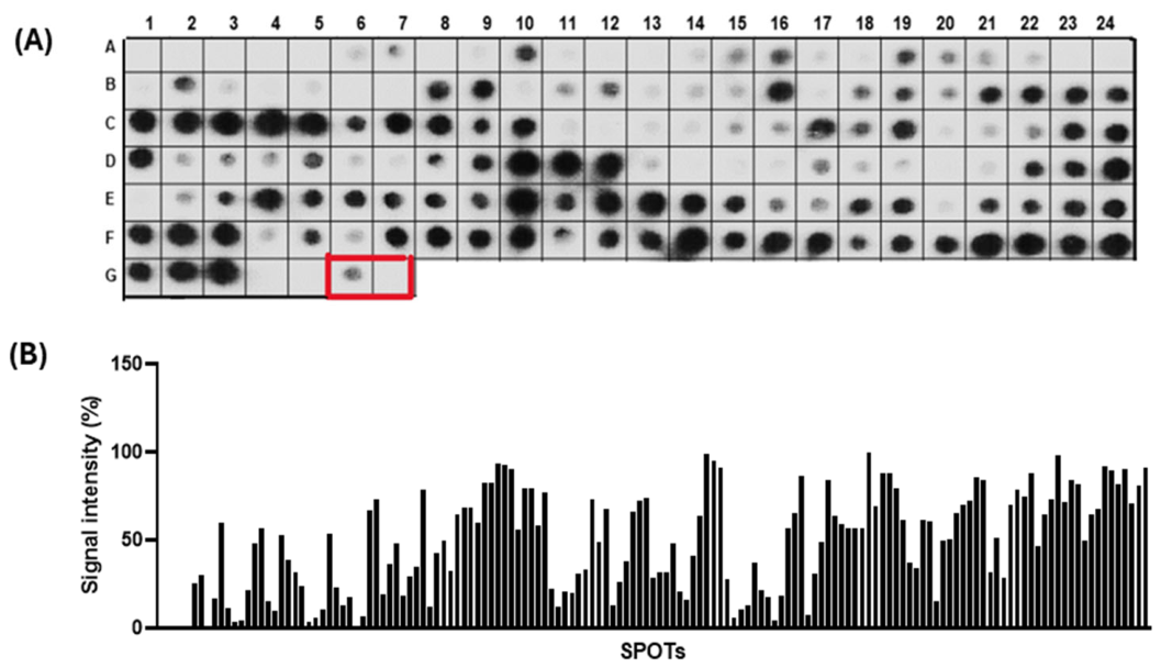
Figure 2.
Three-dimensional structure of the P. falciparum MSP9 protein predicted using the I-TASSER server and a surface model generated in PyMOL, showing the locations of the epitopes selected for validation. The PfMSP9/IgG/23 epitope, highlighted in blue, is mostly exposed on the protein surface. In contrast, the PfMSP9/IgG/27 and PfMSP9/IgG/30 epitopes, highlighted in red and green, respectively, have their entire sequences exposed to the solvent.
Figure 2.
Three-dimensional structure of the P. falciparum MSP9 protein predicted using the I-TASSER server and a surface model generated in PyMOL, showing the locations of the epitopes selected for validation. The PfMSP9/IgG/23 epitope, highlighted in blue, is mostly exposed on the protein surface. In contrast, the PfMSP9/IgG/27 and PfMSP9/IgG/30 epitopes, highlighted in red and green, respectively, have their entire sequences exposed to the solvent.
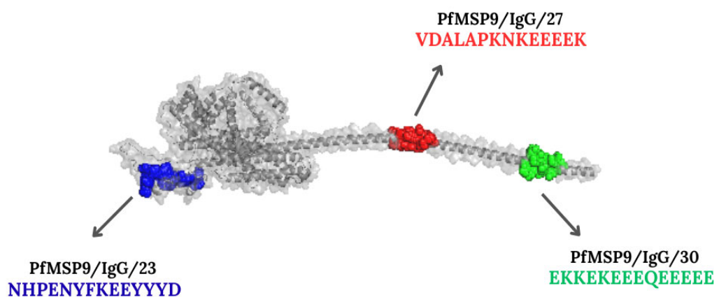
Figure 3.
Reactivity of peptides synthesized using the Fmoc strategy, as assessed by ELISA. We used sera from individuals infected with P. falciparum and P. vivax, as well as sera from blood donors. The data show the reactivity index of the samples analyzed. We analyzed the results using parametric and non-parametric ANOVA tests and considered them statistically significant (p < 0.001). Pf (P. falciparum), Pv (P. vivax), and HD (healthy donors).
Figure 3.
Reactivity of peptides synthesized using the Fmoc strategy, as assessed by ELISA. We used sera from individuals infected with P. falciparum and P. vivax, as well as sera from blood donors. The data show the reactivity index of the samples analyzed. We analyzed the results using parametric and non-parametric ANOVA tests and considered them statistically significant (p < 0.001). Pf (P. falciparum), Pv (P. vivax), and HD (healthy donors).
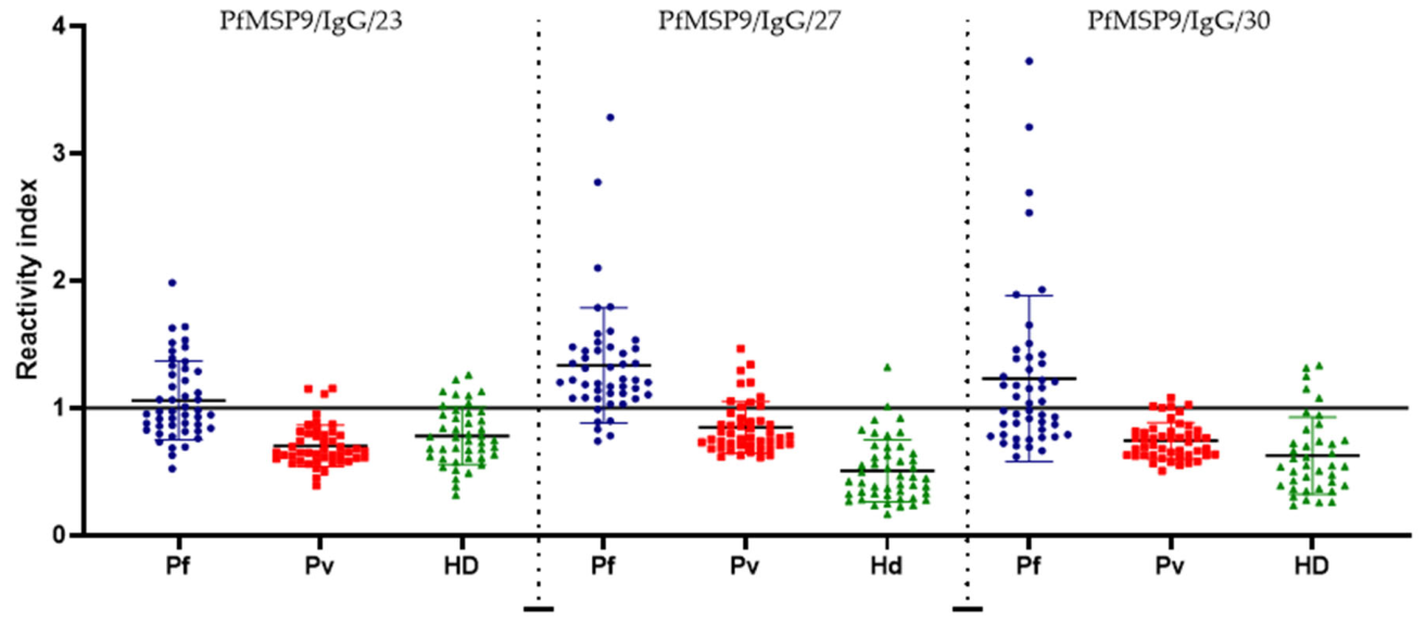
Figure 4.
ROC curve analysis was used to determine the sensitivity and specificity of each peptide tested. (a) PfMSP9/IgG/23: 44.81% sensitivity and 81.82% specificity, AUC = 0.7635; (b) PfMSP9/IgG/27: 87.32% sensitivity and 95.74% specificity, AUC = 0.9765; (c) PfMSP9/IgG/30: 46.81% sensitivity and 89.74% specificity, AUC = 0.8672.
Figure 4.
ROC curve analysis was used to determine the sensitivity and specificity of each peptide tested. (a) PfMSP9/IgG/23: 44.81% sensitivity and 81.82% specificity, AUC = 0.7635; (b) PfMSP9/IgG/27: 87.32% sensitivity and 95.74% specificity, AUC = 0.9765; (c) PfMSP9/IgG/30: 46.81% sensitivity and 89.74% specificity, AUC = 0.8672.
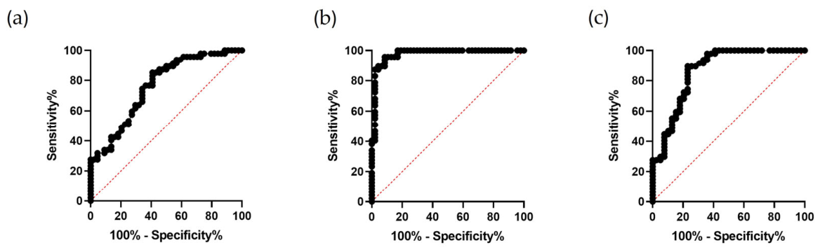

Table 1.
List of IgG-recognized epitopes of the P. falciparum MSP9 protein, signal intensities (RI%) obtained by SPOT synthesis, their structural properties, and in silico cross-reactivity. (BLAST and PIR) (https://blast.ncbi.nlm.nih.gov/Blast.cgi; https://proteininformationresource.org/).
Table 1.
List of IgG-recognized epitopes of the P. falciparum MSP9 protein, signal intensities (RI%) obtained by SPOT synthesis, their structural properties, and in silico cross-reactivity. (BLAST and PIR) (https://blast.ncbi.nlm.nih.gov/Blast.cgi; https://proteininformationresource.org/).
| Epitope code | Sequence | R.I. (%) | Secondary structure | Other Plasmodium sp |
| PfMSP9/IgG/01 | 31GVDMNVLNNYENLF44 | 30,47 | T+H | None |
| PfMSP9/IgG/02 | 46FVKCEYCNEHTYVK59 | 59,93 | H+T+C | None |
| PfMSP9/IgG/03 | 76CKELLKEKQYTDSV89 | 56,49 | H+T | None |
| PfMSP9/IgG/04 | 91YLMDGFKSANNSAN104 | 53,06 | H+T | None |
| PfMSP9/IgG/05 | 126KLIKALKKNIESIQ139 | 53,87 | H | None |
| PfMSP9/IgG/06 | 161KMNMLKENVDYIQK174 | 72,89 | H+T | P. cynomolgi |
| PfMSP9/IgG/07 | 176QNLFKELMNQKATY189 | 47,91 | T+H+C | None |
| PfMSP9/IgG/08 | 196KKIISLKSQGHKKE209 | 78,43 | H+C | None |
| PfMSP9/IgG/09 | 211SQNQNENNDNQKYQ224 | 49,59 | C+T+H | None |
| PfMSP9/IgG/10 | 226VNDEDDVNDEEDTN239 | 68,48 | H | P. brasilianum; P. malariae |
| PfMSP9/IgG/11 | 241DEDTNDEEDTNDDE254 | 82,69 | C+T+H | None |
| PfMSP9/IgG/12 | 257NDDEDTNDDEDTND270 | 93,29 | H+T+C | None |
| PfMSP9/IgG/13 | 271EEDHENNNATAYEL284 | 79,83 | C+T+H | None |
| PfMSP9/IgG/14 | 286IVPVNDVLNVNMKN299 | 77,25 | C+H | None |
| PfMSP9/IgG/15 | 321GSNDLINFLNQGKE334 | 73,59 | H | P. knowlesi; P. vivax |
| PfMSP9/IgG/16 | 361LNMLKDNLINYEFI374 | 74,27 | H+T+C | None |
| PfMSP9/IgG/17 | 406AQEKGLPEPTVTNEEYVEE424 | 99,08 | H+C | None |
| PfMSP9/IgG/18 | 441VKSLLKKLKNKIFP454 | 37,62 | T+H+C | None |
| PfMSP9/IgG/19 | 476QPALRGVEPTEDSN489 | 86,15 | H | None |
| PfMSP9/IgG/20 | 496NVMDEIDFFEKELI509 | 83,97 | H+C+T | P. knowlesi |
| PfMSP9/IgG/21 | 511NNNTPNVVPPTQSK524 | 56,46 | H+C+S | None |
| PfMSP9/IgG/22 | 526KNKNETVSGMDENF539 | 100 | T+C+H+S | None |
| PfMSP9/IgG/23 | 541NHPENYFKEEYYYD554 | 87,75 | S+C+T | None |
| PfMSP9/IgG/24 | 566IGVTLKKFEPLKNG580 | 61,5 | T+H | None |
| PfMSP9/IgG/25 | 641DVLNQETEEEMEKQVEAIT659 | 74,43 | H | None |
| PfMSP9/IgG/26 | 666VDALAPKNKEEEEK679 | 97,95 | H | None |
| PfMSP9/IgG/27 | 680KEKEEKEKE689 | 81,8 | H | None |
| PfMSP9/IgG/28 | 701EEKEKEEKE709 | 89,81 | H | None |
| PfMSP9/IgG/29 | 716EKKEKEEEQEEEEE719 | 90,64 | H | None |
| PfMSP9/IgG/30 | 721EEEQEEEEEEEIVP734 | 70,99 | H | None |
| PfMSP9/IgG/31 | 735EEIVPENLTTEESK743 | 90,9 | H | None |
Disclaimer/Publisher’s Note: The statements, opinions and data contained in all publications are solely those of the individual author(s) and contributor(s) and not of MDPI and/or the editor(s). MDPI and/or the editor(s) disclaim responsibility for any injury to people or property resulting from any ideas, methods, instructions or products referred to in the content. |
© 2026 by the authors. Licensee MDPI, Basel, Switzerland. This article is an open access article distributed under the terms and conditions of the Creative Commons Attribution (CC BY) license (http://creativecommons.org/licenses/by/4.0/).
Copyright: This open access article is published under a Creative Commons CC BY 4.0 license, which permit the free download, distribution, and reuse, provided that the author and preprint are cited in any reuse.