
Submitted:
12 November 2026
Posted:
16 November 2026
You are already at the latest version
Abstract
Tumour extracellular vesicles (EVs) can prepare distant tissue for tumour cells in animal models. That is a claim about tissue. Whether exposed cells keep a lasting phenotype, and whether their daughters inherit it, are separate claims that need their own proof. Here I set out three testable hypotheses and a four-gate framework for the cell-level claims. H1: EVs add a measurable share to tissue priming. H2: an exposed cell keeps a pre-defined phenotype after exposure ends. H3: daughter cells carry it. For each, I state what would support it, what would count against it, and what would leave it open. A label system (P, M, H) grades the evidence for a claim, not the cell. The aim is to stop a tissue result from being read, step by step and unnoticed, as cell memory or inheritance. No new data are reported.
Keywords:
extracellular vesicles
; pre-metastatic niche
; cellular memory
; falsifiability
; clonal selection
; MISEV2023
1. Introduction
In 1889 Paget proposed that a tumour cell, the seed, grows only in an organ that suits it, the soil [1]. Later work showed that the soil can be prepared before the seed arrives. Bone marrow cells help set up a pre-metastatic niche [2], and in mouse models tumour-derived EVs were shown to educate bone marrow progenitors and to mark distant organs [3,4]. Here, EV is a generic term for membrane-bound particles released by cells. It implies no specific biogenesis route and no purified vesicle subtype.
So EVs can help prepare tissue. Two harder questions follow. Do exposed cells keep a state after the EVs are gone? Do their daughter cells inherit it? Neither is the same question as tissue priming, yet papers often slide from one to the next with no test in between. A 2026 review of 120 original in vivo studies, published from January 2010 to April 2025, found that 115 gave tumour-derived EVs to animals from outside. Only five used endogenous models, with EVs released by implanted tumours. The authors name loss-of-function tests of EV necessity as a key open challenge [5].
Even the first step needs care. An EV can bind to a cell, enter it, and release its content inside. These are three events, and the last is the one that counts if cargo is the mechanism [6]. Measuring one does not show the others.
This article keeps three claims apart: tissue priming, persistence of a cell phenotype after exposure ends, and passage of that phenotype to daughter cells. Proof of the first is not proof of the second or third, and the claims need not move together. EVs may prepare tissue with no lasting change in any cell. A cell phenotype may persist with no gain for the tissue. Each claim gets its own hypothesis, its own tests and its own way to fail. I report no new data.
2. Three Claims and a Gate System
Each hypothesis ends in one of three ways: supported within the tested model, not supported in the test as run, or open because needed data are missing or invalid. A test never confirms a claim in general. It speaks only for the model, the conditions and the sensitivity used.
Four gate domains check the cell-level claims. Gate 1 asks whether a pre-defined phenotype persists after exposure ends. Gate 2 has two linked tests. Gate 2A asks whether the change can be tied to the exposed cells and their lineages. Gate 2B asks whether the lineage adds evidence beyond selection of states that already existed. Gate 3 asks whether exposed cells respond differently to a second, identical challenge (recall). Gate 4 asks whether daughter cells carry the phenotype through a set number of divisions. H1 is tested differently, by EV depletion and add-back (Section 4.1).
Three labels sum up what has been shown. P (persistence) needs Gate 1. It means the pre-defined phenotype persists after external exposure has ended. It does not by itself show independence from delivered cargo or ongoing intracellular signalling. M (recall-tested memory) needs Gates 1, 2A, 2B and 3. H (heritable transmission) needs M plus Gate 4. A label grades the evidence for a claim. It is not a trait of the cell, and it can change when new data arrive. “Heritable” here means passed on through cell division. H does not show whether the cause is genetic or not, and it does not name a mechanism. The gates and labels develop two earlier conceptual frameworks by the author, one on EV-effect attribution and one on claims of cellular memory. Neither has been independently validated. They are working tools, not accepted standards. M is a strict, operational definition of memory for this task. It does not replace other definitions of cellular memory.
The EV scheme uses separate codes. P1 is the tissue effect, P2 the cell response during exposure, P3 persistence after exposure, and P4 transmission to daughter cells. These codes are not the same as the labels P, M and H. Figure 1 shows how the parts fit.
3. Three Hypotheses
Table 1 gives each hypothesis its own conditions for support, for failure and for an open result. This keeps a weak or broken experiment from being read as a negative one.
H3 can back a narrower statement, that identified daughter cells carry the phenotype, even before the full H label is earned. The H label also needs M, including recall at Gate 3. Rare starting states that went unmeasured can remain a possible explanation for H3.
4. How to Test
4.1. Tissue Test (H1)
Each independent batch of conditioned medium is split into seven matched conditions, equal in volume and origin: (1) intact medium; (2) mock-depleted medium, which goes through the same steps as depletion but without the EV-capture step; (3) EV-depleted medium, not assumed to be free of residual EVs or other particles; (4) EV-depleted medium with EVs added back; (5) an isolated tumour EV-enriched fraction; (6) an EV-enriched fraction from non-tumour cells; (7) vehicle. Unconditioned medium taken through the same steps is a procedural control.
Use several independent batches, so that reproducibility between batches can be judged and not only the effect within one. Randomise samples, score results blind, and set exclusion rules in advance. Assess depletion for residual EV-associated material and for unintended change in representative non-EV components. Assess isolation and add-back for particle recovery, integrity and retained activity. If depletion removed or changed more than EVs, treat that contrast as open, not as an effect of EV removal. Without these checks, a failed add-back means little.
Choose one measure of EV dose, particles or protein, and use it in every group. Report both, because neither shows that a preparation is pure, and equal particle numbers do not mean equal active EVs. Set the add-back dose by the same measure, matched to the EV content of the original medium. Do not mix particle, protein and activity measures.
The main endpoint is the number of viable tumour cells kept in the pretreated tissue at a fixed early time after a standard injection. Fix the readout and the time point in advance. Early retention is not later survival, growth or metastasis, and these should not be merged into one endpoint. A positive result supports an EV-dependent contribution to this endpoint under the tested conditions. It does not establish niche preparation as a whole.
Also fix, before the experiment, the smallest effect that matters and what counts as recovery on add-back: which contrast, and how large. Partial recovery supports a partial EV contribution. It cannot give the EV share of the effect unless dose and activity match those in the original medium. A result with p > 0.05 does not show that EVs add nothing [7]. A negative call needs an interval estimate, or an equivalence test, that rules out an effect of the pre-set size [8]. A similar effect in the EV-depleted fraction does not refute H1. H1 says EVs add to the effect, not that they alone cause it.
4.2. Cell Test (H2 and H3)
Label recipient cells with heritable barcodes, expose them to EVs, wash, and keep a matched unexposed culture that goes through every step except the EVs.
Gate 1. Fix the phenotype, the main readout, the washout time and the detection limit for leftover EV material before the experiment. Conclusions apply to that phenotype and readout. They do not show a broader cell state, though extra readouts help tell a lasting state from a brief shift. Treat uptake as separate from delivery: if the proposed mechanism needs cargo to reach the cytosol or nucleus, test that step on its own.
If residual EV material or an ongoing signal could still account for the phenotype, Gate 1 cannot show persistence that is independent of continued exposure. A cell process that EVs started may still keep the phenotype going. Until delivered cargo is shown not to be needed for maintenance, call the result cargo-dependent persistence. Report carryover as “not detected by the pre-set assay at a stated detection limit”, never as “excluded”. If the assay cannot rule out a meaningful carryover, the result stays open. Check cell fusion and cell loss too.
Gate 2A. Barcodes follow lineages. They do not show that the state changed in the cell that took up the EV: one clone can hold several states, and some of its cells may die. To link ancestry to state, pair barcodes with single-cell readouts or with live imaging that follows both uptake and the later state.
Gate 2B. Say in advance which data would support a change arising within lineages, and which would be explained by better survival or growth of states that already existed. The claim that can survive is modest: a change within tracked lineages that measured differences in survival and growth do not adequately explain. Rare starting states that went unmeasured can never be fully ruled out. This is the old logic of Luria and Delbrück [9], and drug-tolerant cell states show why it matters [10]. Barcoding [11] and single-cell tools narrow the doubt but do not remove it. Where a genetic cause is plausible, check the relevant genome changes. Finding a mutation does not show that it holds the phenotype. That takes a separate functional test, and a genetic cause means the result is not called non-genetic.
Gate 3. Give exposed and control cells the same pre-set second challenge. Each group needs a challenged and an unchallenged arm. The main readout is the difference in response between exposed and control cells, such as the size of a functional or transcriptional response. The main analysis should estimate the exposure-by-challenge interaction, not compare challenged groups alone. This separates a changed response to the second challenge from a baseline difference in phenotype, growth or viability. Faster growth after a second hit is not memory by itself. An unrelated challenge can serve as a secondary check of specificity.
Gate 4. Follow daughter cells for a fixed number of divisions, chosen and justified for the model. Where feasible, prefer direct parent–daughter tracking to barcode identity alone. A shared barcode shows lineage membership, not transmission of a phenotype. Report the share of daughter cells that carry the phenotype in each lineage, the lineages lost, and any growth-rate differences. If faster growth of a subset of cells explains the enrichment, read the result as selection, not as added transmission. Both can occur, so report phenotype retention within lineages and differential expansion separately.
5. What the Literature Shows, and Its Limits
The founding study of EV education of bone marrow cells reported a lasting effect in mouse melanoma models [3]. This is real evidence of a durable functional effect in that model. It does not meet the gates above, in particular attribution to single cells, contribution beyond selection, recall and direct tracing across divisions. The paper also has a 2016 corrigendum, which belongs in the reference list [12].
The 2015 integrin study of organ targeting [4] now carries a Nature Editorial Expression of Concern about image overlap in some figures [13]. This is not a retraction, but it calls for careful reading. A replication of the melanoma exosome study saw a small rise in metastatic burden with control exosomes, which fell when Met was knocked down. The direction matched the original, but the effects were not significant. The authors listed knockdown efficiency, cell-line drift, sample size, endpoints and variability as possible causes [14]. By the rules above, that result is open, not negative. Together with the 120-study review [5], the picture is this: EV effects on tissue are reported often, tests of necessity are rare, and effects on cell phenotype after exposure are hardly tested.
The limits are plain. This is not a systematic review. I ran no systematic search, and the sources were chosen to show the founding work and its weak points, so opposing or added studies may be missing. The thresholds, times and division counts in the gates are choices that each model must justify. EV preparations also vary between batches and can clump or break down, which shifts particle counts and apparent activity. The plan can fail too: if EVs cannot be depleted in a valid way in a given system, H1 cannot be tested there. And a result at one gate does not carry over to the next.
6. Conclusion
Preparing tissue, keeping a cell phenotype and passing it to daughter cells are three different claims, and each needs its own evidence. A tissue effect is not memory, and persistence is not inheritance. The gates ask for little beyond clear pre-set rules, honest controls and the readiness to report “open” when the data cannot decide. Preparing the soil is not remembering the exposure.
Author Contributions
Said Ganihodjaev: conceptualisation; writing – original draft; writing – review and editing. Sole author.
Funding
None.
Ethics Approval
Not applicable. No new work with humans or animals is reported.
Data Availability
No new data were generated or analysed.
Conflicts of Interest
The author declares no competing interests.
Use of AI
AI was used for translation and formatting of this manuscript.
References
- Paget, S. The distribution of secondary growths in cancer of the breast. Lancet 1889, 133, 571–573. [Google Scholar] [CrossRef]
- Kaplan, R.N.; Riba, R.D.; Zacharoulis, S.; et al. VEGFR1-positive haematopoietic bone marrow progenitors initiate the pre-metastatic niche. Nature 2005, 438, 820–827. [Google Scholar] [CrossRef] [PubMed]
- Peinado, H.; Alečković, M.; Lavotshkin, S.; et al. Melanoma exosomes educate bone marrow progenitor cells toward a pro-metastatic phenotype through MET. Nat. Med. 2012, 18, 883–891. [Google Scholar] [CrossRef] [PubMed]
- Hoshino, A.; Costa-Silva, B.; Shen, T.L.; et al. Tumour exosome integrins determine organotropic metastasis. Nature 2015, 527, 329–335. [Google Scholar] [CrossRef] [PubMed]
- Blavier, L.; Crnjac, A.; DeClerck, Y.A. Contribution of tumor-derived extracellular vesicles in the establishment of the pre-metastatic niche: lessons learned from past experimentations and future directions. Clin. Exp. Metastasis 2026, 43, 16. [Google Scholar] [CrossRef] [PubMed]
- Welsh, J.A.; Goberdhan, D.C.I.; O’Driscoll, L.; et al. Minimal information for studies of extracellular vesicles (MISEV2023): from basic to advanced approaches. J. Extracell. Vesicles 2024, 13, e12404. [Google Scholar] [CrossRef] [PubMed]
- Altman, D.G.; Bland, J.M. Absence of evidence is not evidence of absence. BMJ 1995, 311, 485. [Google Scholar] [CrossRef] [PubMed]
- Lakens, D. Equivalence tests: a practical primer for t tests, correlations, and meta-analyses. Soc. Psychol. Personal. Sci. 2017, 8, 355–362. [Google Scholar] [CrossRef] [PubMed]
- Luria, S.E.; Delbrück, M. Mutations of bacteria from virus sensitivity to virus resistance. Genetics 1943, 28, 491–511. [Google Scholar] [CrossRef] [PubMed]
- Sharma, S.V.; Lee, D.Y.; Li, B.; et al. A chromatin-mediated reversible drug-tolerant state in cancer cell subpopulations. Cell 2010, 141, 69–80. [Google Scholar] [CrossRef] [PubMed]
- Bhang, H.E.; Ruddy, D.A.; Krishnamurthy Radhakrishna, V.; et al. Studying clonal dynamics in response to cancer therapy using high-complexity barcoding. Nat. Med. 2015, 21, 440–448. [Google Scholar] [CrossRef] [PubMed]
- Peinado, H.; Alečković, M.; Lavotshkin, S.; et al. Corrigendum: Melanoma exosomes educate bone marrow progenitor cells toward a pro-metastatic phenotype through MET. Nat. Med. 2016, 22, 1502. [Google Scholar] [CrossRef] [PubMed]
- Hoshino, A.; Costa-Silva, B.; Shen, T.L.; et al. Editorial Expression of Concern: Tumour exosome integrins determine organotropic metastasis. Nature 2026, 656, E44. [Google Scholar] [CrossRef] [PubMed]
- Kim, J.; Afshari, A.; Sengupta, R.; et al. Replication Study: Melanoma exosomes educate bone marrow progenitor cells toward a pro-metastatic phenotype through MET. eLife 2018, 7, e39944. [Google Scholar] [CrossRef] [PubMed]
Figure 1.
Three claims and the evidence each needs. Evidence for one claim does not prove the next. P, M and H grade the evidence for a claim, not the cell.
Figure 1.
Three claims and the evidence each needs. Evidence for one claim does not prove the next. P, M and H grade the evidence for a claim, not the cell.
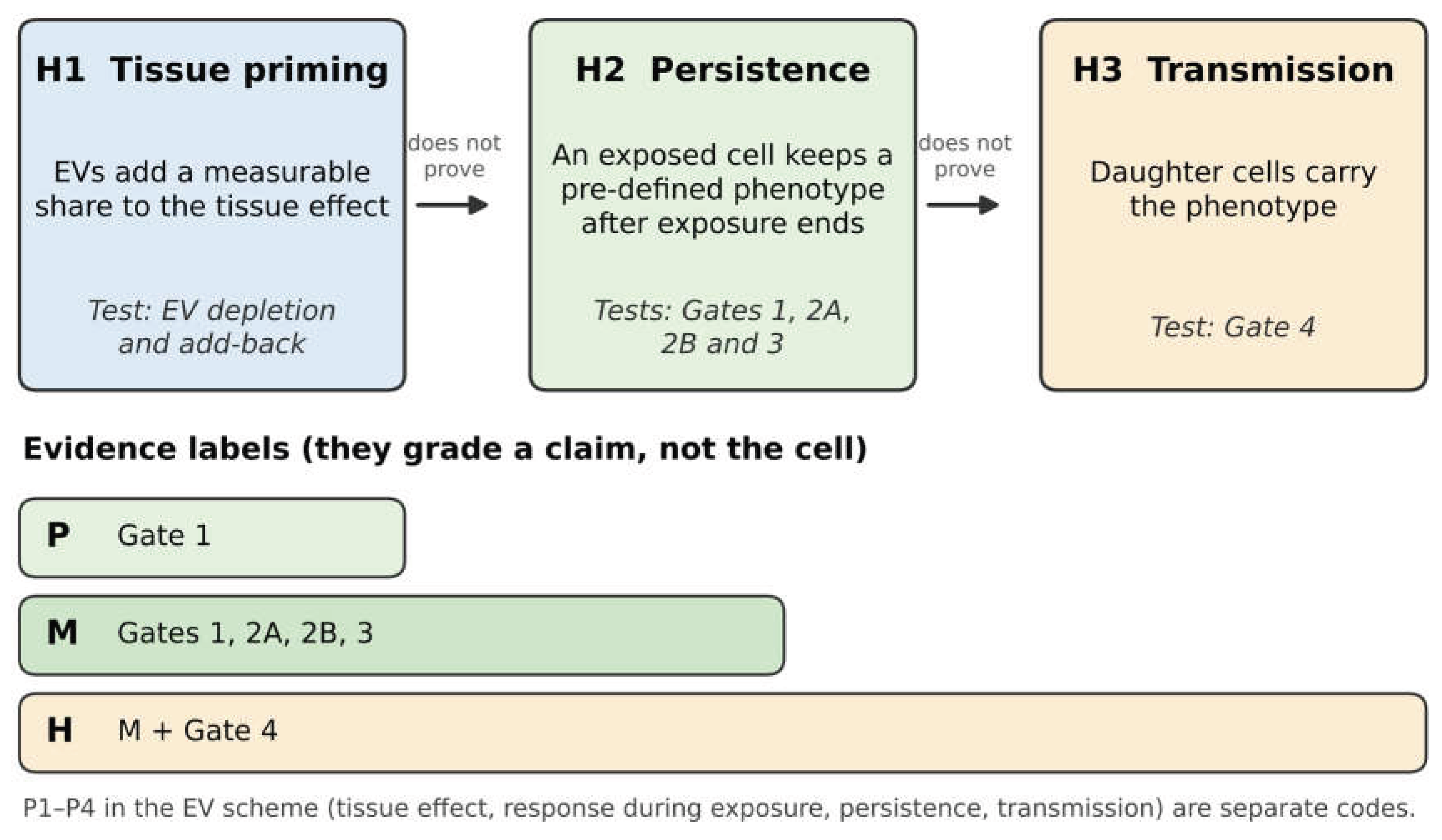

Table 1.
Three hypotheses, the evidence that supports or counts against each, and the conditions that leave the result open.
Table 1.
Three hypotheses, the evidence that supports or counts against each, and the conditions that leave the result open.
| Hypothesis | Maps to | Supported if | Not supported if | Open if |
|---|---|---|---|---|
| H1. EVs make a measurable contribution to tissue priming. | EV scheme: P1. Label system: not used. | Removing EVs lowers the main tissue effect by at least the pre-set amount; add-back reaches the pre-set recovery; all controls are valid. | Confirmed EV removal does not lower the effect by the pre-set amount, the estimate is precise enough to exclude a contribution of that size, and all controls are valid. | EV removal was incomplete, other components were altered, EVs lost activity, add-back is invalid, or the estimate is too imprecise. |
| H2. An exposed cell keeps a pre-defined phenotype after exposure ends. | EV scheme: P3. Label P needs Gate 1; label M adds Gates 2A, 2B and 3. | The phenotype persists after washout for the set time; carryover is not detected by the pre-set assay at a stated detection limit; the assay is sensitive enough. If delivered cargo may still act, the result is read as cargo-dependent persistence. | The phenotype is gone by the set time in a valid assay with live, identified cells. | Residual EVs or cargo, ongoing signalling, cell fusion, or loss of cells cannot be ruled out by the assay; or the need for delivered cargo is untested. |
| H3. The phenotype passes to daughter cells. | EV scheme: P4. Gates 1, 2A and 4; label H also needs M. | The phenotype is found in traced daughter cells after the set number of divisions, and measured survival and growth of pre-existing states do not adequately explain it. | The phenotype is absent from live, identified descendants after the set divisions, or selection explains the result. | Too few divisions, lost lineages, unclear descendant identity, or weak tracking or selection controls. |
Disclaimer/Publisher’s Note: The statements, opinions and data contained in all publications are solely those of the individual author(s) and contributor(s) and not of MDPI and/or the editor(s). MDPI and/or the editor(s) disclaim responsibility for any injury to people or property resulting from any ideas, methods, instructions or products referred to in the content. |
© 2026 by the authors. Licensee MDPI, Basel, Switzerland. This article is an open access article distributed under the terms and conditions of the Creative Commons Attribution (CC BY) license (http://creativecommons.org/licenses/by/4.0/).
Copyright: This open access article is published under a Creative Commons CC BY 4.0 license, which permit the free download, distribution, and reuse, provided that the author and preprint are cited in any reuse.