

Submitted:
11 November 2026
Posted:
12 November 2026
You are already at the latest version
Abstract
Background: In hyperacute and early acute ischaemic stroke interventions in clinical settings, functional near-infrared spectroscopy (fNIRS) must be applied quickly by clinical staff. RETRAIN Phase 2 therefore recorded long-separation channels only. Short-separation (SS) channels, the usual reference for scalp and systemic haemodynamics, were omitted from the montage to allow rapid donning and doffing and because SS coupling through hair was unreliable in acute patients. Without SS channels, superficial and deeper-weighted contributions cannot be separated by standard regression. RETRAIN Phase 2 randomized adults with acute ischaemic stroke to common peroneal nerve neuromuscular electrical stimulation (CPN-NMES, geko™) or intermittent pneumatic compression (IPC). Its primary haemodynamic results have not been published, the primary manuscript is under review.Objective: To prespecify a secondary analysis of RETRAIN Phase 2 that applies a new, frozen long-channel-only method, coherence-volume-phase regression (CVP) with volume/flow indices and multichromophore operational modal analysis (OMA), to test the hypothesis that geko™ and IPC differ in superficial/systemic-weighted versus residual (deeper-weighted) haemodynamic response characteristics in hyperacute stroke. This Stage 1 Registered Report with Data Existing status (RR1-DE) does not concern the trial's primary outcome.Methods: The method was developed and tested without Phase 2 data. In two open fNIRS datasets with measured SS channels (OpenNeuro accession ds005776, n = 9-11 per task, Zenodo record 23163739, n = 22), with SS-regressed signals as the reference, and in RETRAIN Phase 1 recordings from 18 people with subacute stroke. Fifty-three Phase 2 participants were randomized (geko™ 27, IPC 26). Each session included a 5-minute device-off baseline and a 5-minute device-on period. Phase 2 will be analysed with paired Group x Period contrasts, participant-cluster bootstrap intervals, prespecified quality checks (including residual HbO-HbR coherence), comparator corrections (global signal regression, global-HbT regression, uncorrected signals) and null-simulation calibration.Results: Recruitment and data collection are complete (53 of 80 planned participants). No Phase 2 results have been published, the primary results manuscript is under review, and this protocol reports no Phase 2 estimates. In open data, CVP agreed better than global signal regression with SS-regressed HbR in all five test sets (e.g., Zenodo record 23163739 median r 0.88 vs 0.72) and null simulations showed that it does not create spurious HbO-HbR counterphase. In Phase 1, all indices were computable, and stimulation reduced residual HbO-HbR phase concordance (participant-level Δcos φ -0.16, 95% CI -0.28 to -0.05). Conclusions: This protocol tests whether long-channel-only fNIRS, compatible with rapid bedside application in hyperacute stroke, can separate superficial/systemic-weighted from residual responses well enough to compare two venous-return interventions. Results will be reported as estimates with explicit failure checks.
Keywords:
ischaemic stroke
; secondary analysis protocol
; hyperacute stroke
; functional nearinfrared spectroscopy
; short-separation-free fNIRS
; common peroneal nerve stimulation
; intermittent pneumatic compression
; operational modal analysis
; short-separation regression
; haemodynamic oscillations
; RR1-DE
Introduction
Clinical Problem and Rationale
Ischaemic stroke is a major cause of death and disability. Even after reperfusion, collateral-flow limitations, microvascular dysfunction, systemic pressure changes, and impaired vascular regulation may contribute to ongoing tissue vulnerability [1,2,3,4,5]. Bedside physiological monitoring may therefore complement conventional clinical assessments but cannot substitute for direct measurements of cerebral perfusion.
The study addresses the hyperacute and early acute period. The registered RETRAIN Phase 2 eligibility window was 36 hours from onset, approved protocol amendment v3.1 (8 September 2025) extended it to 72 hours [6]. "Hyperacute" refers here to this early bedside-monitoring window, not to a universal time definition. Blood-pressure interventions during early stroke have time- and stroke-subtype-dependent effects [7].
Dynamic cerebral autoregulation refers to the time-dependent buffering of cerebral blood flow against pressure fluctuations and may be impaired after ischaemic stroke [8,9,10]. Transfer-function analysis conventionally relates arterial pressure to flow-sensitive measures such as transcranial Doppler blood-flow velocity [9,11]. fNIRS-derived HbO and HbT instead reflect mixed cortical and extracerebral haemoglobin changes, with sensitivity to CO₂, venous blood volume, systemic rhythms, and local neurovascular activity. Consequently, MAP-fNIRS metrics in this study are described as pressure-haemodynamic coupling or dCA-consistent surrogates, not validated measures of macrovascular autoregulation. Likewise, a serial macrovascular-microvascular transfer-function model is a conceptual hypothesis, not identifiable as separate components without an intermediate flow measurement[11,12,13,14].
Mechanistic Distinction in Lower-Limb Venous-Return Interventions
IPC and CPN-NMES both support peripheral venous return through different mechanisms [15,16]. Their randomized comparison provides a clinical setting in which to examine haemodynamic response characteristics. This secondary-method protocol does not test whether either device improves neurological outcome, nor does it represent the original primary HbT treatment-effect result.
At approximately 1 Hz, geko™ CPN-NMES recruits lower-limb motor axons and likely somatosensory afferents, downstream autonomic or neurovascular consequences remain hypotheses [16,17,18]. RETRAIN Phase 1 provided exploratory evidence of stimulation- and posture-associated haemoglobin and modal-frequency changes, but scalp/systemic contributions and multiple testing constrain mechanistic inference [19,20,21].
IPC augments venous return by externally applied cyclic compression. Prior experimental and fNIRS studies indicate that peripheral pressure perturbations may alter systemic and low-frequency cerebral haemodynamic signals, including wavelet bands III-V[22,23,24]. These studies motivate a comparative physiological hypothesis but do not establish different neural mechanisms for IPC and NMES.
Figure 1 presents a conceptual serial transfer-function framework linking systemic input X(f), an unobserved flow-sensitive intermediate V(f), and measured fNIRS haemodynamic output Y(f). An observed MAP-to-HbO/HbT relationship is estimable, its upstream and downstream factors are not separately identifiable in RETRAIN Phase 2 without direct cerebral blood-flow measurements [25,26].
RETRAIN Phase 1 Exploratory Work and Phase 2 Study
RETRAIN Phase 1 (NCT06614400, n=18 reported participants) established bedside fNIRS feasibility and generated exploratory, posture- and intensity-associated haemodynamic observations [19,20,21]. Because treatment intensity and recording order were confounded and multiple outcomes were investigated, these observations are used to generate hypotheses and estimate feasible signal characteristics.
Clinical rationale and method-development sequence. Bedside fNIRS in hyperacute stroke has to be applied and removed quickly by clinical staff, between assessments, imaging and nursing care. SS channels add optodes that need careful coupling close to each long channel, which is slow and unreliable over hair [27]. RETRAIN Phase 1 (subacute stroke, rehabilitation setting) used a NIRSport2 system with SS capability, placed optodes away from hair-covered areas and did not use SS regression [28]. RETRAIN Phase 2 omitted SS channels from the montage to allow rapid donning and doffing in the hyperacute setting and because reliable SS coupling through hair could not be maintained [29]. A method that recovers SS-like correction from long channels alone would remove this practical barrier to bedside monitoring. We therefore developed and tested CVP in three steps before any application to Phase 2: (1) in two open datasets with measured SS and long channels (OpenNeuro accession ds005776 [30], ZENODO record 23163739 [31]), CVP was benchmarked against the SS-regressed signal with the SS channels withheld, (2) in RETRAIN Phase 1, its computability, behaviour and variability were examined in subacute stroke, and (3) the configuration was frozen and the Phase 2 hypotheses were specified. The SS-regressed residual is a practical benchmark, not anatomical truth: it can remove cerebral haemodynamics driven by systemic physiology (Multimedia Appendix 1).
RETRAIN Phase 2 randomized 53 of 80 planned participants (see Sample Size and Statistical Power). The smaller sample limits precision, particularly for paired periods that pass OMA and physiology eligibility checks, so the analyses emphasize interval estimates and do not treat the shortfall as evidence of efficacy or futility.
Rationale for Low-Frequency Modal Biomarkers
The broad 0.01-0.15-Hz haemodynamic band contains overlapping vasomotor, systemic, superficial and cerebral processes [32,33,34,35,36,37]. Frequency alone cannot identify anatomical origin. Output-only OMA estimates repeatable poles, modal frequencies, damping and mode shapes, which are potentially valuable response descriptors even when the driving oscillations are shared and systemic. Phase 2 tests whether such descriptors are numerically stable and show reproducible period-related differences, it does not assume that an output-only pole is an intrinsic cerebral-autoregulatory mode.
Moving correlation indices such as COx and Mx have been used in cerebrovascular monitoring [11,38,39,40,12,13], but their physiological meaning depends on the measured signal, window length and blood-pressure reference. In RETRAIN, each five-minute period provides at most approximately four minutes after edge exclusions, allowing one exploratory period-level MAP-HbO correlation when synchronized beat-to-beat MAP is available. This is pressure-haemodynamic coupling, not a validated time-resolved COx curve or diagnostic threshold for dynamic cerebral autoregulation.
Methods
Study Design, Governance, and SPIRIT 2025 Compliance
RETRAIN Phase 2 (NCT07197996) was a single-centre randomized, parallel-group, open-label clinical physiology study conducted at Countess of Chester Hospital NHS Foundation Trust. Innovate UK funding was awarded under application 10038715 to Firstkind Ltd. Firstkind Ltd is the trial sponsor. The original registration specified enrolment within 36 hours, approved amendment v3.1 (8 September 2025) extended this to 72 hours [41].
Registered Report status and analysis independence. This manuscript is an RR1-DE secondary analysis protocol. Phase 2 data exist and have been analysed for the trial's primary haemodynamic outcome, that manuscript is under review, and no Phase 2 results have been published or are reported here. The long-channel-only method was developed using open datasets and RETRAIN Phase 1 only, and its configuration was frozen. Its Phase 2 analyses will be run only after Stage 1 acceptance, are secondary and exploratory, and will be reported with the freeze date and code version.
The supplement distinguishes (i) the original Phase 2 clinical/SAP specification, (ii) public-data method development, (iii) Phase 1 exploratory testing, (iv) revision-stage corrections and simulation stress tests, and (v) proposed Phase 2 secondary evaluation.
Protocol and analysis-status audit
Recruitment included 53 randomized participants (27 geko™, 26 IPC), 49 contributed analysable fNIRS to the primary analysis [42]. Before this protocol, Phase 2 data were used for the primary haemodynamic analysis and for a descriptive EEG-fNIRS feasibility analysis of nine sessions from six healthy participants in a separate methods manuscript, neither used CVP or the hypotheses specified here. Dates of database lock, unblinding and each analysis will be listed in the analysis-provenance register submitted with the Stage 2 report.
A versioned analysis-provenance register will accompany later results reporting. Post-submission parameter changes, including CVP, physiological decomposition, SSI-COV, correction of the empirical Hankel rule, and updated classifier descriptions, will be identified as exploratory extensions or methodological corrections.
Patient and Public Involvement (PPIE)
Stroke survivors and carers supported bedside, non-invasive monitoring. Their feedback concerned the total session duration of up to 15 minutes (5 minutes device-off, up to 5 minutes setup/transition and stabilization, and 5 minutes device-on), not 15 minutes of active stimulation. The study team reviewed participant-facing materials with patient and public involvement representatives and will disseminate findings through clinical and public-facing channels.
Participants and Setting
Eligible adults had acute ischaemic stroke and were enrolled within 36 hours of onset under the registered protocol and within 72 hours after approved amendment v3.1 [43], the number enrolled in each interval will be reported. The cohort is described as hyperacute and early acute ischaemic stroke.
Key eligibility and device contraindications included physician-confirmed ischaemic stroke, reduced mobility, absence of intracranial haemorrhage, inability to use the study devices safely, and absence of clinically apparent DVT. Detailed inclusion and exclusion criteria, including any cortical-sign requirement and protocol amendments, will be reported verbatim from the approved protocol and registry history to avoid discrepancies.
Exclusion criteria: absence of cortical signs or TIA, unwitnessed onset with long lie, clinically apparent DVT, expected palliative care within 14 days, anticipated transfer outside local catchment, recent/current conflicting trial participation, geko™ contraindication including hydrogel allergy, IPC contraindications including severe peripheral vascular disease, large leg ulcers requiring extensive bandaging, severe oedema, leg deformity preventing fitting, NYHA IV heart failure, single/double leg amputation, no visible geko™ dorsiflexion response, and current COVID-19 infection.
Randomization, Allocation Concealment, and Blinding
Participants were allocated 1:1 to geko™ CPN-NMES or IPC in an open-label parallel-group design using permuted blocks stratified by baseline NIHSS severity. Participants and bedside staff could not be masked. Whenever feasible, analysis files were treatment-coded and QC decisions made without viewing group-wise treatment effects. Open-label differences in discomfort, breathing, and motion may still influence fNIRS and are addressed by explicit sensitivity analyses.
Interventions
Table 1 summarizes the two VTE-prevention interventions compared in RETRAIN Phase 2 highlighting their distinct peripheral mechanisms and hypothesized cerebrovascular effects during matched stimulation periods.
Measurement Schedule
Each session comprised 5 minutes of device-off recording and 5 minutes with the assigned device active, with a setup, transition, and stabilization interval of up to 5 minutes between periods. Up to four paired sessions were planned over two days. Transition periods are excluded consistently from the time-series analysis, after removing 30 seconds from both ends of each 5-minute epoch, a maximum of approximately 4 minutes per period remains before further artifact loss.
Signal Acquisition
fNIRS was acquired using a NIRSport2 system (760/850 nm, 16 sources, 16 detectors, up to 48 long-separation channels). Phase 2 recordings contained no SS channels: they were omitted from the montage to allow rapid donning and doffing at the bedside and because SS coupling through hair was unreliable in acute patients [44]. Measured SS regression is therefore not possible. The native sampling rate is taken from each file's time vector.
EEG: Concurrent EEG (g.tec, 16 channels, 500 Hz, electrode impedance <10 kΩ) for neurovascular coupling and dynamic connectivity analyses.
MAP was acquired using continuous beat-to-beat monitoring where available, otherwise by intermittent cuff measurements. Only verified synchronized continuous MAP can enter COx and spectral MAP-HbO analyses, cuff observations may be used as session-level covariates.
Available systemic channels included transcutaneous CO₂, SpO₂, and temperature. Period-specific physiological summaries were matched to fNIRS windows. CO₂ may represent both a confounder and a device-mediated pathway, adjustment for concurrent CO₂ therefore changes interpretation of the treatment contrast and is examined alongside an unadjusted sensitivity model.
Preprocessing Pipeline
Preprocessing will follow the documented AnalyzIR-based workflow and modality-specific methods, with algorithms, versions, thresholds, and QC decisions archived. Source, detector, timing, channel-exclusion, and reproducibility tables are provided as supplementary material.
- Raw intensity was screened for saturation, flatlining, discontinuities, and motion-related loss, QC counts and thresholds will be reported by session and group before treatment-effect interpretation.
- Raw light intensity data will be converted to optical density (OD) using nirs.modules.OpticalDensity.
- Channel quality was assessed by scalp-coupling and signal-to-noise criteria (SCI < 0.75 or SNR < 0 dB flagged), with exclusions documented.
- Missing intervals up to 5 seconds were eligible for documented interpolation, longer gaps were excluded rather than filled across sustained physiology.
- Motion artifacts were treated with TDDR, pre-correction artifact burden and residual signal quality were retained for the open-label-bias analysis.
- Optical density signals acquired at 760 and 850 nm will be converted to relative concentration changes in oxyhaemoglobin (HbO) and deoxyhaemoglobin (HbR) using the modified Beer-Lambert law.
- HbT was defined as the sum of relative HbO and HbR concentration changes. Where continuous beat-to-beat MAP exists, a single MAP-HbO correlation is calculated over the usable period.
- OMA channel-eligibility requirements were assessed separately by hemisphere, sample duration, stability, and effective channel rank were documented before applying outcome contrasts.
- Phase 2 contains no SS channels[45]. Consequently, the Brain-Scalp Score is a provisional spatial/temporal classification and cannot establish absolute brain-versus-scalp source separation.
- The 0.01-0.15-Hz bandpass was applied to OMA and the specified low-frequency coupling pipelines, wavelet analysis used appropriate band-specific native-rate data. First-level GLM preprocessing and estimator settings are recorded separately to avoid treating every analysis as having the same frequency response.
- Native acquisition rates were read from each SNIRF time vector, resampling to 4 Hz was restricted to explicitly documented OMA and cross-modal pipelines.
- Recordings will be segmented into predefined baseline (device-off) and active stimulation (device-on) epochs using synchronized acquisition markers.
- Anatomically defined ROIs and channel assignment were kept fixed across periods and arms, remaining channels and coverage were reported.
- All exclusions, missingness patterns, and QC failures were reported in a physiology-specific flowchart, with decisions separated from treatment-effect inspection where feasible.
Outcomes and Endpoints
Table 2 distinguishes the primary haemodynamic estimand from physiologically suggestive but not independently validated secondary or exploratory measures.
Operational Modal Analysis Pipeline
OMA was applied directly to multichannel HbT time series, not to first-level GLM β estimates. Models were fitted within each usable period and anatomical region using N4SID orders 8-20. At the low-frequency boundary, a four-minute period contains only approximately 2.4 cycles at 0.01 Hz, constraining the reliability of frequency and damping estimates.
System identification: identified poles were summarized as natural frequency, damping ratio, and complex output-space mode shape Φj = C vj, where A vj = λj vj. Complex Hermitian MAC was used for mode-shape stability across model orders, the real, phase-aligned representative shape was used for topographic features.
Mode screening: candidate frequencies were restricted to 0.01-0.15 Hz with cross-order tolerances of 0.005 Hz for frequency, 0.1 for damping, and complex Hermitian MAC ≥0.90. Screening is a repeatability criterion.
The original HbT/N4SID pipeline operated on continuous HbT recordings, using orders 8-20, cross-order pole-stability criteria and the reported Brain-Scalp Score.
RETRAIN Phase 2 segment definition: Primary OMA contrast is baseline device-off versus active device-on. Exploratory INIT/LAST analysis within active device-on segments is a secondary analysis, explicitly labelled as such.
Negative-control analyses compared screened brain-like modes with mixed and scalp/systemic-like mode selections. Differential associations provide construct-level evidence, not independent cortical ground truth.
Long-Channel-Only Analysis
Methods and comparators. A volume-associated projection uses V=(HbO+HbR)/(1+rv) and a chromophore-opposition index F=(rv HbO-HbR)/(1+rv), for a reference slope rv, these are invertible transformations. CVP uses a spatially weighted long-channel HbT regressor and its derivative to estimate a shared/fitted component, leaving a residual, global-HbT regression, conventional GSR and uncorrected signals remain comparators. The same fitted predictor space affects both chromophores, so residual HbO-HbR opposition may arise from correction and optical noise, phase is evaluated jointly with residual coherence and simulation controls. The separate multichromophore SSI-COV branch estimates stable pole and HbO-HbR modal-phase properties, using output-power rather than arbitrary eigenvector norm for mode selection.
Development evidence and limitations. With SS channels withheld, CVP agreed better than conventional global signal regression (GSR) with SS-regressed HbR in each of five test sets, and with HbO in ZENODO record 23163739 (Table S-CVP2). In volume-only null simulations, CVP produced spurious HbO-HbR counterphase only when residual HbO-HbR coherence was below 0.24, in Phase 1, residual coherence had a median of 0.50 (5th percentile 0.25) (Table S-CVP5, Figure S-CVP2). In 18 participants with subacute stroke (Phase 1), all indices were computable, and stimulation reduced residual HbO-HbR phase concordance (participant-level Δcos φ -0.16, 95% CI -0.28 to -0.05, 14/18 participants), but baseline always preceded stimulation (Table S-CVP4, Figure S-CVP1). Four-minute OMA feasibility depended strongly on the stability criteria. These are development results, not Phase 2 validation or proof of anatomical recovery (Multimedia Appendix 1).
Phase 2 secondary evaluation. The principal secondary hypothesis (H-E6) is that, relative to IPC, geko™ affects HbO-HbR phase concordance in the CVP residual during the active period. The estimand is Δ(H-E6) = (Qactive - Qbaseline)geko - (Qactive - Qbaseline)IPC, where Q = cos φ is the coherence-weighted circular-mean HbR/HbO phase of the CVP residual in the 0.02-0.05-Hz band for each hemisphere and period, averaged with equal weight over hemispheres and sessions within each participant (spectral estimator, aggregation and missing-period rules: Multimedia Appendix 1, Section S-CVP). The principal estimate uses all technically eligible hemisphere periods. Because residual HbO-HbR coherence could itself change with treatment, the analysis restricted to periods with residual coherence ≥ 0.25 is a robustness analysis, and exclusions are reported by arm and period. Based on the Phase 1 participant-level SD of paired changes (0.26, stimulation versus baseline Δcos φ -0.16), an optimistic two-group approximation with 26 versus 23 analysable participants gives a minimum detectable difference of about 0.21 (80% power, two-sided α = 0.05). Actual precision will depend on the number of eligible paired recordings and their within-participant covariance, so the analysis is estimation-focused. The objectives are (1) to quantify usable paired-period yield and numerical stability of the HbT/N4SID and chromophore-sensitive analyses, (2) to compare CVP, global-HbT regression, conventional GSR and uncorrected signals on prespecified consistency and negative-control measures, and (3) to estimate between-arm differences in the active-minus-baseline change in HbO-HbR phase concordance, spatially shared power and flow/volume-associated indices, accounting for hemisphere and sessions. H-E1-H-E7 in the supplement remain labelled hypothesis-generating. All treatment estimates will include participant-cluster uncertainty and an analysis of technical failures.
Hyperacute checks. Blind to group, each period is checked for a spatially coherent, volume-like fitted component, a residual less coherent than the raw signal, residual HbO-HbR coherence ≥ 0.25, closer tracking of blood pressure, heart rate and SpO₂ by the fitted component than by the residual, and interhemispheric differences in the fitted fraction. Failing periods are flagged and analyses repeated without them, Phase 2 results are conditional on these checks.
Cerebrovascular Reactivity Index Computation
The proposed OMA-derived systemic-to-cortical transfer function treats a scalp-like signal as a surrogate input and a brain-like signal as output. These are mixtures originating from the same fNIRS measurements, feedback, correlated noise, and shared preprocessing limit causal interpretation. The resulting gain and phase will be reported only as exploratory mode-to-mode coupling indices, not quantitative cerebral autoregulation.
Where synchronized beat-to-beat MAP exists, the principal pressure-haemodynamic summary is the period-specific MAP-HbO correlation, computed over the available approximately four-minute window. Spectral gain, phase, and coherence will be reported only when the estimator is sufficiently supported by the number of independent segments. A unity gain after z-normalization has no universal physiological threshold.
Following z-normalisation, hemisphere-averaged signals will undergo discrete Fourier transform and transfer function analysis to derive gain and phase shift metrics [12]. Gain approaching 1 indicates increased passive transmission of systemic oscillations and impaired autoregulatory buffering whereas lower gain and larger phase separation indicate more effective dCA modulation. In participants with continuous arterial blood pressure recordings, guideline-based transfer-function analysis will additionally be performed using arterial blood pressure as input and fNIRS-derived HbO as output [46].
Wavelet Frequency-Band Analysis
Morlet-wavelet amplitude was summarized separately for predefined low-frequency bands III (0.052-0.145 Hz), IV (0.021-0.052 Hz), and V (0.0095-0.021 Hz), using edge/cone-of-influence restrictions. The limited number of cycles in band V constrains inference. The pre-specified second-level analysis tests Group x Period x Band, wavelet results remain secondary and cannot identify unique vascular generators.
Statistical Analysis Plan
General Principles
Randomization defines the intervention comparison, but the physiological primary estimand is restricted to participants with usable recordings rather than the complete randomized population. All 53 randomized participants will appear in the CONSORT flow and baseline/safety descriptions, physiology analysis selection will be reported by arm. Results are presented as effect estimates with confidence intervals, and uncertainty will not be interpreted as evidence of equivalence.
Recruitment reached 53 of the 80 registered participants. The actual number of independent randomized participants, not the total number of repeated sessions or ROI estimates, limits precision for treatment-by-period inference. Retention and missingness will be reported separately for GLM, OMA, continuous-MAP and EEG subsets, no post hoc effect-size target will be used to justify the smaller final sample.
Sample Size and Statistical Power
The trial originally aimed to randomize 80 participants (40 per group). Protocol v3.1 (8 September 2025) revised the sample-size calculation using healthy-volunteer data suggesting Cohen's f = 0.7 and introduced blinded interim reassessment after each 20 participants, interim analyses by the trial statistician were presented to the sponsor and not disclosed to the investigators [47]. Recruitment ultimately stopped at 53 randomized participants, with 49 contributing analysable fNIRS to the separate primary analysis. The sponsor ended the study on 26 May 2026, the actual study completion date recorded on ClinicalTrials.gov (status: completed) [48]. For this secondary analysis, 26 versus 23 analysable participants give 80% power only for a between-participant standardized difference of about 0.8.
Analysis Populations
Table 3 defines the analysis populations for all pre-specified endpoints.
First-Level GLM Specification
The primary fNIRS analysis follows the two-stage NIRS Brain AnalyzIR framework [49]. The first level models the fNIRS time series for each participant and session, the second level analyses the resulting β estimates across participants using mixed-effects models. All first-level analyses will be implemented using the NIRS Brain AnalyzIR Toolbox before any treatment-group comparisons are made.
A first-level GLM was fitted to the preprocessed chromophore time series within each participant/session. Baseline and active periods were represented by non-overlapping boxcars. Where the design contains an intercept and exhaustive period indicators, individual period coefficients are not jointly identifiable, the active-minus-baseline contrast can be interpreted only when it is estimable from the design matrix.
For each participant-session recording, preprocessed HbO, HbR, and HbT time series will be analysed using GLM. Two period regressors will be constructed as boxcar functions, each spanning the duration of the corresponding epoch. A baseline regressor (device-off period) and an active regressor (device-on period). If a recording contains multiple usable sessions (up to 4), each session is analysed as a separate first-level model. This produces one pair of β estimates, βbaseline and βactive, per session, hemisphere/ROI, chromophore, and participant, which are then stacked as the unit of observation for the second-level models.
First-level designs were checked for numerical rank and contrast estimability before inference. A session without valid samples from both periods, an estimable contrast, and finite associated uncertainty was excluded from the relevant physiology analysis with a reason code. The audit reports whether reported period β coefficients represent an intercept-free design or reparameterized period contrasts.
First-Level Estimator: AR-IRLS
AR-IRLS was preferred for serial correlation and outlier resistance. Any OLS fallback was flagged and evaluated separately because OLS standard errors may be anti-conservative in autocorrelated fNIRS data, the estimator and AR order were archived.
First-Level Outputs
Archived first-level outputs include β estimates or estimable contrasts, standard errors, fit quality, and estimator flags, by participant, session, period, hemisphere/ROI and chromophore.
Period-Specific Physiological Covariate Summaries
MAP, tcpCO₂, and SpO₂ were summarized within matching baseline and active windows. Means were the default scalar summaries, robust alternatives were considered only under a documented outcome-independent criterion. Continuous synchronized signals were retained separately for spectral or time-domain coupling analysis.
Continuous synchronized physiological time series will additionally be used as input signals in the secondary pressure-haemodynamic coupling and transfer-function analyses (see Secondary and Exploratory Mechanistic Models), where the temporal resolution of the signal is essential for spectral estimation.
Tiered Primary Haemodynamic Model Architecture
The specified confirmatory endpoint was the Group x Period coefficient on first-level HbT GLM β estimates in Tier 1. Tiers 2 and 3 provide sensitivity checks with different assumptions.
Tier 1 - Confirmatory Primary Model
βHbT ~ Group x Period + SessionIndex + NIHSSStratum + HemROI + tcpCO₂period + (1 | Participant)
The dependent variable is the first-level period-specific HbT β estimate. A positive Group x Period coefficient means that the active-minus-baseline change was larger in geko™ than in IPC, conditional on the listed covariates. The original Tier 1 model adjusts for period-specific tcpCO₂, because tcpCO₂ may change in response to the device, that coefficient is a CO₂-adjusted contrast rather than the total intervention effect. A supplementary model omitting concurrent tcpCO₂ estimates the less-adjusted total contrast, this is labelled as a post-submission sensitivity analysis. Participant-level resampling preserves clustering across sessions and ROIs, complete pairs, interval width and any model-convergence fallback are reported.
Primary estimand: The adjusted mean difference in active-minus-baseline HbT β response (i.e. the Group x Period interaction coefficient) between geko™ NMES and IPC, reported with 95% participant-level cluster bootstrap CI (2000 resamples) and Kenward-Roger adjusted parametric p-value. All second-level models are estimated using REML in lme4 (R ≥ 4.3) and MATLAB fitlme, with verified equivalence of point estimates across platforms before reporting.
Tier 2 is a sensitivity model including day, within-day session, time from stroke onset, NIHSS stratum, hemisphere/ROI and measured systemic covariates. It may be unstable with 53 randomized participants and should not be overinterpreted if confidence intervals widen or coefficients are poorly determined.
βHbT ~ Group x Period + Day + Session + TimeSinceOnset + NIHSSStratum + HemROI + MAPperiod + tcpCO₂period + SpO₂period + (1 | Participant) + (1 | Participant:Session)
Variance inflation and model singularity are evaluated before interpretation. Any removal or reparameterization of correlated physiological covariates is logged, secondary models are not selected by treatment-effect significance.
Before interpreting Tier 2, multicollinearity among the physiological covariates will be assessed using variance inflation factors (VIF). If VIF exceeds 5 for SpO₂period relative to tcpCO₂period and MAPperiod, SpO₂period will be residualized against tcpCO₂period or removed, and this decision will be documented. If Tier 2 converges, the Group x Period point estimates from Tier 1 and Tier 2 will be compared, a change exceeding 20% or a change in effect direction will be reported as material divergence and investigated.
The nested random effect (1 | Participant:Session) is included in Tier 2 subject to the same LRT criterion as in Tier 1. If the LRT supports the nested term in Tier 1, it will also be retained in Tier 2. If the LRT does not support it, Tier 2 will use only (1 | Participant) and the Tier 2 fallback will proceed through the convergence ladder in the same sequence as Tier 1.
Tier 3 models the within-session HbT contrast and can aid numerical stability, but it does not restore information missing from an unusable baseline or active period. Its treatment coefficient is compared with Tier 1, with inference conditional on adequate paired data.
ΔβHbT ~ Group + SessionIndex + NIHSSStratum + HemROI + βHbTbaseline + ΔtcpCO₂ + (1 | Participant)
Here, ΔβHbT = βHbTactive - βHbTbaseline is the within-session, within-HemROI difference between active-period and baseline-period first-level β estimates. Each observation represents one unit, reducing the repeated-measures structure to a single within-session contrast. βHbTbaseline adjusts for the absolute baseline haemodynamic level within that session and HemROI, correcting for regression-to-the-mean effects. ΔtcpCO₂ = tcpCO₂active - tcpCO₂baseline is the within-session change in CO₂, adjusting for shifts in respiratory state across periods.
This delta formulation reduces each session to one within-session contrast and is expected to be more stable in the presence of limited sample size, missing periods, or convergence instability. It is the pre-specified confirmatory fallback model if Tier 1 remains non-estimable after the convergence fallback ladder is exhausted. Otherwise, Tier 3 is reported as a consistency check.
The primary estimand in Tier 3 is the Group main effect coefficient, which directly estimates the adjusted difference in within-session HbT β change between geko™ NMES and IPC. The absence of a Period term follows from the fact that ΔβHbT is itself the baseline-to-active contrast, Period variation is already captured in the outcome construction.
Pre-Specified Random-Effects Selection
The decision whether to include the nested session-level random effect (1 | Participant:Session) is made using the following pre-specified procedure, applied before inspecting treatment-effect estimates and documented in the Stage 2 manuscript regardless of outcome.
- Fit the Tier 1 fixed-effects structure with (1 | Participant) only base Model using REML.
- Fit the same fixed-effects structure with (1 | Participant) + (1 | Participant:Session) nested Model using REML.
- Participant and nested session random intercepts were compared using the same fixed-effects specification, variance-component likelihood-ratio tests at the boundary were interpreted cautiously. The chosen structure and convergence diagnostics are reported without outcome-driven model selection.
- If LRT p ≤ 0.10 and the nested variance estimate is strictly positive and the model is not singular, retain (1 | Participant:Session) in both Tier 1 and Tier 2.
- If the nested variance estimate is zero or near-zero, or if the model is singular (as detected by lme4's isSingular function or equivalent), drop the nested term regardless of LRT p-value.
- The LRT result, the nested variance estimate and its 95% profile CI, the singularity flag, and the final adopted random-effects structure will be reported in the Stage 2 manuscript.
Pre-specified Convergence Fallback Ladder
The pre-existing convergence fallback ladder is retained as a technical contingency. Each invoked simplification is logged with the failed diagnostic, remaining participant count and effect estimate. Failure of every estimable model is reported as non-estimability rather than interpreted as evidence for no effect.
Any invoked fallback step will be reported in the Stage 2 manuscript with the step number, the failed convergence criterion, the optimizer output, and whether results from the fallback step qualitatively agree with the last converging higher-tier model. Tier 3 becomes the confirmatory primary model if Steps 1-5 all fail.
Primary Mechanistic OMA Model
OMA is performed on continuous multichannel HbT, not β estimates. Models of order 8-20 are screened for stable poles and classified into brain-like, mixed and scalp/systemic-like groups using a heuristic score. Results are hypothesis-supportive only, the sample-size and short-window restrictions may substantially reduce the qualified analysis population.
ModalFrequency ~ Group x Period + SessionIndex + NIHSSStratum + ModeQualityScore + (1 | Participant)
The OMA Group x Period model includes session order, NIHSS stratum and an independently defined stability/reliability score, this reliability score is not the Brain-Scalp Score, which contains the frequency prior and is used for mode selection.
The retained historical modal-frequency definition is the maximum-Brain-Scalp-Score selected mode per eligible period. Any rederived five-feature score, changed scaling, mode matching, or revised QC differs potentially from the original analysis and must be identified as a sensitivity/reconstruction rather than silently called the original estimand. ModeQualityScore summarizes independent cross-order numerical reproducibility, not physiological or anatomical validity. [19]
The stability score is the proportion of evaluated model orders supporting a matched pole under the pre-defined screening criteria. Modal damping is secondary. When qualifying modes are unavailable, no replacement is chosen based on treatment association.
Primary OMA inference: participant-level cluster bootstrap (2000 resamples) for the Group x Period interaction. OMA fallback - if no OMA mixed-effects model converges, inference uses a non-parametric permutation test (5000 permutations of Group label at participant level) on the within-participant active-minus-baseline modal frequency difference-in-differences.
Multiplicity Control: Three-Step Hierarchical Testing Strategy
Multiplicity and present interpretation. The three-step plan was Step 1: Group x Period HbT, conditional Step 2: OMA modal frequency, and conditional Step 3: secondary endpoint family. The Step 1 comparison has been analysed and submitted for publication (manuscript under review), its result is not reported here. Because this RR1 is a secondary analysis specified after the primary analysis, none of its analyses supports confirmatory claims under the historical hierarchy: the Step 2 and Step 3 endpoints and all new CVP, volume-flow and multichromophore OMA analyses are reported as secondary and exploratory, with interval estimates, whatever the primary result.
Secondary and Exploratory Mechanistic Models
COx and Pressure-Haemodynamic Coupling
COx is computed on synchronized beat-to-beat MAP and HbO time series, not first-level GLM coefficients. Each 5-minute period yields at most one usable approximately four-minute baseline or active correlation after transition exclusion, a five-minute sliding estimator is not feasible.
For continuous beat-to-beat MAP sessions, COx is the Pearson correlation of appropriately low-pass-filtered MAP and HbO over one usable baseline or active window (approximately four minutes maximum). It is a pressure-oxygenation coupling index, not a moving COx trajectory or directly validated dCA in stroke. Cuff MAP is excluded from this calculation.
COx_period ~ Group x Period + SessionIndex + NIHSSStratum + BaselineMAP + tcpCO₂period + (1 | Participant)
No BPFlag term is included in the beat-to-beat subset because acquisition modality is constant. A threshold such as COx >0.3 is exploratory and not assumed to be diagnostic for this short-window HbO implementation. Sparse pressure variability and correlated estimation error will be flagged.
Wavelet Frequency-Band Analysis
Wavelet amplitudes were calculated on time series in bands III-V, then summarized by participant, session, period, band, and ROI. The model uses ln(mean WA) as the outcome, distinguished explicitly from mean[ln(WA)], zero or invalid amplitudes are handled using a fixed documented rule.
Group x Period x Band tests differential frequency-band effects. Inference near 0.01 Hz is limited by short epochs and cone-of-influence loss, band-specific results are reported with confidence intervals and multiplicity control.
EEG-fNIRS Neurovascular Coupling
EEG-fNIRS coupling is exploratory. Shared autonomic rhythms, global-signal handling, limited short-channel information, and autocorrelated surrogate nulls can influence coherence. Coupling estimates will therefore be reported with appropriately calibrated null models and sensitivity analyses.
Pre-Specified Subgroup Analyses
Subgroup analyses (baseline NIHSS, onset-to-recording time, infarct characteristics, and baseline autoregulatory proxy where measurable) are exploratory with effect estimates and broad confidence intervals. No adequately powered subgroup claim is anticipated from the achieved sample.
Missing Data
Missingness is summarized by group and signal modality. Mixed models use all available estimable periods under a missing-at-random assumption conditional on observed covariates, but signal-quality exclusions may be informative. Complete-case and appropriately structured sensitivity analyses will examine this assumption, multiple imputation will not reconstruct excluded physiological time series.
Robustness and Sensitivity Analyses
Open-label expectancy and signal-quality sensitivity analysis
Open-label expectancy and measurement-quality sensitivity analysis. Motion-artifact counts per minute and the proportion of affected samples will be estimated before correction, by arm and period. Respiratory rate will be included only when an independent respiratory or ECG-derived signal is available and the extraction method is technically validated, a spectral peak in fNIRS alone is not treated as an independent respiratory ground truth. Changes in tcCO₂ and motion burden will be summarized. Group x Period comparisons and fixed sensitivity analyses will assess robustness of the physiological contrasts, because motion, breathing and CO₂ may themselves respond to stimulation, they will not automatically be adjusted away in the primary randomized contrast.
Robustness analysis includes participant-cluster bootstrap, estimator convergence, sensitivity to period-edge exclusion, CO₂ adjustment, spatial signal handling, and the extent to which OMA/COx results depend on available channels and duration. Technical checks introduced after outcome access are labelled as sensitivity analyses.
Reporting Standards
| Analysis | Specification | Purpose |
| Tier 2 full model | Run after Tier 1 converges, convergence assessed first. | Checks whether Tier 1 estimates are biased by omitted covariates including MAP, SpO₂, and TimeSinceOnset. |
| Tier 3 delta model | ΔβHbT formulation, always run regardless of Tier 1 status. | Independent check using within-session β contrasts, most stable formulation for limited sample size. |
| Cluster bootstrap (primary) | 2000 participant-level resamples for Tier 1 Group x Period estimand. | Primary inference tool for the between-subjects contrast, small-sample CIs. |
| Per-protocol analysis | Exclude sessions with device non-adherence, major synchronization failure (>30% session duration), <4 minutes usable fNIRS after preprocessing. | Tests whether effects are attributable to adequate device engagement. |
| AR order sensitivity | Repeat first-level GLM with fixed AR(1) and AR(3) models rather than information-criterion-selected order. | Checks whether AR order selection drives first-level β estimates. |
| OLS vs AR-IRLS sensitivity | Repeat first-level GLM with OLS, compare β estimates and standard errors. | Quantifies the impact of serial-correlation correction on second-level estimates. |
| Alternative OMA pipelines | Brain-like mode (primary), mixed mode (median Brain-Scalp Score), scalp-dominant mode (minimum Brain-Scalp Score). | Tests whether Group x Period effects in OMA are restricted to brain-like modes (negative control, descriptive). |
| CO₂ sensitivity | Tier 1 and OMA models with and without tcpCO₂period covariate. | Quantifies proportion of HbT β or OMA effect attributable to CO₂-mediated vasodilation. |
| Session-stratified | Day 1 vs Day 2, Session 1 vs Session 2 within day. | Tests temporal evolution, mirrors RETRAIN Phase 1 INIT/LAST OMA analysis. |
| SpO₂ exploratory | Tier 1 + SpO₂period as additional covariate, VIF reported. | Tests whether SpO₂ adds independent information beyond tcpCO₂period. |
The Stage 2 results manuscript will report for all primary and secondary second-level models, (1) the fixed-effect Group x Period coefficient with 95% bootstrap CI and KR-adjusted p-value, (2) the convergence tier reached (Steps 1-5) and the LRT result for the nested random effect, (3) the bootstrap robustness proportion (proportion of 2000 resamples in which the bootstrap CI excludes zero, used as a stability metric), (4) standardized effect sizes (Cohen's d computed from the fixed-effect estimate divided by the participant-level SD), (5) the AR model order used in first-level estimation and the proportion estimated by AR-IRLS versus OLS fallback, (6) residual diagnostic figures for Tier 1 and the OMA primary model (Q-Q plots, standardized residuals vs fitted values), and (7) a supplementary one-to-one mapping table linking each pre-specified hypothesis, model, and endpoint to its corresponding output table and figure.
Exploratory Secondary Analysis: SINDYc Multimodal System Identification
SINDYc is exploratory, not part of any confirmatory testing family, and is retained only to evaluate whether multivariate observations support stable descriptive input-response models.
Rationale
This study randomizes device type between people and does not deliver independent within-person perturbations of both inputs. Consequently, SINDYc cannot identify causal, device-specific neurovascular pathways. Feasibility will be assessed with held-out prediction and coefficient stability rather than physiological mechanism claims.
State Space Formulation
A bounded exploratory model has the form dx(t)/dt = f[x(t), u(t)] + ε(t), where x denotes selected EEG, fNIRS, MAP and CO₂ variables and u the recorded device-on/cycle information, model complexity is restricted to the available effective sample.
Sparse coefficients may be estimated by regularized methods with cross-validated prediction. No control policy or clinical efficacy claim will be made from these data.
Interpretation and Validation
Stable parameter selection and sensible held-out prediction are necessary but insufficient for mechanistic interpretation.
Future validation requires controlled within-participant variation, appropriate excitation, independent flow measures and physiologically meaningful recovery intervals.
Ethics, Regulatory Compliance, and Safety Monitoring
RETRAIN Phase 2 received a favourable ethics opinion from the North East - Newcastle & North Tyneside 1 Research Ethics Committee (reference 25/NE/0090, IRAS 355812) on 19 May 2025, protocol ISS-BBF-002 version 3.1 (8 September 2025) is the final approved version [50]. Safety monitoring followed the approved clinical-device procedures, the present secondary RR1 does not assess clinical benefit.
Adverse events and device deficiencies were recorded under the trial safety procedures. Stage 2 reporting will distinguish all randomized participants from those actually exposed and will describe safety data without causal attribution based solely on temporal association.
Data Management, Reproducibility, and Transparency
Reproducibility and analysis provenance. The versioned record will identify public-data subject/task partitions, Phase 1 participants and exclusion decisions, public-data training and held-out comparisons, simulator code and seeds, the historical HbT/N4SID SAP and software settings, revised CVP/SSI-COV code hashes, and all documented Phase 2 data- and allocation-access dates. External SSR agreement is not anatomical validation, internal task-held-out runs sharing subjects are not independent replication cohorts.
The final report will provide a reproducibility appendix with QC decisions, software versions, source-data mapping, exclusions, amendments and figure-to-hypothesis traceability, subject to NHS governance and data-sharing permissions.
Results
Recruitment and data collection are complete (53 randomized against a target of 80, 49 with analysable fNIRS). No Phase 2 results have been published: the primary haemodynamic results manuscript is under review [51], and this protocol reports no Phase 2 estimates. The long-channel-only method was developed and frozen using open datasets and RETRAIN Phase 1, its development results are reported in Multimedia Appendix 1.
The planned secondary analysis will report participant-level available-data counts, paired periods, model convergence, technical failure rates, period-specific component summaries, modal detection and matching yield, between-arm uncertainty intervals, and simulations/negative controls, results will be labelled according to the actual date and level of specification.
Discussion
Anticipated Findings and Interpretation
The principal anticipated contribution is practical. If CVP and the associated volume/flow and modal descriptors behave in hyperacute recordings as they did in open data and in subacute Phase 1 data, bedside fNIRS could be run without SS channels, with fewer optodes and faster application and removal by clinical staff, while still separating superficial/systemic-weighted from residual responses well enough to compare interventions. The analysis will also show where this fails: periods with noise-dominated residuals, low modal yield in four-minute windows, or disagreement between CVP and simpler corrections. The achieved sample and short windows will give wide intervals, these outcomes are part of the method evaluation, not protocol failure. The analysis does not validate anatomical cortical recovery.
The trial's primary comparison is reported in a separate manuscript under review, no Phase 2 results have been published. This RR1 documents a complementary methodological question rather than revising its original hypothesis or declaring a new confirmatory treatment effect. A between-arm difference in corrected fNIRS phase or modal properties would describe a measured haemodynamic response characteristic, not necessarily a change in cerebral blood flow, autoregulatory competence, tissue salvage or disability. Inconclusive intervals would not imply equivalence.
Comparison With Prior Work and Independent Contribution
Relative to analyses that treat spatially coherent fNIRS signals as purely superficial, the proposed framework tests whether global-HbT-informed regressors preserve useful residual signals and whether OMA adds reproducible oscillatory descriptors [16,17,18,19,20,22,23,24]. Its contribution is made falsifiable by including the uncorrected and simpler-regression comparators, participant-level paired estimates, negative controls, modality-specific failure counts and a fixed interpretation hierarchy. The development evidence is not presented as independent anatomical validation.
The primary HbT model is distinct from OMA and spectral analyses: the former uses first-level GLM coefficients, whereas the latter operate on continuous signals. The Brain-Scalp Score is a heuristic classification and cannot replace short-separation regression or independent flow-sensitive validation.
MAP-HbO correlations reflect slow pressure-oxygenation association, approximately four-minute windows cannot establish a time-varying optimal-pressure curve. Likewise, low-frequency OMA poles fitted from two to five observed cycles near the band minimum can have high estimator uncertainty. Negative controls, mode stabilization and transparent uncertainty are necessary but not sufficient physiological validation.
The trial is open label. Breathing, discomfort, and motion may differ between devices or between baseline and stimulation. Pre-correction motion burden and an independently measured or reliably extracted respiratory-rate signal will be compared between periods and groups where such recordings exist, these are exploratory sensitivity analyses, not covariates forced into the primary estimand.
The target of 80 randomized patients was not achieved. Four-minute windows, lack of short channels, missing synchronized MAP, Phase 1 recording-order confounding, and limited stroke-specific calibration restrict source and mechanism claims.
Strengths, Limitations, and Future Directions
Strengths include random treatment allocation, repeated matched recording periods, synchronized physiology where available, explicit handling of variable signal quality and outcome-neutral analysis reporting. Limitations include extracerebral fNIRS sensitivity, open-label behavioral effects, potential CO₂ mediation. The long-channel-only estimators are benchmarked only against SS-regressed residuals in healthy public data and can bias phase toward opposition when residuals are noise-dominated.
The decisive next validation is a direct comparison of CVP against measured SS channels in acute stroke using rapid-application SS optodes. Future trials should also use direct cerebral-flow measurements, longer and independently perturbed recordings, and clinical outcomes such as 90-day disability. Prospective replication of the complete classifier and adequate separation of intervention mechanisms will be needed before any closed-loop clinical application.
Dissemination
Results, including null or inconclusive estimates, will be shared through peer-reviewed publication, clinical meetings and accessible participant-facing summaries, subject to privacy and sponsor review.
Conclusions
This RR1-DE prespecifies a secondary analysis of the completed RETRAIN Phase 2 trial, whose results have not yet been published, using a long-channel-only method designed for rapid bedside fNIRS in hyperacute stroke. The method was benchmarked against measured short-separation channels in open datasets and tested in subacute stroke (RETRAIN Phase 1). Phase 2 will test whether it distinguishes superficial/systemic-weighted from residual responses to geko™ and IPC, with explicit failure checks. Long-channel regression and OMA provide response descriptors, not validated anatomical source separation or a stand-alone measure of cerebral autoregulation.
Trial Registration: ClinicalTrials.gov NCT07197996 (RETRAIN Phase 2), NCT06614400 (RETRAIN Phase 1)
Funding: Innovate UK Application No. 10038715 to Firstkind Ltd. Firstkind Ltd is the trial sponsor.
Trial Registration: ClinicalTrials.gov NCT07197996, exploratory foundation NCT06614400.
International Registered Report Identifier (IRRID): [to be assigned upon acceptance]
Use of Generative Artificial Intelligence
Generative artificial intelligence tools were used to assist with manuscript preparation, methodological clarification, and computational code development. The authors independently reviewed the scientific content, verified the reported analyses against archived code and outputs, and checked the accuracy of references. The authors take full responsibility for the manuscript and its findings. No AI tool was credited as an author.
Data Availability
Anonymized participant data will be accessible only subject to NHS/sponsor data-controller approval and applicable agreements. The analysis code, frozen configuration and figure/table scripts are archived and will be deposited in a public repository, with version identifiers, at Stage 2.
Author Contributions
AD: conceptualization, methods, signal-processing design, drafting and review. KC: clinical oversight, resources, funding acquisition and manuscript review. Both authors take responsibility for the scientific content, including verification of any AI-assisted text or code.
Acknowledgements
The authors thank the participating patients, carers and clinical teams at Countess of Chester Hospital NHS Foundation Trust. Firstkind Ltd provided geko™ devices, support and relevant interests are disclosed below.
Conflicts of Interest
Funding came from Innovate UK (application 10038715) to Firstkind Ltd, manufacturer of the geko™ device and trial sponsor, which supplied the devices. The sponsor and funder are not involved in the secondary analysis or its interpretation. KC has received research funding from Innovate UK. AD is an inventor on PCT patent application PCT/IN2026/050839 concerning modal-analysis methods for cerebrovascular monitoring and physiological control, the application is not assigned to Firstkind Ltd. No other competing interests are declared.
Abbreviations
ADE: adverse device effect, AE: adverse event, AR: autoregressive, AR-IRLS: autoregressive iteratively reweighted least squares, AUROC: area under the receiver operating characteristic curve, BP: blood pressure, BPFlag: binary blood pressure acquisition quality indicator, CBFV: cerebral blood flow velocity, CBSI: chromophore-based signal improvement, CI: confidence interval, CO₂: carbon dioxide, COx: Cerebral Oximetry Index, CPN: common peroneal nerve, CT: computed tomography, dCA: dynamic cerebral autoregulation, DD: device deficiency, DE: Data Existing, DSMC: Data Safety Monitoring Committee, DVT: deep vein thrombosis, EEG: electroencephalography, FDA: Food and Drug Administration, FDR: false discovery rate, fNIRS: functional near-infrared spectroscopy, GDPR: General Data Protection Regulation, GLM: general linear model, HbO: oxyhaemoglobin, HbR: deoxyhaemoglobin, HbT: total haemoglobin, ICA: internal carotid artery, ICC: intra-class correlation, ICH: International Council for Harmonisation, IFESS: International Functional Electrical Stimulation Society, IPC: intermittent pneumatic compression, ISPC: intermittent sequential pneumatic compression, ITT: intention-to-treat, KR: Kenward-Roger, LASSO: least absolute shrinkage and selection operator, LME: linear mixed-effects, LRT: likelihood ratio test, MAC: modal assurance criterion, MAP: mean arterial pressure, MAR: missing at random, MICE: multiple imputation by chained equations, MRI: magnetic resonance imaging, MSC: magnitude-squared coherence, NHS: National Health Service, NIHSS: National Institutes of Health Stroke Scale, NMES: neuromuscular electrical stimulation, NVU: neurovascular unit, NYHA: New York Heart Association, OD: optical density, OLS: ordinary least squares, OMA: operational modal analysis, OR: odds ratio, PCT: Patent Cooperation Treaty, PPIE: patient and public involvement and engagement, REC: Research Ethics Committee, REML: restricted maximum likelihood, ROI: region of interest, SAE: serious adverse event, SADE: serious adverse device effect, SCI: scalp coupling index, SHS: stroke-heart syndrome, SINDYc: Sparse Identification of Nonlinear Dynamics with control, SNR: signal-to-noise ratio, SpO₂: peripheral oxygen saturation, SPIRIT: Standard Protocol Items: Recommendations for Interventional Trials, SS: short separation, StO₂: tissue oxygen saturation, TCD: transcranial Doppler ultrasonography, tcpCO₂: transcutaneous carbon dioxide, TDDR: temporal derivative distribution repair, TF: transfer function, TF0: empirical total transfer function, TF1: upstream macrovascular transfer function, TF2: downstream microvascular transfer function, TFc: cascade transfer function, TIA: transient ischaemic attack, USADE: unanticipated serious adverse device effect, VIF: variance inflation factor, VLF: very low frequency, VTE: venous thromboembolism, WA: wavelet amplitude, XGBoost: extreme gradient boosting.
References
- Patel, A.; Berdunov, V.; Quayyum, Z.; King, D.; Knapp, M.; Wittenberg, R. Estimated societal costs of stroke in the UK based on a discrete event simulation. Age Ageing 2020, 49(2), 270–276. [Google Scholar] [CrossRef] [PubMed]
- Kim, J.; Olaiya, M.T.; De Silva, D.A.; Norrving, B.; Bosch, J.; De Sousa, D.A.; Christensen, H.K.; Ranta, A.; Donnan, G.A.; Feigin, V.; Martins, S.; Schwamm, L.H.; Werring, D.J.; Howard, G.; Owolabi, M.; Pandian, J.; Mikulik, R.; Thayabaranathan, T.; Cadilhac, D.A. Global stroke statistics 2023: Availability of reperfusion services around the world. Int. J. Stroke 2024, 19(3), 253–270. [Google Scholar] [CrossRef] [PubMed]
- GBD 2021 Stroke Risk Factor Collaborators. Global, regional, and national burden of stroke and its risk factors, 1990-2021: a systematic analysis for the Global Burden of Disease Study 2021. Lancet Neurol. 2024, 23(10), 973–1003. [CrossRef] [PubMed]
- Liu, L.; Ding, J.; Leng, X.; Pu, Y.; Huang, L.-A.; Xu, A.; Wong, K.S.L.; Wang, X.; Wang, Y. Guidelines for evaluation and management of cerebral collateral circulation in ischaemic stroke 2017. Stroke Vasc. Neurol. BMJ Spec. J. 2018, 3(3). [Google Scholar] [CrossRef] [PubMed]
- Sun, F.; Zhou, J.; Chen, X.; Yang, T.; Wang, G.; Ge, J.; Zhang, Z.; Mei, Z. No-reflow after recanalization in ischemic stroke: From pathomechanisms to therapeutic strategies. J. Cereb. Blood Flow Metab. 2024, 44(6), 857–880. [Google Scholar] [CrossRef] [PubMed]
- Chatterjee, K.; Dutta, A.; Hettiarachchi, P.; Leason, S.; Davies, A. Common peroneal nerve neuromuscular electrical stimulation versus intermittent pneumatic compression after acute ischaemic stroke: a randomised mechanistic trial. [PubMed]
- Li, G.; Lin, Y.; Yang, J.; Anderson, C.S.; Chen, C.; Liu, F.; Billot, L.; Li, Q.; Chen, X.; Liu, X.; Ren, X.; Zhang, C.; Xu, P.; Wu, L.; Wang, F.; Qiu, D.; Jiang, M.; Peng, Y.; Li, C.; Huang, Y.; Zhao, X.; Liang, J.; Wang, Y.; Wu, X.; Xu, X.; Chen, G.; Huang, D.; Zhang, Y.; Zuo, L.; Ma, G.; Yang, Y.; Hao, J.; Xu, X.; Xiong, X.; Tang, Y.; Guo, Y.; Yu, J.; Li, S.; He, S.; Mao, F.; Tan, Q.; Tan, S.; Yu, N.; Xu, R.; Sun, M.; Li, B.; Guo, J.; Liu, L.; Liu, H.; Ouyang, M.; Si, L.; Arima, H.; Bath, P.M.; Ford, G.A.; Robinson, T.; Sandset, E.C.; Saver, J.L.; Sprigg, N.; van der Worp, H.B.; Song, L. INTERACT4 investigators, INTERACT4 Investigators. Intensive Ambulance-Delivered Blood-Pressure Reduction in Hyperacute Stroke. N Engl. J. Med. 2024, 390(20), 1862–1872. [Google Scholar] [PubMed]
- Reinhard, M.; Roth, M.; Guschlbauer, B.; Harloff, A.; Timmer, J.; Czosnyka, M.; Hetzel, A. Dynamic cerebral autoregulation in acute ischemic stroke assessed from spontaneous blood pressure fluctuations. Stroke 2005, 36(8), 1684–1689. [Google Scholar] [CrossRef] [PubMed]
- Nogueira, R.C.; Aries, M.; Minhas, J.S.; H Petersen, N.; Xiong, L.; Kainerstorfer, J.M.; Castro, P. Review of studies on dynamic cerebral autoregulation in the acute phase of stroke and the relationship with clinical outcome. J. Cereb. Blood Flow Metab. 2022, 42(3), 430–453. [Google Scholar] [CrossRef] [PubMed]
- Chi, N.-F.; Hu, H.-H.; Wang, C.-Y.; Chan, L.; Peng, C.-K.; Novak, V.; Hu, C.-J. Dynamic Cerebral Autoregulation Is an Independent Functional Outcome Predictor of Mild Acute Ischemic Stroke. Stroke 2018, 49(11), 2605–2611. [Google Scholar] [CrossRef] [PubMed]
- Bögli, S.Y.; Smith, C.; Olakorede, I.; Placek, M.M.; Bale, G.; Smielewski, P. On the utility of near-infrared spectroscopy-derived measures for assessing cerebrovascular autoregulation: results from an observational cohort study. J. Clin. Monit. Comput 2026, 40(2), 333–344. [Google Scholar] [CrossRef] [PubMed]
- Becker, S.; Klein, F.; König, K.; Mathys, C.; Liman, T.; Witt, K. Assessment of dynamic cerebral autoregulation in near-infrared spectroscopy using short channels: A feasibility study in acute ischemic stroke patients. Front Neurol. 2022, 13, 1028864. [Google Scholar] [CrossRef] [PubMed]
- Ball, J.D.; Panerai, R.B.; Minhas, J.S.; Edwards, O.; Alshehri, A.; Sensier, Y.; Robinson, T.G.; Beishon, L.C. Simultaneous macrovasculature and microvasculature cerebral autoregulation derived from the transfer function analysis of integrated cerebral haemodynamic data in older adults. Physiol. Meas. 2026. [Google Scholar] [CrossRef] [PubMed]
- Voets, T.; Nilius, B. TRPCs, GPCRs and the Bayliss effect. EMBO J. 2009, 28(1), 4–5. [Google Scholar] [CrossRef] [PubMed]
- Dennis, M.; Caso, V.; Kappelle, L.J.; Pavlovic, A.; Sandercock, P. European Stroke Organisation (ESO) guidelines for prophylaxis for venous thromboembolism in immobile patients with acute ischaemic stroke. Eur. Stroke J. 2016, 1(1), 6–19. [Google Scholar] [CrossRef] [PubMed]
- Dutta, A.; Zhao, F.; Cheung, M.; Das, A.; Tomita, M.; Chatterjee, K. Cerebral and muscle near-infrared spectroscopy during lower-limb muscle activity - volitional and neuromuscular electrical stimulation. Annu Int. Conf. IEEE Eng. Med. Biol. Soc. 2021, 2021, 6577–6580. [Google Scholar] [CrossRef] [PubMed]
- McNulty, C.L.; Moody, W.E.; Wagenmakers, A.J.M.; Fisher, J.P. Effect of muscle metaboreflex activation on central hemodynamics and cardiac function in humans. Appl. Physiol. Nutr. Metab. 2014, 39(8), 861–870. [Google Scholar] [CrossRef] [PubMed]
- Maffiuletti, N.A. Physiological and methodological considerations for the use of neuromuscular electrical stimulation. Eur. J. Appl. Physiol. 2010, 110(2), 223–234. [Google Scholar] [CrossRef] [PubMed]
- Neurovascular Frequency Modulation During Common Peroneal Nerve Stimulation in Ischaemic Stroke: A Prospective Observational Study Using fNIRS-Based Operational Modal Analysis. JMIR Preprints. Available online: https://preprints.jmir.org/preprint/95570 (accessed on 17 May 2026).
- Chatterjee, K.; Leason, S.; Harfoush, A.; Arora, Y.; Dutta, A. Neuromuscular Electrical Stimulation Enhances Cerebral Oxygenation in Subacute Stroke: Insights Using functional Near Infrared Spectroscopy from the RETRAIN Phase 1 Study. medRxiv 2025, 2025.05.10.25327353. [Google Scholar] [CrossRef]
- Chatterjee, K.; Leason, S.; Harfoush, A.; Arora, Y.; Dutta, A. Cerebral Hemodynamics Modulation in Subacute Stroke: Predictors of Responsiveness to Common Peroneal NMES. 2025 IEEE International Conference on E-health Networking, Application & Services (Healthcom), 2025; pp. 1–6. [Google Scholar] [CrossRef]
- Merchant, S.; Medow, M.S.; Visintainer, P.; Terilli, C.; Stewart, J.M. Oscillatory lower body negative pressure impairs working memory task-related functional hyperemia in healthy volunteers. Am. J. Physiol. Heart Circ. Physiol. 2017, 312(4), H672–H680. [Google Scholar] [CrossRef] [PubMed]
- Li, W.; Xu, G.; Huo, C.; Xie, H.; Lv, Z.; Zhao, H.; Li, Z. Intermittent Sequential Pneumatic Compression Improves Coupling between Cerebral Oxyhaemoglobin and Arterial Blood Pressure in Patients with Cerebral Infarction. Biol. Multidiscip. Digit. Publ. Inst. 2021, 10(9), 869. [Google Scholar] [CrossRef] [PubMed]
- Xie, H.; Xu, G.; Huo, C.; Li, W.; Zhao, H.; Lv, Z.; Li, Z. Brain Function Changes Induced by Intermittent Sequential Pneumatic Compression in Patients With Stroke as Assessed by Functional Near-Infrared Spectroscopy. Phys. Ther. 2021, 101(9), pzab140. [Google Scholar] [CrossRef] [PubMed]
- Arora, Y.; Hayashibe, M.; Muthalib, M.; Chowdhury, S.R.; Perrey, S.; Dutta, A. Grey-box linear modeling to analyze functional near-infrared spectroscopy-based cerebrovascular reactivity to anodal HD-tDCS in healthy humans. Res. Sq. 2020. [Google Scholar] [CrossRef] [PubMed]
- Arora, Y.; Walia, P.; Hayashibe, M.; Muthalib, M.; Chowdhury, S.R.; Perrey, S.; Dutta, A. Grey-box modeling and hypothesis testing of functional near-infrared spectroscopy-based cerebrovascular reactivity to anodal high-definition tDCS in healthy humans. PLoS Comput. Biol. Public Libr. Sci. 2021, 17(10), e1009386. [Google Scholar] [CrossRef] [PubMed]
- Gagnon, L.; Cooper, R.J.; Yücel, M.A.; Perdue, K.L.; Greve, D.N.; Boas, D.A. Short separation channel location impacts the performance of short channel regression in NIRS. NeuroImage 2012, 59(3), 2518–2528. [Google Scholar] [CrossRef] [PubMed]
- Dutta, A.; Arora, Y.; Leason, S.; Harfoush, A.; Chatterjee, K. Enhancement of cerebral oxygenation by lower-limb neuromuscular electrical stimulation in stroke survivors. Ann. Biomed. Eng. 2026, 54, 2643–2664. [Google Scholar] [CrossRef] [PubMed]
- Chatterjee, K.; Dutta, A.; Hettiarachchi, P.; Leason, S.; Davies, A. Common peroneal nerve neuromuscular electrical stimulation versus intermittent pneumatic compression after acute ischaemic stroke: a randomised mechanistic trial. [PubMed]
- Yücel, M.A.; Selb, J.; Aasted, C.M.; Petkov, M.P.; Becerra, L.; Borsook, D.; Boas, D.A. Short separation regression improves statistical significance and better localizes the hemodynamic response obtained by near-infrared spectroscopy for tasks with differing autonomic responses. Neurophotonics 2015, 2(3), 035005. [Google Scholar] [CrossRef] [PubMed]
- Raible, S.; Pereira, J.; Kotsogiannis, F.; Direito, B.; Sousa, T.; da Cunha Seiffert, M.; Lavicka, R.; Skeltona, V.; Evenblij, D.; Ciarlo, A.; Heinecke, A.; Gädtke, J.; Tipado, Z.; Mehler, D.M.A.; Kohl, S.H.; Castelo-Branco, M.; Goebel, R.; Lührs, M.; Sorger, B. All signals considered: Data quality partially explains inter-individual task differences in a large, open fNIRS dataset. bioRxiv 2026. [Google Scholar] [CrossRef]
- Reddy, P.; Izzetoglu, M.; Shewokis, P.A.; Sangobowale, M.; Diaz-Arrastia, R.; Izzetoglu, K. Evaluation of fNIRS signal components elicited by cognitive and hypercapnic stimuli; Sci Rep Nature Publishing Group, 6 Dec 2021; Volume 11, 1, p. 23457. [Google Scholar] [CrossRef] [PubMed]
- Aalkjær, C.; Boedtkjer, D.; Matchkov, V. Vasomotion – what is currently thought? Acta Physiol. 2011, 202(3), 253–269. [Google Scholar] [CrossRef] [PubMed]
- Julien, C. The enigma of Mayer waves: Facts and models. Cardiovasc Res. 2006, 70(1), 12–21. [Google Scholar] [CrossRef] [PubMed]
- Stauss, H.M. Identification of Blood Pressure Control Mechanisms by Power Spectral Analysis. Clin. Exp. Pharmacol. Physiol. 2007, 34(4), 362–368. [Google Scholar] [CrossRef] [PubMed]
- Stefanovska, A. Coupled oscillators. Complex but not complicated cardiovascular and brain interactions. IEEE Eng. Med. Biol. Mag. 2007, 26(6), 25–29. [Google Scholar] [CrossRef] [PubMed]
- Zhao, F.; Tomita, M.; Dutta, A. Operational Modal Analysis of Near-Infrared Spectroscopy Measure of 2-Month Exercise Intervention Effects in Sedentary Older Adults with Diabetes and Cognitive Impairment. Brain Sci. 2023, 13(7), 1099. [Google Scholar] [CrossRef] [PubMed]
- Salter, B.; Baron, E. Cerebral Oximetry. In Modern Monitoring in Anesthesiology and Perioperative Care; Leibowitz, A.B., Uysal, S., Eds.; Cambridge University Press: Cambridge, 2020; pp. 20–29. [Google Scholar] [CrossRef]
- Vu, E.L.; Brown, C.H.; Brady, K.M.; Hogue, C.W. Monitoring of cerebral blood flow autoregulation: physiologic basis, measurement, and clinical implications. Br. J. Anaesth. 2024, 132(6), 1260–1273. [Google Scholar] [CrossRef] [PubMed]
- Ince, J.; Panerai, R.B.; Salinet, A.S.M.; Lam, M.Y.; Llwyd, O.; Haunton, V.J.; Robinson, T.G.; Minhas, J.S. Dynamics of Critical Closing Pressure Explain Cerebral Autoregulation Impairment in Acute Cerebrovascular Disease. Cerebrovasc. Dis. 2025, 54(3), 289–297. [Google Scholar] [CrossRef] [PubMed]
- Chatterjee, K.; Dutta, A.; Hettiarachchi, P.; Leason, S.; Davies, A. Common peroneal nerve neuromuscular electrical stimulation versus intermittent pneumatic compression after acute ischaemic stroke: a randomised mechanistic trial. [PubMed]
- Chatterjee, K.; Dutta, A.; Hettiarachchi, P.; Leason, S.; Davies, A. Common peroneal nerve neuromuscular electrical stimulation versus intermittent pneumatic compression after acute ischaemic stroke: a randomised mechanistic trial. [PubMed]
- Chatterjee, K.; Dutta, A.; Hettiarachchi, P.; Leason, S.; Davies, A. Common peroneal nerve neuromuscular electrical stimulation versus intermittent pneumatic compression after acute ischaemic stroke: a randomised mechanistic trial. [PubMed]
- Chatterjee, K.; Dutta, A.; Hettiarachchi, P.; Leason, S.; Davies, A. Common peroneal nerve neuromuscular electrical stimulation versus intermittent pneumatic compression after acute ischaemic stroke: a randomised mechanistic trial. [PubMed]
- Chatterjee, K.; Dutta, A.; Hettiarachchi, P.; Leason, S.; Davies, A. Common peroneal nerve neuromuscular electrical stimulation versus intermittent pneumatic compression after acute ischaemic stroke: a randomised mechanistic trial. [PubMed]
- Panerai, R.B.; Brassard, P.; Burma, J.S.; Castro, P.; Claassen, J.A.; van Lieshout, J.J.; Liu, J.; Lucas, S.J.; Minhas, J.S.; Mitsis, G.D.; Nogueira, R.C.; Ogoh, S.; Payne, S.J.; Rickards, C.A.; Robertson, A.D.; Rodrigues, G.D.; Smirl, J.D.; Simpson, D.M. Cerebrovascular Research Network (CARNet). Transfer function analysis of dynamic cerebral autoregulation: A CARNet white paper 2022 update. J. Cereb. Blood Flow Metab. 2023, 43(1), 3–25. [Google Scholar] [CrossRef] [PubMed]
- Chatterjee, K.; Dutta, A.; Hettiarachchi, P.; Leason, S.; Davies, A. Common peroneal nerve neuromuscular electrical stimulation versus intermittent pneumatic compression after acute ischaemic stroke: a randomised mechanistic trial. [PubMed]
- Firstkind Ltd. The RETRAIN Trial Phase 2. ClinicalTrials.gov. Available online: https://clinicaltrials.gov/study/NCT07197996 (accessed on 10 Nov 2026).
- Santosa, H.; Zhai, X.; Fishburn, F.; Huppert, T. The NIRS Brain AnalyzIR Toolbox. In Algorithms Multidisciplinary Digital Publishing Institute; May 2018; Volume 11, 5. [Google Scholar] [CrossRef] [PubMed]
- Chatterjee, K.; Dutta, A.; Hettiarachchi, P.; Leason, S.; Davies, A. Common peroneal nerve neuromuscular electrical stimulation versus intermittent pneumatic compression after acute ischaemic stroke: a randomised mechanistic trial. [PubMed]
- Chatterjee, K.; Dutta, A.; Hettiarachchi, P.; Leason, S.; Davies, A. Common peroneal nerve neuromuscular electrical stimulation versus intermittent pneumatic compression after acute ischaemic stroke: a randomised mechanistic trial. [PubMed]
Figure 1.
Conceptual model of potential systemic-to-cortical haemodynamic coupling during IPC and CPN-NMES. X(f) represents systemic pressure-related input, V(f) an unmeasured flow-sensitive intermediate, and Y(f) the observed fNIRS HbO/HbT output. The hypothetical cascade TFc(f) = TF1(f) x TF2(f) distinguishes upstream pressure-flow and downstream haemodynamic processes, whereas TF0(f) denotes empirically estimated MAP-fNIRS coupling. Because intermediate flow is not measured, TF1 and TF2 cannot be identified separately, gain and phase are exploratory pressure-haemodynamic descriptors, not proof of autoregulation or neurovascular-unit engagement.
Figure 1.
Conceptual model of potential systemic-to-cortical haemodynamic coupling during IPC and CPN-NMES. X(f) represents systemic pressure-related input, V(f) an unmeasured flow-sensitive intermediate, and Y(f) the observed fNIRS HbO/HbT output. The hypothetical cascade TFc(f) = TF1(f) x TF2(f) distinguishes upstream pressure-flow and downstream haemodynamic processes, whereas TF0(f) denotes empirically estimated MAP-fNIRS coupling. Because intermediate flow is not measured, TF1 and TF2 cannot be identified separately, gain and phase are exploratory pressure-haemodynamic descriptors, not proof of autoregulation or neurovascular-unit engagement.
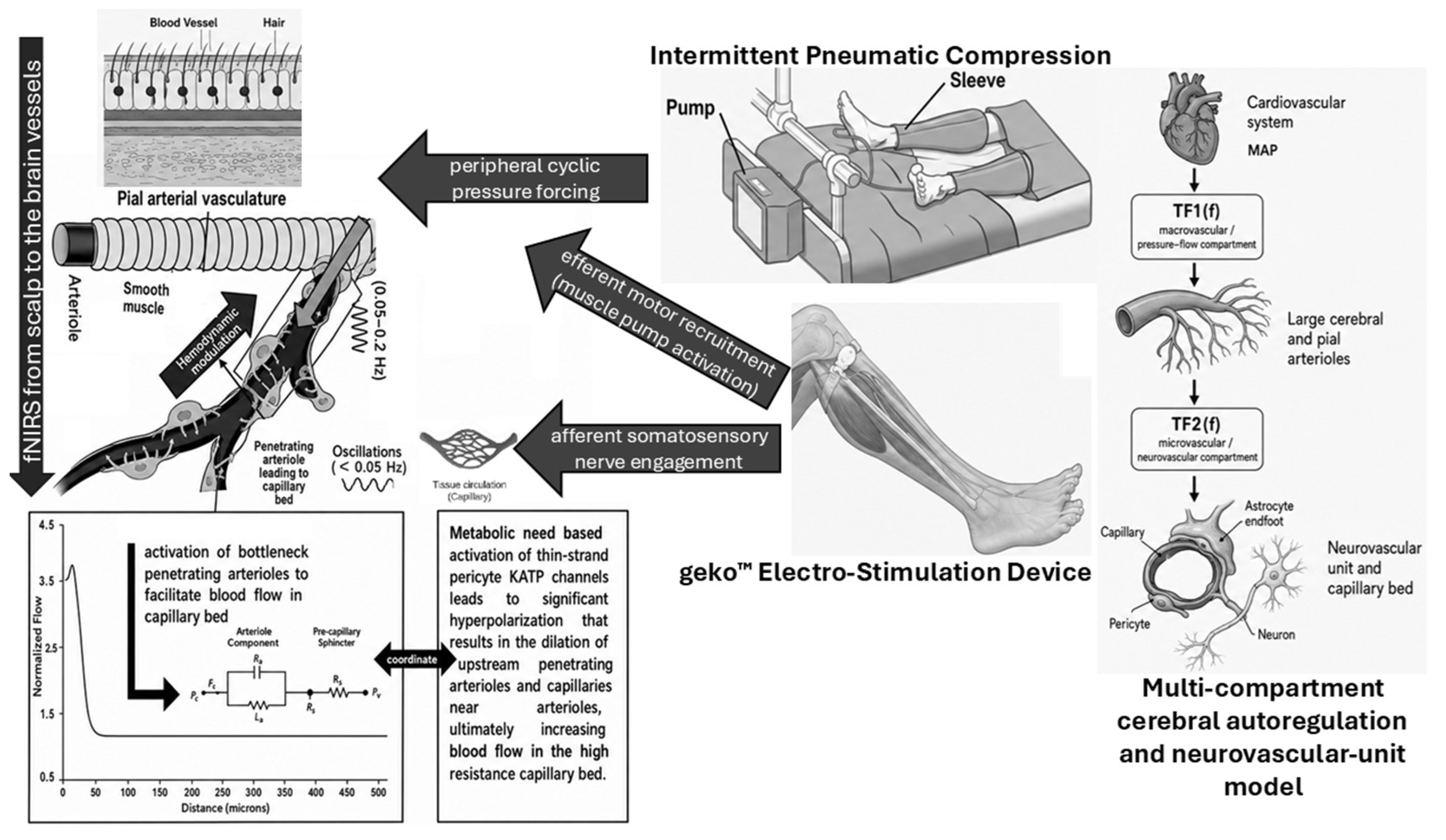

Table 1.
Intervention arms, device characteristics, physiological mechanisms, and matched application protocols for geko™ CPN-NMES and IPC in RETRAIN Phase 2.
Table 1.
Intervention arms, device characteristics, physiological mechanisms, and matched application protocols for geko™ CPN-NMES and IPC in RETRAIN Phase 2.
| Arm | Device | Physiological Mechanism | Application Protocol |
|---|---|---|---|
| geko™ NMES (experimental) | geko™ T-3 (Firstkind Ltd, UK), FDA 510(k) cleared for VTE prevention | Bilateral CPN stimulation evoking rhythmic ankle dorsiflexion. Activates calf/foot muscle venous pump (efferent). Additionally recruits afferent somatosensory fibres engaging muscle metaboreflex and sympathetic baroreflex modulation [17],[18]. Low frequency (1 Hz) optimizes venous refill time. Prior healthy-volunteer fNIRS study confirmed cerebral blood volume increases [16]. | Applied bilaterally at fibular head. Amplitude and pulse width titrated to VTE-optimal visible dorsiflexion (27-54 mA, 35-560 μs). 5-min active period per session. |
| IPC (active comparator) | Bilateral pneumatic compression sleeves (standard care, manufacturer per site protocol) | Cyclic external pneumatic leg compression augments venous return through passive mechanical outflow. No direct afferent nerve recruitment but may modulate low-frequency cerebrovascular oscillations (myogenic, neurogenic, endothelial bands) via arterial pressure pulse transmission and venous capacitance changes as demonstrated in prior fNIRS studies [23],[24]. Temporal forcing pattern and IPC-autoregulation pathway differ from 1 Hz CPN-NMES. | Applied bilaterally per manufacturer instructions and clinical protocol. 5-min active period per session matched to NMES arm. |
Table 2.
Pre-specified RETRAIN Phase 2 endpoints, statistical testing hierarchy, and SPIRIT 2025 endpoint classifications.
Table 2.
Pre-specified RETRAIN Phase 2 endpoints, statistical testing hierarchy, and SPIRIT 2025 endpoint classifications.
| Endpoint Class | Definition | Testing | SPIRIT 2025 Classification |
|---|---|---|---|
| Primary haemodynamic endpoint | First-level GLM-derived HbT β estimates for baseline and active device periods, analysed at second level by treatment arm, period, ROI, and participant, with the primary estimand defined by the Group x Period interaction. | Step 1, confirmatory, α=.05 | Primary |
| Mechanistic OMA endpoint | Active-minus-baseline change in OMA-derived brain-like modal frequencies in the 0.01-0.15 Hz range testing the Group x Period interaction. | Step 2 | Secondary |
| dCA endpoint | Period-specific change in pressure-haemodynamic coupling from synchronized beat-to-beat MAP and HbO, using one usable (~4-min) correlation window per baseline/active period. | Step 3 | Secondary |
| Wavelet frequency-band endpoint | Active-minus-baseline change in HbO/HbT wavelet amplitude within predefined low-frequency oscillatory bands III, 0.052-0.145 Hz, IV, 0.021-0.052 Hz, and V, 0.0095-0.021 Hz. Group x Period effects will be evaluated independently for each band. | Step 3, confirmatory secondary family | Secondary |
| Experimental modal analysis and multimodal coupling | Exploratory multimodal EEG-fNIRS and systemic physiological coupling, with artifact/source sensitivity and autocorrelation-aware surrogates. These analyses are hypothesis-generating, not an independent validation of cortical or neurovascular origin. | Step 3, secondary, hypothesis-generating | Secondary |
| SINDYc/system identification | Exploratory descriptive input-response model feasibility (SINDYc), held out predictive validity and coefficient stability only. | Exploratory, clearly bounded | Exploratory |
| Safety endpoints | Incidence of adverse events, serious adverse events, adverse device effects, serious adverse device effects, device deficiencies, protocol-defined hypotension, skin irritation, unexpected cardiac events, and neurological deterioration temporally associated with device use during or within 30 minutes after intervention. | Safety, all enrolled participants | Safety |
Table 3.
Pre-specified analysis populations for RETRAIN Phase 2.
| Population | Definition | Primary use |
|---|---|---|
| Randomized (ITT) | All 53 randomized participants, analysed according to allocated arm regardless of adherence or data quality. | Baseline characteristics, CONSORT flow, supportive safety summaries. |
| Physiology analysis | Randomized participants with ≥1 qualifying paired fNIRS observation, valid first-level HbT contrast and finite uncertainty, channel, period and artifact losses reported by arm. This observed-data population is not equivalent to all randomized participants. | All haemodynamic primary and secondary endpoints including βHbT, βHbO, βHbR, wavelet and coupling analyses. |
| OMA analysis | Physiology subset with ≥20 quality-approved long-separation channels and an estimable screened brain-like mode in both paired periods. N4SID eligibility uses recorded length, stable modes, cycle coverage and a transparently labelled empirical block-Hankel adequacy check, QC failures are logged. Because the Hankel matrix is built from all quality-approved raw channels rather than the effective rank, this rule is not established for raw multichannel identification (Supplementary Section S-AM). | Mechanistic OMA modal frequency and damping analyses. |
| dCA/pressure-haemodynamic coupling | Synchronized continuous beat-to-beat MAP and fNIRS are required for COx/transfer-function estimation. Cuff-only measurements may support descriptive period covariates, not time-series dCA estimation. | COx, MAP-fNIRS transfer-function and wavelet coupling analyses. |
| Safety | All 53 participants who received at least one session with either device. | All safety, AE, ADE, DD analyses. |
Table 4.
Pre-specified five-step fallback ladder for RETRAIN Phase 2.
| Step | Action | Model formula |
|---|---|---|
| 1 (default) | Apply Tier 1 as specified. | βHbT ~ Group x Period + SessionIndex + NIHSSStratum + HemROI + tcpCO₂period + (1 | Participant) |
| 2 (fallback 1) | Remove tcpCO₂period from fixed effects. | βHbT ~ Group x Period + SessionIndex + NIHSSStratum + HemROI + (1 | Participant) |
| 3 (fallback 2) | Replace NIHSSStratum (2 df) with continuous baselineNIHSS (1 df). | βHbT ~ Group x Period + SessionIndex + baselineNIHSS + HemROI + (1 | Participant) |
| 4 (fallback 3) | Remove HemROI (eliminates within-participant ROI nesting). | βHbT ~ Group x Period + SessionIndex + baselineNIHSS + (1 | Participant) |
| 5 (fallback 4) | Minimal model. Cluster bootstrap is sole inference, parametric p-value not reported. | βHbT ~ Group x Period + (1 | Participant) |
Table 5.
Hierarchical confirmatory testing framework for RETRAIN Phase 2 endpoints.
| Step | Test | Model | Criterion | If Significant | If Not Significant |
|---|---|---|---|---|---|
| 1 | Primary haemodynamic: Group x Period on βHbT | Tier 1 (primary), supported by Tiers 2 and 3 | Two-sided α = 0.05, primary inference from 95% cluster bootstrap CI excluding zero | Proceed to Step 2 | Steps 2 and 3 reported descriptively only, no confirmatory claims. |
| 2 | Primary mechanistic: Group x Period on OMA brain-like ModalFrequency | OMA primary model | Two-sided α = 0.05, 95% cluster bootstrap CI | Proceed to Step 3 | dCA-surrogate, wavelet, oxygenation, damping, coupling endpoints reported descriptively. |
| 3 | Seven supported quantitative secondary tests: COx, wavelet bands III, IV and V, βHbO, βHbR, OMA damping. | Separate LME models per endpoint (Tier 1 covariate structure, appropriate outcome transformation) | BH-FDR q<0.05 across seven supported tests, conditional on Steps 1 and 2, amendment documented. | Mechanistic convergence evidence reported | Descriptive interpretation only. |
Disclaimer/Publisher’s Note: The statements, opinions and data contained in all publications are solely those of the individual author(s) and contributor(s) and not of MDPI and/or the editor(s). MDPI and/or the editor(s) disclaim responsibility for any injury to people or property resulting from any ideas, methods, instructions or products referred to in the content. |
© 2026 by the authors. Licensee MDPI, Basel, Switzerland. This article is an open access article distributed under the terms and conditions of the Creative Commons Attribution (CC BY) license (http://creativecommons.org/licenses/by/4.0/).
Copyright: This open access article is published under a Creative Commons CC BY 4.0 license, which permit the free download, distribution, and reuse, provided that the author and preprint are cited in any reuse.