
Submitted:
11 November 2026
Posted:
12 November 2026
You are already at the latest version
Abstract
Upstream start–stop elements represent an emerging class of translational regulators that can strongly repress downstream protein synthesis. However, the contribution of adjacent RNA secondary structures to their regulatory activity remains poorly understood. Here, we investigated the potential interplay between start–stop elements and downstream RNA G-quadruplexes (rG4s) in human mRNA 5′ untranslated regions (5′-UTRs). By integrating ribosome profiling data with experimentally supported rG4 annotations, we identified candidate start–stop elements associated with nearby rG4-prone sequences. Among these, DUSP4 was selected for further investigation based on its pronounced ribosome occupancy peak at the start–stop element, the proximity of a downstream G-rich motif, and evolutionary conservation of the start–stop element and its context. Using fluorescence spectroscopy, circular dichroism, UV melting, and NMR experiments, we demonstrated that the downstream G-rich motif forms a stable, potassium-dependent, three-layered rG4 in vitro. Functional analysis in HEK293T cells revealed that disruption of the start–stop element significantly increased reporter expression in both plasmid- and mRNA-based systems. Mutations in the rG4-prone sequence enhanced reporter activity in the plasmid system. Together, these findings identify a functional start–stop element in the DUSP4 5′-UTR and a closely positioned, structurally characterized rG4-prone motif, suggesting a potential additional layer of post-transcriptional regulation.
Keywords:
DUSP4
; translational regulation
; start–stop elements
; RNA G-quadruplexes (rG4s)
; 5′ untranslated region (5′-UTR)
; ribosome profiling
; translation initiation
1. Introduction
Translational control is a key stage of gene-expression regulation because it enables cells to rapidly alter protein output without requiring changes in mRNA level. Translation initiation is the most tightly regulated step of protein synthesis. Its efficiency is determined both by the presence of a start codon in an appropriate sequence context and by the combined activity of cis-regulatory elements within the 5′ untranslated region (5′-UTR). These include diverse classes of upstream translons, such as those initiated at upstream AUG codons (uAUGs), upstream open reading frames (uORFs), and start–stop elements; terminal oligopyrimidine tracts (TOP); binding sites for RNA-binding proteins; RNA modifications; and secondary-structure elements, including hairpins, pseudoknots, and RNA G-quadruplexes [1,2,3]. Combined together, these elements can modulate ribosome recruitment, scanning kinetics, selection of alternative initiation sites, translation reinitiation in the 5′-UTR, and transcript stability [1,2,3].
One important mechanism of 5′-UTR-mediated regulation involves upstream translons (uTranslons). Translation initiation at a uAUG generally reduces translation of the downstream CDS by engaging scanning pre-initiation complexes in uORF translation before they reach the main AUG. The value of this effect depends on the sequence context of the uAUG, the length of the uTranslon, the distance between its stop codon and the CDS start site, and the reinitiation competence of the post-termination 40S ribosomal subunit. During cellular stress, changes in activity of translation-initiation factors can alter the balance between translation of uORF and CDS, contributing to the selective translation of specific mRNAs. The functional importance of upstream translation is underscored by the fact that 5′-UTR variants that create uAUGs, alter uORFs, or disrupt their stop codons are associated with inherited diseases, including through changes in CDS translation [4,5,6,7,8,9,10].
A distinct class of uTranslons comprises start–stop elements, in which an initiating AUG codon, or an alternative start codon, is immediately followed by a UAA, UAG, or UGA termination codon. Unlike canonical uORFs, translation of start–stop elements does not include an elongation phase. At a start–stop element, initiation and termination are colocalized: the ribosome assembles at the AUG codon but does not proceed to decode a subsequent codon. This fundamental feature supports the classification of start–stop sequences as an independent type of 5′-UTR regulatory element rather than as extremely short uORFs11.
Previous studies have shown that translation of start–stop elements depends on the non-canonical translation factors DENR and MCTS1 [11,12,13,14]. Reporter-based experiments demonstrated that knockdown of DENR and MCTS1 substantially reduced the translation of start–stop elements, but not that of minimal canonical uORFs; moreover, start–stop elements in stronger Kozak contexts exhibited a greater effect12. Subsequent studies suggested that DENR and MCTS1 are required for reinitiation and that only uTranslons bearing specific penultimate codons, including AUG, depend on these factors [11,13,14].
A recent systematic analysis of the human transcriptome identified start–stop sequences in the 5′-UTRs of more than 1,400 genes11. The authors showed that start–stop elements are associated with reduced initiation at the downstream CDS and, in several reporter systems, repress its translation considerably more strongly than minimal, nine-nucleotide uORFs. They proposed a model in which ribosomes are retained at start–stop elements through recurrent cycles of initiation, termination, ribosome splitting, and 60S-subunit joining. In contrast to minimal uORFs, ribosome retention at start–stop elements was less sensitive to the global reduction in translation initiation induced by cellular stress.
In plants and yeast, metabolite-dependent regulation mediated by start–stop elements has also been described [15,16,17,18]. For example, a global ribosome-profiling analysis in Arabidopsis thaliana showed that high boron availability suppresses translation of transcripts enriched in start–stop elements16. Using NIP5;1, which encodes a boric-acid channel, as a model, it was shown that boric acid induces ribosome stalling at a start–stop element through stable retention of eRF1 on the 80S ribosome, ultimately leading to mRNA degradation18. Moreover, a recent study demonstrated that this start–stop element mediates not only translational repression, but consequently promotes ARGONAUTE1-dependent transcriptional downregulation of NIP5;1 expression [17,18].
It remains unclear whether cis-acting factors can dynamically modulate the probability of initiation specifically at start–stop elements. A characteristic feature of these elements is the suboptimal nucleotide at position +4: because the AUG codon is immediately followed by a stop codon, this position is necessarily occupied by U rather than G, the latter being characteristic of a strong Kozak initiation context. Nevertheless, ribosome-profiling data indicate that initiation at start–stop elements suppresses translation of the downstream CDS more strongly than initiation at minimal 9-nucleotide uORFs11. The efficiency with which a start–stop AUG is recognized may therefore depend on local structural properties of the 5′ leader, including RNA secondary structure.
In this context, a stable secondary structure positioned downstream of the start codon may be particularly important. Classical studies by M. Kozak showed that, for example, a hairpin located downstream of an AUG codon increases recognition of an initiation codon in a weak sequence context19. Such structures are thought to slow the movement of the scanning complex, thereby extending the time available for start-codon recognition. A similar principle has been supported at the transcriptome-wide level: hairpins located immediately downstream of uAUGs promote the use of upstream initiation sites, whereas their unwinding by RNA helicases during the immune response enables ribosomes to bypass uAUGs and translate the downstream CDS in plant cells20. Consistent with a potential link between start–stop regulation and RNA secondary structure, sequences flanking translated start–stop elements exhibit higher GC content than those flanking translated minimal 9-nucleotide uORFs11.
RNA G-quadruplexes (rG4s) are promising candidates for dynamic structural regulators of start–stop elements. rG4s are non-canonical four-stranded RNA structures formed by G-rich sequences through the stacking of G-quartets. When positioned downstream of a start codon, they may alter the probability that the scanning complex recognizes that codon, while their folding state can vary with cellular conditions. In most characterized cases, rG4s in 5′-UTRs repress cap-dependent translation. Such effects have been demonstrated for the 5′-UTRs of ZIC-1, MT3-MMP, NRAS, TERF2, and ADAM10, in which deletion or mutation of a rG4 motif altered translation of reporter constructs with luciferase and/or corresponding protein expression [21,22,23,24,25]. Subsequent studies have indicated that rG4-mediated translational regulation can be performed by RNA-binding proteins. In particular, the helicases DHX36, DDX3X, GRSF1, DDX21, and DHX9 promote translation of mRNAs containing rG4s in their 5′-UTRs by resolving rG4 structures [26,27,28,29,30]. However, rG4 function is not limited to translational repression: several studies have suggested that rG4s in 5′-UTRs can promote translation, including by acting as components of putative internal ribosome entry sites (IRESs) [31,32,33,34].
Based on these observations, we hypothesized that an rG4 forming downstream of a start–stop element could slow scanning by the pre-initiation complex and increase recognition of a suboptimal uAUG. Enhanced initiation at the start–stop element would consequently be expected to strengthen repression of the downstream CDS. Because rG4s can fold and unfold in response to cellular conditions, they may provide a mechanism for dynamic and fine-tuned translational regulation. This model remains hypothetical: published studies convincingly link downstream RNA structures to the selection of upstream start sites and demonstrate the effects of rG4s on uORF translation27, but direct evidence for rG4-mediated regulation of an elongationless start–stop element has not yet been reported. Therefore, in this study, we systematically screened human mRNA 5′-UTRs for potential regulatory combinations of start–stop elements and downstream rG4s, prioritized promising candidates based on bioinformatic analysis, and selected DUSP4 for experimental characterization of their potential roles in translational regulation.
2. Results
2.1. Bioinformatic Search & Characterization
To identify potential regulatory combinations of start–stop elements and rG4s in human mRNA 5′-UTRs, we performed an integrative analysis of publicly available ribosome profiling and high-throughput RNA structure probing datasets. We hypothesized that an rG4 located immediately downstream of a start–stop element could influence recognition of the start codon by the scanning preinitiation complex, thereby modulating the repression of downstream CDS translation.
As a starting point, we used annotations of translated regions from the RiboCrypt database (https://ribocrypt.org/), which integrates data from multiple ribosome profiling experiments. From the Homo sapiens dataset, we extracted start–stop elements characterized by an initiation codon immediately followed by a termination codon, which precludes an intermediate elongation phase. In total, 1652 start–stop elements were identified across 1,468 unique human transcripts.
To identify potential structural regulators, we used the QUADRatlas database35, which integrates experimentally identified and computationally predicted rG4s. The genomic coordinates of all annotated human rG4-forming regions were intersected with those of the selected start–stop elements, taking strand orientation into account (Figure 1). We retained combinations in which the 5′ boundary of an annotated rG4-forming region was located within a window extending from the first nucleotide of the start–stop initiation codon to a position 50 nucleotides downstream of its termination codon.
This distance threshold was chosen based on the proposed regulatory mechanism and to ensure sufficiently broad coverage of potentially functional combinations. Previous studies have demonstrated that stable RNA secondary structures located downstream of an initiation codon can enhance its recognition by slowing PIC scanning. In particular, RNA secondary structures positioned approximately 14–41 nucleotides downstream of an initiation codon can facilitate start codon recognition [19,27,36,37]. However, the optimal spacing between a start–stop element and a downstream rG4 remains unknown. Therefore, we applied a broader 50-nucleotide screening window to account for potential inaccuracies in rG4 boundary annotations and avoid excluding potentially relevant candidates.
The initial intersection identified 299 putative start–stop–rG4 combinations. To increase the confidence of candidate selection, putative rG4-forming sequences supported exclusively by computational predictions were excluded. Only rG4-containing regions with experimental evidence from rG4-seq or RT-stop profiling were retained for further analysis. These approaches identify RNA regions with experimentally detectable rG4-forming potential; however, they do not, by themselves, prove that the corresponding structures are folded in living cells.
Application of this filter yielded 34 combinations in unique transcripts. Thus, by integrating independent transcriptome-wide datasets, we established a set of candidate transcripts containing closely spaced start–stop elements and experimentally identified putative rG4s.
The 34 candidate start–stop–rG4 combinations were subsequently subjected to manual inspection to identify the most promising targets for experimental investigation. First, aggregated human ribosome profiling data available in RiboCrypt were visually examined, with particular attention to the distribution of ribosome-protected fragments (RPFs) around the start–stop elements. Preference was given to candidates displaying a well-defined RPF peak at the start–stop site, clearly distinguishable from surrounding ribosome occupancy signals. Second, the relative positioning of the start–stop elements and downstream G-rich sequences was examined in detail. Because experimentally annotated rG4-forming regions may include additional nucleotides upstream of the actual G-rich motif, the distance to the putative G4-forming core was assessed based on the underlying nucleotide sequence rather than solely on the annotated rG4 boundaries. Based on the combined assessment of ribosome occupancy and proximity of the downstream G-rich motif, several genes, including DUSP4, were selected as the most promising candidates.
Among the shortlisted candidates, DUSP4 was prioritized for further investigation based on the evolutionary conservation of its start–stop element. Nucleotide-level conservation was assessed using phyloP scores from the 100-vertebrate conservation track in the UCSC Genome Browser (Figure 2A). Visual inspection revealed elevated conservation scores across the AUG–UGA start–stop sequence and its context, suggesting evolutionary constraints on this region and supporting its functional significance. Notably, the reduced phyloP score at the second position of the stop codon is consistent with variation between TGA and TAA, which preserves the termination signal despite nucleotide divergence. The sequence logo (Figure 2B) further illustrated the conservation of the start–stop architecture and the downstream G-rich region. Unlike phyloP scores, the sequence logo reflects nucleotide frequencies and information content without accounting for phylogenetic relationships, explaining the differences in their conservation patterns. Together with the pronounced ribosome-protected fragment (RPF) peak at the start–stop element and the close proximity of a downstream G-rich motif, this conservation pattern provided the rationale for selecting DUSP4 for subsequent experimental characterization.
The DUSP4 mRNA 5′-UTR contains several putative regulatory elements (Figure 3). These include a 30-codon-long upstream open reading frame (uORF) initiated at a non-canonical CUG start codon. The uORF begins 49 nucleotides downstream of the 5′ end of the transcript (ENST00000240100). An AUG–UGA start–stop element is located 78 nucleotides downstream of the uORF, followed by a putative rG4-forming region positioned only 12 nucleotides downstream of the stop codon. The main DUSP4 coding sequence begins 186 nucleotides downstream of the start–stop element.
Aggregated human ribosome profiling data available in the RiboCrypt database shows a pronounced peak of ribosome-protected fragments (RPFs) at the DUSP4 start–stop element (Figure 3). The intensity of this peak markedly exceeded the signals observed within the uORF and near the beginning of the coding sequence in the displayed profile. This distribution indicates elevated local ribosome occupancy at the AUG–UGA sequence and is consistent with the accumulation of ribosomal complexes at the start–stop element. Together with the close proximity of the putative rG4-forming region, these observations suggest that the two elements may cooperatively regulate the DUSP4 translation and provide a rationale for investigating their potential functional interplay.
2.2. In Vitro Characterization of a Putative RNA G-Quadruplex in the DUSP4 5′-UTR
The identification of a putative rG4-forming region in close proximity to the DUSP4 start–stop element raised the question of whether this sequence could adopt a stable rG4 structure. Although the transcriptome-wide datasets provided experimental evidence of rG4-forming potential, the structural properties of this region remained to be characterized. Moreover, to avoid overlooking additional rG4-prone motifs that might contribute to translational regulation, we decided to systematically screen the entire DUSP4 5′-UTR and the proximal region of its coding sequence for potential rG4-forming sequences.
Using the QGRS tool38 to search for potential rG4-forming sequences, we found four such sequences in the 5′-UTR and the beginning of the CDS of DUSP4 mRNA (Figure 4). We synthesized oligonucleotides corresponding to these sequences along with their mutated analogs containing G-to-A substitutions, which are unable to form rG4s, as negative controls. The choice of G-to-A substitutions was dictated by the lowest probability of alternative hairpin structure formation in the resulting controls.
First, a thioflavin T assay was performed, which is based on the increase in thioflavin T (ThT) fluorescence intensity upon complex formation with rG4s [39,40]. TERRA rG441 and RNA hairpin rDS2641 served as positive and negative controls, respectively. Only the G3 sequence showed an increase in ThT fluorescence intensity comparable to the TERRA rG4 control (Figure 5A). However, an increase was also observed for G3 MUT, possibly due to the known ability of ThT to light up in the presence of GA-rich DNA duplexes42.
Circular dichroism (CD) spectroscopy also allows the confirmation of rG4 formation by a characteristic signature with a maximum at 263 nm and a minimum at 240 nm (Figure 5B, Figure S1A-C)43. Among the wild-type sequences, only G3 WT (Figure 5B) and G4 WT (Figure S1C) matched this profile. Notably, however, some mutant sequences incapable of forming rG4s also matched this pattern, which is related to the close signals of A-form RNA duplexes43. Therefore, CD spectra were complemented with thermal difference spectra (TDS) analysis, involving subtraction of rG4 spectra in the folded and unfolded states at room temperature and 95 °C, respectively (Figure 5C, Figure S1D-F)44. Even in the case of RNA, this method permits unambiguous determination of whether a rG4 or RNA duplex forms: a rG4 is characterized by maxima at 240 nm and 265 nm and a negative minimum at 295 nm, the latter being absent in RNA duplexes. Among all wild-type and mutant sequences, only G3 WT (Figure 5C), which was the leader in the ThT assay and whose folding was confirmed by CD experiments, exhibited the characteristic negative minimum at 295 nm. This sequence was selected as the most probable and reliable rG4-forming candidate for further studies.
To confirm further rG4 formation by the G3 WT sequence, CD spectra were recorded during potassium chloride titration (Figure 5D) [43,45]. The increase in the maximum with the sequential addition of 1 mM, 10 mM, and 100 mM potassium chloride to G3 WT confirms rG4 formation (Figure 5D), since potassium cations localize between G-tetrads and stabilize the rG4 structure. In addition, we compared CD spectra of all sequences in the presence of 100 mM KCl and 100 mM LiCl. With the exception of G3 WT, none of the wild-type sequences showed an increase in the maximum upon replacing Li+ with K+, which argues against rG4 formation by these sequences under the conditions tested (Figure 5D, Figure S1G-I). In turn, UV melting permits the determination of rG4 thermal stability and discrimination between a rG4 and an RNA duplex, as the former yields a melting curve with a decrease in absorption at 295 nm (Figure 5E)46. First, in the case of G3 WT melting, we observed a melting curve at 295 nm, supporting rG4 formation. Second, the melting temperature increased from 40 °C to 70 °C upon raising the potassium chloride concentration from 14 mM to 140 mM and from 40 °C to 60 °C upon the addition of the G4 stabilizer pyridostatin (PDS) [47,48,49]. These results confirm rG4 formation and stability at the intracellular concentration of potassium cations (140 mM) and temperature (37 °C).
Furthermore, the structure of G3 WT was studied by 1H NMR spectroscopy. Twelve signals characteristic of rG4 formation were observed in the imino proton region of 10–12 ppm (Figure 6). This indicates the formation of three G-tetrads, suggesting that the G3 WT sequence preferentially folds into a three-layered rG4, rather than a two-layered one that could potentially form without the G-tract breakings. However, a broad hump in the imino proton region hinted at the possibility of alternative folding. No signals in the imino proton region were observed in 1H NMR spectra for G3 MUT (Figure S2).
In summary, having confirmed by biophysical methods that a stable rG4 forms at intracellular potassium cation concentrations and temperature, we proceeded to investigate the influence of this rG4 on mRNA translation in vivo.
2.3. Cellular Reporter Assays Reveal Translational Repression of DUSP4 by a Start–Stop Element and a Downstream RNA G-Quadruplex
To investigate the functional contribution of the regulatory elements (start-stop, uORF and rG4) identified within the DUSP4 5′-UTR, we performed dual-luciferase reporter assays in HEK293T cells. A549 cells were selected for 5′-RACE based on the reported expression of DUSP4 mRNA in this cell line according to the Human Protein Atlas (HPA)50. The DUSP4 5′-UTR sequence determined by 5′-RACE, together with an N-terminal fragment of the DUSP4 coding sequence, was fused in-frame to Firefly luciferase (Fluc) lacking its native initiation codon (Figure 7). Thus, Fluc expression depended on translation initiation at the main DUSP4 AUG. Renilla luciferase (Rluc), expressed from an independent promoter, served as an internal control. To examine the contribution of individual 5′-UTR elements, we generated constructs containing a CUG-to-CAG substitution in the initiation codon of the upstream open reading frame (ΔuORF), an AUG-to-AAG substitution in the start–stop element (ΔSt–St), or point G-to-A substitutions in the G3 motif that were shown to disrupt rG4 formation in vitro (ΔrG4). An additional construct combined mutations in the start–stop element and G3 motif (ΔrG4ΔSt–St).
In the plasmid-based reporter system, inactivation of the CUG-initiated uORF did not significantly affect the Fluc/Rluc ratio relative to the wild-type (WT) construct (Figure 7). In contrast, disruption of the start–stop element increased reporter expression, indicating a repressive effect of the native AUG–UGA sequence. This finding is consistent with a recent systematic analysis that identified start–stop elements in more than 1,400 human genes and demonstrated that these elements repress downstream translation more strongly, on average, than minimal nine-nucleotide uORFs11. The authors proposed a ribosome-retention mechanism involving repeated cycles of initiation, immediate termination, ribosomal subunit dissociation, and 60S subunit rejoining. Such a mechanism could account for the pronounced accumulation of ribosome-protected fragments observed at the DUSP4 start–stop element (Figure 3). Mutations in the G3 motif were also associated with increased reporter activity, while the double mutant exhibited the highest Fluc/Rluc ratio among the tested constructs, exceeding the effects of either single mutation. These observations suggest that both the start–stop element and the downstream G3 region contribute to repression of reporter expression in the plasmid-based system. However, the increased activity of the double mutant does not, by itself, establish a cooperative interaction between the two regulatory elements.
To examine whether these effects were maintained independently of plasmid transcription and nuclear RNA processing, we performed analogous experiments using in vitro-transcribed, capped, and polyadenylated reporter mRNAs. This approach bypasses promoter-dependent transcription, splicing, and potential cryptic transcript formation, thereby reducing confounding effects associated with plasmid-based reporter systems34. Fluc mRNAs carrying the WT or mutant DUSP4 5′-UTRs were cotransfected with a separately synthesized Rluc-control mRNA into HEK293T cells (Figure 8). Consistent with the plasmid-based experiments and the previously reported repressive properties of start–stop elements11, disruption of the DUSP4 start–stop element significantly increased the Fluc/Rluc ratio. In contrast, mutations in the CUG-initiated uORF or the G3 motif did not produce statistically significant changes relative to WT. Although the double mutant exhibited higher reporter activity than WT, its effect did not exceed that of the start–stop inactivation alone.
These findings demonstrate that the repressive effect associated with the DUSP4 start–stop element is reproducible in both reporter formats, consistent with its proposed role as an upstream translational repressor. In contrast, the contribution of the G3 region was less consistent; it exhibited an effect observed in the plasmid-based system but no significant independent effect following direct mRNA transfection. This discrepancy may reflect differences in RNA folding pathways between the two experimental systems. In particular, cotranscriptional folding and interactions with RNA-binding proteins during intracellular mRNA biogenesis may favor the formation of specific RNA structures, whereas in vitro-transcribed mRNAs may adopt alternative conformations that persist following transfection. Consequently, the G3 motif may differ in its folding state or structural accessibility between the two reporter systems. Thus, while the start–stop element represents the principal functional repressor within the investigated 5′-UTR, the regulatory contribution of the adjacent rG4-forming sequence appears to depend on the experimental context.
Although disruption of the upstream CUG codon did not significantly affect downstream reporter expression, this observation did not exclude translation of the corresponding uORF. To examine its translational potential, we introduced a HiBiT-tag in-frame immediately upstream of the uORF termination codon. The resulting construct produced a detectable luminescence signal substantially above background (Figure S3), supporting translation initiation within the upstream reading frame and production of a tagged peptide. Thus, the DUSP4 uORF may encode a stable short peptide whose potential function is independent of its effect on the downstream CDS expression.
Previous studies have demonstrated that uORFs can modulate translation under oxidative stress conditions [51,52,53,54], whereas start–stop elements have been reported to maintain their repressive activity despite stress-induced reductions in global translation initiation11. To investigate whether the regulatory activity of the DUSP4 upstream elements could be modulated by cellular stress, plasmid-transfected cells were treated with sodium arsenite (Figure S4A). Arsenite treatment reduced Fluc/Rluc ratios across the tested constructs but did not substantially alter their relative expression patterns: disruption of the CUG-initiated uORF remained without a significant effect, whereas inactivation of the start–stop element continued to enhance reporter expression. Nevertheless, the absence of an apparent stress-dependent change under the tested conditions does not exclude the possibility that start–stop-mediated regulation of DUSP4 responds to other cellular stresses or physiological signals.
We next investigated whether stabilization of the downstream G3 rG4 could influence DUSP4 reporter expression. The positioning of G3 in close proximity to the start–stop element is consistent with a proposed mechanism in which stable downstream RNA structures slow ribosomal scanning, thereby increasing the probability of recognition of an upstream initiation codon. Moreover, previous studies have demonstrated that rG4s can influence upstream initiation, with depletion of the rG4-resolving helicases DHX36 and DHX9 enhancing translation of rG4-associated uORFs while reducing translation of the main CDS 27. Given that G3 forms a stable three-layered rG4 in vitro, we hypothesized that its further stabilization might enhance recognition of the start–stop AUG and consequently strengthen repression of downstream translation. To test this possibility, cells were transfected with plasmids and treated with pyridostatin (PDS), a G4 ligand that increased the melting temperature of G3 in vitro (Figure 5E, Figure S4B). PDS treatment reduced Fluc/Rluc ratios; however, this effect was not selective for reporters containing the intact G3 motif, and the relative pattern of mutational effects remained largely unchanged. The absence of a G3-specific response may be explained by several alternative mechanisms. First, G3 may already form a sufficiently stable rG4 under cellular conditions, leaving little opportunity for further stabilization by PDS. Indeed, the isolated G3 sequence exhibited a melting temperature of approximately 70 °C in the presence of 140 mM KCl, supporting its ability to form a highly stable structure under physiologically relevant ionic conditions. Alternatively, the G3 region may adopt competing RNA conformations in the context of the full-length mRNA, preventing efficient formation or accessibility of the expected rG4. Furthermore, limited intracellular accessibility of G3, insufficient treatment conditions, or effects of PDS on other cellular targets could obscure a potential G3-specific response. Consequently, the lack of a selective effect of PDS neither confirms nor excludes rG4 formation within cellular DUSP4 mRNA. Direct investigation of G3 folding in cells would be required to establish whether this structure contributes to start–stop-mediated translational regulation.
Finally, we explored the possibility of targeted modulation of DUSP4 expression using 2′-O-methyl- and LNA-modified antisense oligonucleotides (ASOs) directed against the upstream regulatory elements (Figure S5-6). Although binding of several ASOs to the target RNA was demonstrated in vitro, none produced consistent enhanced translation of the WT mRNA reporter. Instead, certain ASOs reduced reporter activity, potentially reflecting interference with ribosomal scanning or other effects of RNA–ASO interactions. These experiments are described in detail in the Supplementary Materials (Figure S5-6).
3. Discussion
The present study identifies a previously uncharacterized regulatory region within the DUSP4 5′-UTR containing an AUG–UGA start–stop element and a closely positioned G-rich region capable of forming a stable rG4. Our initial hypothesis was that the downstream rG4 could facilitate recognition of the upstream start–stop initiation codon, thereby enhancing repression of the main coding sequence. Although our research did not provide direct experimental evidence for cooperative interaction between these elements, it revealed a robust repressive contribution of the start–stop element and a potentially context-dependent contribution of the adjacent G-rich region.
The most consistent finding was the increased reporter expression following disruption of the DUSP4 start–stop element in both plasmid- and mRNA-based systems. Together with the pronounced accumulation of ribosome-protected fragments at this site in aggregated Ribo-seq data, these results support the functional importance of the AUG–UGA sequence in controlling downstream gene expression. Recent studies have demonstrated that start–stop elements can act as particularly effective translational repressors, potentially through repeated cycles of initiation, immediate termination, and ribosome recycling11. Our findings are consistent with the proposed repressive function of these elements and extend their functional characterization to DUSP4.
A distinctive feature of the DUSP4 regulatory region is the presence of a G-rich motif immediately downstream of the start–stop element. Biophysical characterization demonstrated that this sequence forms a potassium-dependent, thermally stable rG4 in vitro, consistent with an imperfect three-tetrad structure. Its position relative to the upstream AUG is compatible with the proposed mechanism by which downstream RNA structures facilitate recognition of upstream initiation sites. Previous studies have shown that unresolved rG4s can promote translation initiation at upstream ORFs and consequently reduce translation of downstream coding sequences27. Nevertheless, the functional contribution of G3 was not pronounced across our cellular assays. Whereas disruption of the G3 motif increased reporter expression in the plasmid-based system, a significant independent effect was not detected following direct mRNA transfection. Moreover, although the double mutant exhibited the highest reporter activity in the plasmid-based system, this observation alone does not establish cooperative regulation, and no additional effect beyond start–stop disruption was detected in the mRNA-based system.
Several factors may contribute to the differences between these experimental systems. Plasmid-derived and in vitro-transcribed mRNAs differ in their biogenesis and potentially in their structural and protein-binding environments. In addition, the two reporter formats differ in delivery procedures and incubation times. Differences in mRNA abundance or stability could also contribute to the observed effects. Importantly, G-to-A substitutions in the G3 motif may provoke alternative RNA secondary structures or alter RNA-binding protein interactions independently of rG4 formation. Therefore, although G3 clearly possesses rG4-forming potential in vitro, the extent to which its folding contributes to DUSP4 regulation in living cells remains unresolved.
We also investigated whether the activity of the proposed regulatory module could be modified by cellular conditions. Oxidative stress induced by sodium arsenite did not produce an apparent selective change in the repressive effect of the start–stop element, although overall reporter expression was reduced. This observation is consistent with recent evidence that start–stop-mediated repression can be relatively resistant to changes in global translation initiation11. Our exploratory examination of publicly available ribosome profiling datasets likewise did not reveal an obvious condition-dependent redistribution of ribosome occupancy between the start–stop element and the DUSP4 coding sequence. However, the limited and heterogeneous nature of the available data prevents conclusions about the absence of regulation under other physiological or stress conditions.
Stabilization of rG4s using PDS also failed to produce a G3-specific response in the reporter system, despite the pronounced stabilization of the isolated G3 structure observed in vitro. This lack of selectivity may reflect limited accessibility of the motif, insufficient intracellular target engagement, or the contribution of other cellular RNA structures and PDS targets. Alternatively, the G3 sequence may already adopt a relatively stable structure under the tested conditions, limiting any additional effect of the ligand. These possibilities remain speculative, and the absence of a selective PDS response neither confirms nor excludes G3 folding within cellular DUSP4 mRNA.
An additional observation concerns the CUG-initiated uORF located upstream of the start–stop element. Although mutation of its initiation codon did not significantly affect downstream reporter expression, the HiBiT-tagged construct produced a detectable translation-dependent signal. This suggests that the uORF can support synthesis of a short peptide, potentially providing a function independent of translational repression of the CDS. However, the existence, stability, and biological activity of the corresponding endogenous peptide remain to be established. Further investigation may therefore reveal an additional functional role for this region of the DUSP4 transcript.
DUSP4 encodes a dual-specificity phosphatase involved in the regulation of MAPK signaling, making the mechanisms controlling its expression potentially relevant to cellular signaling responses. Nevertheless, the physiological significance of the identified 5′-UTR elements cannot yet be inferred from reporter experiments alone. Determining whether the start–stop element controls abundance of endogenous DUSP4 protein and whether its activity can be modulated by changes in cellular state will be important directions for future investigation.
In conclusion, our findings establish the DUSP4 AUG–UGA start–stop element as a functional repressive element in cellular reporter systems and identify a stable rG4-forming motif in its immediate downstream vicinity. Although the proposed structural regulation of start–stop activity remains to be demonstrated directly, the coexistence of these elements provides a basis for further investigation of how specific local RNA structure may influence initiation at elongationless upstream regulatory sequences.
4. Materials and Methods
4.1. Bioinformatic Screening and Prioritization of Candidate Regulatory Elements
Annotations of human translated regions were obtained from the RiboCrypt database https://ribocrypt.org/ (all_merged_homo_sapiens dataset, v.3.23). Start–stop elements were identified as regions containing an initiation codon immediately followed by a termination codon. Human rG4 annotations were downloaded from the QUADRatlas database35 in SQL format. Both datasets used the hg38 genome assembly. Data processing and coordinate matching were performed using PostgreSQL.
Genomic coordinates were compared taking strand orientation into account. Candidates were retained if the annotated 5′ boundary of an rG4-containing region was located between the first nucleotide of the start–stop element and a position 50 nucleotides downstream of its termination codon. This window accounted for potential inaccuracies in rG4 boundary annotations. Only rG4 regions supported by experimental rG4-seq or RT-stop profiling data were retained.
Candidates were manually prioritized based on three criteria: (i) the presence of a distinct, well-defined ribosome-protected fragment (RPF) peak at the start–stop element in aggregated RiboCrypt profiling data; (ii) the proximity of the downstream G-rich motif, assessed from its nucleotide sequence rather than the annotated rG4 boundaries; and (iii) evolutionary conservation of the start–stop element, evaluated using phyloP scores from the 100-vertebrate conservation track in the UCSC Genome Browser. Prioritization was performed qualitatively without a numerical scoring system.
Evolutionary conservation of the DUSP4 start–stop element was analyzed using the UCSC Genome Browser (GRCh38/hg38 assembly). A 61-nucleotide region of the human DUSP4 5′-UTR (chr8:29,350,420–29,350,480), encompassing the start–stop element (chr8:29,350,465–29,350,470), was selected for analysis. Multiple sequence alignments were retrieved from the UCSC 100 Vertebrates Multiz Alignment track (multiz100way) in MAF format using the UCSC Table Browser. Alignment blocks were reconstructed according to their positions in the human reference sequence, and all sequences were reverse-complemented to match the transcriptional orientation of DUSP4.
A sequence logo was generated from the aligned nucleotide sequences. Nucleotide frequencies were calculated at each position, excluding alignment gaps and ambiguous nucleotides. Letter heights were expressed as information content (bits), calculated as the difference between the maximum nucleotide entropy (2 bits) and the observed Shannon entropy at each position.
Nucleotide-level evolutionary conservation scores were obtained from the UCSC phyloP 100-way conservation track (phyloP100way) for the same genomic interval. The scores were visualized using custom Python scripts and Matplotlib, with nucleotide symbols from the human reference sequence scaled according to their phyloP scores. Positive and negative scores were displayed above and below the baseline, respectively. The start codon (ATG) and adjacent stop codon (TGA) were highlighted in both visualizations.
4.2. In Vitro Assays
4.2.1. Oligonucleotide Synthesis and Sample Preparation
Oligonucleotides (Table 1 and Table 2) were purchased from Genterra (Moscow, Russia). RNA solutions in a buffer specified for the experiment, were annealed rapidly (heated to 90 °C and then snap-cooled on ice) to facilitate intramolecular folding. Thioflavin T (ThT) and pyridostatin (PDS) were obtained from Shanghai Macklin Biochemical Company (Shanghai, China).
4.2.2. Thioflavin T (ThT) Light-Up Assays
Assays were performed in Corning® 96-well black, flat-bottom polystyrene untreated microplates (Corning, NY, USA) using a buffer composed of 20 mM sodium phosphate (pH 7.4) and 140 mM KCl. The annealed RNA constructs G1–G4 and their corresponding mutants (Table 1) were combined with Thioflavin T (ThT) to yield final concentrations of 2 µM and 1 µM, respectively, and were incubated for 15 min. All fluorescence measurements were acquired at 25 °C using a Tecan M200 PRO microplate reader (Tecan Group Ltd., Männedorf, Switzerland). Emission spectra were recorded over the 470–650 nm range with the excitation wavelength set at 445 nm. Fluorescence intensities at 485 nm, normalized to those of control solutions devoid of oligonucleotides, were employed to construct the bar charts.
4.2.3. Circular Dichroism (CD) and UV Spectroscopy and Melting
CD and UV measurements were carried out using a Chirascan spectropolarimeter (Applied Photophysics, Leatherhead, UK) with a 10 mm optical path quartz cuvette. To compare the CD and TDS spectra of potential rG4-forming sequences G1-G4 and their mutants (Table 1), a buffer containing 20 mM sodium phosphate(pH 7.4), and 140 mM KCl was used. To study the change in the CD spectra of potential rG4-forming sequences G1-G4 (Table 1) in the presence of potassium or lithium cations, 10 mM Tris (pH 7.4), with 100 mM KCl or 100 mM LiCl was used as specified. For the potassium chloride titration of G3 WT, CD spectra were recorded in 10 mM Tris buffer (pH 7.4) containing 100 mM LiCl in the absence of KCl, or in the presence of 1, 10, or 100 mM KCl. CD spectra of annealed 3 µM RNA samples were recorded at 25 °C from 230 to 320 nm. TDS spectra were obtained by subtracting absorption spectra recorded at 95 °C in the range 230 nm to 320 nm from those recorded at 25 °C. Each reported spectrum is an average of two scans.
UV melting experiments were performed on G3 (Table 1) at a heating rate of 1 °C/min from 20 °C to 94 °C, while monitoring absorbance changes at 295 nm. For the UV melting experiments, a buffer containing 20 mM sodium phosphate (pH 7.4) supplemented with either 14 mM or 140 mM KCl was used as specified. For the experiments with PDS, annealed 3 µM RNA samples in buffer containing 20 mM sodium phosphate (pH 7.4), 14 mM KCl were supplemented with PDS (2.5 mM stock solution in DMSO) to a final concentration of 15 µM, or an equivalent DMSO volume.
4.2.4. Nuclear Magnetic Resonance (NMR) Spectroscopy
NMR experiments were performed using the 116 μM solution of the non-labeled oligonucleotides G3 and G3mut (Table 1) in a buffer containing 20 mM sodium phosphate (pH 7.4), and 140 mM KCl, supplemented with 3% (v/v) D2O. The 1D 1H spectra were acquired on a Bruker Avance 700 spectrometer equipped with a cryoprobe at 37 °C. The spectra were recorded using an excitation sculpting water suppression scheme (pulse program zgesgp in the Bruker library) with the following parameters: a spectral width of 12 kHz; a repetition delay of 1.5 s; 32 768 complex points; 5120 scans per FID. Partial aggregation of the G3 oligonucleotide was evident from the presence of a broad signal (hump) at 10.5–11.5 ppm.
4.2.5. Electrophoretic Mobility Shift Assay (EMSA)
For EMSA, 0.5 µM Dusp4_3flank RNA solutions (Table 1) in a buffer containing 20 mM sodium phosphate (pH 7.4), and 140 mM KCl were mixed with 0.5 µM ASOs (Table 2), incubated for 15 min and loaded onto a 15% native polyacrylamide gel. The gel was run at 150 V at room temperature using a standard 1× TBE buffer, stained with SYBR Green II, and visualized using an Amersham Typhoon imager (GE Healthcare) Two independent EMSA repeats were performed.
4.3. In Vivo Assays
4.3.1. Cell Culture
HEK293T and A549 cells (ATCC) were cultured in DMEM/F12 medium (Servicebio, Wuhan, China) supplemented with L-alanyl-L-glutamine (Himedia, Mumbai, India), 10% fetal bovine serum (Himedia, Mumbai, India), 100 U/mL penicillin, and 100 µg/mL streptomycin (Himedia, Mumbai, India) at 37 °C in a humidified atmosphere containing 5% CO2. Cells were routinely tested for mycoplasma contamination using the MycoReport kit (Evrogen, Moscow, Russia).
4.3.2. Reporter Constructs
The 5′-terminal region of DUSP4 mRNA was determined by 5′ rapid amplification of cDNA ends (5′-RACE) using total RNA isolated from A549 cells. The procedure was performed using the 5′ RACE System for Rapid Amplification of cDNA Ends (Thermo Fisher Scientific, Waltham, MA, USA). First-strand cDNA was synthesized using SuperScript II reverse transcriptase and the gene-specific primer DUSP4_5race1 (5′-GTGTTACAGCGCACGTTGAC-3′). Following cDNA purification, a poly(dC) tail was added using terminal deoxynucleotidyl transferase (TdT). The 5′-terminal region was subsequently amplified using the anchor primer AAP (5′-GGCCACGCGTCGACTAGTAC-3′) and the nested gene-specific primer DUSP4_5race2 (5′-CGAACCTAGGATGTAGCCCG-3′), followed by high-fidelity PCR amplification. The resulting product was cloned into the SmaI-digested pUC19 vector and verified by Sanger sequencing. The obtained sequence matched the annotated DUSP4 transcript ENST00000240100 (Ensembl) and was used as the basis for subsequent reporter construction.
Dual-luciferase reporters were generated in a pGL3-derived vector (described in 55) expressing Firefly luciferase (Fluc) and Renilla luciferase (Rluc) from independent promoters. Fluc expression was driven by the EF1α promoter, whereas Rluc expression was driven by the SV40 promoter. Both expression cassettes contained an SV40 polyadenylation signal downstream of their respective coding sequences. The Fluc reporter cassette contained the DUSP4 5′ UTR and a short N-terminal fragment of the DUSP4 coding sequence fused in-frame to the Fluc ORF. The native Fluc AUG codon was removed; therefore, Fluc translation depends on recognition of the native initiation codon of the main DUSP4 ORF. Rluc served as an internal normalization control.
The WT reporter contained the native DUSP4 5′ UTR sequence. Mutant reporters carried a non-initiating substitution in the uORF start codon, the start codon of the start-stop element, or G-to-A substitutions in the G3 quadruplex-forming sequence to disrupt rG4 formation. A double-mutant reporter combining the start-stop and rG4 mutations was also analyzed. All plasmid constructs were verified by Sanger sequencing.
4.3.3. In Vitro mRNA Synthesis
For mRNA-based reporter assays, DNA templates encoding Fluc reporters and Rluc control RNA were amplified by PCR using primers introducing a T7 promoter at the 5′ end and a poly(A) tail of approximately 50 adenosine residues at the 3′ end (primers are listed below). The Fluc templates contained the DUSP4 5′ UTR, a short N-terminal DUSP4 coding-sequence fragment, and the Fluc ORF lacking its native AUG codon.

Table 3.
Oligonucleotide primers used for PCR amplification of DNA templates for in vitro transcription of Firefly and Renilla luciferase reporter mRNAs.
Table 3.
Oligonucleotide primers used for PCR amplification of DNA templates for in vitro transcription of Firefly and Renilla luciferase reporter mRNAs.
| DUSP4_T7_f1 | 5′-GAGTACTTAATACGACTCACTATAGCTGGCTGGGGCAGTGCGCTG-3′ |
| DUSP4_T7_r1 | 5′-TTTTTTTTTTTTTTTTTTTTTTTTTTTTTTTTTTTTTTTTTTTTTTTTTTCCGCCCCGACTCTAGAATTACACGG-3′ |
| hRluc_T7_f1 | 5′-GAGTACTTAATACGACTCACTATAGTGGTTCTTTCCGCCTCAG |
| hRluc_T7_r1 | 5′-TTTTTTTTTTTTTTTTTTTTTTTTTTTTTTTTTTTTTTTTTTTTTTTTTTCTCTAGAATTACTGCTCGTTCTTCAGCACG |
Capped mRNAs were synthesized from PCR templates using the mMESSAGE mMACHINE T7 Transcription Kit (Invitrogen, Waltham, MA, USA) according to the manufacturer’s instructions. DNA templates were removed by TURBO DNase treatment. RNA was purified using the CleanRNA Standard Kit (Evrogen, Moscow, Russia). RNA integrity was assessed by denaturing agarose gel electrophoresis, and RNA concentrations were determined using the Qubit RNA High Sensitivity Assay Kit (Invitrogen, Waltham, MA, USA).
4.3.4. Dual-Luciferase Reporter Assays
HEK293T or A549 cells were seeded in 96-well plates and incubated overnight before transfection. For plasmid-based experiments, 18,000 cells per well were seeded in 100 µL of complete medium, whereas for mRNA-based experiments, 30,000 cells per well were used.
For plasmid-based reporter assays, HEK293T cells were transfected with 50 ng of reporter plasmid DNA per well using 0.2 µL of Lipofectamine 3000 Transfection Reagent (Invitrogen, Waltham, MA, USA) according to the manufacturer’s protocol. Under basal conditions, cells were harvested 48 h after transfection.
For mRNA-based reporter assays, HEK293T cells were cotransfected with 90 ng of Fluc reporter mRNA and 10 ng of Rluc control mRNA per well using Lipofectamine MessengerMAX Transfection Reagent (Thermo Fisher Scientific, Waltham, MA, USA) according to the manufacturer’s protocol. Briefly, Lipofectamine MessengerMAX was diluted in Opti-MEM and incubated for 10 min. Fluc and Rluc mRNAs were separately diluted in Opti-MEM, combined with the diluted transfection reagent, and incubated for an additional 5 min. The resulting transfection mixtures (10 µL per well) were added to the cells, which were incubated for 4 h before preparation of cell lysates.
For experiments involving antisense oligonucleotides (ASOs), HEK293T cells were cotransfected with 90 ng of Fluc reporter mRNA, 10 ng of Rluc control mRNA, and 10 pmol of the indicated 2′-O-methyl- or LNA-modified ASO per well using Lipofectamine MessengerMAX according to the manufacturer’s protocol. Transfection mixtures (10 µL per well) were prepared as described above. Reporter mRNAs and ASOs were cotransfected without prior annealing. A scrambled oligonucleotide (SCR) served as a negative control. Cell lysates were prepared 4 h after transfection.
For pyridostatin (PDS) treatment, the culture medium was replaced 4 h after plasmid transfection with a fresh complete medium containing 10 µM PDS (Shanghai Macklin Biochemical Co., Ltd., Shanghai, China) or an equivalent volume of DMSO. Cells were incubated with PDS for 48 h before harvesting.
For oxidative-stress experiments, plasmid-transfected A549 cells were treated with 40 µM sodium arsenite (NaAsO2; Himedia, Mumbai, India) for 16 h, starting 2 h after transfection. Control wells received the corresponding control medium.
Luciferase activities were measured using the TransDetect Double-Luciferase Reporter Assay Kit (TransGen Biotech, Beijing, China) according to the manufacturer’s instructions in a 384-well assay format. Luminescence was recorded using a Victor X5 plate reader (PerkinElmer, Shelton, CT, USA). Firefly luciferase activity was normalized to Renilla luciferase activity and shown as the Fluc/Rluc ratio.
4.3.5. Exogenous Tagging of uORF and Start–Stop Using Hibit-Tag
To investigate the translation of DUSP4 upstream open reading frames, HiBiT-tagged reporter constructs were generated using the previously described DUSP4 5′-RACE product. First, the unmodified 5′UTR was amplified using the DUSP_UTR_f and DUSP_UTR_r primers (listed below) and cloned into the pUTR-EGFP reporter vector. The pUTR-EGFP plasmid was based on pHaloTag-EGFP (Addgene #86629), where the HaloTag sequence was replaced with a short spacer, introducing two BsmBI sites downstream of the CMV promoter and immediately before EGFP ORF without a start codon. The vector was amplified using primers UTRsite_f and UTRsite_r, followed by self-ligation of the PCR-product.
To introduce HiBiT-tag at the C-terminus of the target uORFs, the initial construct was amplified by inverse PCR using primers tailed with the HiBiT sequence (listed below). Following amplification, the PCR products were self-ligated.
Table 4.
Oligonucleotide primers used for the construction of the pUTR-EGFP reporter vector and HiBiT-tagged DUSP4 uORF and start–stop constructs.
Table 4.
Oligonucleotide primers used for the construction of the pUTR-EGFP reporter vector and HiBiT-tagged DUSP4 uORF and start–stop constructs.
| UTRsite_f | 5‘-AGCACGTCTCCCATGGTGAGCAAGGGCGAG-3‘ |
| UTRsite_r | 5‘-AGCTCGTCTCGGTACACTAAACCAGCTCTGCTTATATAG-3′ |
| HBT_DUSP4_f1 | 5′-TGTTCAAGAAGATTAGCTAAACACACTCTCCTCCACCGG-3′ |
| HBT_DUSP4_f2 | 5′-TGTTCAAGAAGATTAGCTGACCGCGAGGCTGC-3′ |
| HBT_DUSP4_r1 | 5′-GCCGCCAGCCGCTCACCGAGAGAGGACCGCGG-3′ |
| HBT_DUSP4_r2 | 5′-GCCGCCAGCCGCTCACCATAGCAGTCGGAGCGGC-3′ |
| DUSP_UTR_f | 5′-GAACCTCGTCTCTGTACCTGGCTGGGGCAGTGC-3′ |
| DUSP_UTR_r | 5′-TCAATGCGTCTCCCATGTTCATCAGCCTTTTGAGCACACTG-3′ |
The resulting constructs were transfected into HEK293T cells according to the protocol in 3.3.4 and lysis was performed in 24 h. Nano-Glo® HiBit Lytic Detection System (Promega) was used to check the expression of HiBiT transfected cells. The preparation of the Nano-Glo® HiBiT lytic reagent and the detection were performed as instructed by the manufacturer. The luminescence was measured by Perkin Elmer Victor multilabel microplate reader (PerkinElmer, Inc.).
4.3.6. Statistical Analysis
Experiments were performed in at least n=3 independent biological replicates, each including 3 technical replicates per condition. Data are presented as Mean ± SD. Statistical analysis was performed using GraphPad Prism 11. Statistical significance was assessed using ordinary one-way or two-way ANOVA followed by Dunnett’s or Tukey’s multiple comparisons test, and p<0.05 was considered statistically significant.
Supplementary Materials
The following supporting information can be downloaded at the website of this paper posted on Preprints.org.
Author Contributions
Conceptualization, E.A.R., N.M.S., A.V.A., M.P.R.; Methodology, E.A.R., P.N.K., N.M.S., A.V.A., M.P.R.; Formal Analysis, E.A.R.; Investigation, E.A.R., P.N.K., S.D.O., A.I.L., M.N.R., E.T.M.; Resources, M.P.R.; Data Curation, E.A.R.; Writing—Original Draft Preparation, E.A.R and P.N.K.; Writing—Review & Editing, A.I.L., N.M.S., A.V.A., M.P.R.; Visualization, E.A.R., P.N.K. and S.D.O.; Funding Acquisition, M.P.R. and A.V.A. All authors have read and agreed to the published version of the manuscript.
Funding
The study was funded by The Russian Science Foundation grant 24-15-00236 to A.V.A. (the synthesis and characterization of short RNA sequences and ASOs) and 23-14-00058-P to M.P.R. (plasmid construction and cell-based assays) and by the state assignment of Lomonosov Moscow State University, project № 121031300037-7 (bioinformatic target selection).
Institutional Review Board Statement
Not applicable.
Informed Consent Statement
Not applicable.
Data Availability Statement
The original contributions presented in this study are included in the article. Further inquiries can be directed to the corresponding author(s).
Acknowledgments
During the preparation of this manuscript, the authors used ChatGPT (OpenAI) to assist with English-language proofreading and the development of scripts for data analysis and visualization. All AI-assisted outputs were reviewed and verified by the authors, who take full responsibility for the content of the manuscript.
Conflicts of Interest
The authors declare no conflict of interest.
References
- Tidu, A.; Martin, F. The Interplay between Cis- and Trans-Acting Factors Drives Selective mRNA Translation Initiation in Eukaryotes. Biochimie 2024, 217, 20–30. [CrossRef]
- Leppek, K.; Das, R.; Barna, M. Functional 5′ UTR mRNA Structures in Eukaryotic Translation Regulation and How to Find Them. Nat. Rev. Mol. Cell Biol. 2018, 19 (3), 158–174. [CrossRef]
- Razumova, E.; Makariuk, A.; Dontsova, O.; Shepelev, N.; Rubtsova, M. Structural Features of 5′ Untranslated Region in Translational Control of Eukaryotes. Int. J. Mol. Sci. 2025, 26 (5), 1979. [CrossRef]
- Coursimault, J.; Rovelet-Lecrux, A.; Cassinari, K.; Brischoux-Boucher, E.; Saugier-Veber, P.; Goldenberg, A.; Lecoquierre, F.; Drouot, N.; Richard, A.; Vera, G.; Coutant, S.; Quenez, O.; Rolain, M.; Bonnet, C.; Bronner, M.; Lecourtois, M.; Nicolas, G. uORF-introducing Variants in the 5′UTR of the NIPBL Gene as a Cause of Cornelia de Lange Syndrome. Hum. Mutat. 2022, 43 (9), 1239–1248. [CrossRef]
- Occhi, G.; Regazzo, D.; Trivellin, G.; Boaretto, F.; Ciato, D.; Bobisse, S.; Ferasin, S.; Cetani, F.; Pardi, E.; Korbonits, M.; Pellegata, N. S.; Sidarovich, V.; Quattrone, A.; Opocher, G.; Mantero, F.; Scaroni, C. A Novel Mutation in the Upstream Open Reading Frame of the CDKN1B Gene Causes a MEN4 Phenotype. PLoS Genet. 2013, 9 (3), e1003350. [CrossRef]
- Soukarieh, O.; Meguerditchian, C.; Proust, C.; Aïssi, D.; Eyries, M.; Goyenvalle, A.; Trégouët, D.-A. Common and Rare 5′UTR Variants Altering Upstream Open Reading Frames in Cardiovascular Genomics. Front. Cardiovasc. Med. 2022, 9, 841032. [CrossRef]
- Romanelli Tavares, V. L.; Kague, E.; Musso, C. M.; Alegria, T. G. P.; Freitas, R. S.; Bertola, D. R.; Twigg, S. R. F.; Passos-Bueno, M. R. Craniofrontonasal Syndrome Caused by Introduction of a Novel uATG in the 5′UTR of EFNB1. Mol. Syndromol. 2019, 10 (1–2), 40–47. [CrossRef]
- Lee, D. S. M.; Park, J.; Kromer, A.; Baras, A.; Rader, D. J.; Ritchie, M. D.; Ghanem, L. R.; Barash, Y. Disrupting Upstream Translation in mRNAs Is Associated with Human Disease. Nat. Commun. 2021, 12 (1), 1515. [CrossRef]
- Schulz, J.; Mah, N.; Neuenschwander, M.; Kischka, T.; Ratei, R.; Schlag, P. M.; Castaños-Vélez, E.; Fichtner, I.; Tunn, P.-U.; Denkert, C.; Klaas, O.; Berdel, W. E.; Von Kries, J. P.; Makalowski, W.; Andrade-Navarro, M. A.; Leutz, A.; Wethmar, K. Loss-of-Function uORF Mutations in Human Malignancies. Sci. Rep. 2018, 8 (1), 2395. [CrossRef]
- Calvo, S. E.; Pagliarini, D. J.; Mootha, V. K. Upstream Open Reading Frames Cause Widespread Reduction of Protein Expression and Are Polymorphic among Humans. Proc. Natl. Acad. Sci. 2009, 106 (18), 7507–7512. [CrossRef]
- Rendleman, J.; Haizel, S. A.; Wu, S.; Young, L. L.; Liu, J.; Ge, X.; Zou, H.; Mohammad, M. P.; Pressler, M.; Maity, S.; Hronová, V.; Gao, Z.; Herrmannová, A.; Smirnova, A.; Lei, A.; Allgoewer, K.; Sultanov, D.; Hinckley, W. E.; Cheng, Z.; Shelby, L.; Dey, T.; Szkop, K. J.; Topisirovic, I.; Larsson, O.; Hatzoglou, M.; Valášek, L. S.; Vogel, C. Elongationless Start–Stop Elements Are Stress-Resilient Translation Gates That Are More Repressive than uTranslons. Nucleic Acids Res. 2026, 54 (13), gkag627. [CrossRef]
- Schleich, S.; Acevedo, J. M.; Clemm Von Hohenberg, K.; Teleman, A. A. Identification of Transcripts with Short stuORFs as Targets for DENR•MCTS1-Dependent Translation in Human Cells. Sci. Rep. 2017, 7 (1), 3722. [CrossRef]
- Bohlen, J.; Harbrecht, L.; Blanco, S.; Clemm Von Hohenberg, K.; Fenzl, K.; Kramer, G.; Bukau, B.; Teleman, A. A. DENR Promotes Translation Reinitiation via Ribosome Recycling to Drive Expression of Oncogenes Including ATF4. Nat. Commun. 2020, 11 (1), 4676. [CrossRef]
- Meurs, R.; De Matos, M.; Bothe, A.; Guex, N.; Weber, T.; Teleman, A. A.; Ban, N.; Gatfield, D. MCTS2 and Distinct eIF2D Roles in uORF-Dependent Translation Regulation Revealed by in Vitro Re-Initiation Assays. EMBO J. 2025, 44 (3), 854–876. [CrossRef]
- Tsednee, M.; Tanaka, M.; Kasai, K.; Fujiwara, T. Boron-dependent Regulation of Translation through AUGUAA Sequence in Yeast. Yeast 2020, 37 (12), 638–646. [CrossRef]
- Sotta, N.; Chiba, Y.; Miwa, K.; Takamatsu, S.; Tanaka, M.; Yamashita, Y.; Naito, S.; Fujiwara, T. Global Analysis of Boron-induced Ribosome Stalling Reveals Its Effects on Translation Termination and Unique Regulation by AUG-stops in Arabidopsis Shoots. Plant J. 2021, 106 (5), 1455–1467. [CrossRef]
- Tanaka, M.; Sotta, N.; Duncan, S.; Chiba, Y.; Onouchi, H.; Marée, A. F. M.; Naito, S.; Grieneisen, V. A.; Fujiwara, T. Ribosome Stalling-Induced NIP5;1 mRNA Decay Triggers ARGONAUTE1-Dependent Transcription Downregulation. Nucleic Acids Res. 2025, 53 (5), gkaf159. [CrossRef]
- Tanaka, M.; Yokoyama, T.; Saito, H.; Nishimoto, M.; Tsuda, K.; Sotta, N.; Shigematsu, H.; Shirouzu, M.; Iwasaki, S.; Ito, T.; Fujiwara, T. Boric Acid Intercepts 80S Ribosome Migration from AUG-Stop by Stabilizing eRF1. Nat. Chem. Biol. 2024, 20 (5), 605–614. [CrossRef]
- Kozak, M. Downstream Secondary Structure Facilitates Recognition of Initiator Codons by Eukaryotic Ribosomes. Proc. Natl. Acad. Sci. 1990, 87 (21), 8301–8305. [CrossRef]
- Xiang, Y.; Huang, W.; Tan, L.; Chen, T.; He, Y.; Irving, P. S.; Weeks, K. M.; Zhang, Q. C.; Dong, X. Pervasive Downstream RNA Hairpins Dynamically Dictate Start-Codon Selection. Nature 2023, 621 (7978), 423–430. [CrossRef]
- Lammich, S.; Kamp, F.; Wagner, J.; Nuscher, B.; Zilow, S.; Ludwig, A.-K.; Willem, M.; Haass, C. Translational Repression of the Disintegrin and Metalloprotease ADAM10 by a Stable G-Quadruplex Secondary Structure in Its 5′-Untranslated Region. J. Biol. Chem. 2011, 286 (52), 45063–45072. [CrossRef]
- Kumari, S.; Bugaut, A.; Huppert, J. L.; Balasubramanian, S. An RNA G-Quadruplex in the 5′ UTR of the NRAS Proto-Oncogene Modulates Translation. Nat. Chem. Biol. 2007, 3 (4), 218–221. [CrossRef]
- Arora, A.; Dutkiewicz, M.; Scaria, V.; Hariharan, M.; Maiti, S.; Kurreck, J. Inhibition of Translation in Living Eukaryotic Cells by an RNA G-Quadruplex Motif. RNA 2008, 14 (7), 1290–1296. [CrossRef]
- Gomez, D.; Guédin, A.; Mergny, J.-L.; Salles, B.; Riou, J.-F.; Teulade-Fichou, M.-P.; Calsou, P. A G-Quadruplex Structure within the 5′-UTR of TRF2 mRNA Represses Translation in Human Cells. Nucleic Acids Res. 2010, 38 (20), 7187–7198. [CrossRef]
- Morris, M. J.; Basu, S. An Unusually Stable G-Quadruplex within the 5′-UTR of the MT3 Matrix Metalloproteinase mRNA Represses Translation in Eukaryotic Cells. Biochemistry 2009, 48 (23), 5313–5319. [CrossRef]
- McRae, E. K. S.; Dupas, S. J.; Booy, E. P.; Piragasam, R. S.; Fahlman, R. P.; McKenna, S. A. An RNA Guanine Quadruplex Regulated Pathway to TRAIL-Sensitization by DDX21. RNA 2020, 26 (1), 44–57. [CrossRef]
- Murat, P.; Marsico, G.; Herdy, B.; Ghanbarian, A.; Portella, G.; Balasubramanian, S. RNA G-Quadruplexes at Upstream Open Reading Frames Cause DHX36- and DHX9-Dependent Translation of Human mRNAs. Genome Biol. 2018, 19 (1), 229. [CrossRef]
- Chen, X.; Yuan, J.; Xue, G.; Campanario, S.; Wang, D.; Wang, W.; Mou, X.; Liew, S. W.; Umar, M. I.; Isern, J.; Zhao, Y.; He, L.; Li, Y.; Mann, C. J.; Yu, X.; Wang, L.; Perdiguero, E.; Chen, W.; Xue, Y.; Nagamine, Y.; Kwok, C. K.; Sun, H.; Muñoz-Cánoves, P.; Wang, H. Translational Control by DHX36 Binding to 5′UTR G-Quadruplex Is Essential for Muscle Stem-Cell Regenerative Functions. Nat. Commun. 2021, 12 (1), 5043. [CrossRef]
- Nie, J.; Jiang, M.; Zhang, X.; Tang, H.; Jin, H.; Huang, X.; Yuan, B.; Zhang, C.; Lai, J. C.; Nagamine, Y.; Pan, D.; Wang, W.; Yang, Z. Post-Transcriptional Regulation of Nkx2-5 by RHAU in Heart Development. Cell Rep. 2015, 13 (4), 723–732. [CrossRef]
- Varshney, D.; Cuesta, S. M.; Herdy, B.; Abdullah, U. B.; Tannahill, D.; Balasubramanian, S. RNA G-Quadruplex Structures Control Ribosomal Protein Production. Sci. Rep. 2021, 11 (1), 22735. [CrossRef]
- Jodoin, R.; Carrier, J. C.; Rivard, N.; Bisaillon, M.; Perreault, J.-P. G-Quadruplex Located in the 5′UTR of the BAG-1 mRNA Affects Both Its Cap-Dependent and Cap-Independent Translation through Global Secondary Structure Maintenance. Nucleic Acids Res. 2019, 47 (19), 10247–10266. [CrossRef]
- Singha Roy, A.; Majumder, S.; Saha, P. Stable RNA G-Quadruplex in the 5′-UTR of Human cIAP1 mRNA Promotes Translation in an IRES-Independent Manner. Biochemistry 2024. [CrossRef]
- Morris, M. J.; Negishi, Y.; Pazsint, C.; Schonhoft, J. D.; Basu, S. An RNA G-Quadruplex Is Essential for Cap-Independent Translation Initiation in Human VEGF IRES. J. Am. Chem. Soc. 2010, 132 (50), 17831–17839. [CrossRef]
- Agarwala, P.; Pandey, S.; Mapa, K.; Maiti, S. The G-Quadruplex Augments Translation in the 5′ Untranslated Region of Transforming Growth Factor Β2. Biochemistry 2013, 52 (9), 1528–1538. [CrossRef]
- Bourdon, S.; Herviou, P.; Dumas, L.; Destefanis, E.; Zen, A.; Cammas, A.; Millevoi, S.; Dassi, E. QUADRatlas: The RNA G-Quadruplex and RG4-Binding Proteins Database. Nucleic Acids Res. 2023, 51 (D1), D240–D247. [CrossRef]
- Kochetov, A. V.; Palyanov, A.; Titov, I. I.; Grigorovich, D.; Sarai, A.; Kolchanov, N. A. AUG_hairpin: Prediction of a Downstream Secondary Structure Influencing the Recognition of a Translation Start Site. BMC Bioinformatics 2007, 8 (1), 318. [CrossRef]
- Tidu, A.; Alghoul, F.; Despons, L.; Eriani, G.; Martin, F. Critical Cis -Parameters Influence STructure Assisted RNA Translation (START) Initiation on Non-AUG Codons in Eukaryotes. NAR Genomics Bioinforma. 2024, 6 (2), lqae065. [CrossRef]
- Kikin, O.; D’Antonio, L.; Bagga, P. S. QGRS Mapper: A Web-Based Server for Predicting G-Quadruplexes in Nucleotide Sequences. Nucleic Acids Res. 2006, 34 (Web Server issue), W676-682. [CrossRef]
- Wang, J.; Shin, B.-S.; Alvarado, C.; Kim, J.-R.; Bohlen, J.; Dever, T. E.; Puglisi, J. D. Rapid 40S Scanning and Its Regulation by mRNA Structure during Eukaryotic Translation Initiation. Cell 2022, 185 (24), 4474-4487.e17. [CrossRef]
- Turcotte, M.-A.; Perreault, J.-P. RNA G-Quadruplex Reprogramming with Guanine-Rich Antisense Oligonucleotides Inhibits Monoamine Oxidase B’s Translation. ACS Bio Med Chem Au 2025, 5 (3), 403–414. [CrossRef]
- Malina, J.; Scott, P.; Brabec, V. Stabilization of Human Telomeric RNA G-Quadruplex by the Water-Compatible Optically Pure and Biologically-Active Metallohelices. Sci. Rep. 2020, 10, 14543. [CrossRef]
- Liu, S.; Peng, P.; Wang, H.; Shi, L.; Li, T. Thioflavin T Binds Dimeric Parallel-Stranded GA-Containing Non-G-Quadruplex DNAs: A General Approach to Lighting up Double-Stranded Scaffolds. Nucleic Acids Res. 2017, 45 (21), 12080–12089. [CrossRef]
- Kejnovská, I.; Renčiuk, D.; Palacký, J.; Vorlíčková, M. CD Study of the G-Quadruplex Conformation. Methods Mol. Biol. 2019, 2035, 25–44. [CrossRef]
- Mergny, J.-L. Thermal Difference Spectra: A Specific Signature for Nucleic Acid Structures. Nucleic Acids Res. 2005, 33 (16), e138–e138. [CrossRef]
- Kamzeeva, P. N.; Alferova, V. A.; Korshun, V. A.; Varizhuk, A. M.; Aralov, A. V. 5′-UTR G-Quadruplex-Mediated Translation Regulation in Eukaryotes: Current Understanding and Methodological Challenges. Int. J. Mol. Sci. 2025, 26 (3), 1187. [CrossRef]
- Mergny, J.; Lacroix, L. UV Melting of G-Quadruplexes. Curr. Protoc. Nucleic Acid Chem. 2009, 37 (1). [CrossRef]
- Hou, Y.; Gan, T.; Fang, T.; Zhao, Y.; Luo, Q.; Liu, X.; Qi, L.; Zhang, Y.; Jia, F.; Han, J.; Li, S.; Wang, S.; Wang, F. G-Quadruplex Inducer/Stabilizer Pyridostatin Targets SUB1 to Promote Cytotoxicity of a Transplatinum Complex. Nucleic Acids Res. 2022, 50 (6), 3070–3082. [CrossRef]
- Rodriguez, R.; Müller, S.; Yeoman, J. A.; Trentesaux, C.; Riou, J.-F.; Balasubramanian, S. A Novel Small Molecule That Alters Shelterin Integrity and Triggers a DNA-Damage Response at Telomeres. J. Am. Chem. Soc. 2008, 130 (47), 15758–15759. [CrossRef]
- Rodriguez, R.; Miller, K. M.; Forment, J. V.; Bradshaw, C. R.; Nikan, M.; Britton, S.; Oelschlaegel, T.; Xhemalce, B.; Balasubramanian, S.; Jackson, S. P. Small-Molecule–Induced DNA Damage Identifies Alternative DNA Structures in Human Genes. Nat. Chem. Biol. 2012, 8 (3), 301–310. [CrossRef]
- (50) Thul, P. J.; Lindskog, C. The Human Protein Atlas: A Spatial Map of the Human Proteome. Protein Sci. Publ. Protein Soc. 2018, 27 (1), 233–244. [CrossRef]
- Khitun, A.; Ness, T. J.; Slavoff, S. A. Small Open Reading Frames and Cellular Stress Responses. Mol. Omics 2019, 15 (2), 108–116. [CrossRef]
- Watatani, Y.; Ichikawa, K.; Nakanishi, N.; Fujimoto, M.; Takeda, H.; Kimura, N.; Hirose, H.; Takahashi, S.; Takahashi, Y. Stress-Induced Translation of ATF5 mRNA Is Regulated by the 5′-Untranslated Region. J. Biol. Chem. 2008, 283 (5), 2543–2553. [CrossRef]
- Zhang, J.; Shi, Y. An Upstream Open Reading Frame (5′-uORF) Links Oxidative Stress to Translational Control of ALCAT1 through Phosphorylation of eIF2α. Free Radic. Biol. Med. 2024, 214, 129–136. [CrossRef]
- Andreev, D. E.; O’Connor, P. B.; Fahey, C.; Kenny, E. M.; Terenin, I. M.; Dmitriev, S. E.; Cormican, P.; Morris, D. W.; Shatsky, I. N.; Baranov, P. V. Translation of 5′ Leaders Is Pervasive in Genes Resistant to eIF2 Repression. eLife 2015, 4, e03971. [CrossRef]
- Kamzeeva, P.; Shepelev, N.; Zabbarova, V.; Brylev, V.; Chistov, A.; Ryazantsev, D.; Kot, E.; Novopashina, D.; Rubtsova, M.; Aralov, A. Structural Characterization of DDX23 5′ UTR Regulatory Elements and Their Targeting by LNA-Modified Antisense Oligonucleotides. Int. J. Mol. Sci. 2025, 26 (22), 11047. [CrossRef]
Figure 1.
Schematic overview of the integrative identification of potential start–stop–rG4 regulatory combinations. Annotated human start–stop elements derived from aggregated ribosome profiling data (RiboCrypt) were cross-referenced with predicted and experimentally identified rG4 regions from the QUADRatlas database. Candidate combinations containing closely spaced start–stop elements and rG4-forming regions were identified for further analysis. The lower panel illustrates the proposed regulatory mechanism, in which a downstream rG4 may facilitate recognition of the upstream start–stop initiation codon, thereby enhancing repression of translation from the CDS.
Figure 1.
Schematic overview of the integrative identification of potential start–stop–rG4 regulatory combinations. Annotated human start–stop elements derived from aggregated ribosome profiling data (RiboCrypt) were cross-referenced with predicted and experimentally identified rG4 regions from the QUADRatlas database. Candidate combinations containing closely spaced start–stop elements and rG4-forming regions were identified for further analysis. The lower panel illustrates the proposed regulatory mechanism, in which a downstream rG4 may facilitate recognition of the upstream start–stop initiation codon, thereby enhancing repression of translation from the CDS.
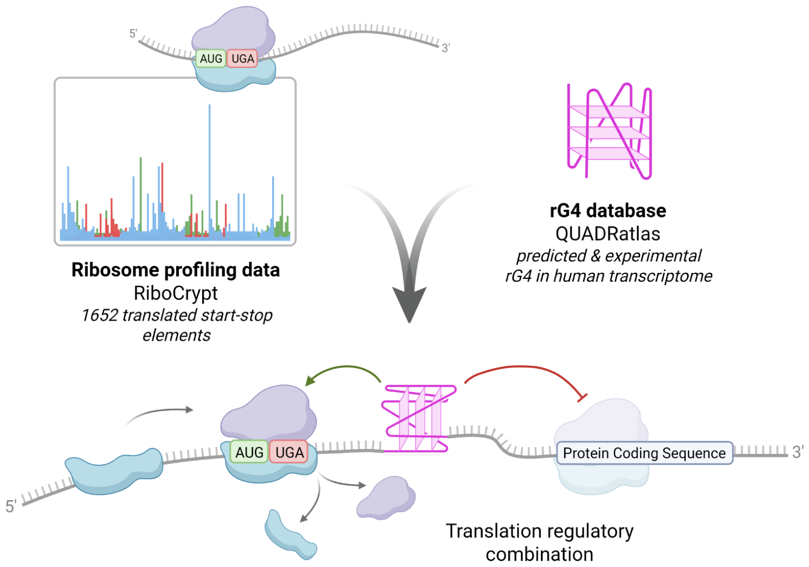
Figure 2.
Evolutionary conservation of the DUSP4 start–stop element. A. Nucleotide-level evolutionary conservation of the same region based on phyloP100way scores. The height of each nucleotide corresponds to its phyloP score, with positive and negative values indicating evolutionary conservation and acceleration, respectively. Elevated phyloP scores within and immediately adjacent to the start–stop element indicate localized evolutionary conservation. B. Sequence logo generated from the UCSC Multiz alignment of 100 vertebrates for a 61-nt region of the human DUSP4 5′-UTR (hg38, chr8:29,350,420–29,350,480). Letter heights represent nucleotide information content (bits), calculated from 67 aligned sequences, excluding gaps and missing nucleotides.
Figure 2.
Evolutionary conservation of the DUSP4 start–stop element. A. Nucleotide-level evolutionary conservation of the same region based on phyloP100way scores. The height of each nucleotide corresponds to its phyloP score, with positive and negative values indicating evolutionary conservation and acceleration, respectively. Elevated phyloP scores within and immediately adjacent to the start–stop element indicate localized evolutionary conservation. B. Sequence logo generated from the UCSC Multiz alignment of 100 vertebrates for a 61-nt region of the human DUSP4 5′-UTR (hg38, chr8:29,350,420–29,350,480). Letter heights represent nucleotide information content (bits), calculated from 67 aligned sequences, excluding gaps and missing nucleotides.
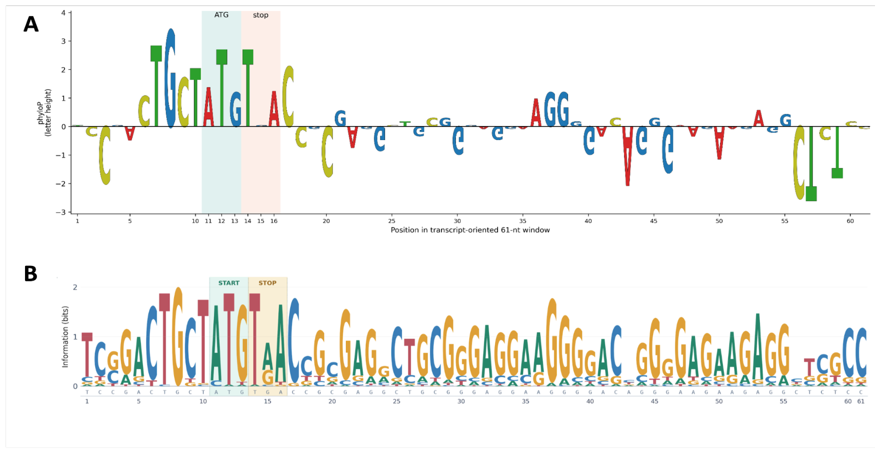
Figure 3.
Organization of the DUSP4 5′-UTR and distribution of ribosome-protected fragments. Upper panel: Schematic representation of the DUSP4 5′-UTR showing the upstream open reading frame (uORF), AUG–UGA start–stop element, putative rG4, and the main coding sequence, with the lengths of and distances between the regulatory elements indicated. Lower panel: Distribution of RPFs along the DUSP4 transcript based on aggregated human Ribo-seq data from the RiboCrypt database. The most prominent peak of ribosome occupancy corresponds to the start–stop element.
Figure 3.
Organization of the DUSP4 5′-UTR and distribution of ribosome-protected fragments. Upper panel: Schematic representation of the DUSP4 5′-UTR showing the upstream open reading frame (uORF), AUG–UGA start–stop element, putative rG4, and the main coding sequence, with the lengths of and distances between the regulatory elements indicated. Lower panel: Distribution of RPFs along the DUSP4 transcript based on aggregated human Ribo-seq data from the RiboCrypt database. The most prominent peak of ribosome occupancy corresponds to the start–stop element.
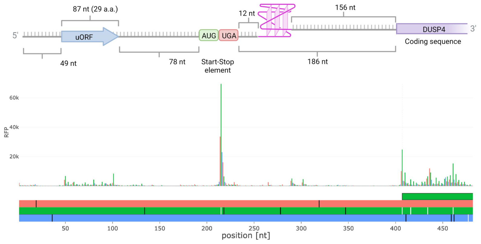
Figure 4.
Putative rG4-prone sequences within 5′-UTR and the proximal region of the coding sequence of DUSP4 mRNA. RNA sequences G1-G4 that contain rG4-forming motifs predicted by QGRS mapper.
Figure 4.
Putative rG4-prone sequences within 5′-UTR and the proximal region of the coding sequence of DUSP4 mRNA. RNA sequences G1-G4 that contain rG4-forming motifs predicted by QGRS mapper.
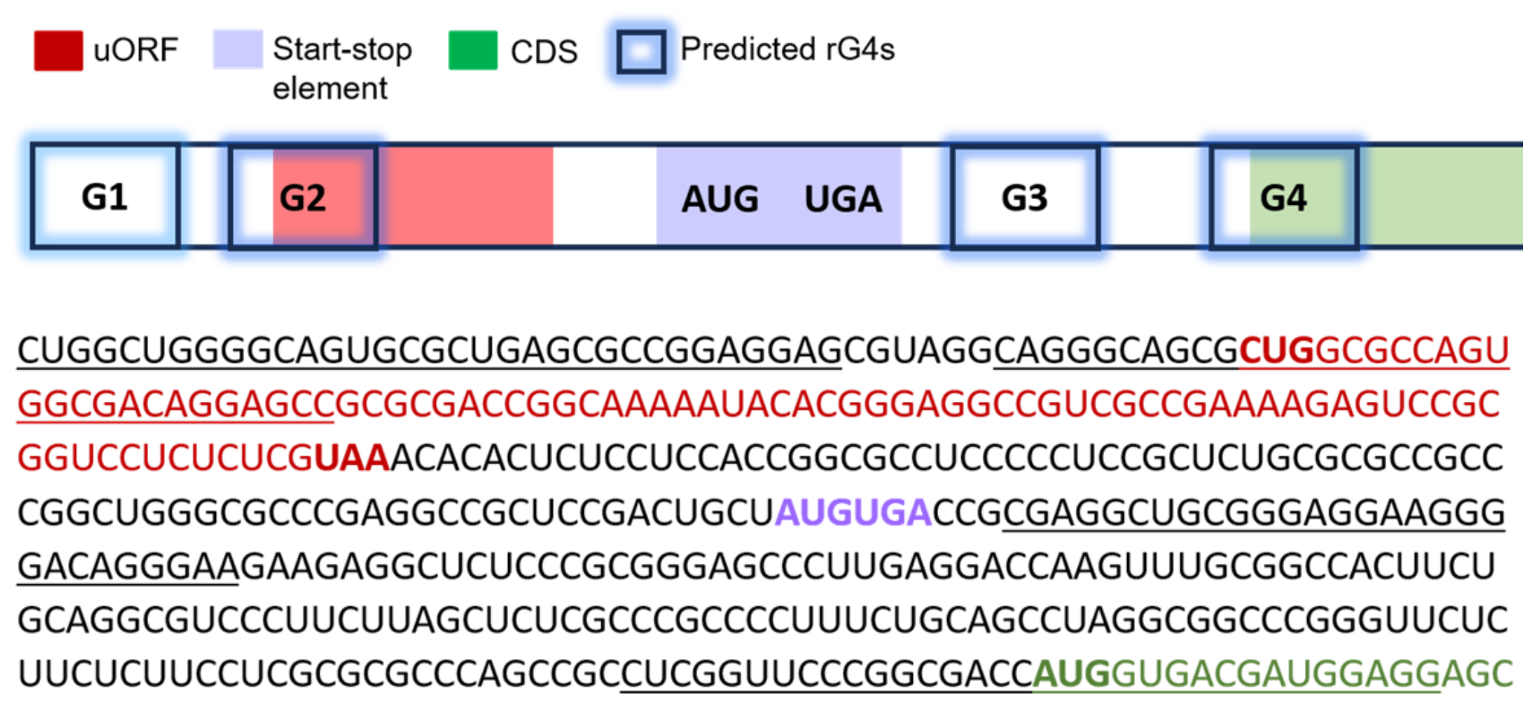
Figure 5.
Characterization of putative rG4-forming sequences using fluorescence, CD and UV spectroscopy. A. Thioflavin T-based (ThT) light-up assay. The ThT fluorescence intensity (FI) was monitored at 485 nm upon excitation at 445 nm. Conditions: 2 µM RNA, 1 µM ThT, 20 mM sodium phosphate buffer, pH 7.4, 140 mM KCl, 25 °C. B. CD spectra of G3 WT and G3 MUT. Conditions: 3 µM RNA, 20 mM sodium phosphate buffer, pH 7.4, 140 mM KCl, 25 °C. C. Thermal difference spectra (TDS) of G3 WT and G3 MUT. Conditions: 3 µM RNA, 20 mM sodium phosphate buffer, pH 7.4, 140 mM KCl. D. CD spectra of G3 WT in the presence of 100 mM LiCl (without KCl) or 1, 10, 100 mM KCl, respectively. Conditions: 3 µM RNA, 10 mM Tris buffer, pH 7.4, 25 °C. E. UV melting curves registered at 295 nm for G3 WT with 140 mM KCl alone, or with 14 mM KCl in the absence or presence of 5 eq. PDS. Conditions: 3 µM RNA, 20 mM sodium phosphate buffer, pH 7.4.
Figure 5.
Characterization of putative rG4-forming sequences using fluorescence, CD and UV spectroscopy. A. Thioflavin T-based (ThT) light-up assay. The ThT fluorescence intensity (FI) was monitored at 485 nm upon excitation at 445 nm. Conditions: 2 µM RNA, 1 µM ThT, 20 mM sodium phosphate buffer, pH 7.4, 140 mM KCl, 25 °C. B. CD spectra of G3 WT and G3 MUT. Conditions: 3 µM RNA, 20 mM sodium phosphate buffer, pH 7.4, 140 mM KCl, 25 °C. C. Thermal difference spectra (TDS) of G3 WT and G3 MUT. Conditions: 3 µM RNA, 20 mM sodium phosphate buffer, pH 7.4, 140 mM KCl. D. CD spectra of G3 WT in the presence of 100 mM LiCl (without KCl) or 1, 10, 100 mM KCl, respectively. Conditions: 3 µM RNA, 10 mM Tris buffer, pH 7.4, 25 °C. E. UV melting curves registered at 295 nm for G3 WT with 140 mM KCl alone, or with 14 mM KCl in the absence or presence of 5 eq. PDS. Conditions: 3 µM RNA, 20 mM sodium phosphate buffer, pH 7.4.
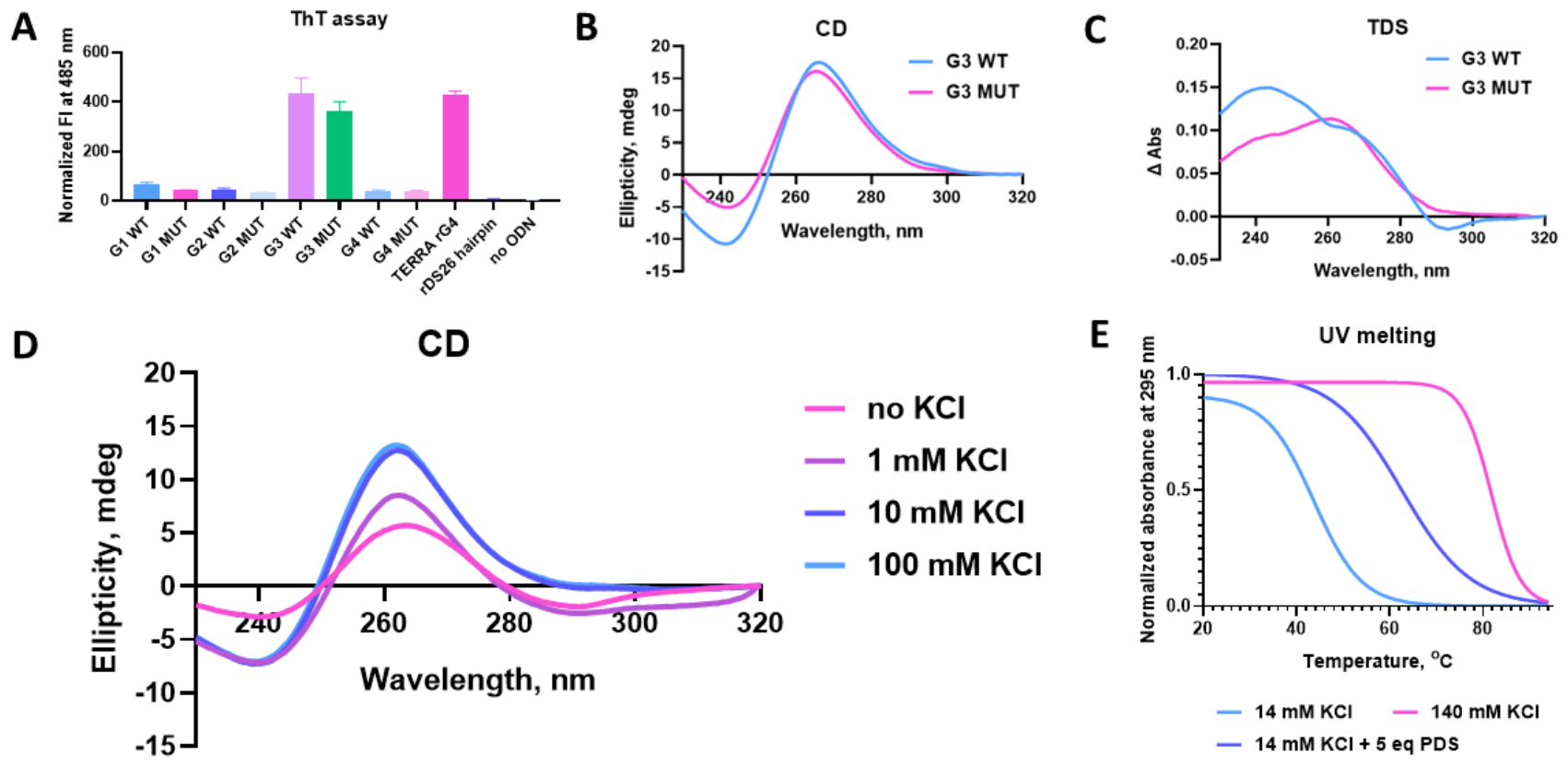
Figure 6.
1H NMR spectrum of the G3 WT sequence. The spectrum of a 116 μM G3 WT solution was recorded in a physiological buffer (20 mM sodium phosphate, pH 7.4, 140 mM KCl, 3% v/v D2O) at 37 °C. Imino proton signals indexed 1 to 12 in the 10–12 ppm region indicate the formation of three G-tetrads.
Figure 6.
1H NMR spectrum of the G3 WT sequence. The spectrum of a 116 μM G3 WT solution was recorded in a physiological buffer (20 mM sodium phosphate, pH 7.4, 140 mM KCl, 3% v/v D2O) at 37 °C. Imino proton signals indexed 1 to 12 in the 10–12 ppm region indicate the formation of three G-tetrads.
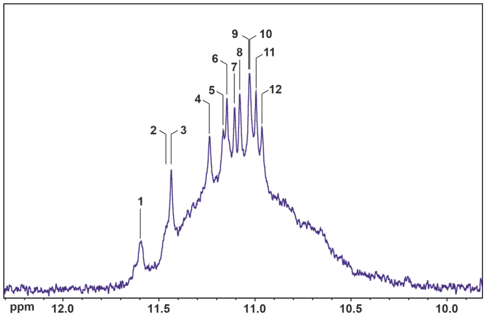
Figure 7.
The DUSP4 5′-UTR start–stop element and rG4 motif repress downstream reporter translation. HEK293T cells were transfected with dual-luciferase reporter plasmids carrying the wild-type (WT) DUSP4 5′-UTR or variants with an inactivated upstream open reading frame (ΔuORF), disrupted start–stop element (ΔSt–St), disrupted rG4 motif (ΔrG4), or combined disruption of the rG4 motif and start–stop element (ΔrG4ΔSt–St). Firefly luciferase activity was normalized to Renilla luciferase activity and is presented as the Fluc/Rluc ratio. Individual dots represent independent biological replicates, n≥8; bars indicate mean ± SD. Statistical significance was assessed using ordinary one-way ANOVA followed by Tukey’s multiple comparisons test. Adjusted p-values are indicated as follows: ns, not significant (p ≥ 0.05); * p < 0.05; ** p < 0.01; *** p < 0.001; **** p < 0.0001.
Figure 7.
The DUSP4 5′-UTR start–stop element and rG4 motif repress downstream reporter translation. HEK293T cells were transfected with dual-luciferase reporter plasmids carrying the wild-type (WT) DUSP4 5′-UTR or variants with an inactivated upstream open reading frame (ΔuORF), disrupted start–stop element (ΔSt–St), disrupted rG4 motif (ΔrG4), or combined disruption of the rG4 motif and start–stop element (ΔrG4ΔSt–St). Firefly luciferase activity was normalized to Renilla luciferase activity and is presented as the Fluc/Rluc ratio. Individual dots represent independent biological replicates, n≥8; bars indicate mean ± SD. Statistical significance was assessed using ordinary one-way ANOVA followed by Tukey’s multiple comparisons test. Adjusted p-values are indicated as follows: ns, not significant (p ≥ 0.05); * p < 0.05; ** p < 0.01; *** p < 0.001; **** p < 0.0001.
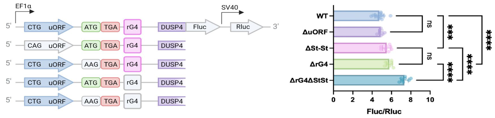
Figure 8.
Disruption of the DUSP4 start–stop element increases reporter translation in the mRNA system. HEK293T cells were transfected with in vitro-transcribed dual-luciferase mRNA reporters containing the wild-type (WT) DUSP4 5′-UTR or the indicated mutant variants. The ΔuORF construct contains an inactivated upstream open reading frame; in the ΔSt–St construct, the AUG codon of the start–stop element was replaced with a non-initiating codon; in the ΔrG4 construct, G-to-A substitutions were introduced into the G-rich motif to disrupt rG4 formation; and the ΔrG4ΔSt–St construct carries both the rG4-disrupting and start–stop mutations. Reporter translation was quantified as the ratio of Firefly to Renilla luciferase activity (Fluc/Rluc). Individual dots represent independent biological replicates, n≥8; bars indicate mean ± SD. Statistical significance was assessed using ordinary one-way ANOVA followed by Tukey’s multiple comparisons test. Adjusted p-values are indicated as follows: ns, not significant (p ≥ 0.05); * p < 0.05; ** p < 0.01; *** p < 0.001; **** p < 0.0001.
Figure 8.
Disruption of the DUSP4 start–stop element increases reporter translation in the mRNA system. HEK293T cells were transfected with in vitro-transcribed dual-luciferase mRNA reporters containing the wild-type (WT) DUSP4 5′-UTR or the indicated mutant variants. The ΔuORF construct contains an inactivated upstream open reading frame; in the ΔSt–St construct, the AUG codon of the start–stop element was replaced with a non-initiating codon; in the ΔrG4 construct, G-to-A substitutions were introduced into the G-rich motif to disrupt rG4 formation; and the ΔrG4ΔSt–St construct carries both the rG4-disrupting and start–stop mutations. Reporter translation was quantified as the ratio of Firefly to Renilla luciferase activity (Fluc/Rluc). Individual dots represent independent biological replicates, n≥8; bars indicate mean ± SD. Statistical significance was assessed using ordinary one-way ANOVA followed by Tukey’s multiple comparisons test. Adjusted p-values are indicated as follows: ns, not significant (p ≥ 0.05); * p < 0.05; ** p < 0.01; *** p < 0.001; **** p < 0.0001.
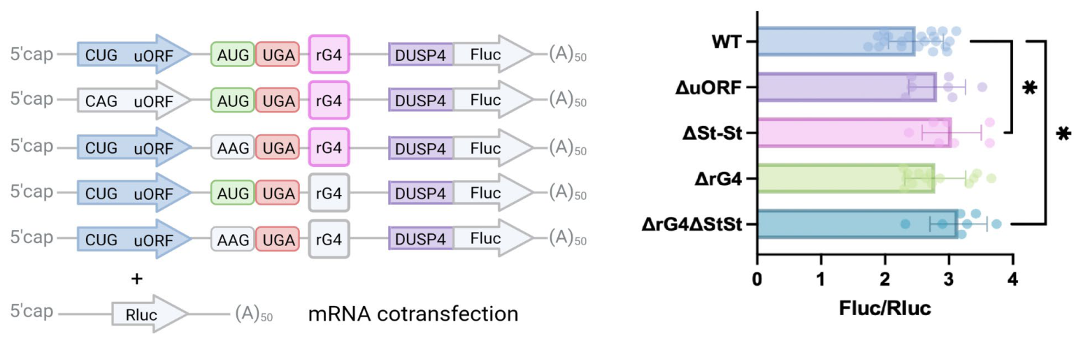
Table 1.
RNA oligonucleotides used in this study. G-tracts longer than two Gs are shown in bold; G-to-A mutations are underlined.
Table 1.
RNA oligonucleotides used in this study. G-tracts longer than two Gs are shown in bold; G-to-A mutations are underlined.
| Code | Sequence 5′→3′ |
|---|---|
| G1 WT | r(CUGGCUGGGGCAGUGCGCUGAGCGCCGGAGGAG) |
| G1 MUT | r(CUGACUAGAGCAGUGCGCUGAGCGCCGAAGAAG) |
| G2 WT | r(CAGGGCAGCGCUGGCGCCAGUGGCGACAGGAGCC) |
| G2 MUT | r(CAGAGCAGCGCUGGCGCCAGUGACGACAGAAGCC) |
| G3 WT | r(CGAGGCUGCGGGAGGAAGGGGACAGGGAA) |
| G3 MUT | r(CGAGACUGCGAGAAGAAGGAGACAGAGAA) |
| G4 WT | r(CUCGGUUCCCGGCGACCAUGGUGACGAUGGAGG) |
| G4 MUT | r(CUCGAUUCCCGACGACCAUGAUGACGAUGAAGG) |
| Dusp4_3flank | r(CGAGGCUGCGGGAGGAAGGGGACAGGGAAGAAGAGGCUCU) |
| TERRA | r(AGGGUUAGGGUUAGGGUUAGGG) |
| rDS26 | r(CAAUCGGAUCGAAUUCGAUCCGAUUG) |
Table 2.
2′-OMe- and LNA-modified ASOs targeting the start-stop element and rG4 in DUSP4 5′-UTR.
| Code | Sequence 5′→3′ (all PO 2′-OMe, except underlined LNA) |
|---|---|
| Act_1 | CAUAGCAGUCGGAGCG |
| Act_1L | CAUAGCAGUCGGAGCG |
| Act_2 | AGAGCCUCUUCUUCCC |
| Act_2L_1 | AGAGCCUCUUCUUCCC |
| Act_2L_2 | AGAGCCUCUUCUUCCC |
| Act_2L_3 | AGAGCCUCUUCUUCCC |
| Act_2L_4 | AGAGCCUCUUCUUCCC |
| Act_2L_5 | AGAGCCUCUUCUUCCC |
| Act_3 | CCCGCAGCCUCGCGGUCACAUAGC |
| SCR | CAUACGUCUAUACGC |
Disclaimer/Publisher’s Note: The statements, opinions and data contained in all publications are solely those of the individual author(s) and contributor(s) and not of MDPI and/or the editor(s). MDPI and/or the editor(s) disclaim responsibility for any injury to people or property resulting from any ideas, methods, instructions or products referred to in the content. |
© 2026 by the authors. Licensee MDPI, Basel, Switzerland. This article is an open access article distributed under the terms and conditions of the Creative Commons Attribution (CC BY) license (http://creativecommons.org/licenses/by/4.0/).
Copyright: This open access article is published under a Creative Commons CC BY 4.0 license, which permit the free download, distribution, and reuse, provided that the author and preprint are cited in any reuse.