

Submitted:
11 November 2026
Posted:
12 November 2026
You are already at the latest version
Abstract
Background/Objectives: Photoplethysmography (PPG) could support unobtrusive respiratory-rate monitoring during breathing-centered rehabilitation. Accuracy measured in one cohort, however, says little about how the same estimator will behave after deployment elsewhere. Confidence estimates may also fail. We tested whether physiology-informed artificial intelligence and a learned error-prediction gate retained their performance across two clinically distinct public datasets. Methods: We conducted a retrospective, subject-separated benchmark using 53 BIDMC intensive-care recordings and 42 CapnoBase anesthesia recordings. From 32 s PPG windows advanced every 8 s, we derived four respiratory modulations and 51 rate, agreement, morphology, and signal-quality features. Six estimators were evaluated by five-fold grouped cross-validation and by bidirectional external transfer. A nested gradient-boosting model predicted each estimate's absolute error to support selective reporting. Performance was summarized with subject-bootstrap confidence intervals, Bland–Altman agreement, and risk–coverage analysis. Results: The analysis included 5262 windows from 95 participants. Within-dataset mean absolute error (MAE) was lowest for random forest on BIDMC (1.65 breaths/min) and Extra Trees on CapnoBase (3.55 breaths/min). The physiology-consensus model achieved 1.70 and 4.02 breaths/min, respectively. Its learned gate reduced MAE at 50% coverage to 0.93 and 2.01 breaths/min. Under external transfer, however, its MAE increased to 6.19 breaths/min for BIDMC→CapnoBase and 4.90 breaths/min for CapnoBase→BIDMC. Uncertainty–error Spearman correlation fell from 0.48 and 0.41 internally to 0.15 and 0.09 externally, and gating yielded only modest improvement. A fixed raw-spectral baseline outperformed the learned model in both transfer directions. Conclusions: Confidence measured within the development cohort did not establish safety after transfer. PPG-derived respiratory rate needs external calibration before it can guide rehabilitation feedback, even when point accuracy appears acceptable internally. Public benchmarks should report bidirectional transfer and risk–coverage behavior alongside random or subject-wise internal splits.
Keywords:
artificial intelligence
; photoplethysmography
; respiratory rate
; selective prediction
; uncertainty
; external validation
; dataset shift
; pulmonary rehabilitation
1. Introduction
Respiratory rate is one of the simplest vital signs to describe and one of the easiest to neglect. It changes early in cardiorespiratory deterioration, responds to exercise load, and provides a compact view of ventilatory demand, yet manual counting is intermittent and often imprecise [1]. For breathing-centered rehabilitation, the measurement has an additional role: it can document whether a participant actually adopts a prescribed pattern, whether ventilatory recovery follows exercise, and whether feedback should be withheld when the signal is unreliable. A useful monitor must therefore do more than emit a number. It must also know when that number should not be trusted.
Contact photoplethysmography is attractive in this setting because the same optical sensor already used for pulse-rate and oxygen-saturation monitoring contains respiratory information. Breathing changes venous return, stroke volume, intrathoracic pressure, and beat timing, producing baseline, amplitude, and frequency modulations in the peripheral pulse waveform [2]. Modern wearable PPG systems can acquire these signals continuously with little additional hardware burden [3]. The recorded waveform reflects both physiology and the measurement system [4]. Reviews of wearable PPG accordingly treat sensing, algorithms, and validation conditions as one design problem [5]. The engineering problem is difficult. Respiratory modulation is weaker than the cardiac component, and it changes with posture, contact pressure, perfusion, motion, age, ventilation mode, and acquisition hardware.
The algorithmic literature has progressed from selecting one spectral peak to combining several respiratory-induced variations. Reviews have catalogued dozens of extraction, estimation, fusion, and quality-assessment strategies [6]. Comparative work has shown that performance depends strongly on the dataset, window definition, reference signal, and fusion rule [7]. Contact-based respiratory sensing reviews place PPG among several indirect approaches with distinct motion and calibration liabilities [8]. Technology surveys reach the same conclusion: respiratory rate is measurable through many surrogates, but none is context-free [9]. Smart Fusion introduced quality-weighted combination of multiple PPG-derived estimates [10]. Subsequent approaches used autoregressive spectra, sparse reconstruction, signal-quality indices, and learned representations [11]. These methods established that consensus among physiological views can be more robust than trusting one modulation, but they did not settle how confidence should be handled when the data-generating process changes.
Recent AI models have added nonlinear feature stacks and end-to-end architectures. A transformer model trained on electrocardiogram and PPG signals reported strong subject-wise performance across BIDMC and CapnoBase [12]. Energy-efficient spiking networks have also been proposed for wearable inference [13]. A newer temporal-fusion study screened respiratory components before principal-component fusion [14]. Pooled or internal validation leaves a practical question unanswered. Will the model still rank its own errors when the target cohort has a different age structure, respiratory distribution, reference modality, and care context? End-to-end PPG learning has already demonstrated how large waveform models can exploit activity-linked structure for heart-rate estimation [15]. Multimodal wearable datasets such as WESAD further illustrate that subject state and sensing context are entangled in ambulatory signals [16].
Selective prediction gives a model an abstention option. A system retains estimates below an uncertainty threshold and withholds the rest. The resulting risk–coverage curve shows the accuracy–availability trade-off [17]. In PPG research, BeliefPPG demonstrated calibrated uncertainty and selective reporting for heart rate [18]. For respiratory rate, however, confidence is usually represented by hand-crafted signal-quality or modulation-quality thresholds. Sparse-reconstruction work used a quality index to decide whether a respiratory estimate should be emitted [19]. Entropy-based and modulation-quality indices provide related gating mechanisms [20]. These methods are close prior art, so the contribution of a learned error predictor cannot simply be described as the first confidence gate. Its value must be tested against distribution shift and reported as selective risk, not inferred from internal accuracy.
Rehabilitation raises the stakes of this test. Pulmonary rehabilitation combines assessment, exercise, education, and behavior change [21]. Contemporary programs increasingly extend beyond supervised centers into home and hybrid care [22]. A wearable respiratory estimate could support these models of care, but an overconfident error could provide misleading feedback precisely when physiology or sensor conditions depart from the development cohort. Guidance for medical AI therefore emphasizes independent evaluation, transparent reporting, and analysis of failure modes [23].
The study asked how fixed spectral, linear, ensemble-tree, and gradient-boosted physiology-consensus estimators compare under subject-separated validation within BIDMC and CapnoBase. We then measured the performance loss when the complete learned pipeline moved from one dataset to the other without adaptation. The final question was whether a learned absolute-error model could still rank errors after that transfer. We expected nonlinear stacking to improve on the raw-spectral baseline internally. We also expected a transfer penalty because the datasets differ materially. The transportability of uncertainty was treated as the central empirical question, not as an assumption.
2. Materials and Methods
2.1. Study Design and Reporting Framework
This was a retrospective secondary analysis of two fully de-identified public physiological datasets. The analysis plan was fixed before inspecting model outputs, although it was not externally preregistered. We followed the reporting priorities of TRIPOD+AI for prediction-model development and evaluation [23]. We also used PROBAST+AI concepts to separate model-development quality from risk of bias in performance evaluation [24]. In particular, all cross-validation folds were grouped by participant, preprocessing was performed identically in development and test data, and no external-test labels were used for model selection, threshold choice, or recalibration.
The analysis had two complementary validation layers. The first estimated subject-independent performance within each dataset using five-fold GroupKFold. The second trained the entire point-prediction and error-prediction pipeline on one dataset and evaluated it, unchanged, on the other. The directional design was essential because a single pooled analysis cannot distinguish genuine transport from learning a compromise between cohorts. External validation was therefore performed as BIDMC→CapnoBase and CapnoBase→BIDMC, not as random folds after concatenation.
2.2. Data Sources and Participants
The BIDMC PPG and Respiration Dataset contains 53 eight-minute recordings collected from adult and older-adult intensive-care patients at 125 Hz. It is distributed through PhysioNet, an established repository for complex physiological signals [25]. Each record includes fingertip PPG and two independent manual breath annotations derived from the respiratory signal. We used the PPG waveform and both breath annotation series from the official release [26].
CapnoBase contains 42 eight-minute recordings acquired during elective surgery and anesthesia, including 29 pediatric and 13 adult cases. The PPG was sampled at 300 Hz and the respiratory reference was derived from capnography. Metadata include age and ventilation mode. We used the official 8 min MATLAB files from the Borealis Dataverse release [27]. The two datasets therefore differ in population, clinical context, sampling rate, reference technology, and respiratory-rate distribution. This combination makes them a realistic stress test of transport, not interchangeable folds of one cohort.
Recordings were segmented into 32 s windows with an 8 s stride. This duration balances frequency resolution against responsiveness and is common in respiratory-rate estimation. BIDMC reference rate was calculated separately for each annotator as 60 divided by the median valid inter-breath interval and then averaged. Windows were excluded if fewer than four breath marks were available, the two estimates differed by more than 5 breaths/min, or the final rate fell outside 4–45 breaths/min. CapnoBase reference rate was linearly interpolated at the window center and excluded outside the same physiological range. No participant was excluded after window-level quality screening.

Table 1.
Dataset characteristics and analytic yield.
| Characteristic | BIDMC | CapnoBase |
| Clinical setting | Adult/older-adult intensive care | Pediatric and adult anesthesia |
| Participants | 53 | 42 (29 pediatric, 13 adult) |
| PPG sampling rate | 125 Hz | 300 Hz |
| Respiratory reference | Two manual breath annotations | Capnography-derived rate |
| Windowing | 32 s; 8 s stride | 32 s; 8 s stride |
| Attempted / included windows | 3021 / 2958 | 2394 / 2304 |
| Reference RR, mean ± SD | 17.62 ± 2.76 breaths/min | 15.02 ± 7.62 breaths/min |
| Reference RR, range | 5.83–26.64 breaths/min | 4.07–44.67 breaths/min |
2.3. Signal Processing and Physiological Views
Each PPG window was linearly detrended and robustly standardized using the median and median absolute deviation. Non-finite samples were interpolated only when at least 80% of the window was finite. Signals were resampled by polyphase filtering to 25 Hz. A third-order zero-phase Butterworth bandpass of 0.08–0.70 Hz defined the candidate respiratory band, corresponding to 4.8–42 breaths/min. The upper analysis limit remained 45 breaths/min to retain clinically plausible high-rate windows while allowing edge estimates to be screened.
We extracted four complementary physiological views. Baseline modulation was the low-frequency component of the standardized PPG. Envelope modulation was obtained by bandpassing the absolute Hilbert envelope of the cardiac-band signal. Pulse-amplitude modulation interpolated detected pulse peak heights to 4 Hz, and pulse-interval modulation interpolated successive peak intervals to 4 Hz. Peak polarity was selected by a plausibility score that combined the proportion of 0.27–1.80 s intervals, interval regularity, and peak prominence. This representation is consistent with the known intensity, amplitude, and frequency pathways through which respiration alters PPG [6]. Published motion-contaminated PPG work shows why respiratory estimation must also consider pulse-detection stability and accelerometry context [28].
For every view, we computed a Welch-spectrum respiratory estimate with parabolic peak refinement and an autocorrelation estimate. Associated features included spectral concentration, peak prominence, normalized spectral entropy, autocorrelation peak height, standard deviation, skewness, and kurtosis. Global features described PPG shape, clipping, derivative variability, pulse count, heart rate, and inter-beat-interval variation. Agreement features summarized the count, median, mean, median absolute deviation, range, and interquartile range of the eight candidate rates. The final predictor matrix contained 51 features. Unlike end-to-end waveform networks, the design kept each respiratory hypothesis inspectable and computationally modest. Signal-quality research has shown that the most useful PPG quality index depends on the downstream task [29].
2.4. Point Estimators and the Learned Confidence Gate
Six point estimators represented increasing model flexibility. Two non-learned comparators were the baseline-modulation spectral peak and the median of all valid physiological candidate rates. Ridge regression used median imputation with missingness indicators, robust scaling, and an L2 penalty of 10. Random forest used 300 trees, 70% feature sampling, and a minimum leaf size of eight. Extra Trees used 350 trees, 80% feature sampling, and a minimum leaf size of five. The designated physiology-consensus model was histogram gradient boosting with learning rate 0.06, 15 terminal leaves, minimum leaf size 25, L2 regularization 2, and 260 boosting iterations. Hyperparameters were chosen a priori for regularization and tractable computation rather than optimized on either test set. Table 2 lists the estimators and their roles.
The confidence gate was a second histogram gradient-boosting regressor trained to predict absolute error. Within each outer grouped fold, the point model first generated participant-separated inner out-of-fold predictions. The error model then received the original features plus the point estimate and its absolute disagreement with the median physiological candidate. Its target was the absolute point-estimation error. At test time, lower predicted error represented higher confidence. The same nested construction was used for external validation: inner grouped predictions were produced only within the source dataset, after which both models were fitted on all source participants and applied unchanged to the target dataset.
Selective performance was evaluated at 100%, 90%, 80%, 70%, and 50% coverage by retaining windows with the lowest predicted errors. We report MAE against retained coverage and Spearman correlation between predicted and observed absolute error. The analysis tests whether the score ranks errors. The score itself has no intrinsic clinical meaning. An uncertainty mechanism can be well calibrated locally and still fail under dataset shift [30].
2.5. Outcomes and Statistical Analysis
The primary performance measure was MAE in breaths/min. Secondary measures were root mean squared error (RMSE), mean signed bias, 95% limits of agreement, Pearson correlation, and the proportions of windows within ±2 and ±3 breaths/min. Bland–Altman plots were used to visualize heteroscedastic and rate-dependent errors because correlation alone does not measure agreement [31]. Confidence intervals were obtained from 2000 bootstrap resamples at the participant level so that overlapping windows from one person were never treated as independent sampling units.
For paired model comparisons, MAE was first calculated within each participant. Two-sided Wilcoxon signed-rank tests compared the physiology-consensus model with each comparator, and Holm adjustment controlled family-wise error within each evaluation setting. Ablations repeated grouped validation using candidate-rate features only, removal of interval modulation, removal of amplitude modulation, and removal of agreement and global signal-quality features. External CapnoBase results were described by pediatric/adult age group and ventilation mode. These subgroup analyses were exploratory and were not used for optimization.
All analyses used Python 3.12 with NumPy 2.5.2, pandas 3.0.5, SciPy 1.18.1, scikit-learn 1.9.0, h5py 3.16.0, and Matplotlib 3.11.1. A fixed seed of 20260831 was used. The complete run took 563.65 s on the analysis workstation. The pipeline saved window-level features, predictions, audit counts, software versions, tables, and publication-resolution figures to support reproducibility.
3. Results
3.1. Analysis Cohorts and Window Yield
The two datasets contributed all 95 available participants. BIDMC yielded 2958 of 3021 attempted windows. Sixty-three windows were excluded because the reference was unavailable, implausible, or too discordant between annotators. CapnoBase yielded 2304 of 2394 attempted windows. Ninety windows were excluded by reference criteria. Feature extraction completed for every included window. BIDMC rates were concentrated around 17.62 ± 2.76 breaths/min (range 5.83–26.64), whereas CapnoBase had a lower median and much broader distribution: mean 15.02 ± 7.62, median 11.57, range 4.07–44.67 breaths/min.
The CapnoBase cohort contained 29 pediatric and 13 adult cases. One record lacked usable age, leaving 41 for age summaries. The median age was 14 years, with a range of 1–63 years. Ventilation metadata described 21 controlled, 20 spontaneous, and one assisted case at the participant level. BIDMC represented critically ill adults and older adults, with respiratory rate obtained from manual breath annotations. Those cohort and reference differences defined the domain shift tested in the external experiment. Table 1 summarizes the two datasets and analytic yield.
3.2. Subject-Separated Within-Dataset Performance
On BIDMC, the random forest achieved the lowest point-estimation MAE at 1.65 breaths/min (95% CI 1.26–2.12), followed by Extra Trees at 1.70 (1.28–2.19) and the physiology-consensus model at 1.70 (1.32–2.15). The confidence intervals overlapped substantially, and participant-level comparisons did not show a Holm-adjusted difference between the consensus model and random forest (adjusted p=0.098) or Extra Trees (adjusted p=0.418). Ridge regression had MAE 2.18, while the raw-spectral baseline had MAE 3.83. The simple median physiological fusion performed poorly at 7.36, showing that unweighted consensus among discordant candidates can be worse than selecting one stable view.
CapnoBase was more difficult. Extra Trees had the lowest MAE at 3.55 breaths/min (95% CI 2.47–4.92), followed by random forest at 3.86 and the consensus model at 4.02 (2.75–5.70). The adjusted participant-level difference between Extra Trees and the consensus model did not cross the 0.05 threshold (adjusted p=0.063), but the effect direction favored Extra Trees. The raw-spectral baseline had MAE 4.65. Median fusion had 4.87. Correlation was also cohort-dependent: 0.37 for the consensus model on BIDMC and 0.55 on CapnoBase, despite the lower BIDMC MAE.
Accuracy within fixed tolerances told the same story. The consensus model was within ±2 breaths/min for 73.0% of BIDMC windows and 49.7% of CapnoBase windows. Within ±3 breaths/min, the proportions were 83.1% and 62.4%. Mean signed bias was small within both cohorts (−0.07 and +0.37 breaths/min), but the 95% limits of agreement were wide: −5.24 to +5.09 on BIDMC and −13.00 to +13.75 on CapnoBase. Small average bias therefore did not imply interchangeable window-level measurements. Figure 1 and Table 3 present the internal comparisons and the external summary.
3.3. Bidirectional External Validation
External transfer reversed the favorable internal picture. Training the physiology-consensus model on BIDMC and testing on CapnoBase produced MAE 6.19 breaths/min (95% CI 4.98–7.51), RMSE 7.75, bias +2.53, and correlation 0.28. Only 23.5% of windows were within ±2 breaths/min. The raw-spectral baseline, which required no source-domain fitting, achieved a lower MAE of 4.65. Random forest also outperformed the consensus model at 5.97. The participant-level difference was significant after Holm adjustment (adjusted p=0.004).
Transfer in the opposite direction was similarly poor but phenotypically different. A CapnoBase-trained consensus model tested on BIDMC had MAE 4.90 breaths/min (95% CI 4.29–5.55), RMSE 6.01, bias −2.36, and correlation −0.14. Only 21.3% of windows were within ±2 breaths/min. Ridge regression transported better than the tree ensembles, with MAE 3.24, and the raw-spectral baseline again performed better at 3.83. Bland–Altman analysis exposed strong rate dependence: the BIDMC-trained model compressed the broad CapnoBase rate range toward roughly 15–20 breaths/min, whereas the CapnoBase-trained model systematically underestimated much of the narrower BIDMC distribution.
The transfer penalty was not symmetric because the source distributions differed. CapnoBase contained high-rate pediatric and controlled-ventilation segments absent from BIDMC, while BIDMC concentrated observations near the adult critical-care range. Nonlinear learners could minimize source-domain error by exploiting those marginal distributions. The external plots show that this behavior was a structured contraction toward source-domain rate regions. Figure 2 shows the directional error structure and agreement limits.
3.4. Selective Prediction and Confidence Transport
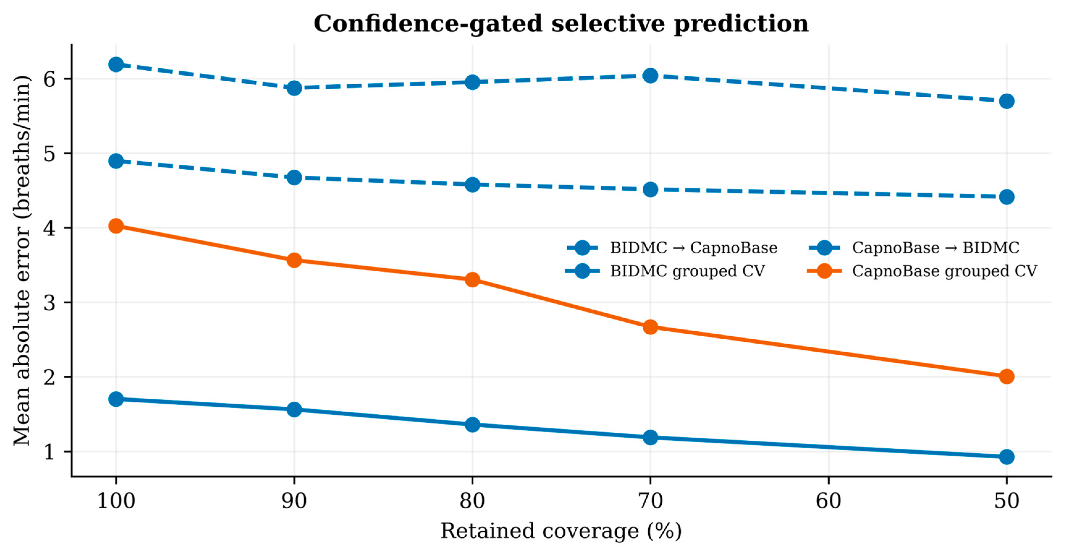
Within BIDMC, predicted absolute error correlated moderately with observed absolute error (Spearman ρ=0.481, p<0.001). Retaining the 80% most confident windows reduced MAE from 1.70 to 1.36, and retaining 50% reduced it to 0.93. Within CapnoBase, correlation was ρ=0.414 (p<0.001). MAE fell from 4.02 to 3.30 at 80% coverage and to 2.01 at 50% coverage. Thus, the learned error model ranked difficulty usefully when development and evaluation data came from the same cohort.
That ranking largely disappeared under transfer. For BIDMC→CapnoBase, uncertainty–error correlation was only ρ=0.146 (p<0.001). MAE was 6.19 at full coverage, 5.95 at 80%, 6.04 at 70%, and 5.70 at 50%. The curve was not monotonic. For CapnoBase→BIDMC, correlation was ρ=0.092 (p<0.001), and MAE changed from 4.90 to 4.58 at 80% and 4.41 at 50%. The very small p-values reflect the number of overlapping windows and should not be mistaken for operationally strong ranking. Effect magnitude and the risk–coverage curve were the clinically relevant findings.
At 50% coverage, the internal gate removed 45.6% of BIDMC absolute error and 50.2% of CapnoBase absolute error relative to full coverage. The corresponding external reductions were 7.9% and 9.8%. Half of the estimates were withheld, yet the remaining predictions were only slightly safer. Confidence learned from source-domain residuals did not travel with the point model. Figure 3 and Table 4 show the resulting risk–coverage gap.
3.5. Ablations and Exploratory Subgroups
Ablations did not support the assumption that more quality descriptors automatically improve the point model. On BIDMC, candidate-rate features alone yielded MAE 1.60, and removing agreement/global signal-quality features yielded 1.55, both slightly below the 1.70 full-feature result. On CapnoBase, the corresponding values were 3.70 and 3.51 versus 4.02. Removing interval modulation changed MAE minimally on BIDMC (1.70) and worsened it on CapnoBase (4.18). Removing pulse-amplitude modulation produced MAE 1.73 and 4.11. These patterns suggest that some quality features encoded cohort-specific structure rather than universally useful reliability. Table 5 provides the full ablation summary.
In BIDMC→CapnoBase transfer, consensus-model MAE was 5.81 among adult windows and 6.38 among pediatric windows. By ventilation label it was 2.45 for assisted, 6.63 for controlled, and 5.91 for spontaneous windows. The assisted category represented only 56 windows from one participant and cannot support a comparative claim. Differences across adult, pediatric, and ventilation groups suggest heterogeneous target subdomains. They remain exploratory because age and treatment are confounded with other acquisition and clinical characteristics.
4. Discussion
4.1. Principal Findings
PPG respiratory-rate confidence failed to transfer with the AI model. Subject-separated internal validation produced acceptable BIDMC accuracy and useful error ranking in both datasets. External transfer produced larger errors, rate-dependent bias, and much weaker uncertainty ranking. A fixed spectral baseline outperformed the designated AI model in both directions. Machine learning was not uniformly inferior. Its flexibility, however, allowed the estimators to use relationships that were predictive inside the source cohort and unstable elsewhere.
The point-estimation and confidence failures were related but not identical. A point model can lose accuracy because P(X), P(Y), or P(Y|X) changes. An error model must also preserve the mapping from features and predictions to residual magnitude. That mapping creates a second transport problem. The internal risk–coverage curves looked reassuring. External testing showed that the gate often rejected windows without isolating the worst errors. Uncertainty should therefore be reported as a tested system property, not as a decorative model output [30].
4.2. Relation to Previous Respiratory-Rate Algorithms
Our work builds on, but does not replace, established PPG respiratory estimation. Multiparameter fusion showed early benefits from combining respiratory-induced intensity, amplitude, and frequency variation [10]. Autoregressive fusion improved robustness by combining spectra rather than committing to one peak [11]. Cross time–frequency methods offered another way to combine source information [32]. A robust fusion model later extended the concept across PPG and electrocardiographic respiratory signals [33].
Quality-aware methods are particularly relevant to our confidence gate. Sparse reconstruction derived a signal-quality index alongside rate [19]. Real-time methods have used spectral and autocorrelation structure to suppress unreliable estimates [34]. Entropy-based screening has been evaluated on BIDMC [20]. Sultan and Saadeh combined robust spectro-temporal features with continuous patient-independent estimation [35]. Lin and colleagues screened six respiratory-modulated components before temporal fusion [14]. These studies show that gating and physiological consensus are not, separately, new. Our contribution is the controlled demonstration that a learned residual gate can appear useful internally while losing most of its discrimination in bidirectional cross-dataset transfer.
Measurement studies show why model choice is only part of the transport problem. Early wearable-PPG reviews described large variation in sensor placement and acquisition conditions [36]. Heart-rate-variability norms demonstrate substantial physiological variation between people and recording contexts [37]. Pulse-rate variability is only an imperfect surrogate for electrocardiographic variability under non-stationary conditions [38]. PPG-based atrial-fibrillation reviews likewise identify motion and signal quality as persistent limitations [39]. Reviews of PPG and oxygen-saturation monitoring emphasize device and population dependencies [40]. Machine-learning studies of cuffless pressure from PPG offer a parallel example of attractive internal accuracy with difficult calibration [41]. Direct comparisons of PPG-only pressure models show that algorithm ranking is dataset dependent [42]. Consumer PPG has also been used for sleep-stage inference, where state, motion, and device become intertwined [43]. Psychophysiology guidance therefore asks authors to report acquisition and preprocessing details rather than treating derived autonomic features as device independent [44].
Wearable medicine has reached the same conclusion from a deployment perspective. Cardiovascular reviews call for stronger evidence and workflow integration before smart devices are used in care [45]. Commentaries on the wearable revolution stress that convenience does not remove validation obligations [46]. Skin-interfaced sensors in neonatal and pediatric care illustrate both the opportunity and the population-specific engineering challenge [47]. Digital-biomarker frameworks recommend explicit verification, analytical validation, and clinical validation [48]. A scoping review of wearable health research found heterogeneous devices, endpoints, and study designs [49]. Athlete monitoring reviews show that even high-frequency physiological data require contextual interpretation [50]. Reviews of AI-enabled wearable sensors identify generalization and power as joint constraints [51]. Broader digital-health reviews place sensing algorithms inside interoperability and governance systems [52]. Connected-health guidance treats sampling, missingness, and metadata as first-order design decisions [53]. The COVID-19 experience showed how rapidly wearable infrastructure can be repurposed, but also how easily evidence lags implementation [54]. Cardiovascular monitoring reviews propose combining new devices with machine learning only after representative evaluation [55]. Mental-health wearable reviews report similar challenges with labeling and context [56]. Data-mining surveys warn that personalization can conceal poor cross-user transport [57]. Emerging-material telehealth sensors add another layer of device-domain variation [58]. Sleep-staging reviews show that ground-truth definitions can dominate model comparisons [59]. Pandemic-era wearable reviews reinforce the need for transparent study quality even when remote measurement is urgently needed [60].
Rehabilitation adds clinical heterogeneity to the technical shift. Mobile-health interventions have shown variable effects across behavior-change and disease-management targets [61]. GOLD guidance positions pulmonary rehabilitation within individualized COPD assessment and management rather than as a uniform breathing prescription [62]. Global physical-activity guidance also requires adaptation for ability and health status [63]. Early mobilization in mechanically ventilated patients demonstrates that rehabilitation may begin in high-acuity settings [64]. Interoception research explains why bodily feedback can be therapeutically meaningful yet psychologically mediated [65]. Training-load research shows that a physiological response must be interpreted relative to exposure and recovery [66]. Exercise guidance for older adults emphasizes multimorbidity and functional capacity [67]. Vagal and brain–gut research provides plausible pathways linking paced breathing with autonomic regulation [68]. Post-resuscitation guidance illustrates the tighter monitoring requirements of medically fragile rehabilitation populations [69]. Systematic reviews of smartphone health applications caution that access and usability do not prove measurement validity [70].
Safe deployment also depends on implementation. Ethical reviews identify privacy, bias, explainability, and accountability as linked regulatory challenges [71]. General healthcare-AI reviews similarly distinguish proof-of-concept models from clinically useful systems [72]. Systematic reviews of wearable health devices report limited prospective evidence despite rapid product growth [73]. Telehealth evidence during the pandemic shows that utilization can expand faster than comparative effectiveness evidence [74]. Materials and device reviews frame wearables as a platform whose accuracy depends on the entire skin–sensor interface [75]. Consumer biofeedback reviews warn against treating proprietary outputs as validated physiological endpoints [76]. A randomized cardiac telerehabilitation trial shows the level of clinical evaluation needed beyond algorithm benchmarking [77]. Remote-patient-monitoring reviews emphasize outcome selection and escalation pathways [78]. Unobtrusive-sensing research demonstrates the trade-off between burden and signal control [79]. Real-time disease-monitoring reviews conclude that wearable value depends on reliable analytics and actionable integration [80].
Reported benchmark numbers across papers should not be ranked as if they came from one competition. Window length, overlap, reference construction, exclusions, modality combinations, and split units differ. Some recent models use both ECG and PPG, whereas our inputs were PPG only. Some pool BIDMC and CapnoBase before cross-validation, whereas we deliberately prevented the target dataset from influencing training. A recent late-fusion multi-task study found that differences between capnographic and impedance references complicate joint learning [81]. Our directional results are consistent with that warning. Scheme 1 summarizes the analytic and intended rehabilitation-facing workflow.
4.3. Why the Dataset Shift Was So Damaging
Several mechanisms can explain the transfer loss. CapnoBase covered a wide pediatric and anesthesia-related respiratory range, whereas BIDMC clustered near adult critical-care values. Tree models do not extrapolate naturally beyond the source response support. The reference also changed, from capnography in CapnoBase to manual annotations associated with respiratory impedance in BIDMC. Latency, smoothing, and the operational definition of a breath can alter the target even when the underlying physiology is similar. Sampling hardware and preprocessing histories differed as well. PPG morphology is sensitive to sensor wavelength, contact, ambient light, skin, and perfusion [82]. Age and ventilation mode further change how respiratory mechanics couple to peripheral pulse modulation.
The error plots are consistent with shortcut learning. BIDMC-trained predictions occupied a compressed band when confronted with the broader CapnoBase range. Conversely, CapnoBase-trained predictions were biased downward on BIDMC. Similar portability failures have been reported across healthcare settings, where high internal performance can coexist with substantial external degradation [83]. Analyses of clinical model transport emphasize that acquisition and care processes can become hidden predictors even when they are not explicitly included [84]. Our quality and agreement features may have carried exactly this kind of contextual information.
The relative resilience of the raw-spectral baseline is instructive. It was not highly accurate, but its rules did not change with the source cohort. This created a smaller gap between internal and external use. In a deployment pipeline, a simple physiology-anchored comparator can therefore serve as a drift reference. Divergence between the AI output and the fixed estimate may flag a change in operating conditions. A hybrid safety architecture can use that disagreement instead of placing all control in the most flexible model.
4.4. Implications for Breathing-Centered Rehabilitation
Breathing-centered rehabilitation spans different goals and populations. Inspiratory muscle training can improve respiratory muscle performance in chronic obstructive pulmonary disease, but protocols and responders vary [85]. Comprehensive pulmonary rehabilitation improves exercise capacity and quality of life [86]. Slow, structured breathing can also influence autonomic and psychological outcomes [87]. Brief controlled-breathing practices have shown effects on mood and physiological arousal [88]. These interventions create precisely the non-stationarity that challenges a rate estimator: respiratory rhythm is intentionally altered while posture, movement, attention, and sensor contact change.
A confidence-gated wearable could still be valuable, but its role should be bounded. It may summarize session-level breathing behavior, flag segments for review, or avoid issuing real-time coaching when confidence is low. It should not be assumed to provide clinically interchangeable breath counts during exercise or disease exacerbation on the basis of anesthesia and intensive-care benchmarks. Wearable rehabilitation systems succeed when sensing, feedback, and clinical workflow are designed together [89]. Biofeedback is most useful when the measured variable is interpretable and the limitations of the feedback loop are explicit [90]. Reviews of wearable vital-sign systems show that integration and validation remain recurring barriers [91]. Machine-learning reviews of healthcare wearables likewise distinguish algorithmic performance from deployable benefit [92].
A rehabilitation study should first establish analytic agreement against capnography or another defensible reference across rest, paced breathing, recovery, and movement. Confidence can then be calibrated at the development site, with thresholds locked before external testing. Reports should pair coverage with accuracy and identify participant groups that are disproportionately rejected. Only then should a trial test whether the monitor changes a meaningful rehabilitation process or outcome. Remote monitoring reviews show that sensor availability alone does not establish clinical benefit [93]. The same caution applies to respiratory-rate AI.
4.5. Methodological and Governance Implications
Participant grouping prevents overlapping windows from one individual entering both training and test folds. It does not separate device, protocol, reference, or clinical context. Directional external validation addresses a different question. Training A→B and B→A exposed failure modes hidden by a pooled score. The uncertainty model also needs testing under the shifts expected after deployment. A confidence score correlated only with internal residuals does not support a safety claim.
These practices are consistent with medical-AI guidance. Responsible machine learning requires attention to the full path from data generation to clinical use [94]. Translation depends on workflow, external evidence, maintenance, and human factors, not only model discrimination [95]. Editorial guidance for digital-medicine studies similarly calls for a test dataset separate from development and validation data [96]. Our results illustrate why these recommendations apply even to a seemingly narrow regression task using familiar vital-sign data. High-performance medicine depends on complementary human and machine strengths, not unqualified automation [97].
A practical deployment architecture would maintain a fixed physiological estimator, a learned point model, an uncertainty model, and a drift monitor as separate auditable components. The system would log coverage, residual audits when reference samples are available, feature-distribution changes, and subgroup rejection rates. Recalibration would be treated as a controlled model update rather than an informal threshold change. Prospective trials involving an AI-supported rehabilitation intervention should describe human–AI interaction, error handling, and access to the algorithm in line with SPIRIT-AI principles [98]. Trial reports should then follow CONSORT-AI so that input data, failure cases, and human handling of AI output are visible to readers [99]. The development protocol for TRIPOD-AI and PROBAST-AI also states that reporting and bias appraisal must evolve together [100].
4.6. Strengths and Limitations
The analysis used two official public datasets and kept participants separated throughout model development and evaluation. External transfer was tested in both directions. The error predictor was trained from nested out-of-fold estimates, intervals were bootstrapped by participant, and paired tests were also performed at the participant level. We retained every intermediate prediction and reported the unfavorable transfer results. Fixed physiological baselines made it possible to compare point accuracy with the operational behavior of abstention.
Several limitations constrain interpretation. Neither dataset was collected during pulmonary or balance rehabilitation, so the clinical implications are pre-deployment inferences. BIDMC and CapnoBase recordings are short, and overlapping windows reduce the number of independent temporal episodes even though statistical resampling occurred at the participant level. The cohorts are small for modern machine learning, and BIDMC lacks detailed demographic metadata. CapnoBase is dominated by children and anesthesia conditions, while BIDMC represents critical care. The design cannot isolate which source of shift caused each error.
The feature pipeline used conventional filters and pulse detection and did not reproduce every published respiratory algorithm. We did not tune models separately for each dataset, use waveform deep learning, perform source-target adaptation, or calibrate on labeled target participants. These choices were deliberate for the transport test but limit claims about the best attainable accuracy. The confidence model predicted absolute error and was assessed by ranking. It did not produce formal prediction intervals or guaranteed coverage. Repeated windows may make window-level Spearman p-values appear more decisive than participant-level evidence, which is why we emphasized effect sizes and risk–coverage curves.
A stronger test requires multicenter rehabilitation data with synchronized respiratory reference, movement annotations, device metadata, skin-tone reporting, and prespecified breathing tasks. Methods should compare target-free drift detection, small labeled calibration sets, conformal or distribution-free intervals, and domain-generalized representations. Success should be judged by whether uncertainty remains informative when the cohort changes. A steep internal curve is not enough.
5. Conclusions
Physiology-informed AI estimated respiratory rate reasonably within BIDMC and less accurately within CapnoBase, and a learned error model supported effective abstention inside each cohort. Neither advantage transported reliably. External transfer produced large, structured errors. The error predictor lost most of its ranking ability, and a fixed spectral baseline outperformed the designated AI model in both directions. For breathing-centered rehabilitation, the result is a practical warning. Confidence gating can support safer monitoring only after the gate itself has been externally validated in the intended population, activity, device, and reference context. Public PPG respiratory-rate benchmarks should routinely report bidirectional transfer and risk–coverage behavior.
Supplementary Materials
The reproducibility package contains the complete analysis script, environment versions, dataset audit, window-level derived features, all prediction files, statistical summaries, and publication figures. Raw BIDMC and CapnoBase signals are not redistributed; they remain available from their official repositories under the respective licenses.
Author Contributions
Conceptualization, S.A.A. and M.M.H.; methodology, S.A.A. and A.N.; software, S.A.A.; validation, S.H.A., A.N., M.M.R. and N.A.; formal analysis, S.A.A. and N.A.; investigation, S.A.A., A.N. and M.M.R.; data curation, S.A.A. and M.M.R.; writing, original draft preparation, S.A.A.; writing, review and editing, S.H.A., A.N., M.M.R., N.A. and M.M.H.; visualization, S.A.A. and S.H.A.; supervision, M.M.H. All authors have read and agreed to the published version of the manuscript.
Funding
This research received no external funding.
Institutional Review Board Statement
Ethical review and approval were waived because this study used only publicly available, de-identified secondary data. The original data providers remain responsible for the ethics approvals and consent procedures associated with primary data collection.
Informed Consent Statement
Patient consent was waived for this secondary analysis because no identifiable information was accessed. Consent procedures for the original datasets are described by their custodians.
Data Availability Statement
BIDMC PPG and Respiration data are available from PhysioNet at https://physionet.org/content/bidmc/1.0.0/ (accessed 31 August 2026). CapnoBase data are available from Borealis Dataverse at https://doi.org/10.5683/SP2/NLB8IT (accessed 31 August 2026). Derived features, predictions, and code accompany this manuscript in the reproducibility package.
Acknowledgments
The authors thank the BIDMC and CapnoBase investigators and repositories for maintaining openly accessible physiological datasets. During preparation of this manuscript, OpenAI Codex/ChatGPT was used to assist with literature discovery, code drafting, figure prototyping, and language editing; Perplexity was used for an additional prior-art search. All citations were checked against primary records, all analyses were executed on the downloaded datasets, and the authors are responsible for the final text, interpretation, and submission.
Conflicts of Interest
The authors declare no conflicts of interest.
Abbreviations
AI, artificial intelligence; CI, confidence interval; CV, cross-validation; MAE, mean absolute error; PPG, photoplethysmography; RMSE, root mean squared error; RR, respiratory rate; SQI, signal-quality index.
References
- Nicolò, A.; Massaroni, C.; Schena, E.; Sacchetti, M.. The Importance of Respiratory Rate Monitoring: From Healthcare to Sport and Exercise. Sensors 2020, 20(21), 6396. [CrossRef] [PubMed]
- Allen, J.. Photoplethysmography and its application in clinical physiological measurement. Physiological Measurement 2007, 28(3), R1-R39. [CrossRef] [PubMed]
- Charlton, P.H.; Allen, J.; Bailón, R.; Baker, S.; Behar, J.A.; Chen, F.; Clifford, G.D.; Clifton, D.A.; Davies, H.J.; Ding, C.; et al.. The 2023 wearable photoplethysmography roadmap. Physiological Measurement 2023, 44(11), 111001. [CrossRef] [PubMed]
- Park, J.; Seok, H.S.; Kim, S.S.; Shin, H.. Photoplethysmogram Analysis and Applications: An Integrative Review. Frontiers in Physiology 2022, 12, 808451. [CrossRef] [PubMed]
- Charlton, P.H.; Kyriacou, P.A.; Mant, J.; Marozas, V.; Chowienczyk, P.; Alastruey, J.. Wearable Photoplethysmography for Cardiovascular Monitoring. Proceedings of the IEEE 2022, 110(3), 355-381. [CrossRef] [PubMed]
- Charlton, P.H.; Birrenkott, D.A.; Bonnici, T.; Pimentel, M.A.F.; Johnson, A.E.W.; Alastruey, J.; Tarassenko, L.; Watkinson, P.J.; Beale, R.; Clifton, D.A.. Breathing Rate Estimation From the Electrocardiogram and Photoplethysmogram: A Review. IEEE Reviews in Biomedical Engineering 2018, 11, 2-20. [CrossRef] [PubMed]
- Charlton, P.H.; Bonnici, T.; Tarassenko, L.; Clifton, D.A.; Beale, R.; Watkinson, P.J.. An assessment of algorithms to estimate respiratory rate from the electrocardiogram and photoplethysmogram. Physiological Measurement 2016, 37(4), 610-626. [CrossRef] [PubMed]
- Massaroni, C.; Nicolò, A.; Lo Presti, D.; Sacchetti, M.; Silvestri, S.; Schena, E.. Contact-Based Methods for Measuring Respiratory Rate. Sensors 2019, 19(4), 908. [CrossRef] [PubMed]
- Liu, H.; Allen, J.; Zheng, D.; Chen, F.. Recent development of respiratory rate measurement technologies. Physiological Measurement 2019, 40(7), 07TR01. [CrossRef] [PubMed]
- Karlen, W.; Raman, S.; Ansermino, J.M.; Dumont, G.A.. Multiparameter Respiratory Rate Estimation From the Photoplethysmogram. IEEE Transactions on Biomedical Engineering 2013, 60(7), 1946-1953. [CrossRef] [PubMed]
- Pimentel, M.A.F.; Johnson, A.E.W.; Charlton, P.H.; Birrenkott, D.; Watkinson, P.J.; Tarassenko, L.; Clifton, D.A.. Toward a Robust Estimation of Respiratory Rate From Pulse Oximeters. IEEE Transactions on Biomedical Engineering 2017, 64(8), 1914-1923. [CrossRef] [PubMed]
- Zhao, Q.; Liu, F.; Song, Y.; Fan, X.; Wang, Y.; Yao, Y.; Mao, Q.; Zhao, Z.. Predicting Respiratory Rate from Electrocardiogram and Photoplethysmogram Using a Transformer-Based Model. Bioengineering 2023, 10(9), 1024. [CrossRef] [PubMed]
- Yang, G.; Kang, Y.; Charlton, P.H.; Kyriacou, P.A.; Kim, K.K.; Li, L.; Park, C.. Energy-Efficient PPG-Based Respiratory Rate Estimation Using Spiking Neural Networks. Sensors 2024, 24(12), 3980. [CrossRef] [PubMed]
- Lin, Y.; Song, X.; Zhao, Y.; Zhang, C.; Ding, X.. Continuous respiratory rate monitoring through temporal fusion of ECG and PPG signals. PLOS One 2025, 20(6), e0325307. [CrossRef] [PubMed]
- Reiss, A.; Indlekofer, I.; Schmidt, P.; Van Laerhoven, K.. Deep PPG: Large-Scale Heart Rate Estimation with Convolutional Neural Networks. Sensors 2019, 19(14), 3079. [CrossRef] [PubMed]
- Schmidt, P.; Reiss, A.; Duerichen, R.; Marberger, C.; Van Laerhoven, K.. Introducing WESAD, a Multimodal Dataset for Wearable Stress and Affect Detection. Proceedings of the 20th ACM International Conference on Multimodal Interaction 2018, 400-408. [CrossRef]
- Geifman, Y.; El-Yaniv, R. SelectiveNet: A Deep Neural Network with an Integrated Reject Option. In Proceedings of the 36th International Conference on Machine Learning, Long Beach, CA, USA, 9–15 June 2019; Volume 97, pp. 2151–2159. https://proceedings.mlr.press/v97/geifman19a.html.
- Bieri, V.; Streli, P.; Demirel, B.U.; Holz, C. BeliefPPG: Uncertainty-Aware Heart Rate Estimation from PPG Signals via Belief Propagation. In Proceedings of the Thirty-Ninth Conference on Uncertainty in Artificial Intelligence, Pittsburgh, PA, USA, 31 July–4 August 2023; Volume 216, pp. 173–183. https://proceedings.mlr.press/v216/bieri23a.html.
- Zhang, X.; Ding, Q.. Respiratory rate monitoring from the photoplethysmogram via sparse signal reconstruction. Physiological Measurement 2016, 37(7), 1105-1119. [CrossRef] [PubMed]
- Iqbal, T.; Elahi, A.; Ganly, S.; Wijns, W.; Shahzad, A.. Photoplethysmography-Based Respiratory Rate Estimation Algorithm for Health Monitoring Applications. Journal of Medical and Biological Engineering 2022, 42(2), 242-252. [CrossRef] [PubMed]
- Spruit, M.A.; Singh, S.J.; Garvey, C.; ZuWallack, R.; Nici, L.; Rochester, C.; Hill, K.; Holland, A.E.; Lareau, S.C.; Man, W.D.C.; et al.. An Official American Thoracic Society/European Respiratory Society Statement: Key Concepts and Advances in Pulmonary Rehabilitation. American Journal of Respiratory and Critical Care Medicine 2013, 188(8), e13-e64. [CrossRef] [PubMed]
- Holland, A.E.; Cox, N.S.; Houchen-Wolloff, L.; Rochester, C.L.; Garvey, C.; ZuWallack, R.; Nici, L.; Limberg, T.; Lareau, S.C.; Yawn, B.P.; et al.. Defining Modern Pulmonary Rehabilitation. An Official American Thoracic Society Workshop Report. Annals of the American Thoracic Society 2021, 18(5), e12-e29. [CrossRef] [PubMed]
- Collins, G.S.; Moons, K.G.M.; Dhiman, P.; Riley, R.D.; Beam, A.L.; Van Calster, B.; Ghassemi, M.; Liu, X.; Reitsma, J.B.; van Smeden, M.; et al.. TRIPOD+AI statement: updated guidance for reporting clinical prediction models that use regression or machine learning methods. BMJ 2024, 385, e078378. [CrossRef] [PubMed]
- Moons, K.G.M.; Damen, J.A.A.; Kaul, T.; Hooft, L.; Andaur Navarro, C.; Dhiman, P.; Beam, A.L.; Van Calster, B.; Celi, L.A.; Denaxas, S.; et al.. PROBAST+AI: an updated quality, risk of bias, and applicability assessment tool for prediction models using regression or artificial intelligence methods. BMJ 2025, 388, e082505. [CrossRef] [PubMed]
- Goldberger, A.L.; Amaral, L.A.N.; Glass, L.; Hausdorff, J.M.; Ivanov, P.C.; Mark, R.G.; Mietus, J.E.; Moody, G.B.; Peng, C.K.; Stanley, H.E.. PhysioBank, PhysioToolkit, and PhysioNet. Circulation 2000, 101(23). [CrossRef] [PubMed]
- Pimentel, M.A.F.; Johnson, A.E.W.; Charlton, P.H.; Birrenkott, D.; Watkinson, P.J.; Tarassenko, L.; Clifton, D.A. BIDMC PPG and Respiration Dataset, version 1.0.0. PhysioNet 2017. [CrossRef]
- Karlen, W.; Turner, M.; Cooke, E.; Dumont, G.A.; Ansermino, J.M. CapnoBase IEEE TBME Respiratory Rate Benchmark. Borealis Dataverse 2021. [CrossRef]
- Jarchi, D.; Charlton, P.; Pimentel, M.; Casson, A.; Tarassenko, L.; Clifton, D.A.. Estimation of respiratory rate from motion contaminated photoplethysmography signals incorporating accelerometry. Healthcare Technology Letters 2019, 6(1), 19-26. [CrossRef] [PubMed]
- Elgendi, M.. Optimal Signal Quality Index for Photoplethysmogram Signals. Bioengineering 2016, 3(4), 21. [CrossRef] [PubMed]
- Kompa, B.; Snoek, J.; Beam, A.L.. Second opinion needed: communicating uncertainty in medical machine learning. npj Digital Medicine 2021, 4(1), 4. [CrossRef] [PubMed]
- Martin Bland, J.; Altman, D.. STATISTICAL METHODS FOR ASSESSING AGREEMENT BETWEEN TWO METHODS OF CLINICAL MEASUREMENT. The Lancet 1986, 327(8476), 307-310. [CrossRef]
- Peláez-Coca, M.D.; Orini, M.; Lázaro, J.; Bailón, R.; Gil, E.. Cross Time-Frequency Analysis for Combining Information of Several Sources: Application to Estimation of Spontaneous Respiratory Rate from Photoplethysmography. Computational and Mathematical Methods in Medicine 2013, 2013, 1-8. [CrossRef] [PubMed]
- Birrenkott, D.A.; Pimentel, M.A.F.; Watkinson, P.J.; Clifton, D.A.. A Robust Fusion Model for Estimating Respiratory Rate From Photoplethysmography and Electrocardiography. IEEE Transactions on Biomedical Engineering 2018, 65(9), 2033-2041. [CrossRef] [PubMed]
- Kim, H.; Kim, J.Y.; Im, C.H.. Fast and Robust Real-Time Estimation of Respiratory Rate from Photoplethysmography. Sensors 2016, 16(9), 1494. [CrossRef] [PubMed]
- Sultan, M.A.; Saadeh, W.. Continuous Patient-Independent Estimation of Respiratory Rate and Blood Pressure Using Robust Spectro-Temporal Features Derived From Photoplethysmogram Only. IEEE Open Journal of Engineering in Medicine and Biology 2024, 5, 637-649. [CrossRef] [PubMed]
- Ghamari, M.. A review on wearable photoplethysmography sensors and their potential future applications in health care. International Journal of Biosensors & Bioelectronics 2018, 4(4). [CrossRef] [PubMed]
- Shaffer, F.; Ginsberg, J.P.. An Overview of Heart Rate Variability Metrics and Norms. Frontiers in Public Health 2017, 5, 258. [CrossRef] [PubMed]
- Gil, E.; Orini, M.; Bailón, R.; Vergara, J.M.; Mainardi, L.; Laguna, P.. Photoplethysmography pulse rate variability as a surrogate measurement of heart rate variability during non-stationary conditions. Physiological Measurement 2010, 31(9), 1271-1290. [CrossRef] [PubMed]
- Pereira, T.; Tran, N.; Gadhoumi, K.; Pelter, M.M.; Do, D.H.; Lee, R.J.; Colorado, R.; Meisel, K.; Hu, X.. Photoplethysmography based atrial fibrillation detection: a review. npj Digital Medicine 2020, 3(1), 3. [CrossRef] [PubMed]
- Tamura, T.. Current progress of photoplethysmography and SPO2 for health monitoring. Biomedical Engineering Letters 2019, 9(1), 21-36. [CrossRef] [PubMed]
- El-Hajj, C.; Kyriacou, P.A.. A review of machine learning techniques in photoplethysmography for the non-invasive cuff-less measurement of blood pressure. Biomedical Signal Processing and Control 2020, 58, 101870. [CrossRef]
- Khalid, S.G.; Zhang, J.; Chen, F.; Zheng, D.. Blood Pressure Estimation Using Photoplethysmography Only: Comparison between Different Machine Learning Approaches. Journal of Healthcare Engineering 2018, 2018, 1-13. [CrossRef] [PubMed]
- Walch, O.; Huang, Y.; Forger, D.; Goldstein, C.. Sleep stage prediction with raw acceleration and photoplethysmography heart rate data derived from a consumer wearable device. Sleep 2019, 42(12), zsz180. [CrossRef] [PubMed]
- Laborde, S.; Mosley, E.; Thayer, J.F.. Heart Rate Variability and Cardiac Vagal Tone in Psychophysiological Research – Recommendations for Experiment Planning, Data Analysis, and Data Reporting. Frontiers in Psychology 2017, 08. [CrossRef] [PubMed]
- Bayoumy, K.; Gaber, M.; Elshafeey, A.; Mhaimeed, O.; Dineen, E.H.; Marvel, F.A.; Martin, S.S.; Muse, E.D.; Turakhia, M.P.; Tarakji, K.G.; et al.. Smart wearable devices in cardiovascular care: where we are and how to move forward. Nature Reviews Cardiology 2021, 18(8), 581-599. [CrossRef] [PubMed]
- Dunn, J.; Runge, R.; Snyder, M.. Wearables and the Medical Revolution. Personalized Medicine 2018, 15(5), 429-448. [CrossRef] [PubMed]
- Chung, H.U.; Rwei, A.Y.; Hourlier-Fargette, A.; Xu, S.; Lee, K.; Dunne, E.C.; Xie, Z.; Liu, C.; Carlini, A.; Kim, D.H.; et al.. Skin-interfaced biosensors for advanced wireless physiological monitoring in neonatal and pediatric intensive-care units. Nature Medicine 2020, 26(3), 418-429. [CrossRef] [PubMed]
- Coravos, A.; Khozin, S.; Mandl, K.D.. Developing and adopting safe and effective digital biomarkers to improve patient outcomes. npj Digital Medicine 2019, 2(1), 14. [CrossRef] [PubMed]
- Huhn, S.; Axt, M.; Gunga, H.C.; Maggioni, M.A.; Munga, S.; Obor, D.; Sié, A.; Boudo, V.; Bunker, A.; Sauerborn, R.; et al.. The Impact of Wearable Technologies in Health Research: Scoping Review. JMIR mHealth and uHealth 2022, 10(1), e34384. [CrossRef] [PubMed]
- Seshadri, D.R.; Li, R.T.; Voos, J.E.; Rowbottom, J.R.; Alfes, C.M.; Zorman, C.A.; Drummond, C.K.. Wearable sensors for monitoring the physiological and biochemical profile of the athlete. npj Digital Medicine 2019, 2(1), 72. [CrossRef] [PubMed]
- Shajari, S.; Kuruvinashetti, K.; Komeili, A.; Sundararaj, U.. The Emergence of AI-Based Wearable Sensors for Digital Health Technology: A Review. Sensors 2023, 23(23), 9498. [CrossRef] [PubMed]
- Senbekov, M.; Saliev, T.; Bukeyeva, Z.; Almabayeva, A.; Zhanaliyeva, M.; Aitenova, N.; Toishibekov, Y.; Fakhradiyev, I.. The Recent Progress and Applications of Digital Technologies in Healthcare: A Review. International Journal of Telemedicine and Applications 2020, 2020, 1-18. [CrossRef] [PubMed]
- Vijayan, V.; Connolly, J.P.; Condell, J.; McKelvey, N.; Gardiner, P.. Review of Wearable Devices and Data Collection Considerations for Connected Health. Sensors 2021, 21(16), 5589. [CrossRef] [PubMed]
- Seshadri, D.R.; Davies, E.V.; Harlow, E.R.; Hsu, J.J.; Knighton, S.C.; Walker, T.A.; Voos, J.E.; Drummond, C.K.. Wearable Sensors for COVID-19: A Call to Action to Harness Our Digital Infrastructure for Remote Patient Monitoring and Virtual Assessments. Frontiers in Digital Health 2020, 2, 8. [CrossRef] [PubMed]
- Krittanawong, C.; Rogers, A.J.; Johnson, K.W.; Wang, Z.; Turakhia, M.P.; Halperin, J.L.; Narayan, S.M.. Integration of novel monitoring devices with machine learning technology for scalable cardiovascular management. Nature Reviews Cardiology 2021, 18(2), 75-91. [CrossRef] [PubMed]
- Hickey, B.A.; Chalmers, T.; Newton, P.; Lin, C.T.; Sibbritt, D.; McLachlan, C.S.; Clifton-Bligh, R.; Morley, J.; Lal, S.. Smart Devices and Wearable Technologies to Detect and Monitor Mental Health Conditions and Stress: A Systematic Review. Sensors 2021, 21(10), 3461. [CrossRef] [PubMed]
- Banaee, H.; Ahmed, M.; Loutfi, A.. Data Mining for Wearable Sensors in Health Monitoring Systems: A Review of Recent Trends and Challenges. Sensors 2013, 13(12), 17472-17500. [CrossRef] [PubMed]
- Vaghasiya, J.V.; Mayorga-Martinez, C.C.; Pumera, M.. Wearable sensors for telehealth based on emerging materials and nanoarchitectonics. npj Flexible Electronics 2023, 7(1), 26. [CrossRef] [PubMed]
- Imtiaz, S.A.. A Systematic Review of Sensing Technologies for Wearable Sleep Staging. Sensors 2021, 21(5), 1562. [CrossRef] [PubMed]
- Channa, A.; Popescu, N.; Skibinska, J.; Burget, R.. The Rise of Wearable Devices during the COVID-19 Pandemic: A Systematic Review. Sensors 2021, 21(17), 5787. [CrossRef] [PubMed]
- Free, C.; Phillips, G.; Galli, L.; Watson, L.; Felix, L.; Edwards, P.; Patel, V.; Haines, A.. The Effectiveness of Mobile-Health Technology-Based Health Behaviour Change or Disease Management Interventions for Health Care Consumers: A Systematic Review. PLoS Medicine 2013, 10(1), e1001362. [CrossRef] [PubMed]
- Agustí, A.; Celli, B.R.; Criner, G.J.; Halpin, D.; Anzueto, A.; Barnes, P.; Bourbeau, J.; Han, M.K.; Martinez, F.J.; Montes de Oca, M.; et al.. Global Initiative for Chronic Obstructive Lung Disease 2023 Report: GOLD Executive Summary. European Respiratory Journal 2023, 61(4), 2300239. [CrossRef] [PubMed]
- Bull, F.C.; Al-Ansari, S.S.; Biddle, S.; Borodulin, K.; Buman, M.P.; Cardon, G.; Carty, C.; Chaput, J.P.; Chastin, S.; Chou, R.; et al.. World Health Organization 2020 guidelines on physical activity and sedentary behaviour. British Journal of Sports Medicine 2020, 54(24), 1451-1462. [CrossRef] [PubMed]
- Schweickert, W.D.; Pohlman, M.C.; Pohlman, A.S.; Nigos, C.; Pawlik, A.J.; Esbrook, C.L.; Spears, L.; Miller, M.; Franczyk, M.; Deprizio, D.; et al.. Early physical and occupational therapy in mechanically ventilated, critically ill patients: a randomised controlled trial. The Lancet 2009, 373(9678), 1874-1882. [CrossRef] [PubMed]
- Khalsa, S.S.; Adolphs, R.; Cameron, O.G.; Critchley, H.D.; Davenport, P.W.; Feinstein, J.S.; Feusner, J.D.; Garfinkel, S.N.; Lane, R.D.; Mehling, W.E.; et al.. Interoception and Mental Health: A Roadmap. Biological Psychiatry: Cognitive Neuroscience and Neuroimaging 2018, 3(6), 501-513. [CrossRef] [PubMed]
- Halson, S.L.. Monitoring Training Load to Understand Fatigue in Athletes. Sports Medicine 2014, 44(S2), 139-147. [CrossRef] [PubMed]
- Izquierdo, M.; Merchant, R.A.; Morley, J.E.; Anker, S.D.; Aprahamian, I.; Arai, H.; Aubertin-Leheudre, M.; Bernabei, R.; Cadore, E.L.; Cesari, M.; et al.. International Exercise Recommendations in Older Adults (ICFSR): Expert Consensus Guidelines. The Journal of nutrition, health and aging 2021, 25(7), 824-853. [CrossRef] [PubMed]
- Breit, S.; Kupferberg, A.; Rogler, G.; Hasler, G.. Vagus Nerve as Modulator of the Brain–Gut Axis in Psychiatric and Inflammatory Disorders. Frontiers in Psychiatry 2018, 9, 44. [CrossRef] [PubMed]
- Nolan, J.P.; Sandroni, C.; Böttiger, B.W.; Cariou, A.; Cronberg, T.; Friberg, H.; Genbrugge, C.; Haywood, K.; Lilja, G.; Moulaert, V.R.M.; et al.. European Resuscitation Council and European Society of Intensive Care Medicine guidelines 2021: post-resuscitation care. Intensive Care Medicine 2021, 47(4), 369-421. [CrossRef] [PubMed]
- Mosa, A.S.M.; Yoo, I.; Sheets, L.. A Systematic Review of Healthcare Applications for Smartphones. BMC Medical Informatics and Decision Making 2012, 12(1), 67. [CrossRef] [PubMed]
- Mennella, C.; Maniscalco, U.; De Pietro, G.; Esposito, M.. Ethical and regulatory challenges of AI technologies in healthcare: A narrative review. Heliyon 2024, 10(4), e26297. [CrossRef] [PubMed]
- Al Kuwaiti, A.; Nazer, K.; Al-Reedy, A.; Al-Shehri, S.; Al-Muhanna, A.; Subbarayalu, A.V.; Al Muhanna, D.; Al-Muhanna, F.A.. A Review of the Role of Artificial Intelligence in Healthcare. Journal of Personalized Medicine 2023, 13(6), 951. [CrossRef] [PubMed]
- Lu, L.; Zhang, J.; Xie, Y.; Gao, F.; Xu, S.; Wu, X.; Ye, Z.. Wearable Health Devices in Health Care: Narrative Systematic Review. JMIR mHealth and uHealth 2020, 8(11), e18907. [CrossRef] [PubMed]
- Garfan, S.; Alamoodi, A.H.; Zaidan, B.B.; Al-Zobbi, M.; Hamid, R.A.; Alwan, J.K.; Ahmaro, I.Y.Y.; Khalid, E.T.; Jumaah, F.M.; Albahri, O.S.; et al.. Telehealth utilization during the Covid-19 pandemic: A systematic review. Computers in Biology and Medicine 2021, 138, 104878. [CrossRef] [PubMed]
- Yetisen, A.K.; Martinez-Hurtado, J.L.; Ünal, B.; Khademhosseini, A.; Butt, H.. Wearables in Medicine. Advanced Materials 2018, 30(33), 1706910. [CrossRef] [PubMed]
- Peake, J.M.; Kerr, G.; Sullivan, J.P.. A Critical Review of Consumer Wearables, Mobile Applications, and Equipment for Providing Biofeedback, Monitoring Stress, and Sleep in Physically Active Populations. Frontiers in Physiology 2018, 9, 743. [CrossRef] [PubMed]
- Maddison, R.; Rawstorn, J.C.; Stewart, R.A.H.; Benatar, J.; Whittaker, R.; Rolleston, A.; Jiang, Y.; Gao, L.; Moodie, M.; Warren, I.; et al.. Effects and costs of real-time cardiac telerehabilitation: randomised controlled non-inferiority trial. Heart 2019, 105(2), 122-129. [CrossRef] [PubMed]
- Vegesna, A.; Tran, M.; Angelaccio, M.; Arcona, S.. Remote Patient Monitoring via Non-Invasive Digital Technologies: A Systematic Review. Telemedicine and e-Health 2017, 23(1), 3-17. [CrossRef] [PubMed]
- Zheng, Y.L.; Ding, X.R.; Poon, C.C.Y.; Lo, B.P.L.; Zhang, H.; Zhou, X.L.; Yang, G.Z.; Zhao, N.; Zhang, Y.T.. Unobtrusive Sensing and Wearable Devices for Health Informatics. IEEE Transactions on Biomedical Engineering 2014, 61(5), 1538-1554. [CrossRef] [PubMed]
- Guk, K.; Han, G.; Lim, J.; Jeong, K.; Kang, T.; Lim, E.K.; Jung, J.. Evolution of Wearable Devices with Real-Time Disease Monitoring for Personalized Healthcare. Nanomaterials 2019, 9(6), 813. [CrossRef] [PubMed]
- Ho, M.N.; Nguyen, K.T.. A late fusion multi-task learning for respiratory waveform and rate estimation from photoplethysmography. PLOS One 2026, 21(7), e0353203. [CrossRef] [PubMed]
- Fine, J.; Branan, K.L.; Rodriguez, A.J.; Boonya-ananta, T.; Ajmal; Ramella-Roman, J.C.; McShane, M.J.; Coté, G.L.. Sources of Inaccuracy in Photoplethysmography for Continuous Cardiovascular Monitoring. Biosensors 2021, 11(4), 126. [CrossRef] [PubMed]
- Yang, J.; Soltan, A.A.S.; Clifton, D.A.. Machine learning generalizability across healthcare settings: insights from multi-site COVID-19 screening. npj Digital Medicine 2022, 5(1), 69. [CrossRef] [PubMed]
- Lasko, T.A.; Strobl, E.V.; Stead, W.W.. Why do probabilistic clinical models fail to transport between sites. npj Digital Medicine 2024, 7(1), 53. [CrossRef] [PubMed]
- Gosselink, R.; De Vos, J.; van den Heuvel, S.P.; Segers, J.; Decramer, M.; Kwakkel, G.. Impact of inspiratory muscle training in patients with COPD: what is the evidence?. European Respiratory Journal 2011, 37(2), 416-425. [CrossRef] [PubMed]
- McCarthy, B.; Casey, D.; Devane, D.; Murphy, K.; Murphy, E.; Lacasse, Y.. Pulmonary rehabilitation for chronic obstructive pulmonary disease. Cochrane Database of Systematic Reviews 2015, 2015(4). [CrossRef] [PubMed]
- Zaccaro, A.; Piarulli, A.; Laurino, M.; Garbella, E.; Menicucci, D.; Neri, B.; Gemignani, A.. How Breath-Control Can Change Your Life: A Systematic Review on Psycho-Physiological Correlates of Slow Breathing. Frontiers in Human Neuroscience 2018, 12, 353. [CrossRef] [PubMed]
- Balban, M.Y.; Neri, E.; Kogon, M.M.; Weed, L.; Nouriani, B.; Jo, B.; Holl, G.; Zeitzer, J.M.; Spiegel, D.; Huberman, A.D.. Brief structured respiration practices enhance mood and reduce physiological arousal. Cell Reports Medicine 2023, 4(1), 100895. [CrossRef] [PubMed]
- Patel, S.; Park, H.; Bonato, P.; Chan, L.; Rodgers, M.. A review of wearable sensors and systems with application in rehabilitation. Journal of NeuroEngineering and Rehabilitation 2012, 9(1), 21. [CrossRef] [PubMed]
- Giggins, O.M.; Persson, U.; Caulfield, B.. Biofeedback in rehabilitation. Journal of NeuroEngineering and Rehabilitation 2013, 10(1), 60. [CrossRef] [PubMed]
- Dias, D.; Paulo Silva Cunha, J.. Wearable Health Devices—Vital Sign Monitoring, Systems and Technologies. Sensors 2018, 18(8), 2414. [CrossRef] [PubMed]
- Sabry, F.; Eltaras, T.; Labda, W.; Alzoubi, K.; Malluhi, Q.. Machine Learning for Healthcare Wearable Devices: The Big Picture. Journal of Healthcare Engineering 2022, 2022, 1-25. [CrossRef] [PubMed]
- Majumder, S.; Mondal, T.; Deen, M.. Wearable Sensors for Remote Health Monitoring. Sensors 2017, 17(1), 130. [CrossRef] [PubMed]
- Wiens, J.; Saria, S.; Sendak, M.; Ghassemi, M.; Liu, V.X.; Doshi-Velez, F.; Jung, K.; Heller, K.; Kale, D.; Saeed, M.; et al.. Do no harm: a roadmap for responsible machine learning for health care. Nature Medicine 2019, 25(9), 1337-1340. [CrossRef] [PubMed]
- Kelly, C.J.; Karthikesalingam, A.; Suleyman, M.; Corrado, G.; King, D.. Key challenges for delivering clinical impact with artificial intelligence. BMC Medicine 2019, 17(1), 195. [CrossRef] [PubMed]
- Kakarmath, S.; Esteva, A.; Arnaout, R.; Harvey, H.; Kumar, S.; Muse, E.; Dong, F.; Wedlund, L.; Kvedar, J.. Best practices for authors of healthcare-related artificial intelligence manuscripts. npj Digital Medicine 2020, 3(1), 134. [CrossRef] [PubMed]
- Topol, E.J.. High-performance medicine: the convergence of human and artificial intelligence. Nature Medicine 2019, 25(1), 44-56. [CrossRef] [PubMed]
- Cruz Rivera, S.; Liu, X.; Chan, A.W.; Denniston, A.K.; Calvert, M.J.; Darzi, A.; Holmes, C.; Yau, C.; et al.. Guidelines for clinical trial protocols for interventions involving artificial intelligence: the SPIRIT-AI extension. Nature Medicine 2020, 26(9), 1351-1363. [CrossRef] [PubMed]
- Liu, X.; Cruz Rivera, S.; Moher, D.; Calvert, M.J.; Denniston, A.K.; Chan, A.W.; Darzi, A.; Holmes, C.; et al.. Reporting guidelines for clinical trial reports for interventions involving artificial intelligence: the CONSORT-AI extension. Nature Medicine 2020, 26(9), 1364-1374. [CrossRef] [PubMed]
- Collins, G.S.; Dhiman, P.; Andaur Navarro, C.L.; Ma, J.; Hooft, L.; Reitsma, J.B.; Logullo, P.; Beam, A.L.; Peng, L.; Van Calster, B.; et al.. Protocol for development of a reporting guideline (TRIPOD-AI) and risk of bias tool (PROBAST-AI) for diagnostic and prognostic prediction model studies based on artificial intelligence. BMJ Open 2021, 11(7), e048008. [CrossRef] [PubMed]
Figure 1.
Participant-separated internal validation. Bars show window-level MAE; error bars show 95% confidence intervals from 2000 participant-level bootstrap resamples. Learned tree ensembles improved on the fixed baselines within BIDMC, while CapnoBase remained more difficult.
Figure 1.
Participant-separated internal validation. Bars show window-level MAE; error bars show 95% confidence intervals from 2000 participant-level bootstrap resamples. Learned tree ensembles improved on the fixed baselines within BIDMC, while CapnoBase remained more difficult.
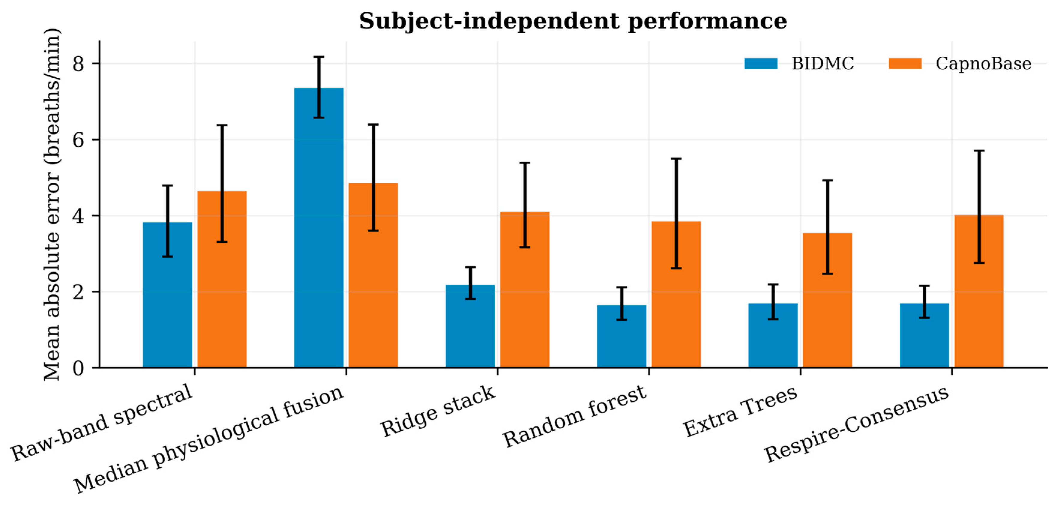
Figure 2.
Bidirectional external validation of the physiology-consensus point model. Top panels compare reference and estimated respiratory rate; bottom panels show Bland–Altman agreement. Transfer caused source-dependent compression and wide limits of agreement.
Figure 2.
Bidirectional external validation of the physiology-consensus point model. Top panels compare reference and estimated respiratory rate; bottom panels show Bland–Altman agreement. Transfer caused source-dependent compression and wide limits of agreement.
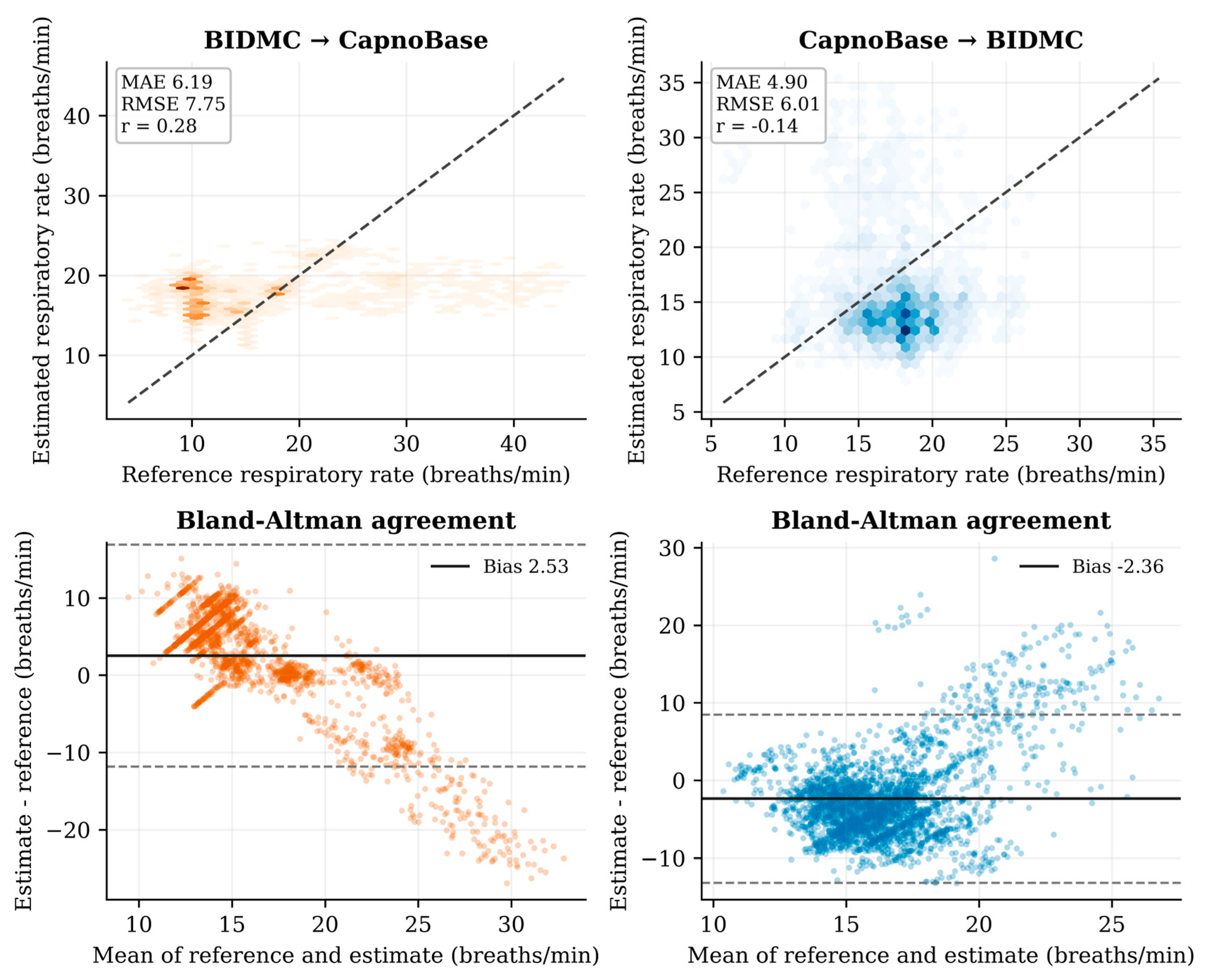
Figure 3.
Selective prediction of the physiology-consensus model. Solid lines show grouped internal validation; dashed lines show external transfer. Decreasing coverage markedly reduced MAE internally but provided little and non-monotonic benefit under BIDMC→CapnoBase transfer.
Figure 3.
Selective prediction of the physiology-consensus model. Solid lines show grouped internal validation; dashed lines show external transfer. Decreasing coverage markedly reduced MAE internally but provided little and non-monotonic benefit under BIDMC→CapnoBase transfer.
Scheme 1.
Conceptual workflow. Contact PPG is decomposed into respiratory modulations, combined by an AI estimator, and passed through a confidence gate before any rehabilitation-facing output. The lower panel summarizes the study's central test: whether point estimates and confidence transfer between BIDMC and CapnoBase. The illustration is conceptual and does not depict a deployed clinical device.
Scheme 1.
Conceptual workflow. Contact PPG is decomposed into respiratory modulations, combined by an AI estimator, and passed through a confidence gate before any rehabilitation-facing output. The lower panel summarizes the study's central test: whether point estimates and confidence transfer between BIDMC and CapnoBase. The illustration is conceptual and does not depict a deployed clinical device.
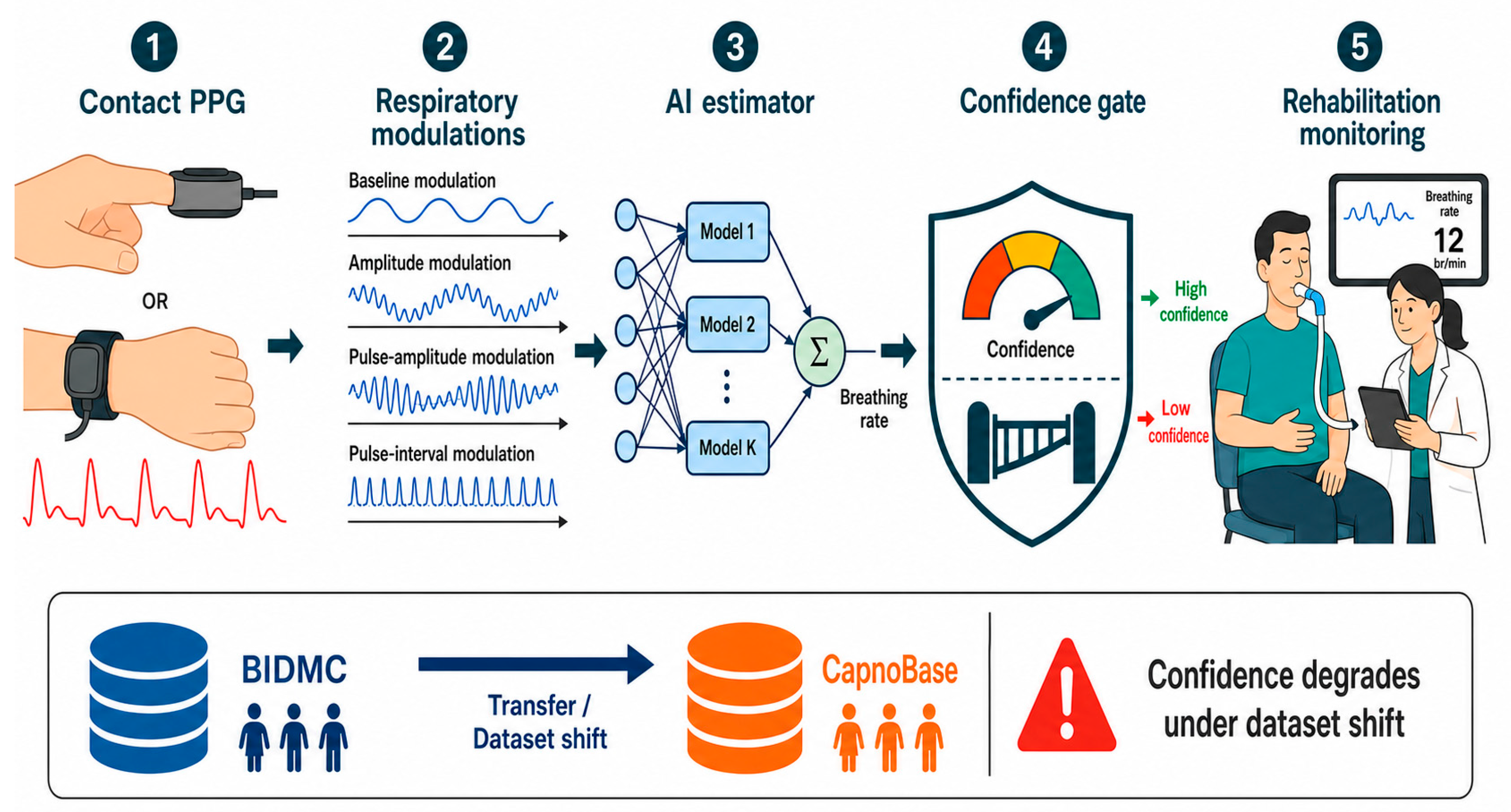
Table 2.
Estimators and roles in the validation framework.
| Estimator | Inputs / specification | Role |
| Raw-band spectral | Peak of baseline-modulation spectrum | Fixed physiological baseline |
| Median fusion | Median of up to eight candidate rates | Unweighted consensus baseline |
| Ridge | 51 features; α=10; robust scaling | Regularized linear stack |
| Random forest | 300 trees; leaf ≥8 | Bagged nonlinear stack |
| Extra Trees | 350 trees; leaf ≥5 | Randomized nonlinear stack |
| Physiology-consensus | Histogram boosting; 260 iterations; leaf ≥25 | Designated point model |
| Error predictor | Nested out-of-fold absolute error model | Ranks outputs for abstention |
Table 3.
Point-estimation performance under internal and external validation.
| Evaluation | Model | MAE (95% CI) | RMSE | Bias | Within ±2 |
| BIDMC grouped CV | Raw spectral | 3.83 (2.92–4.79) | 6.50 | −2.68 | 65.0% |
| BIDMC grouped CV | Random forest | 1.65 (1.26–2.12) | 2.59 | −0.12 | 74.6% |
| BIDMC grouped CV | Physiology-consensus | 1.70 (1.32–2.15) | 2.63 | −0.07 | 73.0% |
| CapnoBase grouped CV | Raw spectral | 4.65 (3.31–6.37) | 7.97 | −2.58 | 50.0% |
| CapnoBase grouped CV | Extra Trees | 3.55 (2.47–4.92) | 5.93 | +0.12 | 51.1% |
| CapnoBase grouped CV | Physiology-consensus | 4.02 (2.75–5.70) | 6.83 | +0.37 | 49.7% |
| BIDMC→CapnoBase | Raw spectral | 4.65 (3.31–6.37) | 7.97 | −2.58 | 50.0% |
| BIDMC→CapnoBase | Physiology-consensus | 6.19 (4.98–7.51) | 7.75 | +2.53 | 23.5% |
| CapnoBase→BIDMC | Raw spectral | 3.83 (2.92–4.79) | 6.50 | −2.68 | 65.0% |
| CapnoBase→BIDMC | Physiology-consensus | 4.90 (4.29–5.55) | 6.01 | −2.36 | 21.3% |
CI, participant-bootstrap 95% confidence interval; CV, cross-validation; MAE, mean absolute error; RMSE, root mean squared error. Errors are in breaths/min.
Table 4.
Selective prediction of the physiology-consensus model.
| Evaluation | MAE at 100% | MAE at 80% | MAE at 50% | ρ(error score, |error|) |
| BIDMC grouped CV | 1.70 | 1.36 | 0.93 | 0.481 |
| CapnoBase grouped CV | 4.02 | 3.30 | 2.01 | 0.414 |
| BIDMC→CapnoBase | 6.19 | 5.95 | 5.70 | 0.146 |
| CapnoBase→BIDMC | 4.90 | 4.58 | 4.41 | 0.092 |
Coverage is the fraction of windows retained after ranking by predicted absolute error. All Spearman correlations had p<0.001 at the window level; effect magnitude is emphasized because windows overlap.
Table 5.
Internal ablations of the physiology-consensus point model.
| Feature configuration | BIDMC MAE | CapnoBase MAE |
| Full feature set (51) | 1.70 | 4.02 |
| Candidate rates only (14) | 1.60 | 3.70 |
| Without interval modulation | 1.70 | 4.18 |
| Without pulse-amplitude modulation | 1.73 | 4.11 |
| Without agreement/global SQI features | 1.55 | 3.51 |
MAE is reported in breaths/min from five-fold participant-grouped cross-validation.
Disclaimer/Publisher’s Note: The statements, opinions and data contained in all publications are solely those of the individual author(s) and contributor(s) and not of MDPI and/or the editor(s). MDPI and/or the editor(s) disclaim responsibility for any injury to people or property resulting from any ideas, methods, instructions or products referred to in the content. |
© 2026 by the authors. Licensee MDPI, Basel, Switzerland. This article is an open access article distributed under the terms and conditions of the Creative Commons Attribution (CC BY) license (http://creativecommons.org/licenses/by/4.0/).
Copyright: This open access article is published under a Creative Commons CC BY 4.0 license, which permit the free download, distribution, and reuse, provided that the author and preprint are cited in any reuse.