Preprint
Article

This version is not peer-reviewed.

Ammonium Magnesium Phosphate Struvite in the Fruiting Body of the Wood-Rotting Fungus Сhondrostereum purpureum: Localization, Morphology and Formation

Submitted:

10 November 2026

Posted:

12 November 2026

You are already at the latest version

Abstract

The present study focuses on phosphate mineral formation under the influence of wood-rotting fungi. Crystals of the ammonium magnesium phosphate struvite were discovered for the first time within the fruiting body of the wood-rotting fungus Chondrostereum purpureum growing on a birch trunk (Russko-Vysotsky Forest Park, Leningrad Region, Russia). Model experiments with Chondrostereum purpureum, Auricularia mesenterica, Auricularia delicata, Auricularia cornea, Inonotus hispidus, Inonotus obliquus, and Serpula himantioides demonstrated the possibility of struvite formation under the action of wood-rotting fungi, provided the pH of the culture medium is near neutral. The morphology of both natural and biomimetic crystals was investigated using a complex of analytical methods, including powder X-ray diffraction, optical microscopy, scanning electron microscopy and EDX spectroscopy. The total quantitative content of extracellular polymeric substance (EPS) in the fungal culture fluid was measured by a gravimetric method. The contribution of EPS, secreted by wood-rotting fungi, in phosphate biomineralization was shown. Wood-rotting fungi inducing struvite precipitation could be used in biotechnological applications for environmental remediation.

Keywords: 
;  ;  ;  ;  

1. Introduction

The first descriptions of mineral crystals on fungal hyphae date back to the late 19th century [1]; however, their role in mineral formation was long underestimated. In the early 2000s, the emergence of geomycology as an independent scientific discipline [2-4] led to significant progress in understanding the role of fungi in mineral formation—primarily that of oxalates and carbonates [5, 6, 3, 7]. It has been shown that, in nature, oxalates of various metals (Ca, Cu, Mn, Mg, Zn, Ni, Pb) frequently form within the thalli of lichens growing on the surfaces of various rocks and minerals [8, 9]. Calcium oxalates (whewellite and weddellite) are the most common forms found in lichens, while moolooite—a copper oxalate—is typically associated with rocks containing copper sulfides [10-12]. Calcium oxalates have also been detected in the fruiting bodies of macromycetes (litter- and humus-decomposing saprotrophs) [13-16]. Furthermore, they have been observed on the hyphae of wood-rotting fungi within a mine gallery [17]. The presence of potassium oxalate (the mineral vertushkovite) has also been documented in growths (pseudosclerotia) on birch wood formed by the wood-rotting fungus Inonotus obliquus (chaga) [18]. There are only a few isolated reports regarding the occurrence of calcite crystals in association with fungal hyphae (without specification of the fungal species) [19, 20]. To date, phosphates—including struvite, the subject of this paper—have not been found in association with fungi in natural environments.
Experimental modeling has contributed significantly to understanding of the mechanisms by which fungi facilitate the formation of oxalates. A wide range of acid-producing fungal species has been shown to induce the formation of oxalates involving divalent and trivalent metals—including some (such as Fe, Cd, and Ce) that have not yet been identified within lichen thalli [7, 9]. The formation of heavy metal oxalates mediated by fungi opens up possibilities for developing biotechnologies for environmental bioremediation and detoxification, as well as for ore enrichment [7, 21]. Calcium carbonate precipitation is traditionally associated with bacterial activity, including the secretion of extracellular polymeric substances (EPS) [22, 23]. In contrast, the production of mycogenic (fungi-derived) calcite has been observed much less frequently in experimental models [24 -26]. Studies by Rusakov et al. [27] and Sazanova et al. [28, 29] have identified the conditions that trigger a transition from oxalate-based to carbonate-based biomineralization, and vice versa.
Phosphate formation mediated by fungi has not been sufficiently studied. Only a few works have been dedicated to modeling fungal-induced phosphate biomineralization. Experimental studies involving microscopic fungi have resulted in the formation of various phosphates, including lead phosphate (pyromorphite) [30], calcium phosphate (hydroxyapatite) [31], uranium phosphates ( [32], and zinc phosphate (hopeite) [33].
The mineral struvite (MgNH₄PO₄·6H₂O), which is the focus of this study, is found in municipal wastewater, biological waste from pig farms, and guano deposits [34, 35]. Struvite is frequently present in human kidney stones and serves as an indicator of inflammatory processes in the body—processes that may arise due to environmental, climatic, or other factors [36, 37].
As demonstrated by Michael Witty [38], the first sketches of struvite crystals—which possess a characteristic “coffin-like” form—were published in Robert Hooke’s famous book, “Micrographia” (1665), as early as the 17th century. The first detailed description of this mineral was provided by the German chemist G. L. Ulex, who discovered it while examining the remains of a medieval sewer system beneath St. Nicholas Church in Hamburg [39]. He also named the new mineral in honor of Heinrich von Struve—a prominent diplomat, geographer, and mineralogist.
According to the classification of the International Mineralogical Association (IMA), struvite holds the official status of a “valid mineral”, as it was first described long before the association itself was established [40]. The crystal structure of struvite was determined by A. Whitaker and J. W. Jeffery [41].
Numerous studies have addressed the formation of struvite in model experiments mediated by bacteria [42-50]. We obtained struvite for the first time through the action of the soil fungus Penicillium chrysogenum [29].
The scientific community’s interest in the microbially mediated formation of struvite is primarily driven by the development of highly efficient and nature-inspired biotechnologies for environmental remediation: specifically, the removal of ammonium nitrogen through precipitation. The struvite obtained in this manner can be utilized as a fertilizer [51-53] This approach can also be applied to the remediation of soils and water bodies contaminated with heavy metals by converting them into insoluble and less mobile forms [54].
The formation of biogenic struvite is linked to the active secretion of the enzymes urease and phosphatase by microorganisms [55]. This process leads to the release of NH₄⁺ and PO₄³⁻ ions, raises the solution’s pH to a slightly alkaline level, and creates favorable conditions for struvite crystallization [56]. The size of the resulting struvite crystals depends on the ratio of urease, phosphatase and extracellular polymeric substances (EPS) [55]. The EPS produced by bacteria acts as a matrix for struvite nucleation [55]. The presence of sodium alginate—a component of EPS—in the medium significantly reduces the growth rate of struvite crystals; this is attributed to the adsorption of alginate molecules onto the crystal surfaces, which blocks active growth sites. Furthermore, evidence suggests that EPS can inhibit crystal growth by stabilizing amorphous phases [57, 55, 58]. The role of microbial EPS in struvite crystallization remains a subject of debate and requires further investigation.
The influence of pH and medium composition on struvite crystallization is examined in detail in a paper by [59]. It is shown that the solubility of struvite increases significantly as the solution pH decreases from 9.0 to 6.5. The morphology of struvite crystals is highly dependent on the relative supersaturation of the solution and its chemical composition—primarily the NH4/Mg ratio.
This study aims to investigate the potential for struvite formation mediated by wood-rotting fungi.
The objectives of this work were as follows:
1. To examine the localization and morphology of struvite found within the fruiting body of the fungus Chondrostereum purpureum (Pers. ex Fr.).
2. To produce struvite in vitro using various species of wood-rotting fungi and to analyze the morphology of the resulting crystals.
3. To determine the content of extracellular polymeric substances (EPS) in the metabolic products of the fungi and to identify any correlation between the amount of EPS produced and the morphology and structural perfection of the formed struvite crystals.

2. Materials and Methods

2.1. Wood-Rotting Fungi in Nature and Experiment

A specimen of the fruiting body (FB) of a wood-rotting fungus—tentatively identified as Chondrostereum purpureum (Table 1)—was collected during fieldwork in July 2023. The collection took place while searching for metal oxalates within the pseudosclerotia of the fungus Inonotus obliquus (chaga) on the trunk of a living birch tree in the Russko-Vysotskoye Forest Park (Leningrad Region, Russia) (Figure 1). The fruiting body was situated within a deep fold of the bark, in an area where both the bark and the underlying wood had sustained damage (Figure 1b).The coordinates of the collection site are 59º41’58.33” N, 29º57’46.16” E.The collected fruiting bodies were represented by resupinate to effused-reflexed basidiomata with small, nail-like, and imbricated pilei. The upper sterile surface of the pileus is tomentose, whitish to pale greyish or yellowish-brown, and has unclear zones. The hymenophore is smooth and violaceous to purplish. The consistency of fresh basidiomata in situ is tough and waxy-cartilaginous, but becomes brittle after drying [60].
Strains of wood-rotting fungi for the model experiment were selected from the Komarov Botanical Institute Basidiomycete Culture Collection (LE-BIN) (Table 1). The samples from which pure cultures were isolated were collected in different years and from various regions. The strains were maintained in the LE-BIN collection using three simultaneous methods: (1) subculturing in test tubes on wort agar slants, stored in a refrigerator at 4–6 °C (14–16 °C for the strain from Vietnam); (2) the disk method, using screw-cap cryovials with the samples submerged in distilled water at 4–6 °C (14–16 °C for the strain from Vietnam); and (3) cryopreservation in screw-cap cryovials containing a 10% aqueous glycerol solution at −80 °C, with a cooling rate of 1 °C per minute. Despite differences in origin and taxonomy, all the species studied are wood-rotting fungi that cause wood rot. A strain of the fungus Chondrostereum purpureum was included in the study because a fruiting body of this species was collected for research purposes. The other selected fungal species are utilized by humans; for instance, the genus Auricularia is the fourth most important cultivated fungal genus globally, with China accounting for over 90% of total world production. Wood-rotting fungi of the genus Inonotus are used in both traditional and conventional medicine in a number of countries, owing to an exceptionally wide range of secondary metabolites [61-64]. The fungus Serpula himantioides is included in the present study because it is a well-known model organism for investigating the role of fungi in the processes of oxalate biomineralization [7, 17]. Thus, all the strains included in this study are of interest due to their biochemical characteristics, practical applications, and role in contemporary ecosystems.

2.2. Model Experiment

To obtain an inoculum, the strains were cultured on Malt Extract Agar (Condalab 1038-500G, India) for two weeks. The screening of wood-rotting fungal strains for their ability to form struvite was conducted using the B-41 growth medium (yeast extract – 4 g/L, MgSO4•7H2O – 2 g/L, KH2PO4 – 2 g/L), which had been previously proposed by researchers [42].Thus, the fungus could obtain Mg2+ and PO43- ions from the nutrient medium, whereas it had to produce the NH4+ ion through its own enzymatic activity. The initial pH of the medium was 6.5. The experiment was conducted at a temperature of 20–22 °C for 30 days. At the end of the experiment, the pH values ​​of the culture medium were recorded (using a Checker 1 pH meter, HI 98103). The resulting crystals were recovered from the culture medium and the mycelium for further analysis. The culture medium was separated from the mycelium in order to determine the amount of EPS produced by the fungi.

2.3. Research Methods

2.3.1. Biological Methods. Identification of Fungal Species and Strains, Study of Their Metabolic Products.

Fungi were identified based on morphological characteristics, as well as through molecular analysis (Table 1).
Verification of Fungal Fruiting Bodies and Strains Using Molecular Methods
Verification of strain samples and the fruiting body specimen was performed via Sanger sequencing of the nuclear rDNA ITS region (and, in some cases, the LSU region). For amplification, a small fragment—ideally from a non-pigmented part of the fruiting body or from the growing edge of a mycelial colony—was selected. The Thermo Scientific Phire Plant Direct PCR Kit (Vilnius, Lithuania) was used, along with standard basidiomycete-specific primers: ITS1F and ITS4B (or ITS4). The ITS4 primer was used for specimens belonging to the genus Auricularia. Primers LR0R and LR5 were used for the LSU region (https://www2.clarku.edu/faculty/dhibbett/protocols_folder/primers/primers.pdf, accessed June 23, 2026). Sample preparation followed the manufacturer’s protocol. Amplification was carried out using a C1000 Touch Thermal Cycler (Bio-Rad, Hercules, CA, USA). The presence of PCR products was confirmed by electrophoresis in a 1% agarose gel using 50× buffer (BE-DNA-500, Biolabmix, Novosibirsk, Russia) diluted 1:49 with H₂O. Electrophoresis results were visualized using a GelDoc Go Imaging System (Bio-Rad, Hercules, CA, USA). PCR products were purified using a commercial diaGene kit (DIA-M, Russia) according to the manufacturer’s protocol. Sample preparation for sequencing was performed using the BigDye™ Terminator Cycle Sequencing Ready Reaction Kit (NimaGen, Netherlands). Sequencing was conducted on an ABI 3130 genetic analyzer (Applied Biosystems, Foster City, CA, USA) at a shared research facility of the V.L. Komarov Botanical Institute of the Russian Academy of Sciences. Raw data and final sequences were processed using the MEGA XI software [65] Closely related sequences were identified using the BLAST tool within the public NCBI database (https://www.ncbi.nlm.nih.gov, accessed June 2026). The obtained sequences were also deposited in the NCBI GenBank database.
Determination of EPS Consentration in Cultural Liquid of the Fungi
The quantitative content of EPS in the cultural liquid of 7 fungal strains was determined gravimetrically according to the method described previously (Sazanova et al., 2020). To determine the amount of EPS, the cultural liquid was centrifuged at 4.000 rpm for 10 min. The collected supernatant was mixed with an equal volume of chilled (−20 °C) ethanol and incubated at 4 °C for 24 hours. The solution was then centrifuged for 20 min. The resulting precipitates were resuspended in distilled water together with an equal volume of ice-cold ethanol and then centrifuged again. The resulting final precipitate was dried on a vacuum evaporator (Labconco, USA) to an absolutely dry weight and weighed.

2.3.2. Methods of Analyzing Crystallization Products

Light Microscopy and Scanning Electron Microscopy (SEM).
Light stereomicroscopy (Leica MZ16 and Leica DM300 LED stereomicroscopes) and scanning electron microscopy (Hitachi TM3000 and Tescan MIRA3 LMU) were used to study the morphology of crystals found in the fungal fruiting body and those obtained in a model experiment.
Crystal dimensions were evaluated based on their length, and the degree of elongation was determined by the ratio l/w, where l = length and w = width of the primary monohedron.
For semiquantitative EDX analysis, the microscope was equipped with the Oxford Inca system (Oxford Instruments, UK) which operated in a low vacuum (60 Pa) mode and at an acceleration voltage of 15 kV. Magnification range varied from 100x to 1000x. To avoid the charging effect on SEM images, the samples were coated with a thin carbon layer (High Vacuum Carbon Sprayer Q150TE).The specimen of the fruiting body was coated with carbon (~ 15 nm).
Powder X-ray Diffraction (PXRD)
The determination of phase composition of the products of biomineralization was carried out using the PXRD method. Powder diffraction measurements were performed using a Rigaku II Miniflex diffractometer with CuKα radiation (2θ = 5–80°, with a step size of 0.02°). The sample was ground in a mortar and deposited onto a glass sample holder from an alcohol suspension. Phase determination was made using the ICDD PDF-2 database (2019).
For the fruiting body, powder X-ray diffraction data were obtained using a Rigaku R-AXIS Rapid II single-crystal diffractometer equipped with a cylindrical “image plate” detector (radius 127.4 mm) in the Debye-Scherrer geometry, with CoKα radiation (generated by a rotating anode with VariMAX microfocus optics) at 40 kV and 15 mA. The angular resolution of the detector is 0.045° 2θ (pixel size 0.1 mm). The data were integrated using the Osc2Tab software package [66]. It should be noted that preferred orientation of the sample during powder diffraction data collection leads to some minor differences in peak intensities between the observed and calculated powder diffraction patterns, while the angular positions of the peaks remain unaffected.

3. Results

3.1. Identification of Fungi

Sequencing results for the natural FB sample, when analyzed using the BLAST algorithm against sequences deposited in the NCBI GenBank, showed that the top 19 sequences with the highest similarity to the nucleotide sequence of the sample under study exhibited 99.59% homology (including MH270439, PP906278, GU062264, etc.) with 100% coverage.
Sequencing of the ITS regions of nrDNA from the LE-BIN collection strains revealed a 98.11–100% similarity to sequences of corresponding species in GenBank, with an overlap of 98–100%. For the Auricularia cornea strain LE-BIN 4803, the sequence of the LSU region was also obtained, showing 99.14% homology with the sequence of the same species in GenBank, with 100% overlap. Thus, the taxonomic identity and authenticity of all the strains studied were confirmed.
The accession numbers for the nucleotide sequences deposited in GenBank, along with a comparison to homologous sequences from the database, are presented in Table 2.

3.2. Struvite Crystals in the FB of Chondrostereum purpureum

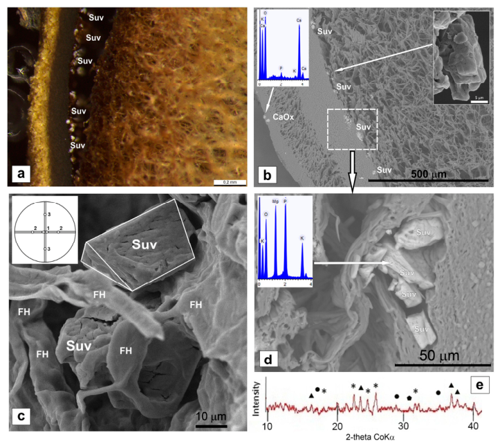
In vertical section, the basidiomata of Chondrostereum purpureum are characterized by a multi-layered structure (Figure 2, a). The whitish tomentum, composed of more-or-less thick-walled hyphae, is separated from the context by a cartilaginous, dark layer consisting of dense, parallel hyphae and visible as a thin black line to the naked eye. Between this dark layer and the hymenium, three layers of the context can be recognized, namely a dense hyaline layer formed by parallel hyphae, a vesicular layer with few hyphae and numerous vesicles, and a subhymenial layer of densely interwoven hyphae.
Struvite crystals measuring 10–30 µm were observed in the cartilaginous, dark layer (Figure 2). They are highly degraded and contain an admixture of potassium (~2–5 wt%, Figure 2d, inset). In the hymenial layer (hymenium) of the fruiting body, small clusters of very fine calcium oxalate crystals are visible (Figure 2, b). The X-ray diffraction pattern of the fruiting body shows, in addition to struvite peaks, faint peaks corresponding to whewellite, weddellite, and quartz (Figure 2, e).
Mycogenic struvite crystals are highly imperfect. They are characterized by extensive fracturing, a blocky and layered nature, a stepped appearance, and the presence of numerous depressions and rounded edges. Systems of subparallel fractures are evident, resulting in a patterned surface structure composed of blocks ranging from 2 to 5 µm in size (Figure 2b, inset). At the same time, individual crystals (~20–30 µm) can be observed, which exhibit simple forms characteristic of struvite (2mm symmetry): a monohedron and two dihedrons, connected to it via shared edges (Figure 2c).

3.3. Formation of Struvite by Wood-Rotting Fungi Under Experimental Conditions. Crystal Morphology.

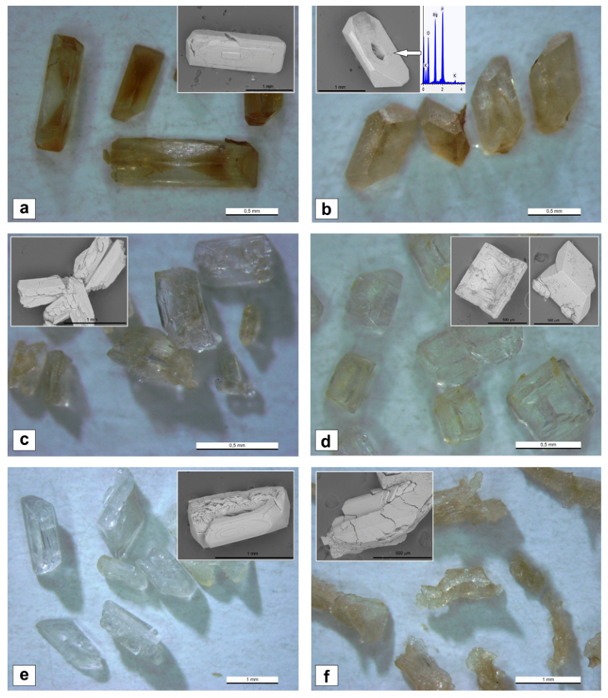
In a model experiment, struvite crystals (ranging from 200 µm to 1–2 mm in length) were observed after 25–30 days at pH 6.5–7.0, produced by six of the seven selected fungal strains (Figure 3, Figure 4, Table 3), including Chondrostereum purpureum, a fungus species in whose fruiting body struvite has been found in a natural setting (see Section 3.2). All struvite crystals contain an admixture of potassium (no more than 2 wt%; Figure 4d, inset) and, with the exception of those formed under the influence of Auricularia delicata, exhibit a characteristic “coffin-like” shape.
In the acidic medium created by the fungus Serpula himantioides (pH = 4.5), no struvite precipitation occurred. Struvite crystals also failed to form in the culture medium that had not been inoculated with the fungus.
Struvite crystals formed under the influence of different fungal strains differ in size, degree of elongation, the development of simple forms, and the degree of perfection (Table 3; Figure 4, Figure 5a and Figure 6).
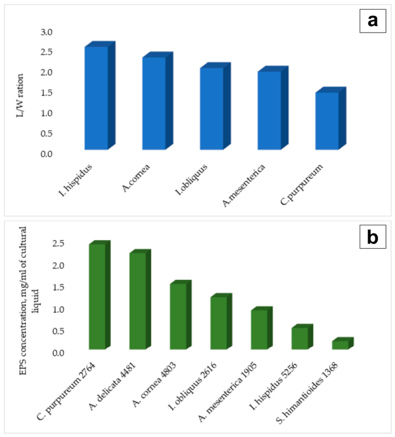
The length of the crystals obtained through the action of each of the fungi varies significantly (Table 3). Furthermore, the struvite crystals obtained under the action of the fungi Inonotus hispidus, Auricularia cornea and Inonotus obliquus are larger than those obtained under the action of Auricularia mesenterica and Chondrostereum purpureum. In terms of the degree of elongation, the studied crystals are ranked as follows: Inonotus hispidus > Auricularia cornea > Inonotus obliquus > Auricularia mesenterica > Chondrostereum purpureum (Figure 5a). In the case of Auricularia delicata, there are no distinct individuals for which length and the degree of elongation can be measured.
The most elongated and largest struvite crystals are those that formed under the influence of Inonotus hispidus, while the least elongated and smallest are those that formed under the influence of Chondrostereum purpureum—a fungus in whose fruiting body struvite has been found in nature.
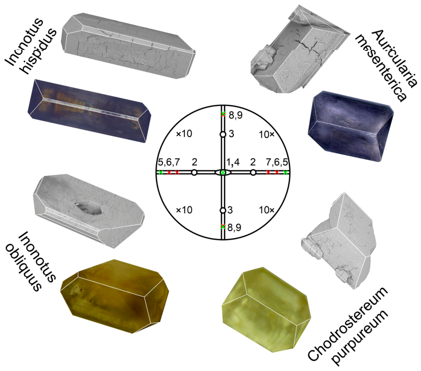
Struvite crystals formed under experimental conditions through the action of Auricularia cornea and Auricularia delicata have virtually no straight edges. The faceting of the other biomimetic crystals formed under the influence of Inonotus hispidus, Inonotus obliquus, Auricularia mesenterica, and Chondrostereum purpureum is more rich (Figure 4 and Figure 6) than that of the crystals formed within the fungal fruiting body (Figure 2). All crystals synthesized under the influence of these strains have a monohedron and two associated dihedrons (connected via shared edges)—simple forms characteristic of natural struvite crystals (Figure 2)—though the degree to which these faces are developed varies (Figure 6). Unlike the crystals formed within the fruiting body of the fungus, synthesized struvite crystals are characterized by the presence of a pyramid (a general simple form). Crystals formed under the influence of fungi of the genus Inonotus, are also have additional dihedrons, while those formed under the influence of Inonotus hispidus have an additional monohedron.
The degree of perfection of biomimetic struvite crystals varies significantly (Figure 4). They have in different degrees the types of defects described above for struvite crystals found in the fruiting body of the fungus Chondrostereum purpureum: fracturing, a blocky structure, depressions and rounded edges. In addition, they exhibit sectoriality, oscillatory zoning, and skeletal forms. Sectoriality is most clearly visible in the darker, unevenly colored struvite crystals that formed under the influence of fungi of the genus Inonotus (Figure 4a, b), while zoning is observed in crystals that formed under the influence of Auricularia cornea (Figure 4e).
Overall, the most perfect crystals are those that formed under the influenve of fungi of the genus Inonotus; they are fully faceted, characterized by straight edges, and exhibit the maximum number of simple forms (Figure 4a, b, 6). The crystals formed under the influence of Inonotus hispidus and Inonotus obliquus are similar in length (Table 3); however, Inonotus hispidus gives rise to elongated crystals with a prismatic habit (Figure 4a), whereas Inonotus obliquus results in less elongated, flattened crystals (Figure 4b). In the case of Inonotus hispidus, subindividuals are also visible on the crystal surfaces (Figure 4a), while in the case of Inonotus obliquus, there are distinct depressions (Figure 4b).
The crystals formed under the influence of Auricularia mesenterica and Chondrostereum purpureum are smaller, less elongated, and less perfect (Table 3, Figure 4c, d, 5a). They are characterized by intense fracturing, a blocky nature, a smaller number of simple forms (Figure 6), as well as the presence of rounded edges and numerous overgrowths and depressions. Auricularia mesenterica is characterized by the formation of aggregates, including subparallel ones. The highest degree of fracturing and blocky structure is observed in crystals that formed under the influence of Chondrostereum purpureum (a fungus in whose fruiting body struvite has been found in nature).
The most defective crystals are those that formed under the influence of the fungi Auricularia cornea and Auricularia delicata. Struvite crystals obtained under the influence of Auricularia cornea are virtually devoid of straight edges, and deep depressions are visible on their faces (Figure 4e). In the case of Auricularia delicata, numerous skeletal forms are visible within the overall mass of the resulting unfaceted crystals (Figure 4f).
Thus, the struvite crystals obtained in experimental conditions under the action of wood-rotting fungi can be ranked according to their degree of perfection as follows: Inonotus hispidus ≈ Inonotus obliquus > Auricularia mesenterica > Chondrostereum purpureum > Auricularia cornea > Auricularia delicata.

3.4. EPS Content in the Crystallization Medium (Model Experiment).

The mycelium of all the fungi produced EPS at concentrations ranging from 0.2 to 2.4 mg/ml of culture medium (Figure 5b). The highest amount of EPS (2.4 mg/ml of cultural fluid) was found in the culture of C. purpureum (in whose fruiting bodies struvite has been observed in nature), while the lowest (0.2 mg/ml) was found in the cultural fluid of S. himantioides (under the influence of which no struvite formation occurred). The EPS precipitate (as well as the culture medium) from fungi of the genus Inonotus exhibited a darker coloration than that of the other fungal strains, which is likely due to the presence of the pigment melanin.
At the level of a trend, there is an inverse relationship between the EPS (extracellular polymeric substance) content in the crystallization medium and the perfection of the resulting crystals, as well as their size and degree of elongation. More perfect crystals obtained under the influence of Inonotus hispidus, I. obliquus, and Auricularia mesenterica are formed when the EPS concentration is between 0.5 and 1.2 mg/ml, whereas less perfect crystals—obtained in the presence of Chondrostereum purpureum, Auricularia cornea, and Auricularia delicata—are formed at EPS concentrations ranging from 1.5 to 2.4 mg/ml. Within these groups, however, this correlation does not hold consistently. In the first group, this is due to Auricularia mesenterica: the struvite crystals formed with its involvement are less perfect than would be expected given the amount of EPS secreted. In the second group, this is due to Chondrostereum purpureum: the struvite crystals formed with its involvement are more perfect than would be expected based on the amount of EPS secreted. The largest and most elongated crystals—those with a prismatic habitwere obtained under influence of Inonotus hispidus at an EPS concentration of 0.5 mg/ml. Conversely, the smallest and least elongated crystals were obtained in the presence of Chondrostereum purpureum at an EPS concentration of 2.4 mg/ml.

4. Discussion

The discovery of struvite in the fruiting body of Chondrostereum purpureum, combined with the results of model experiments, has demonstrated the ability of wood-rotting fungi—including Chondrostereum purpureum—to participate in the formation of struvite. A model experiment also demonstrated that struvite formation occurs only when a near-neutral medium (pH 6.5–7.0) is established in the culture fluid (Table 3). The narrower pH range for mycogenic struvites—shifted toward lower values ​​compared to abiogenic ones (pH 6.5–9.0; [59]—is well explained by the influence of organic acids present in the metabolic products of wood-rotting fungi [67] As demonstrated by the fungus Serpula himantioides, the formation of struvite under the influence of wood-rotting fungi occurs only when the effect of the organic acids secreted by the fungus on the environmental pH is outweighed by the influence of metabolic products (such as ammonium ions, EPS, etc.) that promote an increase in pH (alkalinization).
It is known that Chondrostereum purpureum, in whose fruiting body struvite crystals were found, produces enzymes as well as acid and alkaline phosphatase [68]. There are no data of the accumulation of urea in the fruiting body of Chondrostereum purpureum or of the mechanisms of its decomposition. Our discovery of this mineral in the fruiting body of this fungus showed that Chondrostereum purpureum accumulates urea and implements one of the possible physiological mechanisms of its decomposition with the formation of ammonium.
The significant defectiveness of struvite crystals in the fruiting body of Chondrostereum purpureum (fracturing, blockiness, numerous depressions, layering, and stepped surfaces, Figure 2) could have formed under strong supersaturation and high growth rates [69]. However, real crystals with similar types of defectiveness also form during crystallization from viscous solutions in a diffusion-limited regime with significant supersaturation gradients along the surface of the growing faces [70].
The model experiment revealed relationships between the EPS content in the fungal culture liquid and the perfection of the resulting struvite crystals, as well as their size and habit, demonstrating (Figure 4, Figure 5 and Figure 6) that the presence of EPS in the medium (along with the presence of NH4+, Mg2+, and [PO4]3- ions) is one of the most important factors of mycogenic struvite formation. This result and the few literature data on the influence of bacterial EPS on the morphology and growth of struvite crystals [55, 57, 58, 71] allow preference to be given to a model of mycogenic struvite formation based on a direct relationship between EPS content and the viscosity of the medium, which substantially affects the diffusion processes controlling crystallization [69, 70].
The formation of struvite in the model experiment only on days 25–30 can be explained not only by the slow accumulation of NH4+ ions in the medium, which were formed under the action of the fungi via the decomposition of urea by means of specific enzymes, but also by the inhibitory effect of the increasing viscosity of the medium due to the input of EPS. The surface-active molecules composing the EPS can hinder incipient spontaneous crystallization by adsorbing on crystal nuclei and inhibiting their further growth [69].
The formation, under the action of fungi of the genus Inonotus at a low EPS content (from 0.5 to 1.2 mg/ml), of fairly large (up to 2 mm, Table 3) and perfect fully faceted struvite crystals with straight edges and the maximum number of simple forms formed (Figure 4a,b, 6) indicates that in this case the viscosity of the medium was insignificant and crystal growth occurred at a rate that was limited predominantly by the rate of incorporation of particles (ions and their associations) arriving from the solution. The contribution of diffusion limitations to crystallization is manifested in the sectorial structure of the crystals, the presence of isolated depressions on their faces, and their variable habit (Figure 4a, b, 5a). The sectorial structure of the synthesized struvite crystals indicates heterometry [72]— an inhomogeneous distribution of impurities (in our case, K+ ions, Figure 2d) over the faces of different simple forms, leading to variations in the crystal lattice parameters in different parts of a single crystal; the presence of isolated depressions indicates insignificant supersaturation gradients along the surface of the growing faces; the variable habit indicates selective adsorption of surface-active molecules on the faces of the growing crystal, which leads to anisotropy of growth of different faces and radically changes their habit. The latter is clearly seen in the example of struvite crystals formed under the action of Inonotus hispidus and Inonotus obliquus (see Figure 4a,b), whose degree of elongation is different (Figure 5a).
Crystals formed under the action of the species Chondrostereum purpureum, Auricularia cornea, and Auricularia delicata at an EPS content greater than 1.5 mg/ml are very imperfect (Figure 4d, e, f). They are characterized by strong fracturing, oscillatory zoning, blockiness, and the presence of deep depressions on the faces and skeletal growth forms.
All these features indicate crystallization in a substantially viscous medium — a pronounced diffusion-limited process in which crystal growth occurs at a rate that is limited almost entirely by the rate of supply of material to the crystal. This slows down the overall growth rate and provokes a transition to unstable skeletal growth. Deep depressions on the faces and skeletal forms of the synthesized crystals (Figure 4e,f) indicate significant supersaturation gradients along the surface of the growing faces; oscillatory zoning (Figure 4 c) indicates a periodic alternation of regimes of almost complete starvation and intense growth, which is accompanied by the capture of molecules composing the EPS; fracturing and blockiness (Figure 4d,e) indicate internal stresses leading to the destruction of crystals. All crystals of this group, like those described above, have a sectorial structure, but the insignificant potassium content (no more than 2 wt%) indicates that the contribution of the uneven distribution of K+ ions to their stressed state is not the main one. This suggests that during crystallization in a diffusion-limited regime, the main contribution to the generation of stresses is made by the selective adsorption of surface-active molecules composing the EPS, which leads to significant anisotropy of growth rates in different parts of the crystal and, as a consequence, to the appearance of strong internal stresses. The presence of selective adsorption is confirmed by the localization of zoning structures on certain faces of the crystal, as well as by the variable habit of the crystals, whose degree of elongation varies (Figure 5a)
All mycogenic struvite crystals — both those found in the fruiting body of the fungus and those obtained in the experiment — display faces of the monohedron and of two dihedra that share edges with it (numbered 1, 2, 3 in Figure 2c and Figure 6), which indicates their stability and low formation rates, probably caused by intense adsorption of surface-active molecules. Changes in the habit of the synthesized mycogenic crystals (variations in the degree of elongation, Figure 5a) are determined by the ratio of the sizes of these faces, which depends on their growth rates controlled by the intensity of adsorption of surface-active molecules on them. The more uniformly the molecules are distributed over the surface of these three faces, the closer the shape of the monohedron approaches a square.
Increasing of EPS concentration should lead not only to a higher degree of defectiveness in struvite crystals but also to a reduction in their size, due to the emergence of a greater number of nucleation sites [55, 57, 58]. This approach effectively explains why the influence of Inonotus hispidus results in the formation of crystals that are both the most structurally perfect and the largest in size, whereas the influence of Chondrostereum purpureum leads to the formation of crystals that are both highly defective and very small.
All violations of the above-described patterns of mycogenic struvite formation (see Section 3.2 and Section 3.3) are due to the multifactorial nature of the crystallization processes occurring with the participation of fungi. For example, the defectiveness of the resulting struvite crystals is greatly influenced by the ratio of ammonium and magnesium ions [59]. As the ammonium content increases (as in the case of increasing EPS), a transition is observed from fully faceted crystals to crystals with depressions on the faces and then to skeletal forms. The manifestation of relationships between EPS content and the perfection of struvite crystals synthesized with the participation of wood-rotting fungi indirectly indicates a similar ammonium ion content in the culture liquid of most of the wood-rotting fungi involved in the experiment. The greater (Auricularia mesenterica) / lesser (Chondrostereum purpureum) degree of defectiveness of mycogenic struvite crystals compared with that which should be expected taking into account only the EPS activity of the fungi is well explained by the corresponding variations in the ammonium-producing capabilities of these strains.
Struvite crystals formed in the experiment under the action of Chondrostereum purpureum are close in the character and degree of defectiveness to the crystals found in the fruiting body of this fungus. This allows the crystals synthesized under laboratory conditions with the participation of Chondrostereum purpureum to be considered analogues of those formed under the action of wood-rotting fungi in nature, and the patterns of mycogenic struvite formation revealed in the model experiment to be used for characterizing natural processes.
Future findings of phosphates and other minerals in the fruiting bodies of wood-rotting fungi should expand our understanding of the mechanisms of mycogenic mineral formation and find their worthy application in the developing nature-like technologies.

5. Conclusions

The discovery of struvite in the fruiting body of Chondrostereum purpureum has demonstrated that this mineral can form through the action of fungi, and has highlighted the significant role played by wood-rotting fungi in its formation. A model experiment made it possible to study in detail the morphology of struvite crystals formed under the influence of various species of wood-rotting fungi, and to advance the understanding of the mechanisms behind mycogenic struvite formation. It has been shown that the growth of mycogenic struvite crystals is controlled not only by the presence of ammonium, magnesium and phosphate ions, but also by the viscosity of the medium, which is determined by the secretion of EPS by fungi. The obtained results were shown that wood-rotting fungi, along with bacteria, would be used in biotechnologies for cleaning the environment of organic pollutants.

Author Contributions

Conceptualization, M.S.Z., D.Y.V., K.V.S. and O.V.F.-K.; methodology, M.S.Z., K.V.S., A.R.I., S.V.V. and N.V.P ; software, N.V.P; validation, M.S.Z., K.V.S., A.R.I., S.V.V. and N.V.P.; investigation, M.S.Z., K.V.S., D.K.K., A.R.I. and S.V.V.; writing— original draft preparation, M.S.Z., D.Y.V., K.V.S. and O.V.F.-K.; writing—review and editing, M.S.Z., D.Y.V., K.V.S. and O.V.F.-K.; visualization, M.S.Z., K.V.S., D.K.K., A.R.I.; funding acquisition, M.S.Z. All authors have read and agreed to the published version of the manuscript.

Funding

The research was funded by the Russian Science Foundation (grant 25-24-01035 to M.S.Z., K.V.S. and D.Yu.V, D.K. K.).

Data Availability Statement

The original contributions presented in the study are included in the article. The accession numbers for the nucleotide sequences of fungal specimens and strains have been deposited in GenBank.

Acknowledgments

The laboratory researches were carried out in the Research Resource Centers of Saint Petersburg State University: SEM and EDX investigations - in the Resource Centers “Development of molecular and cell technologies” and “Resource Center Microscopy and Microanalysis” and XRD measurements – in the X-ray Diffraction Centre.

Conflicts of Interest

The authors declare no conflicts of interest. .

References

  1. Whitney, K.D.; Arnott, H.J. Calcium Oxalate Crystal Morphology and Development in Agaricus bisporus. Mycologia 1987, 79, 180–187. [Google Scholar] [CrossRef]
  2. Sterflinger, K. Fungi as geologic agents. Geomicrobiol. J. 2000, 17, 97–124. [Google Scholar] [CrossRef]
  3. Gadd, M.G. Geomycology: biochemical transformation of rocks, minerals, metals and radionuclides by fungi, bioweathering and bioremediation. Mycol. Res. 2007, 111, 3–49. [Google Scholar] [CrossRef] [PubMed]
  4. Gorbushina, A.A. Life on the rocks. Environ. Microbiol. 2007, 9, 1613–1631. [Google Scholar] [CrossRef] [PubMed]
  5. Burford, E.P.; Kierans, M.; Gadd, J.M. Geomycology: Fungi in mineral substrata. Mycologist 2003, 17, 98–107. [Google Scholar] [CrossRef]
  6. Burford, E.P.; Hillier, S.; Gadd, G.M. Biomineralization of Fungal Hyphae with Calcite (CaCO3) and Calcium Oxalate Mono- and Dihydrate in Carboniferous Limestone Microcosms. Geomicrobiol. J. 2006, 23, 599–611. [Google Scholar] [CrossRef]
  7. Gadd, G.M.; Bahri-Esfahani, J.; Li, Q.; Rhee, Y.J.; Wei, Z.; Fomina, M.; Liang, X. Oxalate production by fungi: Significance in geomycology, biodeterioration and bioremediation. Fungal Biol. Rev. 2014, 28, 36–55. [Google Scholar] [CrossRef]
  8. Adamo, P.; Violante, P. Weathering of rocks and neogenesis of minerals associated with lichen activity. Appl. Clay Sci. 2000, 16, 229–256. [Google Scholar] [CrossRef]
  9. Frank-Kamenetskaya, O.; Vlasov, D. Crystallization induced by fungi and bacteria. Acta Crystallogr. Sect. B 2025, 81, 1–10. [Google Scholar] [CrossRef] [PubMed]
  10. Frank-Kamenetskaya, O.V.; Ivanyuk, G.Y.; Zelenskaya, M.S.; Izatulina, A.R.; Kalashnikov, A.O.; Vlasov, D.J.; Polyanskaya, E.I. Calcium Oxalates in Lichens on Surface of Apatite-Nepheline Ore (Kola Peninsula, Russia). Minerals 2019, 9, 656. [Google Scholar] [CrossRef]
  11. Frank-Kamenetskaya, O.V.; Zelenskaya, M.S.; Izatulina, A.R.; Vereshchagin, O.S.; Vlasov, D.Y.; Himelbrant, D.E.; Pankin, D.V. Copper oxalate formation by lichens and fungi. Sci. Rep. 2021, 11, 24239. [Google Scholar] [CrossRef]
  12. Vlasov, D.Y.; Frank-Kamenetskaya, O.V.; Zelenskaya, M.S.; Sazanova, K.V.; Rusakov, A.V.; Izatulina, A.R. The use of Aspergillus niger in modeling of modern mineral formation in lithobiotic systems. In Aspergillus niger: Pathogenicity, Cultivation and Uses; Baughan, E., Ed.; Nova Science Publishers: New York, NY, USA, 2020; pp. 1–123. ISBN 978-1-53618-080-0. [Google Scholar]
  13. Whitney, K.D.; Arnott, H.J. Calcium Oxalate Crystals and Basidiocarp Dehiscence in Geastrum saccatum (Gasteromycetes). Mycologia 1986, 78, 649–656. [Google Scholar] [CrossRef]
  14. Horner, H.T.; Tiffany, L.H.; Knaphus, G. Oak-leaf-litter rhizomorphs from Iowa and Texas: Calcium oxalate producers. Mycologia 1995, 87, 34–40. [Google Scholar] [CrossRef]
  15. Horner, H.T.; Tiffany, L.H.; Cody, A.M. Formation of Calcium Oxalate Crystals Associated with Apothecia of the Discomycete Dasyscypha capitata. Mycologia 1983, 75, 423–435. [Google Scholar] [CrossRef]
  16. Tewari, J.P.; Shinners, T.C.; Briggs, K.G. Production of Calcium Oxalate Crystals by Two Species of Cyathus in Culture and Infested Plant Debris. Z. Für Naturforschung C 1997, 52, 421–425. [Google Scholar] [CrossRef]
  17. Vlasov, D.Y.; Zelenskaya, M.S.; Izatulina, A.R.; Janson, S.Y.; Frank-Kamenetskaya, O.V. Oxalate Crystallization under the Action of Brown Rot Fungi. Crystals 2023, 13, 432. [Google Scholar] [CrossRef]
  18. Erokhin, Y.V.; Shorin, Y.V. Grigory Nikolaevich Vertushkov (1909–1994) and former vertushkovite (mineral not approved). News Ural State Min. Univ. 2022, 3(67), 173–183. [Google Scholar] [CrossRef]
  19. Masaphy, S.; Zabari, L.; Pastrana, J.; Dultz, S. Role of Fungal Mycelium in the Formation of Carbonate Concretions in Growing Media—An Investigation by SEM and Synchrotron-Based X-Ray Tomographic Microscopy. Geomicrobiol. J. 2009, 26, 442–450. [Google Scholar] [CrossRef]
  20. Bindschedler, S.; Cailleau, G.; Verrecchia, E. Role of Fungi in the Biomineralization of Calcite. Minerals 2016, 6, 41. [Google Scholar] [CrossRef]
  21. Ayilara, M.S.; Babalola, O.O. Bioremediation of Environmental Wastes: The Role of Microorganisms. Front. Agron. 2023, 5, 1183691. [Google Scholar] [CrossRef]
  22. Zhu, T.; Dittrich, M. Carbonate Precipitation through Microbial Activities in Natural Environment, and Their Potential in Biotechnology: A Review. Front. Bioeng. Biotechnol. 2016, 4, 4. [Google Scholar] [CrossRef] [PubMed]
  23. Eltarahony, M.; Jestrzemski, D.; Hassan, M.A. A comprehensive review of recent advancements in microbial-induced mineralization: biosynthesis and mechanism, with potential implementation in various environmental, engineering, and medical sectors. Sci. Total Environ. 2025, 978, 179426. [Google Scholar] [CrossRef] [PubMed]
  24. Li, Q.; Csetenyi, L.; Paton, G.I.; Gadd, G.M. CaCO₃ and SrCO₃ bioprecipitation by fungi isolated from calcareous soil. Environ. Microbiol. 2015, 17, 3082–3097. [Google Scholar] [CrossRef] [PubMed]
  25. Dhami, N.K.; Quirin, M.E.C.; Mukherjee, A. Carbonate biomineralization and heavy metal remediation by calcifying fungi isolated from karstic caves. Ecol. Eng. 2017, 103, 106–117. [Google Scholar] [CrossRef]
  26. Zhao, J.; Csetenyi, L.; Gadd, G.M. Fungal-induced CaCO₃ and SrCO₃ precipitation: a potential strategy for bioprotection of concrete. Sci. Total Environ. 2022, 816, 151501. [Google Scholar] [CrossRef] [PubMed]
  27. Rusakov, A.V.; Vlasov, A.D.; Zelenskaya, M.S.; Frank-Kamenetskaya, O.V.; Vlasov, D.Y. The crystallization of calcium oxalate hydrates formed by interaction between microorganisms and minerals. In Biogenic—Abiogenic Interactions in Natural and Anthropogenic Systems. Lecture Notes in Earth System Sciences; Frank-Kamenetskaya, O., Panova, E., Vlasov, D., Eds.; Springer: Cham, Switzerland, 2016; pp. 357–377. [Google Scholar] [CrossRef]
  28. Sazanova, K.V.; Frank-Kamenetskaya, O.V.; Vlasov, D.Y.; Zelenskaya, M.S.; Vlasov, A.D.; Rusakov, A.V.; Petrova, M.A. Carbonate and oxalate crystallization by interaction of calcite marble with Bacillus subtilis and Bacillus subtilis–Aspergillus niger association. Crystals 2020, 10, 756. [Google Scholar] [CrossRef]
  29. Sazanova, K.V.; Zelenskaya, M.S.; Izatulina, A.R.; Korneev, A.V.; Vlasov, D.Y.; Frank-Kamenetskaya, O.V. Carbonate and Oxalate Crystallization Effected by the Metabolism of Fungi and Bacteria in Various Trophic Conditions: The Case of Penicillium chrysogenum and Penicillium chrysogenum with Bacillus subtilis. Crystals 2023, 13, 94. [Google Scholar] [CrossRef]
  30. Rhee, Y.J.; Hillier, S.; Gadd, G.M. Lead transformation to pyromorphite by fungi. Curr. Biol. 2012, 22, 237–241. [Google Scholar] [CrossRef] [PubMed]
  31. Ennever, J.; Summers, F.E. Calcification by Candida albicans. J. Bacteriol. 1975, 122, 1391–1393. [Google Scholar] [CrossRef] [PubMed]
  32. Liang, X.; Hillier, S.; Pendlowski, H.; et al. Uranium phosphate biomineralization by fungi. Environ. Microbiol. 2015, 17, 2064–2075. [Google Scholar] [CrossRef] [PubMed]
  33. Sazanova, K.V.; Zelenskaya, M.S.; Korneev, A.V.; Bakhvalova, E.V.; Vlasov, D.Y.; Frank-Kamenetskaya, O.V. Effect of fungal metabolism on Zn minerals formation: the case of Aspergillus niger and Penicillium chrysogenum. Crystals 2025, 15, 118. [Google Scholar] [CrossRef]
  34. Korago, L.A. Introduction to Biomineralogy; In Russ.; Nedra: St. Petersburg, Russia, 1992; p. 280 p. [Google Scholar]
  35. Burns, R.T.; Moody, L.B.; Walker, F.R.; Raman, D.R. Laboratory and in-situ reductions of soluble phosphorus in liquid swine waste slurries. Environ. Technol. 2001, 22, 1273–1278. [Google Scholar] [CrossRef] [PubMed]
  36. Rivadeneyra, M.A.; Gutierrez-Calderón, A.; Rivadeneyra, A.M.; Ramos-Cormenzana, A. A Study of Struvite Precipitation and Urease Activity in Bacteria Isolated from Patients with Urinary Infections and Their Possible Involvement in the Formation of Renal Calculi. Urol. Int. 1999, 63, 188–192. [Google Scholar] [CrossRef] [PubMed]
  37. Golovanova, O.A. Pathogenic Minerals in the Human Body; In Russ.; Omsk State University Publishing House: Omsk, Russia, 2006; p. 400 p. [Google Scholar]
  38. Witty, M. Hooke’s gravel was struvite. Notes Queries 2016, 63, 569–570. [Google Scholar] [CrossRef]
  39. Ulex, G.L. On struvite, a new mineral. Lond. Edinb. Dublin Philos. Mag. J. Sci. 1846, 29, 124–128. [Google Scholar] [CrossRef]
  40. Palache, C.; Berman, H.; Frondel, C. The System of Mineralogy of James Dwight Dana and Edward Salisbury Dana, Yale University 1837-1892, 7th ed.; John Wiley and Sons, Inc.: New York, NY, USA, 1951; pp. 715–717. [Google Scholar]
  41. Whitaker, A.; Jeffery, J.W. The crystal structure of struvite, MgNH4PO4.6H2O. Acta Crystallogr. Sect. B Struct. Crystallogr. Cryst. Chem. 1970, 26, 1429–1440. [Google Scholar] [CrossRef]
  42. Rivadeneyra, M.A.; Ramos-Cormenzana, A.; García-Cervigón, A. Bacterial formation of struvite. Geomicrobiol. J. 1983, 3, 151–163. [Google Scholar] [CrossRef]
  43. Nelson, B.; Struble, J.; McCarthy, G. In vitro production of struvite by Bacillus pumilus. Can. J. Microbiol. 1991, 37, 978–983. [Google Scholar] [CrossRef]
  44. Da Silva, S.; Bernet, N.; Delgenes, J.P.; Moletta, R. Effect of culture conditions on the formation of struvite by Myxococcus xanthus. Chemosphere 2000, 40, 1289–1296. [Google Scholar] [CrossRef]
  45. Jimenez-Lopez, C.; Jroundi, F.; Rodriguez-Gallego, M.; Arias, J.M.; Gonzalez-Munoz, M.T. Biomineralization induced by Myxobacteria. Commun. Curr. Res. Educ. Top. Trends Appl. Microbiol. 2007, 1, 143–154. [Google Scholar]
  46. Sharma, A.; Angulo-Bejaran, P.I.; Fierros-Romero, G.; Flores-Vallejo, R.C.; Gomez-García, K.R.; Collin, G.R. Struvite production by Pseudomonas syringae pv phaseolicola. J. Microbiol. Biotechnol. Food Sci. 2018, 8, 812–814. [Google Scholar] [CrossRef]
  47. Leng, Y.; Colston, R.; Soares, A. Understanding the biochemical characteristics of struvite bio-mineralising microorganisms and their future in nutrient recovery. Chemosphere 2020, 247, 125799. [Google Scholar] [CrossRef] [PubMed]
  48. Leng, Y.; Soares, A. The mechanisms of struvite biomineralization in municipal wastewater. Sci. Total Environ. 2021, 799, 149261. [Google Scholar] [CrossRef] [PubMed]
  49. Soares, А.; Veesam, M.; Simoes, F.; Wood, E.; Parsons, S.A.; Stephenson, T. Bio-Struvite: A New Route to Recover Phosphorus from Wastewater. Clean – Soil Air Water 2014, 42, 994–997. [Google Scholar] [CrossRef]
  50. Leng, Y.; Soares, A. Microbial phosphorus removal and recovery by struvite biomineralisation in comparison to chemical struvite precipitation in municipal wastewater. J. Environ. Chem. Eng. 2023, 11, 109208. [Google Scholar] [CrossRef]
  51. Doyle, J.; Oldring, K.; Churchley, J.; Parsons, S. Struvite formation and the fouling propensity of different materials. Water Res. 2002, 36, 3971–3978. [Google Scholar] [CrossRef]
  52. Hertzberger, A.J.; Cusick, R.D.; Margenot, A.J. A review and meta-analysis of the agricultural potential of struvite as a phosphorus fertilizer. Soil Sci. Soc. Am. J. 2020, 84, 653–671. [Google Scholar] [CrossRef]
  53. Boubehziz, S.; Guerrero-Gallardo, L.; Barrón, V.; del Campillo, M.C.; Martín Santos, M.A.; Gutiérrez Martín, M.C.; Sánchez-Rodríguez, A.R. Soil-specific effects of biobased fertilizers and struvite on phosphorus dynamics and wheat growth. Front. Soil Sci. 2026, 6, 1937202. [Google Scholar] [CrossRef]
  54. Yu, Y.; Liu, G.; Wang, W.; Liu, X. Struvite as soil conditioner for the effective remediation of heavy metals in acidic soil: Performance and mechanism. Process Saf. Environ. Prot. 2026, 209, 108562. [Google Scholar] [CrossRef]
  55. Toprak, P.; Dubey, A.A.; Mukherjee, A.; Pring, A.; Rodriguez-Navarro, C.; Dhami, N.K. Microbial factors behind struvite precipitation: enzymes and extracellular polymeric substances (EPS) as key players. Appl. Microbiol. Biotechnol. 2025, 109, 266. [Google Scholar] [CrossRef] [PubMed]
  56. Le Corre, K.S.; Valsami-Jones, E.; Hobbs, P.; Parsons, S.A. Phosphorus recovery from wastewater by struvite crystallization: A review. Crit. Rev. Environ. Sci. Technol. 2009, 39, 433–477. [Google Scholar] [CrossRef]
  57. Braissant, O.; Cailleau, G.; Dupraz, C.; Verrecchia, E.P. Bacterially induced mineralization of calcium carbonate in terrestrial environments: the role of exopolysaccharides and amino acids. J. Sediment. Res. 2003, 73, 485–490. [Google Scholar] [CrossRef]
  58. Li, H.; Zhao, T.-L.; Qian, F.-J.; Jiang, H.-F.; Yao, Q.-Z.; Luo, Y.; Fu, S.-Q.; Zhou, G.-T. A model of extracellular polymeric substances on crystal growth and morphogenesis of struvite: Effects of sodium alginate. Powder Technol. 2021, 380, 80–88. [Google Scholar] [CrossRef]
  59. Kuzmina, M.A.; Zhuravlev, S.V.; Yelnikov, V.Yu.; Frank-Kamenetskaya, O.V. The influence of impurities on the crystallization and morphology of struvite in renal calculi under experimental conditions (minerals of human kidney stones). Proc. Russ. Mineral. Soc. In Russ.). 2009, 138(6), 89–95. [Google Scholar]
  60. Bernicchia, A.; Gorjón, S.P. Corticiaceae s.l . In Fungi Europaei; Edizioni Candusso: Alassio, Italy, 2010; Volume 12, pp. 1–1008. ISBN 978-88-901057-9-1. [Google Scholar]
  61. Worrall, J.J.; Anagnost, S.E.; Zabel, R.A. Comparison of wood decay among diverse lignicolous fungi. Mycologia 1997, 89, 199–219. [Google Scholar] [CrossRef]
  62. Miao, J.; Regenstein, J.M.; Qiu, J.; Zhang, J.; Zhang, X.; Li, H.; Zhang, H.; Wang, Z. Isolation, structural characterization and bioactivities of polysaccharides and its derivatives from Auricularia — A review. Int. J. Biol. Macromol. 2020, 150, 102–113. [Google Scholar] [CrossRef] [PubMed]
  63. Kou, R.W.; Du, S.T.; Xia, B.; Zhang, Q.; Yin, X.; Gao, J.M. Phenolic and Steroidal Metabolites from the Cultivated Edible Inonotus hispidus Mushroom and Their Bioactivities. J. Agric. Food Chem. 2021, 69, 668–675. [Google Scholar] [CrossRef] [PubMed]
  64. Balandaykin, M.E.; Zmitrovich, I.V. Review on Chaga Medicinal Mushroom, Inonotus obliquus (Higher Basidiomycetes): Realm of Medicinal Applications and Approaches on Estimating its Resource Potential. Int. J. Med. Mushrooms 2015, 17, 95–104. [Google Scholar] [CrossRef] [PubMed]
  65. Tamura, K.; Stecher, G.; Kumar, S. MEGA11: Molecular evolutionary genetics analysis version 11. Mol. Biol. Evol. 2021, 38, 3022–3027. [Google Scholar] [CrossRef]
  66. Britvin, S.N.; Dolivo-Dobrovolsky, D.V.; Krzhizhanovskaya, M.G. A software package for processing X-ray powder diffraction data obtained using the cylindrical detector of a Rigaku RAXIS Rapid II diffractometer. Proc. Russ. Mineral. Soc. In Russ.. 2017, 146(3), 104–107. [Google Scholar]
  67. Dutton, M.V.; Evans, C.S. Oxalate production by fungi: its role in pathogenicity and ecology in the soil environment. Can. J. Microbiol. 2016, 42, 881–895. [Google Scholar] [CrossRef]
  68. Shamoun, S.F.; Wall, R.E. Characterization of Canadian isolates of Chondrostereum purpureum by protein content, API ZYM and isozyme analyses. Eur. J. For. Pathol. 1996, 26, 333–342. [Google Scholar] [CrossRef]
  69. Petrov, T.G.; Treivus, E.B.; Kasatkin, A.P. Crystal Growing from Solutions; Academic Press: New York, NY, USA, 1969; p. 224 p. [Google Scholar]
  70. Chernov, A.A.; Givargizov, E.I.; Bagdasarov, H.S.; Kuznetsov, V.A.; Demianets, L.N.; Lobachev, A.N. Modern Crystallography . In Crystal Formation; In Russ.; Nauka: Moscow, Russia, 1980; Volume 3, p. 401 p. [Google Scholar]
  71. Wei, L.; Hong, H.; Liu, H.; Chen, T. The effect of sodium alginate on struvite crystallization in aqueous solution: A kinetics study. J. Cryst. Growth 2017, 473, 60–65. [Google Scholar] [CrossRef]
  72. Shternberg, A.A. On the relationship between fracturing, crystal morphology, and impurities (heterometry). Kristallografiya 1962, 7(1), 114–120. [Google Scholar]
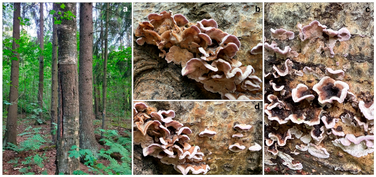
Figure 1. Habitat and basidiomas of Chondrostereum purpureum: a – birch trunk with signs of damage to the bark, on which basidiomas were collected (Russko-Vysotsky forest park, Leningrad region, Russia); b, c, d – basidiomas in situ.
Preprints 243566 g001
Figure 2. Minerals in the fruiting body of the fungus Chondrostereum purpureum: a, b – localization of struvite and calcium oxalates crystals (light stereomicroscopy and SEM, respectively); c, d – struvite crystals among the fungal hyphae (SEM images); e – X-ray diffraction pattern. The insets in figures b and d show typical EDX spectra for calcium oxalates and struvite, respectively; the inset in figure b also shows a struvite crystal with clearly defined layering and stepped features. Abbreviations: SUV – struvite, CaOx – calcium oxalates, FH – fungal hyphae. In figure e: ∗– struvite, ●– weddellite, •– whewellite, ⬟– quartz. The inset in figure c shows a projection of simple forms of a struvite crystal: ○– upper faces, ×– lower faces; 1 – monohedron, 2, 3 – dihedrons. 3.3.
Preprints 243566 g002
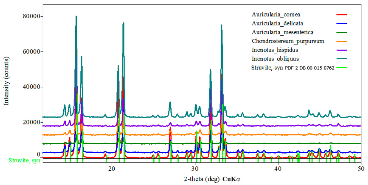
Figure 3. X-ray diffraction patterns of struvite formed in experiment with wood-rotting fungi.
Preprints 243566 g003
Figure 4. Struvite crystals and their aggregates formed under the influence of various species of wood-rotting fungi in experimental condition, results of light microscopy: a – Inonotus hispidus, b – Inonotus obliquus, c – Auricularia mesenterica, d – Chondrostereum purpureum, e – Auricularia cornea, f – Auricularia delicata. The insets show representative SEM images. The inset in panel (b) demonstrate a typical EDX spectrum.
Preprints 243566 g004
Figure 5. The degree of elongation L/W (where L = length and W = width of the primary monohedron) of struvite crystals synthesized under the action of wood-rotting fungi (a) and EPS content in their culture liquid (b) (standard error of the mean: 10%).
Preprints 243566 g005
Figure 6. Generalized projection of the simple forms of synthesized mycogenic crystals (2mm). Legend: ○– upper faces, ×– lower faces; 1, 4 – monohedrons; 2, 5–9 – dihedrons; 10 – rhombic pyramid; additional faces formed under the influence of Inonotus hispidus are marked in green, while those formed under the influence of Inonotus obliquus are marked in red.
Preprints 243566 g006
Table 1. Origination of studied specimens.
Specimen/ number Species Origination Year of deposition into LE-BIN
Nature (FB) LECB F000501 Chondrostereum purpureum (Pers. exFr.) the Russko-Vysotskoye Forest Park (Leningrad Region, Russia) Date of collection: July 2023 2026
LE-BIN 1905 Auricularia
mesenterica (Dicks.) Pers.
ex basidiospores, on fallen trunk of deciduous tree, Russia: Samara Region, Zhigulevsky State Nature Reserve. Coll. 17.08.2004 2006
LE-BIN 4481 Auricularia delicata (Mont. ex Fr.) Henn. ex basidiospores, on fallen mossy trunk, Việt Nam: Cao Bằng Province, Phia Oắc - Phia Đén National Park. Coll. 05.10.2019 2020
LE-BIN 4803 Auricularia cornea Ehrenb. ex basidiospores, on fallen wood of deciduous tree, Russia: Primorye Territory, Ussurisky State Nature Reserve, Coll. 25.08.2021 2022
LE-BIN 5256 Inonotus hispidus (Bull.) P. Karst. ex basidiome, on living drying-up poplar Populus sp., Russia: Republic of Dagestan, Samursky State Sanctuary, Coll. 28.10.2023 2024
LE-BIN 2616 Inonotus obliquus (Fr.) Pilát ex stroma, on dead-standing birch Betula sp., Russia: Khanty-Mansi Autonomous, Yugra region. Coll. 21.08.2010 2011
LE-BIN 1368 Serpula
himantioides (Fr.) P. Karst
Received from: Culture Collection of the F. Schiller University, Biology and Pharmacology Department, Jena, Weimar, Germany 1997
LE-BIN 2764 Chondrostereum purpureum (Pers.) Pouzar ex basidiospores, on branch of alder Alnus sp., Russia: Primorye Territory
Ussurisky State Nature Reserve. Coll. 16.08.2011
2012
Table 2. Verification results of studied fungi.
Specimen
NCBI acc. num. Query cover, % Identity, % Sbjct NCBI acc. num.
Nature FB
LECB F000501
Chondrostereum purpureum
QB051556 100 99.57 MH270439, PP906278, GU062264 et al.
LE-BIN 1905 Auricularia mesenterica PZ501454 100 99.07 KR336698, OR250341
LE-BIN 4481 Auricularia delicata OP975777 100 99.48 KF297963, KF297965
LE-BIN 4803 Auricularia cornea PZ556323
PZ556299
98
100
100
99.14
LC373468,
MH213340
LE-BIN 5256 Inonotus hispidus PQ601119 100 98.85 KX881610, KF446596
LE-BIN 2616 Inonotus obliquus MG722729 100 99.82 PV388883, KY949235
LE-BIN 1368 Serpula
himantioides
KY352492
99
99
98.31
98.11
ON561763
ON561769
LE-BIN 2764 Chondrostereum purpureum OR475683
OQ023318
100
100
99.56
99.57
KR264909
PP849909
Table 3. Values of pH ​​of the crystallization medium at the end of the experiment (30 days) and the sizes of the crystals formed under fungal activity.
Fungal species рН
Crystals sizes**
l, mm
l/w
Inonotus hispidus 6.5
0.5– 2
2.51
Auricularia cornea 6.5 2.25
Inonotus obliquus 6.5 1.99
Auricularia mesenterica 6.5 0.2 – 1.25 1.90
Chondrostereum purpureum 7.0 0.3 – 1 1.39
* The table have not data for crystals formed under the influence of Auricularia delicata, which appear as skeletal forms. ** l – length, w = width of the primary monohedron.
Disclaimer/Publisher’s Note: The statements, opinions and data contained in all publications are solely those of the individual author(s) and contributor(s) and not of MDPI and/or the editor(s). MDPI and/or the editor(s) disclaim responsibility for any injury to people or property resulting from any ideas, methods, instructions or products referred to in the content.
Copyright: This open access article is published under a Creative Commons CC BY 4.0 license, which permit the free download, distribution, and reuse, provided that the author and preprint are cited in any reuse.