

Submitted:
09 November 2026
Posted:
12 November 2026
You are already at the latest version
Abstract
Extracellular vesicles (EVs), as endogenous mediators of intercellular communication, have been extensively characterized in mammalian systems; however, EVs from non-mammalian medicinal organisms remain comparatively undercharacterized. Despite the well-established medicinal value of the invertebrate H. nipponia—largely attributed to the complex bioactive secretions of its salivary glands—the characterization of its salivary gland-derived extracellular vesicles has yet to be fully explored, with current studies still in the nascent stages. In this study, four types of extracellular vesicles were isolated from the salivary glands of living H. nipponia and the heads of lyophilized H. nipponia using differential ultracentrifugation. Their morphological features and particle size distributions were characterized via transmission electron microscopy (TEM) and nanoparticle tracking analysis (NTA), respectively. Finally, their protein compositions were comprehensively analyzed using proteomic techniques. Characterization by TEM and NTA confirmed that microvesicles (MVs) and exosomes (EXOs) from both origins displayed classical saucer-like vesicular structures, with diameters consistently ranging from 100 to 300 nm. Notably, proteomic analysis indicated that EVs harvested from live H. nipponia salivary glands preserved a broader profile of proteins with reported pharmacological activities than the LP-derived preparations. In conclusion, the successful isolation and profiling of H. nipponia salivary gland-derived EVs provide a foundational framework and fresh perspectives for exploring the biological roles of extracellular vesicles in invertebrates.
Keywords:
proteomics
; salivary gland
; lyophilized head tissue
; extracellular vesicles
; Hirudo nipponia
1. Introduction
Extracellular vesicles (EVs) are lipid bilayer-enclosed nanoscale particles released by cells into the extracellular space and function as key mediators of intercellular communication [1,2,3]. They participate in a wide range of physiological and pathological processes by transferring proteins, nucleic acids, and lipids between cells [4,5]. Based on their biogenesis and size, EVs are generally classified into exosomes (EXOs, 30-150 nm) and microvesicles (MVs, 100-1000nm), which differ in their molecular composition and functional properties. In experimental practice, EVs are commonly separated into large and small fractions by differential ultracentrifugation (typically at approximately 10,000×g and 100,000-200,000×g, respectively [6]). These operational fractions do not strictly correspond to vesicles of distinct biogenetic origins [7,8,9,10].
In addition to their biological roles, EVs have attracted increasing attention as natural delivery vehicles due to their ability to transport bioactive molecules and interact with recipient cells [11,12]. However, to investigate their molecular composition and functional properties, EVs must be isolated from complex biological environments. Such isolation remains challenging, particularly in non-model organisms and heterogeneous tissue-derived samples, due to issues such as sample heterogeneity, co-isolation of non-vesicular components, and the lack of standardized purification strategies [13,14,15].
EVs derived from natural biological sources have recently gained interest for their potential applications, yet current research has largely focused on mammalian cell-derived and plant-derived EVs. For example, mammalian mesenchymal stem cell-derived EVs are extensively utilized in tissue engineering to promote fibroblast proliferation and wound healing [16,17], while plant-derived EVs are increasingly explored for their anti-inflammatory properties [18,19].
In contrast, EVs from animal-based medicinal materials, particularly invertebrates, remain poorly characterized. As the source of EVs critically influences their composition, safety, and potential applications, the identification of suitable biological systems and reliable isolation strategies is essential for expanding EV-based research in these underexplored contexts. Although EV secretion has been documented across parasitic helminths, protozoa, and blood-feeding arthropods [20,21], vesicles derived from animal materials used in traditional medicine remain largely unexplored; notably, exosome-like nanoparticles isolated from the medicinal insect Periplaneta americana retained wound-healing activity, indicating that pharmacologically active vesicles can be recovered from medicinal animal sources [22].
H. nipponia represents a suitable biological system for exploring EVs from animal-derived sources due to its well-developed secretory system and the availability of both secretion-derived and tissue-derived materials [23]. The salivary gland system is characterized by active secretion processes and continuous release of protein-rich components [24], providing a biologically relevant source for investigating EV-enriched preparations. In contrast, tissue-derived samples, such as the leech head, represent a more complex biological matrix that may contain EVs originating from multiple cell types. This dual-source characteristic enables the investigation of EVs across both secretion-based and tissue-derived contexts within the same system. In blood-feeding arthropods, vesicles released into saliva and salivary glands have been shown to deliver immunomodulatory and wound-healing-related cargo to the host skin interface [21,25], which supports the view that secretory organs of hematophagous invertebrates are a productive source of functionally active EVs.
Despite these advantages, it remains unclear whether EVs can be reliably isolated from these biological materials, to compare operationally separated EV-enriched fractions, and how EV composition varies between sources and vesicle subtypes. In addition, tissue-derived matrices release intracellular and extracellular matrix components upon dissociation, so that dedicated enrichment strategies are required to obtain preparations that fulfill current consensus criteria [26,27]. In the present study, we aimed to isolate EVs from salivary gland secretions (SGS) and lyophilized head tissue (LP) of H. nipponia and to characterize their structural features and molecular composition. By combining morphological characterization and proteomic analysis, this study establishes an experimental framework for EV isolation from invertebrate medicinal materials and enables a comparative assessment of EV composition across different biological sources and vesicle subtypes.
2. Materials and Methods
2.1. Sample Preparation and Isolation of Extracellular Vesicles
H. nipponia specimens were obtained from Liwen Aquaculture Facility (Dalian, Liaoning, China) Live leeches were maintained in a dark-controlled environment at China Jiliang University and housed in transparent culture tanks (dimensions 5L). The animals were kept in freshwater with daily water replacement under controlled temperature conditions (22 ± 2℃). H. nipponia were fasted for one month before the experiment, and six leeches of similar size and weighing about 0.5g were selected for the experiment.
Extracellular vesicles (EVs) were isolated from both live and freeze-dried materials. For live specimens, salivary glands were dissected following the method described by Zhu et al [28]. H. nipponia were anesthetized by immersion in a 10% (v/v) ethanol solution for 10 minutes. Subsequently, surface mucus was gently removed using a sterile cotton swab, and the animals were secured dorsal-side-up on a dissecting foam board with fine insect pins. A midline incision was made along the ventral abdominal wall under a stereomicroscope, and salivary gland tissue was carefully excised following meticulous removal of adjacent connective tissue using ophthalmic scissors(Figure 1).
The isolated tissues were incubated in 6 cm culture dishes containing 10⁻⁶ mol/L serotonin to stimulate secretion, and salivary gland secretions (SGS) were collected as described by Marshall and Lent [29].
For freeze-dried samples, lyophilized head tissue (LP)was separated and ground into a fine powder. The powder was resuspended in phosphate-buffered saline (PBS) at a ratio of 1 g per 10 mL and left at 4°C overnight (About 12 hours). The supernatant was then collected as the head extract.
EVs were isolated using a differential ultracentrifugation protocol (Figure 2), and two EV-enriched fractions were operationally separated based on centrifugal force: a large-vesicle fraction (hereafter termed MV-enriched fraction) at 10,000 x g, and a small-vesicle fraction (hereafter termed EXO-enriched fraction) at 130,000 x g. Briefly, both SGS and LPS were first centrifuged at 300 × g for 10 min, followed by 3000 × g and 5000 × g for 20 min each, to remove cells and large debris. The resulting supernatant was then centrifuged at 10,000 × g for 45 min to pellet the MV-enriched fraction. Subsequently, the supernatant was further ultracentrifuged at 130,000 × g for 70 min to obtain the EXO-enriched fraction. All centrifugation steps were performed at 4°C using an ultracentrifuge (Hitachi, CP100MX) [30,31].
2.2 Morphological and Size Characterization of Extracellular Vesicles
EV samples were deposited onto 200-mesh copper grids coated with carbon support films and allowed to adsorb for 1 min at room temperature. Excess liquid was carefully removed using filter paper, and the grids were negatively stained with 2% uranyl acetate [32]. After air drying, the samples were examined using a transmission electron microscope (Talos L120C, Thermo Fisher Scientific), and representative images were acquired.
For nanoparticle tracking analysis (NTA), the detection was completed by ZetaView-PMX120-Z (Particle Metrix) instrument and its companion software ZetaView(version 8.05). EV samples were diluted in PBS to an appropriate concentration prior to measurement. The instrument was calibrated using 100 nm polystyrene bead standards. During acquisition, images were recorded at 11 different positions for each sample and analyzed automatically by the software. The measurement temperature was maintained at approximately 23°C. Particle size distribution and mean particle diameter were calculated using the instrument software [33,34,35].
2.3. Proteomic Analysis and Functional Annotation of Extracellular Vesicles
Proteomic analysis of EV samples was performed by Shanghai Applied Protein Technology Co., Ltd. using data-independent acquisition (DIA) mass spectrometry [36,37]. All analyses were conducted with three independent biological replicates for each group. EV samples were lysed in SDT buffer (4% sodium dodecyl sulfate, 100 mM Tris-HCl, pH 7.6) and boiled for protein denaturation. After centrifugation at 14,000 × g, the supernatant was collected and protein concentration was determined using the bicinchoninic acid (BCA) assay. A total of 15 μg protein per sample was separated by SDS-PAGE following mixing with loading buffer and heat denaturation, and visualized by Coomassie staining. Proteins were reduced with tris (2-carboxyethyl) phosphine (TCEP) and alkylated with iodoacetamide prior to enzymatic digestion. Samples were digested using trypsin and Lys-C at 37°C for 3 h.
The resulting peptides were desalted using strong cation exchange solid-phase extraction (SPE MCX cartridges) and concentrated by vacuum centrifugation. Peptides were reconstituted in 0.1% formic acid, quantified by UV absorbance at 280 nm, and spiked with indexed retention time (iRT) peptides (Biognosys, Switzerland) for retention time calibration. Peptide separation was performed using an Evosep One liquid chromatography system (Evosep, Denmark), and mass spectrometry analysis was conducted on a timsTOF Pro 2 instrument (Bruker Daltonics, Germany) in DIA mode. MS1 spectra were acquired over an m/z range of 100–1700 using four acquisition windows with an accumulation time of 100 ms per window. Data were collected using the dia-PASEF acquisition method, with collision energy linearly correlated to the inverse ion mobility (1/K0).
Raw data were processed using Spectronaut software (version 19.7, Biognosys, Switzerland),The protein reference database for mass spectrometry searching was downloaded from the National Center for Biotechnology Information (NCBI Hirudinidae-1414-20230529). A maximum of two missed cleavages was allowed. Carbamidomethylation of cysteine was set as a fixed modification, while oxidation of methionine and protein N-terminal acetylation were set as variable modifications. Protein identifications were accepted at a false discovery rate (FDR) ≤ 1%. For differential expression analysis, proteins with a fold change (FC) > 1.5 or < 0.67 and p value < 0.05 were considered significantly differentially expressed, and proteins showing more than 10-fold changes were highlighted. Domain enrichment analysis was performed using Fisher’s exact test with multiple testing correction. Functional annotation of differentially expressed proteins was conducted using Gene Ontology (GO) analysis (biological process) and Kyoto Encyclopedia of Genes and Genomes (KEGG) pathway analysis, implemented through the OmicsBean platform.
3. Results
3.1. Morphology, Particle Size and Concentration Characterization of H. Nipponia-Derived Extracellular Vesicles
Transmission electron microscopy (TEM) (Figure 3) and nanoparticle tracking analysis (NTA) (Figure 4) showed that all EV samples exhibited typical morphological characteristics consistent with extracellular vesicles. Both MV and EXO populations displayed membrane-bound vesicular structures with sizes within the expected nanoscale range. SGS-MVs exhibited irregular spherical or ellipsoidal morphology, with an average particle diameter of 228.0 ± 122.9 nm. In comparison, LP-MVs were generally spherical or near-spherical and showed a smaller average diameter of 193.3 ± 82.5 nm. Both SGS-EXOs and LP-EXOs displayed typical cup-shaped morphology with intact membrane structures. The average particle diameters of SGS-EXOs and LP-EXOs were 191.2 ± 77.7 nm and 182.1 ± 60.4 nm, respectively, with relatively narrower size distributions compared to MVs.
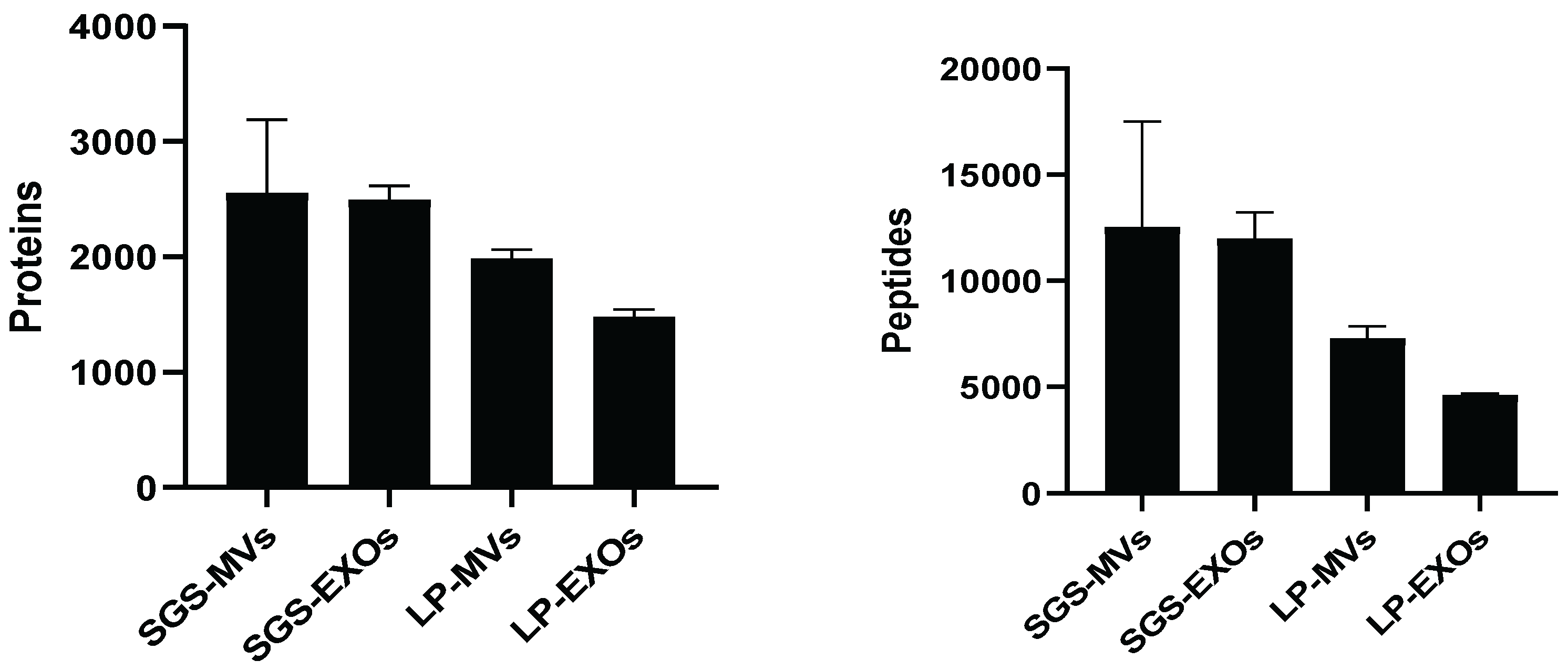
Proteomic Characterization of EV-Enriched FractionsA total of 3,340 proteins and 20,351 peptides were identified across all EV samples, indicating successful protein extraction and detection. Among these, 1,171 proteins were shared across all four EV populations, representing proteins shared across all four preparations. These shared proteins were primarily associated with fundamental cellular functions, including protein folding, energy metabolism, cytoskeleton organization, and signal transduction.
Figure 5.
Number of identified proteins and peptides in four types of extracellular vesicles from Hirudo nipponica.
Figure 5.
Number of identified proteins and peptides in four types of extracellular vesicles from Hirudo nipponica.
In addition, several proteins with reported pharmacological activities were identified [38,39,40], including Saratin, Theromyzin, Hyaluronidase, Hirudin, Destabilase I, and Eglin C. Among these, Saratin, Theromyzin, and Hyaluronidase were detected in all EV populations, whereas Hirudin, Destabilase I, and Eglin C were detected only in SGS-derived EVs.
Six proteins with potential pharmacological activity were screened from all identified proteins, and their distribution characteristics in the four types of EVs are shown in Table 1. These proteins are involved in functions such as anticoagulation, antithrombosis, anti-inflammation, antibiosis, and extracellular matrix degradation, and exhibit differential distribution among the four types of EVs. Among them, Saratin, Theromyzin and Hyaluronidase were all identified in the four types of EVs; while Hirudin, Destabilase I and Eglin C were only detected in SGS-derived EVs (SGS-MVs, SGS-EXOs).
3.2. Proteomic Differences Between SGS- and LP-Derived EVs
Differential expression analysis identified 561 proteins between SGS-MVs and LP-MVs, including 267 upregulated and 294 downregulated proteins. Among these, 16 proteins were highly upregulated and 38 were highly downregulated. In the EXO populations, 669 differentially expressed proteins were identified between SGS-EXOs and LP-EXOs, including 219 upregulated and 450 downregulated proteins, with 15 highly upregulated and 37 highly downregulated proteins (Figure 6).
Domain enrichment analysis showed that proteins differing between SGS-MVs and LP-MVs were primarily associated with ion binding and signal transduction, lipid and carbohydrate metabolism, protein interaction, and membrane trafficking. In SGS-EXOs versus LP-EXOs, enriched domains were mainly related to protease regulation, vesicle transport, calcium signaling, and extracellular matrix remodeling (Figure 7A and 7C).
GO and KEGG enrichment analyses indicated that differentially expressed proteins between SGS-MVs and LP-MVs were mainly involved in metabolic processes, transmembrane transport, and signaling pathways. In contrast, proteins differing between SGS-EXOs and LP-EXOs were associated with immune-related processes, protein turnover, extracellular matrix interactions, and signal transduction pathways (Figure 8A and 8C) (Figure9A and 9C).
Upregulated: proteins with upregulated differential expression; Downregulated: proteins with downregulated expression.
3.3. Proteomic Differences Between EV Subtypes (MVs vs EXOs)
Differential expression analysis identified 619 proteins between SGS-EXOs and SGS-MVs, including 275 upregulated and 344 downregulated proteins. Among these, 28 proteins were highly upregulated and 45 were highly downregulated. In LP-derived EVs, 318 differentially expressed proteins were identified between LP-EXOs and LP-MVs, including 177 upregulated and 141 downregulated proteins, with 6 highly upregulated and 2 highly downregulated proteins (Figure 6).
Domain enrichment analysis showed that proteins differing between SGS-EXOs and SGS-MVs were mainly associated with extracellular matrix organization, protease regulation, protein interaction, ion transport, and glycosylation-related functions. In LP-derived EVs, enriched domains between EXOs and MVs were related to membrane trafficking, cytoskeleton organization, calcium signaling, protease activity, and immune recognition (Figure7B and 7D).
GO and KEGG enrichment analyses indicated that proteins differing between SGS-EXOs and SGS-MVs were mainly involved in ion transport, mitochondrial function, metabolic pathways, and glycosylation processes. In LP-derived EVs, enriched functions included RNA regulation, intracellular trafficking, cell signaling, and metabolic pathways (Figure 8B and 8D) (Figure 9B and 9D).
4. Discussion
This study provides a systematic characterization of extracellular vesicles (EVs) derived from H. nipponia. EVs were successfully isolated from both salivary gland secretions and head-derived materials, and distinct vesicle subtypes were obtained. Proteomic analysis further revealed variations in EV composition associated with both tissue origin and vesicle subtype.
This study identified several proteins with documented pharmacological activities—including hirudin, destabilase I, eglins C, saratin, theromyzin, and hyaluronidase—in extracellular vesicles (EVs) isolated from four leech salivary gland samples. These proteins are well-established bioactive constituents of leech saliva [41,42]. The findings corroborate that EVs serve as carriers for functionally relevant molecules—particularly anticoagulant, antithrombotic, and anti-inflammatory agents—secreted by salivary gland cells. Whether these proteins are encapsulated within EVs or merely associated with the vesicle surface remains to be determined, as does whether EV association influences their stability or biological activity. These findings provide a proteomic basis for further functional investigation of EV-associated bioactive molecules from leech salivary glands [43,44,45,46].
Saratin, theromycin, and hyaluronidase were consistently identified across all four extracellular vesicle (EV) preparations. Saratin exhibits potent antiplatelet activity by inhibiting both platelet adhesion and aggregation, thereby suppressing thrombus formation [47,48]. Theromycin is an anionic antimicrobial peptide demonstrating robust antibacterial efficacy specifically against Gram-positive bacteria [49]. Hyaluronidase catalyzes the hydrolysis of hyaluronic acid in the extracellular matrix, compromising structural integrity of host tissue barriers and facilitating enhanced diffusion and tissue penetration of bioactive salivary components from leeches [50,51]. Collectively, these findings suggest that the EVs serve as natural carriers for salivary gland-derived bioactive molecules and possess multifunctional therapeutic potential—including antithrombotic, antimicrobial, and extracellular matrix–modulating activities. Moreover, Hirudin, Destabilase I, and Eglin C were exclusively detected in EVs isolated from freshly collected leech salivary gland secretions (SGS), whereas they were undetectable in EVs derived from lyophilized leech powder (LP). These proteins were detected in SGS-derived EV preparations but not detected in the LP-derived preparations under the present proteomic conditions. In contrast, SGS-derived EVs are obtained directly from the physiologically active secretion of leech salivary glands, The protein composition of SGS-derived preparations reflects the dissected and 5-HT-stimulated salivary gland secretion. Functionally, these three proteins act at distinct steps within the coagulation and inflammatory cascades: Hirudin—recognized as the most potent and specific thrombin inhibitor identified to date—irreversibly inhibits thrombin, thereby blocking the final common pathway of both intrinsic and extrinsic coagulation and serving as the principal mediator of anticoagulant activity [52,53,54,55]. Destabilase I exerts thrombolytic effects via enzymatic degradation of cross-linked fibrin and demonstrates broad-spectrum antibacterial activity through both enzymatic and non-enzymatic mechanisms [56,57,58]. Eglin C, a serine protease inhibitor, suppresses inflammation-associated proteases—including neutrophil elastase and cathepsin G—thereby attenuating inflammatory responses [59,60]. Collectively, these findings indicate that SGS-derived EV preparations contained a broader detectable profile of proteins with reported anticoagulant, thrombolytic, and anti-inflammatory activities compared with LP-derived preparations.
5. Conclusions
This study successfully isolated and characterized extracellular vesicles (EVs) from H. nipponia, and systematically analyzed the proteomic profiles of microvesicles (MVs) and exosomes (EXOs) derived from salivary gland secretions (SGS) and lyophilized head powder (LP). Proteomic analysis revealed that the 1171 core proteins shared by the four EV subsets primarily sustain the fundamental biological functions of EVs, whereas subset-specific proteins reflect heterogeneity in biogenesis and protein sorting across different EV subtypes. A range of pharmacologically active proteins, represented by Hirudin, Destabilase I, and Eglin C, were successfully identified, confirming that EVs serve as critical functional carriers for the transport of bioactive substances in leeches.
Comparative analyses further demonstrated that SGS-derived EVs exhibit a more intact pharmacological component profile compared to those isolated from the traditional LP source, suggesting that research directly targeting the salivary gland secretory system more closely aligns with the physiological and pharmacological essence of leech bioactivity. Overall, the present study reveals proteomic differences among source- and centrifugation-defined EV-enriched fractions. SGS-derived preparations exhibited a broader detectable profile of pharmacologically relevant proteins than LP-derived preparations.
Supplementary Materials
The data supporting the findings of this study are available within the main article and its supplementary materials.
Author Contributions
Conceptualization, Meng-Xiang Jia and Xia Qiu; Methodology,.Meng-Xiang Jia, Ya-Li Zhu and Xia Qiu; Formal analysis, Meng-Xiang Jia and Ya-Li Zhu; Investigation, Meng-Xiang Jia; Resources, Yuan-Yuan Luo; Data curation, Qian Li and Wei-Ming Wang; Writing – original draft, Meng-Xiang Jia and Ya-Li Zhu; Writing – review & editing, Ya-Li Zhu, Xia Qiu, Qian Li and Yuan-Yuan Luo; Visualization, Wei-Ming Wang; Supervision, Xia Qiu, Qian Li, Wei-Ming Wang and Yuan-Yuan Luo; Project administration, Yuan-Yuan Luo; Funding acquisition, Yuan-Yuan Luo.
Funding
This research was funded by the China Jiliang University Science and Technology Cooperation Project, grant number 03106–231601 (to Yuan-Yuan Luo).
Informed Consent Statement
Not applicable.
Data Availability Statement
The datasets generated and analyzed during the current study are not publicly available due to ongoing follow-up research, but are available from the corresponding author upon reasonable request.
Acknowledgments
We thank Yu-Chen Zhang at the Center for CryoElectron Microscopy, Zhejiang University, for her technical assistance with transmission.
Conflicts of Interest
The authors declare no conflicts of interest.
References
- Raposo, G.; Stoorvogel, W. Extracellular vesicles: Exosomes, microvesicles, and friends. J. Cell Biol. 2013, 200, 373–383. [Google Scholar] [CrossRef]
- Kalluri, R.; LeBleu, V.S. The biology, function, and biomedical applications of exosomes. Science 2020, 367, eaau6977. [Google Scholar] [CrossRef] [PubMed]
- El Andaloussi, S.; Mäger, I.; Breakefield, X.O.; Wood, M.J.A. Extracellular vesicles: biology and emerging therapeutic opportunities. Nat. Rev. Drug Discov. 2013, 12, 347–357. [Google Scholar] [CrossRef] [PubMed]
- Van Niel, G.; D’Angelo, G.; Raposo, G. Shedding light on the cell biology of extracellular vesicles. Nat. Rev. Mol. Cell Biol. 2018, 19, 213–228. [Google Scholar] [CrossRef] [PubMed]
- Colombo, M.; Raposo, G.; Théry, C. Biogenesis, secretion, and intercellular interactions of exosomes and other extracellular vesicles. Annu. Rev. Cell Dev. Biol. 2014, 30, 255–289. [Google Scholar] [CrossRef] [PubMed]
- Thery, C.; Clayton, A.; Amigorena, S.; Raposo, G. Isolation and characterization of exosomes from cell culture supernatants and biological fluids. Curr. Protoc. Cell Biol. 2006, 3. [Google Scholar] [CrossRef] [PubMed]
- Théry, C.; Witwer, K.W.; Aikawa, E.; Alcaraz, M.J.; Anderson, J.D.; Andriantsitohaina, R.; Antoniou, A.; Arab, T.; Archer, F.; Atkin-Smith, G.K.; et al. Minimal information for studies of extracellular vesicles 2018 (MISEV2018): A position statement of the International Society for Extracellular Vesicles and update of the MISEV2014 guidelines. J. Extracell. Vesicles 2018, 7, 1535750. [Google Scholar] [CrossRef] [PubMed]
- Lötvall, J.; Hill, A.F.; Hochberg, F.; Buzás, E.I.; Di Vizio, D.; Gardiner, C.; Gho, Y.S.; Kurochkin, I.V.; Mathivanan, S.; Quesenberry, P.; et al. Minimal experimental requirements for definition of extracellular vesicles and their functions: A position statement from the International Society for Extracellular Vesicles. J. Extracell. Vesicles 2014, 3, 26913. [Google Scholar] [CrossRef] [PubMed]
- Witwer, K.W.; Théry, C. Extracellular vesicles or exosomes? On primacy, precision, and popularity influencing a choice of nomenclature. J. Extracell. Vesicles 2019, 8, 1648167. [Google Scholar] [CrossRef] [PubMed]
- Welsh, J.A.; Goberdhan, D.C.I.; O'DRiscoll, L.; Buzas, E.I.; Blenkiron, C.; Bussolati, B.; Cai, H.; Di Vizio, D.; Driedonks, T.A.P.; Erdbrügger, U.; et al. Minimal information for studies of extracellular vesicles (MISEV2023): From basic to advanced approaches. J. Extracell. Vesicles 2024, 13, e12404. [Google Scholar] [CrossRef] [PubMed]
- Herrmann, I.K.; Wood, M.J.A.; Fuhrmann, G. Extracellular vesicles as a next-generation drug delivery platform. Nat. Nanotechnol. 2021, 16, 748–759. [Google Scholar] [CrossRef] [PubMed]
- Elsharkasy, O.M.; Nordin, J.Z.; Hagey, D.W.; De Jong, O.G.; Schiffelers, R.M.; Andaloussi, S.E.; Vader, P. Extracellular vesicles as drug delivery systems: Why and how? Adv. Drug Deliv. Rev. 2020, 159, 332–343. [Google Scholar] [CrossRef] [PubMed]
- Gardiner, C.; Di Vizio, D.; Sahoo, S.; Théry, C.; Witwer, K.W.; Wauben, M.; Hill, A.F. Techniques used for the isolation and characterization of extracellular vesicles: Results of a worldwide survey. J. Extracell. Vesicles 2016, 5, 32945. [Google Scholar] [CrossRef] [PubMed]
- Van Deun, J.; Mestdagh, P.; Sormunen, R.; Cocquyt, V.; Vermaelen, K.; Vandesompele, J.; Bracke, M.; De Wever, O.; Hendrix, A. The impact of disparate isolation methods for extracellular vesicles on downstream RNA profiling. J. Extracell. Vesicles 2014, 3. [Google Scholar] [CrossRef] [PubMed]
- Böing, A.N.; van der Pol, E.; Grootemaat, A.E.; Coumans, F.A.W.; Sturk, A.; Nieuwland, R. Single-step isolation of extracellular vesicles by size-exclusion chromatography. J. Extracell. Vesicles 2014, 3. [Google Scholar] [CrossRef] [PubMed]
- Kou, M.; Huang, L.; Yang, J.; Chiang, Z.; Chen, S.; Liu, J.; Guo, L.; Zhang, X.; Zhou, X.; Xu, X.; et al. Mesenchymal stem cell-derived extracellular vesicles for immunomodulation and regeneration: a next generation therapeutic tool? Cell Death Dis. 2022, 13, 1–16. [Google Scholar] [CrossRef] [PubMed]
- Zhang, Y.; Pan, Y.; Liu, Y.; Li, X.; Tang, L.; Duan, M.; Li, J.; Zhang, G. Exosomes derived from human umbilical cord blood mesenchymal stem cells stimulate regenerative wound healing via transforming growth factor-β receptor inhibition. Stem Cell Res. Ther. 2021, 12, 1–14. [Google Scholar] [CrossRef] [PubMed]
- Teng, Y.; Ren, Y.; Sayed, M.; Hu, X.; Lei, C.; Kumar, A.; Hutchins, E.; Mu, J.; Deng, Z.; Luo, C.; et al. Plant-Derived Exosomal MicroRNAs Shape the Gut Microbiota. Cell Host Microbe 2018, 24, 637–652.e8. [Google Scholar] [CrossRef] [PubMed]
- Dad, H.A.; Gu, T.-W.; Zhu, A.-Q.; Huang, L.-Q.; Peng, L.-H. Plant Exosome-like Nanovesicles: Emerging Therapeutics and Drug Delivery Nanoplatforms. Mol. Ther. 2021, 29, 13–31. [Google Scholar] [CrossRef] [PubMed]
- Coakley, G.; Maizels, R.M.; Buck, A.H. Exosomes and Other Extracellular Vesicles: The New Communicators in Parasite Infections. Trends Parasitol. 2015, 31, 477–489. [Google Scholar] [CrossRef] [PubMed]
- Zhou, W.; Tahir, F.; Wang, J.C.-Y.; Woodson, M.; Sherman, M.B.; Karim, S.; Neelakanta, G.; Sultana, H. Discovery of Exosomes From Tick Saliva and Salivary Glands Reveals Therapeutic Roles for CXCL12 and IL-8 in Wound Healing at the Tick–Human Skin Interface. Front. Cell Dev. Biol. 2020, 8, 554. [Google Scholar] [CrossRef] [PubMed]
- Liao, Q.; Su, L.; Pang, L.; Li, J.; Li, H.; Li, J.; Liu, Y.; Zhang, J. Natural exosome-like nanoparticles derived from ancient medicinal insect Periplaneta americana L. as a novel diabetic wound healing accelerator. J. Nanobiotechnology 2023, 21, 1–16. [Google Scholar] [CrossRef] [PubMed]
- Dong, H.; Ren, J.-X.; Wang, J.-J.; Ding, L.-S.; Zhao, J.-J.; Liu, S.-Y.; Gao, H.-M. Chinese Medicinal Leech: Ethnopharmacology, Phytochemistry, and Pharmacological Activities. Evid.-Based Complement Altern. Med. 2016, 2016, 7895935. [Google Scholar] [CrossRef] [PubMed]
- Lu, Z.; Shi, P.; You, H.; Liu, Y.; Chen, S. Transcriptomic analysis of the salivary gland of medicinal leech Hirudo nipponia. PLoS ONE 2018, 13, e0205875. [Google Scholar] [CrossRef] [PubMed]
- Chávez, A.S.O.; Wang, X.; Marnin, L.; Archer, N.K.; Hammond, H.L.; Carroll, E.E.M.; Shaw, D.K.; Tully, B.G.; Buskirk, A.D.; Ford, S.L.; et al. Tick extracellular vesicles enable arthropod feeding and promote distinct outcomes of bacterial infection. Nat. Commun. 2021, 12, 1–17. [Google Scholar] [CrossRef] [PubMed]
- White, R.; Sotillo, J.; Ancarola, M.E.; Borup, A.; Boysen, A.T.; Brindley, P.J.; Buzás, E.I.; Cavallero, S.; Chaiyadet, S.; Chalmers, I.W.; et al. Special considerations for studies of extracellular vesicles from parasitic helminths: A community-led roadmap to increase rigour and reproducibility. J. Extracell. Vesicles 2023, 12, e12298. [Google Scholar] [CrossRef] [PubMed]
- Vella, L.J.; Scicluna, B.J.; Cheng, L.; Bawden, E.G.; Masters, C.L.; Ang, C.; Willamson, N.; McLean, C.; Barnham, K.J.; Hill, A.F. A rigorous method to enrich for exosomes from brain tissue. J. Extracell. Vesicles 2017, 6, 1348885. [Google Scholar] [CrossRef] [PubMed]
- Zhu, Y.-L.; Jia, M.-X.; Qu, R.-Y.; Li, Q.; Qiu, X.; Zhao, W.-C.; Luo, Y.-Y. The unique biogenesis pathway of extracellular vesicles in Hirudo nipponia salivary gland cells. Tissue Cell 2025, 99, 103296. [Google Scholar] [CrossRef] [PubMed]
- Marshall, C.G.; Lent, C.M. Excitability and Secretory Activity in the Salivary Gland Cells of Jawed Leeches (Hirudinea: Gnathobdellida). J. Exp. Biol. 1988, 137, 89–105. [Google Scholar] [CrossRef] [PubMed]
- Baranyai, T.; Herczeg, K.; Onódi, Z.; Voszka, I.; Módos, K.; Marton, N.; Nagy, G.; Mäger, I.; Wood, M. J.; El Andaloussi, S.; Pálinkás, Z.; Kumar, V.; Nagy, P.; Kittel, Á.; Buzás, E. I.; Ferdinandy, P.; Giricz, Z. Isolation of Exo-somes from Blood Plasma: Qualitative and Quantitative Comparison of Ultracentrifugation and Size Exclusion Chromatog-raphy Methods. PLoS ONE 2015, 10, e0145686. [Google Scholar] [CrossRef] [PubMed]
- Nordin, J.Z.; Lee, Y.; Vader, P.; Mäger, I.; Johansson, H.J.; Heusermann, W.; Wiklander, O.P.B.; Hällbrink, M.; Seow, Y.; Bultema, J.J.; et al. Ultrafiltration with size-exclusion liquid chromatography for high yield isolation of extracellular vesicles preserving intact biophysical and functional properties. Nanomed. Nanotechnol. Biol. Med. 2015, 11, 879–883. [Google Scholar] [CrossRef] [PubMed]
- Jung, M.K.; Mun, J.Y. Sample Preparation and Imaging of Exosomes by Transmission Electron Microscopy. J. Vis. Exp. 2018, e56482–e56482. [Google Scholar] [CrossRef] [PubMed]
- Dragovic, R.A.; Gardiner, C.; Brooks, A.S.; Tannetta, D.S.; Ferguson, D.J.P.; Hole, P.; Carr, B.; Redman, C.W.G.; Harris, A.L.; Dobson, P.J.; et al. Sizing and phenotyping of cellular vesicles using Nanoparticle Tracking Analysis. Nanomed. Nanotechnol. Biol. Med. 2011, 7, 780–788. [Google Scholar] [CrossRef] [PubMed]
- Gardiner, C.; Ferreira, Y.J.; Dragovic, R.A.; Redman, C.W.G.; Sargent, I.L. Extracellular vesicle sizing and enumeration by nanoparticle tracking analysis. J. Extracell. Vesicles 2013, 2, 19671. [Google Scholar] [CrossRef] [PubMed]
- Szatanek, R.; Baj-Krzyworzeka, M.; Zimoch, J.; Lekka, M.; Siedlar, M.; Baran, J. The Methods of Choice for Extracellular Vesicles (EVs) Characterization. Int. J. Mol. Sci. 2017, 18, 1153. [Google Scholar] [CrossRef] [PubMed]
- Krasny, L.; Huang, P.H. Data-independent acquisition mass spectrometry (DIA-MS) for proteomic applications in oncology. Mol. Omics 2020, 17, 29–42. [Google Scholar] [CrossRef] [PubMed]
- Fröhlich, K.; Fahrner, M.; Brombacher, E.; Seredynska, A.; Maldacker, M.; Kreutz, C.; Schmidt, A.; Schilling, O. Data-Independent Acquisition: A Milestone and Prospect in Clinical Mass Spectrometry–Based Proteomics. Mol. Cell. Proteom. 2024, 23, 100800. [Google Scholar] [CrossRef] [PubMed]
- Lemke, S.; Vilcinskas, A. European Medicinal Leeches—New Roles in Modern Medicine. Biomedicines 2020, 8, 99. [Google Scholar] [CrossRef] [PubMed]
- Montinari, M.R.; Minelli, S. From ancient leech to direct thrombin inhibitors and beyond: New from old. Biomed. Pharmacother. 2022, 149, 112878. [Google Scholar] [CrossRef] [PubMed]
- Whitaker, I.S.; Oboumarzouk, O.; Rozen, W.M.; Naderi, N.; Balasubramanian, S.; Azzopardi, E.A.; Kon, M. The efficacy of medicinal leeches in plastic and reconstructive surgery: A systematic review of 277 reported clinical cases. Microsurgery 2012, 32, 240–250. [Google Scholar] [CrossRef] [PubMed]
- Müller, C.; Mescke, K.; Liebig, S.; Mahfoud, H.; Lemke, S.; Hildebrandt, J.-P. More than just one: multiplicity of Hirudins and Hirudin-like Factors in the Medicinal Leech, Hirudo medicinalis. Mol. Genet. Genom. 2016, 291, 227–240. [Google Scholar] [CrossRef] [PubMed]
- Harvey, R.P.; Degryse, E.; Stefani, L.; Schamber, F.; Cazenave, J.P.; Courtney, M.; Tolstoshev, P.; Lecocq, J.P. Cloning and expression of a cDNA coding for the anticoagulant hirudin from the bloodsucking leech, Hirudo medicinalis. Proc. Natl. Acad. Sci. USA 1986, 83, 1084–1088. [Google Scholar] [CrossRef] [PubMed]
- Rosa-Fernandes, L.; Rocha, V.B.; Carregari, V.C.; Urbani, A.; Palmisano, G. A Perspective on Extracellular Vesicles Proteomics. Front. Chem. 2017, 5, 102–102. [Google Scholar] [CrossRef] [PubMed]
- Vagner, T.; Chin, A.; Mariscal, J.; Bannykh, S.; Engman, D.M.; Di Vizio, D. Protein Composition Reflects Extracellular Vesicle Heterogeneity. Proteomics 2019, 19, e1800167. [Google Scholar] [CrossRef] [PubMed]
- Chitti, S.V.; Gummadi, S.; Kang, T.; Shahi, S.; Marzan, A.L.; Nedeva, C.; Sanwlani, R.; Bramich, K.; Stewart, S.; Petrovska, M.; et al. Vesiclepedia 2024: an extracellular vesicles and extracellular particles repository. Nucleic Acids Res. 2023, 52, D1694–D1698. [Google Scholar] [CrossRef] [PubMed]
- Bandu, R.; Oh, J.W.; Kim, K.P. Mass spectrometry-based proteome profiling of extracellular vesicles and their roles in cancer biology. Exp. Mol. Med. 2019, 51, 1–10. [Google Scholar] [CrossRef] [PubMed]
- Barnes, C.S.; Krafft, B.; Frech, M.; Hofmann, U.R.; Papendieck, A.; Dahlems, U.; Gellissen, G.; Hoylaerts, M.F. Production and Characterization of Saratin, an Inhibitor of von Willebrand Factor-Dependent Platelet Adhesion to Collagen. Semin. Thromb. Hemost. 2001, 27, 337–348. [Google Scholar] [CrossRef] [PubMed]
- White, T.C.; Bernyl, M.A.; Robinson, D.K.; Yin, H.; DeGrado, W.F.; Hanson, S.R.; McCarthy, O.J.T. The leech product saratin is a potent inhibitor of platelet integrin α2β1 and von Willebrand factor binding to collagen. FEBS J. 2007, 274, 1481–1491. [Google Scholar] [CrossRef] [PubMed]
- Tasiemski, A.; Vandenbulcke, F.; Mitta, G.; Lemoine, J.; Lefebvre, C.; Sautière, P.-E.; Salzet, M. Molecular Characterization of Two Novel Antibacterial Peptides Inducible upon Bacterial Challenge in an Annelid, the Leech Theromyzon tessulatum. J. Biol. Chem. 2004, 279, 30973–30982. [Google Scholar] [CrossRef] [PubMed]
- Yuki, H.; Fishman, W.H. Purification and Characterization of Leech Hyaluronic Acid-endo-β-glucuronidase. J. Biol. Chem. 1963, 238, 1877–1879. [Google Scholar] [CrossRef]
- Sun, R.; Fu, X.; Ai, R.; Huang, Z.; Li, H.; Tang, L.; Chen, H.; Zhang, X.; Zhao, F.; Lin, G. Feeding Strategy Shapes the Evolution of the Hyaluronidase Gene Family in Medicinal Leeches. Ecol. Evol. 2026, 16, e74049. [Google Scholar] [CrossRef] [PubMed]
- Markwardt, F. Hirudin As Alternative Anticoagulant- A Historical Review. Semin. Thromb. Hemost. 2002, 28, 405–414. [Google Scholar] [CrossRef] [PubMed]
- Warkentin, T.E.; Greinacher, A. The direct thrombin inhibitor hirudin. Thromb. Haemost. 2008, 99, 819–829. [Google Scholar] [CrossRef] [PubMed]
- Cheng, B.; Liu, F.; Guo, Q.; Lu, Y.; Shi, H.; Ding, A.; Xu, C. Identification and characterization of hirudin-HN, a new thrombin inhibitor, from the salivary glands ofHirudo nipponia. PeerJ 2019, 7, e7716. [Google Scholar] [CrossRef] [PubMed]
- Shi, P.; Wei, J.; You, H.; Chen, S.; Tan, F.; Lu, Z. Cloning, characterization, and heterologous expression of a candidate Hirudin gene from the salivary gland transcriptome of Hirudo nipponia. Sci. Rep. 2023, 13, 1–11. [Google Scholar] [CrossRef] [PubMed]
- Baskova, I.P.; I Nikonov, G. Destabilase: an enzyme of medicinal leech salivary gland secretion hydrolyzes the isopeptide bonds in stabilized fibrin. Biokhimiia 1985, 50, 424–31. [Google Scholar] [PubMed]
- Zavalova, L.L.; Lazarev, V.N.; Levitsky, S.A.; Yudina, T.G.; Baskova, I.P. Destabilase-lysozyme of medicinal leech. Multifunctionality of recombinant protein. Biochem. 2010, 75, 1173–1181. [Google Scholar] [CrossRef] [PubMed]
- Marin, E.; Kornilov, D.A.; Bukhdruker, S.S.; Aleksenko, V.A.; Manuvera, V.A.; Zinovev, E.V.; Kovalev, K.V.; Shevtsov, M.B.; Talyzina, A.A.; Bobrovsky, P.A.; et al. Structural insights into thrombolytic activity of destabilase from medicinal leech. Sci. Rep. 2023, 13, 1–11. [Google Scholar] [CrossRef] [PubMed]
- Seemüller, U.; Meier, M.; Ohlsson, K.; Müller, H.-P.; Fritz, H. Isolation and Characterisation of a Low Molecular Weight Inhibitor (of Chymotrypsin and Human Granulocytic Elastase and Cathepsin G) from Leeches. Hoppe-Seyler´s Z. Für Physiol. Chem. 1977, 358, 1105–1118. [Google Scholar] [CrossRef] [PubMed]
- RENESTO, P.; FERRERLOPEZ, P.; CHIGNARD, M. INTERFERENCE OF RECOMBINANT EGLIN-C, A PROTEINASE-INHIBITOR EXTRACTED FROM LEECHES, WITH NEUTROPHIL-MEDIATED PLATELET ACTIVATION. 1990, 62, 409–416. [Google Scholar] [PubMed]
Figure 1.
Schematic diagram of the anatomy of the H. nipponia (A) Anatomical locality of the salivary gland tissue of Hirudo japonica; (B) Salivary gland tissues and jaw structures; j: jaw structures; sgc: Salivary gland cells.
Figure 1.
Schematic diagram of the anatomy of the H. nipponia (A) Anatomical locality of the salivary gland tissue of Hirudo japonica; (B) Salivary gland tissues and jaw structures; j: jaw structures; sgc: Salivary gland cells.
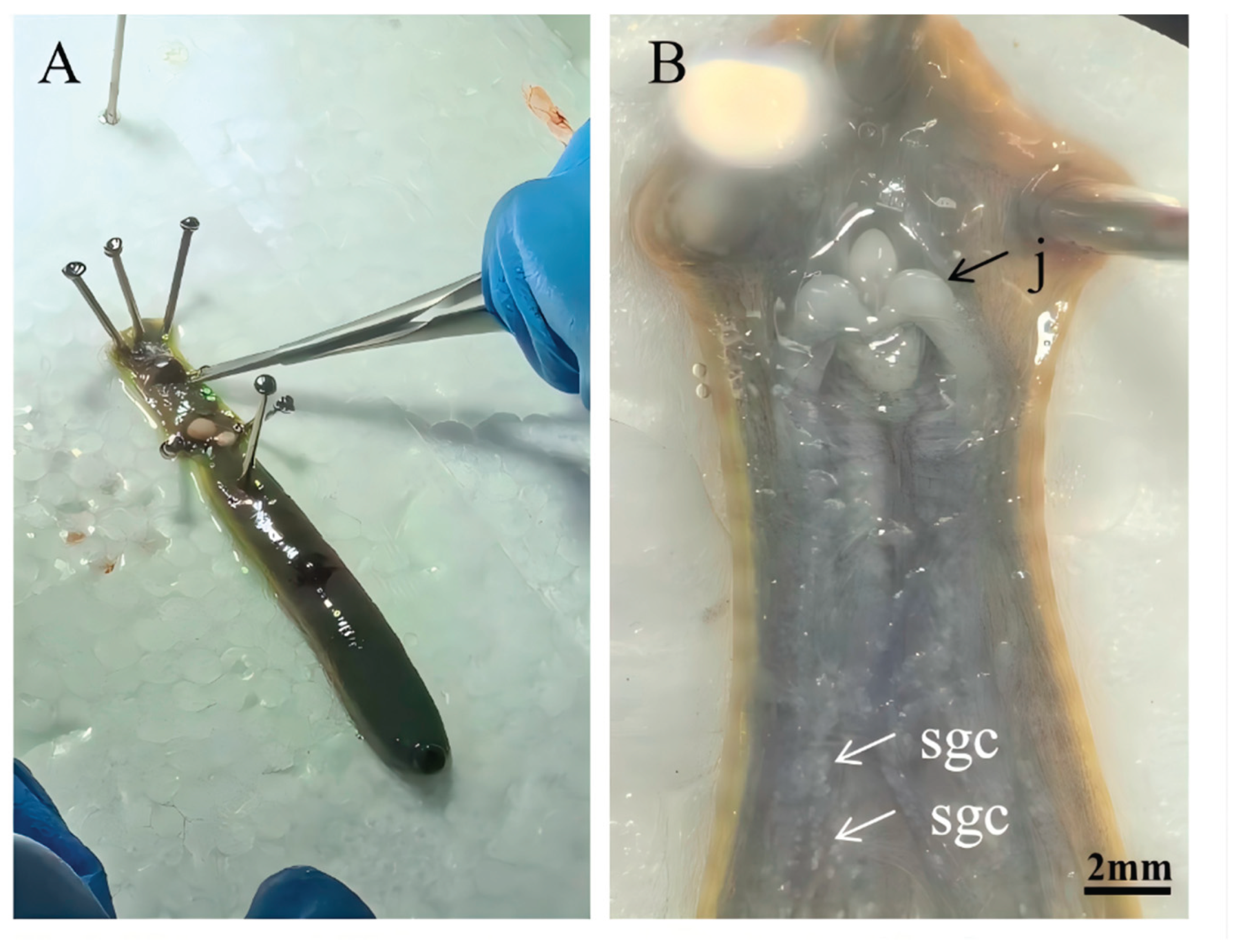
Figure 2.
Flow chart of the isolation of four extracellular vesicles by differential ultracentrifugation.
Figure 2.
Flow chart of the isolation of four extracellular vesicles by differential ultracentrifugation.
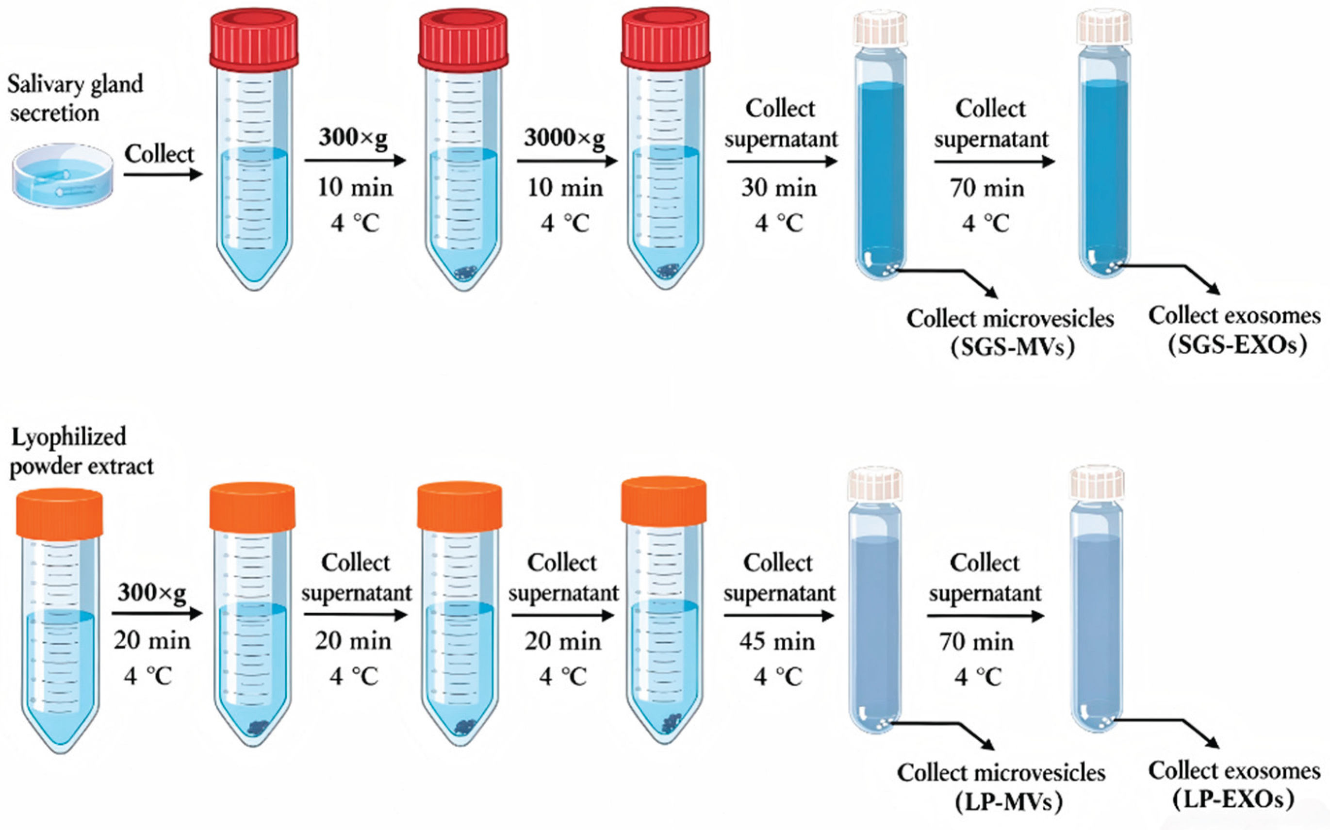
Figure 3.
Morphology of four types of extracellular vesicles from the salivary glands of Hirudo nipponica under transmission electron microscope. (A) Morphological diagram of salivary gland microvesicles; (B) Morphological diagram of microvesicles in extract of lyophilized powder of head; (C) Morphological diagram of exosomes in salivary gland secretion ;(D) Morphological diagram of exosomes extracted from lyophilized powder of head;.
Figure 3.
Morphology of four types of extracellular vesicles from the salivary glands of Hirudo nipponica under transmission electron microscope. (A) Morphological diagram of salivary gland microvesicles; (B) Morphological diagram of microvesicles in extract of lyophilized powder of head; (C) Morphological diagram of exosomes in salivary gland secretion ;(D) Morphological diagram of exosomes extracted from lyophilized powder of head;.
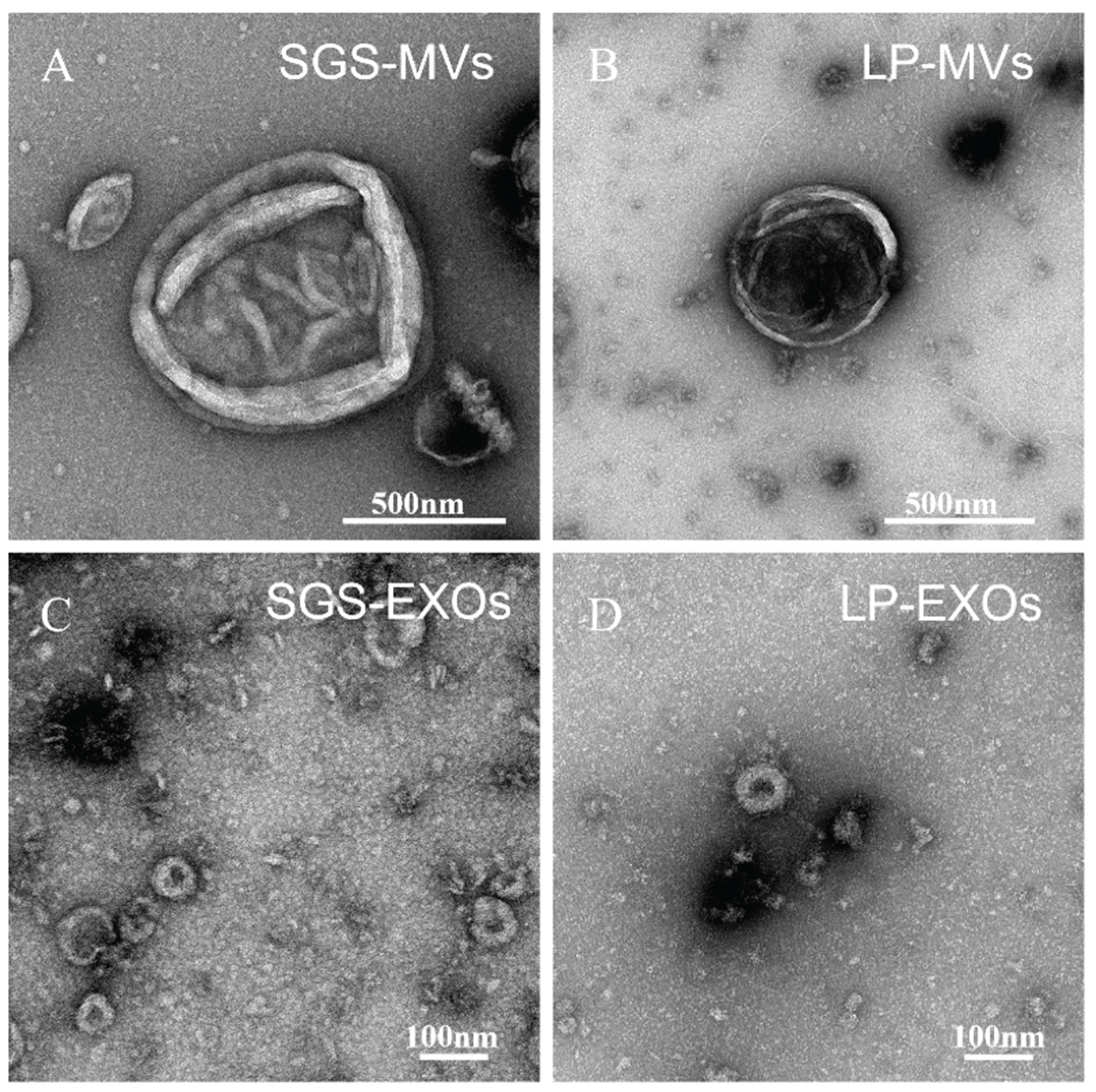
Figure 4.
Particle size distribution curves of four kinds of extracellular vesicles in the salivary gland of Hirudo nipponica. (A) Size distribution of salivary gland secretory microvesicles (SGS-MVs); (B) size distribution curve of exosomes derived from salivary gland secretion (SGS-EXOs); (C) Particle size distribution curve of microvesicles (LP-MVs) in lyophilized powder extract of head; (D) Particle size distribution curve of exosomes extracted from lyophilized head powder (LP-EXOs).
Figure 4.
Particle size distribution curves of four kinds of extracellular vesicles in the salivary gland of Hirudo nipponica. (A) Size distribution of salivary gland secretory microvesicles (SGS-MVs); (B) size distribution curve of exosomes derived from salivary gland secretion (SGS-EXOs); (C) Particle size distribution curve of microvesicles (LP-MVs) in lyophilized powder extract of head; (D) Particle size distribution curve of exosomes extracted from lyophilized head powder (LP-EXOs).
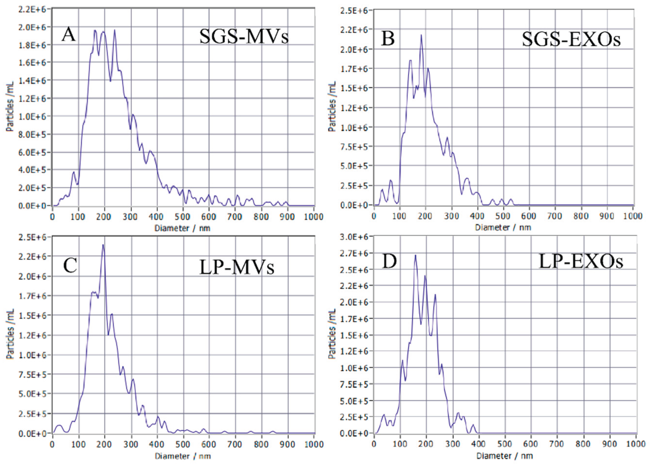
Figure 6.
Bar chart of protein quantification differential results.
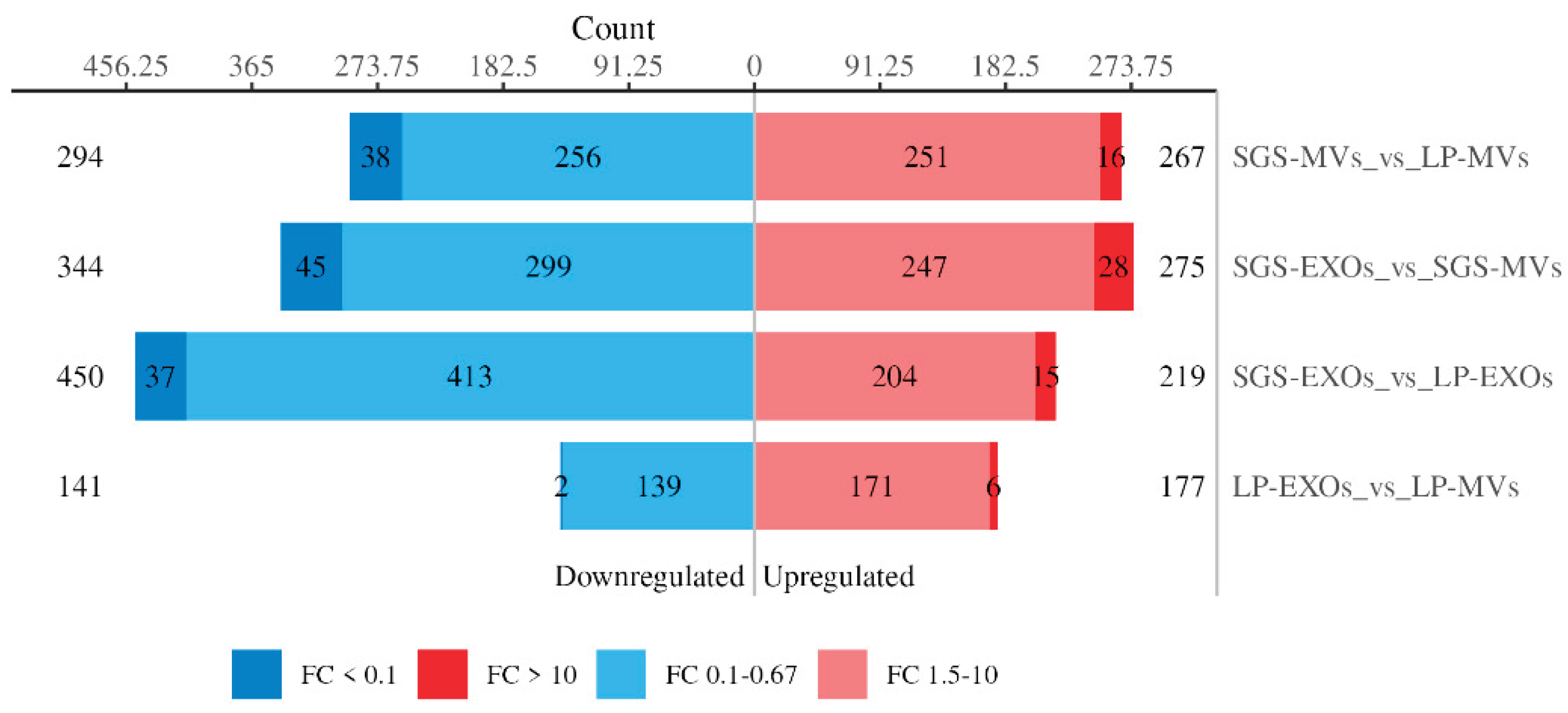
Figure 7.
Bubble plots of domain enrichment analysis for each comparison group. (A) Domain enrichment analysis comparing SGS-MVs and LP-MVs groups; (B) Domain enrichment analysis comparing SGS-EXOs and SGS-MVs groups; (C) Domain enrichment analysis comparing SGS-EXOs and LP-EXOs groups; (D) Domain enrichment analysis comparing LP-EXOs and LP-MVs groups. The horizontal axis displays the P-value (converted to –log₁₀) calculated using Fisher's exact test; a higher value indicates a greater statistical significance of enrichment in the corresponding domain category. The vertical axis lists domain names, where the size of each bubble represents the number of differentially expressed proteins, and the color indicates the Rich Factor—where colors closer to red correspond to higher Rich Factor values.
Figure 7.
Bubble plots of domain enrichment analysis for each comparison group. (A) Domain enrichment analysis comparing SGS-MVs and LP-MVs groups; (B) Domain enrichment analysis comparing SGS-EXOs and SGS-MVs groups; (C) Domain enrichment analysis comparing SGS-EXOs and LP-EXOs groups; (D) Domain enrichment analysis comparing LP-EXOs and LP-MVs groups. The horizontal axis displays the P-value (converted to –log₁₀) calculated using Fisher's exact test; a higher value indicates a greater statistical significance of enrichment in the corresponding domain category. The vertical axis lists domain names, where the size of each bubble represents the number of differentially expressed proteins, and the color indicates the Rich Factor—where colors closer to red correspond to higher Rich Factor values.
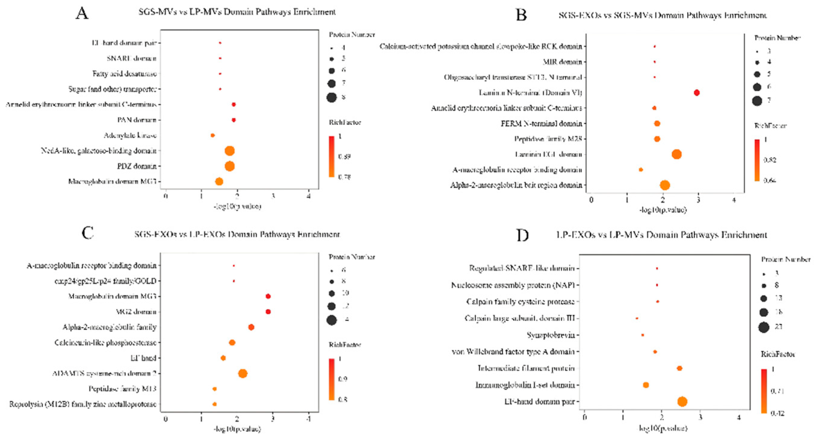
Figure 8.
GO functional enrichment bar plots of differentially expressed proteins across all comparison groups (BP). (A) GO functional enrichment analysis for the SGS-MVs vs. LP-MVs groups; (B) GO functional enrichment analysis for the SGS-EXOs vs. SGS-MVs groups; (C) GO functional enrichment analysis for the SGS-EXOs vs. LP-EXOs groups; (D) GO functional enrichment analysis for the LP-EXOs vs. LP-MVs groups. The horizontal axis displays GO functional annotation names, with color indicating the enrichment degree (Rich Factor)—the closer the color to blue, the greater the Rich Factor value; the number above each bar indicates the number of differentially expressed proteins contained in that GO functional annotation; the vertical axis shows the P-value (converted to-log₁₀) calculated using Fisher's exact test, reflecting the statistical significance of the enrichment.
Figure 8.
GO functional enrichment bar plots of differentially expressed proteins across all comparison groups (BP). (A) GO functional enrichment analysis for the SGS-MVs vs. LP-MVs groups; (B) GO functional enrichment analysis for the SGS-EXOs vs. SGS-MVs groups; (C) GO functional enrichment analysis for the SGS-EXOs vs. LP-EXOs groups; (D) GO functional enrichment analysis for the LP-EXOs vs. LP-MVs groups. The horizontal axis displays GO functional annotation names, with color indicating the enrichment degree (Rich Factor)—the closer the color to blue, the greater the Rich Factor value; the number above each bar indicates the number of differentially expressed proteins contained in that GO functional annotation; the vertical axis shows the P-value (converted to-log₁₀) calculated using Fisher's exact test, reflecting the statistical significance of the enrichment.
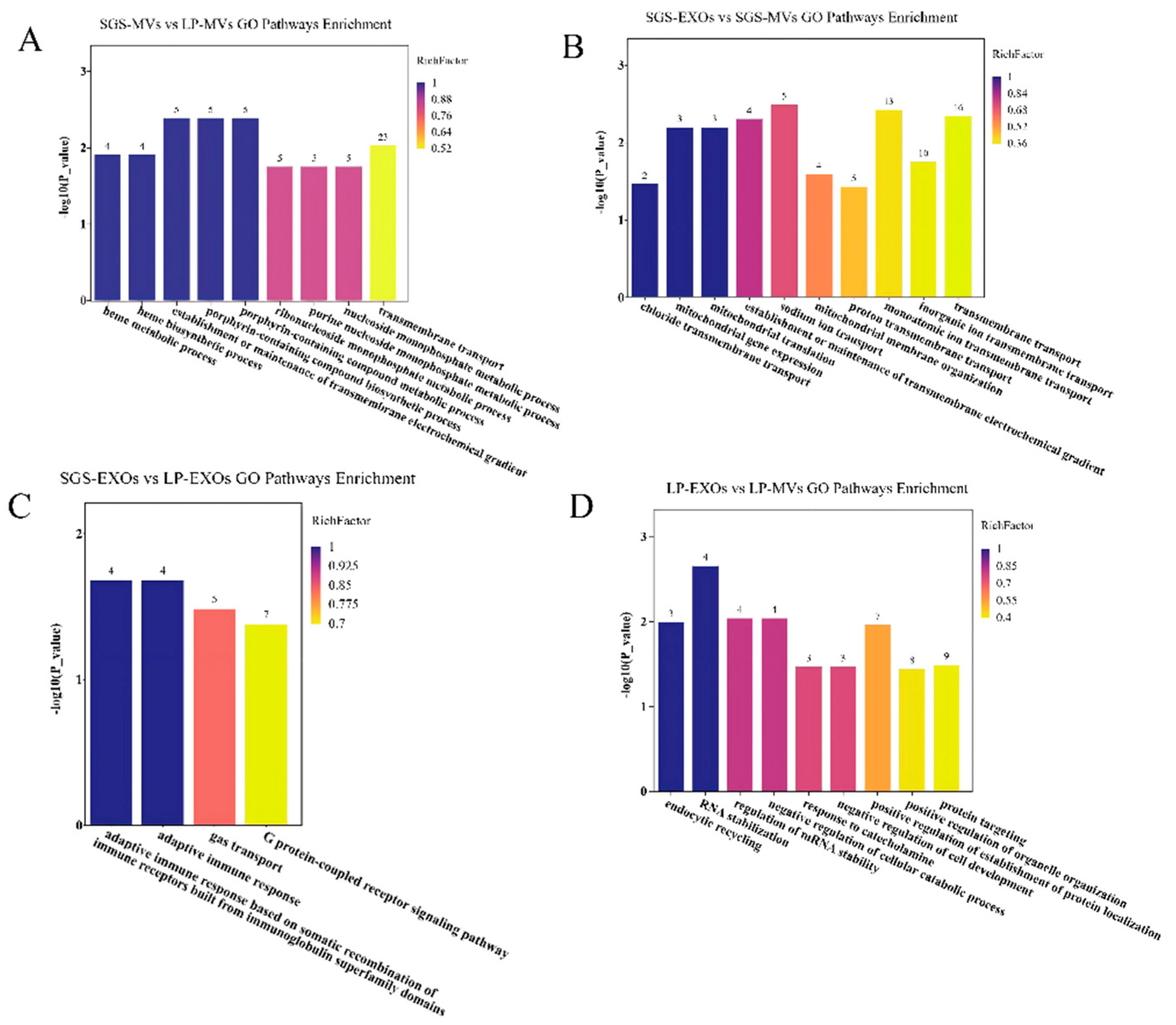
Figure 9.
KEGG pathway enrichment bar plots of differentially expressed proteins across all comparison groups. (A) KEGG pathway enrichment analysis for the SGS-MVs vs. LP-MVs groups; (B) KEGG pathway enrichment analysis for the SGS-EXOs vs. SGS-MVs groups; (C) KEGG pathway enrichment analysis for the SGS-EXOs vs. LP-EXOs groups; (D) KEGG pathway enrichment analysis for the LP-EXOs vs. LP-MVs groups. The horizontal axis displays the names of KEGG pathways, with colors indicating the enrichment degree (Rich Factor)—the closer the color to red, the greater the Rich Factor value. The number above each bar indicates the number of differentially expressed proteins contained in that pathway. The vertical axis shows the P-values (converted to-log₁₀) calculated using Fisher's exact test, reflecting the statistical significance of the enrichment results.
Figure 9.
KEGG pathway enrichment bar plots of differentially expressed proteins across all comparison groups. (A) KEGG pathway enrichment analysis for the SGS-MVs vs. LP-MVs groups; (B) KEGG pathway enrichment analysis for the SGS-EXOs vs. SGS-MVs groups; (C) KEGG pathway enrichment analysis for the SGS-EXOs vs. LP-EXOs groups; (D) KEGG pathway enrichment analysis for the LP-EXOs vs. LP-MVs groups. The horizontal axis displays the names of KEGG pathways, with colors indicating the enrichment degree (Rich Factor)—the closer the color to red, the greater the Rich Factor value. The number above each bar indicates the number of differentially expressed proteins contained in that pathway. The vertical axis shows the P-values (converted to-log₁₀) calculated using Fisher's exact test, reflecting the statistical significance of the enrichment results.
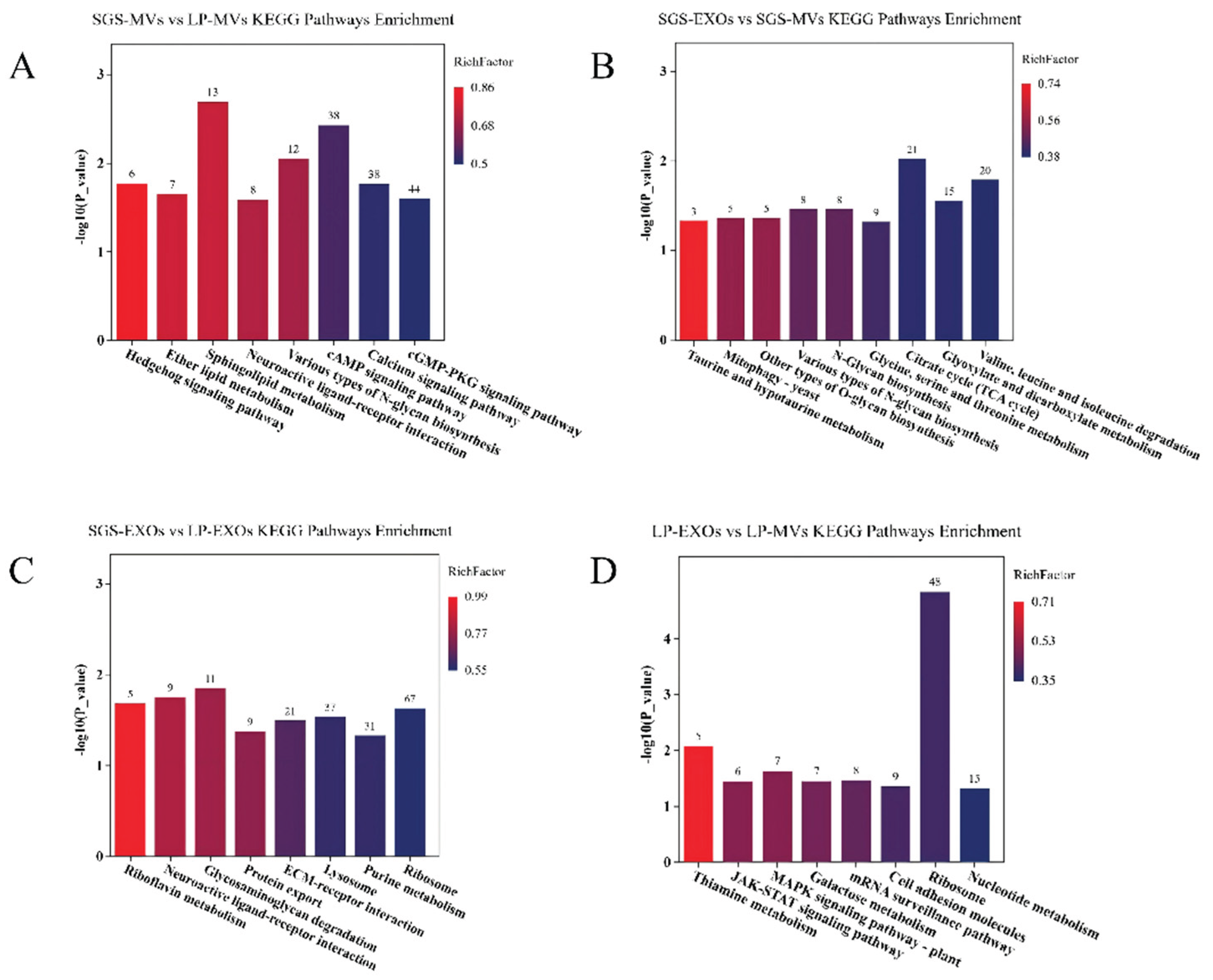

Table 1.
Distribution Characteristics of Key Active Proteins in Extracellular Vesicles from Salivary Glands of Hirudo nipponica.
Table 1.
Distribution Characteristics of Key Active Proteins in Extracellular Vesicles from Salivary Glands of Hirudo nipponica.
| Protein ID | Protein | SGS-MVs | SGS-EXOs | LP-MVs | LP-EXOs |
|---|---|---|---|---|---|
| QDZ37419.1 | Hirudin | √ | √ | ||
| AAA96144.1 | Destabilase I | √ | √ | ||
| P01051.1 | Eglin C | √ | √ | ||
| Q6T6C1 | Theromyzin | √ | √ | √ | √ |
| WBW88639.1 | Saratin | √ | √ | √ | √ |
| AHV78514.1 | Hyaluronidase | √ | √ | √ | √ |
Disclaimer/Publisher’s Note: The statements, opinions and data contained in all publications are solely those of the individual author(s) and contributor(s) and not of MDPI and/or the editor(s). MDPI and/or the editor(s) disclaim responsibility for any injury to people or property resulting from any ideas, methods, instructions or products referred to in the content. |
© 2026 by the authors. Licensee MDPI, Basel, Switzerland. This article is an open access article distributed under the terms and conditions of the Creative Commons Attribution (CC BY) license (http://creativecommons.org/licenses/by/4.0/).
Copyright: This open access article is published under a Creative Commons CC BY 4.0 license, which permit the free download, distribution, and reuse, provided that the author and preprint are cited in any reuse.