

Submitted:
09 November 2026
Posted:
12 November 2026
You are already at the latest version
Abstract
In previous cancer research, cancer stem cells (CSCs) have generally been defined as undifferentiated, whereas somatic bulk tumor cells —by analogy with the somatic cells of the human body—have been considered differentiated and, in some contexts, referred to as non-stem cells. However, somatic cancer cells are not differentiated progeny generated through asymmetric cell division of self-renewing CSCs. Rather, they arise through cellular plasticity and mesenchymal-to-epithelial transition (MET) from CSCs, whereas new CSC clones and sublines can arise through the opposing epithelial-to-mesenchymal transition (EMT). These processes therefore do not represent conventional differentiation and dedifferentiation, but rather an ancestral form of “germ-to-soma” and “soma-to-germ” conversion that may serve to maintain proliferation and survival in unicellular systems or, in cancer, to enable continuous adaptation to microenvironmental and therapeutic stress. The hypoxic CSC-stemgermline that coordinates the cancer-cell life cycle is continuously challenged and damaged by the hyperoxic microenvironment and by the excessively high oxygen levels encountered in blood and tissue, particularly above 6.0% oxygen. Irreversibly damaged stemgermline DNA must therefore be rapidly repaired or replaced. Dysfunctional stemgermlines are continuously replaced by functional “replacement clones” generated from somatic tumor cells or revitalized through hyperpolyploid DNA-damage-response mechanisms involving genome reconstruction. Following conversion, oxygen-resistant somatic cancer cells protect the stemgermline genome and may therefore function as a virtually unlimited genomic reservoir for the generation of new stemgermlines and CSC clones. The large spectrum of intermediate and transient phenotypes arising during both conversion processes contributes to the intratumoral heterogeneity characteristic of cancer cell populations.
Keywords:
cancer
; CSCs
; somatic tumor cells
; cellular plasticity
; ecDNA
; genomic stability
1. Introduction
This article adopts a dialectical perspective on the dual cancer-cell system by juxtaposing established concepts and terminology in cancer biology with insights emerging from the more recent framework of Evolutionary Cancer Cell Biology (ECCB) [1,2,3,4].
The evolutionary hypothesis proposed here is that, analogous to unicellular systems that preceded modern multicellularity, cancer-cell systems comprise two “germ” and “soma” subpopulations characterized by distinct genomic properties. One subpopulation, consisting of CSC-germ cells, possesses a stable yet expandable genome, whereas the other, represented by somatic bulk tumor cells, carries a highly unstable and crisis-prone genome.
The CSC-germline retains the ancestral gene-regulatory network, whereas somatic subpopulations harbor a highly dysregulated hybrid genome that remains continuously subject to genomic change. This distinction exposes a conflict between archaic unicellular genes (UGs) and active multicellular genes (MGs) following malignant transformation. The functional relationship between the two populations can be illustrated metaphorically by the anecdote “The Cook and the Waiter,” in which the chef ultimately remains in command.
Many researchers regard somatic cancer cells as differentiated tumor cells, whereas CSCs are generally considered undifferentiated cells that generate somatic cancer cells through asymmetric cell division (ACD). However, the ECCB framework, supported by numerous studies, challenges this interpretation. Instead, somatic tumor cells are proposed to arise through phenotype conversion via germ-to-soma transition (GST), also referred to as mesenchymal-to-epithelial transition (MET) [1,2,3,4]. This phenotypic conversion does not involve differentiation through asymmetric cell division.
Taken together, the ECCB framework supports the conclusion of a dual cancer-cell organization in which regulatory CSC-stemgermline lineages and intratumoral somatic lineages are not hierarchically separated populations but dynamically interconnected compartments. Their continuous reciprocal conversion, genome repair, replacement, and adaptation provide a potential cellular and evolutionary framework for understanding cancer as a highly plastic system maintained under persistent environmental and therapeutic stress.
1.1. The CSC -Germline and its Function
Unlike evolutionarily established human embryonic or adult stem cells, CSCs are proposed to represent primitive stem cells that are monopotent and generate progenitor cells through an archaic mechanism of hyperpolyploidization termed “cystic polyploidy.”
The approximately 1–2% CSC population, described from an evolutionary perspective as the hypoxic CSC-stemgermline, or simply the stemgermline, is characterized by a distinctive life cycle regulated by the tumor microenvironment (TME) and its oxygen gradients. Even relatively small increases above an oxygen concentration of 1.0% O₂ are proposed to alter the stemgermline phenotype, initially shifting it from a functional, stemness-positive ACD (asymmetric cell-dividing) state toward a stemness-negative ACD phenotype. Exposure to oxygen concentrations exceeding 6.0% O₂, corresponding to systemic cancer cell hyperoxia, causes irreversible DNA damage within the stemgermline and ultimately renders its genome dysfunctional.
Yet, without the “cook,” the system cannot continue to operate and must therefore establish a new one. Two ancient mechanisms conserved within the ancestral genome compartment may provide this replacement capacity: (1) DNA-damage response (DDR) accompanied by hyperpolyploid genome reconstruction and, alternatively, (2) generation of a replacement stemgermline through SGT processes, known in cancer biology as epithelial-to-mesenchymal transitions (EMTs) (Figure 1)
The first pathway proceeds through distinct stemgermline phenotypes. These include a senescent phenotype that may remain for an extended period in a reparative senescence state before transitioning into a senescence-bypassing phenotype. The latter subsequently proliferates through atypical and defective symmetric cell divisions, generating a dysfunctional DSCD phenotype. Once the DSCD progeny reach a critical size, the cells become fusion-competent and form multinucleated genome repair structures (MGRSs), also known as polyploid giant cancer cells (PGCCs), as a consequence of their hyperpolyploid state.
MGRSs/PGCCs attempt, through two distinct polyploidization phases, to revive the stemgermline genome and generate multiple progenitors capable of establishing new stemgermlines through reductive nuclear division. In this manner, a new stemgermline may be initiated, either during carcinogenesis (nascent stemgermline) or during tumorigenesis and metastasis (secondary stemgermlines). A classical description of the CSC system is provided in detail by Walcher et al. [5].
The researchers confirmed that CSCs exhibit substantial plasticity, resulting in changes in their phenotypic and functional characteristics. Such changes may be induced by chemotherapy and radiotherapy as well as by senescent tumor cells, which alter the tumor microenvironment. Therapy-induced senescence (TIS) can initially contribute to tumor regression by creating an anti-tumorigenic environment characterized by growth arrest and immune-cell recruitment. According to Walcher [5], TIS may also exert adverse, post-treatment effects. These include enhanced CSC plasticity, activation of stemness pathways in previously non-CSC populations, and promotion of senescence-escape followed by renewed activation of stemness-associated pathways. Senescence can therefore also exert a pronounced pro-tumorigenic effect by supporting the generation and maintenance of CSC phenotypes
1.2. “Soma” Cell Lines Preserve a Functional, Silent “Germ”- Line Genome
Somatic intra-tumoral heterogeneity is not considered here to be a consequence of CSC differentiation. Rather, somatic cancer-cell lineages arise through cellular plasticity and phenotype conversion and continue to evolve through specific evolutionary programs, which will be examined in greater detail in the following chapters.
Over the past decades, a major objective of conventional cancer research has been to characterize the unusual processes occurring within the bulk tumor population, which constitutes approximately 98% of the tumor. Particular attention has been devoted to understanding how genomically altered bulk tumor populations selectively survive, adapt, and acquire resistance to anticancer therapies. The evolution of the “mutated” tumor has therefore dominated cancer-cell research and clinical oncology for decades. This emphasis has progressively shifted attention toward bulk-tumor genomics and contributed to the development of concepts such as genomic instability, genomic chaos, and chromothripsis.
Paradoxically, the amount of genomic knowledge available for the two highly interconnected cancer-cell populations is almost inversely related to their relative abundance within the tumor. Whereas an extensive body of genomic data exists for somatic bulk tumor cell lineages, comparatively little is known about the genome of the 1–2% CSC-population Consequently, the term “cancer-cell genome” is frequently used to describe genomic data derived predominantly from the aberrant somatic bulk-tumor population.
1.3. “Germ-to-soma” Circuitry
From a functional perspective, the dual cancer-cell system consists of two interconnected circuits. The first comprises the CSC-stemgermline, its intra-tumoral evolution and life cycles, as well as the repair and genome-reconstruction mechanisms that maintain primary and secondary genomic stability and integrity. The second comprises the somatic bulk-tumor lineage, which performs the system’s vegetative functions, develops secondary genomic stability following chromothripsis, and acquires resistance to chemotherapeutic agents.
At the same time, the somatic lineage preserves the hypoxic stemgermline genome through epigenetic mechanisms. The interaction between these two circuits therefore constitutes the broader circuitry of a highly complex and therapeutically difficult-to-control cancer-cell system [1,2,3,4].
The present study therefore reviews current knowledge of both somatic and CSC genomics and interprets these findings from the perspective of Evolutionary Cancer Cell Biology.
2. Somatic Cancer Genomics
To date, cancer research has primarily focused on the genomes of somatic bulk tumor cells. In this context, the cancer cell system — which is highly complex and well-organized — and its genome are considered an altered version of the original host DNA. It has undergone significant changes, to the point that the cancer cell system behaves like a completely different organism.
This formulation implies that the cancer genome undergoes extensive reorganization and rewriting, leading to massive structural rearrangements, which can apparently circumvent conventional chromosomal organization by generating small independent circles of extrachromosomal DNA, and undergoes extensive epigenetic modification, including changes in DNA methylation that can silence protective genes and activate growth-promoting genes.
The standard answer to the question of why these genetic changes occur to a comparable extent in all cancer patients is significantly less precise. Most researchers believe that these alterations (mutations) provide the biological advantage necessary for diseased cells to survive and proliferate. One theory is that driver mutations act as a permanently activated accelerator, forcing the cell toward uncontrolled proliferation. Consequently, only cells that acquire an appropriate combinations of driver mutations can overcome cellular defenses and survive natural selection.
2.1. The “Blueprint” Concept
Many cancers follow a characteristic pattern. However, this does not refer to a specific genetic code, but rather to a set of functional patterns. Although every tumor is genetically unique, cancer cells must overcome similar biological and genomic challenges in order to survive, proliferate, and spread throughout the body.
Regardless of whether the tumor originates in the lung, breast, or colon, cancer cells employ a limited number of adaptive strategies to circumvent the regulatory mechanisms of the host organism. These include: (i) sustained proliferative signaling; (ii) evasion of growth-inhibitory signals, such as those mediated by p53, which normally prevents damaged cells from continuing to divide; (iii) resistance to programmed cell death; (iv) unlimited replicative potential; (v) angiogenesis, tissue invasion, and metastasis; and—of particular importance—(vi) convergent intra-tumoral evolution. The latter phenomenon demonstrates that, despite highly divergent underlying mutations, different cancer cells or tumor types may ultimately activate the same central signaling pathways, such as the MAPK pathway.
Under the selective pressure imposed by the immune system and the host environment, tumor evolution tends to favor the most effective mechanisms of survival and proliferation. Since there are biologically only a limited number of routes toward uncontrolled growth, different cancer types may ultimately converge on similar functional patterns.
Nevertheless, the concept of a cancer "blueprint" is highly dynamic and can be internally contradictory. Although functional blueprints may be similar, the genetic architecture of a tumor can differ substantially from one patient to another, as demonstrated by Aitken et al. [6] in 2026. Studies on somatic-germline epistasis further support this principle. However, in this context, the term "germline" refers to the host organism germline and not the cancer specific CSCs germline proposed in the present framework. From an evolutionary perspective, however, a distant evolutionary connection between the two germline compartments may exist.
Somatic-germline epistasis describes a phenomenon in which an acquired, non-heritable genetic alteration, such as a somatic mutation, is significantly influenced by the genetic information encoded in the host organism’s germline background. Epistasis refers to interactions between genes in which the presence or activity of one gene modifies, suppresses, or enhances the effect of another gene.
Therefore, the consequences of a somatic mutation or an induced genetic defect can vary substantially depending on the genetic background of the host organism. The host germline may influence how aggressively a tumor develops, how rapidly it progresses, and which signaling pathways are most likely to be disrupted. Consequently, modern cancer therapies increasingly rely on tumor profiling to identify specific genetic alterations, particularly driver mutations, within this functional framework and to exploit tumor-specific vulnerabilities.
2.2. Instability and Genomic Chaos through Chromothripsis and Genomic Catastrophe
Permanent alterations in the DNA sequence that persist after replication or repair and can be transmitted to daughter cells during cell division are defined as acquired mutations. The increased propensity to acquire mutations, chromosomal alterations, and other disturbances of genome stability is referred to as genomic instability. Moreover, to describe the state of massive, rapid, and unpredictable restructuring of the somatic cancer cell genome, current cancer research uses the term genomic chaos. It represents an extreme manifestation of genomic instability in which the alterations extend far beyond individual point mutations. Entire chromosomes may break, undergo erroneous reassembly, or experience profound changes in copy number and structure. One important example is chromothripsis [7-13].
Chromothripsis refers to extensive chromosomal rearrangement that is restricted to one or a few chromosomes and can generate a poorly characterized spectrum of structural abnormalities. The process involves numerous DNA double-strand breaks (DSBs) and may contribute to malignant transformation and cancer progression. Such catastrophic genomic events may occur under conditions of extreme cellular stress or as a consequence of a single catastrophic event during the life cycle of a cell, including events associated with cancer development or, potentially, aggressive anticancer therapies
Already in 2012, Forment et al. [14] demonstrated that chromothripsis may contribute to cellular transformation through the loss of tumor-suppressor functions, the generation of oncogenic fusions, and oncogene amplification via double-minute chromosomes, with micronuclei playing a potentially important role. According to this model, chromosomes trapped within micronuclei may undergo aberrant DNA replication and subsequent fragmentation. Derivative chromosome material may then be reincorporated into the main nucleus, potentially resulting in chromothripsis and contributing to the development of cancer.
According to this model, genomic alterations leading to oncogene activation and loss of tumor-suppressor function represent important driving forces in cancer development. Such alterations may accumulate progressively during cancer evolution and thereby contribute to the emergence and selection of malignant cell populations.
More recently, Simovic-Lorenz and Ernst (2025) [15] described the prevalence of chromothripsis across different cancer types and discussed its contribution to cancer development, the selective advantages that cancer cells may derive from chromothripsis, and the evolutionary trajectories of chromothriptic tumors. From this perspective, chromothripsis appears less likely to represent a primary transformation driver than a genomic event that facilitates and promotes subsequent cancer progression.
A reversion from genuine genomic chaos to the original state of genetic integrity and genomic stability is, at the cellular level, biologically extremely difficult and may be effectively impossible once extensive genomic damage has occurred. Two major explanations have been proposed for this apparent irreversibility: first, the loss of the original genomic blueprint and, second, the irreversible nature of many genomic alterations [16,17]. Once the genome has been extensively fragmented and incorrectly reassembled, the cell may lack the molecular template required to reconstruct the original genomic state with complete accuracy.
2.3. New Genomic Stability After Chromothripsis
Rather than simply collapsing into genomic chaos, the mutated cell may subsequently seek to establish a new state of secondary genomic stability. Two different mechanisms may contribute to the termination and stabilization of the chaotic state. One of these is karyotype selection. Cells carrying the most severe genomic alterations are frequently unable to survive and are eliminated. A small proportion of cells, however, may succeed in stabilizing their extensively rearranged chromosomes. These cells do not necessarily restore their former genomic integrity through repair. Instead, they may acquire new functional properties and cellular identities, including, for example, resistance to anticancer drugs. This process can be considered a form of functional compensation.
In some cases, a secondary mutation occurring at another genomic location may functionally compensate for the consequences of an earlier genetic alteration. Such a suppressor mutation may restore or stabilize a cellular phenotype even though the underlying genome remains substantially altered. Thus, new phenotypic stability does not necessarily imply restoration of the original genomic state.
For a long time, chromothripsis was regarded primarily as a major driving force of genomic instability and cancer development [18]. Two major models have been proposed to explain its origin. The first is the “micronucleus hypothesis” in which chromothripsis arises following chromosome mis-segregation during mitosis and the subsequent formation of micronuclei. The second is based on “telomere crisis,” which can result from dysfunctional telomere maintenance and defective cell-cycle checkpoint pathways. Both models, however, primarily interpret chromothripsis as a process of genome destabilization that contributes to malignant transformation and cancer progression.
More recent developments in biomedical research indicate that cells possess specific molecular mechanisms of containment, processing, and repair that can allow the cell to emerge from such genomic chaos with a new, although profoundly altered, state of genomic stability. Rather than inevitably undergoing cell death or losing the fragmented DNA entirely, the cell may retain, reorganize, and permanently reassemble substantial portions of the fragmented genome.
Several years ago, cancer researchers including Cortés-Ciriano [19,20] and Lin [21] demonstrated that DNA fragments generated by chromothripsis can undergo complex rearrangements and subsequently be transmitted through cell division. In cancer, chromothripsis is frequently associated with substantial DNA loss. In certain cases, however, including some congenital disorders and tumors, chromothripsis can result in extensive genomic shuffling with relatively limited loss of DNA. This observation suggests the existence of mechanisms capable of retaining and physically associating fragmented DNA molecules rather than allowing them to be completely lost.
These mechanisms may prevent the irreversible loss of fragmented DNA and resulting micronuclei from being irreversibly lost following chromosome mis-segregation. During mitosis, chromosomes that fail to enter the main nucleus can become enclosed within micronuclei and remain separated from the primary nuclear compartment. The micronuclear envelope may subsequently collapse during the cell cycle [22,23]. Micronuclei may lack essential nuclear pore proteins and structural components of the nuclear, leaving the enclosed chromosomes particularly vulnerable to nuclease degradation and DNA damage. The relationship between micronuclei, chromothripsis, and malignancy has been described in detail by McNeil et al. in 2025 [10].
The enzyme N4BP2 has been implicated as a factor capable of entering micronuclei and contributing to extensive fragmentation of the exposed DNA. The resulting DNA fragments may subsequently be processed and clustered by protein complexes, including CIP2A and TOPBP1, and undergo aberrant ligation [19,20]. These rearranged DNA structures can then be transmitted to daughter cells and subsequently stabilized. Although the order and orientation of genomic segments may appear extensively scrambled, including through inversions and other rearrangements, the DNA molecules can nevertheless become physically reconnected and structurally stabilized [11,24].
However, not all fragmented chromosomal material can be reincorporated into the main chromosome. Portions of the fragmented genome may instead form independent extrachromosomal DNA (ecDNA) molecules capable of autonomous replication during the cell cycle. In cancer cells, such ecDNA can provide a highly dynamic form of genetic stability by maintaining oncogenes at exceptionally high copy numbers and thereby substantially increasing the capacity of tumor cells to adapt to environmental stress and anticancer treatment [25,26].
2.4. ecDNA remodels the cancer genome and leads to transitory drug resistance
Drug resistance is increasingly understood as one of the functional consequences of the genomic instability and selection processes associated with micronucleus formation and chromothripsis. Genomic rearrangements generated during these processes may provide cancer cells with a substantial selective advantage, thereby reducing the effectiveness of anticancer therapy.
Fragments of displaced, broken, or excised DNA, including ecDNA molecules, may frequently contain oncogenes and genes associated with drug resistance, such as MYC or genes encoding drug-efflux transporters. Because ecDNA molecules are distributed unevenly between daughter cells during cell division, these genes can become massively enriched in individual tumor cell populations.
As a consequence, selected cancer cells may carry exceptionally high copy numbers of resistance-associated genes and express the corresponding resistance factors at very high levels. This provides a powerful mechanism by which a small subpopulation of tumor cells can survive chemotherapy and subsequently expand under therapeutic selection.
Such mechanisms enable ecDNA tumours to adapt extremely rapidly and dynamically — though often reversibly — to cancer therapies. ecDNA rings frequently contain driver oncogenes and are small, circular structures with extremely open and accessible chromatin. This leads to massive overproduction of oncogenes. Within just a few cell divisions, daughter cells can emerge with up to 100 copies of resistance and oncogenes. [25,26]
As reported by Pecorino et al. [27] in 2022, ecDNA elements were not found in normal cells. Analysis of clinical samples reveal they are common in most cancers and their presence indicates poor prognosis. They often contain enhancers and driver oncogenes that are highly expressed. The circular ecDNA topology leads to an open chromatin conformation and generates new gene regulatory interactions, including with distal enhancers. The absence of centromeres leads to random distribution of ecDNA rings during cell division and genes encoded on them are transmitted in a non-mendelian manner. ecDNA can integrate into and exit from chromosomal DNA. The numbers of specific ecDNA rings can change in response to treatment. This dynamic ability to remodel the cancer genome challenges long-standing fundamentals, providing new insights into tumor heterogeneity, cancer genome remodeling, and drug resistance.
Even in 2013 Nathanson et al. [28] has demonstrated that tumor cells reversibly up-regulate or suppress mutant EGFR expression, conferring distinct cellular phenotypes to reach optimal tumor cell growth. Resistance to EGFR tyrosine kinase inhibitors (TKIs) occurs by elimination of mutant EGFR from ecDNA. After drug withdrawal, reemergence of clonal EGFR mutants on ecDNA follows. These results indicate a highly specific, dynamic, and adaptive route by which cancers can evade therapies that target oncogenes maintained on ecDNA. Searching on glioblastoma, researchers found that EGFR is frequently mutated, commonly giving rise to the constitutively active oncogenic variant EGFR-vIII. This factor accelerates tumor but makes tumor cells more sensitive to EGFR – TKIs.
Lange et al. (2022) [29] also argues that the amplification of oncogenes on extrachromosomal DNA (ecDNA) is a widespread phenomenon that encourages aggressive tumour growth, drug resistance and reduced survival rates, however the impact of ecDNA on somatic variation and selection remains unclear. The authors discussed the random segregation of ecDNA in human cancer cells and the drug-induced selection of ecDNA, and demonstrate that random ecDNA inheritance results in extensive intratumoral ecDNA copy number heterogeneity and rapid adaptation to metabolic stress and targeted treatment. The random inheritance by descent, enhanced variation through random segregation and selection, thereby accelerating tumor cell evolution and increasing adaptability.
Observed ecDNAs benefit cell survival and growth and can change within a single cell cycle. ecDNA inheritance can predict, a priori, some of the aggressive features of ecDNA-containing cancers. These properties are facilitated by the ability of ecDNA to rapidly adapt genomes in a way that is not possible through chromosomal oncogene amplification. These results show how the nonchromosomal random inheritance pattern of ecDNA contributes to poor outcomes for patients with cancer.
More recently Haughey et al. (2025) [30] stated that ecDNA would progressively emerge as a crucial driver of cancer evolution, albeit only in around 20% of tumours. The researchers discussed open questions concerning the evolutionary role of ecDNA in tumor development, including tumorigenesis and metastatic seeding, the mutational landscape on ecDNA, the dynamic ecDNA genotype–phenotype map, the structural evolution of ecDNA, and how knowledge of tissue-specific ecDNA evolutionary paths can be leveraged to deliver more effective clinical treatment
2.5. Molecular “DNA-Glu” and the new heritable state of genomic organization.
In 2021, Shoshani et al. [31] demonstrated that therapeutic agents used to treat cancer, paradoxically promote the development of permanent resistance, do so by triggering the reintegration of extrachromosomal DNA (ecDNA). Under the influence of therapy, free-floating ecDNA rings jump into chromosome breakpoints and seal the damage. As a result, the ecDNA becomes firmly and permanently integrated into the normal chromosome.
Although free ecDNAs are dynamic and potentially unstable, reintegration results in the permanent fixation of the oncogene in the genome. The result is irreversible, lasting resistance to the inducible therapeutic agent. The authors state: "The landscape of therapy resistance is not purely volatile. Acting effectively as a DNA-Glu, this structural reinsertion into chromosomes stabilizes the previously transient oncogenic activation, transforming dynamic, heterogeneous drug tolerance into permanent, genetically fixed therapeutic resistance."
This shows, that cancer cells possess molecular mechanisms which can maintain the physical proximity of shattered chromosomal fragments and thereby facilitate their subsequent inheritance and reassembly. This process has been described as mitotic tethering or mitotic clustering of chromosomal fragments.
Tethering and clustering
CIP2A–TOPBP1 complex, together with MDC1, has been shown to associate with damaged chromatin within micronuclei and to tether the resulting chromosome fragments during mitosis. Rather than allowing the acentric fragments to disperse randomly throughout the cytoplasm, this molecular tethering keeps them in close spatial proximity and promotes their coordinated segregation into the same daughter cell
This mechanism is particularly important because most fragments generated by chromosome shattering are acentric and therefore lack their own centromere-dependent mechanism for faithful mitotic segregation. Without physical tethering, the fragments could be distributed randomly between daughter cells or lost entirely. CIP2A–TOPBP1-mediated clustering instead promotes the asymmetric inheritance of the fragmented chromosome by a single daughter cell, thereby preserving a substantial proportion of the original genetic material [25]. As revealed by Trivedi et al [32] tethering is shown to be crucial for the viability of cells undergoing chromosome missegregation and shattering after transient inactivation of the spindle assembly checkpoint. Chromatin-bound tethers maintain proximity of fragments of a shattered chromosome enabling their re-encapsulation into, and religation within, a daughter cell nucleus.
Importantly, tethering does not constitute complete DNA repair. The fragmented chromosome remains extensively damaged during mitosis. Following its reincorporation into the main nucleus, the DNA fragments can subsequently undergo processing and ligation, with non-homologous end joining (NHEJ) representing an important mechanism contributing to their reassembly. The resulting chromosome is therefore not restored to its original configuration. Instead, the fragments may be joined in a new order and orientation, generating a highly rearranged but nevertheless stable and heritable chromosome.
The somatic cancer cell does not necessarily recover the original genomic state after chromothripsis. Rather, it can transform a state of extreme genomic disruption into a new state of genomic organization and stability. Chromosomal fragments that are not reincorporated into a rearranged chromosome may circularize and persist as autonomous DNA molecules. Recent work has additionally identified the nuclease N4BP2 as an important mediator of DNA fragmentation within ruptured micronuclei and linked its activity to chromothripsis and ecDNA formation. Thus, chromosome fragmentation, molecular tethering, reassembly, and ecDNA formation can be viewed as interconnected processes through which a severely disrupted cancer-cell genome can acquire a new and heritable state of organization.
As stated by Li et al [33] ecDNA exhibits a unique structure that can be representative of high chromosome accessibility, contributing to hyperactivated proto-oncogenes and malignant behaviours. Meanwhile, nonchromosomal inheritance and recurrent mutations of ecDNA fuel tumour heterogeneity and evolution. Recent studies have demonstrated that ecDNA drives tumorigenesis and progression and is related to poor clinical outcomes and drug resistance across widespread cancers. The work of Li et al. also summarized the current understanding of the origin, biogenesis process, discovery history, molecular mechanisms, and physiological functions of ecDNA in cancer.
2.6. Chromosome Breakage and Gene Fusions as Evolutionary Mechanisms Conserved in the Cancer Genome
2.6.1. Macroevolutionary Shifts in Classical Evolutionary Theories
Classical evolutionary theory has traditionally emphasized the gradual accumulation of small genetic changes, particularly point mutations, whereas chromosome breakage and gene fusion are recognized as mechanisms capable of producing large-scale genomic reorganization, sometimes described under the concept of “genome rescheduling” and macroevolutionary jumps. In contrast, mechanisms such as ecDNA biogenesis and extensive chromosome breakage can produce rapid and even seismic restructuring of the genome.
In a 2019 study, Pellestor and Gatinois [34] argued that chromothripsis may represent a powerful molecular mechanism underlying macroevolutionary transitions and the evolutionary model known as punctuated equilibrium. The authors describe how a single catastrophic event result in reproductive isolation and, ultimately, the emergence of a new species. Their analysis of the etiology of chromothripsis identified several cellular processes capable of generating catastrophic genomic restructuring, including premature chromosome condensation, telomere dysfunction, abortive apoptosis, and micronucleus formation. Importantly, these mechanisms may occur during early embryonic germline development, suggesting that chromothripsis may constitute an unexpectedly powerful mechanism for profound genome modification.
The occurrence of chromothripsis is therefore compatible with macroevolutionary models proposed as an alternative or complement to phyletic gradualism. Cases of chromosomal speciation and short-term adaptation may likewise be associated with chromothripsis-mediated mechanisms. The emergence of this apparently chaotic genomic phenomenon may consequently contribute to our understanding of how chromosome rearrangements can participate in the speciation process.
Pellestor and Gatinois [34] grouped chromothripsis, chromoanasynthesis, and chromoplexy under the broader term chromoanagenesis This concept encompasses rapid and profound genomic alterations, particularly within the germlines of early developmental stages, and provides a plausible pathway for genomic evolution and speciation. Because these phenomena contrast with models based on the progressive accumulation of driver mutations or rearrangements, they provide conceptual support for macroevolutionary models such as punctuated equilibrium. According to the authors, numerous examples of chromosomal speciation and short-term adaptation could potentially be related to previously unexpected chromoanagenesis-associated mechanisms.
More recently, Gisselsson [36] compared mechanisms of chromosomal instability (CIN) with processes involved in tumor evolution and demonstrated striking similarities between the structural genome alterations associated with speciation in animals and plants and those observed during the development of therapeutic resistance in tumors. In both contexts, the evolutionary trajectory may converge on a similar set of cytogenetic and cytological events. These include excessive shortening of telomeric repeats, disruption of normal mitotic processes, chromosome breakage-fusion-bridge events, and extensive genomic imbalance.
Telomere dysfunction may, in turn, lead to cytokinesis failure, resulting in tetraploidization and the accumulation of supernumerary centrosomes. These alterations can subsequently promote multipolar cell division and major abnormalities in chromosome number. Several established cancer risk factors may contribute to these processes. For example, breakage-fusion-bridge instability can be directly induced by clastogenic agents, whereas papillomavirus infection has been shown to interfere with centrosome function. Partial impairment of the DNA-damage response and mitotic checkpoint functions appears to be a prerequisite for the development of chromosome instability through these mechanisms. Conversely, telomerase overexpression may play an important role in modulating the rate at which chromosomal evolution proceeds.
2.6.2. Genomic Plasticity in ecDNA Populations and the ecDNA “Plasmids” of Eukaryotes
Several very recent and potentially paradigm-shifting studies are beginning to transform the way. ecDNA is viewed within evolutionary biology, particularly with regard to the evolutionary significance of plasmid-like extrachromosomal elements in eukaryotes. One of these studies, from the group of Shaw and Hennsen [37], proposes that tumor-associated ecDNA can function in a manner analogous to plasmids at so-called “contingency loci.” Such a configuration provides a level of ultrafast genomic plasticity that would not be achievable through conventional chromosomal organization alone.
From this evolutionary perspective, cancer can be viewed as resembling a unicellular organism exposed to intense selective pressure. Because ecDNA molecules are distributed asymmetrically during cell division, they can generate substantial population-level heterogeneity and thereby fulfill an evolutionary role analogous to that of bacterial plasmids. Although the analogy between plasmids and ecDNA has been recognized previously, its evolutionary implications have not been explored in comparable depth.
The authors compare plasmids and ecDNA with respect to their formation, expression, segregation, and evolutionary dynamics and identify common principles operating across the two systems. They propose that both plasmids and ecDNA can function as “contingency loci” that permit rapid adaptation. From this perspective, extrachromosomal genetic elements may represent an example of convergent evolutionary solutions to the problem of rapid adaptation under changing selective conditions.
2.6.3. Phenotypic Heterogeneity in Somatic ecDNA Populations
Studies of ecDNA indicate that phenotypic heterogeneity can emerge even among genetically identical or closely related somatic cancer cells within the same tumor. Such cells may acquire markedly different phenotypes, biological properties, and patterns of gene expression. This diversity appears to be driven, at least in part, by the distinctive physical and inheritance properties of circular ecDNA molecules.
Several mechanisms contribute to this phenotypic heterogeneity, including asymmetric segregation and dynamic transcriptional regulation. Because ecDNA molecules can be distributed unevenly between daughter cells, substantial variation in gene copy number and oncogene dosage can arise rapidly [38-41]. The highly accessible chromatin structure associated with ecDNA further promotes strong transcriptional activity of the oncogenes carried on these molecules. In addition, ecDNA molecules can cluster within the nucleus to form ecDNA hubs, which function as transcriptional super-enhancers and can strongly increase or modulate the expression of target genes in a cell-dependent manner.
Consequently, unequal ecDNA inheritance can alter the global transcriptional state of individual tumor cells and thereby influence metabolic pathways, signaling networks, and cell-cycle regulation.
The number of ecDNA copies can also have a major influence on somatic cancer-cell fate. Cells carrying extremely high numbers of ecDNA copies, for example MYCN-amplified ecDNA, may undergo apoptosis and die in response to genotoxic stress. By contrast, subpopulations carrying lower ecDNA copy numbers may enter a state of therapy-induced senescence. Such senescent cells can subsequently acquire resistance and provide a reservoir from which later tumor recurrence may arise.
At the same time, very high oncogene dosage can provide a substantial selective advantage under conditions of therapeutic or environmental stress. When the selective pressure is removed, however, the metabolic burden associated with excessive oncogene expression may become disadvantageous. Tumor cells may therefore exploit their phenotypic plasticity to modulate ecDNA copy number dynamically and reversibly, increasing or reducing dosage according to the prevailing selective environment. ecDNA-driven variation in oncogene dosage may even alter the morphology of cancer cells.
2.6.4. Graded ecDNA Dosage and “Distributed Evolution”
Wang et al. [42] provide an additional perspective by showing that ecDNA-bearing cell populations can maintain a continuous spectrum of heritable states. This graded distribution may confer a form of deterministic adaptive capacity on the population and potentially challenges the classical concept of evolution based predominantly on clonal selection.
The researchers analyzed ecDNA levels across thousands of individual cells and found that increasing ecDNA dosage was accompanied by proportional changes in transcript abundance, chromatin accessibility, protein levels, cell-cycle progression, and proliferation. Importantly, when the naturally occurring dosage continuum was experimentally disrupted by separating cells into low- and high-copy-number populations, the resulting populations rapidly reconstructed the original continuous ecDNA distribution.
Taken together, these observations suggest that restoration of the ecDNA dosage spectrum is not simply a passive consequence of random segregation. Rather, it appears to represent an active and deterministic process. ecDNA-mediated gene expression may therefore constitute an intrinsic evolutionary mechanism through which the population continually reconstructs its evolutionary landscape, thereby providing rapid population-level adaptability. This mechanism may help explain why ecDNA-driven cancers can display particularly aggressive and treatment-resistant phenotypes.
According to the researchers, ecDNA-driven tumors do not necessarily converge toward a single dominant clonal state. Instead, they maintain a broad and ordered continuum of oncogene dosage across individual cells. This continuum represents a structured and heritable feature capable of generating a wide range of molecular and phenotypic states.
Unlike conventional clonal systems, which depend on the emergence and subsequent expansion of rare advantageous variants, ecDNA-bearing populations may respond to environmental pressures by simultaneously sampling a broad spectrum of phenotypic states. Graded ecDNA dosage generates coordinated continua across genomic, epigenomic, transcriptomic, and proteomic levels, thereby reorganizing regulatory networks governing proliferation, signaling, and chromatin control.
Importantly, the amplification architecture itself appears to encode regulatory information. Genes amplified on ecDNA follow transcriptional scaling patterns that differ from those of the same genes amplified within homogeneously staining regions (HSRs). EcDNA amplification is therefore not simply an increase in gene copy number. Rather, the architecture of amplification changes how gene dosage is interpreted and reshapes the quantitative relationship between genotype and phenotype.
The ecDNA dosage continuum is actively maintained. When cells representing the extremes of the copy-number distribution were isolated, the full spectrum was rapidly re-established within only a few generations. The population therefore appears to possess intrinsic mechanisms capable of reconstructing its characteristic evolutionary landscape.
The authors consequently conclude that ecDNA-driven populations contain an intrinsic capacity to generate and preserve adaptive potential. ecDNA can thus be understood not merely as a vehicle for oncogene amplification, but as a structured evolutionary system. The aggressiveness of ecDNA-driven cancers may therefore depend not only on the quantity of amplified oncogenes but also on the architecture of amplification itself, which confers enhanced evolvability. From this perspective, targeting the architecture, dynamics, or regulatory consequences of ecDNA could potentially disrupt not only oncogene dosage but also the broader “evolutionary engine” sustaining these cancers.
3. The CSC-Stemgermline – the Enduring Comand Center
As described by Zeng et al. [43], the collective term CSCs refers to a subpopulation of stem-like cells characterized by self-renewal capacity and differentiation potential and by their significant contribution to tumor-cell proliferation, metastasis, and tumor growth. The regulatory networks governing CSCs encompass transcriptional and post-transcriptional control, epigenetic modification, interactions with the tumor microenvironment (TME), and regulation through epithelial-mesenchymal transition (EMT). Several of these regulatory mechanisms are also closely associated with therapeutic resistance.
However, the effective eradication of CSCs remains challenging. First, a definitive set of surface markers that uniquely identifies CSCs has not yet been established, in part because CSCs can alter their surface-marker profiles and thereby evade immune recognition. In addition, no effective therapy is currently available that specifically targets the quiescent CSC state.
Understanding the biological characteristics of CSCs is therefore considered essential for investigating tumorigenesis and tumor development. Self-renewal and differentiation are commonly regarded as two defining properties of CSCs. Similar to non-cancerous stem cells, CSCs have been reported to undergo both symmetric and asymmetric cell division in response to regulatory signaling pathways [44]. According to the ECCB, during symmetric cell division (SCD), a CSC divides into two identical daughter cells that contribute to the maintenance or expansion of the CSC pool. By contrast, during asymmetric cell division (ACD), the daughter cells differ from each other. One remains self-renewing, while the other becomes committed to differentiation.
From the perspective of Evolutionary Cancer Cell System Biology (ECCB), however, the collective term CSCs corresponds more closely to the evolutionary concept of the ancestral stemgermlines. This terminology emphasizes the genomic relationship between CSCs and ancent stem-cell systems and may provide a framework for understanding the physiological and evolutionary potential of this determinant cell lineage. In particular, the stemgermline concept brings its characteristic life cycle and its stemness-loss-and-recovery pathway into focus, including stemness-positive and dysfunctional stemness negative phenotypes, mechanisms of genome repair and reconstruction, and the subsequent establishment of new genomic stability [1,2,3,4].
3.1. Sustained Self-Renewal as Supposed “Dysregulation” of Asymmetric Cell Division
Cancer stem cells may more closely resemble ancestral germ stem cells and protist stemgermlines, characterized by limited differentiation potential, than the pluripotent embryonic stem cells (ESCs) or multipotent adult stem cells (ASCs) of multicellular organisms. Both the functional ACD phenotype and the dysfunctional DSCD state are regarded within ECCB as manifestations of a common stemgermline cellular system. Persistent self-renewal is interpreted as evidence of ancestral unicellular roots, whereas multicellular stem cells such as ESCs and ASCs generally exhibit temporally restricted self-renewal.
When the functionality of primitive stemgermlines is maintained, these cells may possess an apparently unlimited capacity for self-renewal. Following severe stress, however, stemgermlines lose their functionality and stemness, potentially in an irreversible manner. They may nevertheless continue to proliferate in the dysfunctional stemness-negative DSCD state for the subsequent restoration of genomic function and recover stemness. [1-4].
Already in 2010, Pannuti et al. [45] reported that CSCs employ a persistent self-renewal strategy resembling that of unicellular systems rather than that of conventional multicellular cell systems. The authors described “evolutionarily ancient pathways” that regulate self-renewal and cell-fate decisions in undifferentiated stem cells. They proposed that CSCs may hijack ancient evolutionary toolkits to regulate the tightly time-limited process of multicellular self-renewal.
Nevertheless, more recent studies have repeatedly discussed the apparent “dysregulation” of cancer-associated ACD [47-51). Within the conventional interpretation, loss of multicellular control over cancer ACD contributes to the continuous production of progenitor cells, which may subsequently generate more aggressive intratumoral clones with metastatic and drug-resistant properties. These researchers therefore propose that the asymmetric division in tumor cells and during early tumorigenesis becomes dysregulated [52]. From the ECCB perspective, however, the concept of ACD “dysregulation” may reflect a limited understanding of the persistent ACD proliferation of primitive cancer stemgermlines, for which sustained self-renewal is itself considered a unlimited l characteristic [1,2,3,4].
It has even been proposed that CSCs specifically undergo continuous ACD in a manner that generates intratumoral heterogeneity and contributes to treatment refractoriness. However, the heterogeneity observed within the somatic tumor population may instead arise through other mechanisms, including graded ecDNA dosage and the previously described process of distributed evolution within ecDNA-bearing populations.
Despite these interpretive differences, ACD is increasingly recognized as a dynamic and regulated feature of cancer biology, particularly under stress conditions such as hypoxia and radiation [47]. ACD is consequently regarded as an important contributor to tumor progression. Recent studies have further presented ACD as a potential therapeutic vulnerability rather than merely a static biological characteristic. By shifting attention from CSC surface markers toward the mode of cell division itself, this perspective introduces a potentially useful framework for targeting tumor hierarchy and cellular plasticity. Acccordingly, a more precise understanding of ACD and the ability to manipulate its regulation could therefore represent a promising frontier in precision oncology. Such an approach may provide new possibilities for influencing cell-fate decisions and proliferation, reducing tumor relapse, and ultimately limiting the biological complexity of cancer.
3.2. Drug Resistance through Increased ABC Expression
Multiple molecular mechanisms contribute to multidrug resistance, including alterations in membrane transporters belonging primarily to the ATP-binding cassette (ABC) transporter family. ABC transporters constitute a protein family involved in the release and transport of specific bioactive signaling molecules, including prostaglandins, leukotrienes, and interleukin-1β. These activities may contribute to the survival and growth of CSCs. ABC transporters also influence redox homeostasis, glutathione transport, and the lipid composition of the cellular membrane, thereby supporting aggressive phenotypes in malignant cancer cells.
Begicevic and Falasca [50] proposed that this characteristic expression of ABC transporters may be associated with a slow-cycling CSC subpopulation endowed with enhanced tumorigenic potential and multidrug resistance. These CSCs are characterized by elevated expression of ABC transporters. Although research has traditionally focused on their role in cancer chemoresistance, increasing evidence suggests that these proteins may also perform more active biological functions by releasing specific bioactive molecules into the extracellular environment.
Taken together, these observations suggest that increased ABC-transporter expression in CSCs may serve specific functional purposes while simultaneously providing potential therapeutic targets. The authors therefore propose that selective inhibition of ABC transporters could be incorporated into combination-treatment strategies designed to target resistant CSC populations.
3.3. Highly Efficient DNA-Damage Response
CSCs exhibit substantial resistance to radiotherapy and to treatment with genotoxic agents despite the DNA damage produced by these interventions. They possess sophisticated mechanisms for recognizing and processing DNA lesions. This machinery, collectively referred to as the DNA-damage response (DDR), detects DNA damage and coordinates its repair. A particularly important mechanism contributing to CSC resistance against DNA-damaging agents is therefore their highly efficient DDR [51]. It has been proposed that CSCs possess enhanced DDR activity, enabling them to process DNA damage more efficiently than bulk cancer cells. Studies of glioma stem cells (GSCs), for example, have indicated that enhanced DNA repair contributes to tumor regeneration Studies have shown that enhanced DNA repair contributes to tumor regeneration and, consequently, resistance after treatment.
Although DDR is essential for maintaining genome stability, highly efficient DNA-repair machinery can simultaneously reduce the therapeutic efficacy of DNA-damaging agents. Cancer therapies based on DNA damage can produce a wide range of lesions, including covalent crosslinks between DNA bases, as induced by agents such as cisplatin, carboplatin, oxaliplatin, and mitomycin C, as well as single-strand breaks (SSBs) and double-strand breaks (DSBs), for example following treatment with camptothecin or etoposide. The DDR pathways operating in cancer cells can therefore strongly influence the outcome of treatment with DNA-damaging agents.
At the same time, dysregulated DDR is observed in many cancers and contributes to the genomic instability associated with tumorigenesis and tumor progression. The enhanced DDR capacity observed in CSCs is consequently considered an important protective mechanism against genotoxic stress.
Although numerous studies indicate that competent DNA-repair machinery enables CSCs to survive treatment with DNA-damaging agents, contradictory findings have also been reported. Enhanced DDR may contribute to resistance in some CSC populations but may not apply uniformly to all CSCs. Given the genetic heterogeneity of cancer, CSCs derived from different cell lines or patients may exhibit distinct responses to DNA damage. Furthermore, heterogeneity within CSC populations and uncertainty surrounding CSC markers may result in different CSC subpopulations being isolated from the same tumor or cell line, each with different DDR characteristics. Differences in experimental culture conditions may provide an additional explanation for the observed variation in DNA-damage responses.
The DDR of CSCs is influenced by multiple factors, among which chromatin remodeling plays an important role in facilitating DNA repair following damage. Although stem cells generally exhibit relatively open chromatin conformations, comparatively little is known about the precise chromatin organization of CSCs. Understanding how chromatin structure and remodeling influence the CSC DDR following genotoxic treatment may therefore provide new opportunities for interfering with this resistance mechanism.
The DDR of CSCs is influenced by multiple factors, among which chromatin remodeling plays an important role in facilitating DNA repair following damage. Although stem cells generally exhibit relatively open chromatin conformations, comparatively little is known about the precise chromatin organization of CSCs. Understanding how chromatin structure and remodeling influence the CSC DDR following genotoxic treatment may therefore provide new opportunities for interfering with this resistance mechanism.
In addition, the CSC niche may protect CSCs from diverse genotoxic insults by physically and biologically shielding them from environmental and therapeutic stress. This protective microenvironment can consequently contribute to enhanced therapeutic resistance. Future strategies aimed at eliminating CSCs will therefore need to consider the tumor microenvironment as well as the phenotypic heterogeneity of CSCs across different tumors and even within the same tumor. Identifying dysregulated DDR components in distinct CSC subpopulations may be essential for selecting targets capable of effectively disrupting their specific DNA-repair capacity.
3.4. DNA-Damage Tolerance and Translesion Synthesis
Cells experiencing genomic injury can be arrested at specific checkpoints, thereby allowing time for DNA lesions to be repaired before they become fixed as permanent mutations. When DNA damage exceeds the cell’s capacity for repair, the cell may instead undergo apoptosis, thereby preventing the transmission of damaged DNA to its progeny.
Translesion synthesis (TLS) represents an important DNA-damage tolerance mechanism that enables the replication machinery to bypass unrepaired DNA lesions during DNA replication rather than stalling and ultimately collapsing the replication fork [52]. TLS allows cells to continue replicating across sites of DNA damage. Lesions that would normally block replication can be bypassed by specialized, low-fidelity DNA polymerases capable of synthesizing DNA across damaged templates.
Within the ECCB framework, stemgermline cells that survive and bypass such damage have been described during both carcinogenesis and tumorigenesis. These cells may subsequently proliferate through defective symmetric cell division (DSCD), become fusion-competent, and form hyperpolyploid MGRS/PGCC syncytia. These structures are proposed to participate in DNA repair, reconstruction of the dysfunctional stemgermline genome, and the generation of progenitor cells capable of establishing new replacement-stemgerm clones.
3.5. CSCs as the Genetic Tumor Core and Command Center
The classical model describes the CSC subpopulation as the functional genetic core and command center of the tumor, consisting of self-renewing cells, while the bulk of the tumor is composed of rapidly and symmetrically proliferating somatic cells. Within this conventional framework, somatic cells are regarded as differentiated, whereas CSCs are considered undifferentiated and multipotent and are therefore thought to determine the overall cellular hierarchy and heterogeneity of the tumor [53-55].
From the perspective of ECCB, this description is conceptually confusing and, in part, inaccurate. Somatic cancer cells are not considered differentiated cells in the conventional developmental sense; rather, they represent a somatic cellular state that arises through cellular conversion and plasticity from the CSC-stemgermline via MET processes, understood as the counterpart to EMT.
Accordingly, CSCs and somatic bulk tumor cells represent two tightly interconnected phenotypes that can shift between states through epigenetic mechanisms. Their respective sensitivities to oxygen gradients further distinguish them: the stemgermline is highly sensitive to increasing oxygen concentrations, whereas the somatic lineage is comparatively resistant. The two populations also complement one another through several compensatory mechanisms that generate stemgerm or somatic replacement clones.
ECCB therefore provide a framework for interpreting the relationship between the stemgermline (CSCs), somatic bulk tumor cells, and MET/EMT processes—a relationship that was previously described only in part. Mani et al. already demonstrated in 2008 [55] that EMT-converted cells can express stem-cell markers and that the mesenchymal-transition phenotype possesses a substantially increased capacity to initiate tumors in vivo. From the ECCB perspective, such EMT-converted phenotypes can therefore be interpreted as replacement stemgerm clones generated through an SGT process [1,2,3,4].
A very recent study by Gajon et al. [56] proposed a comprehensive model reflecting the involvement of CSCs across several interconnected contexts, including major signaling pathways, their relationship with the suppressive microenvironment, and interactions with surrounding stromal components. Through this triaxial conceptual framework, the authors emphasize the central role of stemness architecture and demonstrate a close relationship between several stem-cell phenotypes.
In contrast to a rigid hierarchical model, the plasticity model proposes that stemness is not restricted to a permanently defined cellular subset. Instead, it may represent a dynamic and reversible state that can be acquired or lost at different stages of carcinogenesis in response to microenvironmental signals such as hypoxia, inflammation, nutritional depletion, stromal remodeling, or therapeutic stress.
This model approaches the permanent stemness-loss-and-recovery model described within ECCB. As outlined above, the stemgermline loses functional capacity and its monopotent progenitor-generating ability when the microenvironment becomes even moderately less hypoxic. The resulting partially differentiated ACD-derived phenotypes are no longer capable of generating progenitor daughter cells. This loss of function is nevertheless considered reversible and may be restored when more severe hypoxia returns.
If, however, the period of reduced hypoxia is followed by an environment containing more than approximately 6.0% O₂, the stemgermline and its genome may undergo irreversible damage. Under these conditions, the system is proposed to respond through EMT-mediated generation of replacement stemgerm clones and, alternatively or additionally, through DDR- and hyperpolyploidization-associated mechanisms that revitalize the damaged and dysfunctional genome.
In addition, CSCs are proposed to reside preferentially within the central regions of the tumor, where a hypoxic microenvironment is generated through oxygen consumption by the large intratumoral somatic lineage. In this model, the somatic subpopulation therefore contributes to the establishment and maintenance of the hypoxic microenvironment required for optimal stemgermline function.
3.6. Regulatory CSC Networks
Tumor-associated CSCs are regulated by complex molecular networks and a broad range of interacting cellular processes.
As described by Zeng et al. [43], CSCs exhibit self-renewal and differentiation-associated properties and are regulated through interconnected networks involving transcriptional factors, post-transcriptional mechanisms, epigenetic control, and interactions with the tumor microenvironment. CSC characteristics are therefore inseparable from the surrounding microenvironment. In this section, the regulatory networks of CSCs are considered from five complementary perspectives: transcriptional control, post-transcriptional control, epigenetic modification, TME regulation, and EMT-associated control.
Transcriptional Control
Transcription factors (TFs) are defined in non-cancerous cellular systems as proteins that bind specific genomic sequences and regulate transcription, thereby activating or repressing gene expression. In cancer, aberrant transcription-factor activity can drive CSC-associated self-renewal and differentiation programs while simultaneously promoting uncontrolled proliferation, metastasis, angiogenesis, and survival.
Among the major stemness-associated transcription factors, six have been identified as particularly relevant to CSC transcriptional regulation: Oct4, Sox2, c-Myc, KLF4, Nanog, and PBX1 [57-59].
Post-transcriptional Control
Post-transcriptional mechanisms contribute to the maintenance of CSC characteristics by regulating RNA processing and gene expression at the RNA level. Dysregulation of post-transcriptional control can promote uncontrolled proliferation, vascular sprouting, EMT, and other tumorigenic processes [60,61,62].
Epigenetic Modification Control
Epigenetic mechanisms regulate specific genomic loci through transcriptional control and nucleosome remodeling without altering the underlying DNA sequence. DNA modification, histone modification, and chromatin remodeling constitute three major classes of epigenetic regulation involved in maintaining CSC stemness [63].
DNA Modification
In non-cancerous cell systems, DNA methylation and demethylation represent major epigenetic mechanisms involved in development, apoptosis, and differentiation. In many cancers, alterations in DNA methylation and demethylation modify the expression of specific genes and thereby influence CSC characteristics. Excessive DNA methylation can be established by the tumor itself and may contribute to the maintenance of CSC stemness and therapy resistance [64-66].
From the ECCB perspective, epigenetic modifications also include the proposed protection of the CSC genome through epigenetic shields established during MET (GST) processes. Conversely, removal of these protective mechanisms is associated with EMT (SGT) [1,2,3,4]
EMT Control of CSCs
Epithelial-mesenchymal transition (EMT) is commonly viewed as a process through which epithelial cells acquire a mesenchymal phenotype. According to the prevailing interpretation, conversion from an epithelial to a mesenchymal state can confer multiple properties, including stemness [67].
This interpretation is inconsistent with the ECCB framework presented in this study [1,2,3,4]. From the ECCB perspective, EMT and the conversion of a somatic phenotype toward a stemgermline (CSC) phenotype do not constitute dedifferentiation in the conventional developmental sense. Rather, they represent manifestations of cellular plasticity associated with EMT.
As emphasized throughout this article, somatic cancer cells are not regarded as the products of ACD-mediated differentiation. Instead, monopotent differentiation within the self-renewing stemgermline generates progenitor cells that may contribute to the establishment of replacement stemgermline clones.
The 2017 study by Shibue and Weinberg [68] provides experimental support relevant to this ECCB interpretation. The authors demonstrated that EMT-inducing transcription factors (EMT-TFs) promote the EMT program, resulting in the loss of epithelial characteristics accompanied by acquisition of mesenchymal properties and stemness. EMT can therefore promote the expression of stem-cell markers and contribute to the maintenance of CSC-associated stemness, tumor metastasis, and therapeutic resistance.
Tumor Microenvironment (TME)
Regulation by the tumor microenvironment (TME) represents another major component of the regulatory network governing CSCs. The TME comprises a complex cellular and molecular environment that influences tumor growth and metastasis through continuous interactions among tumor cells and surrounding non-tumor cells. Among the stromal and immune components of the TME, cancer-associated fibroblasts (CAFs) represent major stromal constituents, whereas tumor-associated macrophages (TAMs) constitute important immune-cell populations capable of influencing CSCs and promoting tumor progression [69].
From the ECCB perspective [1-4], the hypoxic intra-tumoral microenvironment required by the CSC-stemgermline is shaped to a significant extent by the somatic bulk-tumor population. The somatic population therefore contributes to establishing and maintaining the hypoxic environment required for stemgermline function.
4. CSC Genomics: A Debate on Chromothripsis and CSC-Clone Heterogeneity
Current research on the CSC genome provides surprisingly limited information about the origin and characteristics of individual CSC clones. It has been reported that a CSC clone can arise from a non-stem cancer cell. This observation has contributed to the assumption that the genome of a CSC may not differ substantially from that of the remaining “differentiated” tumor cells (i.e., somatic tumor cells), from which CSCs are thought to arise through EMT/SGT conversion. According to this interpretation, the major differences between CSCs and somatic cancer cells would therefore reside primarily at the epigenetic level, reflecting which genes are activated or silenced during the conversion process.
4.1. Epigenetic Considerations
According to previous studies, Yasuda et al. (2010) [70] reported that CSCs display substantially lower levels of DNA methylation together with repressive histone marks such as H3K27me3 and the associated enzyme EZH2. These findings suggest that, during the EMT/SGT process, parts of the CSC genome may be released from epigenetic repression, resulting in increased activity of CSC-associated genes. As a consequence, CSCs remain in a more transcriptionally active chromatin state than differentiate somatic bulk tumor cells.
Flavahan et al. (2017) [71] further described how non-mutated epigenetic regulators become specifically upregulated in CSCs during the conversion process, thereby supporting CSC function and survival, whereas somatic bulk tumor cells lose this regulatory capacity.
Wainwright and Scaffidi [72] moved the discussion further toward the concept of a nascent CSC-stemgermline. They showed that, during carcinogenesis, alterations in chromatin organization and DNA methylation resulting from genetic lesions can unleash cellular plasticity and favor oncogenic cellular reprogramming. At later stages of tumor growth and progression, additional epigenetic changes triggered by interactions with the microenvironment modulate cancer-cell phenotypes and properties and shape tumor architecture. Their work emphasizes the close interplay between epigenetics, genetics, and cell-to-cell signaling in cancer, particularly in mechanisms relevant to cancer stem-cell formation and function.
4.2. Stemness and Heterogeneity
In a 2024 study, Tu et al. [73] highlighted cellular plasticity—the ability of cancer cells to convert into diverse cellular lineages—as a central characteristic of stemness and an important contributor to tumor heterogeneity. Similarly, Kumari et al. [74] identified chromosomal instability (CIN) as one of the causal factors that may contribute to CSC formation. CIN has also been proposed to support the maintenance of CSCs and to contribute to their heterogeneity.
Previous CSC studies have demonstrated that not all stem cells are equivalent. In addition to the existence of CSC phenotypes that are either quiescent or highly proliferative, not all CSC clones are genomically identical. Differences have been described in DNA-repair capacity, epigenetic plasticity, and genomic instability [75,76].
Within the ECCB framework, such differences can be interpreted as distinct stemgermline phenotypes. For example, highly proliferative and genomically unstable CSCs may represent dysfunctional DSCD phenotypes progressing through the DDR/PGCC circuitries. Through hyperpolyploidization and MGRS/PGCC-mediated repair mechanisms, these cells may process damaged DNA material and reconstruct dysfunctional CSC genomes, ultimately replacing them with new, fully functional stemgermline clones.
Differences in epigenetic plasticity may likewise be temporary rather than constitutive. They may emerge as intermediate, fractal GST phenotypes during the generation of new somatic clones. In this interpretation, phenotypic diversity within the CSC compartment does not necessarily represent the accumulation of permanently distinct CSC lineages, but may instead reflect different functional states within a dynamic stemgerm lineage.
4.3. Chromothripsis and Secondary Genomic Stability
In 2025, a debate emerged concerning the phenomenon of secondary genomic stability experienced by CSC clones following chromothripsis. According to Xian (2025) [75], CSCs can acquire remarkable intrinsic stability following radical genomic perturbations, such as those generated by a chromothripsis “jump.” This secondary stability enables highly complex chromosomal aberrations, once established, to be accurately replicated and maintained across generations rather than resulting in persistent genomic chaos or cell death.
Through sequencing of individually isolated stem-cell clones, the authors demonstrated that precursor lesions and tumors from the same patient are not necessarily homogeneous but may consist of multiple coexisting, genetically distinct clones. From these data, they reconstructed an evolutionary lineage showing how genomic alterations accumulated progressively over decades within stem-cell lineages before carcinogenesis occurred. The reconstructed evolutionary tree corresponded closely to the different stages of disease development.
The authors further emphasized that the tumors investigated contained numerous coexisting, genetically distinct CSC clones that did not arise through conventional differentiation mechanisms. They proposed that chromothripsis may generate rapid, punctuated evolutionary changes within a single cell cycle.
From an ECCB perspective, this observation is particularly relevant because it challenges a strictly linear model of CSC evolution. The coexistence of genetically distinct CSC clones, together with the possibility of rapid genomic reorganization within a single cell cycle, is compatible with an evolutionary system in which genome restructuring, cellular plasticity, and population-level heterogeneity operate simultaneously rather than sequentially.
5. CONCLUSIONS
We are currently entering a post-mutational era of cancer biology. Increasingly, recent findings challenge several fundamental assumptions of classical oncology, particularly the concept of linear clonal evolution, and indicate that major mechanisms underlying cancer origin and progression may derive from evolutionary historic processes that extend far back in time—both preceding and accompanying the early transition toward multicellular life.
Evolutionary Cancer Cell Biology (ECCB) [1,2,3,4] proposes that the dual germ-and-soma cell system may have originated in the common ancestor of amoebozoans, metazoans, and fungi (AMF) and subsequently persisted as an ancestral genomic compartment within the genomes of metazoans, from invertebrates to mammals.
Comparisons with the genomics and life cycles of unicellular organisms, including human-associated parasitic amoebae, together with the findings summarized in this work, allow several fundamental conclusions to be drawn.
First, primitive dual germ-and-soma cell systems such as the cancer cell system display a fundamentally unicellular organization while maintaining a strong functional interrelationship between the two lineages. Within this system, the functional stemgermline acts as the regulatory and reproductive core that coordinates the survival and continuity of the entire population.
Second, the ancestral genomes present during the period surrounding the Great Cambrian Explosion must have undergone further evolutionary development. Mechanisms such as chromosome breakage and ecDNA formation may have emerged or become increasingly exploited during this evolutionary transition, allowing ancestral and newly evolved genes to be incorporated into rapidly changing genomic structures [77-80]. Such mechanisms could have generated clonal heterogeneity within the mixed cell populations of the transition period and thereby contributed to the evolutionary emergence of increasingly complex organisms.
Third, the common ACD ancestor may have developed, prior to major evolutionary branching, genome-restructuring mechanisms such as chromothripsis capable of producing profound genomic reorganization within a single cell cycle. Such mechanisms would have provided an exceptionally rapid evolutionary route for restructuring genomes under strong environmental or developmental selection.
Fourth, activation of conserved unicellular genomic programs—or of specific components of such programs—within a multicellular organism may provide a mechanistic basis for cancer. In this sense, cancer can be viewed as a process of unicellularization, in which damaged or stressed multicellular cells gain access to conserved ancestral genomic programs that had remained embedded within the metazoan genome.
These considerations suggest a three-phase model of genome evolution.
The first two phases occurred during the unicellular era and during the transition toward multicellularity. The third phase began with the emergence of early metazoans, when multicellular organisms developed evolutionary mechanisms that, under specific circumstances, allowed a return toward unicellular organizational principles [1–4, 81].
In this third phase, cancerous transformation may begin with the activation of conserved unicellular mechanisms and programs within seriously damaged multicellular cells. These cells gain access to an ancestral genomic compartment that has remained conserved within the multicellular genome. The subsequent phase encompasses adaptive responses to the newly established microenvironment within the diseased organism. Importantly, this environment may reproduce, at a smaller spatial and temporal scale, several of the fundamental stresses and life-cycle signals characteristic of ancestral hypoxic/hyperoxic macroenvironments.
The dual cancer-cell system can therefore be understood as retaining the two ancestral organizational levels characteristic of the late AMF ancestor.
At the first level is a hypoxic stemgermline, represented by the CSC lineage, which continuously coordinates the maintenance and survival of the entire system. The nascent cancer stemgermline establishes, through cellular plasticity and germ-to-soma transition (GST/MET), a somatic cell line. Over time, this somatic cell line expands into an intratumorally heterogeneous somatic subpopulation composed of multiple cellular states and lineages. Chromothripsis, intrinsic genomic mechanisms, and external selective pressures—including chemotherapeutic treatment—contribute to the generation and maintenance of this somatic cell heterogeneity.
The two organization levels of the cancer cell system are connected through a profound and continuous exchange of information mediated by two plasticity channels belonging to the same system: the GST (MET) channel and the SGT (EMT) channel. (Figure 2)
These channel transfer functions can become activated when one of the two cellular levels is challenged by environmental factors, becomes dysfunctional, or enters an apoptotic or senescent state and generates corresponding signals.
When the somatic level is severely stressed and enters apoptotic senescence, signals may be transmitted to the stemgermline. In response, one or more functional stemgerm cells may enter the GST channel, where epigenetic resistance mechanisms can be activated, providing partial, intermediate, or full resistance according to the degree of environmental stress—a process that may be described as fractal GST conversion. The GST channel may therefore simultaneously contribute to the protection of the hypoxic stemgermline genome against hyperoxic blood stressors.
The somatic cell lineage can consequently be regarded as a virtually unlimited reservoir of replacement CSC-stemgermline clones. Importantly, somatic cancer cells are not generated through differentiation from functional, stemness-positive stemgermline cells. Rather, they arise through cellular conversion and plasticity.
Conversely, when nascent or secondary stemgermlines are damaged by hyperoxic blood oxygen levels, they may be rapidly replaced through soma-to-germ transition (SGT/EMT). Somatic cells enter the SGT channel, may undergo a fractal transition, and ultimately generate new functional CSC-stemgermline clones.
A second mechanism can address DNA damage within the stemgermline. The DDR pathway, within a unicellular-like cancer-cell system, may enable genome reconstruction through the fusion of proliferating, stemness-negative DSCD cells that have exited the restorative senescence phase. These cells become proliferative and, through cell fusion, generate hyperpolyploid MGRS/PGCC syncytia capable of genome reconstruction [1,2,3,4]. The progeny of these syncytia can subsequently provide progenitors for the generation of new functional stemgermlines.
An important question remains unresolved. It is not yet clear to what extent chromosome breakage and gene fusion—which appear to predominantly affect the somatic compartment and contribute to heterogeneity and secondary stability following chromothripsis—are transferred through SGT/EMT processes into replacement stemgermlines. Such transfer could potentially generate CSCs with progressively expanding genomes. Alternatively, the stemgermline itself may, like an ancestral stemgermline, employ chromothripsis as an evolutionary mechanism and thereby further develop its own genome.
The stable post-chromothripsis phenotypes, however, should not be equated with the intermediate stemness-negative DSCD phenotypes that have exited the senescence phase and are progressing through the MGRS/PGCC pathway toward genomic stability and integrity.
Taken together, these observations support a model in which cancer is not adequately described as a linear succession of mutations followed by clonal selection. Instead, cancer may represent the reactivation of a deeply conserved evolutionary architecture in which a functional stemgermline and a somatic population form a dynamically interconnected dual system.
Within this framework, cellular plasticity, genome reconstruction, chromothripsis, ecDNA-mediated evolution, and environmental adaptation are not independent phenomena. They represent interconnected components of an ancient evolutionary system that has been repurposed within the multicellular organism.
The ECCB framework therefore provides the evolutionary and cellular foundation for the emerging of an Ancestral Genomic Theory of Cancer. Rather than treating cancer as an exclusively pathological deviation from multicellular organization, this perspective considers cancer as a state in which ancestral evolutionary programs become reactivated and reorganized within the ecological constraints of the host organism.
In this view, the cancer cell does not simply acquire mutations and progressively become malignant. It enters, or partially re-enters, an ancestral evolutionary state in which genomic plasticity, cellular conversion, environmental sensing, and population-level adaptation are integrated into a coherent life cycle.
This provides a conceptual bridge between cancer cell biology, evolutionary biology, genome evolution, and the ecological dynamics within the tumor microenvironment, providing the basis for an eco-evolutionary cancer theory.
Author Contributions
The author confirms sole responsibility for this work. The author approves of this work and takes responsibility for its integrity.
Funding
There are no sources of funding to declare.
Institutional Review Board Statement
Not applicable.
Informed Consent Statement
Not applicable.
Data Availability Statement
Data supporting these findings are available within the article or upon request.
Conflicts of Interest
The author declares no conflict of interest.
References
- Niculescu, V.F. Non-genetic mechanisms of ancestral origin involved in cancer drug resistance. Tumor Discov. 2026, 026140030. [Google Scholar] [CrossRef]
- Niculescu, V.F. From multicellular constraint to unicellular control: Ancient mechanisms of genome reconstruction, repair, and expansion in cancer evolution. Cancer Plus 2025, 7(3), 42–72. [Google Scholar] [CrossRef]
- Niculescu, V.F. Non-genetic mechanisms in cancer evolution: Senescence, unicellularization, and cycles of stemness recovery. Acad. Mol. Biol. Genom. 2025, 2(2). [Google Scholar] [CrossRef]
- Niculescu, V.F. Understanding cancer from an evolutionary perspective: High-risk reprogramming of genome-damaged stem cells. Acad. Med. 2024, 1(1). [Google Scholar] [CrossRef]
- Walcher, L.; Kistenmacher, A.-K.; Suo, H.; Kitte, R.; Dluczek, S.; et al. Cancer stem cells—origins and biomarkers: Perspectives for targeted personalized therapies. Front Immunol. 2020, 11, 1280. [Google Scholar] [CrossRef] [PubMed]
- Aitken, S.J.; Connor, F.; Feig, C.; Rayner, T.F.; Lukk, M.; et al. Genetic background sets the trajectory of experimental cancer evolution. Nature 2026, 657(8133), 1035–1044. [Google Scholar] [CrossRef] [PubMed]
- Krupina, K.; Goginashvili, A.; Baughn, M.W.; Moore, S.; Steele, C.D.; et al. Chromothripsis and ecDNA initiated by N4BP2 nuclease fragmentation of cytoplasm-exposed chromosomes. Science 2025, 390(6778), 1156–1163. [Google Scholar] [CrossRef] [PubMed]
- Chen, R.; Duffy, Á.; Do, R. Genomics of drug target prioritization for complex diseases. Nat. Rev. Genet. 2026, 27(3), 231–245. [Google Scholar] [CrossRef] [PubMed]
- Shoshani, O.; Brunner, S.F.; Yaeger, R.; Ly, P.; Nechemia-Arbely, Y.; Kim, D.H.; et al. Publisher correction: Chromothripsis drives the evolution of gene amplification in cancer. Nature 2021, 591(7850). [Google Scholar] [CrossRef] [PubMed]
- McNeil, T.R.; Sikder, S.; Dalal, Y. Cancer cells’ chamber of secrets: The link between micronuclei, chromothripsis and malignancy. Open Biol. 2025, 15(5), 240388. [Google Scholar] [CrossRef] [PubMed]
- Stephens, P.J.; Greenman, C.D.; Fu, B.; Yang, F.; Bignell, G.R.; et al. Massive genomic rearrangement acquired in a single catastrophic event during cancer development. Cell 2011, 144(1), 27–40. [Google Scholar] [CrossRef] [PubMed]
- Baca, S.C.; Prandi, D.; Lawrence, M.S.; Mosquera, J.M.; Romanel, A.; Drier, Y.; et al. Punctuated evolution of prostate cancer genomes. Cell 2013, 153(3), 666–677. [Google Scholar] [CrossRef] [PubMed]
- Mardin, B.R.; Drainas, A.P.; Waszak, S.M.; et al. A cell-based model system links chromothripsis with hyperploidy. Mol. Syst. Biol. 2015, 11. [Google Scholar] [CrossRef] [PubMed]
- Forment, J.V.; Kaidi, A.; Jackson, S.P. Chromothripsis and cancer: Causes and consequences of chromosome shattering. Nat. Rev. Cancer 2012, 12, 663–670. [Google Scholar] [CrossRef] [PubMed]
- Simovic-Lorenz, M.; Ernst, A. Chromothripsis in cancer. Nat. Rev. Cancer 2025, 25(2), 79–92. [Google Scholar] [CrossRef] [PubMed]
- Gesundheitsfundament. Genomische Instabilität. Gesundheitsfundament. Available online. 2026.
- Ye, C.J.; Sharpe, Z.; Alemara, S.; Mackenzie, S.; Liu, G.; Abdallah, B.; Horne, S.; Regan, S.; Heng, H.H. Micronuclei and genome chaos: Changing the system inheritance. Genes 2019, 10(5), 366. [Google Scholar] [CrossRef] [PubMed]
- Agrawal, I. Chromothripsis: A cause of genome instability in cancer. Biotechnol. Kiosk 2021, 3(4). [Google Scholar] [CrossRef]
- Cortés-Ciriano, I.; Lee, J.J.; Xi, R.; Jain, D.; Jung, Y.L.; et al. Comprehensive analysis of chromothripsis in 2,658 human cancers using whole-genome sequencing. Nat. Genet. 2020, 52, 1258–1266. [Google Scholar] [CrossRef]
- Cortés-Ciriano, I.; Steele, C.D.; Piculell, K.; Al-Ibraheemi, A.; Eulo, V.; et al. Genomic patterns of malignant peripheral nerve sheath tumor (MPNST) evolution correlate with clinical outcome and are detectable in cell-free DNA. Cancer Discov. 2023, 13(3), 654–671. [Google Scholar] [CrossRef] [PubMed]
- Lin, Y.F.; Hu, Q.; Mazzagatti, A.; Espejo Valle-Inclán, J.; Maurais, E.G.; Dahiya, R.; et al. Mitotic clustering of pulverized chromosomes from micronuclei. Nature 2023, 618(7967), 1041–1048. [Google Scholar] [CrossRef] [PubMed]
- Hatch, E.M.; Fischer, A.H.; Hetzer, M.W. Catastrophic nuclear envelope collapse in cancer cell micronuclei. Cell 2013, 154(1), 47–60. [Google Scholar] [CrossRef] [PubMed]
- Di Bona, M.; Chen, Y.; Agustinus, A.S.; Mazzagatti, A.; Duran, M.A.; et al. Micronuclear collapse from oxidative damage. Science 2024, 385(6712). [Google Scholar] [CrossRef] [PubMed]
- Nazaryan-Petersen, L.; Bjerregaard, V.A.; Nielsen, F.C.; Tommerup, N.; Tümer, Z. Chromothripsis and DNA repair disorders. J. Clin. Med. 2020, 9(3), 613. [Google Scholar] [CrossRef] [PubMed]
- Umbreit, N.T.; Zhang, C.Z.; Lynch, L.D.; Blaine, L.J.; Cheng, A.M.; et al. Mechanisms generating cancer genome complexity from a single cell division error. Science 2020, 368(6488). [Google Scholar] [CrossRef] [PubMed]
- Sankar, V.; Hung, K.L.; Gnanasekar, A.; Wong, I.T.L.; Shi, Q.; et al. Genetic elements promote retention of extrachromosomal DNA in cancer cells. Nature 2026, 649(8095), 152–160. [Google Scholar] [CrossRef] [PubMed]
- Pecorino, L.T.; Verhaak, R.G.W.; Henssen, A.; Mischel, P.S. Extrachromosomal DNA (ecDNA): An origin of tumor heterogeneity, genomic remodeling, and drug resistance. Biochem Soc. Trans. 2022, 50(6), 1911–1920. [Google Scholar] [CrossRef] [PubMed]
- Nathanson, D.A.; Gini, B.; Mottahedeh, J.; Visnyei, K.; Koga, T.; et al. Targeted therapy resistance mediated by dynamic regulation of extrachromosomal mutant EGFR DNA. Science 2014, 343(6166), 72–76. [Google Scholar] [CrossRef] [PubMed]
- Lange, J.T.; Rose, J.C.; Chen, C.Y.; Pichugin, Y.; Xie, L.; et al. The evolutionary dynamics of extrachromosomal DNA in human cancers. Nat. Genet. 2022, 54(10), 1527–1533. [Google Scholar] [CrossRef] [PubMed]
- Haughey, M.; Noorani, I.; Swanton, C.; Mischel, P.S.; Werner, B. Extrachromosomal DNA: Shaping the evolutionary dynamics of cancer. Trends Cancer 2025, 11(9), 901–916. [Google Scholar] [CrossRef] [PubMed]
- Shoshani, O.; Bakker, B.; de Haan, L.; Tijhuis, A.E.; Wang, Y.; et al. Transient genomic instability drives tumorigenesis through accelerated clonal evolution. Genes Dev. 2021, 35(15–16), 1093–1108. [Google Scholar] [CrossRef] [PubMed]
- Trivedi, P.; Steele, C.D.; Au, F.K.C.; Alexandrov, L.B.; Cleveland, D.W. Mitotic tethering enables inheritance of shattered micronuclear chromosomes. Nature 2023, 618(7967), 1049–1056. [Google Scholar] [CrossRef] [PubMed]
- Li, Z.; Wang, B.; Liang, H.; Han, L. Pioneering insights of extrachromosomal DNA (ecDNA) generation, action and its implications for cancer therapy. Int. J. Biol. Sci. 2022, 18(10), 4006–4025. [Google Scholar] [CrossRef] [PubMed]
- Pellestor, F.; Gatinois, V. Chromothripsis, a credible chromosomal mechanism in evolutionary process. Chromosoma 2019, 128(1), 1–6. [Google Scholar] [CrossRef] [PubMed]
- Pellestor, F.; Gatinois, V. Chromoanagenesis: A piece of the macroevolution scenario. Mol. Cytogenet. 2020, 13, 3. [Google Scholar] [CrossRef] [PubMed]
- Gisselsson, D. Chromosome instability in cancer: How, when, and why? Adv. Cancer Res. 2003, 87, 1–29. [Google Scholar] [CrossRef] [PubMed]
- Shaw, L.P.; Henssen, A.G. The shared evolutionary capacities of plasmids and extrachromosomal DNA. Nat. Rev. Genet. 2026, 14. [Google Scholar] [CrossRef] [PubMed]
- Montuori, G.; Tu, F.; Qin, D.; Schmargon, R.; Rodriguez-Fos, E.; et al. Extrachromosomal DNA-driven oncogene dosage heterogeneity promotes rapid adaptation to therapy in MYCN-amplified cancers. Cancer Discov. 2025, 15(10), 2054–2077. [Google Scholar] [CrossRef] [PubMed]
- Fiorini, E.; Malinova, A.; Schreyer, D.; Pasini, D.; Bevere, M.; et al. MYC-ecDNA promotes intratumour heterogeneity and plasticity in PDAC. Nature 2025, 640, 811–820. [Google Scholar] [CrossRef] [PubMed]
- Stöber, M.C.; Chamorro González, R.; Brückner, L.; Conrad, T.; Wittstruck, N.; et al. Intercellular extrachromosomal DNA copy-number heterogeneity drives neuroblastoma cell state diversity. Cell Rep. 2024, 43(9), 114711. [Google Scholar] [CrossRef] [PubMed]
- Turner, K.M.; Deshpande, V.; Beyter, D.; Koga, T.; Rusert, J.; et al. Extrachromosomal oncogene amplification drives tumour evolution and genetic heterogeneity. Nature 2017, 543(7643), 122–125. [Google Scholar] [CrossRef] [PubMed]
- Wang, Y.; Cope, O.; Chen, J.; Mehta, A.; Fleifel, D.; et al. Extrachromosomal DNA gives cancer a new evolutionary pathway. Res. Sq. 2026, rs.3.rs–9477622. [Google Scholar] [CrossRef] [PubMed]
- Zeng, Z.; Fu, M.; Hu, Y.; Wei, Y.; Wei, X.; Luo, M. Regulation and signaling pathways in cancer stem cells: implications for targeted therapy for cancer. Mol. Cancer 2023, 22(1), 172. [Google Scholar] [CrossRef] [PubMed]
- Marjanovic, N.D.; Weinberg, R.A.; Chaffer, C.L. Cell plasticity and heterogeneity in cancer. Clin. Chem. 2013, 59, 168–179. [Google Scholar] [CrossRef] [PubMed]
- Hannuti, A.; Foreman, K.; Rozzo, P.; Osipo, C.; Goldé, T.; et al. Targeting Notch to target cancer stem cells. Clin. Cancer Res. 2020, 16(12), 3141. [Google Scholar] [CrossRef] [PubMed]
- Li, Z.; Zhang, Y.Y.; Zhang, H.; Yang, J.; Chen, Y.; Lu, H. Asymmetric cell division and tumor heterogeneity. Front Cell Dev. Biol. 2022, 10, 938685. [Google Scholar] [CrossRef] [PubMed]
- Chao, S.; Yan, H.; Bu, P. Asymmetric division of stem cells and its cancer relevance. Cell Regen. 2024, 13, 5. [Google Scholar] [CrossRef] [PubMed]
- Majumdar, S.; Liu, S.T. Cell division symmetry control and cancer stem cells. AIMS Mol. Sci. 2020, 7(2), 82–98. [Google Scholar] [CrossRef] [PubMed]
- Latifi, M.; Sani, M.; Sani, F. Targeting asymmetric division in cancer stem cells: Implications for relapse and resistance. Cancer Cell Int. 2025, 25(1), 434. [Google Scholar] [CrossRef] [PubMed]
- Begicevic, R.R.; Falasca, M. ABC transporters in cancer stem cells: Beyond chemoresistance. Int. J. Mol. Sci. 2017, 18(11), 2362. [Google Scholar] [CrossRef] [PubMed]
- Wang, Q.E. DNA damage responses in cancer stem cells: Implications for cancer therapeutic strategies. World J. Biol. Chem. 2015, 6(3), 57–64. [Google Scholar] [CrossRef] [PubMed]
- Bi, X. Mechanism of DNA damage tolerance. World J. Biol. Chem. 2015, 6(3), 48–56. [Google Scholar] [CrossRef] [PubMed]
- Batlle, E.; Clevers, H. Cancer stem cells revisited. Nat. Med. 2017, 23(10), 1124–1134. [Google Scholar] [CrossRef] [PubMed]
- Nassar, D.; Blanpain, C. Cancer stem cells: Basic concepts and therapeutic implications. Annu Rev. Pathol. 2016, 11, 47–76. [Google Scholar] [CrossRef] [PubMed]
- Mani, S.A.; Guo, W.; Liao, M.J.; Eaton, E.N.; Ayyanan, A.; et al. The epithelial-mesenchymal transition generates cells with properties of stem cells. Cell 2008, 133(4), 704–715. [Google Scholar] [CrossRef] [PubMed]
- Gajón, J.A.; Esperante, D.; Ko, N.; Jimenez-Miranda, A.; Muñoz-Cruz, S.; et al. The resilient subset: Cancer stem cells at the core of immunotherapy resistance. Immunother. Adv. 2026, 6(1). [Google Scholar] [CrossRef] [PubMed]
- Okita, K.; Ichisaka, T.; Yamanaka, S. Generation of germline-competent induced pluripotent stem cells. Nature 2007, 448(7151), 313–317. [Google Scholar] [CrossRef] [PubMed]
- van Schaijik, B.; Davis, P.F.; Wickremesekera, A.C.; Tan, S.T.; Itinteang, T. Subcellular localisation of the stem cell markers OCT4, SOX2, NANOG, KLF4 and c-MYC in cancer: A review. J. Clin. Pathol. 2018, 71, 88–91. [Google Scholar] [CrossRef] [PubMed]
- Takahashi, K.; Yamanaka, S. Induction of pluripotent stem cells from mouse embryonic and adult fibroblast cultures by defined factors. Cell 2006, 126(4), 663–676. [Google Scholar] [CrossRef] [PubMed]
- Grifone, R.; Shao, M.; Saquet, A.; Shi, D.L. RNA-binding protein Rbm24 as a multifaceted post-transcriptional regulator of embryonic lineage differentiation and cellular homeostasis. Cells 2020, 9(8), 1891. [Google Scholar] [CrossRef] [PubMed]
- Herbert, S.P.; Stainier, D.Y.R. Molecular control of endothelial cell behaviour during blood vessel morphogenesis. Nat. Rev. Mol. Cell Biol. 2011, 12, 551–564. [Google Scholar] [CrossRef] [PubMed]
- Haruehanroengra, P.; Zheng, Y.Y.; Zhou, Y.; Huang, Y.; Sheng, J. RNA modifications and cancer. RNA Biol. 2020, 17(11), 1560–1575. [Google Scholar] [CrossRef] [PubMed]
- Dawson, M.A.; Kouzarides, T. Cancer epigenetics: From mechanism to therapy. Cell 2012, 150(1), 12–27. [Google Scholar] [CrossRef] [PubMed]
- Dai, X.; Ren, T.; Zhang, Y.; Nan, N. Methylation multiplicity and its clinical values in cancer. Expert Rev. Mol. Med. 2021, 23. [Google Scholar] [CrossRef] [PubMed]
- Angeloni, A.; Bogdanovic, O. Sequence determinants, function, and evolution of CpG islands. Biochem Soc. Trans. 2021, 49(3), 1109–1119. [Google Scholar] [CrossRef] [PubMed]
- Prasetyanti, P.R.; Medema, J.P. Intra-tumor heterogeneity from a cancer stem cell perspective. Mol. Cancer 2017, 16(1), 4. [Google Scholar] [CrossRef] [PubMed]
- Wittmer, J. Breast cancer stem cells: Features, key drivers and treatment options. Semin Cancer Biol. 2018, 53, 59–74. [Google Scholar] [CrossRef] [PubMed]
- Shibue, T.; Weinberg, R.A. EMT, CSCs, and drug resistance: The mechanistic link and clinical implications. Nat. Rev. Clin. Oncol. 2017, 14, 611–629. [Google Scholar] [CrossRef] [PubMed]
- Jin, M.Z.; Jin, W.L. The updated landscape of tumor microenvironment and drug repurposing. Signal Transduct. Target Ther. 2020, 5(1), 166. [Google Scholar] [CrossRef] [PubMed]
- Yasuda, H.; Soejima, K.; Watanabe, H.; Kawada, I.; Nakachi, I. A distinct epigenetic regulation of tumor suppressor genes in putative cancer stem cells of solid tumors. Int. J. Oncol. 2010, 37(6), 1537–1546. [Google Scholar] [CrossRef] [PubMed]
- Flavahan, W.A.; Gaskell, E.; Bernstein, B.E. Epigenetic plasticity and the hallmarks of cancer. Science 2017, 357(6348). [Google Scholar] [CrossRef] [PubMed]
- Wainwright, E.N.; Scaffidi, P. Epigenetics and cancer stem cells: Unleashing, hijacking, and restricting cellular plasticity. Trends Cancer 2017, 3(5), 372–386. [Google Scholar] [CrossRef]
- Tu, S.M.; Zhang, M.; Wood, C.G.; Pisters, L.L. Stem cell theory of cancer: Origin of tumor heterogeneity and plasticity. Cancers 2021, 13, 4006. [Google Scholar] [CrossRef] [PubMed]
- Kumari, L.; Munjal, S.; Yadav, R.; Kumar, Y.; Bhatia, A. Cancer stem cell, chromosomal instability, and cancer immunity. Explor Immunol. 2024, 4, 169–188. [Google Scholar] [CrossRef]
- Xian, W.; Wang, S.; Xie, J.; Yamamoto, Y.; Khorrami, M.; et al. Evolution of esophageal adenocarcinoma from precursor lesion stem cells. Gastroenterology 2025, 169(2), 277–293.e4. [Google Scholar] [CrossRef]
- Yu, X.; Cai, W.; Wang, W. Reconsidering genomic stability in esophageal cancer stem cells: Insights from chromothripsis. Gastroenterology 2025, 170(1), 224–225. [Google Scholar] [CrossRef] [PubMed]
- Domazet-Lošo, T.; Tautz, D. Phylostratigraphic tracking of cancer genes suggests a link to the emergence of multicellularity in metazoa. BMC Biol. 2010, 8, 66. [Google Scholar] [CrossRef] [PubMed]
- Domazet-Lošo, T.; Tautz, D. A phylogenetically based transcriptome age index mirrors ontogenetic divergence patterns. Nature 2010, 468(7325), 4–12. [Google Scholar] [CrossRef] [PubMed]
- Trigos, A.S.; Pearson, R.B.; Papenfuss, A.T.; Goode, D.L. Somatic mutations in early metazoan genes disrupt regulatory links between unicellular and multicellular genes in cancer. eLife 2019, 8. [Google Scholar] [CrossRef] [PubMed]
- Trigos, A.S.; Pearson, R.B.; Papenfuss, A.T.; Goode, D.L. Altered interactions between unicellular and multicellular genes drive hallmarks of transformation in a diverse range of solid tumors. Proc. Natl. Acad. Sci. USA 2017, 114(24), 6406–6411. [Google Scholar] [CrossRef] [PubMed]
- Vincent, M.D. Cancer: A super-organism elicited by an evolutionary reversion to unicellularity. BioEssays 2012, 34(12), 1018–1028. [Google Scholar]
Figure 1.
The life cycle of the CSC stem germline in the context of stemness loss and recovery cycles. (i) The red trajectory represents a damaged stemgermline with a long-lasting senescent phenotype before exiting the restorative senescence phase. Furthermore, the senescence exiter as a dysfunctional stem germline phenotype that is capable of defective symmetric cell division (DSCD), cell fusion, and the formation of regenerative hyperpolyploid, multinucleated genome repair syncytia (MGRS). Green trajectory: Progenitor cells (MGRS progeny) form functional stem germline clones and sublines that can continuously undergo damage due to stressors and hyperoxia (O₂ ranges up to 6.0%) and become dysfunctional. In the lower figure, (ii) genomic dysfunction in the stem germline due to irreversible DNA damage and stemness loss leads to compensatory EMT processes (blue), which generate replacement, fully functional stem germ sublines or clones. Therapeutic stress, somatic cell damage, and apoptosis initiate opposing EMT (GST) processes (yellow) to create more resilient somatic clones.
Figure 1.
The life cycle of the CSC stem germline in the context of stemness loss and recovery cycles. (i) The red trajectory represents a damaged stemgermline with a long-lasting senescent phenotype before exiting the restorative senescence phase. Furthermore, the senescence exiter as a dysfunctional stem germline phenotype that is capable of defective symmetric cell division (DSCD), cell fusion, and the formation of regenerative hyperpolyploid, multinucleated genome repair syncytia (MGRS). Green trajectory: Progenitor cells (MGRS progeny) form functional stem germline clones and sublines that can continuously undergo damage due to stressors and hyperoxia (O₂ ranges up to 6.0%) and become dysfunctional. In the lower figure, (ii) genomic dysfunction in the stem germline due to irreversible DNA damage and stemness loss leads to compensatory EMT processes (blue), which generate replacement, fully functional stem germ sublines or clones. Therapeutic stress, somatic cell damage, and apoptosis initiate opposing EMT (GST) processes (yellow) to create more resilient somatic clones.
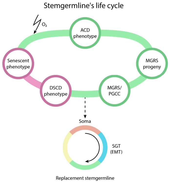
Figure 2.
The two organizational "germ" and "soma" levels of the cancer cell system - specifically, CSC germline and somatic BLK tumor cells - are connected by two reciprocal conversion "channels": the GST/MET channel (left) and the SGT/EMT channel (right). Top: Heterogeneous, secondarily stable ecDNA cell lines. Bottom: Stemness regeneration cycle carried out by the CSC- stemgermline.
Figure 2.
The two organizational "germ" and "soma" levels of the cancer cell system - specifically, CSC germline and somatic BLK tumor cells - are connected by two reciprocal conversion "channels": the GST/MET channel (left) and the SGT/EMT channel (right). Top: Heterogeneous, secondarily stable ecDNA cell lines. Bottom: Stemness regeneration cycle carried out by the CSC- stemgermline.
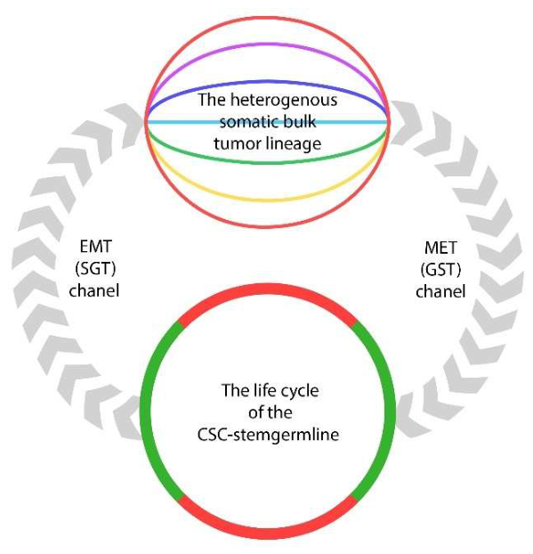
Disclaimer/Publisher’s Note: The statements, opinions and data contained in all publications are solely those of the individual author(s) and contributor(s) and not of MDPI and/or the editor(s). MDPI and/or the editor(s) disclaim responsibility for any injury to people or property resulting from any ideas, methods, instructions or products referred to in the content. |
© 2026 by the authors. Licensee MDPI, Basel, Switzerland. This article is an open access article distributed under the terms and conditions of the Creative Commons Attribution (CC BY) license (http://creativecommons.org/licenses/by/4.0/).
Copyright: This open access article is published under a Creative Commons CC BY 4.0 license, which permit the free download, distribution, and reuse, provided that the author and preprint are cited in any reuse.