
Submitted:
09 November 2026
Posted:
10 November 2026
You are already at the latest version
Abstract
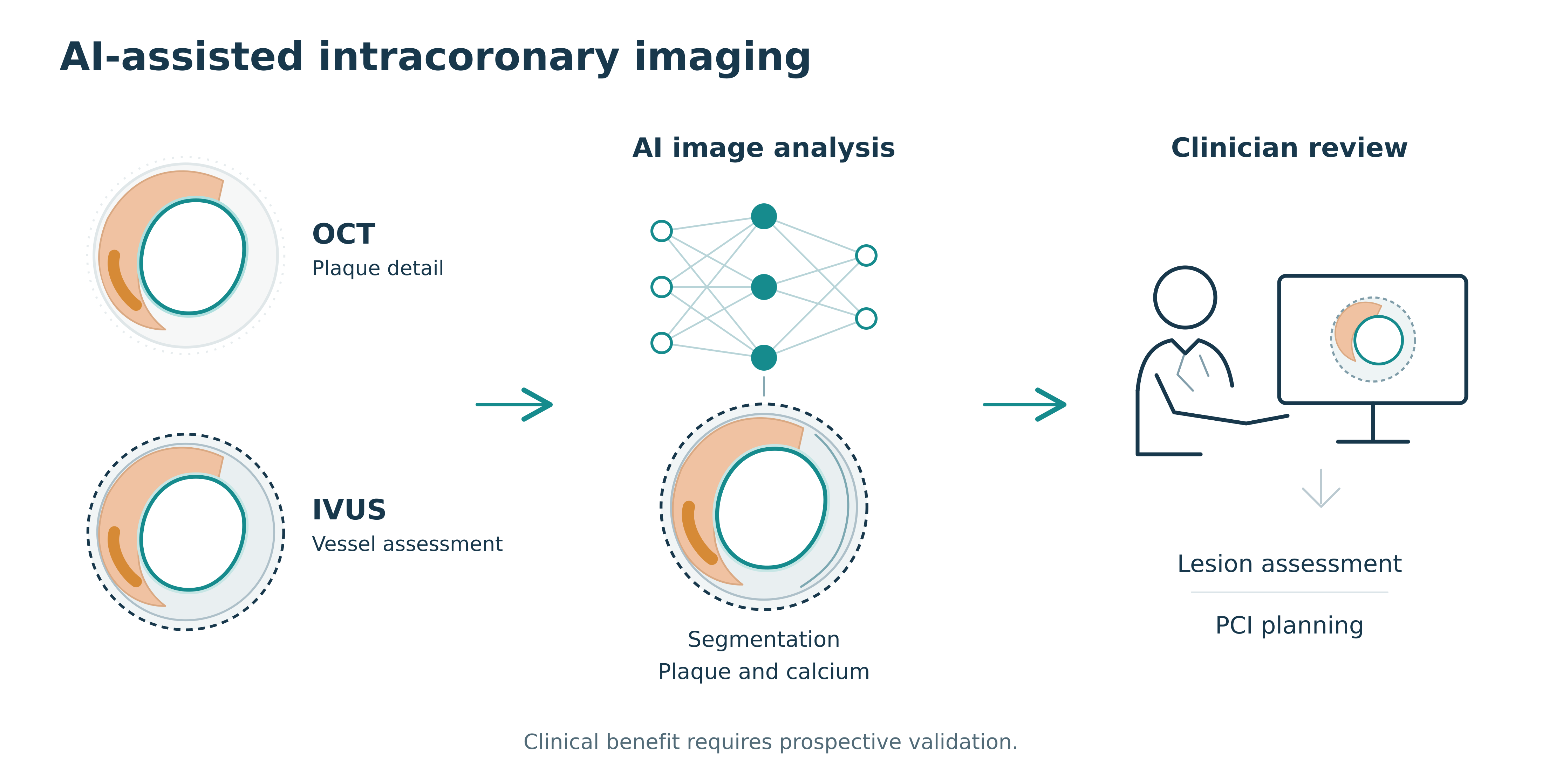
Artificial intelligence (AI) is being developed to analyze intracoronary optical coherence tomography (OCT) and intravascular ultrasound (IVUS) images. This narrative review examines applications in vessel segmentation, plaque and calcium characterization, plaque phenotyping, physiological assessment, and percutaneous coronary intervention (PCI) planning. OCT provides high-resolution assessment of superficial plaque and stent structures, whereas IVUS provides deeper vessel-wall information. Automated segmentation and quantitative measurements show promising technical performance, although results vary by model, imaging feature, and validation setting. Evidence for physiological inference and prognostic assessment remains largely observational, and diagnostic agreement or association with events does not establish that AI-guided care improves clinical outcomes. These tools may support individualized lesion assessment and procedural planning, but their clinical value requires prospective evaluation. At present, AI should be used as clinician-supervised decision support, with attention to image quality, uncertainty, and model limitations.
Keywords:
artificial intelligence
; optical coherence tomography
; intravascular ultrasound
; coronary artery disease
; plaque characterization
; percutaneous coronary intervention
Copyright: This open access article is published under a Creative Commons CC BY 4.0 license, which permit the free download, distribution, and reuse, provided that the author and preprint are cited in any reuse.