
Submitted:
07 November 2026
Posted:
10 November 2026
You are already at the latest version
Abstract
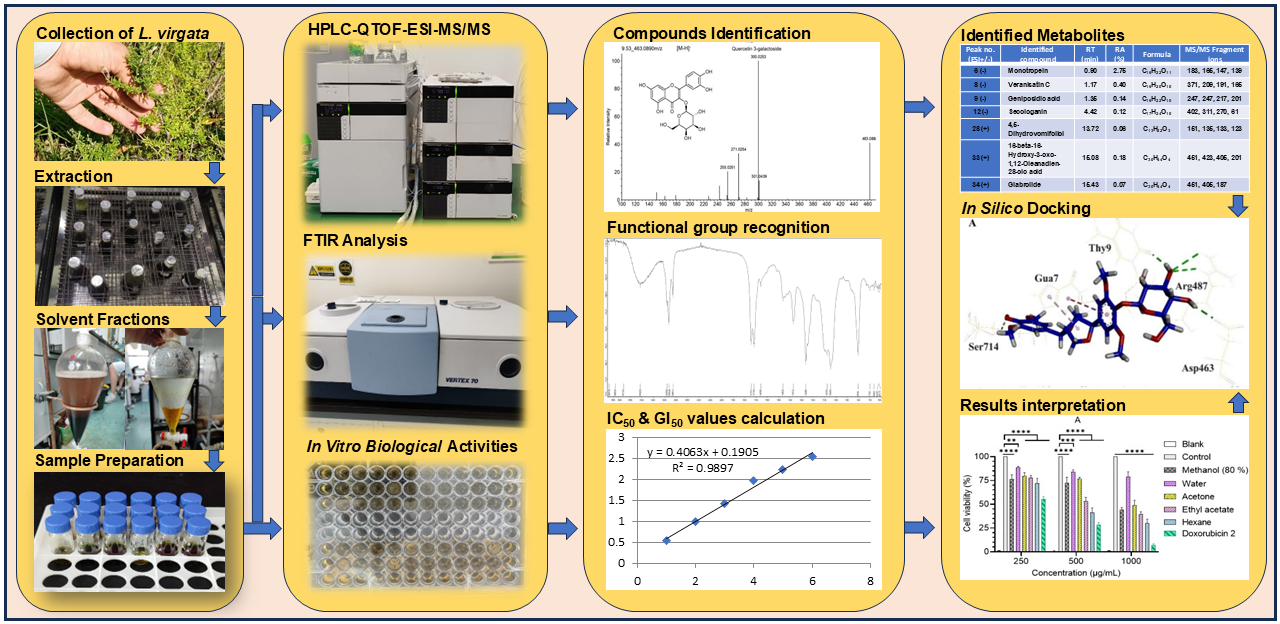
Despite its therapeutic potential, the metabolomic profile and biomedical efficacy of L. virgata remain entirely unexplored. This study was designed to comprehensively map the metabolomic profile of L. virgata leaves and evaluate their biomedical efficacy. The leaf extract was prepared in methanol (80%) and fractionated with hexane, ethyl acetate, acetone, and distilled water, and evaluated for biological activities, including antioxidant (DPPH), antimicrobial, and in vitro and in silico AChE inhibitory and cytotoxic effects. Phytochemical profiling was conducted using ATR-FTIR and UHPLC-ESI-QTOF-MS/MS (LC-MS). The crude extract showed the highest DPPH scavenging activity (88.11 ± 3.45%). Antibacterial and antifungal MIC values ranged from 375 ± 125 to 1500 ± 217 μg/mL. The acetone fraction showed strong AChE inhibition (IC50: 805.63 ± 5.87 μg/mL), while the hexane fraction showed notable anti-leukemic cytotoxic activity against U937 cells (GI50: 596.74 ± 2.24 μg/mL). Untargeted LC-MS profiling identified 194 compounds, dominated by terpenoids (69.32%), followed by lipids (7.93%), steroids (6.40%), flavonoids (4.13%), quinones (2.72%), coumarins (2.17%), and carbohydrates (1.87%). Oleanolic acid (15.84%) was the most abundant metabolite. In silico docking revealed strong binding affinities of 4,5-dicaffeoylquinic acid and Asperuloside for AChE, and Acanthoside B for DNA topoisomerase II. These findings indicate that L. virgata leaves are rich sources of bioactive Rubiaceae-specific metabolites, supporting their potential for developing natural therapeutic and nutraceutical products.
Keywords:
Leptodermis virgata
; antimicrobial study
; AChE inhibition
; leukemia U937 cell line
; LC-MS metabolomic profiling
; molecular docking
1. Introduction
Throughout human civilization, various diseases and disorders have been a persistent challenge for mankind [1,2]. Presently, cancer, diabetes, cardiovascular disorders, microbial infections and antibiotic resistance, Alzheimer's, and other memory-related disorders are the major causes of death globally [3,4,5]. Additionally, reactive oxygen species (ROS), metabolic abnormalities, and microbial infections are important factors in the onset, development, and progression of various health problems [2,6,7]. Pathogens, like viruses, bacteria, and fungi, are the main cause of various infectious diseases [8]. Similarly, ROS such as hydroxyl (OH), peroxide, and superoxide are formed during cellular respiration [9]. This causes numerous disorders in the body, such as cardiovascular disorders, carcinogenesis, diabetes, liver damage, the aging process, and neurodegenerative disorders [10,11]. In recent years, tremendous efforts have been made to develop several new medications and remedies for treating these life-threatening conditions [12]. A serious rise in drug resistance to various deadly pathogens, as well as several adverse effects from the excessive use of these medications, is also observed. Thus, there is always room for improvement [13,14,15]. In contrast, natural therapies, such as plant-based remedies and medications, are substitutes with reduced side effects of these synthetic drugs and are also a source of new and comparatively inexpensive medicines [16,17]. Therefore, the discovery and bioactivity evaluation of medicinal compounds from natural sources, particularly plants, has gained significant recognition within the scientific community [18]. According to the WHO, about 60% of the global population and 80% of the population in developing countries rely on medicinal herbs to treat various illnesses [19]. Currently, Japan, the UK, China, and the USA are the major markets for medicinal plants [20].
The healing and therapeutic properties of medicinal plants are due to their phytochemical compounds, also known as secondary metabolites. A variety of these compounds have been extracted from different plants, including saponins, tannins, phenolics, flavonoids, alkaloids, and essential oils [21,22]. These secondary metabolites exhibit numerous medicinal and pharmacological properties, including anti-inflammatory and antimicrobial activities. Furthermore, these bioactive plant compounds also have antidiabetic, anticancer, and anti-Alzheimer properties and are helpful in many other disorders [22,23,24].
Pakistan has an altitude range of 0-8611 meters and therefore has distinct climate zones with diverse biodiversity. It is blessed with 6000 species of plants, and almost 1000 of which have been reported to have medicinal properties [25,26]. Over the past 2-3 decades, researchers have investigated various Pakistani traditional medicinal plant species for their medicinal properties. For example, Caralluma tuberculata, locally known as Pamankay, is commonly used to treat Jaundice, stomach pain, dysentery, and other conditions. Another herb, Thymus serpyllum, is reported to be effective in poor vision, hepatic disorders, as an antiseptic, to aid wound healing, and to regulate the menstrual cycle. Similarly, Fagonia cretica has been abundantly used to cure abdominal pain, smallpox, asthma, vomiting, dysentery, typhoid, toothache, and stomach troubles. Furthermore, its aqueous extract has also been used to treat breast cancer [27].
However, some species remain underexplored or uninvestigated for their therapeutic potential. Leptodermis virgata is one of them; it is an erect, much-branched shrub up to 70 cm tall, confined mostly to the Western Himalayas. In Pakistan, it is distributed in the lower Himalayas (Mansehra and Murree), Kashmir, and in the dry stony soils of the Salt Range [28,29,30]. L. virgata is a member of the family Rubiaceae, which contains approximately 630 genera and about 13,500 species. Most of these possess a diverse range of metabolites, including terpenoids, alkaloids, anthraquinones, flavonoids, and other phenolic derivatives, with a vast range of medicinal properties, including antimicrobial, anti-inflammatory, and analgesic [28,31,32].
Despite its vast ecological presence, the medicinal and chemical virtues of L. virgata remain largely unexplored in the scientific literature. To date, only a single documented investigation has examined this species, reporting preliminary antibacterial and antifungal zone inhibition data for a crude ethanolic extract against Candida species and select pathogenic bacteria [33]. While this work provided the first evidence of the plant's antimicrobial activity, it was limited to a single crude solvent extraction and lacked comprehensive chemical characterization. Consequently, the literature remains completely void of information on fraction-specific bioactivities, quantitative multi-solvent enzyme inhibitory profiles, and the advanced metabolomic architecture of L. virgata. To address this gap and unveil the true therapeutic potential of this species, the present study was designed to provide a detailed metabolomic profile of the numerous primary and secondary metabolites present in the leaves of this novel plant, and to assess biological potential, including antioxidant and antibacterial activities, as well as in vitro and in silico acetylcholinesterase (AChE) inhibitory and cytotoxic effects. The research findings will provide a strong foundation for future use of L. virgata leaves in herbal products, including the herbal pharma and food industries.
2. Experimental
2.1. Chemicals and Solvents
Quercetin, gallic acid, ascorbic acid, linalool, Folin-Ciocalteu (FC) reagent, 1,1-diphenyl-2-picrylhydrazyl (DPPH), and other reference chemicals were sourced from Sigma-Aldrich Co., St. Louis, USA. Potato dextrose agar, nutritional agar, and Mueller-Hinton broth were obtained from Mark Chemicals. The microbial strains were provided by the Microbiology and Biochemistry departments of Hazara University, Mansehra, Pakistan. The substrates p-nitrophenyl-α-D-glucopyranoside (PNPG) and dinitrosalicylic acid (DNSA), along with the enzymes α-amylase and α-glucosidase, were acquired from Sigma-Aldrich in Germany. Acetylthiocholine iodide (AChI), acetylcholinesterase (AChE), and 5,5′-dithiobis (2-nitrobenzoic acid) (DTNB) were products of Sigma USA. The human leukemia U937 cell line was obtained from the Human Sciences Research Resource Bank, Human Sciences Foundation, Tokyo, Japan. Gibco Cell Culture Solutions (Thermo Fisher Scientific, USA) provided the supplement, comprising 100 mg/mL of streptomycin and 100 units/mL of penicillin, 1% (v/v) fetal bovine serum (FBS), Roswell Park Memorial Institute (RPMI) 1640 medium, and phosphate-buffered saline (PBS, pH 7.4), and the trypsin-EDTA (ethylenediaminetetraacetic acid) solution was made by Beyotime Biotechnology USA.
2.2. Plant Material
Leptodermis virgata plants were collected from Mang, a village 4 km from Balakot city in the district Mansehra, KPK, Pakistan. After the taxonomic examination, a voucher specimen of the plant was submitted to the Hazara University (HUP) Herbarium under the number HUP15656. The plant material was transported to the Foodomics laboratory at the University of Haripur. The leaves were washed with water after being separated from the stems and dried in a shed for two weeks. The dried leaves were ground into powder with a lab grinder (Model: HMD-177, BIOBASE China). The powder material was stored in an airtight container after sieving through a 100-micron mesh [34].
2.2.1. Extraction and Fractionation
100 g of the powdered leaves was separately mixed with 1 L of methanol (80%) in 250 mL conical flasks. The flasks were shaken at 28°C on an orbital shaker (Irmeco GmbH, Germany) for 24 h. Whatman No. 1 filter paper was used to filter the resultant crude extract, and the excess solvent was removed using a rotary evaporator (Model RE 301, BIOBASE, China) under reduced pressure at 40°C. The resulting semi-solid extract was then dried at -50°C in an Alpha 1-2 LD plus a freeze-dryer (Crist, Germany), and the percentage yield was determined [34]. One portion of the obtained crude extract was stored in airtight vials for metabolomic characterization and biological evaluation. The other part was fractionated with four different solvents, such as hexane, ethyl acetate, acetone, and distilled water, in increasing order of polarity, repeating a previously reported procedure [35].
2.3. Antioxidant Activity
The well-known DPPH free radical scavenging assay was used to estimate the antioxidant activity of the L. virgata leaf extract and its fractions. The 0.1 mM stock solution of DPPH in methanol and the three dilutions (200, 400, and 800 µg/mL) of the extracts were prepared in DMSO. For analysis, 125 µL of each sample solution and 500 µL of DPPH solution were mixed separately in a microtiter plate. The microplate was covered and kept in the dark for half an hour at 25°C. Finally, the UV/Vis absorbance was taken using a multimode microplate reader (BioTek Synergy HTX, USA) at 515 nm. Lower absorbance values for the test samples indicate higher antioxidant activity. Ascorbic acid was used as the standard, and a blank solution with DMSO instead of the extract served as a control [36].
2.4. Antimicrobial Activity
The antibacterial potential of the extracts under investigation was assessed using four clinically significant bacterial strains: Bacillus cereus (ATCC 21366) and Staphylococcus aureus (ATCC 29213), two Gram-positive bacteria, and Escherichia coli (ATCC 25922) and Pseudomonas aeruginosa (ATCC 27853), two Gram-negative bacteria. Similarly, two fungal strains, Candida albicans (ATCC 60193) and Candida parapsilosis (ATCC 7330), were evaluated for antifungal activity. The microdilution method was used to determine the minimal concentration of the extract that inhibits microorganism growth (MIC). Moxifloxacin was used as the reference standard for bacteria, and fluconazole was used as the reference drug for fungi [37,38].
2.5. Acetylcholinesterase Inhibition Activity
To evaluate the AChE inhibitory effect, in a 96-well microplate, 40 µL of the AChE enzyme solution (0.2 units/mL) and 300 µL of 0.1 M sodium phosphate buffer (pH 7.8) were mixed with 20 µL of the extract solution (200-800 µg/mL). The assay plate was incubated for 15 min. at room temperature, followed by the addition of 20 µL of 5 mM 5,5′-dithiobis (2-nitrobenzoic acid) (DTNB) and 20 µL of 10 mM acetylthiocholine iodide solution. The plate was again incubated for 10 min. to complete the reaction; DTNB and thiocholine reacted, and the enzyme hydrolyzed them, producing a colored product known as the 5-thio-2-nitrobenzoate anion. DMSO was used as the negative control, and galantamine as the positive control, and the proportion of AChE inhibition was calculated using the following formula [39].
2.6. Anticancer Activity
2.6.1 Cell Culture
The U937 leukemia cells were cultivated in RPMI 1640 medium to evaluate cytotoxic potential. 10% (v/v) fetal bovine serum, 1% (v/v) penicillin, and 100 units/mL of streptomycin were added as supplements. The cell culture was incubated with 5% CO2 at 37 °C. Before the experiment, the cultures were separated using a 0.25% trypsin-ethylenediaminetetraacetic acid (EDTA) solution, sprayed with phosphate-buffered saline (PBS, pH 7.4), spun at 1000 rpm for five minutes, and then returned to suspension [40].
2.6.2 Cell Counting Kit-8 (CCK-8) Assay
For the assay, a 96-well plate was seeded with around 5000 cells per well and incubated for 24 h at 37 °C in humidified air with 5% CO2. The culture medium was supplemented with extract solutions at concentrations of 250, 500, and 1000 μg/mL, which were then diluted with the corresponding culture fluid. Each measurement was taken in triplicate, and the assay plates were incubated overnight under identical conditions. As a standard medication, an injection of doxorubicin hydrochloride (2 mg/mL) was co-incubated. The CCK-8 (10 μL/well) solution was added, and the assay plates were incubated for an additional two hours under the same conditions. Lastly, absorbance at 450 nm was used to quantify the optical density and percentage cell viability for each concentration, and GI50 values were calculated [40].
2.7. Fourier Transform Infrared (FTIR) Spectrometry
An Attenuated Total Reflectance Fourier Transform Infrared (ATR-FTIR) spectrophotometer (Vertex 70 series, Bruker Co. Ltd., Germany) was used to record the infrared spectra of a solvent-free crude extract of L. virgata leaves. With a resolution of 4 cm−1 and 32 scans, the scan range was 600–4000 cm-1. Using PeakFit 4.04 software, all spectra were examined to find and compare peaks, and corresponding functional groups were identified [41].
2.8. Metabolite Profiling by Liquid Chromatography-Mass Spectrometry (LC-MS)
A previously described procedure was used to perform untargeted metabolite profiling of the methanolic (80 %) extract of L. virgata leaves. using an Ultra-High-Performance Liquid Chromatography Quadrupole Time-of-Flight Mass Spectrometry (UHPLC-ESI-QTOF-MS/MS, simply LC-MS). An HPLC system with an electrospray ionization source (LC-30A, Shimadzu, USA) was used for this study. The extract sample was centrifuged at 5 °C and 10,000 rpm for 10 min, filtered through a 0.22 μm syringe filter, and 2 µL was loaded into an autosampler. The LC system was equipped with a Hypersil GOLD RP C18 column of Thermo Fisher Scientific, USA (length 100, diameter 2.1 mm, with 1.9 µm particle size, and pore size 80 Å) and an electrospray ionizer (ESI), coupled to an X500R QTOF MS/MS (AB SCIEX, USA). A binary gradient mobile phase system, with solvent A comprising formic acid and Milli-Q water (0.5:99.5 v/v respectively) and solvent B made up of formic acid and acetonitrile (0.1:99.9 v/v). Each solvent mixture was vacuum-degassed for 15 min. at 25 °C, and the solvent flow rate was set to 0.3 mL/min. For MS/MS fragmentation, an automatic mode with collision energies of 20, 40, and 60 eV was employed, and peak identification was carried out in both positive and negative ionization modes with the m/z range from 100 to 1500 amu. The source temperature was 550 °C, and the flow pressures for the carrier and the curtain gases were maintained at 55 and 35 psi, respectively. For ESI- and ESI+, the voltages were 5500 V and 4500 V, accordingly [35].
2.8.1. Data Analysis
After baseline filtering, peak identification, peak integration, alignment, and retention time correction, the LC-MS raw data were processed into a standard format using the Progenesis QI software (Waters, Milford, USA). After that, the data matrix containing peak intensities, retention times, m/z values, and metabolite names was exported for examination. The Human Metabolite Database (HMDB) (http://www.hmdb.ca/), METLIN (https://metlin.scripps.edu/), and a self-compiled Majorbio Database (MJDB) from Majorbio Biotechnology Co., Ltd. (Shanghai, China) were the three databases examined for metabolite identification.
2.9. Molecular Docking Study
In silico docking is an effective and cost-efficient method for screening lead compounds. Therefore, to further confirm the in vitro cytotoxic and AChE-inhibitory effects, docking studies were conducted on certain bioactive metabolites identified in the L. virgata leaf extract. ChemDraw Pro 8.0 was used to draw the ligand structures, and ACD (3D viewer) software was employed for optimization. The Protein Data Bank was searched to obtain the crystal structures of AChE and human DNA topoisomerase II (PDB IDs 1EVE and 5GWK, respectively). The native ligands were redocked to evaluate docking reliability, and their root-mean-square deviation (RMSD) was within a suitable range (< 2.0 Å). Prior to the interaction study, water molecules along with conventional inhibitor ligands were removed, and polar hydrogens were incorporated into the protein structures. The enzyme grid box center was set at x: 23.35, y: 38.58, and z: 59.57, with dimensions of 40 × 40 × 40 points and a spacing of 0.375 Å. Docking was performed using AutoDock Vina software, and Biovia Discovery Studio Visualizer was used to display ligand-enzyme interactions (BIOVIA, D.S. 2017) [37].
2.10. Statistical Analysis
The experimental readings were taken in triplicate, and the data were recorded as mean ± standard deviation (SD). The R2 (coefficient of determination) was calculated with Microsoft Excel 2020. For LSD for mean values, one-way ANOVA was used in Statistics 8.1. Except for cytotoxicity, for which the significance of differences among means (p < 0.05) was determined using two-way ANOVA, with a post hoc test, on GraphPad Prism 9.0.
3. Results and Discussion
3.1. Extract Yield and Antioxidant Activity
Ethyl acetate produced the highest yield (12.41%) among all the fractions, while the hexane fraction accounted for just 6.53% of the dried plant material utilized. The crude extract yield was 14.40%. Comparably, at 800 μg/mL (the maximum tested concentration), the crude extract exhibited the highest antioxidant potential (88.11 ± 3.45%), followed by the acetone (76.98 ± 2.26%) and ethyl acetate (71.78 ± 3.18%) fractions (Table 1). The reference standard ascorbic acid (1 mg/mL) had an antioxidant activity of 93.65 ± 1.07%, while the hexane fraction had the lowest antioxidant activity (62.84 ± 5.42%). The strong ability of the crude extract and its acetone and ethyl acetate fractions to scavenge free radicals is likely due to the presence of considerable amounts of antioxidative compounds like phenols, flavonoids, and tannins. Additionally, several metabolites identified in the metabolite profiling of the studied extract (section 3.7) are also reported to have antioxidant properties. Despite the lack of prior data for comparable research on L. virgata, our findings are similar to those published for other plants in the same family [42,43].
3.2. Antimicrobial Activity
3.2.1. Antibacterial Activity
The MIC method was used for the assessment of antimicrobial activity. This is a gold standard for in vitro antimicrobial susceptibility testing that provides an exact and quantifiable value for the lowest concentration of an antimicrobial drug required to limit the observable microbial growth [44]. According to the results (Figure 1A), the hexane extract exhibited intermediate MIC values (750 ± 00 to 1000 ± 217 μg/mL) for all tested bacteria, while the acetone fraction displayed the lowest activity (highest MIC values: 1000 ± 217 to 1375 ± 125 μg/mL). The crude methanolic extract (MIC = 500 ± 125 μg/mL) and its aqueous fraction showed stronger efficacy against B. Cereus (MIC = 375 ± 125 μg/mL). However, the ethyl acetate fraction presented the lowest MIC against the other two (Gram-negative) bacteria, E. coli (MIC = 750 ± 125 μg/mL) and P. aeruginosa (MIC = 625 ± 217 μg/mL). The standard antibacterial drug, moxifloxacin, had the least bactericidal impact in terms of MIC (6.25 ± 1.25 μg/mL to 8.75 ± 125 μg/mL).
3.2.2. Antifungal Activity
According to the results (Figure 1B), the acetone fraction displayed the maximum fungicidal activity (MIC = 750 ± 125 μg/mL) against C. parapsilosis. At the same time, the hexane fraction was highly effective against C. albicans, with a MIC = 625 ± 125 μg/mL. In contrast, the crude methanolic extract exhibited the least activity (MIC = 1250 ± 125 μg/mL) for C. parapsilosis, and the distilled water fraction exhibited the lowest MIC (1500 ± 217 μg/mL) against C. albicans. The conventional fungicidal drug, fluconazole, exhibited the lowest MIC values for both C. albicans (7.5 ± 1.25 μg/mL) and C. parapsilosis (11.25 ± 1.25 μg/mL).
The metabolomics study (section 3.6) revealed that the studied extracts contain significant amounts of various compounds that may contribute to their potent antimicrobial properties. It is worth mentioning that there have been no prior reports of L. virgata's antibacterial activity. These findings are highly consistent with the antimicrobial properties seen in extracts from some other plants of the Rubiaceae family [31,45].
3.3. AChE Inhibition Study
Alzheimer's is a degenerative neurological condition associated with reduced acetylcholine (ACh) neurotransmitter levels and the death of cholinergic neurons in the brain. The mainstay of Alzheimer's treatment involves inhibiting AChE, which degrades ACh. [46]. The present results of in vitro AChE inhibition assays (Figure 2) show that, with increasing concentration, the enzyme inhibition activity increased considerably for most of the extract fractions of L. virgata leaves (Figure 2A). At 1000 μg/mL (the highest evaluated concentration), acetone and ethyl acetate were the most active fractions; only these two inhibited more than 50% of the enzyme, with IC50 values of 805.6 ± 5.9 μg/mL and 886.2 ± 3.1 μg/mL (Figure 2B), respectively and the IC50 value for the pure and standard drug galantamine was 39.8 ± 1.1 μg/mL. The acetone fraction possessed the maximum activity (56.5 ± 0.4%), whereas the crude methanolic extract showed the lowest AChE inhibitory activity (33.6 ± 1.2%) at 1000 μg/mL. These findings align with those from similar extracts of Chebula fruits (which also belong to the same plant family), except that the aqueous fraction showed lower activity in our study [47]. The results also exhibit a significant correlation with the AChE inhibitory potential reported for many other plants belonging to the same family, Rubiaceae [42].
Metabolomic analyses by LC-MS (Section 3.7) confirmed the presence of several such compounds reported to have AChE inhibitory potential. These include 4-Methylesculetin [48], Asperuloside [49], arjunolic acid [50,51], Hederagenin [52,53], morin [54], and oleanolic acid, etc. [55]. The overall finding indicates that the acetone and ethyl acetate fractions predominantly shared these bioactive compounds, with potential AChE inhibitory activity. Furthermore, in silico studies (section 3.7.1) have validated the ability of some of these compounds to inhibit AChE.
3.4. Anticancer Activity
The cytotoxic activity (Figure 3) showed that all the extract fractions showed a positive response against the U937 human leukemia cell line; the decrease in viability of cancer cells was significantly increased with a rise in dose concentration (Figure 4). Among all tested extracts, the aqueous fraction presented the least cytotoxic effects, i.e., about 21 % reduction in cell viability at up to the maximum tested concentration (1000 μg/mL). Subsequently, the acetonic fraction exhibited lower cytotoxicity (Figure 4A), with a GI50 of 986.87 ± 3.73 μg/mL (Table 2). However, the crude methanolic extract and its ethyl acetate and hexane fractions presented substantial cytotoxic potential; among these, the hexane fraction (GI50: 596.74 ± 2.24 μg/mL) was most potent. Its activity increased substantially with extract concentration and was close to that of the standard drug Doxorubicin (Figure 4B), which exhibited the highest anticancer potential (GI50: 410.67 ± 2.07) at the same concentrations.
As far as we can tell, this is the first study on the cytotoxic properties of leaf extracts of L. virgata. Nonetheless, our results closely relate to the previously published studies on other plant extracts for the same leukemia U937 cell line [56,57]. Furthermore, it is worth highlighting that the extract fractions in hexane and ethyl acetate in the current study demonstrated a meaningfully superior cytotoxic activity compared to other plants of the Rubiaceae family reported by different researchers [58,59].
Metabolomic profiling (Section 3.7) revealed that the crude leaf extract of L. virgata is highly rich in biologically active compounds of various classes that possess anticancer properties. These include phenols like 4-hydroxycinnamic acid [60] and Gingerol [61]. Several flavonoids, for instance, Isorhamnetin [62], Luteolin [63], Morin [54], Tamarixetin [64], Tricin [65]. Furthermore, many other identified compounds, such as Quercetin and its derivatives [62], Kaempferol and its derivatives [66], Steroids such as 27-Hydroxyiso-mangiferolic acid [67] and Stigmasterol [68] have anticancer potential. Similarly, several terpenoids, such as Beta-Elemonic acid [69], Ganoderic acid Y [70], Ganoderiol F [71]. Ursolic Acid, Oleanolic acid [72] and many others, Coumarins [73], Quinones [74], some lipids [75], Lignans [76], and carbohydrates [77] have also been reported for different anticancer activities.
3.5. Fourier Transform Infrared Spectrometry (FTIR)
A quick and non-invasive IR technique called Attenuated Total Reflection Fourier Transform Infrared Spectroscopy (ATR-FTIR) was used for the evaluation and identification of the particular characteristics of the extract [78]. The ATR-FTIR spectrum of L. virgata leaves’ extract revealed several functional groups through characteristic absorption peaks associated with different stretching vibrations. Figure S-1 (Supplementary Materials Figure 1) shows the spectrum, with frequency assignments for various peaks and the identification of multiple stretching vibrations (functional groups) representing different classes and groups of phytochemicals, as listed in Table 3. The results show O–H stretching at 3422, 2959, and 2924 cm-1, indicating the presence of carboxylic acids, alcohols, and phenolic substances, and the usual C–H stretching of alkanes was seen as a band at 2851 cm-1. While the C=C stretching at 1656 cm-1 indicates aromatic compounds, the C=O stretching at 1716 cm-1 and 1741 cm-1 suggest aldehydes, ketones, and organic acids. The band observed at 1260 cm-1 corresponds to C–H wagging of alkyl halides (probably fluoride). Next, the C–N stretching, consistent with aromatic amines, was associated with the band at 1372 cm-1. Similarly, the C–N stretching of aliphatic amines was attributed to the band at 1045 cm-1, while ethers and esters were related to the C–O stretch at the same wavelength. The N-H stretching vibrations in amines were associated with a strong peak at 804 cm-1, and finally, the weaker bands at 702, 626, and 613 cm-1 were assigned to alkyl halides (typically chlorides and iodides). Overall, these results are consistent with earlier findings for a variety of plant extracts [35,79,80].
3.6. Metabolite Profiling by Liquid Chromatography Mass Spectrometry (LC-MS)
UHPLC-ESI-QTOF-MS/MS (or simply LC-MS) was used to analyze the phytochemical composition of the 80% methanolic extract of L. virgata leaves to discover as many metabolites as feasible. Figure 5 shows the total ion chromatogram in both negative-ion (Figure 5A) and positive-ion (Figure 5B) modes of analysis, indicating that superior resolution was achieved in the positive-ion mode. The retention time, parent ions (adducts), secondary MS fragment ions, and m/z values of each metabolite were compared with the databases, and a total of 194 compounds with accurate formulas and masses were identified. Figure 6 represents the variation in metabolite composition of L. virgata leaf extract. Including the percentage of primary and secondary metabolites in total identified metabolic content (Figure 6A), percentage of different classes of primary (Figure 6B) and secondary (Figure 6C) metabolites, and overall percentage of all classes belonging to both primary and secondary metabolite groups identified in the evaluated extract (Figure 6D).
3.6.1. Carbohydrates, Amino Acids, and Vitamins
Carbohydrates are vital for cellular respiration and energy production and facilitate cell recognition and communication. They also supply essential nutrients to the body and serve as one of the main calorie sources; fibers are a type of carbohydrate that aids digestion and cholesterol regulation [83,84]. We found that 1.87% of the metabolites identified in the L. virgata leaf extract are carbohydrates. These comprise 12 compounds (Table 4); 6 were detected in positive (ESI +) and 6 in negative (ESI -) ionization modes. Peak 11 (ESI -), identified as Asperuloside (C18H22O11, RT 3.27 min., [M+FA-H]-, m/z 459.1151), was found as the most abundant carbohydrate (RA 4.19%). Next, at RT 35.43 min. the peak 119 (ESI +) was identified as Neomycin (C23H46N6O13, [M+Na]+, m/z 637.3017), with an RA of 1.11%, followed by Gentiobiose (Peak 2 ESI-, C12H22O11, RT 0.80 min., [M-H]-, m/z 449.1302), which had an RA of 0.72%. The remaining carbohydrates were found in relatively low quantities.
Amino acids are involved in protein synthesis, enzyme activity, cell signaling, and the formation of neurotransmitters and hormones [85]. The analyzed extract revealed two amino acids (Table 4), including peak 1 ESI+ recognized as Betaine (C5H11NO2, RT 0.82 min., [M-H]-, m/z 118.0585) and Peak 22 ESI-, identified as Ustiloxin D (C23H34N4O8, RT 13.13 min., [M-H]-, m/z 493.2298). Both amino acids were found in relatively low quantities, i.e., 0.17% and 0.06%, respectively.
Vitamins are essential for immune function, energy production, and cellular health. Although small amounts are necessary for proper metabolic function and overall health, the body is unable to make sufficient amounts; hence, these should be regularly provided by diet [86,87]. The peak 124 ESI + (Table 4) was confirmed as vitamin 25-hydroxycholecalciferol (C27H44O2, RT 37.01 min., [M+H-H2O]+, m/z 383.3301, RA 0.46 %), which is an active form of vitamin D3 with anti-inflammatory, antioxidant, and immunomodulatory properties [88].
3.6.2. Lipids
Lipids play important functions in energy storage, membrane construction, and signaling pathways [89]. The total metabolic content of L. virgata leaf extract contained the second-highest amount of lipids (7.93%). Among the total 194 recognized compounds, 32 were confirmed as lipids (Table S1). Fatty acid Fa (18:2+1O), discovered at RT 21.29 min. for peak 34 (ESI -) with the chemical formula C18H32O3 ([M-H]-, m/z 295.2289), was found as a predominant lipid (RA 1.59%). [90]. Next, peak 114 (ESI +), identified as a monoacylglyceride (18:0/0:0/0:0), molecular formula C21H42O4 (RT 34.47 min., [M+H]+, m/z 359.3154), was present at 1.43 % (RA). Subsequently, peak 94 (ESI +), confirmed as Glyceryl palmitate (C19H38O4, RT 30.00 min., [M+H]+, m/z 331.2845, RA 1.29 %). The other lipids present in relatively good quantities are Araliacerebroside (RA 0.70 %), 16-hydroxyhexadecanoic acid (RA 0.65 %), Elaidic acid (RA 0.52 %), and 2-Hydroxylinolenic acid (RA 0.46 %). The other lipid compounds were present in relatively small amounts. The literature attests to the biological significance of the number of discovered lipids.
3.6.3. Phenols and Phenolic Acids
Phenols are known for their anti-inflammatory, antioxidant (scavenging free radicals), antimicrobial, and anticancer (by inducing apoptosis) properties, and are sometimes also employed as disinfectants. Phenols are aromatic alcohols, meaning that a hydroxyl group is directly connected to a benzene ring [91,92,93]. The results (Table 5) show that the peak 149 ESI + ([M+H-H2O]+) at RT 46.80 min was identified as Gingerol with molecular formula C17H26O4, RA 0.23%, and m/z 277.1803. Next, the peak at 25 ESI + ([M+CH3OH+H]+) at RT 11.42 min was Sesamolinol, with molecular formula C20H20O7, m/z 405.1537, and RA 0.03%. Similarly, at RT 34.62 min, peak 116 (ESI +, [M+H-2H2O]+) was Mangiferdesmethyl-ursanone (C29H48O2, m/z 393.3509, RA 0.03 %), which is an aliphatic cyclic alcohol. Among phenolic acids, two quinic acid derivatives including peak 10 (ES -, [M-H]-, RT 2.86 min.) as Caffeoylquinic acid (C16H18O9, m/z 353.0885), which was present in abundance (RA 1.19 %), and peak 18 (ESI -, [M-H]-, RT 11.12 min.) as 4,5-Dicaffeoyl quinic acid (C25H24O12, m/z 515.1202, RA 0.11 %). The other phenolic acids, including 4-hydroxycinnamic acid, Trans-Caffeic acid [apiosyl-(1->6)-glucosyl] ester, and Dibutyl phthalate, were also identified with their RT, m/z, precursor ions, and MS/MS fragments. Most of the identified phenolic compounds possess noteworthy medicinal and therapeutic properties [91,92,93].
3.6.4. Flavonoids and Quinones
The antioxidative properties of flavonoids are widely recognized; they protect cells from oxidative damage and decrease the chances of various chronic illnesses, including heart disease and some types of cancer [94,95]. 19 compounds (4.13 %) of the total identified metabolites in the evaluated extract of L. virgata leaves were found to be flavonoids (Table 6). Among those, four were quercetin and its derivatives, among which peak 17 (ESI –, [M-H]-, RT 11.12 min.), recognized as Quercetin 3-galactoside (C25H24O12, m/z 463.0890), was the most abundant flavonoid (RA 1.47%). Peak 24 (ESI -, [M-H]-, RT 13.85 min.) was recognized as Kaempferol (C15H10O6, m/z 285.0411, RA 0.58 %), and the peaks 18 (ESI +), 19 (ESI +), and 19 (ESI -) were confirmed as derivatives of Kaempferol. Among the others, Morin (peak 47, ESI +, RA 0.20 %) and Isorhamnetin (peak 25, ESI -, RA 0.42 %) were two flavanols.
Numerous fruits and vegetables contain morin, which is also a component of certain herbal remedies. Isorhamnetin is an essential monomethoxyflavonol and one of the main ingredients in Ginkgo biloba leaf extract [96]. Likewise, peaks 23, 27, and 28 (ESI-) and 29 (ESI+) were identified as flavones, specifically Glycitein, Tricin, Cirsimaritin, and Tamarixetin, respectively. Figure 7 shows the structure, m/z value, and MS/MS fragmentation patterns of compounds Alatanin C and Quercetin 3-galactoside. Similarly, the other flavonoid compounds were identified by their parent ions (adducts), MS/MS fragment ions, and m/z. Quinones are essential for biological processes like cellular respiration and electron transport. They also have antiparasitic, anticancer, and antibacterial properties [97]. Additionally, at RT 14.76 min, peak 36 (ESI +, [M+H]+) was identified as Decylubiquinone, with molecular formula C11H16O2, m/z 181.1214, and RA 0.48 %. Likewise, at RT 19.29 min, peak 52 (ESI +, [M+H]+) was recognized as Ubiquinol (QH2), molecular formula C14H20O4. [C5H8]n, m/z 321.2059 with RA 0.14 %. were identified as quinones.
3.6.5. Amines, Lignans, and Others
Amines are organic compounds derived from ammonia, naturally present in proteins, some vitamins, and hormones [98]. Three peaks at 39, 41, and 151 (Table S-2), corresponding to 2-aminooctadecane-1,3-diol, Phytosphingosine, and L-Histidinol, respectively, are amines. All were identified in positive ion mode. Among these, at RT 47.44 min, L-Histidinol (C6H11N3O, [M+H-H2O]+, m/z 124.0863) was relatively present in abundance (RA 1.43%). Lignans are polyphenolic compounds that display antioxidant and anticancer properties and enhance cardiovascular health [99]. The two lignans identified for peaks 20 (ESI -, [M-H]-) and 21 (ESI +, [M+NH4]+) are Acanthoside B (C28H36O13, RT 10.76 min., m/z 579.2095, RA 0.52%) and Myristicanol A (C23H30O8, RT 10.90 min., m/z 452.2278, RA 0.04%).
The other compounds, like for peak 5 (ESI +, [M+NH4]+), Cerulenin (C12H17NO3, RT 0.89 min., m/z 241543, RA 0.04%), is an epoxide, whereas peak 7 (ESI -, [M+FA-H]-), Gardenoside (C17H24O11, RT 1.01 min., m/z 511.3418, RA 0.05%), is an iridoid. A fatty acid ester, myristic acid ethyl ester (C16H32O2, RT 32.65 min.), is identified by comparison with peak 39 (ESI -, [M+H]+, m/z 255.2338, RA 0.50%). In the same way, peaks 35, 115, 146, and 153 (ESI +) are identified as Hydroxydestruxin B, Pyro-pheophytin B, Nervonoylacetone, and (5Z,8Z)-1,5,8-Heptadecatriene, respectively. They are members of the depsipeptides, olefins, ketones, and styrenes.
3.6.6. Benzenoids, Benzofuran, Benzopyran, and Coumarins
The LC-MS study revealed that some compounds (present in relatively small quantities in the evaluated extracts) belong to Benzenoids, Benzofuran, Benzopyran, and Coumarins (Table S-2). Out of these, peak 6 (ESI +, [M+H]+), identified as Noreugenin (C10H8O4, RT 1.01 min., m/z 193.0492, RA 0.14 %), and peak 8 (ESI + [M+H]+), known as 7-Hydroxy-2-methyl-4-oxo-4H-1-benzopyran-5-acetic acid (C12H10O5, RT 3.29 min., m/z 235.0601, RA 0.29 %), were two benzopyrans. Similarly, among other compounds, peak 32 (ESI +, [M+H]+) is a benzofuran and was recognized as Dihydroactinidiolide (C11H16O2, RT 14.76 min., m/z 181.1224, RA 0.48 %). And peak 41 (ESI -, [M+Na-2H]-), identified as Idarubicin (C26H27NO9, RT 34.40 min., m/z 518.1443, RA 0.03 %), while peak 63 (ESI +, [M+Na]+) is identified as 5-(16-Heneicosenyl) resorcinol (C27H46O2, RT 21.08 min., m/z 425.3407, RA 0.11 %). However, peaks 9, 10, 11, 12, and 46 (ESI +) are coumarins. Among these, peak 46 ([M+NH4]+), identified as Pectachol (C26H34O6; RT 17.79 min, m/z 460.2694), was found in a considerable quantity (RA 1.99%). The MS/MS fragments and structures of Idarubicin and Pectachol are presented in Figure S-2. Dihydroactinidiolide has a tea-resembling smell and is often used as a fragrance [100], and 5-(16-Heneicosenyl)resorcinol possesses an anticancer effect [101].
3.6.7. Steroids
Steroids play vital roles in biology; they are a major component of cell membranes, which influence membrane fluidity. They also function as signaling molecules that regulate metabolism, inflammation, and immunological processes. Additionally, steroids, such as estrogen and testosterone, are essential for reproductive and developmental functions [102,103]. The metabolomic analysis of L. virgata leaves identified 29 phytochemicals as steroids and their derivatives (Table S-3). All these steroid compounds were observed in positive ionization mode and accounted for 6.40% of the total identified metabolites. The peak 138 (ESI +, [M+H]+), identified as Schleicherastatin 5 (C29H48O3, RT 40.72 min., m/z 445.3674), was present at 1.77 % (RA). At RT 39.92 min., the peak 118 (ESI+, [M+NH4]+) was confirmed as (3beta,22R,23R,24S)-3,22,23-Trihydroxystigmastan-6-one, (C30H50O3, m/z 463.3778, RA 1.04 %) also called 28-Homoteasterone. Subsequently, compounds identified for peaks 72, 73, and 143, namely Etiocholanolone (RA 0.48 %), 5-Androstenediol (RA 0.69 %), and Corbisterol (RA 0.63%), were present in substantial quantities.
Schleicherastatin 5 is a derivative of a hydride of stigmastane and has been reported in Schleichera oleosa [104] and 28-homoteasterone is also a naturally occurring brassinosteroid in plants [105]. Likewise, Etiocholanolone is a 5β-androstane produced by the metabolism of testosterone and is used to improve adrenal cortex and bone marrow function and to stimulate the immune system in neoplastic disease [106]. Androstenediol is a neurosteroid that may shield the brain by lowering inflammation and cell death markers in the brain, as well as the harmful effects of some drugs on dopamine levels. Etiocholanolone is one of the precursors in the synthesis of testosterone and also functions as an agonist for estrogen receptors, specifically the beta receptor [107,108]. Corbisterol, also known as D7-stigmasterol, is a stigmasterane derivative reported to have antioxidant and anti-inflammatory activities [109]. Other steroids, such as Cinobufagin, Campesterol, Momordenol, Cycloeucalenone, 7-alpha-hydroxycholesterol, Ouabain, Stigmasterol, Corbisterol, Campestanol, and Schottenol, were also identified in the same way by confirming their m/z, precursor ions, and secondary MS fragments.
3.6.8. Terpenoids
Terpenoids are probably the largest group of phytochemicals produced by most plants. They possess a wide range of biological activities against malaria, inflammation, tuberculosis, and numerous infectious diseases, as well as being ingredients of various Indian and Traditional Chinese medicines [110,111]. With the increase in cancer and diabetes patients in the modern world, terpenes also have the potential to serve as anticancer and antidiabetic agents. Recent studies on plant-based compounds have explored a variety of terpenoids effective against metastasis in cancer [111].
The LC-MS study shows that L. virgata is also one of those plants that possess terpenoids as the majority among phytochemical classes, i.e., comprising about 69.32 % of the total identified metabolites. Of all the metabolites found in L. virgata, at RT 28.18 min., peak 89 (ESI +, [M+H-H2O]+), which was recognized as oleanolic acid (C30H48O3, m/z 439.3567), was present in the greatest concentration (RA 15.84%). Next, the terpenoids present in significant quantities include, at RT 27.60 min., peak 86 (ESI +, [M+H-H2O]+), was identified as Alpha-Elemolic acid (C30H48O3, m/z 439.3568, RA 6.55%), and at RT 28.20 min, peak 90 (ESI +, [M+H]+) as Ursolic acid (C30H48O3, m/z 457.3670, RA 4.85%). Figure 8 shows the secondary MS fragments of these two (Alpha-Elemolic acid and Ursolic acid). Likewise, at RT 26.52 min., the peak 84 (ESI +, [M+H]+), Betulonic acid (C30H46O3, m/z 455.3517, RA 3.59 %), and at RT 0.90 min., peak 6 (ESI -, [M-H]-) Monotropein (C16H22O11, m/z 389.1097, RA 2.75 %) were present in substantial quantities. The other terpenoids, such as Ganodermic acid Jb (RA 2.64 %), Ganoderiol I (2.51 %), Beta-carotene (RA 2.20 %), Desoxoglabrolide (1.31 %), Beta-Elemonic acid (1.13 %), Ganoderiol F (1.01 %), and Maslinic acid (0.51 %), etc., were present in considerable amounts (Table 7). The secondary MS fragmentation patterns and structures of two of these, i.e., Maslinic acid and Ganoderiol I, are shown in Figure S-3.
Among all the identified metabolites in the examined extracts of the L. virgata leaves, Oleanolic acid was the most prevalent compound. It is a pentacyclic triterpenoid that has been shown to have several pharmacological and therapeutic properties, including antioxidant, anti-inflammatory, and anticancer effects [112,113]. Subsequently, a weak basic triterpenoid, Alpha-elemolic acid, alternatively referred to as alpha-elemolate, was the metabolite found in the second-highest quantity [114]. Some studies indicate its presence in some plant oils and extracts with anti-inflammatory and anti-Alzheimer activities [115,116].
Ursolic acid, the next prevalent compound, is likewise known for therapeutic potential, including anticancer activity through multiple biological processes [117]. Similarly, Betulonic acid is also a potential anticancer compound. For instance, Betulonic acid derived from Toona sinensis and Belamcanda chinensis exhibited significant cytotoxic effects against a number of cancers, including MGC-803 gastric cancer, MCF-7 breast cancer, and PC3 prostate cancer. Furthermore, it can also induce apoptosis in different cancer cell types. [118]. In comparison with the literature, many other identified terpenoids also have noteworthy biological and pharmacological importance.
3.7. Molecular Docking Studies
3.7.1. Docking Analysis on AChE
4,5-Dicaffeoyl quinic acid was found to be the prime metabolite among these docked compounds. According to the 3D pictures (Figure 9) of molecular modeling of the evaluated compounds against AChE, 4,5-Dicaffeoyl quinic acid (Figure 9A) formed a conventional H-bond with Tyr70 and two π-π stacking interactions with Tyr334 and Trp279 in the peripheral anionic site (PAS) of AChE. Furthermore, a hydrogen bond was also observed with Glu199 in the acyl pocket and Gly119 in the oxyanion hole. With an RMSD of 1.14 Å, 4,5-Dicaffeoyl quinic acid demonstrated a strong binding energy (BE-10.44 kcal/mol). Similarly, the Caffeoyl quinic acid (Figure 9B) formed several hydrogen bonds and π-interactions with important residues in the PAS and CAS (catalytic anionic site) of AChE. It established a traditional H-bond with Tyr70 and two π-π linkages with Trp279 and Tyr334. Additionally, hydrogen bond interactions were observed by Gly119 in the oxyanion hole and with Glu199 in the acyl pocket, yielding a BE of -8.73 kcal/mol in addition to a root mean square deviation (RMSD) of 1.11 Å. Next, Asperuloside (Figure 9C) formed multiple hydrogen bonds with Tyr70, Gly118, Gly119, Phe288, and Arg289 in the oxyanion hole and acyl pocket, combined with a π-alkyl interaction by Phe330 and Phe331 in CAS, which produced an RMSD of 1.23 Å and a BE of -10.13 kcal/mol. Lastly, the well-known AChE inhibitor Galantamine (Figure 9D), which serves as the standard reference for in vitro analysis, was also docked into the active site (CAS and PAS) of AChE. It exhibits two H-bonds with Ser286 and Arg289 and three π-alkyl linkages with Phe331, Tyr334, and Trp279, with BE -8.76 kcal/mol and RMSD 0.66 Å.
4,5-Dicaffeoyl quinic acid was found to be the prime metabolite among these docked compounds. It has high binding energy and complex interactions in both the CAS and PAS, including π-π stacked and T-shaped interactions. This indicates its strong potential as a lead compound for further investigation as a beneficial compound in Alzheimer's and other conditions associated with AChE dysfunction. Compared to the reference drug, galantamine, which had a BE of -8.76 kcal/mol, its BE (-10.44 kcal/mol) was noticeably higher. However, a comparatively greater RMSD value of it suggests that the ligand-protein complex has some flexibility.
3.7.2. Molecular Docking Analysis of DNA Topoisomerase-II
Both the DNA topoisomerase I and II are widely distributed enzymes that untwist DNA, a process essential for many biological processes. These topoisomerases significantly influence DNA replication and other vital functions and help maintain DNA topology. Moreover, topoisomerase II expression is often increased in cancer cells. Therefore, topoisomerase II is a promising target for several anticancer drugs. As a result, DNA topoisomerase II was selected for the current study for in silico validation of the cytotoxic activity of the identified compounds [119].
The results (Figure 9) show that the steroid lactone Acanthoside B (Figure 10A), as indicated by its structure, forms typical hydrogen bonds with Ser714, Arg487, Asp463, and Thy9, along with a π-π T-shaped linking with Gua7 and π-alkyl bindings with Gua7. The measured RMSD values are 1.60Å and BE -10.4 Kcal/mol, indicating strong binding affinity and spatial stability. Similarly, Idarubicin (Figure 10 B) exhibits hydrogen bonds with Arg487, Asp463, and Thy9, as well as π-π T-shaped, π-alkyl, and π-lone pair linking with Cyt8, with BE -8.5 Kcal/mol and RMSD 1.5, demonstrating robust binding properties. Additionally, Tamarixetin (Figure 10C) forms conventional hydrogen bonds with Thy15, Cyt14, and Gua7, a π-alkyl interaction with Arg713 and Lys489m, as well as a π-π T-shaped interaction with His759. It has a BE of -8.93 Kcal/mol with an RMSD of 1.06Å, indicative of moderate affinity and stable interactions.
Finally, as a reference standard, Doxorubicin (Figure 10D) was evaluated, showing hydrogen bond interactions with Arg713, Gly760, Asp545, Ser763, Glu461, Asp543, Cyt14, Asp541, Gua13, and Lys489, along with π-π T-shaped interactions for Ade6 and π-alkyl interactions for Arg713. The RMSD was 0.8 Å, and the BE was -10.3 kcal/mol, representing a high binding affinity consistent with its known inhibitory effects on topoisomerase II.
Overall, these compounds exhibit interactions similar to those observed for the standard drug Doxorubicin. Hence, molecular docking studies also validate the cytotoxic potential of L. virgata leaf extracts, which are rich in the evaluated compounds. In conclusion, Acanthoside B showed the most potent binding interactions with DNA topoisomerase II (very close to Doxorubicin), displaying strong binding energy and multiple interaction types, indicating its potential as a lead compound for further investigation. The other compounds, although exhibiting moderate to strong interactions, may be candidates for optimization in future drug design efforts.
4. Conclusion
The crude extract of a novel medicinal plant, L. virgata, and its various fractions were evaluated for in vitro biological activities, along with in silico assessments of AChE inhibition and anticancer potential against the human monocytic leukemia U937 cell line, as well as detailed metabolomic profiling using FTIR and LC-MS. Regarding biomedicinal properties, the crude extract shows significant antioxidant activity, and most fractions exhibit notable antimicrobial and antifungal effects. The methanolic extract displayed both the highest (against B. cereus) and lowest (against P. aeruginosa) bactericidal activity. Similarly, aqueous, acetone, and hexane fractions demonstrated promising antifungal activity. Additionally, AChE inhibition indicates that the acetone fraction has the lowest IC50 value (highest cytotoxicity), subsequent to the ethyl acetate extract fraction. The aqueous, ethyl acetate, and hexane fractions exhibited strong cytotoxicity against the U937 tumor cell line, with the lowest GI50 value. The untargeted metabolite profiling with UHPLC-QTOF-ESI-MS/MS identified 194 compounds spanning various classes of metabolites, including phenolics, flavonoids, terpenoids, lipids, carbohydrates, and steroids. The literature confirms that many of these compounds exhibit one or more biological activities tested in this study. The docking studies of selected compounds further confirmed the in vitro findings. Among these compounds, 4,5-Dicaffeoyl quinic acid and Acanthoside B showed notable efficacy against AChE and DNA topoisomerase II, respectively, highlighting their potential anti-Alzheimer and cytotoxic activities. This research, for the first time, identified several bioactive compounds in L. virgata leaves with novel in vitro and in silico activities, thereby contributing a valuable addition to the existing literature. Overall, this study not only reveals the comprehensive profile of metabolites present in L. virgata leaves but also indicates their richness in potent health-promoting and medicinal compounds, suggesting their potential applications in the nutraceutical and therapeutic industry. However, isolation and characterization of individual compounds and further exploration of them using in vivo models and advanced studies are recommended for further validation of the usefulness of the identified metabolites.
Supplementary Materials
The following supporting information can be downloaded at the website of this paper posted on Preprints.org.
Acknowledgments
The authors are grateful to the Microbiology Department of Hazara University, Mansehra, KPK, Pakistan, for facilitation in the assessment of antimicrobial activity.
References
- Chakarova, B.; Kichukova, K. Ancient and newly emerged diseases affecting the course of human history . Trakia J. Sci. 2021, 19(2), 91–105. [Google Scholar]
- Sigerist, H.E. Civilization and disease; Cornell University Press, 2018. [Google Scholar]
- Shi, Y.; Hu, F.B. The global implications of diabetes and cancer . The lancet 2014, 383(9933), 1947–1948. [Google Scholar] [CrossRef] [PubMed]
- Naghavi, M.; et al. Global burden of bacterial antimicrobial resistance 1990–2021: a systematic analysis with forecasts to 2050 . The Lancet 2024, 404(10459), 1199–1226. [Google Scholar] [CrossRef] [PubMed]
- Organization, W.H. Global health risks: mortality and burden of disease attributable to selected major risks; World Health Organization, 2009. [Google Scholar]
- Jomova, K.; et al. Reactive oxygen species, toxicity, oxidative stress, and antioxidants: chronic diseases and aging . Arch. Toxicol. 2023, 97(10), 2499–2574. [Google Scholar] [CrossRef] [PubMed]
- Omenn, G.S. Evolution and public health . Proc. Natl. Acad. Sci. 2010, 107 (suppl_1), 1702–1709. [Google Scholar] [CrossRef] [PubMed]
- Meena, M.; et al. A review on infectious pathogens and mode of transmission . J. Plant Pathol. Microbiol. 2019, 10, 472. [Google Scholar]
- Mueed, A.; et al. Effect of flaxseed oil cyclolinopeptides on lipid oxidation, protein oxidation, and lipid profile during in vitro digestion of high-fat beef . Food Chem. 2025, 463, 141256. [Google Scholar] [CrossRef] [PubMed]
- Meena, H.; et al. Evaluation of antioxidant activity of two important memory enhancing medicinal plants Baccopa monnieri and Centella asiatica . Indian J. Pharmacol. 2012, 44(1), 114. [Google Scholar] [CrossRef] [PubMed]
- Iqbal, E.; Salim, K.A.; Lim, L.B. Phytochemical screening, total phenolics and antioxidant activities of bark and leaf extracts of Goniothalamus velutinus (Airy Shaw) from Brunei Darussalam . J. King Saud Univ.-Sci. 2015, 27(3), 224–232. [Google Scholar] [CrossRef]
- Rao, B.N. Bioactive phytochemicals in Indian foods and their potential in health promotion and disease prevention . Asia Pac. J. Clin. Nutr. 2003, 12(1). [Google Scholar] [PubMed]
- Ayukekbong, J.A.; Ntemgwa, M.; Atabe, A.N. The threat of antimicrobial resistance in developing countries: causes and control strategies . Antimicrob. Resist. Infect. Control 2017, 6(1), 1–8. [Google Scholar] [CrossRef] [PubMed]
- Llor, C.; Bjerrum, L. Antimicrobial resistance: risk associated with antibiotic overuse and initiatives to reduce the problem . Ther. Adv. Drug Saf. 2014, 5(6), 229–241. [Google Scholar] [CrossRef] [PubMed]
- Ali, S.; et al. Exploring Medicinal Plants: A Gateway to Modern Drug Discovery Innovations . Annu. Methodol. Arch. Res. Rev. 2025. 3, 11, 573–593. [Google Scholar]
- Nasim, N.; Sandeep, I.S.; Mohanty, S. Plant-derived natural products for drug discovery: current approaches and prospects . The Nucleus 2022, 65(3), 399–411. [Google Scholar] [CrossRef] [PubMed]
- Ashraf, M.R.; Nasrullah, M.; Hanif, M.; Javaid, B.; Ali, S.; Qayyum, M. A. Phytochemical, antioxidant and cytotoxicity studies of Calendula officinalis (Pot marigold) leaves extracts . Oxid. Commun. 2017, 40(1-I), 120–130. [Google Scholar]
- Saleem, H.; et al. In vitro biological propensities and chemical profiling of Euphorbia milii Des Moul (Euphorbiaceae): A novel source for bioactive agents . Ind. Crops Prod. 2019, 130, 9–15. [Google Scholar] [CrossRef]
- Khan, M.S.A.; Ahmad, I. Herbal medicine: current trends and future prospects, in New look to phytomedicine; Elsevier, 2019; pp. 3–13. [Google Scholar]
- Ullah, N. Medicinal plants of Pakistan: challenges and opportunities . Int. J. Complement. Alt. Med. 2017, 6(4), 00193. [Google Scholar] [CrossRef]
- Penumala, M.; et al. In vitro screening of three Indian medicinal plants for their phytochemicals, anticholinesterase, antiglucosidase, antioxidant, and neuroprotective effects . In BioMed research international; 2017. [Google Scholar]
- Ali, S.; et al. Evolution of olive farming, industry, and usage in Pakistan: A comprehensive review . J. Agric. Food Res. 2024, 16, 101091. [Google Scholar] [CrossRef]
- Bhatti, M.Z.; Ismail, H.; Kayani, W.K. Plant secondary metabolites: therapeutic potential and pharmacological properties, in Secondary metabolites-trends and reviews; IntechOpen, 2022. [Google Scholar]
- Pandita, D.; Pandita, A. Secondary metabolites in medicinal and aromatic plants (MAPs): potent molecules in nature’s arsenal to fight human diseases, in Medicinal and Aromatic Plants: Healthcare and Industrial Applications; Springer, 2021; pp. 41–84. [Google Scholar]
- Ahmad, M.; et al. Interdependence of biodiversity, applied ethnobotany, and conservation in higher ecosystems of northern Pakistan under fast climatic changes, in Climate change impacts on high-altitude ecosystems; Springer, 2015; pp. 455–489. [Google Scholar]
- Hamayun, M. Ethnobotanical profile of Utror and Gabral valleys, district Swat, Pakistan . Ethnobot. Leafl. 2005, 2005(1), 9. [Google Scholar]
- Aziz, M.A.; et al. Traditional uses of medicinal plants practiced by the indigenous communities at Mohmand Agency, FATA, Pakistan . J. Ethnobiol. Ethnomedicine 2018, 14(1), 1–16. [Google Scholar] [CrossRef] [PubMed]
- Nasir, E.; et al. Flora of Pakistan: Rubiaceae; Department of Botany, University of Karachi, 1989. [Google Scholar]
- Rahman, I.U.; et al. Classification and Characterization of the Manoor Valley’s (Lesser Himalaya) Vegetation from the Subtropical-Temperate Ecotonal Forests to the Alpine Pastures along Ecological Variables . Plants 2021, 11(1), 87. [Google Scholar] [CrossRef] [PubMed]
- Malik, A.H.; et al. The woody flora of Jammu and Kashmir State, India: an updated checklist . J. Econ. Taxon. Bot. 2010, 34(2), 274–297. [Google Scholar]
- Kala, S.C. Medicinal attributes of family Rubiaceae . Int. J. Pharm. Biol. Sci. 2015, 5(2), 179–181. [Google Scholar]
- Martins, D.; Nunez, C.V. Secondary metabolites from Rubiaceae species . Molecules 2015, 20(7), 13422–13495. [Google Scholar] [CrossRef] [PubMed]
- Shah, A.H.H.; Ul, Zia. Antifungal and Antibacterial Activity of Leptodermis virgata Ethanolic Extract Against Candida Species and Pathogenic Bacteria . Biomed. J. Sci. Tech. Res. 2026, 67(1), 14. [Google Scholar] [CrossRef]
- Baba, S.A.; Malik, S.A. Determination of total phenolic and flavonoid content, antimicrobial and antioxidant activity of a root extract of Arisaema jacquemontii Blume . J. Taibah Univ. Sci. 2015, 9(4), 449–454. [Google Scholar] [CrossRef]
- Ali, S.; et al. In Vitro and In Silico Biological Activities and Comprehensive Metabolomic Profiling Using UHPLC-ESI-QTOF-MS/MS of Himalrandia Tetrasperma: A Novel Source of Bioactive Metabolites from Pakistan . J. Agric. Food Res. 2026, 102633. [Google Scholar] [CrossRef]
- Ashraf, A.; et al. Antioxidant, antimicrobial, antitumor, and cytotoxic activities of an important medicinal plant (Euphorbia royleana) from Pakistan . J. Food Drug Anal. 2015, 23(1), 109–115. [Google Scholar] [CrossRef] [PubMed]
- Baccari, W.; et al. The root essential oil from the Tunisian endemic plant Ferula tunetana: Chemical composition, biological evaluation, molecular docking analysis and drug-likeness prediction . Arab. J. Chem. 2023, 105044. [Google Scholar] [CrossRef]
- Balouiri, M.; Sadiki, M.; Ibnsouda, S.K. Methods for in vitro evaluating antimicrobial activity: A review . J. Pharm. Anal. 2016, 6(2), 71–79. [Google Scholar] [CrossRef] [PubMed]
- Hasnat, M.A.; Pervin, M.; Lim, B.O. Acetylcholinesterase inhibition and in vitro and in vivo antioxidant activities of Ganoderma lucidum grown on germinated brown rice . Molecules 2013, 18(6), 6663–6678. [Google Scholar] [CrossRef] [PubMed]
- Guo-Fang, D.; et al. Anticancer activity of an oligopeptide isolated from hydrolysates of Sepia ink . Chin. J. Nat. Med. 2011, 9(2), 151–155. [Google Scholar]
- Wang, H.; et al. Biofilm formation by meat-borne Pseudomonas fluorescens on stainless steel and its resistance to disinfectants . Food Control 2018, 91, 397–403. [Google Scholar] [CrossRef]
- Tasdemir, D.; et al. Evaluation of biological activity of Turkish plants. Rapid screening for the antimicrobial, antioxidant, and acetylcholinesterase inhibitory potential by TLC bioautographic methods . Pharm. Biol. 2004, 42(4-5), 374–383. [Google Scholar] [CrossRef]
- Fratianni, F.; et al. Potential therapeutic benefits of unconventional oils: assessment of the potential in vitro biological properties of some Rubiaceae, Cucurbitaceae, and Brassicaceae seed oils . Front. Nutr. 2023, 10, 1171766. [Google Scholar] [CrossRef] [PubMed]
- Kadeřábková, N.; Mahmood, A.J.; Mavridou, D.A. Antibiotic susceptibility testing using minimum inhibitory concentration (MIC) assays . npj Antimicrob. Resist. 2024, 2(1), 37. [Google Scholar] [CrossRef] [PubMed]
- Praptiwi, P.; et al. Antibacterial activity, antioxidant potential, total phenolic and flavonoids of three plant species of Rubiaceae from Banggai Island, Indonesia . Biodiversitas J. Biol. Divers. 2021, 22(5). [Google Scholar] [CrossRef]
- Colovic, M.B.; et al. Acetylcholinesterase inhibitors: pharmacology and toxicology . Curr. Neuropharmacol. 2013, 11(3), 315–335. [Google Scholar] [CrossRef] [PubMed]
- Li, Y.; Li, Y.; Chen, J. Screening and identification of acetylcholinesterase inhibitors from Terminalia chebula fruits based on ultrafiltration and ultra-performance liquid chromatography-quadrupole time-of-flight mass spectrometry . Microchem. J. 2021, 168, 106438. [Google Scholar] [CrossRef]
- Shirole, T.; et al. PP264—4-Methylesculetin a dual acting inhibitor of acetylcholinesterase and xanthine oxidase . Clin. Ther. 2013, 35(8), e100. [Google Scholar] [CrossRef]
- Park, J.H.; Whang, W.K. Bioassay-guided isolation of anti-Alzheimer active components from the aerial parts of Hedyotis diffusa and simultaneous analysis for marker compounds . Molecules 2020, 25(24), 5867. [Google Scholar] [CrossRef] [PubMed]
- Hemalatha, T.; et al. Arjunolic Acid. A Nov. Phytomed. With Multifunct. Ther. Appl. 2010. [CrossRef] [PubMed]
- Ghosh, J.; Sil, P.C. Arjunolic acid: a new multifunctional therapeutic promise of alternative medicine . Biochimie 2013, 95(6), 1098–1109. [Google Scholar] [CrossRef] [PubMed]
- Alsenani, F. Unraveling potential neuroprotective mechanisms of herbal medicine for Alzheimer’s diseases through comprehensive molecular docking analyses . Saudi J. Biol. Sci. 2024, 103998. [Google Scholar] [CrossRef] [PubMed]
- Rauter, A.P.; et al. A new lupene triterpenetriol and anticholinesterase activity of Salvia sclareoides . Fitoterapia 2007, 78(7-8), 474–481. [Google Scholar] [CrossRef] [PubMed]
- Wu, M.; et al. Monotropein: A comprehensive review of biosynthesis, physicochemical properties, pharmacokinetics, and pharmacology . Front. Pharmacol. 2023, 14, 1109940. [Google Scholar] [CrossRef] [PubMed]
- Şenol, H.; et al. Synthesis of nitrogen-containing oleanolic acid derivatives as carbonic anhydrase and acetylcholinesterase inhibitors . Med. Chem. Res. 2023, 32(4), 694–704. [Google Scholar] [CrossRef]
- Maizan, M.; et al. In-Vitro Screening of Cytotoxicity and Potential Anti-Cancer Activity of Mangrove Crab Soup on Human Lymphoma Cell Line U937 . Adv. Sci. Lett. 2018, 24(9), 6696–6698. [Google Scholar] [CrossRef]
- Bakheit, H.F.; et al. Acacia nilotica Pod Extract has an Anti-cancer Effect on the U937 Cell Line . Pharmacogn. Res. 2023, 15(4). [Google Scholar] [CrossRef]
- Chaniad, P.; et al. Antiplasmodial activity and cytotoxicity of plant extracts from the Asteraceae and Rubiaceae families . Heliyon 2022, 8(1). [Google Scholar] [CrossRef] [PubMed]
- Rupachandra, S.; Sarada, D. Anticancer activity of methanol extract of the seeds of Borreria hispida and Momordica dioica . J. Pharm. Res. 2013, 6(5), 565–568. [Google Scholar] [CrossRef]
- Coman, V.; Vodnar, D.C. Hydroxycinnamic acids and human health: Recent advances . J. Sci. Food Agric. 2020, 100(2), 483–499. [Google Scholar] [CrossRef] [PubMed]
- de Lima, R.M.T.; et al. Toxic, cytogenetic and antitumor evaluations of [6]-gingerol in non-clinical in vitro studies. Biomed. Pharmacother. 2019, 115, 108873. [Google Scholar] [CrossRef] [PubMed]
- Olennikov, D.N.; et al. Isorhamnetin and quercetin derivatives as anti-acetylcholinesterase principles of marigold (Calendula officinalis) flowers and preparations . Int. J. Mol. Sci. 2017, 18(8), 1685. [Google Scholar] [CrossRef] [PubMed]
- Li, R.; et al. The natural (poly) phenols as modulators of microglia polarization via TLR4/NF-κB pathway exert anti-inflammatory activity in ischemic stroke . Eur. J. Pharmacol. 2022, 914, 174660. [Google Scholar] [CrossRef] [PubMed]
- Li, C.; et al. Research Progress of Tamarixetin and its Glycosides . Mini Rev. Med. Chem. 2024, 24(7), 689–703. [Google Scholar] [CrossRef] [PubMed]
- Duarte-Almeida, J.M.; et al. Antiproliferative and antioxidant activities of a tricin acylated glycoside from sugarcane (Saccharum officinarum) juice . Phytochemistry 2007, 68(8), 1165–1171. [Google Scholar] [CrossRef] [PubMed]
- Radziejewska, I.; et al. Anti-cancer potential of afzelin towards ags gastric cancer cells . Pharmaceuticals 2021, 14(10), 973. [Google Scholar] [CrossRef] [PubMed]
- Nguyen, H.X.; et al. Chemical constituents of propolis from Vietnamese Trigona minor and their antiausterity activity against the PANC-1 human pancreatic cancer cell line . J. Nat. Prod. 2017, 80(8), 2345–2352. [Google Scholar] [CrossRef] [PubMed]
- O’Callaghan, Y.C.; et al. Cytotoxic and apoptotic effects of the oxidized derivatives of stigmasterol in the U937 human monocytic cell line . J. Agric. Food Chem. 2010, 58(19), 10793–10798. [Google Scholar] [CrossRef] [PubMed]
- Bao, X.; et al. β-elemonic acid inhibits growth and triggers apoptosis in human castration-resistant prostate cancer cells through the suppression of JAK2/STAT3/MCL-1 and NF-ĸB signal pathways . Chem.-Biol. Interact. 2021, 342, 109477. [Google Scholar] [CrossRef] [PubMed]
- Hsu, C.-L.; Yen, G.-C. Ganoderic acid and lucidenic acid (triterpenoid), in The enzymes; Elsevier, 2014; pp. 33–56. [Google Scholar]
- Cör, D.; Knez, Ž.; Hrnčič, M. Knez. Antitumour, antimicrobial, antioxidant and antiacetylcholinesterase effect of Ganoderma lucidum terpenoids and polysaccharides: A review . Molecules 2018, 23(3), 649. [Google Scholar] [CrossRef] [PubMed]
- Yan, S.-l.; et al. Oleanolic acid and ursolic acid induce apoptosis in four human liver cancer cell lines . Toxicol. Vitr. 2010, 24(3), 842–848. [Google Scholar] [CrossRef] [PubMed]
- Tailulu, A.; et al. Antimicrobial and anticancer activities of Hainan dry noni fruit alcoholic extracts and their novel compounds identification using UPLC-Q-Exactive Obitrap-MS/MS . J. Pharm. Biomed. Anal. 2022, 220, 114989. [Google Scholar] [CrossRef] [PubMed]
- Lu, J.-J.; et al. Quinones derived from plant secondary metabolites as anti-cancer agents . Anti-Cancer Agents Med. Chem. (Formerly Current Medicinal Chemistry-Anti-Cancer Agents) 2013, 13(3), 456–463. [Google Scholar] [CrossRef]
- Christensen, L.P. Bioactive C17 and C18 acetylenic oxylipins from terrestrial plants as potential lead compounds for anticancer drug development . Molecules 2020, 25(11), 2568. [Google Scholar] [CrossRef] [PubMed]
- Zhao, D.; et al. New 2-Benzoxazolinone derivatives with cytotoxic activities from the roots of Acanthus ilicifolius . Chem. Pharm. Bull. 2015, 63(12), 1087–1090. [Google Scholar] [CrossRef] [PubMed]
- Artanti, N.; et al. Isolation of an anti-cancer asperuloside from Hedyotis corymbosa L . J. Trop. Life Sci. 2015, 5(2), 98–101. [Google Scholar] [CrossRef]
- Khanmohammadi Khorrami, M.M.; et al. Attenuated Total Reflection-Fourier Transform Infrared (ATR-FTIR) Spectroscopy Analysis of Saliva as a Diagnostic Specimen for Rapid Classification of Oral Squamous Cell Carcinoma Using Chemometrics Methods . Cancer Investig. 2024, 42(10), 815–826. [Google Scholar] [CrossRef] [PubMed]
- Sahithya, S.; Krishnaveni, C. FT-IR and GC-MS Analysis of Stem Extract of Ethanomedicinal Plant: Bridelia Montana (Roxb.) Willd . Asian J. Biol. Life Sci. 2022, 2(2). [Google Scholar] [CrossRef]
- Wongsa, P.; Phatikulrungsun, P.; Prathumthong, S. FT-IR characteristics, phenolic profiles and inhibitory potential against digestive enzymes of 25 herbal infusions . Sci. Rep. 2022, 12(1), 6631. [Google Scholar] [CrossRef] [PubMed]
- Mohrig, J.R.; Hammond, C.N.; Schatz, P.F. Techniques in organic chemistry; Macmillan, 2010. [Google Scholar]
- Pratheeba, T.; et al. Chemical constituents and larvicidal efficacy of Naringi crenulata (Rutaceae) plant extracts and bioassay guided fractions against Culex quinquefasciatus mosquito (Diptera: Culicidae) . Biocatal. Agric. Biotechnol. 2019, 19, 101137. [Google Scholar] [CrossRef]
- Stallforth, P.; et al. Carbohydrates: a frontier in medicinal chemistry . J. Med. Chem. 2009, 52(18), 5561–5577. [Google Scholar] [CrossRef] [PubMed]
- Villar, P.S.; Vergara, C.; Bacigalupo, J. Energy sources that fuel metabolic processes in protruding finger-like organelles . FEBS J. 2021, 288(12), 3799–3812. [Google Scholar] [CrossRef] [PubMed]
- Paudel, S.; Wu, G.; Wang, X. Amino acids in cell signaling: Regulation and function, in Amino Acids in Nutrition and Health: Amino Acids in Gene Expression, Metabolic Regulation, and Exercising Performance; Springer, 2021; pp. 17–33. [Google Scholar]
- Hoque, M. A review on different dietary sources of important vitamins and electrolytes . Int. J. Res. Publ. Rev. 2023, 4(8), 731–736. [Google Scholar] [CrossRef]
- Tamanna, T.; et al. Role of Vitamins in Health and Disease: A Vitamin B Perspective, in Hydrophilic Vitamins in Health and Disease; Springer, 2024; pp. 67–86. [Google Scholar]
- Szymczak, I.; Pawliczak, R. The active metabolite of vitamin D3 as a potential immunomodulator . Scand. J. Immunol. 2016, 83(2), 83–91. [Google Scholar] [CrossRef] [PubMed]
- Yao, Y.; Ding, L.; Huang, X. Diverse functions of lipids and lipid metabolism in development . Small Methods 2020, 4(7), 1900564. [Google Scholar] [CrossRef]
- Tsugawa, H.; et al. A cheminformatics approach to characterize metabolomes in stable-isotope-labeled organisms . Nat. Methods 2019, 16(4), 295–298. [Google Scholar] [CrossRef] [PubMed]
- Scott, K.A.; Cox, P.B.; Njardarson, J.T. Phenols in pharmaceuticals: analysis of a recurring motif . J. Med. Chem. 2022, 65(10), 7044–7072. [Google Scholar] [CrossRef] [PubMed]
- Parham, S.; et al. Antioxidant, antimicrobial and antiviral properties of herbal materials . Antioxidants 2020, 9(12), 1309. [Google Scholar] [CrossRef] [PubMed]
- Rahman, M.M.; et al. Role of phenolic compounds in human disease: current knowledge and future prospects . Molecules 2021, 27(1), 233. [Google Scholar] [CrossRef] [PubMed]
- Swallah, M.S.; et al. Antioxidant potential overviews of secondary metabolites (polyphenols) in fruits . Int. J. Food Sci. 2020, 2020(1), 9081686. [Google Scholar] [CrossRef] [PubMed]
- Ullah, A.; et al. Important flavonoids and their role as a therapeutic agent . Molecules 2020, 25(22), 5243. [Google Scholar] [CrossRef] [PubMed]
- Brahmachari, G. Discovery and development of anti-breast cancer agents from natural products; Elsevier, 2021. [Google Scholar]
- Dar, U.A.; Shahnawaz, M.; Hakeem, K.R. Quinone-Based Compounds in Drug Discovery: Trends and Applications; Elsevier, 2024. [Google Scholar]
- Glória, M.B.A. Bioactive amines . Handb. Food Sci. Technol. Eng. 2005. [Google Scholar] [CrossRef]
- Singh, S.; Sharma, S. Lignans: Advances in Biosynthesis, Bioavailability, and Pharmacological Activity . Comb. Chem. High Throughput Screen. 2025, 28(8), 1331–1344. [Google Scholar] [CrossRef] [PubMed]
- Feng, Z.; et al. Characterization of the key aroma compounds in infusions of four white teas by the sensomics approach . Eur. Food Res. Technol. 2022, 248(5), 1299–1309. [Google Scholar] [CrossRef]
- Liu Lei, L.L.; et al. Wheat Bran Lipophilic Compd. With Vitr. Anticancer Eff. 2011. [CrossRef]
- Sahu, P.; Gidwani, B.; Dhongade, H. Pharmacological activities of dehydroepiandrosterone: A review . Steroids 2020, 153, 108507. [Google Scholar] [CrossRef] [PubMed]
- Crowley, J.; Withana, M.; Deplazes, E. The interaction of steroids with phospholipid bilayers and membranes . Biophys. Rev. 2022, 14(1), 163–179. [Google Scholar] [CrossRef] [PubMed]
- Pettit, G.R.; et al. Isolation and structures of schleicherastatins 1− 7 and schleicheols 1 and 2 from the teak forest medicinal tree Schleichera oleosa . J. Nat. Prod. 2000, 63(1), 72–78. [Google Scholar] [CrossRef] [PubMed]
- Schmidt, J.r.; et al. 28-Homoteasterone, a naturally occurring brassinosteroid from seeds of Raphanus sativus 1993. [CrossRef]
- Reddy, D.S. Neurosteroids: endogenous role in the human brain and therapeutic potentials . In Progress in brain research; 2010; Volume 186, pp. 113–137. [Google Scholar]
- Smurawa, T.M.; Congeni, J.A. Testosterone precursors: use and abuse in pediatric athletes . Pediatr. Clin. N. Am. 2007, 54(4), 787–796. [Google Scholar] [CrossRef] [PubMed]
- Bourque, M.; Morissette, M.; Di Paolo, T. Neuroactive steroids and Parkinson’s disease: a review of human and animal studies . Neurosci. Biobehav. Rev. 2023, 105479. [Google Scholar] [CrossRef] [PubMed]
- Rosenberg, H.R. The antirachitic activity of irradiated corbisterol . Arch. Biochem. Biophys. 1955, 56(1), 256–258. [Google Scholar] [CrossRef] [PubMed]
- Masyita, A.; et al. Terpenes and terpenoids as main bioactive compounds of essential oils, their roles in human health and potential application as natural food preservatives . Food Chem. X 2022, 13, 100217. [Google Scholar] [CrossRef] [PubMed]
- Cox-Georgian, D.; et al. Therapeutic and medicinal uses of terpenes . Med. Plants From Farm To Pharm. 2019, 333–359. [Google Scholar] [CrossRef]
- Jayasuriya, R.; et al. Targeting Nrf2/Keap1 signaling pathway by bioactive natural agents: Possible therapeutic strategy to combat liver disease . Phytomedicine 2021, 92, 153755. [Google Scholar] [CrossRef] [PubMed]
- Ray, S.D.; et al. Toxic. Nat. Prod. 2023. [CrossRef]
- National Center for Biotechnology Information; P. National Library of Medicine, PubChem Compound Summary for CID 441677, alpha-Elemolic acid . 2024. Available online: https://pubchem.ncbi.nlm.nih.gov/compound/alpha-Elemolic-acid.
- Ammon, H. Boswellic acids and their role in chronic inflammatory diseases . In Anti-inflammatory Nutraceuticals and Chronic Diseases; 2016; pp. 291–327. [Google Scholar]
- Siddiqui, A.; et al. Mechanistic role of boswellic acids in Alzheimer’s disease: Emphasis on anti-inflammatory properties . Biomed. Pharmacother. 2021, 144, 112250. [Google Scholar] [CrossRef] [PubMed]
- Shahabipour, F.; et al. Naturally occurring anti-cancer agents targeting EZH2 . Cancer Lett. 2017, 400, 325–335. [Google Scholar] [CrossRef] [PubMed]
- Lombrea, A.; et al. Anticancer potential of betulonic acid derivatives . Int. J. Mol. Sci. 2021, 22(7), 3676. [Google Scholar] [CrossRef] [PubMed]
- Srinivasan, R.; et al. Antioxidant and antiproliferative potential of bioactive molecules ursolic acid and thujone isolated from Memecylon edule and Elaeagnus indica and their inhibitory effect on topoisomerase II by molecular docking approach . In BioMed research international; 2020. [Google Scholar]
Figure 1.
Antibacterial (A) and antifungal (B) potential of L. virgata leaf extract and fractions, assessed as MIC μg/mL.
Figure 1.
Antibacterial (A) and antifungal (B) potential of L. virgata leaf extract and fractions, assessed as MIC μg/mL.
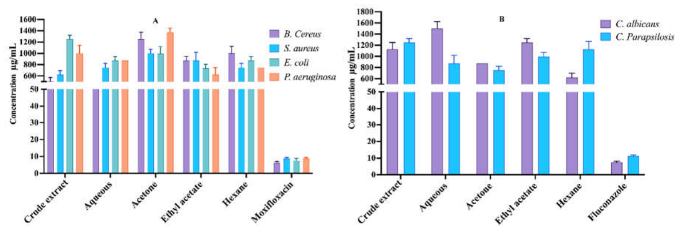
Figure 2.
AChE inhibition potential of L. virgata leaf extracts at various concentrations (A), and extracts that possessed more than 50% inhibition (IC50) at the tested concentration (B).
Figure 2.
AChE inhibition potential of L. virgata leaf extracts at various concentrations (A), and extracts that possessed more than 50% inhibition (IC50) at the tested concentration (B).
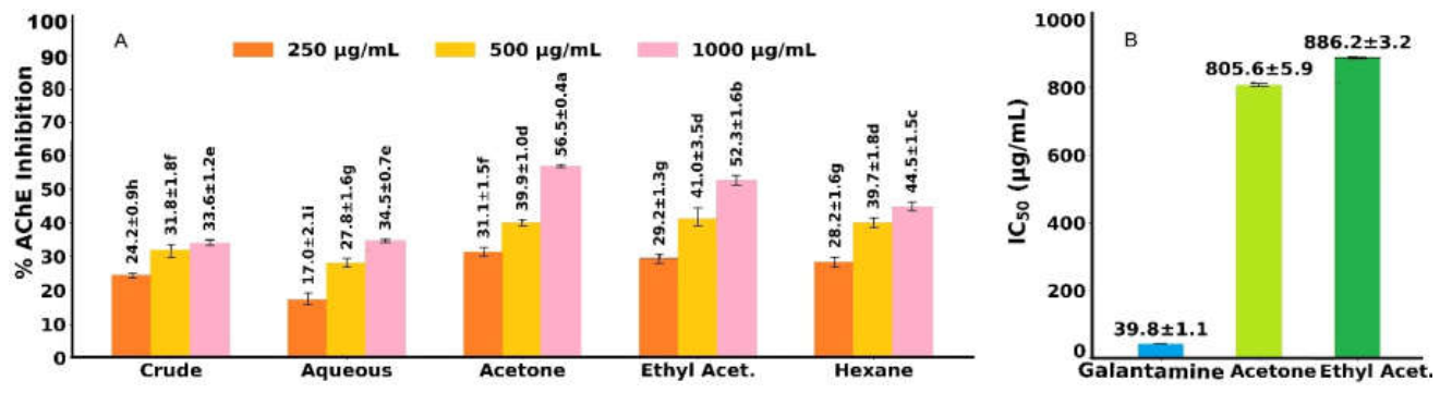
Figure 3.
Images captured under a microscope indicating the cytotoxic effects of L. virgata leaf extract and its fractions at various concentrations against the leukemia U937 cell line.
Figure 3.
Images captured under a microscope indicating the cytotoxic effects of L. virgata leaf extract and its fractions at various concentrations against the leukemia U937 cell line.
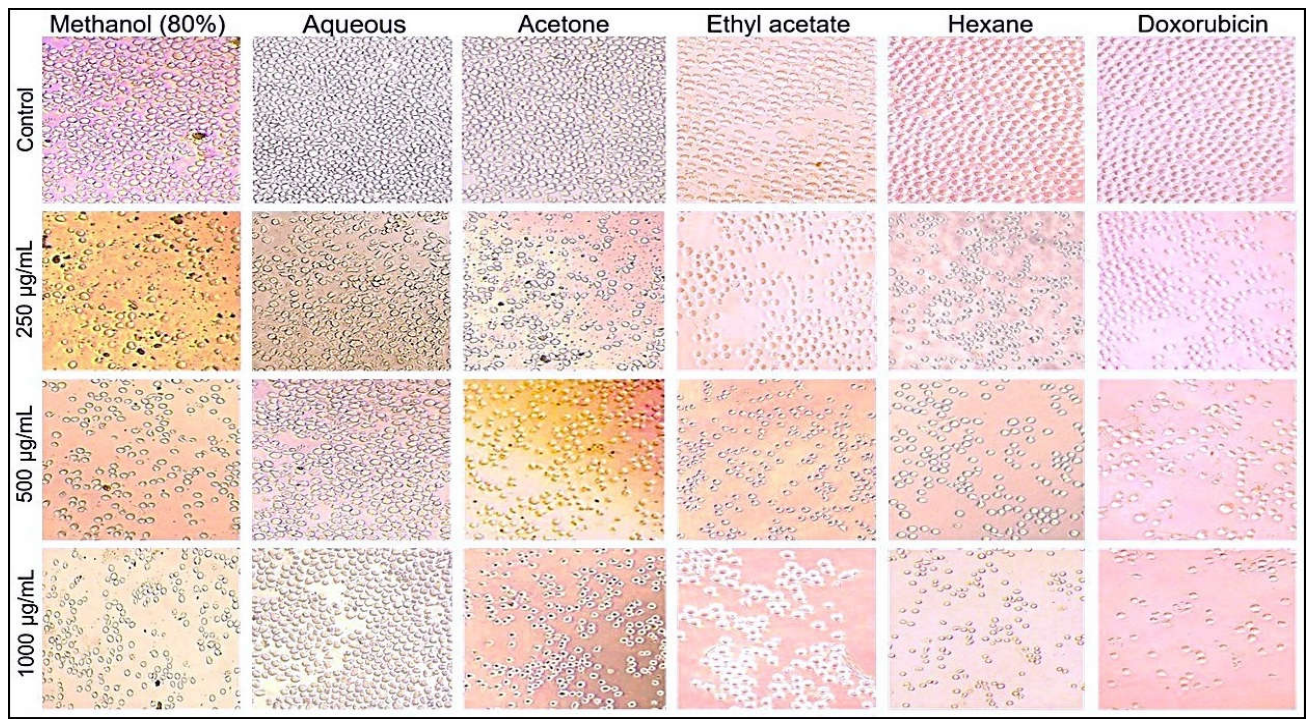
Figure 4.
The cytotoxic potential of L. virgata leaf extract and its fractions (A), and fractions exhibiting dose-dependent and significant cytotoxic activity (B) at various dose concentrations against the leukemia U937 cell line. Mean ± SD, two-way ANOVA substantially different from control (****p < 0.0001, ***p < 0.0005, **p < 0.01, *p < 0.05).
Figure 4.
The cytotoxic potential of L. virgata leaf extract and its fractions (A), and fractions exhibiting dose-dependent and significant cytotoxic activity (B) at various dose concentrations against the leukemia U937 cell line. Mean ± SD, two-way ANOVA substantially different from control (****p < 0.0001, ***p < 0.0005, **p < 0.01, *p < 0.05).
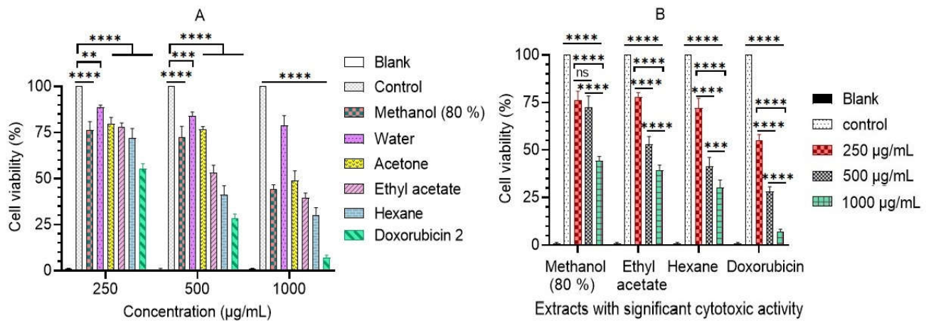
Figure 5.
Total ion chromatogram in the negative (A) and positive (B) ionization modes for LC-MS analysis of L. virgata leaf extract.
Figure 5.
Total ion chromatogram in the negative (A) and positive (B) ionization modes for LC-MS analysis of L. virgata leaf extract.
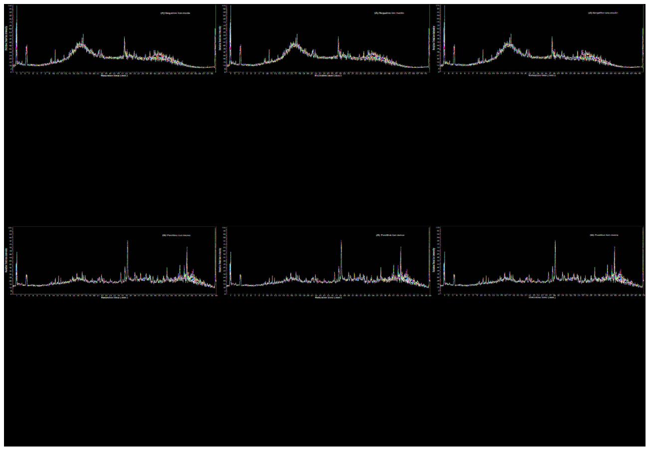
Figure 6.
The relative abundance (%) of various metabolite groups identified by LC-MS in L. virgata leaf extract: overall primary and secondary metabolites (A), various groups of primary metabolites (B), different classes of secondary metabolites (C), and overall classes of total identified metabolites (D).
Figure 6.
The relative abundance (%) of various metabolite groups identified by LC-MS in L. virgata leaf extract: overall primary and secondary metabolites (A), various groups of primary metabolites (B), different classes of secondary metabolites (C), and overall classes of total identified metabolites (D).
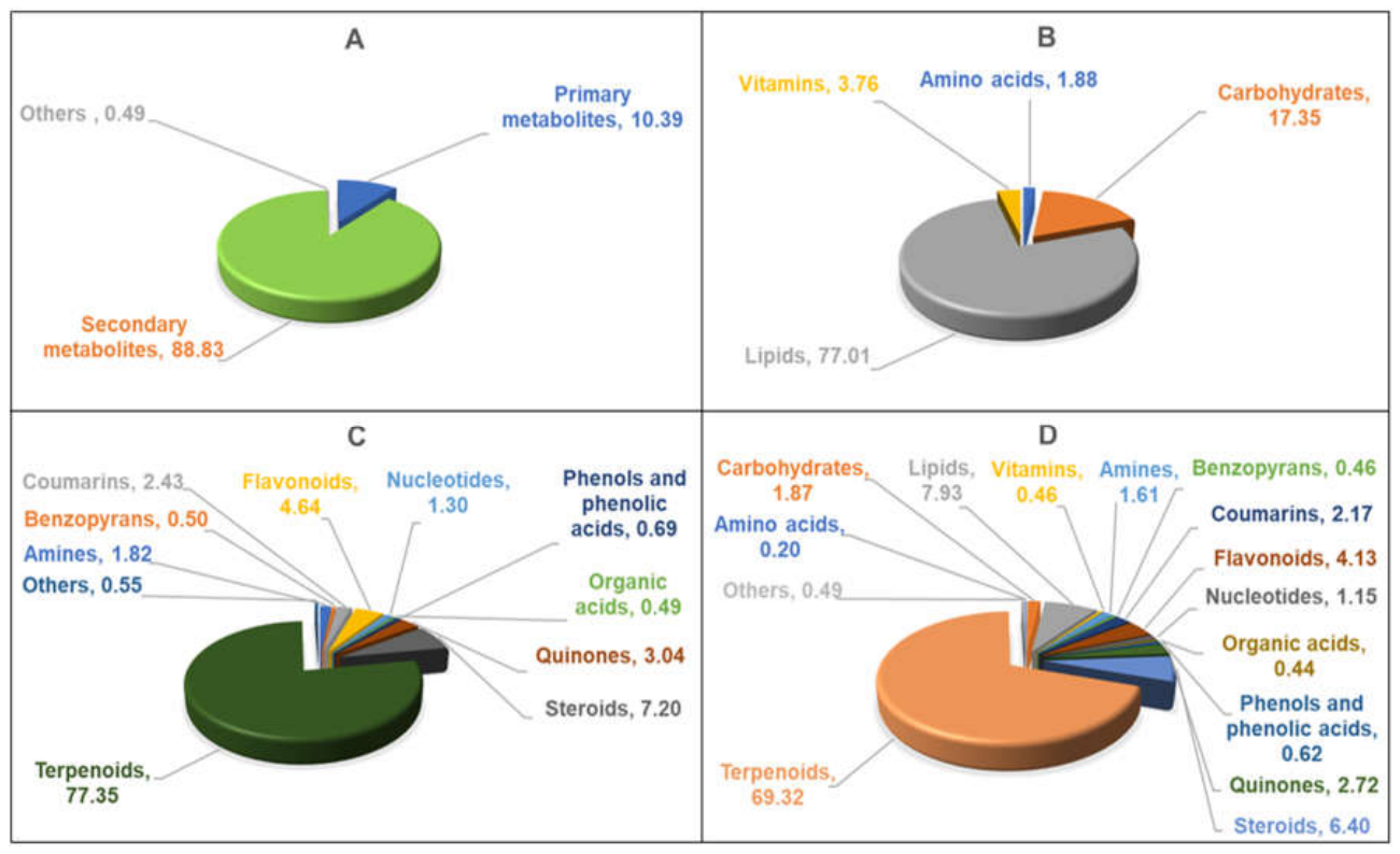
Figure 7.
MS/MS fragment patterns of flavonoids: Quercetin 3-galactoside and Alatanin C identified in the L. virgata leaf extract by LC-MS analysis.
Figure 7.
MS/MS fragment patterns of flavonoids: Quercetin 3-galactoside and Alatanin C identified in the L. virgata leaf extract by LC-MS analysis.
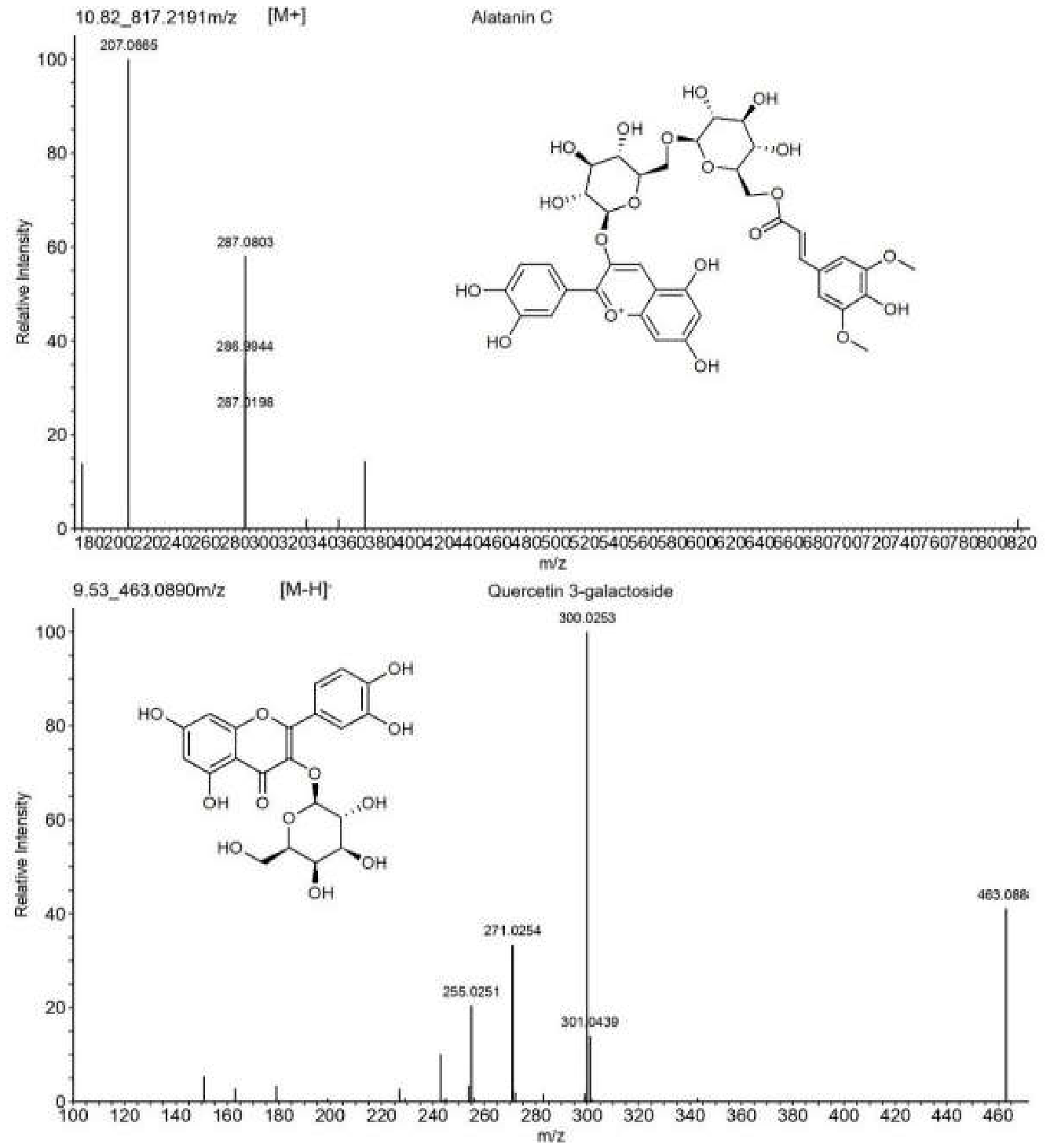
Figure 8.
MS/MS fragments of α-Elemolic acid and Ursolic acid identified in L. virgata leaf extract by LC-MS analysis.
Figure 8.
MS/MS fragments of α-Elemolic acid and Ursolic acid identified in L. virgata leaf extract by LC-MS analysis.
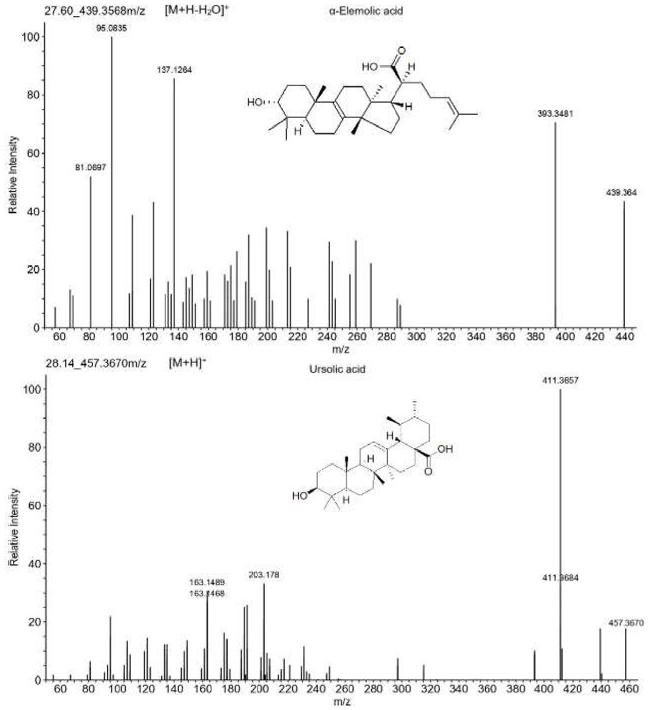
Figure 9.
3D binding approaches of 4,5-Dicaffeoyl quinic acid (A), Caffeoyl quinic acid (B), Asperuloside (C), and Galantamine (D) into the active site of AChE and associated interactions with the residues.
Figure 9.
3D binding approaches of 4,5-Dicaffeoyl quinic acid (A), Caffeoyl quinic acid (B), Asperuloside (C), and Galantamine (D) into the active site of AChE and associated interactions with the residues.
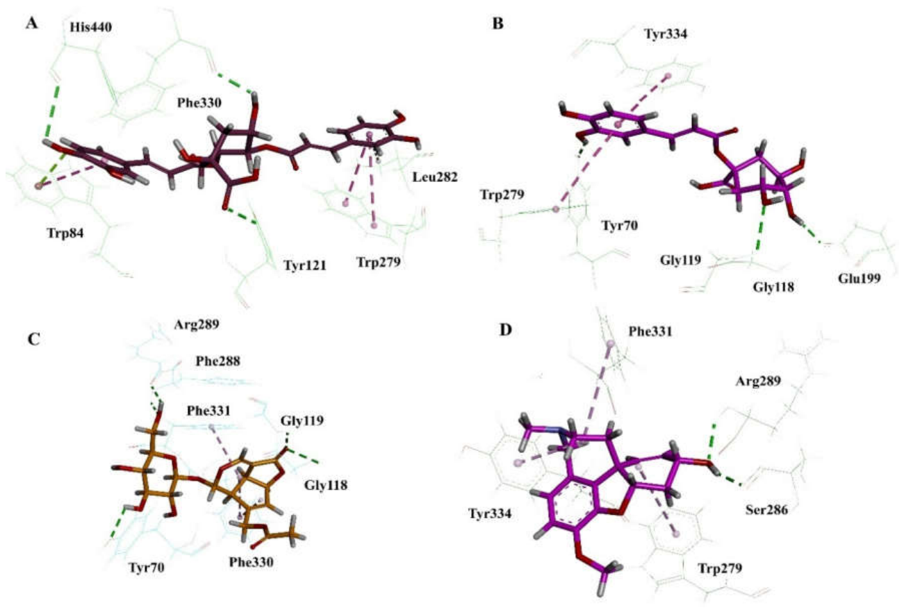
Figure 10.
3D binding modes of Acanthoside B (A), Idarubicin (B), Tamarixetin (C), and Doxorubicin (D) within the topoisomerase II active site, along with their interactions with residues.
Figure 10.
3D binding modes of Acanthoside B (A), Idarubicin (B), Tamarixetin (C), and Doxorubicin (D) within the topoisomerase II active site, along with their interactions with residues.
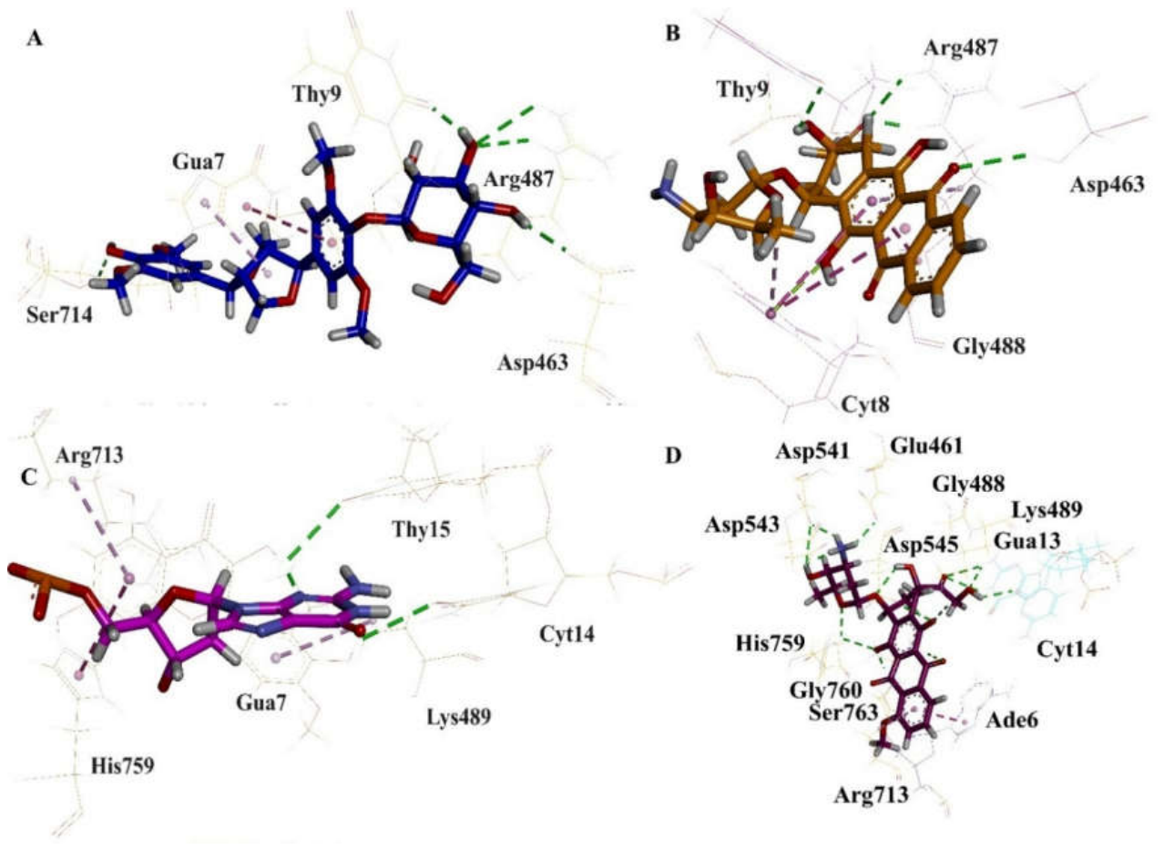

Table 1.
Yield of extract and fractions of L. virgata leaves and their antioxidant activity.
| Sample | Obtained yield (%) | Antioxidant activity (%) | ||
| 200 μg/mL | 400 μg/mL | 800 μg/mL | ||
| Crude extract | 14.40 | 58.01 ± 2.10ef | 69.42 ± 1.47c | 88.11 ± 3.45a |
| Aqueous | 10.07 | 42.98 ± 3.21h | 55.47 ± 1.93f | 69.33 ± 0.92c |
| Acetone | 11.82 | 51.10 ± 2.54g | 64.58 ± 2.84d | 76.98 ± 2.26b |
| Ethyl acetate | 12.41 | 36.94 ± 1.27i | 59.42 ± 3.42e | 71.78 ± 3.18c |
| Hexane | 6.53 | 37.79 ± 1.14i | 49.27 ± 2.65g | 62.84 ± 5.42d |
Reference standard (ascorbic acid 1 mg/mL) showed 93.65 ± 1.07 % antioxidant activity.
Table 2.
Cytotoxic activity (in terms of GI50 values) of different extracts of L. virgata.
| Sample/Standard | Cytotoxicity GI50 (μg/mL) |
| Crude extract | 799.80 ± 3.21e |
| Aqueous | NA |
| Acetone | 986.87 ± 3.73g |
| Ethyl acetate | 730.96 ± 3.85d |
| Hexane | 596.74 ± 2.24c |
| Doxorubicin | 410.67 ± 2.07a |
NA: No GI50 was observed at the tested concentrations.
Table 3.
Absorption bands and frequencies of crude methanolic extract of L. virgata leaves observed by FTIR analysis.
Table 3.
Absorption bands and frequencies of crude methanolic extract of L. virgata leaves observed by FTIR analysis.
| Frequency Range (cm-1) |
Observed frequency (cm-1) |
Band intensity |
Functional groups/ Stretch | Projected compounds |
| 3000-3550 | 3423 | Strong, broad | O–H | Alcohols, Phenols |
| 2800-3200 | 2959, 2924 | Medium | O–H | Carboxylic acids |
| 2800-2990 | 2851 | Weak | C–H | Alkanes |
| 1600-1850 | 1716, 1741 | Strong, sharp | C=O | Aldehydes, Ketones and Carboxylic acids |
| 1440-1625 | 1656 | Weak | C=C | Aromatics |
| 1300-1400 | 1372 | Medium | C–N | Aromatic amines |
| 1000-1400 | 1260 | Strong, sharp | C–F | Alkyl halides |
| 1000-1350 | 1076 | Medium | C–N | Aliphatic amines |
| 1000-1200 | 1045 | Very strong | C–O | Ethers and Esters |
| 660-910 | 804 | Medium | N–H | Amines |
| 600-840 | 702 | Weak | C–Cl | Alky halides |
| < 600 | 613, 626 | Weak | C–I | Alkyl halides |
Table 4.
Carbohydrates, amino acids, and vitamins identified by LC-MS analysis in L. virgata leaf extract.
Table 4.
Carbohydrates, amino acids, and vitamins identified by LC-MS analysis in L. virgata leaf extract.
| Peak no. (ESI +/-) |
Name of identified compound | RT (min) | RA (%) | Chemical formula | MS/MS Product ions |
Adducts (Mode) |
m/z value |
Error (ppm) |
| 2 (-) | Gentiobiose | 0.80 | 0.72 | C12H22O11 | 403, 371, 241, 191, 147 | [M-H]- | 449.1302 | -3.14 |
| 3 (-) | Gluconic Acid | 0.81 | 0.05 | C6H12O7 | 129, 87, 75, 74, 59 | [M-H]- | 195.0510 | 1.45 |
| 1 (+) | Betaine | 0.82 | 0.17 | C5H11NO2 | 117, 59, 58, 56 | [M+H]+ | 118.0585 | -3.53 |
| 4 (-) | 2,3-Butanediol glucoside | 0.84 | 0.05 | C10H20O7 | 297, 118, 119, 89, 85, | [M+FA-H]- | 297.1194 | 1.24 |
| 2 (+) | Lotaustralin | 0.85 | 0.04 | C11H19NO6 | 244, 216, 198, 119. 97 | [M+H]+ | 262.1284 | -0.58 |
| 5 (-) | Lepidimoic acid | 0.86 | 0.04 | C12H18O10 | 281, 175, 113, 86, 85 | [M-H]- | 321.0828 | 0.55 |
| 7 (+) | Methyl salicylate O-[rhamnosyl-(1->6)-glucoside] | 2.24 | 0.10 | C20H28O12 | 193 | [M+NH4]+ | 478.1913 | -1.24 |
| 11 (-) | Asperuloside | 3.27 | 4.19 | C18H22O11 | 459, 414, 413, 251, 233 | [M+FA-H]- | 459.1151 | 1.63 |
| 15 (-) | Linalool 3,7-oxide beta-primeveroside | 9.18 | 0.04 | C21H36O11 | 464, 463, 331, 161, 89 | [M+FA-H]- | 509.2255 | 3.37 |
| 27 (+) | Salidroside | 12.88 | 0.03 | C14H20O7 | 217, 205, 190, 173, 156 | [M+H-2H2O]+ | 265.1073 | 0.99 |
| 22 (-) | Ustiloxin D | 13.13 | 0.06 | C23H34N4O8 | 447, 315, 161, 101, 89 | [M-H]- | 493.2298 | -1.21 |
| 55 (+) | Kanamycin | 20.00 | 0.03 | C18H36N4O11 | 256, 239, 238, 225, 117 | [M+Na]+ | 507.2273 | -0.05 |
| 31 (+) | Arbutin | 14.65 | 0.04 | C12H16O7 | 193, 149, 65, 121, 93 | [M+H-2H2O]+ | 237.0759 | 0.66 |
| 119 (+) | Neomycin | 35.43 | 1.11 | C23H46N6O13 | 637, 619, 587, 559, 549 | [M+Na]+ | 637.3017 | 0.37 |
| 124 (+) | 25-hydroxycholecalciferol | 37.01 | 0.46 | C27H44O2 | 365, 239 | [M+H-H2O]+ | 383.3301 | -1.90 |
Table 5.
Phenols and phenolic acid identified by LC-MS analysis in L. virgata leaf extract.
| Peak no. (ESI +/-) |
Name of identified compound | RT (min) | RA (%) | Chemical formula | MS/MS Product ions |
Adducts (Mode) |
m/z value |
Error (ppm) |
| 4 (+) | 4-hydroxycinnamic acid | 0.89 | 0.05 | C9H8O3 | 147, 119, 118, 89, 77 | [[M+H-H2O]+ | 147.0436 | -2.75 |
| 10 (-) | Caffeoylquinic acid | 2.86 | 1.19 | C16H18O9 | 191, 179, 161, 135, 93 | [M-H]˗ | 353.0885 | 2.07 |
| 13 (+) | Trans-Caffeic acid [apiosyl-(1->6)-glucosyl] ester | 9.31 | 0.07 | C20H26O13 | 235, 193, 187, 175, 165 | [M+H-H2O]+ | 457.1337 | -0.75 |
| 18 (-) | 4,5-Dicaffeoyl quinic acid | 11.12 | 0.11 | C25H24O12 | 353, 173, 191, 179, 155 | [M-H]- | 515.1202 | 3.29 |
| 25 (+) | Sesamolinol | 11.42 | 0.03 | C20H20O7 | 387, 233, 181, 167, 153 | [M+CH3OH+H]+ | 405.1537 | -1.74 |
| 70 (+) | Dibutyl phthalate | 22.95 | 0.68 | C16H22O4 | 201, 149, 121, 93. 65 | [M+H]+ | 279.1592 | 0.54 |
| 116 (+) | Mangiferdesmethylursanone | 34.62 | 0.03 | C29H48O2 | 201, 107, 95, 79, 69 | [M+H-2H2O]+ | 393.3509 | -1.57 |
| 149 (+) | Gingerol | 46.80 | 0.23 | C17H26O4 | 137, 91, 81 | [M+H-H2O]+ | 277.1803 | 1.26 |
Table 6.
Flavonoids and quinones identified by LC-MS analysis in L. virgata leaf extract.
| Peak no. (ESI +/-) |
Name of identified compound | RT (min) | RA (%) | Chemical formula | MS/MS Product ions |
Adducts (Mode) |
m/z value |
Error (ppm) |
| 16 (-) | Quercetin 3-glucoside-7-rhamnoside | 9.29 | 0.22 | C27H30O16 | 300, 301, 271, 255, 88 | [M-H]- | 609.1487 | 4.17 |
| 14 (+) | Vitamin p | 9.33 | 0.14 | C27H30O16 | 358, 356, 305, 304, 303 | [M+H]+ | 611.1608 | 0.27 |
| 17 (-) | Quercetin 3-galactoside | 9.53 | 1.47 | C21H20O12 | 301, 300, 271, 255, 243 | [M-H]- | 463.0890 | 1.62 |
| 16 (+) | Isoquercitrin | 9.55 | 0.56 | C21H20O12 | 304, 303, 302, 257, 229 | [M+H]+ | 465.1025 | -0.52 |
| 17 (+) | Hieracin | 10.47 | 0.11 | C15H10O7 | 257, 229, 201, 155, 153 | [M+H]+ | 303.0498 | -0.26 |
| 18 (+) | Kaempferide 3 [rhamnopyranosyl-(1->6)-glucoside] 7-rhamnoside | 10.47 | 0.05 | C34H42O19 | 773, 309, 303, 165, 147 | [M+ACN+Na]+ | 818.2512 | 4.47 |
| 19 (-) | Kaempferol-3-O-glucoside | 10.48 | 0.08 | C21H20O11 | 284, 285, 256, 255, 227 | [M-H] - | 447.0942 | 2.11 |
| 19 (+) | Kaempferol 3-O-galactoside | 10.50 | 0.05 | C21H20O11 | 309, 288, 287, 185 | [M+H] + | 449.1074 | -1.06 |
| 20 (+) | Alatanin C | 10.82 | 0.05 | C38H41O20+ | 369, 351, 286, 287, 207 | [M+H]+ | 817.2192 | -0.03 |
| 22 (+) | 2''-(6''-p-Coumaroylglucosyl) quercitrin | 11.15 | 0.26 | C36H36O18 | 309, 291, 287, 165, 147 | [M+H]+ | 757.1973 | -0.13 |
| 23 (+) | Luteolin | 11.15 | 0.02 | C15H10O6 | 270, 213, 153, 121, 69 | [M+H]+ | 287.0550 | 0.07 |
| 24 (+) | Vestitone 7-glucoside | 11.39 | 0.05 | C22H26O9 | 417, 367, 263, 233, 181 | [M+H]+ | 435.1641 | -2.02 |
| 21 (-) | Quercetin | 12.62 | 0.80 | C15H10O7 | 301, 178, 151, 121, 107 | [M-H]- | 301.0362 | 2.74 |
| 27 (+) | Morin | 12.63 | 0.20 | C15H10O7 | 285, 257, 229, 201, 165 | [M+H]+ | 303.0500 | 0.18 |
| 23 (-) | Glycitein | 13.85 | 0.04 | C16H12O5 | 268, 240, 239, 238, 227 | [M-H]- | 283.0617 | 1.82 |
| 24 (-) | Kaempferol | 13.85 | 0.58 | C15H10O6 | 239, 229, 185, 211, 163 | [M-H] - | 285.0411 | 2.07 |
| 25 (-) | Isorhamnetin | 14.06 | 0.42 | C16H12O7 | 300, 283, 271, 163, 151 | [M-H]- | 315.0521 | 3.30 |
| 29 (+) | Tamarixetin | 14.08 | 0.14 | C16H12O7 | 302, 285, 274, 257, 245 | [M+H]+ | 317.0657 | 0.40 |
| 27 (-) | Tricin | 14.44 | 0.05 | C17H14O7 | 314, 299, 271, 215 | [M-H]- | 329.0672 | 1.70 |
| 28 (-) | Cirsimaritin | 14.57 | 0.05 | C17H14O6 | 298, 284, 283, 267, 240 | [M-H]+ | 313.0725 | 2.31 |
| 36 (+) | Decylubiquinone | 16.14 | 0.04 | C19H30O4 | 305, 119, 107, 105, 95 | [M+H]+ | 323.2214 | -0.98 |
| 52 (+) | Ubiquinol (QH2) | 19.29 | 0.14 | C14H20O4. [C5H8]n |
289, 271, 159, 153, 147 | [M+H]+ | 321.2059 | -0.43 |
Table 7.
Terpenoids and their derivatives identified by LC-MS analysis in L. virgata leaf extract.
| Peak no. (ESI +/-) |
Name of identified compound | RT (min) | RA (%) | Chemical formula | MS/MS Product ions |
Adducts (Mode) |
m/z value |
Error (ppm) |
| 6 (-) | Monotropein | 0.90 | 2.75 | C16H22O11 | 183, 165, 147, 139, 137 | [M-H]- | 389.1097 | 1.85 |
| 8 (-) | Veranisatin C | 1.17 | 0.40 | C16H20O10 | 371, 209, 191, 165, 147 | [M+FA-H]- | 417.1039 | 0.06 |
| 9 (-) | Geniposidic acid | 1.35 | 0.14 | C16H22O10 | 247, 247, 217, 201, 187 | [M-H]- | 373.1143 | 0.00 |
| 12 (-) | Secologanin | 4.42 | 0.12 | C17H24O10 | 402, 311, 270, 61 | [M+Hac-H]- | 447.1512 | 0.95 |
| 28 (+) | 4,5-Dihydrovomifoliol | 13.72 | 0.06 | C13H22O3 | 151, 135, 133, 123, 111 | [M+H-H2O]+ | 209.1536 | -0.12 |
| 33 (+) | 16-beta-16-Hydroxy-3-oxo- 1,12-Oleanadien-28-oic acid |
15.08 | 0.18 | C30H44O4 | 451, 423, 405, 201, 145 | [M+H]+ | 469.3307 | -1.20 |
| 34 (+) | Glabrolide | 15.43 | 0.07 | C30H44O4 | 451, 405, 187 | [M+H]+ | 469.3304 | -1.76 |
| 37 (+) | 4-Hydroxy-3-polyprenylbenzoate | 16.44 | 0.16 | C17H22O3 | 289, 271, 233, 159, 135 | [M+CH3OH+H]+ | 307.1904 | 0.20 |
| 38 (+) | (3-alpha,20R,24Z)-3-Hydroxy-21-oxoeupha-8,24-dien-26-oic acid | 17.05 | 0.19 | C30H46O4 | 453, 407, 219, 201, 189 | [M+H]+ | 471.3466 | -0.70 |
| 42 (+) | Arjunolic acid | 17.34 | 0.14 | C30H48O5 | 453, 407, 205, 201, 189 | [M+H]+ | 489.3571 | -0.73 |
| 43 (+) | 16-Hydroxy-3-oxo-12-oleanen-28-oic acid | 17.35 | 0.51 | C30H46O4 | 205, 201, 187, 511 | [M+H]+ | 471.3461 | -1.47 |
| 44 (+) | Tyromycic acid | 17.35 | 0.46 | C30H44O3 | 205, 201, 187 | [M+H]+ | 453.3358 | -1.24 |
| 45 (+) | Isomenthone | 17.50 | 0.07 | C10H18O | 77, 67, 55 | [M+H-H2O]+ | 137.1330 | 3.08 |
| 51 (+) | 11-Deoxoglycyrrhetinic acid | 19.09 | 0.05 | C30H48O3 | 439, 421, 203, 191, 159 | [M+H]+ | 457.3668 | -1.84 |
| 53 (+) | Betulinic Acid | 19.72 | 0.10 | C30H48O3 | 439, 411, 203, 191, 189 | [M+H] | 457.3669 | -1.56 |
| 32 (-) | Pomonic acid | 19.81 | 0.06 | C30H46O4 | 425 | [M-H]- | 469.3304 | 2.74 |
| 33 (-) | Hederagenin | 19.94 | 0.04 | C30H48O4 | 453, 471, 407 | [M+FA-H]+ | 517.3550 | 3.22 |
| 56 (+) | Abscisic acid | 20.22 | 0.05 | C15H20O4 | 247, 149, 159, 121, 107 | [M+H]+ | 265.1439 | 1.79 |
| 58 (+) | Glypallidifloric acid | 20.32 | 0.38 | C30H46O3 | 437, 391, 215, 201, 189 | [M+H]+ | 455.3514 | -1.63 |
| 60 (+) | Sugeonol | 20.77 | 0.10 | C15H22O2 | 179, 57 | [M+H]+ | 235.1695 | 1.02 |
| 61 (+) | (R)-ar-Turmerone | 20.82 | 0.06 | C15H20O | 193, 161 | [M+CH3OH+H]+ | 249.1848 | -0.42 |
| 62 (+) | Ganoderiol F | 20.87 | 1.01 | C30H46O3 | 438, 410, 409, 201, 191 | [M+H]+ | 455.3516 | -0.91 |
| 64 (+) | Ganodermic acid Jb | 21.20 | 2.64 | C30H46O4 | 471, 425, 407, 235, 217 | [M+H]+ | 471.3463 | -1.25 |
| 65 (+) | Desoxoglabrolide | 21.79 | 1.31 | C30H46O3 | 437, 409, 205, 203, 201 | [M+H]+ | 455.3514 | -1.22 |
| 66 (+) | Maslinic acid | 21.79 | 0.51 | C30H48O4 | 409, 205, 203, 177, 149 | [M+H]+ | 473.3619 | -1.34 |
| 67 (+) | Beta-Elemonic acid | 22.07 | 1.13 | C30H46O3 | 437, 409, 205, 203, 201 | [M+H]+ | 455.3520 | -1.35 |
| 68 (+) | 3-alpha-Corosolic acid | 22.11 | 0.32 | C30H48O4 | 437, 410, 409, 205, 201 | [M+H]+ | 473.3620 | -1.20 |
| 71 (+) | Ursonic acid | 23.60 | 0.16 | C30H46O3 | 438, 437, 409, 203, 201 | [M+H]+ | 455.3515 | -1.11 |
| 77 (+) | Euscaphic acid | 25.06 | 0.13 | C30H48O5 | 435, 425, 407, 247, 189 | [M+H-H2O]+ | 471.3515 | -1.47 |
| 83 (+) | Azukisapogenol | 26.00 | 0.11 | C30H48O4 | 427, 203, 189, 187, 177 | [M+H]+ | 473.3620 | -1.14 |
| 84 (+) | Betulonic acid | 26.52 | 3.59 | C30H46O3 | 437, 409, 229, 203, 201 | [M+H]+ | 455.3517 | -0.69 |
| 36 (-) | Ganoderiol I | 27.49 | 2.51 | C31H50O5 | 456, 455, 372 | [M-H]- | 501.3605 | 3.81 |
| 86 (+) | Alpha-Elemolic acid | 27.60 | 6.55 | C30H48O3 | 393, 199, 137, 123, 109 | [M+H-H2O]+ | 439.3568 | -0.64 |
| 87 (+) | Isopimaric acid | 27.68 | 0.03 | C20H30O2 | 257, 147, 173, 161, 149 | [M+H]+ | 303.2318 | -0.11 |
| 88 (+) | Carindone | 27.82 | 0.09 | C31H44O6 | 485, 337, 189, 149, 133 | [M+NH4]+ | 530.3484 | 1.56 |
| 89 (+) | Oleanolic acid | 28.18 | 15.84 | C30H48O3 | 205, 203, 201, 191, 189 | [M+H-H2O]+ | 439.3567 | -0.76 |
| 90 (+) | Ursolic acid | 28.20 | 4.85 | C30H48O3 | 439, 411, 203, 189, 163 | [M+H]+ | 457.3760 | -1.37 |
| 91 (+) | 3-Oxo-12,18-ursadien-28-oic acid | 28.86 | 0.16 | C30H44O3 | 407, 277, 181, 145, 119 | [M+H]+ | 453.3361 | -0.56 |
| 92 (+) | Ganoderic acid Y | 29.93 | 0.87 | C30H46O3 | 437, 409, 189, 177 | [M+H]+ | 455.3517 | -0.58 |
| 95 (+) | Tsugaric acid C | 30.16 | 0.19 | C32H50O5 | 470, 469, 308, 201 | [M+H]+ | 515.3724 | -1.38 |
| 96 (+) | Tomentosolic acid | 30.49 | 0.78 | C30H46O3 | 437, 353, 205, 203, 201 | [M+H]+ | 455.3515 | -0.97 |
| 97 (+) | Anhydrosophoradiol | 30.72 | 0.42 | C30H48O | 407, 217, 205, 203, 191 | [M+H]+ | 425.3772 | -1.32 |
| 98 (+) | Betulin | 30.77 | 0.18 | C30H50O2 | 425, 217, 203, 191, 189 | [M+H]+ | 443.3878 | -1.27 |
| 100 (+) | Olean-12-ene-3b,28-diol | 31.37 | 0.18 | C30H50O2 | 425, 407, 339, 161, 147 | [M+H]+ | 443.3879 | -0.93 |
| 38 (-) | Canavalioside | 31.90 | 0.08 | C26H42O12 | 559, 516, 515, 500 | [M+FA-H]- | 591.2640 | -3.28 |
| 101 (+) | 16-Acetylpriverogenin A | 32.54 | 0.49 | C32H50O5 | 437, 203, 201, 189, 147 | [M+H-H2O]+ | 497.3615 | -2.08 |
| 102 (+) | Brein | 32.54 | 0.02 | C30H50O2 | 443, 425, 203, 191, 149 | [M+H]+ | 443.3875 | -1.87 |
| 104 (+) | Phytol | 32.97 | 0.04 | C20H40O | 281, 248, 251, 205, 190 | [M+H]+ | 297.3162 | 3.46 |
| 105 (+) | Erythrodiol | 32.98 | 0.05 | C30H50O2 | 287, 269 | [M+H-H2O]+ | 425.3770 | -1.83 |
| 108 (+) | Soyasapogenol A | 33.68 | 0.08 | C30H50O4 | 479, 437, 329, 247, 203 | [M+Na]+ | 497.3601 | -0.14 |
| 111 (+) | Ursololactone | 33.86 | 0.12 | C32H50O4 | 439, 249, 203, 191, 189 | [M+H]+ | 499.3780 | -0.33 |
| 112 (+) | Astaxanthin | 33.90 | 0.05 | C40H52O4 | 578, 537, 477 | [M+Na]+ | 619.3785 | 4.64 |
| 117 (+) | Lupenone | 34.86 | 0.05 | C30H48O | 325, 203, 145, 135, 109 | [M+H-H2O]+ | 407.3665 | -1.66 |
| 120 (+) | Oleanolic aldehyde | 35.61 | 0.57 | C30H48O2 | 423, 301, 229, 189, 177 | [M+H]+ | 441.3722 | -1.17 |
| 126 (+) | Gamma-Taraxasterone | 37.49 | 0.53 | C30H48O | 407, 149, 143, 69 | [M+H]+ | 425.3772 | -1.90 |
| 127 (+) | Heliantriol C | 37.49 | 0.06 | C30H50O3 | 407 | [M+K]+, [M+NH4]+ | 476.4089 | -2.00 |
| 130 (+) | Heliantriol B2 | 38.22 | 0.13 | C30H50O3 | 441, 135, 133, 121, 107 | [M+H]+ | 459.3826 | -1.47 |
| 133 (+) | Coriandrinonediol | 39.36 | 0.13 | C30H50O3 | 343, 269, 147, 137, 133 | [M+NH4]+ | 476.4092 | -1.39 |
| 134 (+) | Momodicaursenol | 39.98 | 0.38 | C30H48O | 277, 189, 159, 151, 119 | [M+H]+ | 425.3773 | -1.26 |
| 136 (+) | Alpha-Tocopherolquinone | 40.05 | 0.13 | C29H50O3 | 430, 413, 412, 411, 394 | [M+H]+ | 447.3826 | -1.47 |
| 139 (+) | Mangiferoleanone | 40.88 | 0.05 | C30H48O | 204, 161 | [M+H]+ | 425.3773 | -1.32 |
| 143 (+) | Beta-Citraurin | 43.22 | 0.05 | C30H40O2 | 171 | [M+CH3O+H]+ | 465.3353 | -2.36 |
| 144 (+) | Beta-Amyrin | 43.49 | 0.54 | C30H50O | 95, 145, 189, 135, 81 | [M+H-H2O]+ | 409.3823 | -1.32 |
| 150 (+) | Beta-carotene | 47.01 | 2.20 | C40H56 | 444, 281, 185, 171, 145 | [M+]+ | 536.4373 | -1.75 |
| 152 (+) | Lucidenic acid N | 47.75 | 0.12 | C27H40O6 | 905, 904, 903, 844, 564 | [2M+H]+ | 921.5743 | 2.28 |
Disclaimer/Publisher’s Note: The statements, opinions and data contained in all publications are solely those of the individual author(s) and contributor(s) and not of MDPI and/or the editor(s). MDPI and/or the editor(s) disclaim responsibility for any injury to people or property resulting from any ideas, methods, instructions or products referred to in the content. |
© 2026 by the authors. Licensee MDPI, Basel, Switzerland. This article is an open access article distributed under the terms and conditions of the Creative Commons Attribution (CC BY) license (http://creativecommons.org/licenses/by/4.0/).
Copyright: This open access article is published under a Creative Commons CC BY 4.0 license, which permit the free download, distribution, and reuse, provided that the author and preprint are cited in any reuse.