

Submitted:
06 November 2026
Posted:
09 November 2026
You are already at the latest version
Abstract
Despite four decades of therapeutic progress, Helicobacter pylori eradication continues to decline in high-resistance regions, where clarithromycin resistance exceeds the 15% threshold that renders empirical therapy unreliable. Maastricht VI and the 2024 American College of Gastroenterology guideline mandate susceptibility-directed therapy wherever predicted efficacy falls below 80%, yet no existing method delivers phenotypic susceptibility results within the endoscopy encounter: culture requires 5–7 days and succeeds in roughly half of biopsies, while molecular assays interrogate only known mutations and cannot assess bacterial viability. The result is a persistent “open loop” of biopsy, delayed reporting, empirical therapy, and recurrent failure. Fluorescence rapid on-site evaluation (F-ROSE) can close this loop. In published cohorts, dual-fluorescence viability staining visualized viable organisms—including viable but non-culturable coccoid forms—within 25–30 minutes, with a sensitivity of 0.947 in a 317-patient comparison. A peer-reviewed proof-of-concept study showed that the fluorescence residual rate after 1 hour of antibiotic exposure provides a culture-free phenotypic susceptibility readout directly from biopsies, with 100% sensitivity for amoxicillin and levofloxacin. Combined with same-visit tailored therapy and a structured training program that raised interpretation accuracy from 58.1% to 86.8%, these steps form an integrated paradigm we designate HP-REVISE (H. pylori real-time evaluation by visual imaging and phenotypic susceptibility estimation): SEE, TEST, TREAT, TEACH—all at the endoscope tip. Here we set out the rationale, the published evidence, the unresolved biological and standardization questions, and the registered multicentre trial that will determine whether the loop improves eradication at scale.
Keywords:
Helicobacter pylori
; antibiotic resistance
; rapid on-site evaluation
; fluorescence microscopy
; antimicrobial susceptibility testing
; endoscopy
Introduction: The Dilemma at the Endoscope Tip
The endoscope has undergone a remarkable transformation over the past half-century—from a diagnostic instrument that revealed mucosal morphology, to a therapeutic platform that enables resection of lesions, to the present moment, where it is poised to resolve microbiology in real time. Yet for Helicobacter pylori, the most common chronic bacterial infection, affecting half of the world’s population (1–3), this evolution has not kept pace with clinical need (4).
The problem is structural, not incidental. Clarithromycin resistance exceeds the 15% threshold above which empirical therapy is no longer reliable (5), and exceeds 30% in many Chinese regions (5,6). Maastricht VI and the 2024 American College of Gastroenterology guideline mandate susceptibility-directed therapy wherever predicted eradication rates fall below 80% (7,8); nevertheless, clinicians lack immediate susceptibility results at endoscopy. Culture-based testing—the reference standard—requires 5–7 days of microaerophilic incubation plus 72 hours of drug exposure, succeeds in roughly half of endoscopic biopsies, and is simply not performed in most endoscopy units (9). Molecular assays are faster but interrogate only known resistance mutations. For clarithromycin and levofloxacin, where key mutations predict phenotype reasonably well, this limitation is modest; for amoxicillin, by contrast, genotype–phenotype agreement was nearly absent in a large single-cohort analysis (κ < 0.2, not statistically significant) (10). No molecular method can assess bacterial viability, including the viable but non-culturable (VBNC) coccoid state (10,11).
In short, the test the guidelines assume does not exist at the point where the guidelines demand it. This review focuses on the population in whom this gap is most consequential: patients who already undergo endoscopy—for dyspepsia, peptic ulcer disease, or gastric cancer screening—and in whom H. pylori infection is detected at that encounter, because for them endoscopy is often the last contact with the healthcare system before treatment begins. HP-REVISE is not proposed as a population-wide replacement for non-invasive test-and-treat strategies; it is proposed as a closed loop for the endoscopy suite.
The result of the current setup is a persistent “open loop”: biopsy, delayed reporting, empirical therapy, and frequent failure. This review examines the strategy that aims to close this loop—fluorescence rapid on-site evaluation (F-ROSE)—integrates the published evidence, and makes the case that the field should now test it at scale. Because the technique and the pathway built on it are easily conflated, we propose a formal distinction: F-ROSE is the enabling technique; the integrated same-encounter pathway we designate HP-REVISE (H. pylori Real-time Evaluation by Visual Imaging and phenotypic Susceptibility Estimation).
Why Traditional Paths Have Failed
Understanding why the diagnostic–therapeutic loop remains open requires appreciating the biological and practical limitations of existing susceptibility testing methods.
Culture-Based Susceptibility Testing
The reference standard has two fundamental limitations that are biological rather than logistical. First, H. pylori is a fastidious microaerophile with a slow growth rate; culture requires 5–7 days of incubation followed by 72 hours of drug exposure to determine minimum inhibitory concentrations (9). Second, culture success is intrinsically limited: only about half of endoscopic biopsies yield viable organisms for testing. This failure is not incidental—H. pylori can enter a VBNC coccoid state under stress, including antibiotic exposure or suboptimal sampling. These coccoid forms are metabolically active, can reactivate, and are associated with recurrent infection and resistance emergence (12), yet they are invisible to culture-based methods. The practical consequence is that most endoscopy units simply do not perform culture-based susceptibility testing; even when they do, results arrive 7–10 days after the procedure, long after the patient has left and empirical therapy has begun.
Molecular Resistance Testing
Molecular methods (PCR-based or next-generation sequencing) can return results within hours, and for some drugs they perform well: the 23S rRNA mutations underlying clarithromycin resistance predict phenotype with high concordance, and mutation detection has even been validated on tissue by fluorescence in situ hybridisation (13). The trade-offs matter most elsewhere. For antibiotics such as amoxicillin, where genotype–phenotype correlation is poor, molecular assays offer little guidance (10). And because molecular tests detect nucleic acid rather than living organisms, they cannot distinguish viable from non-viable bacteria—a clinically important limitation when a patient harbours both viable resistant and non-viable susceptible organisms, or viable coccoid forms that can later reactivate.
The Empirical Therapy Gap
Empirical therapy—the default approach in most settings—assumes that resistance remains below the threshold at which population-level efficacy exceeds 80%. That assumption no longer holds in many regions of China, where several provinces report clarithromycin resistance >30% (14) and metronidazole resistance often exceeds 60% (15). The predictable consequence is a cascade of failed eradication attempts, each with a lower success probability as resistance emerges, and rising costs of repeated treatment (16).
Rapid Methods That Exist—And the Gap They Leave
Fairness requires acknowledging what already moves quickly. Real-time PCR protocols that include extraction and mutation analysis can be completed in about 2 hours (9); fluorescence in situ hybridisation delivers a same-day clarithromycin answer from biopsy tissue (13); and string test– or stool-based molecular assays extend resistance testing to patients who never undergo endoscopy (17). In clinical microbiology at large, automated platforms such as the Accelerate Pheno system return phenotypic susceptibility results from positive blood cultures within hours (18), and single-cell imaging has demonstrated phenotypic susceptibility readouts in under 30 minutes for conventional pathogens (19).
None of these, however, delivers what the guidelines need at the point where our patients are: a phenotypic, viability-based susceptibility result obtained from a gastric biopsy during the endoscopy encounter. Commercial rapid platforms are validated for bloodstream infections, not for H. pylori in gastric mucosa (18); single-cell methods require pure cultures rather than biopsies (19); and molecular assays inherit the genotype–phenotype and viability constraints described above. The contribution of F-ROSE is therefore not the invention of rapid fluorescence readouts—which exist elsewhere in microbiology—but their integration, validation, and publication for H. pylori at the endoscope tip (20–22). Figure 1 quantifies the global magnitude of the resistance crisis and the diagnostic reality that no conventional method delivers phenotypic susceptibility results at the point of care within a single encounter.
Hp-Revise: Closing the Loop at The Endoscope Tip
Figure 2 contrasts the persistent open loop of conventional practice with the HP-REVISE closed-loop pathway. HP-REVISE comprises four synergistic steps—SEE, TEST, TREAT, TEACH—that together close the diagnostic–therapeutic gap within a single clinical encounter.
SEE: Real-Time Visualisation of Viable H. Pylori
Figure 3 illustrates the technical workflow of the HP-REVISE pathway, from endoscopic biopsy sampling through dual-fluorescence viability staining to real-time microscopic evaluation and, when indicated, same-day phenotypic antimicrobial susceptibility testing. Viability fluorescent probes (e.g., acridine orange/ethidium bromide or EthD-III/DMAO) render viable H. pylori—including coccoid forms—directly visible under 460–490 nm excitation within 25–30 minutes of biopsy: organisms with intact membranes fluoresce green, whereas organisms with compromised membranes fluoresce red.
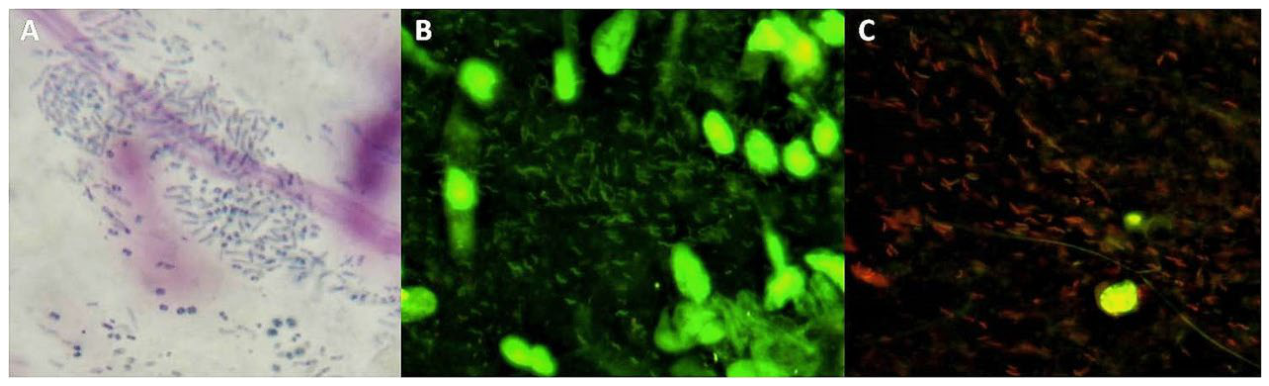
In a published 317-patient comparison against histopathology, F-ROSE achieved a sensitivity of 0.947, specificity of 0.920, and negative predictive value of 0.970, with the highest Youden index (0.867) among all comparators (20). In an independent 180-patient comparison with the rapid urease test, F-ROSE achieved a sensitivity of 93.6%, specificity of 94.2%, and AUC of 0.954 (21). The second study illustrates why the F-ROSE advantage is visual rather than merely statistical: the stable-positive threshold was approximately 4 organisms per high-power field, versus approximately 22 for the rapid urease test; among 63 patients with low bacterial loads, F-ROSE detected infection in 61 where the urease test detected 38 (21). F-ROSE thus sees precisely the low-load and coccoid populations that culture-dependent methods miss (12). Figure 4 contrasts conventional Giemsa staining, which requires fixation, sectioning, and expert pathological review (Figure 4A), with real-time fluorescence imaging (Figure 4B); the shift from green to red fluorescence after antibiotic exposure (Figure 4C) is the signal on which phenotypic susceptibility assessment rests.
TEST: Culture-Free Phenotypic Susceptibility Testing
The same viability readout can be repeated after one hour of exposure to antibiotics at EUCAST breakpoint concentrations (23). The reduction in viable-organism fluorescence—the fluorescence residual rate (FRR)—provides a quantitative phenotypic readout of susceptibility: resistant organisms continue to fluoresce, whereas susceptible organisms lose membrane integrity and switch to red.
In a peer-reviewed proof-of-concept study, biopsies from 40 H. pylori–positive patients were homogenised and co-incubated for 1 hour with amoxicillin, clarithromycin, or levofloxacin at EUCAST breakpoint concentrations (22). Culture failed in 14 of the 40 patients—the familiar attrition of the reference standard. In the 26 culture-evaluable samples, concordance between the fluorescence readout and E-test was 84.6% for amoxicillin, 76.9% for levofloxacin, and 69.2% for clarithromycin (κ = 0.523, 0.539, and 0.412, respectively). Two findings deserve emphasis. First, because the assay reads viability directly, a susceptibility result was obtained for all 40 patients—including the 14 in whom culture failed; for amoxicillin, an antibiotic for which no reliable genetic resistance marker exists, this is arguably the study’s most consequential result. Second, sensitivity and negative predictive value were 100% for amoxicillin and levofloxacin (90.9% sensitivity for clarithromycin), meaning that no resistant isolate was missed for the two agents on which eradication regimens most depend (22). The entire process, from biopsy to result, takes 55–65 minutes—compatible with the workflow of a high-volume endoscopy unit, where patients typically remain in recovery before discharge.
A subsequent two-round comparison, currently available as a preprint, found that sharper imaging and an upgraded recognition algorithm raised pooled sensitivity from 92.9% to 100%, with negative predictive values of 100% for all three agents; receiver-operating-characteristic analysis suggested that drug-specific FRR cut-offs (0.52–0.58) outperform the uniform 50% threshold—most clearly for levofloxacin, where specificity rose from 71.4% to 90.5% (24). Specificity remained moderate and positive predictive values low in both rounds—limitations these studies report transparently and to which we return below.
TREAT: Same-Visit Tailored Therapy, with a Feedback Channel
A one-hour turnaround converts susceptibility testing from a retrospective audit into a prospective decision tool: the patient has not yet left the endoscopy suite when the regimen is chosen. This matters most where it is needed most—in high-resistance regions where empirical therapy already fails the 80% benchmark (25) and where endoscopy is often the only contact point before treatment begins (26).
The loop does not end at the prescription. Because viability is the readout, the same assessment can be repeated during and after therapy to verify that organisms are actually dying. In a published 224-patient cohort spanning the full course of bismuth quadruple therapy, the viable proportion fell from 57.3% before treatment to 5.8% early after treatment, while coccoid formation peaked at 65.3% during treatment; in the subgroup with recurrence or resistance, viable organisms rebounded to 38.2%, and a viable proportion above 30% during treatment predicted eradication failure with an AUC of 0.887 (27). The closed loop thus acquires a feedback channel that neither culture nor molecular testing can offer at any speed.
TEACH: Endoscopic Pathogen Visualisation Literacy
Technology without literacy does not disseminate. Fluorescence microscopy requires a skill set that is not part of standard gastroenterology training, and interpreting fluorescent images, distinguishing viable from non-viable organisms, and quantifying the FRR cannot be assumed. We therefore developed Endoscopic Pathogen Visualisation Literacy (EPVL), a structured 16-hour, six-module curriculum covering the principles of fluorescence microscopy, image acquisition and quality control, viability probe interpretation, FRR quantification, and quality assurance (28). In a quasi-experimental study of 54 trainees (currently available as a preprint), EPVL raised image-interpretation accuracy from 58.1% to 86.8% (versus +6.9% in controls; Cohen’s d = 1.48), shortened reading time from 33.4 to 19.0 seconds per image, and improved inter-observer agreement from κ = 0.56 to 0.91 (29). As depicted in Figure 2, the competency gains from TEACH feed back to raise the quality of SEE—a self-reinforcing cycle that strengthens the loop over time.
Why This Loop will Only Get Stronger
One of the most compelling features of HP-REVISE is that its diagnostic performance is not fixed by biology, as with culture. Fluorescence images are exactly the type of input at which deep learning excels (30,31). Each additional case refines the algorithm: automated segmentation, bacterial counting, and FRR computation will standardise interpretation, reduce observer variability, and lower the training threshold that EPVL now addresses.
This creates a self-reinforcing cycle that conventional antimicrobial susceptibility testing cannot match. As the technology disseminates and image libraries grow, diagnostic performance should rise continuously—not plateau or decline. This is a strong rationale for building standardised, annotated image libraries now, before the technology becomes widely adopted. In high-resistance regions, where empirical therapy is increasingly ineffective, HP-REVISE offers a pragmatic solution that does not require changing the clinical workflow: it adds a fluorescence readout to the biopsies that would otherwise be sent for histopathology, and returns the result within the same visit.
Challenges and Limitations
We are not claiming an established standard of care. The susceptibility data derive from small, single-center cohorts; multicenter validation and, critically, eradication-rate outcome trials are required and are underway (ChiCTR2600125137). Four challenges deserve explicit discussion.
Evidence base. The AST studies enrolled 40 and 29 culture-evaluable patients respectively, with wide confidence intervals; the algorithm optimisation in the two-round study was performed and evaluated within the same small cohort, so the post-optimisation performance estimates are likely optimistic and require independent confirmation (22,24).
Biological validity of the FRR readout. The assay assumes that loss of membrane integrity within 1 hour tracks susceptibility, but antibiotic classes kill differently. β-lactams act only on dividing cells, so metabolically quiescent organisms may register falsely resistant; agents that do not directly disrupt the membrane (macrolides, fluoroquinolones) may register falsely susceptible after a single hour of exposure—exactly the discordance pattern observed for clarithromycin in the published study (22). The VBNC organisms that SEE makes visible complicate the readout further: their low metabolic activity can mimic either non-viability or drug tolerance, and the conditions under which an FRR-susceptible result predicts sterilisation rather than transient suppression remain undefined. Drug-specific cut-offs partially mitigate, but do not resolve, this uncertainty (24,25).
Standardisation. FRR interpretation requires control of imaging conditions, dye concentrations, and exposure times across centres. AI-assisted quantification offers a path to standardisation that does not depend on expert human interpretation (30).
Equipment, cost, and training. Fluorescence microscopy requires equipment that is not standard in most endoscopy units, together with bench space, quality control, and trained staff; the dyes themselves are inexpensive, but the platform is a real investment. Its cost must be weighed against the cost of repeated failed eradication attempts (32)—lost work days, additional consultations, and repeat endoscopies—and formal cost-effectiveness analyses are needed but not yet available (33,34). EPVL addresses the training barrier directly (29).
Finally, we do not see fluorescence-based testing and molecular assays as rivals. For targets with tight genotype–phenotype coupling, such as clarithromycin 23S rRNA mutations, molecular methods remain valuable complements (10,13); what we propose is a division of labour in time—an immediate phenotypic answer at the endoscope, with molecular refinement where indicated.
Future Directions
The immediate priority is to complete the multicentre validation study (ChiCTR2600125137) that will provide eradication-rate outcomes—the endpoint that matters most to clinicians and patients. If the preliminary sensitivity and specificity findings are confirmed, three steps follow. Dissemination: EPVL provides a scalable training model whose next challenge is deployment within gastroenterology fellowship curricula and continuing medical education. Digital and AI integration: the self-reinforcing cycle of AI improvement requires annotated image libraries; we call for a collaborative, multicentre effort to build them using standardised protocols (30,31). Guideline incorporation: for HP-REVISE to become routine, it must be recognised in clinical guidelines with clear guidance on when and how to use it; we anticipate that the next iterations of the Maastricht and ACG guidelines will address point-of-care susceptibility testing (35,36). Integrating the viability readout into post-treatment monitoring—already demonstrated in the 224-patient cohort (27)—may prove as clinically important as the index-visit diagnosis.
Conclusion
HP-REVISE, summarised in Figure 2, transforms the endoscopy suite from a site of diagnostic delay into a hub of same-visit decision-making. The history of digestive endoscopy is a history of expanding what can be accomplished through the scope: from seeing morphology, to resecting lesions, to—now—resolving microbiology. For H. pylori, the organism that taught gastroenterology the cost of treating blind, the endoscope can finally answer two questions before the patient leaves the room: is the organism there, and which drug will kill it?
Closing that loop is, we believe, the pragmatic optimal solution currently available in high-resistance settings—and a direction the field should now test at scale. The alternative—continuing to treat empirically despite rising resistance rates—is not sustainable. HP-REVISE leverages the existing endoscopy infrastructure, requires modest additional investment, and provides immediate clinical returns. The time to test it at scale is now.
Author Contributions
LZ and YH conceived the review, drafted the manuscript, and are guarantors. BL, KW, JZ, and MY contributed to data interpretation and critical revision. All authors approved the final version.
Financial support: None.
Potential competing interests: None declared.
References
- Malfertheiner, P.; Camargo, M.C.; El-Omar, E.; et al. Helicobacter pylori infection. Nat. Rev. Dis. Primers 2023, 9, 19. [Google Scholar] [CrossRef] [PubMed]
- Li, Y.; Choi, H.; Leung, K.; et al. Global prevalence of Helicobacter pylori infection between 1980 and 2022: A systematic review and meta-analysis. Lancet Gastroenterol. Hepatol. 2023, 8, 553–64. [Google Scholar] [CrossRef] [PubMed]
- Chen, Y.C.; Malfertheiner, P.; Yu, H.T.; et al. Global prevalence of Helicobacter pylori infection and incidence of gastric cancer between 1980 and 2022. Gastroenterology 2024, 166, 605–19. [Google Scholar] [CrossRef] [PubMed]
- Hong, T.C.; El-Omar, E.M.; Kuo, Y.T.; et al. Primary antibiotic resistance of Helicobacter pylori in the Asia-Pacific region between 1990 and 2022: An updated systematic review and meta-analysis. Lancet Gastroenterol. Hepatol. 2024, 9, 56–67. [Google Scholar] [CrossRef] [PubMed]
- Savoldi, A.; Carrara, E.; Graham, D.Y.; et al. Prevalence of antibiotic resistance in Helicobacter pylori: A systematic review and meta-analysis in World Health Organization regions. Gastroenterology 2018, 155, 1372–82.e17. [Google Scholar] [CrossRef] [PubMed]
- Schulz, C.; Liou, J.M.; Alboraie, M.; et al. Helicobacter pylori antibiotic resistance: A global challenge in search of solutions. Gut 2025, 74, 1561–70. [Google Scholar] [CrossRef] [PubMed]
- Malfertheiner, P.; Megraud, F.; Rokkas, T.; et al. Management of Helicobacter pylori infection: The Maastricht VI/Florence consensus report. Gut 2022, 71, 1724–62. [Google Scholar] [CrossRef] [PubMed]
- Chey, W.D.; Howden, C.W.; Moss, S.F.; et al. ACG Clinical Guideline: Treatment of Helicobacter pylori Infection. Am. J. Gastroenterol. 2024, 119, 1730–53. [Google Scholar] [CrossRef] [PubMed]
- De Francesco, V.; Zullo, A.; Ierardi, E.; et al. Phenotypic and genotypic Helicobacter pylori clarithromycin resistance and therapeutic outcome: Benefits and limits. J. Antimicrob. Chemother. 2010, 65, 327–32. [Google Scholar] [CrossRef] [PubMed]
- Wang, H.; Li, C.; Peng, R.; et al. Phenotypic and genotypic analysis of Helicobacter pylori resistance patterns, molecular characteristics, and CYP2C19 polymorphisms in Nanjing: A retrospective cross-sectional study. Front Cell Infect. Microbiol. 2026, 16, 1852534. [Google Scholar] [CrossRef] [PubMed]
- Elbaiomy, R.G.; Luo, X.; Guo, R.; et al. Antibiotic resistance in Helicobacter pylori: A genetic and physiological perspective. Gut Pathog. 2025, 17, 35. [Google Scholar] [CrossRef] [PubMed]
- Sadeghloo, Z.; Fatemi, N.; Raeisi, H.; et al. Coccoid H. pylori: A key driver of recurrent infections and a target for novel therapies. Virulence 2025, 16, 2566964. [Google Scholar] [CrossRef] [PubMed]
- Trebesius, K.; Panthel, K.; Strobel, S.; et al. Rapid and specific detection of Helicobacter pylori macrolide resistance in gastric tissue by fluorescent in situ hybridisation. Gut 2000, 46, 608–14. [Google Scholar] [CrossRef] [PubMed]
- Xu, W.; Yang, B.; Lin, L.; et al. Antibiotic resistance of Helicobacter pylori in Chinese children: A multicenter study from 2016 to 2023. Helicobacter 2024, 29, e13038. [Google Scholar] [CrossRef] [PubMed]
- Chen, J.; Li, P.; Huang, Y.; et al. Primary antibiotic resistance of Helicobacter pylori in different regions of China: A systematic review and meta-analysis. Pathogens 2022, 11, 786. [Google Scholar] [CrossRef] [PubMed]
- Lee, J.H.; Min, B.H.; Gong, E.J.; et al. Culture-based susceptibility-guided tailored versus empirical concomitant therapy as first-line Helicobacter pylori treatment: A randomized clinical trial. United Eur. Gastroenterol. J. 2024, 12, 941–50. [Google Scholar] [CrossRef] [PubMed]
- Wang, L.; Li, Z.; Tay, C.Y.; et al. Multicentre, cross-sectional surveillance of Helicobacter pylori prevalence and antibiotic resistance to clarithromycin and levofloxacin in urban China using the string test coupled with quantitative PCR. Lancet Microbe 2024, 5, e512–3. [Google Scholar] [CrossRef] [PubMed]
- Pancholi, P.; Carroll, K.C.; Buchan, B.W.; et al. Multicenter evaluation of the Accelerate PhenoTest BC kit for rapid identification and phenotypic antimicrobial susceptibility testing of Gram-negative bacilli and Enterococcus species isolated from blood cultures. J. Clin. Microbiol. 2018, 56, e01329-17. [Google Scholar] [PubMed]
- Baltekin, Ö.; Boucharin, A.; Tano, E.; et al. Antibiotic susceptibility testing in less than 30 min using direct single-cell imaging. Proc. Natl. Acad. Sci. USA 2017, 114, 9170–5. [Google Scholar] [CrossRef] [PubMed]
- Zhang, L.; Hou, Y.; Li, B.; et al. Comparative study of fluorescence ROSE quantitative detection of Helicobacter pylori and traditional histopathological method. Arab J. Gastroenterol. 2026, S1687-1979(26), 00086–9. [Google Scholar] [CrossRef] [PubMed]
- Wan, M.; Zhang, L.; Hou, Y.H.; et al. Comparison of clinical diagnostic values between fluorescence rapid on-site evaluation and rapid urease test for Helicobacter pylori infection. Chin. J. Microecol. (in Chinese). 2026, 38, 959–65. [Google Scholar]
- Li, B.; Zhang, L.; Hou, Y.; et al. Culture-Free Rapid Phenotypic Antimicrobial Susceptibility Testing for Helicobacter pylori Based on Fluorescence Rapid On-Site Evaluation Technology: A Preliminary Study. bioRxiv 2026, 2026.07.06.736681. [Google Scholar] [CrossRef]
- Ma, D.; Fang, Y.; Wang, Z.; et al. Helicobacter pylori antimicrobial susceptibility testing-guided eradication therapy in the southeast region of China: A retrospective study. Infect. Drug Resist 2024, 17, 5079–86. [Google Scholar] [CrossRef] [PubMed]
- Li, B.; Zhang, L.; Hou, Y.; et al. Diagnostic performance of fluorescence-based rapid on-site specimen evaluation for Helicobacter pylori antimicrobial susceptibility testing before and after algorithm optimization: A two-round comparative study. bioRxiv [Preprint] 2026. [Google Scholar] [CrossRef]
- Zhang, L.; Wu, K.; Wang, C.; et al. Fluorescence ROSE for rapid detection of Helicobacter pylori: A comparative study with histopathology and 13C-UBT. Mod. Dig. Interv. (in Chinese). 2026, 31, 597–603. [Google Scholar]
- Li, C.; Zhou, K.; Suo, B.; et al. Tailored therapy guided by genotypic resistance from gastric mucosa samples in the first-line treatment of Helicobacter pylori infection: A systematic review and meta-analysis. Ther. Adv. Gastroenterol. 2025, 18, 17562848251348826. [Google Scholar] [CrossRef] [PubMed]
- Gisbert, J.P.; Parra, P.; Nyssen, O.P. Classic bismuth quadruple therapy for Helicobacter pylori infection—questions focused on clinical practice. Aliment Pharmacol. Ther. 2026, 63, 616–36. [Google Scholar] [CrossRef] [PubMed]
- Ismail, F.W.; Afzal, A.; Durrani, R.; et al. Exploring endoscopic competence in gastroenterology training: A simulation-based comparative analysis of GAGES, DOPS, and ACE assessment tools. Adv. Med. Educ. Pract. 2024, 15, 75–84. [Google Scholar] [CrossRef] [PubMed]
- Zhang, L.; Hou, Y.; Li, B.; et al. Establishment and efficacy of an Endoscopic Pathogen Visualization Literacy (EPVL) training program for gastroenterologists based on fluorescence rapid on-site evaluation (ROSE) technology. medRxiv [Preprint] 2026. [Google Scholar] [CrossRef]
- Tian, Y.; Zhang, J.; Rao, X.; et al. AI-assisted rapid on-site evaluation ROSE of EUS-FNA cytopathology for pancreatic solid lesions: A two-stage deep learning approach. Endosc. Ultrasound 2025, 14, 314–27. [Google Scholar] [CrossRef] [PubMed]
- Ibrahim, A.U.; Dirilenoğlu, F.; Hacisalihoğlu, U.P.; et al. Classification of H. pylori infection from histopathological images using deep learning. J. Imaging Inf. Med. 2024, 37, 1177–86. [Google Scholar] [CrossRef] [PubMed]
- Kim, J.W.; Kim, A.C. Can patients with clarithromycin-resistant Helicobacter pylori infection be treated with a 7-day bismuth-based quadruple therapy? Gut Liver 2024, 18, 931–3. [Google Scholar] [CrossRef] [PubMed]
- Kim, J.S.; Lee, H.; Jeong, Y.; et al. Cost-effectiveness of tailored vs empirical therapy for H. pylori: A decision-tree analysis. Helicobacter 2025, 30, e70081. [Google Scholar] [CrossRef] [PubMed]
- Aumpan, N.; Gamnarai, P.; Wongcha-Um, A.; et al. The use of real-world evidence to generate cost analysis of antibiotic susceptibility testing (AST) in patients with Helicobacter pylori treatment failure in Thailand: A large population-based study. Heliyon 2024, 10, e39189. [Google Scholar] [CrossRef] [PubMed]
- Liou, J.M.; Malfertheiner, P.; Hong, T.C.; et al. Screening and eradication of Helicobacter pylori for gastric cancer prevention: Taipei Global Consensus II. Gut 2025, 74, 1767–91. [Google Scholar] [CrossRef] [PubMed]
- Isomoto, H.; Shimoyama, T.; Ito, M.; et al. Guidelines for the management of Helicobacter pylori infection in Japan: 2024 revised edition. J. Gastroenterol. 2026, 61, 841–62. [Google Scholar] [CrossRef] [PubMed]
Figure 1.
The global burden of Helicobacter pylori antibiotic resistance and the diagnostic gap. (A) Primary resistance rates to clarithromycin, levofloxacin, and metronidazole by WHO region. Most regions now exceed the 15% threshold at which Maastricht VI and the 2024 ACG guideline mandate susceptibility-directed therapy; the 80% eradication failure risk line underscores the clinical consequence. (B) Decadal trend of rising resistance in China (2013–2023); genotypic clarithromycin resistance reached 50.8% in a 2023 nationwide survey of 12,902 urban residents, determined by string test–coupled quantitative PCR—a molecular estimate that, given the genotype–phenotype discordance documented for this organism, may not translate one-to-one into phenotypic treatment failure. (C) Comparative matrix of current diagnostic methods across eight critical dimensions. No existing method simultaneously delivers phenotypic resistance assessment, bacterial viability evaluation, and same-visit result compatibility. Culture-based AST requires 5–7 days with suboptimal success rates; molecular methods interrogate only known mutations and cannot assess viability; histopathology and rapid urease tests provide no susceptibility information. (D) Key statistics underscoring the persistent open loop: most WHO member states exceed the 15% clarithromycin resistance threshold, China’s national urban rate stands at 50.8% (genotypic), and culture success from endoscopic biopsies remains below 50%.
Figure 1.
The global burden of Helicobacter pylori antibiotic resistance and the diagnostic gap. (A) Primary resistance rates to clarithromycin, levofloxacin, and metronidazole by WHO region. Most regions now exceed the 15% threshold at which Maastricht VI and the 2024 ACG guideline mandate susceptibility-directed therapy; the 80% eradication failure risk line underscores the clinical consequence. (B) Decadal trend of rising resistance in China (2013–2023); genotypic clarithromycin resistance reached 50.8% in a 2023 nationwide survey of 12,902 urban residents, determined by string test–coupled quantitative PCR—a molecular estimate that, given the genotype–phenotype discordance documented for this organism, may not translate one-to-one into phenotypic treatment failure. (C) Comparative matrix of current diagnostic methods across eight critical dimensions. No existing method simultaneously delivers phenotypic resistance assessment, bacterial viability evaluation, and same-visit result compatibility. Culture-based AST requires 5–7 days with suboptimal success rates; molecular methods interrogate only known mutations and cannot assess viability; histopathology and rapid urease tests provide no susceptibility information. (D) Key statistics underscoring the persistent open loop: most WHO member states exceed the 15% clarithromycin resistance threshold, China’s national urban rate stands at 50.8% (genotypic), and culture success from endoscopic biopsies remains below 50%.
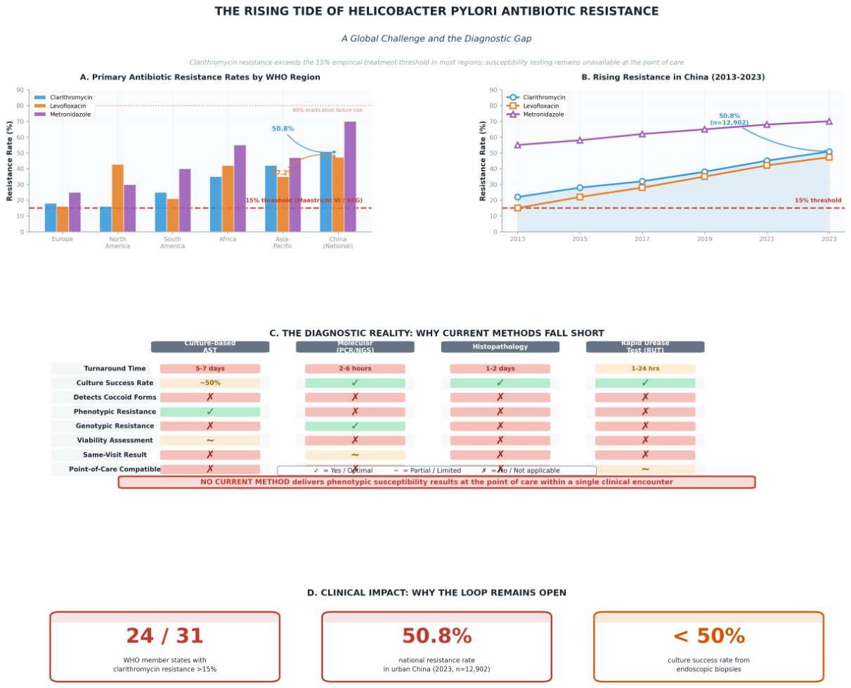
Figure 2.
The closed diagnostic–therapeutic loop for Helicobacter pylori at the endoscope tip: the HP-REVISE pathway. (Left) The traditional pathway follows a linear, delayed sequence: biopsy sampling during endoscopy → UBT/histology confirmation → culture-based reference AST (5–7 days microaerophilic incubation) → drug exposure readout (+72 hours) → empirical eradication without susceptibility data. The inevitable result is treatment failure, resistance selection, and repeated empirical cycles—an open loop. (Center) HP-REVISE (H. pylori real-time evaluation by visual imaging and phenotypic susceptibility estimation) catalyses the shift from delayed retrospective testing to immediate prospective decision-making. (Right) The HP-REVISE closed loop integrates four synergistic steps—SEE (real-time viability visualisation of viable H. pylori, including coccoid forms, within 25–30 minutes), TEST (culture-free phenotypic antimicrobial susceptibility testing via fluorescence residual rate readout within ~1 hour), TREAT (same-visit tailored eradication regimen prescribed before the patient leaves, with viability-based monitoring during follow-up), and TEACH (structured EPVL training and competency assurance that feeds back to raise SEE quality). The cycle closes the diagnostic–therapeutic gap within a single clinical encounter: Diagnosis → Susceptibility → Tailored therapy—all at the endoscope tip.
Figure 2.
The closed diagnostic–therapeutic loop for Helicobacter pylori at the endoscope tip: the HP-REVISE pathway. (Left) The traditional pathway follows a linear, delayed sequence: biopsy sampling during endoscopy → UBT/histology confirmation → culture-based reference AST (5–7 days microaerophilic incubation) → drug exposure readout (+72 hours) → empirical eradication without susceptibility data. The inevitable result is treatment failure, resistance selection, and repeated empirical cycles—an open loop. (Center) HP-REVISE (H. pylori real-time evaluation by visual imaging and phenotypic susceptibility estimation) catalyses the shift from delayed retrospective testing to immediate prospective decision-making. (Right) The HP-REVISE closed loop integrates four synergistic steps—SEE (real-time viability visualisation of viable H. pylori, including coccoid forms, within 25–30 minutes), TEST (culture-free phenotypic antimicrobial susceptibility testing via fluorescence residual rate readout within ~1 hour), TREAT (same-visit tailored eradication regimen prescribed before the patient leaves, with viability-based monitoring during follow-up), and TEACH (structured EPVL training and competency assurance that feeds back to raise SEE quality). The cycle closes the diagnostic–therapeutic gap within a single clinical encounter: Diagnosis → Susceptibility → Tailored therapy—all at the endoscope tip.
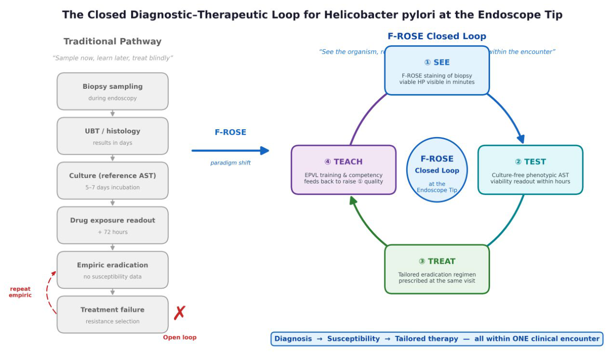
Figure 3.
The HP-REVISE technical workflow. (Left) Endoscopic biopsy sampling. (Centre left) Dual-fluorescence viability staining with acridine orange (AO) and ethidium homodimer-III (EthD-III) for 25–30 minutes. (Centre) Fluorescence microscopy under 460–490 nm excitation renders viable H. pylori—including coccoid forms—visible in real time; viable organisms fluoresce green, non-viable organisms fluoresce red. (Top right) Immediate diagnosis by direct visualisation (SEE). (Bottom right) Same-day phenotypic antimicrobial susceptibility testing via fluorescence residual rate (FRR) after 1-hour antibiotic exposure at EUCAST breakpoint concentrations; resistant organisms retain green fluorescence (R), whereas susceptible organisms lose viability signal (S). The entire SEE→TEST→TREAT loop is closed within a single clinical encounter.
Figure 3.
The HP-REVISE technical workflow. (Left) Endoscopic biopsy sampling. (Centre left) Dual-fluorescence viability staining with acridine orange (AO) and ethidium homodimer-III (EthD-III) for 25–30 minutes. (Centre) Fluorescence microscopy under 460–490 nm excitation renders viable H. pylori—including coccoid forms—visible in real time; viable organisms fluoresce green, non-viable organisms fluoresce red. (Top right) Immediate diagnosis by direct visualisation (SEE). (Bottom right) Same-day phenotypic antimicrobial susceptibility testing via fluorescence residual rate (FRR) after 1-hour antibiotic exposure at EUCAST breakpoint concentrations; resistant organisms retain green fluorescence (R), whereas susceptible organisms lose viability signal (S). The entire SEE→TEST→TREAT loop is closed within a single clinical encounter.
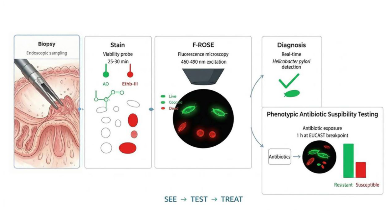
Figure 4.
Representative images comparing conventional histopathology and F-ROSE viability assessment. (A) Helicobacter pylori identified by Giemsa staining of gastric mucosal biopsy (original magnification ×1000). (B) F-ROSE image of viable H. pylori under 460–490 nm excitation after dual-fluorescence viability staining. Note bright green fluorescence of rod-shaped and coccoid organisms. (C) F-ROSE image after antibiotic exposure, showing loss of green fluorescence and uptake of red dye in non-viable organisms, with residual green signal indicating viable resistant organisms. Images are from published studies by our group (20,22).
Figure 4.
Representative images comparing conventional histopathology and F-ROSE viability assessment. (A) Helicobacter pylori identified by Giemsa staining of gastric mucosal biopsy (original magnification ×1000). (B) F-ROSE image of viable H. pylori under 460–490 nm excitation after dual-fluorescence viability staining. Note bright green fluorescence of rod-shaped and coccoid organisms. (C) F-ROSE image after antibiotic exposure, showing loss of green fluorescence and uptake of red dye in non-viable organisms, with residual green signal indicating viable resistant organisms. Images are from published studies by our group (20,22).
Disclaimer/Publisher’s Note: The statements, opinions and data contained in all publications are solely those of the individual author(s) and contributor(s) and not of MDPI and/or the editor(s). MDPI and/or the editor(s) disclaim responsibility for any injury to people or property resulting from any ideas, methods, instructions or products referred to in the content. |
© 2026 by the authors. Licensee MDPI, Basel, Switzerland. This article is an open access article distributed under the terms and conditions of the Creative Commons Attribution (CC BY) license (http://creativecommons.org/licenses/by/4.0/).
Copyright: This open access article is published under a Creative Commons CC BY 4.0 license, which permit the free download, distribution, and reuse, provided that the author and preprint are cited in any reuse.