

Submitted:
06 November 2026
Posted:
09 November 2026
You are already at the latest version
Abstract
Anemia is one of the most common disorders identified in the children, representing a significant global health concern. Delays in diagnosis and treatment may put anemic patients at risk of developing rare and serious complications, including chronic and irreversible cognitive impairment. Risk factors that contribute to the development of anemia in children include eating disorders, chronic comorbidities, inflammation and infection as well as congenital disorders. Pediatricians play a crucial role in identifying these risk factors, which is essential for developing appropriate and timely therapeutic interventions and prevention strategies. Biological sex or gender may influence the risk of anemia, the presentation of symptoms, and the response to therapy, but these are often underexplored in clinical practice and research.
Keywords:
anemia
; children
; anemia risk factors
; anemia and sex differences
1. Introduction
Anemia is a common disorder among children and represents a major global health problem [1]. Delayed diagnosis and treatment of this disease may expose patients to the risk of developing rare and serious complications, including chronic and irreversible cognitive impairment [2].
In 2019, the Global Burden of Disease (GBD) study estimated that there were 1.8 billion cases of anemia worldwide with the highest prevalence among 5- to 9-year-old children of both sexes [3,4]. Anemia is characterized by a reduction in the number of red blood cells (RBCs) or hemoglobin (Hb) levels which leads to impaired oxygen transport. It can cause fatigue, weakness, dizziness, and breathlessness and can affect academic performance and socio-emotional development [1]. These factors have a negative impact on physical growth, cognitive development, and immune function. Risk factors that contribute to the development of anemia in children include nutritional deficiencies, environmental factors, chronic comorbidities, and congenital hemoglobin or RBCs disorders [1].
According to the guidelines of World Health Organization (WHO), anemia is classified as i) mild (Hb between 10 and 12.9 g/dl in males and 11.9 g/dl in females); ii) moderate (Hb between 8 and 9.9 g/dl); and iii) severe (Hb less than 7 g/dl). Based on the mean corpuscular volume (MCV) of red blood cells, anemia can also be categorized morphologically as normocytic (MCV between 80 and 100 femtoliters); microcytic (MCV less than 80 femtoliters); and macrocytic (MCV greater than 100 femtoliters) [5]. Normocytic anemia is frequently present in hospitalized patients and is often due to acute blood loss or secondary to another pathology, such as renal failure, hypothyroidism or multiple myeloma [6]. Microcytic anemia may be due to iron deficiency (IDA), chronic disease, hemoglobinopathies, or sideroblastic anemias. Macrocytic anemia can be divided into megaloblastic and non-megaloblastic types. The former is characterized by a deficiency of vitamin B12 and folate (vitamin B9). The letter can be caused by alcohol, liver disease, myelodysplastic syndrome (MDS), hemolysis, hypothyroidism, and congenital bone marrow failure syndromes.
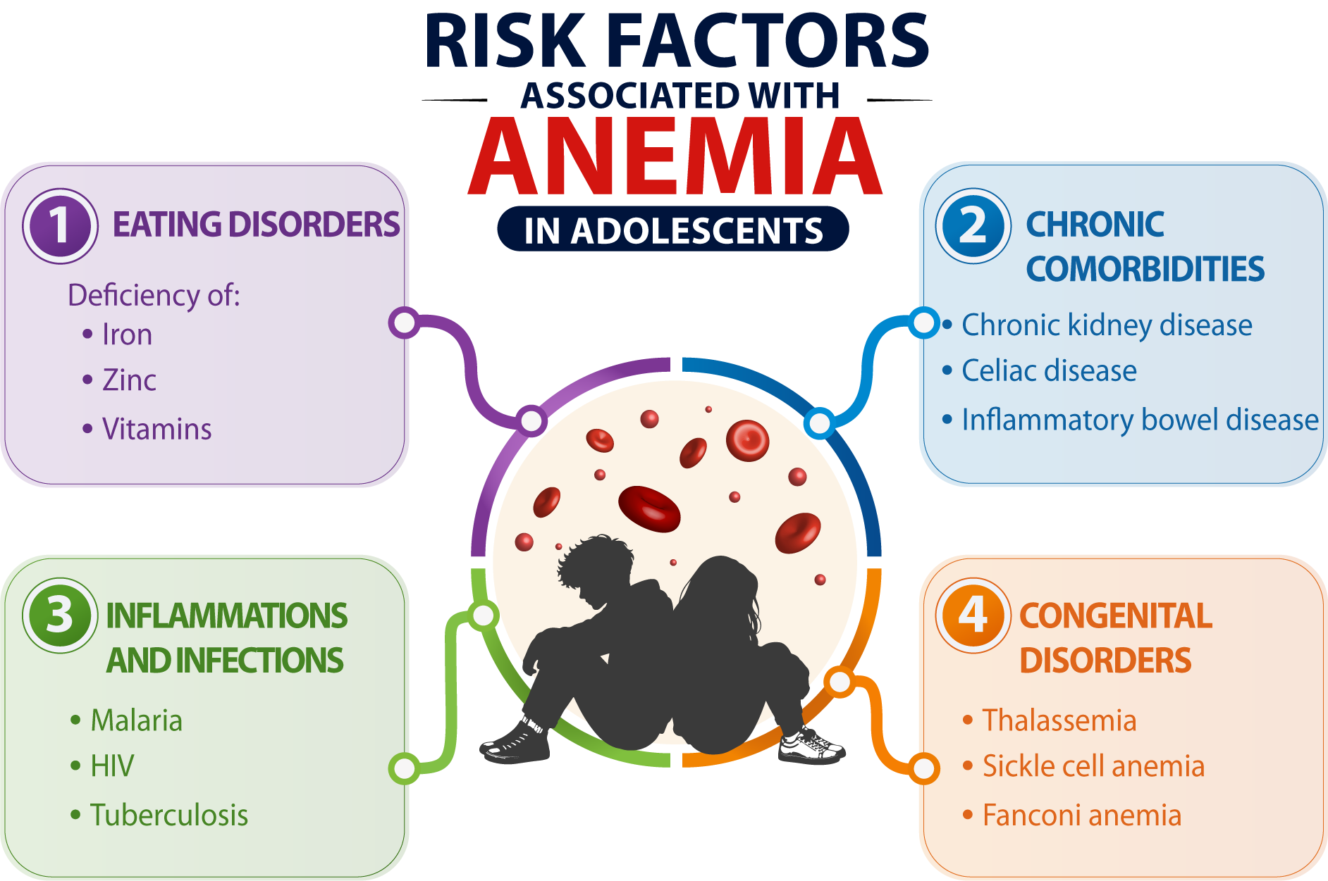
During adolescence, anemia can lead to poor concentration, reduced learning ability, and delayed mental development, which can persist into adulthood. Therefore, it is important to study anemia in adolescents because it affects their development and health in adult life. Risk factors that contribute to the development of anemia in adolescents include eating disorders (EDs), chronic comorbidities, inflammation and infection as well as congenital disorders. Biological sex or gender may influence the risk of anemia, the presentation of symptoms, and the response to therapy, but these are often underexplored in clinical practice and research.
2. Eating Disorders and Anemia
Eating disorders in adolescents can lead to nutritional deficiencies, due to maladaptive dietary restriction [7]. A reduction in essential nutrients such as iron, folic acid (vitamin B9), and vitamin D, essential for blood production in the body, can lead to a low red blood cell count: condition known as nutritional anemia.
Early studies on anemia in adolescents with EDs were not disaggregated by sex and included exclusively or predominantly female patients [8]. In adolescents with a mean age of 12.5 years, the prevalence of EDs ranges from 1% to 22.7% in girls and from 0.3% to 0.6% in boys [9].
Until the publication of the fifth edition of Diagnostic and Statistical Manual of Mental Disorders (DSM-5), amenorrhea was considered a diagnostic criterion for EDs and for this reason these disorders were considered typical of the females [10,11]. EDs are often underdiagnosed in boys, and delays in identification and diagnosis can often lead to increased disease severity. Boys require longer medical hospitalizations and need for more nutrition to establish medical stability. In adolescents, most EDs involve an obsessive focus on body image, weight, and food, which can have consequences for nutrition, growth, and development. Boys have a different perception of weight and body shape. They tend to focus more on developing upper-body muscles, whereas girls tend to want to be thin [12].
2.1. Iron Deficiency
Iron deficiency (ID) is the most common nutritional deficiency worldwide and one of the leading causes of anemia in children [13]. In Europe, its prevalence is estimated at 2-6% among preschool children and 8-20% among adolescent girls [14,15]. Beyond inadequate dietary intake, ID may result from gastrointestinal disorders, chronic inflammation, recurrent infections, and autoimmune diseases [16]. In pediatric populations, iron deficiency is associated with fatigue, impaired concentration, cognitive dysfunction, and reduced academic performance. When occurring during early childhood, it may also lead to neurodevelopmental alterations that are not always fully reversible [17].
Approximately 75% of total body iron is incorporated into hemoglobin and myoglobin, while the remaining fraction is stored in ferritin and hemosiderin, which protect cells from the toxic effects of excess free iron [16]. Accordingly, low ferritin concentrations are indicative of iron deficiency, whereas elevated ferritin levels generally reflect increased iron stores or iron overload. The maintenance of systemic iron homeostasis relies on the coordinated activity of several proteins, including transferrin, the major plasma iron carrier responsible for iron transport between tissues and for scavenging potentially toxic free iron [18]. At the cellular level, the maintenance of systemic iron homeostasis is tightly regulated by the iron regulatory protein/iron-responsive element (IRP/IRE) system. In the event of iron deficiency, activated IRP1 and IRP2 bind to IREs within target mRNA, thereby repressing the translation of ferritin, ferroportin, δ-aminolevulinate synthase 2 (ALAS2) and hypoxia-inducible factor 2 alpha (HIF2α), while simultaneously stabilizing transcripts that encode transferrin receptor 1 (TfR1) and divalent metal transporter 1 (DMT1). This coordinated response maximizes iron uptake and conserves intracellular iron [18,19]. Conversely, when iron is abundant, IRP1 binds to the iron-sulphur [4Fe-4S] cluster, losing its ability to bind to IREs [7]. Meanwhile, IRP2 undergoes FBXL5-mediated proteasomal degradation [20].
Another adaptive mechanism that is activated during iron deficiency is ferritinophagy, a selective autophagic process that mobilizes iron that is stored in ferritin. This pathway is mediated by the nuclear receptor coactivator 4 (NCOA4), which targets ferritin to lysosomes for degradation, thereby releasing iron into the cytosolic labile iron pool. The expression and activity of NCOA4 increase during iron deprivation and hypoxia, thereby contributing to the maintenance of cellular iron availability [21,22].
Growth and iron status are closely interconnected throughout childhood and adolescence. Holmlund-Suila and colleagues demonstrated that, in males, both growth and ferritin concentrations are age-dependent, whereas in females’ growth is associated with lower ferritin levels [23]. Consequently, iron-deficiency anemia (IDA) is more common among adolescent girls than boys, likely reflecting the increased iron requirements associated with pubertal development and hemoglobin expansion [24]. During adolescence, males generally exhibit higher hemoglobin concentrations than females because prostaglandins stimulate erythropoiesis both directly and indirectly through cyclic AMP pathways, while androgens enhance erythropoietin production and estrogens exert inhibitory effects [25]. At the systemic level, iron metabolism is governed by the hepcidin-ferroportin axis. Hepcidin, which is produced primarily by the liver, binds to ferroportin, inducing its internalization and degradation. This limits the export of iron from enterocytes and macrophages. During iron deficiency or increased erythropoietic demand, hepcidin synthesis is suppressed, thereby enhancing systemic iron availability. Concurrently, iron deficiency and hypoxia stabilize HIF2α, which stimulates the expression of intestinal iron transport genes, including DMT1, Dcytb and ferroportin, while also promoting renal erythropoietin (EPO) production [26]. EPO, in turn, induces erythroblasts to secrete erythroferrone (ERFE), which suppresses hepatic hepcidin synthesis and ensures an adequate iron supply for erythropoiesis [27]. The physiological importance of these regulatory pathways is particularly evident during erythroid maturation. Indeed, iron deficiency impairs hem and hemoglobin synthesis by reducing the formation of iron-sulphur (Fe-S) clusters, which, as mentioned earlier, are essential for the activity of key enzymes such as ALAD and FECH. In parallel, IRP1 represses ALAS2 translation, further limiting hem biosynthesis. These coordinated alterations ultimately result in the development of the characteristic microcytic and hypochromic anemia associated with iron deficiency [21].
In addition to nutritional iron deficiency, chronic inflammation is a significant cause of functional iron deficiency. Pro-inflammatory cytokines, particularly IL-6, activate the JAK2/STAT3 signaling pathway in hepatocytes, inducing excessive hepcidin production. Elevated hepcidin levels promote ferroportin degradation, causing iron to be sequestered within macrophages and enterocytes and reducing its availability for erythropoiesis, even when total body iron stores are adequate or increased. This condition is the underlying cause of the anemia associated with chronic inflammatory states [28].
Epidemiological data further highlight the burden of IDA in adolescence. A study conducted among Chinese adolescent girls reported an overall anemia prevalence of 8.5% in individuals aged 10-17 years, with mild, moderate, and severe anemia accounting for 65.9%, 31.8%, and 2.3% of cases, respectively [29].
From a diagnostic perspective, serum ferritin, transferrin saturation, soluble transferrin receptor (sTfR), and the sTfR/ferritin index are considered the most informative biomarkers of iron-deficiency anemia [30]. However, except for sTfR, these parameters may be influenced by inflammation and chronic disease. Measurement of reticulocyte hemoglobin content has also emerged as a valuable indicator of iron availability and an early marker for the diagnosis of IDA [31].
2.2. Zinc Deficiency
Zinc (Zn) is the second most abundant trace element in the body. Its homeostasis is primarily regulated by the ZIP (SLC39A) and ZnT (SLC30A) transporter families. ZIP transporters facilitate zinc entry into the cytosol, while ZnT transporters promote its export or storage within intracellular organelles. Under zinc-deficient conditions, the expression of import transporters, particularly ZIP4, increases, while the expression of ZnT1 and metallothioneins is suppressed, thereby reducing cellular zinc losses. Furthermore, intracellular transporters such as ZIP7 and ZIP13 regulate zinc distribution within organelles and are essential for the proper functioning of the endoplasmic reticulum, development, and the immune response [32].
Zn is involved in several metabolic processes, particularly those related to children's growth and development. However, its deficiency has only recently been associated with anemia and the literature on this topic is very limited [33]. Plasmatic Zn can influence hemoglobin synthesis and stimulate erythropoiesis through several zinc-dependent enzyme systems [26]. During terminal erythroid differentiation, erythroblasts accumulate high levels of zinc, which are necessary for cell proliferation and hem synthesis [34]. Furthermore, Zn plays an important role in stabilizing RBC membranes and contributing to their integrity. Reduced plasma Zn affects the half-life of RBCs, making them more susceptible to hemolysis [35]. Zinc deficiency must cooperate with other factors to cause anemia [36]. The major cooperative factor may be iron deficiency, but it can also cooperate with deficiencies of folate, vitamin B12, vitamin B6, riboflavin, and vitamin A [36]. From a clinical perspective, zinc deficiency often occurs alongside iron deficiency, particularly in pregnant women, children and patients with chronic kidney disease. This is due to shared dietary sources and common inhibitors of intestinal absorption, such as phytates [34,37]. While isolated zinc deficiency rarely causes severe anemia, it can exacerbate iron deficiency and other nutritional or inflammatory disorders, thereby further impairing erythropoiesis [34]. During anemia, systemic zinc redistribution also occurs, with zinc being mobilized from plasma and bone reserves towards the bone marrow to support erythropoietic activity. This redistribution results in the characteristic finding of reduced plasma Zn concentrations alongside elevated RBC zinc content, reflecting the high Zn requirements of reticulocytes compared to mature RBCs [34]. A retrospective study conducted of the medical records of 87 males and 450 females aged 9–25 years who were hospitalized for eating disorders has shown that, although the prevalence of zinc deficiency did not differ by sex, it was associated with a greater likelihood of anemia in males but not in females [38].
2.3. Vitamins Deficiency
In addition to deficiencies in iron and zinc, anemia can be caused by deficiencies in certain vitamins, including vitamins A, B12, B6, C, D, E, and folate. Vitamins A, B6, and B12, as well as folate (vitamin B9), are necessary for erythropoiesis and iron metabolism. Deficiency in these vitamins can lead to specific types of anemia via distinct molecular mechanisms.
In particular, vitamin B12 and vitamin B9 work together in one-carbon metabolism, which is necessary for nucleotide synthesis and the re-methylation of homocysteine. A vitamin B12 deficiency blocks the activity of methionine synthase, whereby folate becomes trapped. This results in defects in DNA synthesis and the abnormal incorporation of uracil, as well as genomic damage and the arrest of erythroblast proliferation. [39]. The outcome is megaloblastic anemia, a type of macrocytic anemia characterized by a low RBC count, macrocytic RBCs, fewer immature RBCs (e.g. reticulocytes) and fewer platelets. A defining feature of megaloblastosis is the asynchronous maturation of the cytoplasmic and nuclear compartments of erythroid progenitors, resulting in abnormal morphology [40].
Vitamin B6 is an essential cofactor for ALAS2, the rate-limiting enzyme in hem biosynthesis. A deficiency of this vitamin compromises the production of hemoglobin, resulting in sideroblastic hypochromic microcytic anemia. This condition is characterized by iron accumulation in erythroblastic mitochondria and the formation of ring-shaped sideroblasts. Unlike iron-deficiency anemia, this condition is not associated with iron depletion, nor does it trigger the HIF2α-mediated intestinal response. Therefore, the activity of this factor could be used as a biomarker to distinguish between iron-responsive and non-iron-deficiency anemias [41].
Vitamins C and E, on the other hand, can protect RBCs due to their antioxidant properties [42].
It has been demonstrated that serum vitamin A levels are significantly and independently inversely associated with anemia risk in children and adolescents. This association was more pronounced in males, suggesting a potential role for vitamin A in the prevention of sex-specific anemia. Serum vitamin B12 levels have been shown to decrease in adults and the elderly. Although no sex differences have been observed, males appear to have a higher frequency of vitamin B12 deficiency [43]. In developing countries such as Indonesia, there is an association between micronutrient deficiencies and growth retardation. A study by Ernawati et al., showed that children aged 6-59 months from a low socioeconomic group had significantly lower mean values of hemoglobin, ferritin, and retinol values [44]. In a study of Korean children, Lee et al. observed that vitamin A deficiency was associated with an increased risk of IDA only in female children and adolescents [45]. Tsui and Nordstrom showed that adolescent males had significantly higher folate levels in both serum and RBCs than adolescent girls [46]. In Table 1 sex differences in anemia associated with nutrient are shown.
3. Anemia Associated to Comorbidities
Anemia often occurs alongside chronic health conditions, worsening the overall prognosis of the disease, increasing the risk of hospitalization and accelerating functional decline. It commonly arises from chronic kidney disease (CKD), celiac disease (CD), Crohn's disease, and inflammatory bowel disease (IBD).
3.1. Chronic Kidney Disease
Chronic kidney disease (CKD) is a progressive condition characterized by irreversible loss of kidney function. It is associated with chronic inflammation, the accumulation of uremic toxins, and systemic metabolic abnormalities.
Globally, the incidence and prevalence of CKD are more common in women, while mortality is higher in men. CKD and its progression are likely independently modulated by a series of physiological risk factors such as obesity, hypertension, and socio-economic factors that differ between men and women. Many women live in poverty and have less access to healthcare than men. Meanwhile, men are more likely to be injured at work [47].
Anemia is one of its most common complications, with an increasing prevalence as estimated glomerular filtration rate declines. It eventually affects the majority of patients in advanced stages and on dialysis. While it can be caused by a shortened lifespan of red blood cell and iron deficiency, decreased production of erythropoietin by the kidneys is the primary cause [48]. In addition to iron deficiency, anemia in children with CKD may be due to deficiencies in vitamin B12, folate, and possibly vitamin D, that may interfere with the production of healthy, mature red blood cells [49]. In both children and adults with CKD, anemia increases with advancing stage of disease. The North cardiovascular disease American Pediatric Renal Trials and Collaborative Studies (NAPRTCS) have demonstrated that the prevalence of anemia is 73% in children at stage 3 CKD; 87% at stage 4 and > 93% at stage 5 [50]. In children with CKD, anemia is associated with morbidity, an increased risk of cardiovascular disease and reduced exercise capacity [51].
Among adolescents on hemodialysis, anemia is associated with an increased frequency of hospitalization and an increased risk of mortality [52]. Using data from the Global Burden of Disease 2021, Qi and colleagues found that the incidence, prevalence, mortality, and years of life lost due to CKD have increased steadily from 1999 to 2021. Furthermore, they discovered that from the age of 1, the incidence and prevalence of anemia in children with CKD is higher in girls, while the mortality rate is higher in boys [53].
3.2. Celiac Disease
Celiac disease (CD) is a systemic autoimmune disorder triggered by gluten in genetically predisposed individuals who carry the HLA-DQ2 or HLA-DQ8 haplotypes. When gliadin peptides cross the intestinal barrier and are modified by tissue transglutaminase 2 (TG2), they trigger both adaptive and innate immune responses. The IL-15/MICA/NKG2D axis promotes the destruction of enterocytes by intraepithelial lymphocytes, resulting in villous atrophy, infiltration of the lamina propria by immune cells, crypt hyperplasia and a reduction in the intestinal absorptive surface area [54,55]. CD can also present with extraintestinal manifestations including fatigue, weight loss, skin rashes, anemia, and osteoporosis [56]. A systematic review and meta-analysis revealed that the prevalence of CD worldwide ranged from 0.05% to 2.6% in the 2018, with an increase observed in countries such as China, India, and Iran due to higher wheat consumption [57].
In CD patients, anemia can be due to malabsorption of iron and vitamins such as folic acid, and vitamin B12; chronic intestinal inflammation that is typical of CD; and extraintestinal diseases (e.g., arthritis and dermatitis) [58,59]. In CD patients, malabsorption of iron is due to atrophy of the mucosa of the duodenum and proximal jejunum, which are the main sites of iron absorption. This atrophy is caused by gliadin or its derived peptides, such as p31-49 and p57-73 [60]. Although a gluten-free diet promotes recovery of the intestinal mucosa and resolution of inflammation, anemia may persist in 15–46 per cent of patients one year after diagnosis. This is attributed to factors such as slow mucosal recovery, concomitant blood loss, dietary contamination, the absence of iron fortification in gluten-free products and microscopic alterations to the microvilli that impair micronutrient absorption despite histological normalization [54,61].
Although the association between anemia and CD has been reported in the literature, it remains rare in children [62]. In a cross-sectional study of both adults and children with CD, Valvano et al., found that anemia prevalence was higher in adults than in pediatric patients [63]. These differences could be due to the recovery of the duodenal mucosa recovering more rapidly and completely in children than in adults or to a diagnostic delay in adults [64,65]. Although the reason is still unknown, the prevalence of CD is higher in girls than in boys [66,67].
3.3. Inflammatory Bowel Diseases
Inflammatory bowel diseases (IBDs) are chronic immune-mediated disorders of the gastrointestinal tract. The two main types are Crohn's disease and ulcerative colitis (UC). Crohn's disease can affect any part of the gastrointestinal tract, whereas UC can affect only the colon [68]. Up to 25% of patients with IBD are diagnosed in childhood or adolescence. The highest incidence rates of IBD are observed in in Europe and North America [69]. The most common metabolic complication of IBD is IDA and anemia of chronic disease (ACD), which are associated with quality of life and cognitive function [70]. The iron-deficiency component is caused by chronic gastrointestinal blood loss, inflammation and damage to the intestinal mucosa, reduced iron absorption, and inadequate dietary iron intake due to malnutrition and dietary restrictions. Meanwhile, chronic inflammation alters the distribution and availability of iron, impairing erythropoiesis and contributing to the development of ACD [70].
Male patients over 30 years of age have a lower risk of anemia than younger patients (under 30 years of age), while the risk of anemia is the same for women at all ages [71]. In a retrospective cross-sectional study female sex was found to be the most significant risk factor for anemia in both Crohn's disease and UC [72].
In Western countries, anemia is more prevalent among adolescents, particularly females (3.5-13% in males and 11-33% in females). Table 2 shows sex differences in anemia associated with comorbidities.
4. Anemia Associated with Inflammation and Infection.
Inflammation and infection are among the most common causes of acquired anemia, particularly in patients with chronic diseases. This type of anemia arises from the interplay between immune activation, iron dysregulation, impaired erythropoiesis, and reduced erythrocyte survival. This highlights the close relationship between the immune and hematopoietic systems.
4.1. Inflammation
Anemia associated with inflammation (inflammatory anemia, IA) or ACD is one of the most common forms of anemia associated with acute and chronic inflammatory conditions. It has a multifactorial pathogenesis resulting from the coordinated action of pro-inflammatory cytokines, including IL-6, IL-1β, TNF-α and IFN-γ. These cytokines profoundly alter iron metabolism, erythropoietin production and RBC survival [73,74].
Anemia of inflammation can present as mild normocytic or normochromic anemia with Hb levels between 8 and 10 g/dL, elevated inflammatory markers, reduced serum iron concentration, and transferrin saturation (e.g., serum ferritin levels greater than100 ng/mL). It is difficult to distinguish from IDA, and the two conditions can often coexist [75]. In preschool children, IA can be assessed through the measurement of C-reactive protein (CRP). This often manifests as an increase in inflammatory cytokines, such as IL-6, TNF-α, and IFN-γ, which reduce iron availability and inhibit the maturation of erythroid precursor cells in the bone marrow. Furthermore, IFN-γ, TNF-α and IL-1β inhibit the production of EPO in the kidneys by suppressing HIF-2α-mediated pathway. This results in EPO levels that are inappropriately low given the severity of the anemia. In the bone marrow, these same cytokines reduce the response of erythroblasts to EPO and promote the apoptosis of erythroid progenitors. This leads to reduced erythroid proliferation and differentiation [76].
4.2. Parasitic Infection
Parasitic infections represent an important cause of anemia, particularly in children living in endemic areas, where they often coexist with nutritional deficiencies and chronic inflammation. Several intestinal parasites have been associated with deficiencies of micronutrients essential for erythropoiesis, including iron, zinc, and vitamins A and D. In particular, infections caused by Giardia lamblia and Blastocystis spp. have been linked to deficiencies in vitamins A and D, whereas Entamoeba coli infection is predominantly associated with vitamin D deficiency and Iodamoeba bütschlii infection with zinc deficiency [77]. These nutritional alterations can contribute directly or indirectly to impaired red blood cell production and the development of anemia.
Evidence from pediatric populations supports a strong association between parasitic infections and anemia. In a cross-sectional study conducted in Argentina involving 162 children aged between 6 months and 14 years, nearly half of the participants (n = 76) presented with predominantly mild anemia (92.1%). Blastocystis hominis was identified as the most frequent parasitic infection, followed by Entamoeba coli, hookworms, and Ascaris lumbricoides. Multivariable analysis demonstrated a significant association between anemia, intestinal parasitosis, and male sex [78]. The higher prevalence and intensity of parasitic infections observed in males have been attributed, at least in part, to sex-related immunological and hormonal differences that may influence susceptibility to infection and disease severity [79].
Beyond nutrient depletion, parasitic infections contribute to anemia through activation of inflammatory pathways that alter systemic iron homeostasis. Similar to bacterial and viral infections, parasitic diseases induce a host defense response known as nutritional immunity, which limits iron availability to restrict pathogen growth. This response is mediated by pro-inflammatory cytokines, particularly IL-6, IFN-γ, and TNF-α, which stimulate hepatic hepcidin production. Elevated hepcidin promotes the degradation of ferroportin, the major cellular iron exporter expressed on enterocytes, macrophages, and hepatocytes, thereby inhibiting intestinal iron absorption and iron release from tissue stores. The resulting hypoferremia and iron sequestration within ferritin stores reduce iron availability for erythropoiesis and contribute to the development of inflammatory anemia [80].
4.3. Malaria
Malaria is one of the primary causes of severe anemia worldwide. It is particularly prevalent in Africa, but due to increased travel to endemic areas and the significant influx of immigrants, the number of malaria cases has also increased in non-endemic countries [81]. There are six species of Plasmodium parasites that infect humans, all of which cause anemia. P falciparum is the most prevalent parasite in Africa and is responsible for the majority of malaria-related deaths [82].
Malaria-induced anemia arises from a complex interaction between parasite-mediated RBC destruction and host immune responses that collectively impair erythropoiesis and disrupt iron metabolism [83]. Following invasion of RBCs by merozoites, parasitic proteins such as P. falciparum RBCs membrane protein 1 (PfEMP1) remodel the RBC membrane, promoting cyto-adhesion, microvascular sequestration, and reduced cellular deformability. Subsequent rupture of infected RBC releases merozoites, free hemoglobin, and hemozoin, generating substantial oxidative stress and depleting intracellular antioxidant defenses, including glutathione [83]. A hallmark of severe malarial anemia is the disproportionate loss of uninfected RBCs. In addition to the clearance of parasitized RBCs, many non-infected RBCs are removed because of reduced deformability, complement activation, antibody-mediated opsonization, and accelerated eryptosis. Indeed, it has been estimated that up to ten uninfected RBCs may be destroyed for every infected cell eliminated. Increased eryptosis and premature senescence of non-parasitized erythrocytes have been documented in both P. falciparum and P. vivax infections, further contributing to anemia severity [84,85]. These events contribute not only to RBCs destruction, but also to tissue damage and systemic inflammation. Indeed, it has been reported that the innate immune system induces a strong inflammatory response after Plasmodium infection to eliminate infected RBCs. Monocytes/macrophages produce mediators such as TNF-α and IFN-γ, which are potent inhibitors of erythropoiesis. TNF-α significantly decreases the proliferation of human erythroid progenitor cell, while IFN-γ inhibits the proliferation and the differentiation of erythroblasts [86,87].
Certain populations are particularly vulnerable to malaria-associated complications, including newborns, people with primary sclerosing cholangitis, and pregnant women. Children under 5 years of age are at increased risk of death. Malaria has a higher incidence and/or prevalence in males, beginning in late childhood, persisting through puberty and most of adulthood (except during pregnancy, when it is more prevalent in females). The lower prevalence in women is due to the rapid elimination of asymptomatic infections [88].
4.4. HIV Infection
Worldwide, there are 1.7 million children under 15 years of age infected with the human immunodeficiency virus (HIV), and 91% of whom are in sub-Saharan Africa [89]. It is estimated that 39.7% of infected children suffer from anemia, which significantly impacts quality of life and health outcome [89]. In HIV infected patients, severe anemia is associated with reduced survival rates and is considered a prognostic marker of disease progression [90]. The pathogenesis of HIV-associated anemia is multifactorial and reflects the combined effects of chronic inflammation, direct bone marrow injury, impaired erythropoiesis, altered iron metabolism, opportunistic infections, nutritional deficiencies, and antiretroviral drug toxicity [91,92,93,94].
Inflammatory anemia is the most common hematological manifestation of HIV infection, particularly in advanced stages of the disease [95]. Elevated concentrations of pro-inflammatory cytokines, particularly IL-6, stimulate hepatic hepcidin production, thereby disrupting systemic iron homeostasis. Hepcidin promotes the internalization and degradation of ferroportin, which is the main cellular iron exporter found in enterocytes and macrophages. Consequently, intestinal iron absorption and iron mobilization from body stores are reduced, leading to hypoferremia and iron sequestration within macrophage ferritin stores, despite total body iron reserves being adequate or increased. This functional iron deficiency restricts the availability of iron for erythropoiesis and substantially contributes to HIV-associated anemia [95].
Effective antiretroviral therapy attenuates systemic inflammation, improves iron metabolism and partially restores hematological parameters in patients with advanced HIV infection [96]. However, even in individuals with sustained virological control, functional iron deficiency may persist, characterized by reduced serum iron concentrations and transferrin saturation, despite normal or elevated ferritin levels [94].
Beyond disturbances in iron metabolism, HIV directly impairs hematopoiesis. The virus itself and the inflammatory microenvironment can both damage hematopoietic stem and progenitor cells, inducing apoptosis, cellular senescence and defective erythroid, myeloid and megakaryocytic differentiation [96]. Inflammatory cytokines such as TNF-α and IFN-γ further suppress erythropoiesis by inhibiting erythroid progenitor proliferation and reducing responsiveness to EPO, thereby limiting the production of effective red blood cells [92].
Antiretroviral therapy itself may also contribute to anemia development. Zidovudine (AZT), one of the most widely used nucleoside reverse transcriptase inhibitors, is particularly associated with bone marrow suppression and impaired erythroid cell production [94]. Furthermore, zidovudine and other nucleoside analogues can induce mitochondrial toxicity, resulting in macrocytic anemia characterized by an increased MCV and ineffective erythropoiesis [94].
In both adolescents and adults, gender has a significant effect on the prevalence of HIV-associated anemia. Compared to men, women bear a greater burden of anemia due to factors such as blood loss during menstruation and hormonal changes. A cross-sectional study of 631 HIV patients aged 18 years or older found that females exhibited a higher overall prevalence of anemia (41.1%) than males (27.1%). The prevalence of mild anemia was equal in both sexes (21.1%), while moderate anemia was more common in females (15%) than in males (5.6%). Severe anemia was significantly higher in females (5%) than in males (0.4%) [98].
4.5. Tubercolosis
Tuberculosis (TB) is a chronic infectious disease caused by Mycobacterium tuberculosis, which requires iron for its growth and reproduction. Due to their immature immune systems, children are more susceptible to infection [99]. TB is considered a global pediatric emergency that must be prevented. Children under 15 years of age represent 11% of those affected [100]. The countries with the highest number of cases are Africa, Asia and China [101]. Prevalence of TB infection varies by age and sex. A study conducted in Brazil found that, among 5-14 years old, there was a more significant increase in infections among females, whereas among 15-39 years old, there was a more significant increase in infections among males. No gender differences were found after the age of 40 [102]. These findings suggest that biological and socio-environmental factors may contribute differently to TB susceptibility across the lifespan.
Anemia is a common complication of TB and inhibition of RBC production and alteration of iron metabolism by inflammatory mediators are the main causes [103].
The pathogenesis of anemia during TB is closely linked to the host immune response and the pathogen's dependence on iron. M. tuberculosis infection triggers the production of pro-inflammatory cytokines, including IL-6 and TNF-α. These cytokines lead to a reduction in intestinal iron absorption and prevent the release of iron from macrophages and reticuloendothelial stores. Consequently, circulating iron levels decline while iron accumulates in ferritin-rich macrophages, resulting in hypoferremia and restricted iron availability for erythropoiesis [104]. This response is an important component of nutritional immunity, which aims to limit pathogen access to iron. However, prolonged iron sequestration can paradoxically favor the intracellular persistence and virulence of M. tuberculosis. This increases the risk of disease progression and adverse clinical outcomes. Conversely, reduced iron delivery to the bone marrow can impair erythroid differentiation and hemoglobin synthesis [104].
Anemia in TB is clinically relevant because it is associated with increased mortality, treatment failure and disease relapse in patients with both isolated TB and HIV co-infection [95,104].
Literature data show that anemia is prevalent in 30%–94% of TB patients [105]. A retrospective study reported that most children with TB-related anemia had mild to moderate microcytic hypochromic anemia, and that there was a strong correlation between the severity of anemia and tuberculosis-induced inflammation. This study observed that, in the 1 to 8 age group, anemic girls outnumbered boys [106]. The study has also demonstrated that, after one month of antitubercular therapy, anemic children with TB returned normal.
Sex-related differences in anemia associated with infectious diseases are summarized in Table 3.
5. Anemia Associated to Congenital Disorders
Congenital disorders are an important cause of anemia, encompassing a diverse group of inherited conditions that affect the production, structure, function or survival of red blood cells. The clinical manifestations of these disorders can vary widely, ranging from mild, asymptomatic forms to severe, life-threatening conditions that require lifelong management.
5.1. Thalassemia
Thalassemia is a heterogeneous group of autosomal recessive hemoglobinopathies characterized by defective synthesis of the α- or β-globin chains of hemoglobin. α-Thalassemia results from deletions affecting one or more α-globin genes, leading to reduced or absent α-globin production, and is particularly prevalent in African and Southeast Asian populations. In contrast, β-thalassemia is caused by mutations in the HBB gene that reduce or abolish β-globin synthesis and occurs predominantly in populations of Mediterranean, African, Middle Eastern, and Southeast Asian origin [107]. According to the severity of the genetic defect and the resulting clinical phenotype, β-thalassemia is generally classified into β-thalassemia minor (trait), β-thalassemia intermedia, and β-thalassemia major. β-Thalassemia minor is usually asymptomatic and often identified incidentally, whereas β-thalassemia intermedia and β-thalassemia major are characterized by clinically significant anemia and frequently require regular blood transfusions. In particular, β-thalassemia major represents the most severe form of the disease and is characterized by chronic hemolysis, ineffective erythropoiesis, and progressive iron overload [108].
At the molecular level, reduced β-globin synthesis creates a marked imbalance between α- and β-globin chain production. Excess unpaired α-globin chains accumulate within erythroid precursors and mature RBCs, where they form unstable aggregates that generate reactive oxygen species and induce severe oxidative stress. The resulting membrane damage promotes eryptosis, premature erythrocyte destruction, and chronic hemolysis, ultimately leading to the characteristic microcytic hypochromic anemia of β-thalassemia [109,110,111].
Sex-related differences have also been reported in the clinical manifestations of β-thalassemia. Although both males and females are exposed to similar risks of iron accumulation, females appear to exhibit greater tolerance to iron-induced oxidative damage. A study published in 2011 demonstrated that males with β-thalassemia major had significantly higher frequencies of cardiac dysfunction and arrhythmias than females across multiple age groups. These findings suggest that sex-specific differences in oxidative stress responses may influence susceptibility to iron toxicity and disease complications [112,113].
5.2. Sickle Cell Anemia
Sickle cell disease (SCD) is a chronic inherited hemoglobinopathy caused by a single point mutation in the HBB gene, resulting in the substitution of glutamic acid with valine at position 6 of the β-globin chain and the formation of sickle hemoglobin (HbS) [114]. This disease is particularly prevalent in sub-Saharan Africa, India, and the Middle East, where it represents a major cause of morbidity and mortality [89].
Under physiological conditions, HbS can adequately transport oxygen; however, under hypoxic conditions, HbS polymerizes into rigid intracellular fibers that distort RBCs into the characteristic sickle shape. The structural change reduces RBC deformability, promotes cellular dehydration, and accelerates RBCs destruction, ultimately leading to chronic hemolytic anemia [114]. The development of anemia in SCD is driven by persistent hemolysis and marked oxidative stress. The intrinsic instability of HbS, together with activation of NADPH oxidase, enhances the generation of reactive oxygen species (ROS), while antioxidant defenses within RBCs are simultaneously compromised. Excess ROS induces membrane lipid peroxidation, disruption of phospholipid asymmetry, and exposure of phosphatidylserine on the RBC surface, thereby promoting eryptosis and premature removal of RBCs by the reticulo-endothelial system [114].
Children with sickle cell anemia (SCA) have elevated levels of adiponectin and anti-inflammatory adipokines. In SCA patients, RBCs are sickle-shaped, rigid, and prone to intravascular hemolysis, causing vaso-occlusive episodes [90,116]. These are associated with an increased risk of acute kidney injury (AKI) and subsequent mortality [116,117].
Sex differences in SCA remain unknown, although females have a longer life expectancy than males [118]. In a study conducted on a cohort of 185 Ugandan children (41.6% females and 58.4% males) aged between 2 and 18 years, it was observed that females were at risk of developing chronic kidney disease, while males were at risk of rapid progression to renal failure and mortality [119]. Research in this field has often reported a higher prevalence of SCA in females than in males [120].
Notably higher sex differences in AKI risk were observed in adolescence, suggesting that sex hormones may contribute to the increased AKI risk in this susceptible population. Additional research is required to better understand the pathobiology of sex impacts on clinical outcomes for children with SCA and AKI. Compared to boys, girls with sickle cell anemia may be at increased risk of AKI and have a higher rate of acute chest syndrome [121,122].
5.3. Fanconi Anemia
Fanconi anemia (FA) is a [123,124inherited chromosomal instability syndrome caused by germline mutations in genes of the FANC pathway, which is responsible for the repair of DNA damage, particularly interstrand crosslinks. FA is a rare genetic disorder involving defects in the DNA repair. It is characterized by early bone marrow failure and pancytopenia, which can progress to aplastic anemia. The disease has an estimated incidence of approximately 1 in 136,000 live births, with higher prevalence in certain populations, including individuals of South African, sub-Saharan African, and Spanish Gypsy ancestry [123,124].
The disease is characterized by progressive bone marrow failure, pancytopenia, congenital abnormalities, and a marked predisposition to malignancy. Severe anemia remains one of the leading causes of death during childhood [123,124]. At the molecular level, FANC proteins coordinate a complex DNA damage response pathway that safeguards genomic stability during cell replication. The pathophysiology of FA is closely linked to the accumulation of endogenous genotoxic damage and oxidative stress [123,124].
FA affects both sexes equally because it is predominantly inherited as an autosomal recessive disorder, although clinically relevant sex-specific differences have been reported [125]. However, significant gender differences exist in terms of clinical presentation, disease progression, cancer risk, and fertility. Compared to females, males with FA have a higher risk of developing bone marrow failure, are less fertile, and suffer from hypogonadism [125]. However, females, have higher levels of spontaneous chromosomal breakage and an increased pro-oxidant status compared to males [126]. Approximately 50% of females are infertile, and those who are fertile experience early menopause. During pregnancy, fertile women are at an increased risk of severe anemia and thrombocytopenia [127]. Table 4 shows sex differences in anemia associated with congenital disorders
6. Conclusion and Future Perspectives
The prevalence of anemia among adolescents represents a widespread and significant global health problem, particularly in low- and middle-income countries, where progress is slow and uneven. If left undiagnosed or untreated, anemia, can increase the adolescents' risk of developing complications such as chronic and irreversible cognitive impairment and impact development and health in adulthood. Various risk factors contribute to the development of anemia in children, including nutritional deficiencies, environmental factors, chronic comorbidities, and congenital disorders of hemoglobin or red blood cells. In addition to these risk factors, biological sex and gender can influence how symptoms manifest and how adolescents respond to treatment, but these aspects are often overlooked in clinical practice and research.
In this review, we present data from studies on pediatric anemia conducted in various countries worldwide, with a focus, on gender differences. Our research has shown that, in recent years, both sex and gender have been considered in relation to many anemia risk factors in pediatric patients.
This could pave the way for targeted interventions to optimize health outcomes from early childhood onwards.
Author Contributions
Conceptualization, I.T.D.J., G.P., R.V., E.S. Investigation, E.S., R.V. and C.C. Drafting, L.G., M.E., E.I., L.G. Revising, R.V. and E.S. Final approval of the version to be published, E.S. All authors have read and agreed to the published version of the manuscript. All authors have read and agreed to the published version of the manuscript.
Funding
This research received no external funding.
Institutional Review Board Statement
Not applicable.
Informed Consent Statement
Not applicable.
Data Availability Statement
No new data were created or analyzed in this study. Data sharing is not applicable to this article.
Conflicts of Interest
The authors declare that the research was conducted in the absence of any commercial or financial relationships that could be construed as potential conflicts of interest.
Abbreviations
The following abbreviations are used in this manuscript:
| ACD | Anemia of Chronic Disease |
| ALAD | Delta-Aminolevulinic Acid Dehydratase |
| ALAS2 | δ-Aminolevulinate synthase 2 |
| AMP | Adenosine Monophosphate |
| AKI | Acute Kidney Injury |
| AZT | Zidovudine |
| CD | Celiac Disease |
| CKD | Chronic Kidney Disease |
| CRP | C-Reactive Protein |
| Dcytb | Duodenal Cytochrome b |
| DMT1 | Divalent Metal Transporter 1 |
| DSM-5 | Diagnostic and Statistical Manual of Mental Disorders |
| EDs | Eating Disorders |
| EPO | Renal Erythropoietin |
| ERFE | Erythroferrone |
| FA | Fanconi Anemia |
| FECH | Ferrochelatase |
| Fe-S | Iron-Sulphur |
| GBD | Global Burden of Disease |
| HB | Hemoglobin |
| HbS | Sickle hemoglobin |
| HIF2α | Hypoxia-inducible factor 2 alpha |
| HIV | Human immunodeficiency virus |
| IA | Inflammatory Anemia |
| IBDs | Inflammatory Bowel Diseases |
| ID | Iron Deficiency |
| IDA | Iron Deficiency anemia |
| IFN-δ | Interferon gamma |
| IL-6 | Interleukin 6 |
| IRP | Iron Regulatory Protein |
| IRE | Iron-Responsive Element |
| MCV | Mean Corpuscular Volume |
| MDS | Myelodysplastic Syndrome |
| NAPRTCS | The North cardiovascular disease American Pediatric Renal Trials and Collaborative Studies |
| NCOA4 | Nuclear Receptor Coactivator 4 |
| PfEMP1 | P. falciparum RBCs membrane protein 1 |
| PGE | Prostaglandins |
| RBCs | Red Blood Cells |
| ROS | Reactive Oxygen Species |
| SCA | Sickle Cell Anemia |
| SCD | Sickle Cell Disease |
| sTfR | soluble Transferrin Receptor |
| TB | Tuberculosis |
| TfR1 | Transferrin Receptor 1 |
| TG2 | Tissue Transglutaminase 2 |
| TNF-α | Tumor Necrosis Factor alpha |
| UC | Ulcerative Disease |
| VDA | Vitamin A Deficiency |
| Zn | Zinc |
| ZnT | Zinc Transporter |
| WHO | World Health Organization |
References
- Martinez-Torres, V.; Torres, N.; Davis, J.A.; Corrales-Medina, F.F. Pediatr. Health Med. Ther. 2023, 14, 267–280.
- Gemechu, K.; Asmerom, H.; Sileshi, B.; Belete, R.; Ayele, F.; Nigussie, K.; Bete, T.; Negash, A.; Sertsu, A.; Mekonnen, S.; Arkew, M. Anemia and associated factors among under-five children attending public Hospitals in Harari Regional State, eastern Ethiopia: A cross-sectional study. Medicine 2024, 103, e38217. [Google Scholar] [CrossRef] [PubMed]
- GBD 2019 Mental Disorders Collaborators. Global, regional, and national burden of 12 mental disorders in 204 countries and territories, 1990-2019: a systematic analysis for the Global Burden of Disease Study 2019. Lancet Psychiatry 2022, 9, 137–150. [Google Scholar] [CrossRef] [PubMed]
- Safiri, S.; Kolahi, A.A.; Noori, M.; Nejadghaderi, S.A.; Karamzad, N.; Bragazzi, N.L.; Sullman, M.J.M.; Abdollahi, M.; Collins, G.S.; Kaufman, J.S.; Grieger, J.A. Burden of anemia and its underlying causes in 204 countries and territories, 1990-2019: results from the Global Burden of Disease Study 2019. J. Hematol. Oncol. 2021, 14, 185. [Google Scholar] [CrossRef] [PubMed]
- Vohra, R.; Hussain, A.; Dudyala, A.K.; Pahareeya, J.; Khan, W. Multi-class classification algorithms for the diagnosis of anemia in an outpatient clinical setting. PLoS ONE 2022, 17, e0269685. [Google Scholar] [CrossRef] [PubMed]
- Newhall, D.A.; Oliver, R.; Lugthart, S. Anaemia: A disease or symptom. Neth. J. Med. 2020, 78, 104–110. [Google Scholar] [PubMed]
- Peterson, C.M.; Tissot, A.M.; Matthews, A.; Hillman, J.B.; Peugh, J.L.; Rawers, E.; Tong, J.; Mitan, L. Impact of short-term refeeding on appetite and meal experiences in new onset adolescent eating disorders. Appetite 2016, 105, 298–305. [Google Scholar] [CrossRef] [PubMed]
- Kennedy, A.; Kohn, M.; Lammi, A.; Clarke, S. Iron status and haematological changes in adolescent female inpatients with anorexia nervosa. J. Paediatr. Child Health 2004, 40, 430–432. [Google Scholar] [CrossRef] [PubMed]
- Lindvall Dahlgren, C.; Wisting, L. Transitioning from DSM-IV to DSM-5: A systematic review of eating disorder prevalence assessment. Int. J. Eat. Disord. 2016, 49, 975–997. [Google Scholar] [CrossRef] [PubMed]
- Neale, J.; Hudson, L.D. Anorexia nervosa in adolescents. Br. J. Hosp. Med. (Lond.) 2020, 81, 1–8. [Google Scholar] [CrossRef] [PubMed]
- Attia, E.; Roberto, C.A. Should amenorrhea be a diagnostic criterion for anorexia nervosa? Int. J. Eat. Disord. 2009, 42, 581–589. [Google Scholar] [CrossRef] [PubMed]
- Varnado-Sullivan, P.J.; Horton, R.; Savoy, S. Differences for gender, weight and exercise in body image disturbance and eating disorder symptoms. Eat. Weight Disord. 2006, 11, 118–125. [Google Scholar] [CrossRef] [PubMed]
- Werner, R.; Luo, H.; Liu, L.; Wang, Y.; Geng, J.; Ko, Y.A.; Suchdev, P.S.; Addo, Y.; Bhutta, Z.A.; Temple, V.; Wieringa, F.; Rohner, F.; Ramirez-Luzuriaga, M.J.; Engle-Stone, R.; Williams, A.; Young, M.F.; BRINDA Workgroup. Micronutrients Associated with Anemia in School-age Children and Adolescents 2005-2018: Biomarkers Reflecting Inflammation and Nutritional Determinants of Anemia (BRINDA) Project. Curr. Dev. Nutr. 2025, 9, 107502. [Google Scholar] [CrossRef] [PubMed]
- Domellöf, M.; Braegger, C.; Campoy, C.; Colomb, V.; Decsi, T.; Fewtrell, M.; Hojsak, I.; Mihatsch, W.; Molgaard, C.; Shamir, R.; Turck, D.; van Goudoever, J. ESPGHAN Committee on Nutrition. Iron requirements of infants and toddlers. J. Pediatr. Gastroenterol. Nutr. 2014, 58, 119–129. [Google Scholar] [PubMed]
- Sekhar, D.L.; Kunselman, A.R.; Chuang, C.H.; Paul, I.M. Optimizing hemoglobin thresholds for detection of iron deficiency among reproductive-age women in the United States. Transl. Res. 2017, 180, 68–76. [Google Scholar] [CrossRef] [PubMed]
- Chaber, R.; Helwich, E.; Lauterbach, R.; Mastalerz-Migas, A.; Matysiak, M.; Peregud-Pogorzelski, J.; Styczyński, J.; Szczepański, T.; Jackowska, T. Diagnosis and Treatment of Iron Deficiency and Iron Deficiency Anemia in Children and Adolescents: Recommendations of the Polish Pediatric Society, the Polish Society of Pediatric Oncology and Hematology, the Polish Society of Neonatology, and the Polish Society of Family Medicine. Nutrients 2024, 16, 3623. [Google Scholar] [CrossRef] [PubMed]
- Leung, A.K.C.; Lam, J.M.; Wong, A.H.C.; Hon, K.L.; Li, X. Iron Deficiency Anemia: An Updated Review. Curr. Pediatr. Rev. 2024, 20, 339–356. [Google Scholar] [CrossRef] [PubMed]
- Elsayed, M.E.; Sharif, M.U.; Stack, A.G. Transferrin Saturation: A Body Iron Biomarker. Adv. Clin. Chem. 2016, 75, 71–97. [Google Scholar] [PubMed]
- Rouault, T.A. The role of iron regulatory proteins in mammalian iron homeostasis and disease. Nat. Chem. Biol. 2006, 2, 406–414. [Google Scholar] [CrossRef] [PubMed]
- Wang, H.; Shi, H.; Rajan, M.; Canarie, E.R.; Hong, S.; Simoneschi, D.; Pagano, M.; Bush, M.F.; Stoll, S.; Leibold, E.A.; et al. FBXL5 Regulates IRP2 Stability in Iron Homeostasis via an Oxygen-Responsive [2Fe2S] Cluster. Mol. Cell. 2020, 78, 31–41. [Google Scholar] [CrossRef] [PubMed]
- Liu, Y.; Li, Y.; Yang, L.; Shen, J.; Zhao, H.; Dong, W.; Chang, Y.; Qiao, T.; Li, K. Stimulation of Hepatic Ferritinophagy Mitigates Irp2 Depletion-Induced Anemia. Antioxidants 2023, 12(3), 566. [Google Scholar] [CrossRef] [PubMed]
- Kuno, S.; Fujita, H.; Tanaka, Y.K.; Ogra, Y.; Iwai, K. Iron-induced NCOA4 condensation regulates ferritin fate and iron homeostasis. EMBO Rep. 2022, 23, e54278. [Google Scholar] [CrossRef] [PubMed]
- Holmlund-Suila, E.M.; Hauta-Alus, H.H.; Enlund-Cerullo, M.; Rosendahl, J.; Valkama, S.M.; Andersson, S.; Mäkitie, O. Iron status in early childhood is modified by diet, sex and growth: Secondary analysis of a randomized controlled vitamin D trial. Clin. Nutr. 2022, 41, 279–287. [Google Scholar] [CrossRef] [PubMed]
- Deivita, Y.; Syafruddin, S.; Andi Nilawati, U.; Aminuddin, A.; Burhanuddin, B.; Zahir, Z. Overview of Anemia; risk factors and solution offering. Gac. Sanit. 2021, 35 Suppl 2, S235–S241. [Google Scholar] [CrossRef] [PubMed]
- De Andrade Cairo, R.C.; Rodrigues Silva, L.; Carneiro Bustani, N.; Ferreira Marques, C.D. Iron deficiency anemia in adolescents; a literature review. Nutr. Hosp. 2014, 29, 1240–1249. [Google Scholar] [PubMed]
- Das, N.K.; Schwartz, A.J.; Barthel, G.; Inohara, N.; Liu, Q.; Sankar, A.; Hill, D.R.; Ma, X.; Lamberg, O.; Schnizlein, M.K.; et al. Microbial Metabolite Signaling Is Required for Systemic Iron Homeostasis. Cell Metab. 2020, 31, 115–130. [Google Scholar] [CrossRef] [PubMed]
- Nemeth, E.; Ganz, T. Hepcidin and Iron in Health and Disease. Annu. Rev. Med. 2022, 74, 261–277. [Google Scholar] [CrossRef] [PubMed]
- Nemeth, E.; Rivera, S.; Gabayan, V.; Keller, C.; Taudorf, S.; Pedersen, B.K.; Ganz, T. IL-6 mediates hypoferremia of inflammation by inducing the synthesis of the iron regulatory hormone hepcidin. J. Clin. Investig. 2004, 113, 1271–1276. [Google Scholar] [CrossRef]
- Li, S.; Zhao, L.; Yu, D.; Ren, H. Attention Should Be Paid to Adolescent Girl Anemia in China: Based on China Nutrition and Health Surveillance (2015-2017). Nutrients 2022, 14, 2449. [Google Scholar] [CrossRef] [PubMed]
- Lopez, A.; Cacoub, P.; Macdougall, I.C.; Peyrin-Biroulet, L. Iron deficiency anaemia. Lancet 387 2016, 907–916. [Google Scholar] [CrossRef] [PubMed]
- Gelaw, Y.; Woldu, B.; Melku, M. The Role of Reticulocyte Hemoglobin Content for Diagnosis of Iron Deficiency and Iron Deficiency Anemia, and Monitoring of Iron Therapy: a Literature Review. Clin. Lab. 2019, 65. [Google Scholar] [CrossRef] [PubMed]
- Liu, H.; Chen, M.; Duan, J.; Lu, R.; Li, L. ZnTs: Key Regulators of Zn2+ Homeostasis in Diseases. Cell Biol. Int. 2025, 49, 1211–1232. [Google Scholar] [CrossRef] [PubMed]
- Jeng, S.S.; Chen, Y.H. Association of Zinc with Anemia. Nutrients 2022, 14, 4918. [Google Scholar] [CrossRef] [PubMed]
- Basmajian, K.; Dermarderosian, A.; Thoreson, R.J.; Adams, J.; Farjami, C.; Yang, A.; Akhtari, E.; Araji, S.; Khan, Z.; Saadat, S.; Arabian, S.; Tansy, C.; Knutzen, G.; Akhtari, M. A Narrative Review of Zinc Deficiency-Associated Anemia. Hematol. Rep. 2026, 18, 60. [Google Scholar] [CrossRef] [PubMed]
- Powell, S.R. The antioxidant properties of zinc. J. Nutr. 2000, 130, 1447S–154S. [Google Scholar] [CrossRef] [PubMed]
- Greffeuille, V.; Fortin, S.; Gibson, R.; Rohner, F.; Williams, A.; Young, M.F.; Houghton, L.; Ou, J.; Dijkhuizen, M.A.; Wirth, J.P.; Lander, R.L.; McDonald, C.M.; Suchdev, P.S.; Berger, J.; Wieringa, F.T. Associations between Zinc and Hemoglobin Concentrations in Preschool Children and Women of Reproductive Age: An Analysis of Representative Survey Data from the Biomarkers Reflecting Inflammation and Nutritional Determinants of Anemia (BRINDA) Project. J. Nutr. 2021, 151, 1277–1285. [Google Scholar] [CrossRef] [PubMed]
- Ergul, A.B.; Turanoglu, C.; Karakukcu, C.; Karaman, S.; Torun, Y.A. Increased Iron Deficiency and Iron Deficiency Anemia in Children with Zinc Deficiency. Eurasian. J. Med. 2018, 50, 34–37. [Google Scholar] [CrossRef]
- Nagata, J.M.; Bojorquez-Ramirez, P.; Nguyen, A.; Ganson, K.T.; McDonald, C.M.; Machen, V.I.; Downey, A.; Buckelew, S.M.; Garber, A.K. Sex differences and associations between zinc deficiency and anemia among hospitalized adolescents and young adults with eating disorders. Eat. Weight Disord. 2022, 27, 2911–2917. [Google Scholar] [CrossRef] [PubMed]
- Parakh, N.; Dewan, P. Revisiting the hematological manifestations of vitamin B12 deficiency. Front Nutr. 2026, 13, 1898533. [Google Scholar] [CrossRef] [PubMed]
- Koury, M.J.J.; Ponka, P. New insights into erythropoiesis: the roles of folate, vitamin B12, and iron. Annu. Rev. Nutr. 2004, 24, 105–131. [Google Scholar] [CrossRef] [PubMed]
- Alexopoulos, A.; Nahiyan, R.; Swaminathan, T.; Roddur, S.; Das, N.K.; Shah, Y.M. Vitamin B6 deficiency anemia resembles iron-deficiency anemia but does not activate intestinal HIF2⍺. Exp. Hematol. 2026, 156, 105367. [Google Scholar] [CrossRef] [PubMed]
- Shiva Shankar Reddy, C.S.; Subramanyam, M.V.; Vani, R.; Asha Devi, S. In vitro models of oxidative stress in rat erythrocytes: effect of antioxidant supplements. Toxicol. Vitr. 2007, 21, 1355–1364. [Google Scholar] [CrossRef] [PubMed]
- Shwan, A.O.A.; Najmuldeen, H.H.; Abdalrahman, N.A.; Hamafaraj, H.J.; Alghofaili, F. Prevalence of Vitamin B12 Deficiency and Its Association with Age and Gender in a Healthy Population from Sulaymaniyah City, Iraq. FASEB J. 2025, 39, e71157. [Google Scholar] [CrossRef] [PubMed]
- Ernawati, F.; Syauqy, A.; Arifin, A.Y.; Soekatri, M.Y.E.; Sandjaja, S. Micronutrient Deficiencies and Stunting Were Associated with Socioeconomic Status in Indonesian Children Aged 6-59 Months. Nutrients 2021, 13, 1802. [Google Scholar] [CrossRef] [PubMed]
- Lee, J.A.; Hwang, J.S.; Hwang, I.T.; Kim, D.H.; Seo, J.H.; Lim, J.S. Low vitamin D levels are associated with both iron deficiency and anemia in children and adolescents. Pediatr. Hematol. Oncol. 2015, 32, 99–108. [Google Scholar] [CrossRef] [PubMed]
- Tsui, J.C.; Nordstrom, J.W. Folate status of adolescents: effects of folic acid supplementation. J. Am. Diet. Assoc. 1990, 90, 1551–1556. [Google Scholar] [CrossRef]
- García, G.G.; Iyengar, A.; Kaze, F.; Kierans, C.; Padilla-Altamira, C.; Luyckx, V.A. Sex and gender differences in chronic kidney disease and access to care around the globe. Semin. Nephrol. 2022, 42, 101–113. [Google Scholar] [CrossRef] [PubMed]
- Helmy, R.; Fadel, F.I.; Galal, R.E.; Mohammed, A.M.; Sayed, S. Erythropoietin resistance among pediatric patients on chronic hemodialysis: A cross-sectional study. Pediatr. Nephrol. 2025, 40, 3725–3732. [Google Scholar] [CrossRef] [PubMed]
- Altemose, K.E.; Kumar, J.; Portale, A.A.; Warady, B.A.; Furth, S.L.; Fadrowski, J.J.; Atkinson, M.A. Vitamin D insufficiency, hemoglobin, and anemia in children with chronic kidney disease. Pediatr. Nephrol. 2018, 33, 2131–2136. [Google Scholar] [CrossRef] [PubMed]
- Atkinson, M.A.; Furth, S.L. Anemia in children with chronic kidney disease. Nat. Rev. Nephrol. 2011, 7, 635–641. [Google Scholar] [CrossRef] [PubMed]
- Staples, A.O.; Wong, C.S.; Smith, J.M.; Gipson, D.S.; Filler, G.; Warady, B.A.; Martz, K.; Greenbaum, L.A. Anemia and risk of hospitalization in pediatric chronic kidney disease. Clin. J. Am. Soc. Nephrol. 2009, 4, 48–56. [Google Scholar] [CrossRef] [PubMed]
- Amaral, S.; Hwang, W.; Fivush, B.; Neu, A.; Frankenfield, D.; Furth, S. Association of mortality and hospitalization with achievement of adult hemoglobin targets in adolescents maintained on hemodialysis. J. Am. Soc. Nephrol. 2006, 17, 2878–2885. [Google Scholar] [CrossRef] [PubMed]
- Qi, Q.; Hu, Y.; Shen, Q.; Tang, K.; Yu, J.; Xu, Y.; Huang, Q.; Hou, B.; Hao, Z. Global, regional, and national burden of chronic kidney disease and its associated anemia, 1990 to 2021 and predictions to 2050: an analysis of the global burden of disease study 2021. BMC Nephrol. 2025, 26, 495. [Google Scholar] [CrossRef] [PubMed]
- Lamjadli, S.; Oujamaa, I.; Souli, I.; Eddehbi, F.E.; Lakhouaja, N.; M'raouni, B.; Salami, A.; Guennouni, M.; Belghali, M.Y.; Hazime, R.; Admou, B. Micronutrient deficiencies in patients with celiac disease: A systematic review and meta-analysis. Int. J. Immunopathol. Pharmacol. 2025, 39, 3946320241313426. [Google Scholar] [CrossRef] [PubMed]
- Austin, K.; Deiss-Yehiely, N.; Alexander, J.T. Diagnosis and Management of Celiac Disease. JAMA 2024, 332, 249–250. [Google Scholar] [CrossRef] [PubMed]
- Daley, S.F.; Haseeb, M. Celiac Disease. In StatPearls [Internet]; StatPearls Publishing: Treasure Island (FL), 2026. [Google Scholar]
- King, J.A.; Jeong, J.; Underwood, F.E.; Quan, J.; Panaccione, N.; Windsor, J.W.; Coward, S.; deBruyn, J.; Ronksley, P.E.; Shaheen, A.A.; Quan, H.; Godley, J.; Veldhuyzen van Zanten, S.; Lebwohl, B.; Ng, S.C.; Ludvigsson, J.F.; Kaplan, G.G. Incidence of Celiac Disease Is Increasing Over Time: A Systematic Review and Meta-analysis. Am. J. Gastroenterol. 2020, 115, 507–525. [Google Scholar] [CrossRef] [PubMed]
- Martín-Masot, R.; Nestares, M.T.; Diaz-Castro, J.; López-Aliaga, I.; Alférez, M.J.M.; Moreno-Fernandez, J.; Maldonado, J. Multifactorial Etiology of Anemia in Celiac Disease and Effect of Gluten-Free Diet: A Comprehensive Review. Nutrients 2019, 11, 2557. [Google Scholar] [CrossRef] [PubMed]
- Stefanelli, G.; Viscido, A.; Longo, S.; Magistroni, M.; Latella, G. Persistent Iron Deficiency Anemia in Patients with Celiac Disease Despite a Gluten-Free Diet. Nutrients 2020, 2, 2176. [Google Scholar] [CrossRef] [PubMed]
- Hershko, C.; Patz, J. Ironing out the mechanism of anemia in celiac disease. Haematologica 2008, 93, 1761–1765. [Google Scholar] [CrossRef] [PubMed]
- Seidita, A.; Mansueto, P.; Compagnoni, S.; Castellucci, D.; Soresi, M.; Chiarello, G.; Cavallo, G.; De Carlo, G.; Nigro, A.; Chiavetta, M.; Mandreucci, F.; Giuliano, A.; Disclafani, R.; Carroccio, A. Anemia in Celiac Disease: Prevalence, Associated Clinical and Laboratory Features, and Persistence after Gluten-Free Diet. J. Pers. Med. 2022, 12(10), 1582. [Google Scholar] [CrossRef] [PubMed]
- Moreira, T.; Simões, D.; Ferreira, F.; Trindade, E.; Carvalho, I. A Rare Association of Celiac Disease and Aplastic Anemia. Pediatr. Dev. Pathol. 2025, 28, 321–325. [Google Scholar] [CrossRef] [PubMed]
- Valvano, M.; Giansante, C.; Vinci, A.; Maurici, M.; Fabiani, S.; Stefanelli, G.; Cesaro, N.; Viscido, A.; Caloisi, C.; Latella, G. Persistence of anemia in patients with Celiac disease despite a gluten free diet: a retrospective study. BMC Gastroenterol. 2025, 25, 128. [Google Scholar] [CrossRef] [PubMed]
- Wahab, P.J.; Meijer, J.W.R.; Mulder, C.J.J. Histologic Follow-up of people with Celiac disease on a Gluten-Free diet. Am. J. Clin. Pathol. 2002, 118, 459–463. [Google Scholar] [CrossRef] [PubMed]
- Sansotta, N.; Amirikian, K.; Guandalini, S.; Jericho, H. Celiac disease symptom resolution: effectiveness of the Gluten-free diet. J. Pediatr. Gastroenterol. Nutr. 2018, 66, 48–52. [Google Scholar] [CrossRef] [PubMed]
- Wingren, C.J.; Agardh, D.; Merlo, J. Sex differences in coeliac disease risk: a Swedish sibling design study. Dig. Liver Dis. 2012, 44, 909–913. [Google Scholar] [CrossRef] [PubMed]
- Zifman, E.; Schujovitzky, D.; Moskovitz-Hivert, Y.; Galai, T. The Association Between Iron Deficiency at Diagnosis, Female Sex, and Tissue Transglutaminase Antibody Normalization in Pediatric Celiac Disease. Cureus 2024, 16, e62951. [Google Scholar] [CrossRef] [PubMed]
- Abraham, C.; Cho, J.H. Inflammatory bowel disease. N. Engl. J. Med. 2009, 361, 2066–2078. [Google Scholar]
- Mak, W.Y.; Zhao, M.; Ng, S.C.; Burisch, J. The epidemiology of inflammatory bowel disease: East meets west. J. Gastroenterol. Hepatol. 2020, 35, 380–389. [Google Scholar] [CrossRef] [PubMed]
- Akhuemonkhan, E.; Parian, A.; Miller, K.; Hanauer, S.; Hutfless, S. Prevalence and screening for anaemia in mild to moderate Crohn's disease and ulcerative colitis in the United States, 2010-2014. BMJ Open Gastroenterol. 2017, 4, e000155. [Google Scholar] [CrossRef] [PubMed]
- Filmann, N.; Rey, J.; Schneeweiss, S.; Ardizzone, S.; Bager, P.; Bergamaschi, G.; Koutroubakis, I.; Lindgren, S.; Morena Fde, L.; Moum, B.; Vavricka, S.R.; Schröder, O.; Herrmann, E.; Blumenstein, I. Prevalence of anemia in inflammatory bowel diseases in european countries: a systematic review and individual patient data meta-analysis. Inflamm. Bowel Dis. 2014, 20, 936–945. [Google Scholar] [PubMed]
- Lucendo, A.J.; Arias, Á.; Roncero, Ó.; Hervías, D.; Verdejo, C.; Naveas-Polo, C.; Bouhmidi, A.; Lorente, R.; Alcázar, L.M.; Salueña, I.; García-Quiñones, J.A.; Carrillo-Ramos, M.J. Anemia at the time of diagnosis of inflammatory bowel disease: Prevalence and associated factors in adolescent and adult patients. Dig. Liver Dis. 2017, 49, 405–411. [Google Scholar] [CrossRef] [PubMed]
- Weiss, G.; Ganz, T.; Goodnough, L.T. Anemia of inflammation. Blood 2019, 133, 40–50. [Google Scholar] [CrossRef] [PubMed]
- de Paiva Lourenção, L.F.; Suano-Souza, F.I.; Fonseca, F.L.A.; Simões, T.M.R.; da Silva, R.; Sarni, R.O.S. Impact of inflammation on anemia in children: a cross-sectional study. BMC Pediatr. 2025, 25, 272. [Google Scholar] [CrossRef] [PubMed]
- Langer, A.L.; Ginzburg, Y.Z. Role of hepcidin-ferroportin axis in the pathophysiology, diagnosis, and treatment of anemia of chronic inflammation. Hemodial. Int. 2017, 21, S37–S46. [Google Scholar] [CrossRef] [PubMed]
- Girndt, M.; Stenvinkel, P.; Ulrich, C.; Axelsson, J.; Nordfors, L.; Barany, P.; Carrero, J.J.; Heine, G.H.; Kaul, H.; Köhler, H. Influence of cytokine gene polymorphisms on erythropoetin dose requirements in chronic haemodialysis patients. Nephrol. Dial. Transplant. 2007, 22, 3586–3592. [Google Scholar] [CrossRef] [PubMed]
- Cuna, W.R.; Passera, R.; Rodriguez, C. Interplay of Gastrointestinal Parasites, Micronutrient Deficiencies, and Anemia in Children from the Bolivian Highlands. Microorganisms 2026, 14, 511. [Google Scholar] [CrossRef] [PubMed]
- Deschutter, E.J.; Marczuk, R.K.; Blanco, N.G.; Ramos-Rincón, J.M. Anemia and intestinal parasites in Mbya Guarani children, Misiones, Argentina. Rev. Inst. Med. Trop. Sao Paulo 2024, 66, e47. [Google Scholar] [CrossRef] [PubMed]
- Klein, S.L. Hormonal and immunological mechanisms mediating sex differences in parasite infection. Parasite Immunol. 2004, 26, 247–264. [Google Scholar] [CrossRef] [PubMed]
- Casals-Pascual, C.; Huang, H.; Lakhal-Littleton, S.; Thezenas, M.L.; Kai, O.; Newton, C.R.; Roberts, D.J. Hepcidin demonstrates a biphasic association with anemia in acute Plasmodium falciparum malaria. Haematologica 2012, 97, 1695–1698. [Google Scholar] [CrossRef] [PubMed]
- Huang, Z.; Tatem, A.J. Global malaria connectivity through air travel. Malar. J. 2013, 12, 269. [Google Scholar] [CrossRef] [PubMed]
- Agnandji, S.T.; Recker, M.; Mordmüller, B.; Glöckner, S.; Adegnika, A.A.; Lell, B.; Otieno, L.; Otieno, W.; Owusu-Agyei, S.; Asante, K.P.; Agbenyega, T.; Ansong, D.; Macete, E.; Aide, P.; Sorgho, H.; Tinto, H.; Mturi, N.; Lusingu, J.P.A.; Gesase, S.; Hoffman, I.; Masoud, N.S.; Newton, C.R.; Bojang, K.; Krause, G.; Kremsner, P.G. Prostration and the prognosis of death in African children with severe malaria. Int. J. Infect. Dis. 2023, 134, 240–247. [Google Scholar] [CrossRef] [PubMed]
- Okagu, I.U.; Aguchem, R.N.; Ezema, C.A.; Ezeorba, T.P.C.; Eje, O.E.; Ndefo, J.C. Molecular mechanisms of hematological and biochemical alterations in malaria: A review. Mol. Biochem. Parasitol. 2022, 247, 111446. [Google Scholar] [CrossRef] [PubMed]
- Totino, P.R.; Magalhães, A.D.; Silva, L.A.; Banic, D.M.; Daniel-Ribeiro, C.T.; Ferreira-da-Cruz Mde, F. Apoptosis of non-parasitized red blood cells in malaria: a putative mechanism involved in the pathogenesis of anaemia. Malar. J. 2010, 9, 350. [Google Scholar] [CrossRef] [PubMed]
- Omodeo-Salè, F.; Motti, A.; Basilico, N.; Parapini, S.; Olliaro, P.; Taramelli, D. Accelerated senescence of human erythrocytes cultured with Plasmodium falciparum. Blood 2003, 102, 705–711. [Google Scholar] [CrossRef] [PubMed]
- Felli, N.; Pedini, F.; Zeuner, A.; Petrucci, E.; Testa, U.; Conticello, C.; Biffoni, M.; Di Cataldo, A.; Winkles, J.A.; Peschle, C.; De Maria, R. Multiple members of the TNF superfamily contribute to IFN-gamma-mediated inhibition of erythropoiesis. J. Immunol. 2005, 175, 1464–1472. [Google Scholar] [CrossRef] [PubMed]
- Clark, I.A.; Chaudhri, G. Tumour necrosis factor may contribute to the anaemia of malaria by causing dyserythropoiesis and erythrophagocytosis. Br. J. Haematol. 1988, 70, 99–103. [Google Scholar] [CrossRef] [PubMed]
- Briggs, J.; Teyssier, N.; Nankabirwa, J.I.; Rek, J.; Jagannathan, P.; Arinaitwe, E.; Bousema, T.; Drakeley, C.; Murray, M.; Crawford, E.; Hathaway, N.; Staedke, S.G.; Smith, D.; Rosenthal, P.J.; Kamya, M.; Dorsey, G.; Rodriguez-Barraquer, I.; Greenhouse, B. Sex-based differences in clearance of chronic Plasmodium falciparum infection. Elife 2020, 9, e59872. [Google Scholar] [CrossRef] [PubMed]
- Lubega, J.; Grimes, A.; Airewele, G.; Bulsara, S.; Kim, T.O.; Haq, H.; Peckham-Gregory, E.; Wanless, S.R.; Elyanu, P.; Musoke, P.; Lumumba, M.; Kekitiinwa, A.; Matshaba, M.; Despotovic, J.; Scheurer, M. Risk factors and prognostic significance of anemia in children with HIV infection on antiretroviral therapy. AIDS 2022, 36, 2139–2146. [Google Scholar] [CrossRef] [PubMed]
- Cao, G.; Wang, Y.; Wu, Y.; Jing, W.; Liu, J.; Liu, M. Prevalence of anemia among people living with HIV: A systematic review and meta-analysis. EClinical Med. 2022, 44, 101283. [Google Scholar] [CrossRef] [PubMed]
- Tsukamoto, T. Hematopoietic Stem/Progenitor Cells and the Pathogenesis of HIV/AIDS. Front. Cell Infect. Microbiol. 2020, 10, 60. [Google Scholar] [CrossRef] [PubMed]
- Demitto, F.O.; Araújo-Pereira, M.; Schmaltz, C.A.; Sant'Anna, F.M.; Arriaga, M.B.; Andrade, B.B.; Rolla, V.C. Impact of Persistent Anemia on Systemic Inflammation and Tuberculosis Outcomes in Persons Living With HIV. Front. Immunol. 2020, 11, 588405. [Google Scholar] [CrossRef] [PubMed]
- Volberding, P. Consensus statement: anemia in HIV infection--current trends, treatment options, and practice strategies. Anemia HIV Work. Group. Clin. Ther. 2000, 22, 1004–1020. [Google Scholar] [CrossRef] [PubMed]
- Sharma, S.K. Zidovudine-induced anaemia in HIV/AIDS. Indian J. Med. Res. 2010, 132, 359–361. [Google Scholar] [PubMed]
- Garrido-Rodríguez, V.; Álvarez-Ríos, A.I.; Olivas-Martínez, I.; Pozo-Balado, M.D.M.; Bulnes-Ramos, Á.; Leal, M.; Pacheco, Y.M. Dysregulation of iron metabolism modulators in virologically suppressed HIV-infected patients. Front. Immunol. 2022, 13, 977316. [Google Scholar] [CrossRef] [PubMed]
- Szymczak, A.; Zalewska, M.; Rymer, W.; Jankowska, E.A. Asymptomatic Human Immunodeficiency Virus-1 Infection with High CD4+ T Cell Count Does Not Alter Iron Metabolism or Hepcidin Levels: The Pilot Study. Infect. Dis. Ther. 2022, 11, 265–275. [Google Scholar] [CrossRef] [PubMed]
- Tsukamoto, T. Hematopoietic Stem/Progenitor Cells and the Pathogenesis of HIV/AIDS. Front. Cell Infect. Microbiol. 2020, 10, 60. [Google Scholar] [CrossRef] [PubMed]
- Kamvuma, K.; Hamooya, B.M.; Chiyenu, K.O.R.; Ademola, Y.U.; Mudenda, S.; Machiko, A.; Masenga, S.K.; Munsaka, S.M. Sex differences in the risk profiles for anemia in people living with HIV, A cross sectional study. PLoS ONE 2025, 20, e0319611. [Google Scholar] [CrossRef] [PubMed]
- Pelosi, U.; Pintus, R.; Savasta, S.; Fanos, V. Pulmonary Tuberculosis in Children: A Forgotten Disease? Microorganisms 2023, 11, 1722. [Google Scholar] [CrossRef] [PubMed]
- World Health Organization. Global tuberculosis report. 2021. [Google Scholar]
- Dodd, P.J.; Yuen, C.M.; Sismanidis, C.; Seddon, J.A.; Jenkins, H.E. The global burden of tuberculosis mortality in children: a mathematical modelling study. Lancet Glob. Health 2017, 5, e898–e906. [Google Scholar] [CrossRef] [PubMed]
- Fernandes, P.; Ma, Y.; Gaeddert, M.; Tsacogianis, T.; Marques-Rodrigues, P.; Fregona, G.; Loomans, A.; Jones-López, E.C.; Dietze, R.; Ellner, J.J.; White, L.F.; Hochberg, N.S. Sex and age differences in Mycobacterium tuberculosis infection in Brazil. Epidemiol. Infect. 2018, 146, 1503–1510. [Google Scholar] [CrossRef] [PubMed]
- Luo, M.; Liu, M.; Wu, X.; Wu, Y.; Yang, H.; Qin, L.; Yu, F.; Hu, Y.; Liu, Z. Impact of anemia on prognosis in tuberculosis patients. Ann. Transl. Med. 2022, 10, 329. [Google Scholar] [CrossRef] [PubMed]
- Bashir, B.A.; Mohamed, H.M.; Hassan, M.M.; Ali, W.Y.; Moglad, E.; Hussain, M.A.; Osman, W.; Alsiyud, D.F.; Mohamed, G.A.; Ibrahim, S.R.M. Role of Iron Indices in Anemia in Patients with Pulmonary Tuberculosis. Interdiscip. Perspect. Infect. Dis. 2025, 2025, 2583917. [Google Scholar] [CrossRef] [PubMed]
- Hella, J.; Cercamondi, C.I.; Mhimbira, F.; Sasamalo, M.; Stoffel, N.; Zwahlen, M.; Bodmer, T.; Gagneux, S.; Reither, K.; Zimmermann, M.B.; Risch, L.; Fenner, L. Anemia in tuberculosis cases and household controls from Tanzania: Contribution of disease, coinfections, and the role of hepcidin. PLoS ONE 2018, 13, e0195985. [Google Scholar] [CrossRef] [PubMed]
- Han, C.; Fang, Y.; Dong, L.; Guo, D.; Lei, M.; Guo, W.; Cai, C. Correlation of tuberculosis-related anemia severity with tuberculosis-induced inflammation in children: a six-year retrospective study. Ital. J. Pediatr. 2024, 50, 116. [Google Scholar] [CrossRef] [PubMed]
- Pepe, A.; Gamberini, M.R.; Missere, M.; Pistoia, L.; Mangione, M.; Cuccia, L.; Spasiano, A.; Maffei, S.; Cadeddu, C.; Midiri, M.; Borgna, C.; Meloni, A.; Working Group of Gender Medicine of Italian Society of Cardiology. Gender differences in the development of cardiac complications: a multicentre study in a large cohort of thalassaemia major patients to optimize the timing of cardiac follow-up. Br. J. Haematol. 2018, 80, 879–888. [Google Scholar] [CrossRef] [PubMed]
- Weatherall, D.J.; Clegg, J.B. The Thalassemia Syndromes. Tex. Rep. Biol.Med. 1980, 40, 323–33. [Google Scholar] [PubMed]
- Martinez, P.A.; Li, R.; Ramanathan, H.N.; Bhasin, M.; Pearsall, R.S.; Kumar, R.; Suragani, R.N.V.S. Smad2/3-pathway ligand trap luspatercept enhances erythroid differentiation in murine β-thalassaemia by increasing GATA-1 availability. J. Cell Mol. Med. 2020, 24, 6162–6177. [Google Scholar] [CrossRef] [PubMed]
- Meloni, A.; Pistoia, L.; Spasiano, A.; Cossu, A.; Casini, T.; Massa, A.; Bagnato, S.; Putti, M.C.; Maffei, S.; Positano, V.; Pepe, A.; Cademartiri, F.; Vassalle, C. Oxidative Stress and Antioxidant Status in Adult Patients with Transfusion-Dependent Thalassemia: Correlation with Demographic, Laboratory, and Clinical Biomarkers. Antioxidants 2024, 13, 446. [Google Scholar] [CrossRef] [PubMed]
- Suriyun, T.; Winichagoon, P.; Fucharoen, S.; Sripichai, O. Impaired Terminal Erythroid Maturation in β0-Thalassemia/HbE Patients with Different Clinical Severity. J. Clin. Med. 2022, 11, 1755. [Google Scholar] [CrossRef] [PubMed]
- Kander, M.C.; Cui, Y.; Liu, Z. Gender difference in oxidative stress: a new look at the mechanisms for cardiovascular diseases. J. Cell Mol. Med. 2017, 21, 1024–1032. [Google Scholar] [CrossRef] [PubMed]
- Jose, A.; Bodade, A.; Madkaikar, M.R. Assessing and managing iron deficiency anemia in sickle cell disease: Insights from a systematic review and meta-analysis. J. Postgrad. Med. 2025, 71, 33–40. [Google Scholar] [CrossRef] [PubMed]
- Vona, R.; Sposi, N.M.; Mattia, L.; Gambardella, L.; Straface, E.; Pietraforte, D. Sickle Cell Disease: Role of Oxidative Stress and Antioxidant Therapy. Antioxidants 2021, 10(2), 296. [Google Scholar] [CrossRef] [PubMed]
- Ulasi, A.E.; Nri-Ezedi, C.A.; Okpara, H.C.; Ikefuna, A.; Ulasi, T.O. Adiponectin levels during steady state and vaso-occlusive crisis in Nigerian children with sickle cell anaemia. Ann. Hematol. 2026, 105, 196. [Google Scholar] [CrossRef] [PubMed]
- McCormick, M.; Richardson, T.; Warady, B.A.; Novelli, E.M.; Kalpatthi, R. Acute kidney injury in paediatric patients with sickle cell disease is associated with increased morbidity and resource utilization. Br. J. Haematol. 2020, 189, 559–565. [Google Scholar] [CrossRef] [PubMed]
- Baddam, S.; Aban, I.; Hilliard, L.; Howard, T.; Askenazi, D.; Lebensburger, J.D. Acute kidney injury during a pediatric sickle cell vaso-occlusive pain crisis. Pediatr. Nephrol. 2017, 32, 1451–1456. [Google Scholar] [CrossRef] [PubMed]
- Masese, R.V.; Bulgin, D.; Knisely, M.R.; Preiss, L.; Stevenson, E.; Hankins, J.S.; Treadwell, M.J.; King, A.A.; Gordeuk, V.R.; Kanter, J.; Gibson, R.; Glassberg, J.A.; Tanabe, P.; Shah, N. Sickle Cell Disease Implementation Consortium. Sex-based differences in the manifestations and complications of sickle cell disease: Report from the Sickle Cell Disease Implementation Consortium. PLoS ONE 2021, 16, e0258638. [Google Scholar] [CrossRef] [PubMed]
- Wyld, M.L.R.; Mata, N.L.; Viecelli, A.; Swaminathan, R.; O'Sullivan, K.M.; O'Lone, E.; Rowlandson, M.; Francis, A.; Wyburn, K.; Webster, A.C. Sex-Based Differences in Risk Factors and Complications of Chronic Kidney Disease. Semin. Nephrol. 2022, 42, 153–169. [Google Scholar] [CrossRef] [PubMed]
- Nyante, G.G.; Oppong, C.; Bonney, E. Sex differences in physical activity among Ghanaian patients with sickle cell disease. Pan. Afr. Med. J. 2019, 32, 63. [Google Scholar] [CrossRef] [PubMed]
- Weckman, A.; McDonald, C.R.; Naggayi, S.K.; Soranno, D.E.; Conroy, A.L.; Batte, A. Sex-disaggregated analysis of acute kidney injury in hospitalized children with sickle cell anemia in Uganda. Am. J. Physiol. Renal. Physiol. 2024, 327, F304–F313. [Google Scholar] [CrossRef] [PubMed]
- Casimir, G.J.; Mulier, S.; Hanssens, L.; Knoop, C.; Ferster, A.; Hofman, B.; Duchateau, J. Chronic inflammatory diseases in children are more severe in girls. Shock 2010, 34, 23–26. [Google Scholar] [CrossRef] [PubMed]
- Bhandari, J.; Thada, P.K.; Killeen, R.B.; Puckett, Y. Fanconi Anemia; StatPearls Publishing: StatPearls Treasure Island (FL), 2024. [Google Scholar]
- Tipping, A.J.; Pearson, T.; Morgan, N.V.; Gibson, R.A.; Kuyt, L.P.; Havenga, C.; Gluckman, E.; Joenje, H.; de Ravel, T.; Jansen, S.; Mathew, C.G. Molecular and genealogical evidence for a founder effect in Fanconi anemia families of the Afrikaner population of South Africa. Proc. Natl. Acad. Sci. U S A 2001, 98, 5734–5739. [Google Scholar] [CrossRef] [PubMed]
- Luo, Y.; Hartford, S.A.; Zeng, R.; Southard, T.L.; Shima, N.; Schimenti, J.C. Hypersensitivity of primordial germ cells to compromised replication-associated DNA repair involves ATM-p53-p21 signaling. PLoS Genet. 2014, 10, e1004471. [Google Scholar] [CrossRef] [PubMed]
- Petrovic, S.; Leskovac, A.; Kotur-Stevuljevic, J.; Joksic, J.; Guc-Scekic, M.; Vujic, D.; Joksic, G. Gender-related differences in the oxidant state of cells in Fanconi anemia heterozygotes. Biol. Chem. 2011, 392, 625–632. [Google Scholar] [CrossRef] [PubMed]
- Rose, S.R.; Stratakis, C. A. Fanconi Anemia Clinical Care Guidelines. In Fanconi Anemia Research Fund (FARF), 4th ed.; Eugene, Oregon, USA, 2014; pp. 133–150. [Google Scholar]

Table 1.
Sex difference in anemia associated with nutrient deficiency.
| Nutrient deficiency | Sex differences | Males | Females |
|---|---|---|---|
| Iron | Yes | Yes | Higher prevalence among adolescents |
| ZN | Yes | Greater likelihood of anemia | |
| VA | Yes | Prevalent in children and adolescents | Prevalent among adolescents |
| VB12 | Yes | Higher frequency | |
| VB9 | Yes | Prevalent among adolescents | |
| VD | Yes | Prevalent in children and adolescents |
Zn, zinc; VA, vitamin A; VB12, vitamin B12; VB9, vitamin B9; VD, vitamin D.
Table 2.
Sex differences in anemia associated with comorbidities.
| Comorbidity | Sex differences | Males | Females |
|---|---|---|---|
| CKD | Yes | Higher mortality | Higher incidence and prevalence |
| CD | Yes | Higher prevalence | |
| Crohn's disease | Yes | Higher prevalence | |
| UC | Yes | Higher prevalence |
CKD, chronic kidney disease; CD, celiac disease; UC, ulcerative colitis.
Table 3.
Sex differences in anemia associated whit infection.
| Infection | Sex differences | Males | Females |
|---|---|---|---|
| Parasitic infection | Yes | Higher prevalence | |
| Malaria | Yes | Higher incidence and prevalence | |
| HIV | Yes | Higher prevalence | |
| TB | Yes | Higher prevalence (1 to 8 years) |
HIV, Human immunodeficiency virus; TB, Tuberculosis.
Table 4.
Sex differences in anemia associated with congenital disorders.
| Congenital Disorder | Sex differences | Males | Females |
|---|---|---|---|
| Talassemia Mayor | Yes | Higher frequency Cardiac dysfunction Arrhythmia |
Reduced sensitivity to oxidative stress Improved tolerability to iron toxicity |
| SCA | Yes | Higher risk for AKI Higher prevalence Higher rate of acute chest syndrome |
|
| FA | Yes | Higher risk of bone marrow failure Hypogonadism |
Higher levels of spontaneous chromosomal breakage Increased pro-oxidant status; Infertility |
SCA, Sickle cell anemia; FA, Fanconi anemia; AKI, Acute kidney injury.
Disclaimer/Publisher’s Note: The statements, opinions and data contained in all publications are solely those of the individual author(s) and contributor(s) and not of MDPI and/or the editor(s). MDPI and/or the editor(s) disclaim responsibility for any injury to people or property resulting from any ideas, methods, instructions or products referred to in the content. |
© 2026 by the authors. Licensee MDPI, Basel, Switzerland. This article is an open access article distributed under the terms and conditions of the Creative Commons Attribution (CC BY) license.
Copyright: This open access article is published under a Creative Commons CC BY 4.0 license, which permit the free download, distribution, and reuse, provided that the author and preprint are cited in any reuse.