

Submitted:
05 November 2026
Posted:
06 November 2026
You are already at the latest version
Abstract
Testicular torsion mainly affects children and adolescents, and the ischemia/reperfusion (I/R) injury that follows surgical detorsion can impair later fertility. Melatonin studies in this setting have used histological, hormonal, and semen endpoints, which cannot show whether the spermatozoa can still fertilize oocytes and support embryo development. Forty-eight male Wistar rats were assigned to sham, sustained ischemia (I), and I/R (720° torsion for 90 min, then detorsion) groups, each with or without a single intraperitoneal dose of melatonin (10 mg/kg; n = 8/group). After 20 days, testes were evaluated by Johnsen scoring and caspase-3 immunohistochemistry, serum testosterone was measured, and epididymal spermatozoa were analyzed and used for in vitro fertilization (IVF) of pooled oocytes from superovulated females. Both injuries lowered Johnsen scores, testosterone, and sperm concentration, motility, and normal morphology and increased caspase-3 immunoreactivity, with sustained ischemia causing greater damage. Fertilization fell from 55.0% in sham animals to 10.0% (I) and 15.0% (I/R), and blastocyst yield from 35.0% of inseminated oocytes to 2.5% and 7.5%, respectively. Melatonin improved all parameters, increasing fertilization to 30.0% (I+MLT) and 35.0% (I/R+MLT) and blastocyst yield to 17.5% and 25.0%, although most values remained below sham levels. A single melatonin dose therefore partially preserves the fertilizing and embryo-supporting capacity of spermatozoa after testicular ischemic injury.
Keywords:
testicular torsion
; ischemia/reperfusion injury
; melatonin
; in vitro fertilization
; blastocyst
; sperm quality
; caspase-3
; Johnsen score
; testosterone
1. Introduction
Infertility affects a significant proportion of couples worldwide, and in about half of the cases, the male factor plays a role [1]. Testicular torsion most often occurs in the neonatal and adolescent periods, before reproductive capacity is acquired, and therefore holds special importance among the causes of acquired male infertility. Currently, the only treatment option for testicular torsion is surgical detorsion, which must be performed urgently. However, restoring testicular blood flow through surgery can cause additional tissue damage known as ischemia/reperfusion (I/R) injury. The ischemia and subsequent reperfusion during this process lead to excessive production of reactive oxygen species (ROS), microcirculatory dysfunction, and germ cell apoptosis in the tissues, thereby disrupting spermatogenesis [2,3]. As a result of I/R damage, testosterone production, which is essential for the maintenance of spermatogenesis in Leydig cells, decreases; this can further exacerbate germ cell damage [2,3]. Even a short-term torsion of the testis can suppress the expression of genes associated with spermatogenesis, although the tubular structure appears preserved microscopically [4].
Damage after I/R is not limited to the tissue; it also affects the function of spermatozoa produced in response to the damage, disrupting sperm energy metabolism and motility [5]. Oxidative stress, a central mechanism of I/R injury, also compromises the fertilizing capacity of spermatozoa: in animal IVF studies, sperm oxidative damage has been associated with lower fertilization rates and, even more consistently, with reduced blastocyst development [6]. Recent studies investigating antioxidant agents against I/R damage in the testes have largely assessed fertility using in vivo mating-based indices [7,8,9]. Since these indices are also affected by mating behavior and female-related factors, they cannot separately indicate the fertilization capacity of the male gamete itself. In vitro fertilization (IVF) allows for a direct assessment of this capacity, reflecting the fertilization rate—the ability of sperm to fertilize the oocyte. The blastocyst development rate indicates the extent to which the paternal genome supports preimplantation development. Since the effects of sperm-derived damage often become apparent after embryonic genome activation, blastocyst development is a particularly sensitive indicator.
Melatonin is an indoleamine hormone secreted by the pineal gland and synthesized locally in the testis; it has potent free-radical-scavenging, antioxidant-enzyme-stimulating, antiapoptotic, and anti-inflammatory effects [1,10]. In experimental testis I/R models, melatonin has been reported to reduce oxidative damage, caspase-3-mediated germ cell apoptosis, and histological deterioration, and to improve the Johnsen score, testicular testosterone levels, and sperm morphology [11,12,13,14,15,16,17,18]. A recent meta-analysis of rodent testicular injury models also confirmed these benefits but noted that the risk of bias was high in most studies [19].
However, when the available findings are evaluated, inconsistencies are observed in oxidative markers, ischemia duration, and hormonal [12,14,15,20,21]. Improvements in histological parameters, hormone levels, or semen parameters shown in the studies do not indicate that spermatozoa have the capacity to fertilize the oocyte and that the resulting embryo can develop to the blastocyst stage. To our knowledge, no study in the literature has yet examined the effect of melatonin on the IVF performance of spermatozoa from testes with I/R damage. However, data in the field of assisted reproduction suggest that such an effect is possible. Treatment of sperm with melatonin in vitro improves fertilization and embryo development [22,23]; in clinical assisted reproduction, a meta-analysis of randomized trials and a review likewise report higher fertilization rates and better embryo quality with melatonin supplementation [10,24]. However, these data were obtained either from in vitro sperm treatment or from female patients.
This study aimed to investigate the effects of melatonin on testicular tissue, testosterone production, and the IVF capacity of spermatozoa in rats subjected to ischemia alone or ischemia followed by reperfusion. In this way, separate ischemia and I/R groups were created to differentiate the effects of ischemic damage from those of reperfusion. Testicular damage was assessed using the Johnsen score and caspase-3 immunohistochemistry; Leydig cell function was evaluated by serum testosterone levels. Epididymal sperm concentration, motility, and morphology were also examined. Using the spermatozoa obtained, IVF was performed on oocytes from female rats in the study; fertilization and blastocyst development rates were determined. This study tested the hypothesis that melatonin would reduce germ cell apoptosis and I/R-induced spermatogenic damage, preserve testosterone production, and that this effect would be reflected in higher fertilization and blastocyst development rates.
2. Materials and Methods
2.1. Ethics Committee and Animals
The study was conducted in the Sakarya University SÜDETAM laboratory in accordance with the decision of the Sakarya University Experimental Animals Ethics Committee dated 01.02.2023 and numbered 06. All animal experiments were performed in accordance with the approval of the Sakarya University Experimental Animals Ethics Committee and applicable institutional and national guidelines for the care and use of laboratory animals. The study was designed, conducted, and reported in accordance with the ARRIVE 2.0 guidelines for animal research to increase methodological transparency and reproducibility [25] .
A total of 60 Wistar albino rats (48 males and 12 females), aged 10–12 weeks and weighing 250–300 g, were included in the study. All animals were obtained from the Sakarya University Experimental Animal Unit (SÜDETAM). They were clinically healthy and had no prior history of participation in experimental studies or procedures. The animals were housed in eight separate cages under controlled environmental conditions, with a 12-h light/dark cycle, an ambient temperature of 20–24 °C, and a relative humidity of 50–60%. Standard laboratory chow and tap water were provided ad libitum. Before the experimental procedures, all animals underwent a 7-day acclimatisation period.
2.2. Study Design
The present study investigated whether melatonin could preserve male reproductive function following experimentally induced testicular ischemia–reperfusion (I/R) injury. Unlike earlier investigations, which predominantly examined short-term biochemical alterations and tissue-level damage, this study emphasized the functional reproductive potential of spermatozoa collected from the affected testis. Fertility-related outcomes were determined using an in vitro fertilization (IVF) model, with fertilization success and subsequent progression of fertilized oocytes to the blastocyst stage defined as the primary endpoints. Additional assessments included epididymal sperm concentration, motility, and morphology; testicular tissue integrity, evaluated using the Johnsen scoring system; germ cell apoptosis, detected by caspase-3 immunohistochemical staining; and circulating testosterone levels.
The sample size was determined a priori with G*Power (version 3.0.10) for a one-way fixed-effects ANOVA comparing the six groups. The effect size (f = 0.716) was derived from a previous study of melatonin in a rat testicular torsion–detorsion model [26]. With α = 0.05, a power of 0.95 and six groups, the required total sample size was 48 rats (n = 8 per group; actual power = 0.966). Because several outcomes deviated from normality, the data were analyzed with nonparametric tests (Section 2.10). A sensitivity analysis showed that, with n = 8 per group (two-sided α = 0.05, power = 0.80), the pairwise Mann–Whitney U comparisons could detect effect sizes of d ≥ 1.5.
2.3. Experimental Protocol and Study Groups
The 48 male rats were individually identified and randomly assigned to six study arms, each containing eight animals. Animals were allocated to groups using a computer-generated random sequence prepared by an investigator who was not involved in surgery, treatment or outcome assessment. To minimize possible confounding arising from cage placement, the cages were distributed without regard to treatment assignment. All operations were performed by the same surgeon to ensure procedural consistency.
The experimental protocols were as follows. For the sham control (S), the right testis was accessed via a scrotal incision, briefly exteriorized, and returned to its normal position without twisting the spermatic cord. In the ischemia group (I), the right testis was rotated 720° in a clockwise direction, attached to the inner scrotal wall with a fixation suture, and kept torsioned for the 20-day observation period. In the ischemia–reperfusion group (I/R), 720° clockwise torsion was maintained for 90 minutes. At the end of this interval, the fixation suture was removed, the testis was untwisted, and the organ was repositioned anatomically. Rats in the sham plus melatonin group (S+MLT) received melatonin 40 minutes before undergoing the sham procedure. In the ischemia plus melatonin group (I+MLT), melatonin was administered 40 minutes before torsion induction, and the testis remained torsioned throughout the observation period. In the I/R plus melatonin group (I/R+MLT), the drug was given 50 minutes after ischemia began, equivalent to 40 minutes before the planned detorsion at the end of the 90-minute ischemic interval.
Melatonin (Sigma-Aldrich, St. Louis, Missouri, USA; catalog no. M5250) was dissolved in ethanol and then diluted using 0.9% saline, yielding a final ethanol concentration of 5%. The solution was prepared immediately before use and kept protected from light. A single intraperitoneal injection was administered to each treated animal at a dose of 10 mg/kg and a total volume of 2.5 mL. The corresponding control animals in groups S, I, and I/R received 2.5 mL of the ethanol–saline vehicle by the same route at the designated time points. Anaesthesia was maintained for equivalent durations in the sham and other experimental groups.
The selected melatonin dose, intraperitoneal route, and administration interval were informed by an earlier rat study [14]. In that investigation, administration of 10 mg/kg melatonin 40 minutes before detorsion was associated with reduced germ cell apoptosis, better preservation of testicular ultrastructure, and higher testicular testosterone levels following torsion–detorsion. The 720° rotation was adopted from previously reported rat testicular torsion models [18,27], and the 90-minute ischemic period was chosen within the 1–2 h range used in these models.
2.4. Surgical Protocol
Surgical interventions were carried out under aseptic conditions with general anesthesia induced by intraperitoneal administration of ketamine (90 mg/kg; Ketalar, Eczacıbaşı) and xylazine (10 mg/kg; Rompun, Bayer). Throughout surgery, a heating pad was used to prevent decreases in body temperature. The right inguinoscrotal area was shaved and prepared with an antiseptic solution before a midline incision was created in the scrotum. The tunica vaginalis was opened to expose the right testis, which was then rotated 720° clockwise and secured to the internal scrotal wall using 6/0 polyglactin sutures (Vicryl; Ethicon Inc., Somerville, NJ, USA). In the I and I+MLT groups, torsion was maintained until tissue harvesting on the 20th experimental day. For animals assigned to the I/R and I/R+MLT groups, the fixation suture was removed after 90 minutes of ischemia, followed by detorsion and repositioning of the testis within the scrotum. The scrotal incision was subsequently closed with 6/0 polyglactin sutures.
Postoperative analgesia was administered to every animal using subcutaneous butorphanol (Butomidor, Richter Pharma, Wels, Austria) at a dose of 2 mg/kg, four times daily over 4 days. On experimental day 20, blood samples were obtained by cardiac puncture under deep anesthesia for serum testosterone determination. Euthanasia was then performed by intravenous injection of T-61 solution (1 mL/kg; Intervet International GmbH, Unterschleißheim, Germany). Following euthanasia, the right testis was excised for histological and immunohistochemical evaluation. The corresponding cauda epididymis was also dissected and collected for sperm parameter analysis and in vitro fertilization procedures [28].
2.5. Sperm Collection and Analysis
On the 20th day of the experiment, the ipsilateral cauda epididymis was collected immediately after euthanasia and carefully separated from the surrounding fatty tissue. The tissue was transferred to 1 mL of human tubal fluid (HTF) medium (Coopersurgical) that had been equilibrated to 37 °C. Spermatozoa were released into the medium by gently scraping the epididymal tissue. The resulting suspension was maintained for 30 minutes at 37 °C in a humidified atmosphere containing 5% CO₂ to facilitate sperm dispersion and capacitation. The prepared suspension was subsequently used for both sperm quality assessment and in vitro fertilization (IVF).
Sperm concentration was determined using a Makler counting chamber. A 50-µL sample was introduced into the chamber, and spermatozoa within 10 grid squares were enumerated under a light microscope. Concentration was calculated using the counting procedure described by Yokoi et al.[29] , with adjustment for the dilution factor where applicable. For IVF, the sperm suspension was prepared at a concentration of (5 \times 10^6) spermatozoa/mL. Motility was evaluated using the same chamber, with a minimum of 100 spermatozoa assessed per sample. Each spermatozoon was categorized as progressively motile, non-progressively motile, or immotile. Total motility was determined by calculating the percentage of spermatozoa displaying either progressive or non-progressive movement relative to the total number examined.
Sperm morphology was evaluated using smears prepared from the suspension. After air-drying, the smears were stained with hematoxylin and eosin and examined by light microscopy at ×1000 magnification. One hundred spermatozoa per sample were assessed and classified as normal or abnormal according to their morphological appearance. The results were expressed as the proportion of morphologically normal spermatozoa. To ensure consistency and minimize observer bias, all sperm assessments were performed under standardized laboratory conditions by a single experienced investigator who was unaware of the animals’ experimental group assignments.
2.6. Superovulation, In Vitro Fertilization and Embryo Culture
Culture media and conditions. HTF medium (Cat. No. 90166; Irvine Scientific, Santa Ana, CA, USA), enriched with human serum albumin (HSA) at 4 mg/mL, was used throughout the sperm preparation, oocyte retrieval, fertilization, and embryo culture procedures. Medium droplets were prepared in volumes appropriate to each stage: 200 µL for sperm pre-incubation, 100 µL for oocyte collection and insemination, 500 µL for washing, and 150 µL for embryo culture. Each droplet was dispensed into a 35-mm culture dish (Nunc, Roskilde, Denmark), covered with liquid paraffin (Cat. No. 9305; Irvine Scientific), and allowed to equilibrate overnight before the experiment. Incubations were carried out at 37 °C under humidified conditions with 5% CO₂, while medium pH was maintained between 7.25 and 7.35
Superovulation and oocyte collection. After acclimatization, estrous cycles were monitored by daily vaginal cytology, and rats with fewer than three consecutive regular cycles were excluded a priori. In all included animals, ovarian hyperstimulation was initiated in diestrus, as confirmed by vaginal cytology. Superovulation was achieved using an intraperitoneal injection of PMSG (Folligon; Intervet, Istanbul, Türkiye) at 300 IU/kg, followed 48 hours later by an intraperitoneal injection of hCG (Chorulon; Intervet, Istanbul, Türkiye) at the same dose [30]. Between 17 and 19 hours after the hCG injection, the females were euthanized [31]. The reproductive tract was accessed through a ventral midline incision, and cumulus–oocyte complexes were retrieved from the ampullary portions of the oviducts into pre-warmed HTF medium. Oocyte maturation was classified as GV, MI, or MII according to the criteria reported by Casillas et al. [32], Only oocytes at the MII stage were selected for use in IVF.
To limit inter-donor variation, MII oocytes collected from 12 female rats were pooled and randomly assigned to the experimental groups. Each male rat was allocated five oocytes, resulting in a total of 40 oocytes per group. The number of oocytes was deliberately limited because more intensive ovarian stimulation was considered likely to compromise oocyte quality.
In vitro fertilization: Five MII oocytes were placed in a fertilization droplet for each male rat. The capacitated sperm suspension, prepared as detailed in Section 2.5, was added to obtain a final sperm concentration of approximately (1 \times 10^6) spermatozoa/mL. The gametes were incubated together for 1 hour. After co-incubation, the oocytes were sequentially transferred through four separate droplets of fresh medium to remove residual spermatozoa. The washed oocytes were then placed in the designated embryo culture droplets.
Determination of Fertilization Success and Embryo Development: Fertilization was assessed 16–18 hours after insemination using the predefined fertilization criterion described in the study protocol and supported by reference [28]. Embryos were subsequently cultured, and blastocyst formation was evaluated on day 5. The presence of a discernible blastocoel cavity was required for an embryo to be classified as a blastocyst. Embryo transfer was not performed.
Fertilization efficiency was expressed as the percentage of inseminated MII oocytes that met the fertilization criterion. Blastocyst development was calculated as the percentage of fertilized oocytes that progressed to the blastocyst stage. Both outcomes were determined separately for each male rat. Accordingly, the individual male rat, rather than the individual oocyte, was treated as the experimental unit in the statistical analysis. All assessments of fertilization and embryo development were performed by the same embryologist, who remained blinded to the treatment assignments.
2.7. Testicular Histopathology
Following collection, the right testicular specimens were fixed in Bouin’s fluid and subsequently subjected to standard tissue-processing procedures before paraffin embedding. Serial sections of 4–5 µm were prepared using a rotary microtome (Microm HM 340E; Thermo Scientific, Walldorf, Germany), mounted onto gelatin-coated slides, and stained with hematoxylin and eosin. Spermatogenic activity was graded using the Johnsen scoring method [33]. For each specimen, randomly chosen seminiferous tubules were examined at ×400 magnification, and the mean score was calculated to represent the overall degree of spermatogenesis in the testis. All microscopic assessments were performed by an examiner unaware of the experimental group assignments to minimize potential observer bias.
2.8. Caspase-3 Immunohistochemistry
Testicular tissue sections (4 µm) obtained from paraffin-embedded specimens were subjected to xylene deparaffinization, followed by rehydration using graded ethanol solutions. Heat-induced antigen retrieval was performed in citrate buffer at pH 6.0 by microwave treatment for 20 minutes. Endogenous peroxidase was inactivated by exposure to 3% hydrogen peroxide for 8 minutes, after which a protein-blocking reagent was applied to reduce nonspecific staining. Sections were then incubated with a rabbit polyclonal anti-caspase-3 antibody (Cat. No. GTX110543; GeneTex, Irvine, CA, USA) at a dilution of 1:200 overnight at 4 °C in a humidified chamber. Immunoreactive signals were visualized using the appropriate detection kit and 3,3′-diaminobenzidine (DAB), followed by counterstaining with Mayer’s hematoxylin. For negative controls, the primary antibody was omitted and replaced with phosphate-buffered saline. Positive control tissue was included if specified in the original experimental protocol.
The extent of caspase-3 staining was quantified semi-quantitatively using the previously reported assessment criteria [27]. In each section, 50 randomly selected seminiferous tubules were examined at ×400 magnification, and immunoreactivity was scored as follows: 0, no staining; 1, [mild staining (<25% of germ cells)]; 2, [moderate staining (25–50%)]; 3, [intense staining (>50%)]. To minimize observer-related bias, scoring was conducted by an investigator blinded to the experimental group allocation
2.9. Hormone Analysis
Blood samples were collected from rats at the start of the study and on the 20th day after surgery. Samples were centrifuged at 3,500 rpm for 20 minutes at 4 °C, and the separated serum was stored at −80 °C until analysis. Serum testosterone concentrations were measured using a commercial ELISA kit (Endocrine Technologies, USA) according to the manufacturer's instructions. Absorbance was measured at 450 nm with a microplate spectrophotometer (BIO-TEK µQuant; BIO-TEK Instruments, USA).
2.10. Statistical Analysis
Statistical analyses were performed using IBM SPSS Statistics version 25.0 (IBM Corp., Armonk, NY, USA). Normality was assessed with the Shapiro–Wilk test; given deviations from normality and the small group sizes (n = 8), nonparametric tests were used. Overall differences among the six groups were evaluated with the Kruskal–Wallis test. When significant, a predefined set of pairwise comparisons (S vs. S+MLT, S vs. I, S vs. I/R, I vs. I/R, I vs. I+MLT, I/R vs. I/R+MLT, S vs. I+MLT, S vs. I/R+MLT) was performed using the two-tailed Mann–Whitney U test (asymptotic significance, corrected for ties); p values are reported without adjustment for multiple comparisons. As a sensitivity analysis, the three treatment comparisons (S vs. S+MLT, I vs. I+MLT, I/R vs. I/R+MLT) were additionally evaluated with Bonferroni adjustment; all melatonin-associated differences remained significant (adjusted p ≤ 0.02). Baseline and day-20 testosterone levels within each group were compared with the Wilcoxon signed-rank test. Data are presented as mean ± standard deviation (SD), and p < 0.05 was considered significant.
3. Results
3.1. Sperm Concentration
Sperm concentration differed significantly between groups (p < 0.001; Figure 1A). There was no difference between the S (31.75 ± 1.39 × 10⁶/mL) and S+MLT (32.00 ± 2.00 × 10⁶/mL) groups (p = 0.789). Compared to the S group, sperm concentration was significantly lower in group I (6.75 ± 1.04 × 10⁶/mL; p < 0.001) and group I/R (13.63 ± 0.92 × 10⁶/mL; p < 0.001). Concentration in group I was also lower than in group I/R (p < 0.001). Melatonin significantly increased sperm concentration in the I+MLT group (12.63 ± 1.77 × 10⁶/mL) compared to the I group (p < 0.001). In the I/R+MLT group, concentration increased significantly (16.38 ± 1.92 × 10⁶/mL) compared to the I/R group (p = 0.006). In both melatonin-treated damage groups, sperm concentration remained below that of the S group (p < 0.001 for both groups).
3.2. Sperm Motility
Sperm motility differed significantly between groups (p < 0.001; Figure 1B). There was no difference between the S (52.88 ± 4.73%) and S+MLT (53.63 ± 4.47%) groups (p = 0.712). Motility was significantly decreased in the I group (10.88 ± 0.99%; p < 0.001) and the I/R group (16.13 ± 4.39%; p < 0.001) compared to the S group. Motility was also significantly lower in the I group than in the I/R group (p = 0.031). Melatonin significantly increased motility in the I+MLT group (17.13 ± 1.55%) compared to the I group (p < 0.001) and in the I/R+MLT group (26.13 ± 1.25%) compared to the I/R group (p < 0.001). In both melatonin-treated damage groups, motility remained below that of group S (p < 0.001 for both).
3.3. Normal Sperm Morphology
The proportion of spermatozoa exhibiting normal morphology varied significantly across the study groups (p < 0.001; Figure 1C). No statistically significant difference was found between the sham-operated groups, with values of 85.13 ± 3.04% in the S group and 82.38 ± 2.39% in the S+MLT group (p = 0.079). Testicular injury was associated with a substantial reduction in normal morphology compared with the S group, reaching 35.00 ± 4.63% in the I group (p < 0.001) and 47.50 ± 2.67% in the I/R group (p < 0.001). The I group also had a significantly lower percentage of morphologically normal sperm than the I/R group (p < 0.001). Melatonin treatment significantly improved these values, from 35.00 ± 4.63% to 52.13 ± 4.52% in the I+MLT group compared with the I group (p < 0.001), and from 47.50 ± 2.67% to 67.50 ± 4.21% in the I/R+MLT group compared with the I/R group (p < 0.001). Despite these improvements, both melatonin-treated injury groups continued to show lower percentages of normal morphology than the S group (p < 0.001 for both comparisons).
3.4. Testicular Histopathological Findings
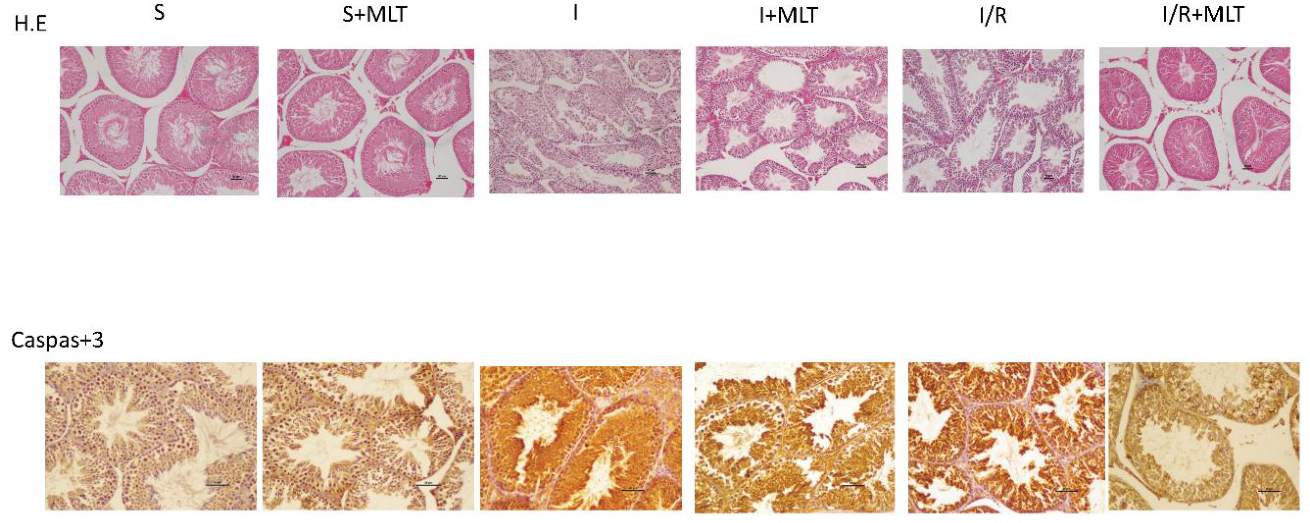
A statistically significant variation in Johnsen scores was identified among the study groups (p < 0.001; Figure 1D and Figure 2). The S group and the S+MLT group exhibited nearly identical scores (9.08 ± 0.16 and 9.11 ± 0.14, respectively; p = 0.699). By comparison, markedly lower scores were recorded in the I group (5.61 ± 0.30) and the I/R group (6.35 ± 0.18) relative to the S group (p < 0.001 for both comparisons), indicating substantial disruption of spermatogenesis. Furthermore, the score in the I group was significantly lower than that in the I/R group (p < 0.001). Treatment with melatonin partially attenuated these changes: the mean score increased to 7.36 ± 0.22 in the I+MLT group and to 8.21 ± 0.17 in the I/R+MLT group, with both values significantly exceeding those of their respective untreated controls (p < 0.001 for both comparisons). Despite this improvement, scores in both melatonin-treated groups remained significantly lower than those in the S group (p < 0.001).
3.5. Caspase-3 Immunohistochemical Findings
A significant difference in Caspase-3 immunoreactivity was observed across the study groups (p < 0.001; Figure 1E and Figure 2). The two control groups showed comparable immunoreactivity scores (0.50 ± 0.53 and 0.25 ± 0.46, respectively; p = 0.317). In comparison with the control group scoring 0.50 ± 0.53, immunoreactivity was significantly greater in the I group (2.63 ± 0.52; p < 0.001) and the I/R group (1.63 ± 0.52; p = 0.003). Moreover, the I group had a significantly higher score than the I/R group (p = 0.005). Melatonin administration was associated with lower caspase-3 immunoreactivity in the I+MLT group (1.13 ± 0.35) than in the I group (p < 0.001). The I/R+MLT group also exhibited a score of 0.63 ± 0.52, representing a significant reduction compared with the I/R group (p = 0.005), and no longer differed significantly from the S group (p = 0.626).
Figure 1.
Effects of melatonin on epididymal sperm parameters and testicular histology after sustained ischemia (I) and ischemia/reperfusion (I/R). (A) Sperm concentration; (B) total sperm motility; (C) normal sperm morphology; (D) Johnsen score; (E) caspase-3 immunoreactivity score. Each symbol represents one animal (n = 8 per group); horizontal lines indicate mean ± SD. Pairwise comparisons were made with the two-tailed Mann–Whitney U test after a significant Kruskal–Wallis test; p values are unadjusted. ns, not significant. S, sham; MLT, melatonin.
Figure 1.
Effects of melatonin on epididymal sperm parameters and testicular histology after sustained ischemia (I) and ischemia/reperfusion (I/R). (A) Sperm concentration; (B) total sperm motility; (C) normal sperm morphology; (D) Johnsen score; (E) caspase-3 immunoreactivity score. Each symbol represents one animal (n = 8 per group); horizontal lines indicate mean ± SD. Pairwise comparisons were made with the two-tailed Mann–Whitney U test after a significant Kruskal–Wallis test; p values are unadjusted. ns, not significant. S, sham; MLT, melatonin.
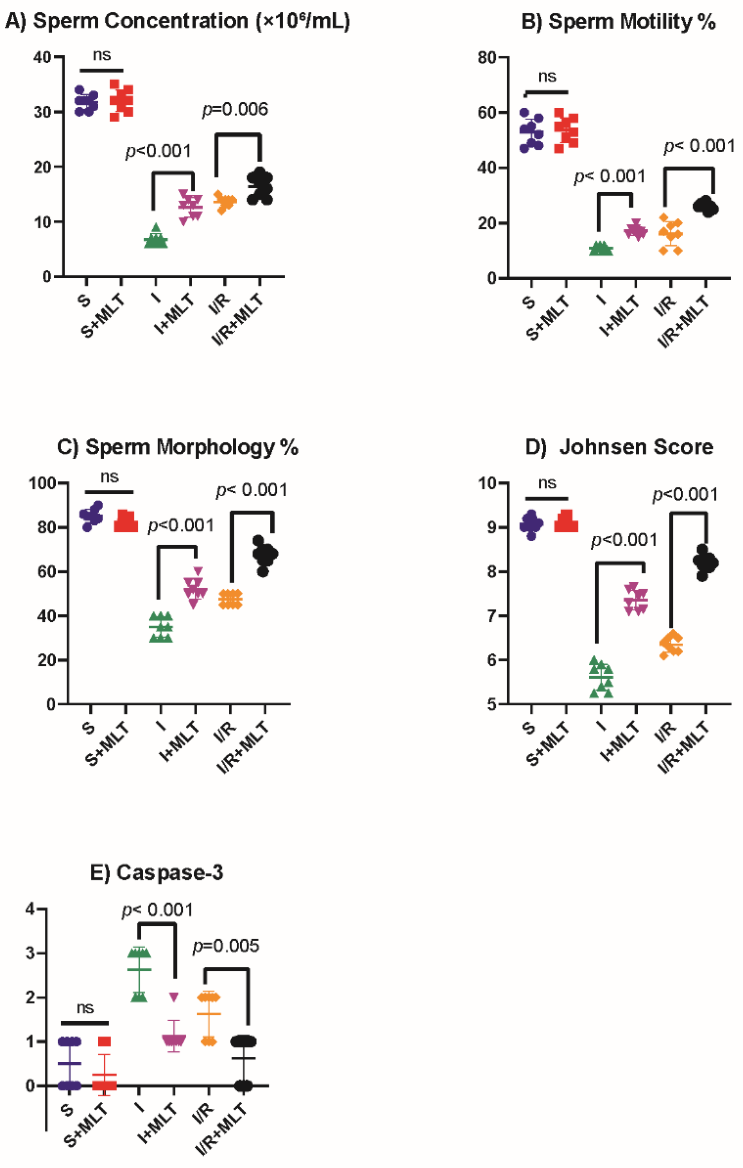
Figure 2.
Representative photomicrographs of testicular sections stained with hematoxylin and eosin (H&E; upper row) and immunohistochemically for caspase-3 (lower row). In the S and S+MLT groups, seminiferous tubules show normal architecture with orderly germinal epithelium and abundant spermatozoa in the lumen, and caspase-3 immunoreactivity is weak. Sustained ischemia (I) causes marked disorganization and loss of the germinal epithelium with intense caspase-3 immunoreactivity; changes after ischemia/reperfusion (I/R) are similar but less severe. Melatonin treatment (I+MLT, I/R+MLT) partially preserves tubular architecture and reduces caspase-3 immunoreactivity, most evidently in the I/R+MLT group. Mean Johnsen scores: S 9.08, S+MLT 9.11, I 5.61, I+MLT 7.36, I/R 6.35, I/R+MLT 8.21; mean caspase-3 scores: 0.50, 0.25, 2.63, 1.13, 1.63 and 0.63, respectively (n = 8 per group; see Figure ). H&E, original magnification [×100]; caspase-3 IHC, original magnification [×200]; scale bars = 50 µm. S, sham; MLT, melatonin; I, ischemia; I/R, ischemia/reperfusion.
Figure 2.
Representative photomicrographs of testicular sections stained with hematoxylin and eosin (H&E; upper row) and immunohistochemically for caspase-3 (lower row). In the S and S+MLT groups, seminiferous tubules show normal architecture with orderly germinal epithelium and abundant spermatozoa in the lumen, and caspase-3 immunoreactivity is weak. Sustained ischemia (I) causes marked disorganization and loss of the germinal epithelium with intense caspase-3 immunoreactivity; changes after ischemia/reperfusion (I/R) are similar but less severe. Melatonin treatment (I+MLT, I/R+MLT) partially preserves tubular architecture and reduces caspase-3 immunoreactivity, most evidently in the I/R+MLT group. Mean Johnsen scores: S 9.08, S+MLT 9.11, I 5.61, I+MLT 7.36, I/R 6.35, I/R+MLT 8.21; mean caspase-3 scores: 0.50, 0.25, 2.63, 1.13, 1.63 and 0.63, respectively (n = 8 per group; see Figure ). H&E, original magnification [×100]; caspase-3 IHC, original magnification [×200]; scale bars = 50 µm. S, sham; MLT, melatonin; I, ischemia; I/R, ischemia/reperfusion.
3.6. Serum Testosterone Concentrations
No significant differences in pretreatment serum testosterone levels were detected among the six groups (p = 0.643). Mean baseline concentrations were 3.90 ± 0.17 ng/mL for S, 3.92 ± 0.05 ng/mL for S+MLT, 3.95 ± 0.08 ng/mL for I, 3.98 ± 0.07 ng/mL for I+MLT, 3.89 ± 0.16 ng/mL for I/R, and 3.92 ± 0.06 ng/mL for I/R+MLT. Comparison of baseline measurements with those obtained on day 20 revealed no significant changes in either the S group (p = 0.944) or the S+MLT group (p = 0.888). Conversely, serum testosterone concentrations declined significantly from baseline in each of the four groups exposed to testicular injury (p = 0.012 in all four groups).
Testosterone levels differed significantly between groups on the twentieth day (p < 0.001; Figure 3). Testosterone levels were 3.91 ± 0.08 ng/mL in group S, 1.33 ± 0.13 ng/mL in group I, and 1.94 ± 0.08 ng/mL in group I/R (p < 0.001 for both). On day 20, there was no difference between the S (3.91 ± 0.08 ng/mL) and S+MLT (3.92 ± 0.08 ng/mL) groups (p = 0.562). Levels in group I were also lower than in group I/R (p < 0.001). Melatonin increased testosterone levels in the I+MLT group (1.93 ± 0.14 ng/mL) and the I/R+MLT group (2.40 ± 0.17 ng/mL) compared to the respective untreated groups (p < 0.001 for both). However, levels in both groups remained below those of the S group (p < 0.001). The level in the S+MLT group on day 20 was 3.92 ± 0.08 ng/mL.
3.7. IVF Outcomes
Fertilized Oocytes: The mean number of fertilized oocytes per male differed significantly across the six experimental groups (p < 0.001; Figure 4A). In the control groups, fertilization occurred in 2.75 ± 0.46 oocytes per male in the S group and 2.63 ± 0.52 oocytes per male in the S+MLT group, with no significant difference between them (p = 0.602). These results corresponded to overall fertilization rates of 55.0% (22/40) and 52.5% (21/40), respectively. Fertilization outcomes were markedly poorer following testicular injury: the I and I/R groups had means of 0.50 ± 0.53 and 0.75 ± 0.46 fertilized oocytes per male, corresponding to overall fertilization rates of 10.0% (4/40) and 15.0% (6/40), respectively. Compared with the S group, both groups showed significantly lower fertilization numbers (p < 0.001 for each comparison), while no significant difference was observed between the I and I/R groups (p = 0.317). Melatonin administration improved fertilization in both injury models. The mean number of fertilized oocytes increased to 1.50 ± 0.53 per male in the I+MLT group and to 1.75 ± 0.46 per male in the I/R+MLT group, significantly exceeding the corresponding untreated groups (p = 0.006 and p = 0.003, respectively). The overall fertilization rates in the treated groups reached 30.0% (12/40) and 35.0% (14/40), respectively. However, fertilization remained significantly lower than in the S group in both melatonin-treated injury groups (p = 0.002 for I+MLT and p = 0.003 for I/R+MLT).
Blastocyst Yield:A significant difference in blastocyst production per male was detected among the experimental groups (p < 0.001; Figure 4B). The S group yielded 1.75 ± 0.46 blastocysts per male, compared with 1.50 ± 0.53 in the S+MLT group; this difference was not statistically significant (p = 0.317). Based on the total number of inseminated oocytes, blastocyst formation rates were 35.0% (14/40) and 30.0% (12/40), respectively. Only one blastocyst was obtained in the I group (0.13 ± 0.35 per male; 1/40, 2.5%) and three in the I/R group (0.38 ± 0.52 per male; 3/40, 7.5%). Blastocyst numbers in both injury groups were significantly lower than in the S group (p < 0.001 and p = 0.001, respectively), but did not differ significantly from each other (p = 0.264). In the I+MLT group, blastocyst production increased to 0.88 ± 0.35 per male, equivalent to 7/40 oocytes (17.5%), which was significantly higher than in the untreated I group (p = 0.004). Likewise, the I/R+MLT group yielded 1.25 ± 0.46 blastocysts per male, corresponding to 10/40 oocytes (25.0%); this was significantly greater than the yield in the untreated I/R group (p = 0.007). Despite these gains, blastocyst production in the I+MLT group remained significantly below that of the S group (p = 0.003), whereas the I/R+MLT group no longer differed significantly from the S group (p = 0.053).
Progression of Fertilized Oocytes to the Blastocyst Stage
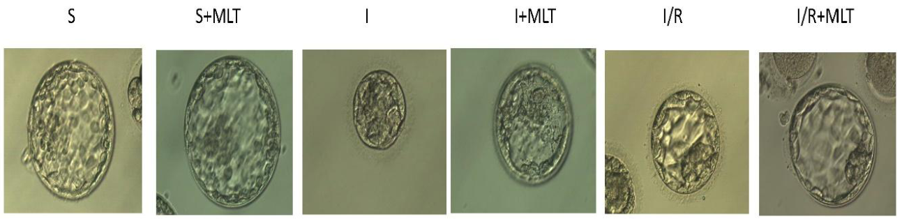
In the S group, 14 of 22 fertilized oocytes developed into blastocysts, giving a developmental rate of 63.6% (Figure 5). In the S+MLT group, 12 of 21 fertilized oocytes reached this stage (57.1%). In the injury groups, one of four fertilized oocytes in the I group (25.0%) and three of six in the I/R group (50.0%) progressed to this stage. The corresponding blastocyst development rates in the melatonin-treated groups were 58.3% (7/12) for I+MLT and 71.4% (10/14) for I/R+MLT. Because the number of fertilized oocytes in the injury groups was small, these proportions are reported descriptively and were not tested statistically.
4. Discussion
The present study assessed the protective effects of melatonin across multiple aspects of testicular function, including histological integrity, serum testosterone concentrations, sperm quality, and the fertilization and developmental potential of spermatozoa in rat models of sustained testicular ischemia and ischemia/reperfusion (I/R) injury. Both experimental conditions resulted in impaired spermatogenesis, greater germ cell apoptosis, and reduced circulating testosterone levels. In parallel, sperm concentration, motility, and the proportion of spermatozoa with normal morphology declined substantially. These reproductive disturbances were also reflected in poorer IVF outcomes, with reduced fertilization success and lower blastocyst yield. Administration of a single dose of melatonin significantly improved every parameter evaluated. However, the observed benefits were incomplete, as the treated injury groups remained below sham values for most parameters; only caspase-3 immunoreactivity and blastocyst yield in the I/R+MLT group no longer differed significantly from the sham group. Importantly, melatonin administration in the absence of testicular injury did not significantly alter any of the measured outcomes, suggesting that the dose used had no detectable adverse effect on the evaluated reproductive parameters in healthy testes. To our knowledge, this is the first study to investigate whether melatonin can improve the IVF performance of spermatozoa collected from injured testes in an experimental model of testicular ischemia and I/R injury.
A notable aspect of this investigation is the comparative assessment of prolonged testicular ischemia and ischemia followed by reperfusion. Compared with the I/R group, the I group had significantly lower Johnsen scores, lower serum testosterone concentrations, reduced sperm counts, and a smaller proportion of morphologically normal spermatozoa. Conversely, caspase-3 immunoreactivity was more pronounced in the I group. Collectively, these findings indicate that uninterrupted ischemia over 20 days may cause more severe and sustained testicular damage than ischemia followed by reperfusion, notwithstanding the additional oxidative stress that can accompany reperfusion [2,3]. These results are consistent with the clinical principle that testicular detorsion should be performed as early as possible. Previous studies have further suggested that persistent torsion and associated testicular necrosis may exert detrimental effects extending beyond the affected testis [34].
Interestingly, melatonin treatment improved the Johnsen score, testosterone concentration, and sperm quality indices, while attenuating caspase-3 immunoreactivity in the I+MLT group, in which testicular blood flow remained interrupted. As melatonin was administered before torsion, this benefit may be attributable to its capacity to limit early ischemia-induced oxidative and apoptotic injury in germ cells and Leydig cells. Melatonin's direct free-radical-scavenging activity, which does not depend on receptor activation, together with its mitochondrial-protective properties, provides a plausible biological explanation for these observations [1,10]. However, the present findings do not directly establish the underlying mechanism. Studies incorporating serial measurements of oxidative stress biomarkers and mitochondrial function are needed to determine how melatonin mediates its protective effects during sustained testicular ischemia.
Our histological observations corroborate previous experimental evidence indicating that melatonin mitigates spermatogenic injury associated with testicular ischemia/reperfusion [13,14,15,19]. In both injury models examined here, melatonin treatment resulted in higher Johnsen scores than those observed in the corresponding untreated groups. Nevertheless, the scores remained below those of the sham controls, indicating that the protective effect of a single melatonin dose was incomplete. The accompanying decline in caspase-3 immunoreactivity suggests that attenuation of apoptosis may represent an important component of this protection. In support of this interpretation, Kanter [14] , using the same melatonin dose and administration schedule, demonstrated reduced TUNEL staining and preservation of mitochondrial integrity in Sertoli cells and spermatids. Chen et al. [11] similarly reported that melatonin attenuated the expression of mitochondrial apoptosis-related factors, including caspase-3, Bax, and cytochrome c, in an experimental torsion model. In addition, Çil et al. [12] documented lower caspase-3 expression and higher SOX-9 expression following melatonin treatment in torsion-injured testes. When considered alongside these earlier findings, our results suggest that melatonin may reduce germ cell loss, in part, by inhibiting mitochondria-mediated apoptotic signaling.
Administration of melatonin significantly enhanced sperm concentration, motility, and the percentage of morphologically normal spermatozoa in both testicular injury groups. These findings are partly consistent with those of Kurcer et al.[16] , who found that melatonin reduced sperm morphological abnormalities in an experimental I/R model but did not prevent the reduction in sperm concentration. Koksal et al. [15] likewise demonstrated that melatonin restored sperm morphology to normal levels. The improvement in sperm concentration observed in our investigation, unlike the findings reported by Kurcer et al., may be attributable to differences in ischemia duration, observation period length, or the timing of melatonin administration. Previous research has established that testicular torsion followed by detorsion can compromise sperm motility, potentially through depletion of intracellular ATP [5]. Moreover, evidence that melatonin protects sperm mitochondria from oxidative injury provides a plausible explanation for the improved motility observed in our study [23,35]. Accordingly, the beneficial effect of melatonin on sperm movement may be associated with preservation of mitochondrial integrity and the cellular energy supply necessary for normal motility.
The absence of significant differences in pretreatment serum testosterone concentrations suggests that the experimental groups had comparable baseline hormonal profiles. By the end of the 20-day observation period, testosterone levels had fallen significantly in all four groups exposed to testicular injury. However, the decline was less marked in the melatonin-treated animals. Previous research by Kanter [14], using an identical melatonin administration protocol, demonstrated an increase in intratesticular testosterone concentrations. In contrast, Çil et al.[12], detected no significant differences in serum testosterone, FSH, or LH between melatonin-treated and untreated animals following testicular torsion. Variation in these findings may be explained by differences in the type of sample analyzed, the duration of torsion, and the interval between injury and sample collection.
The potential influence of melatonin on testosterone production may involve several cellular mechanisms. Melatonin has been reported to modulate the mitochondrial Bax/Bcl-2 signaling pathway, thereby reducing Leydig cell apoptosis and enhancing the expression of steroidogenesis-associated genes, including StAR and SF-1 [36] . Furthermore, activation of the KEAP1–NRF2 pathway may help protect Leydig cells from oxidative stress and preserve the expression of enzymes involved in steroid hormone synthesis [37]. The identification of melatonin receptors in Leydig and Sertoli cells also suggests that melatonin may exert local regulatory effects within the testicular microenvironment [38]. The significant reduction in circulating testosterone despite unilateral testicular injury warrants further consideration. Previous studies have suggested that I/R injury affecting one testis can trigger oxidative changes in the contralateral gonad [18], impair the integrity of the blood–testis barrier, and potentially reduce serum testosterone concentrations [39]. However, whether unilateral torsion consistently causes clinically relevant damage to the opposite testis remains unresolved [34,40], Accordingly, the hormonal changes observed in our study may reflect systemic responses to injury and/or alterations in the contralateral testis. Since the opposite testis was not included in our histological or biochemical assessments, these potential explanations remain unconfirmed.
A major finding of the present study is the demonstration of testicular injury-related functional impairment through direct assessment of IVF outcomes, together with the beneficial effects associated with melatonin treatment. In the I and I/R groups, the mean number of fertilized oocytes fell to 0.50 and 0.75 per male, respectively, and the overall fertilization rate declined substantially, from 55.0% in the sham group to 10.0% and 15.0%. These observations are consistent with evidence from animal IVF studies showing that oxidative damage to spermatozoa reduces fertilization rates and blastocyst development [6] . Blastocyst yield was likewise reduced, from 35.0% of inseminated oocytes in the sham group to 2.5% and 7.5% in the I and I/R groups. Among fertilized oocytes, 63.6% developed into blastocysts in the sham group, compared with 25.0% (1/4) in the I group and 50.0% (3/6) in the I/R group; given the small number of fertilized oocytes, the reduction in blastocyst yield appears to be driven largely by impaired fertilization. Because all oocytes originated from a common pool and were randomly allocated among groups, variation in oocyte source is an unlikely primary explanation for these differences. This pattern instead suggests an important contribution from injury-related alterations in sperm function.
Damage associated with the paternal genome may become apparent only after fertilization, particularly during the period of embryonic genome activation. This delayed manifestation has been termed the “late paternal effect”[41,42]. Consistent with this concept, increased sperm DNA fragmentation has been linked to lower blastocyst formation rates, even when fertilization remains relatively unaffected [43]. Sperm DNA integrity may be compromised by apoptosis in germ cells, defective chromatin packaging, or oxidative stress encountered during epididymal transit [44]. Experimental evidence from mice further indicates that severe sperm DNA damage can delay paternal DNA replication in zygotes and prevent embryos from reaching the blastocyst stage [45]. Taken together, the increased caspase-3 immunoreactivity and reduced proportion of morphologically normal sperm observed in our injured groups, alongside the impaired IVF outcomes, suggest that testicular I/R injury may affect both successful fertilization and the capacity of spermatozoa to support continued embryonic development. Nevertheless, because sperm DNA fragmentation and chromatin integrity were not directly assessed, the specific contribution of paternal genomic damage remains to be established.
In both groups exposed to testicular injury, melatonin administration was associated with a significant increase in the mean number of fertilized oocytes per male (p = 0.006 for I+MLT and p = 0.003 for I/R+MLT). Accordingly, the overall fertilization rate improved from 10.0% to 30.0% in the ischemia model and from 15.0% to 35.0% in the I/R model. A significant increase in blastocyst numbers was also observed following treatment, with differences between the I+MLT and I groups (p = 0.004) and between the I/R+MLT and I/R groups (p = 0.007). The proportion of fertilized oocytes progressing to the blastocyst stage reached 58.3% and 71.4% in the I+MLT and I/R+MLT groups, respectively. Collectively, these results suggest that melatonin offers partial protection against injury-related reductions in sperm fertilizing potential and subsequent embryonic developmental competence.
Protective effects on IVF outcomes have also been documented in other experimental models of testicular toxicity, including those induced by aflatoxin B1 [46], cyclophosphamide [47] and doxorubicin [48] in which systemic melatonin treatment improved fertilization and blastocyst formation. In the aflatoxin B1 model, improved reproductive outcomes were associated with reduced sperm DNA damage [46]. Similarly, findings from cadmium-induced testicular injury indicate that melatonin may preserve the integrity of the blood–testis barrier and enhance sperm DNA integrity [49]. Research involving direct in vitro melatonin exposure has demonstrated increased monospermic fertilization and more favorable embryo development [22]; These observations may be related to decreased sperm ROS concentrations and altered expression of genes involved in embryonic genome activation [23]. Clinical investigations have additionally reported higher fertilization rates and greater numbers of good-quality embryos following melatonin supplementation [24]. The present study adds to these findings by demonstrating a potential reproductive benefit of melatonin in the setting of testicular ischemia and I/R-associated injury.
Earlier studies investigating antioxidant protection against testicular I/R injury have commonly relied on in vivo mating outcomes to assess reproductive function [7,8,9] . Such endpoints, however, are influenced by factors beyond sperm quality, including male mating behavior and female reproductive characteristics, and therefore cannot be used to isolate the fertilizing competence of spermatozoa. By using IVF with oocytes collected into a common pool and randomly assigned to the experimental groups, the present study provides a more direct means of evaluating sperm-associated effects on fertilization and early embryonic development. Despite the improvements observed after melatonin administration, fertilization remained below sham-group values in both treated injury groups, and blastocyst yield remained lower in the I+MLT group, although it no longer differed significantly from sham in the I/R+MLT group. These findings therefore indicate that a single melatonin dose confers incomplete protection and does not fully restore reproductive function following testicular injury.
Several limitations should be acknowledged. First, the allocation of five oocytes per male was intended to maintain oocyte quality; nevertheless, the relatively small number of oocytes may have constrained the statistical power of the IVF analyses. This limitation is particularly relevant to the injury groups, in which low fertilization rates further reduced the number of observable outcomes and may have obscured modest treatment-related differences; for the same reason, the proportion of fertilized oocytes reaching the blastocyst stage could only be reported descriptively. Second, neither sperm DNA fragmentation nor biochemical indicators of oxidative stress in testicular tissue were evaluated. Accordingly, the mechanisms proposed to explain the reduction in blastocyst formation remain speculative and are supported only by indirect evidence. Third, the treatment protocol involved a single administration of melatonin. Melatonin regimens in previous torsion studies have varied widely: a single low dose (25 µg/kg) improved spermatogenesis without reducing tissue malondialdehyde or restoring tubular diameter [20], whereas daily administration of 10 mg/kg for seven days improved Johnsen scores, oxidative markers and inhibin B levels [50]. Single and repeated dosing have not been compared directly. Future studies should therefore investigate alternative melatonin regimens, particularly repeated-dose protocols, to determine whether they offer greater benefits for fertilization success and subsequent embryo development.
5. Conclusions
This investigation shows that prolonged testicular ischemia and ischemia–reperfusion injury adversely affect spermatogenesis, germ cell survival, endocrine function, and sperm quality, ultimately compromising fertilization and blastocyst formation in IVF. The extent of histological damage and testosterone reduction was greater in animals subjected to sustained ischemia than in those undergoing reperfusion. A single intraperitoneal injection of melatonin at 10 mg/kg attenuated germ cell apoptosis and mitigated the deterioration in spermatogenic activity, testosterone levels, and sperm concentration, motility, and morphology in both injury models. Furthermore, melatonin treatment improved sperm fertilizing capacity and enhanced the progression of fertilized oocytes to the blastocyst stage. Despite these benefits, most reproductive and testicular parameters did not return to the levels observed in the sham groups, indicating that melatonin provided partial rather than complete protection.
Overall, these findings identify melatonin as a potential adjunctive strategy to limit testicular damage and preserve reproductive function following torsion-associated ischemia. However, its efficacy and safety in clinical practice require further validation. Future research should compare single-dose and repeated-dose protocols, directly assess sperm DNA damage and integrity, and examine embryo-transfer outcomes to determine whether the observed improvements in IVF translate into better subsequent reproductive outcomes.
Author Contributions
Conceptualization, Ö.B. and M.S.B.; methodology, Ö.B. and M.S.B.; formal analysis, Ö.B.; investigation, Ö.B., M.S.B., E.Y., M.D., E.Ç., H.Ç., O.K., E.Ş. and Y.Y.; writing—original draft preparation, Ö.B. and M.S.B.; writing—review and editing, Ö.B., M.S.B., E.Y.,M.D., E.Ç., H.Ç., O.K., E.Ş. and Y.Y.; supervision, Ö.B. and M.S.B. All authors have read and agreed to the published version of the manuscript.
Funding
This research received no external funding.
Institutional Review Board Statement
The animal study protocol was approved by the Sakarya University Experimental Animals Ethics Committee (decision no. 06, dated 1 February 2023). The study was conducted in accordance with institutional and national guidelines for the care and use of laboratory animals and reported in accordance with the ARRIVE 2.0 guidelines.
Data Availability Statement
The data presented in this study are available on request from the corresponding author.
Acknowledgements
During the preparation of this manuscript, the authors used Consensus (Consensus NLP) for literature searching and verification of cited references.
Conflicts of Interest
The authors declare no conflicts of interest.
Abbreviations
| Abbreviation | Definition |
| ARRIVE | Animal Research: Reporting of In Vivo Experiments |
| DAB | 3,3′-diaminobenzidine |
| DNA | deoxyribonucleic acid |
| ELISA | enzyme-linked immunosorbent assay |
| H&E | hematoxylin and eosin |
| HSA | human serum albumin |
| HTF | human tubal fluid |
| I | sustained ischemia group |
| I+MLT | ischemia + melatonin group |
| I/R | ischemia/reperfusion |
| I/R+MLT | ischemia/reperfusion + melatonin group |
| IHC | immunohistochemistry |
| IVF | in vitro fertilization |
| MII | metaphase II |
| MLT | melatonin |
| ns | not significant |
| S | sham group |
| S+MLT | sham + melatonin group |
| SD | standard deviation |
References
- Heidarizadi, S.; Rashidi, Z.; Jalili, C.; Gholami, M. Overview of biological effects of melatonin on testis: A review. Andrologia 2022, 54, e14597. [CrossRef]
- Akhigbe, R.E.; Odetayo, A.F.; Akhigbe, T.M.; Hamed, M.A.; Ashonibare, P.J. Pathophysiology and management of testicular ischemia/reperfusion injury: Lessons from animal models. Heliyon 2024, 10, e27760. [CrossRef]
- Minas, A.; Mahmoudabadi, S.; Gamchi, N.S.; Antoniassi, M.P.; Alizadeh, A.; Bertolla, R.P. Testicular torsion in vivo models: Mechanisms and treatments. Andrology 2023, 11, 1267-1285. [CrossRef]
- Katami, H.; Suzuki, S.; Fujii, T.; Ueno, M.; Tanaka, A.; Ohta, K.-i.; Miki, T.; Shimono, R. Genetic and histopathological analysis of spermatogenesis after short-term testicular torsion in rats. Pediatric Research 2023, 94, 1650-1658. [CrossRef]
- Hsiao, C.-H.; Ji, A.T.-Q.; Chang, C.-C.; Chien, M.-H.; Lee, L.-M.; Ho, J.H.-C. Mesenchymal stem cells restore the sperm motility from testicular torsion-detorsion injury by regulation of glucose metabolism in sperm. Stem Cell Research & Therapy 2019, 10, 270. [CrossRef]
- Ribas-Maynou, J.; Yeste, M.; Salas-Huetos, A. The Relationship between Sperm Oxidative Stress Alterations and IVF/ICSI Outcomes: A Systematic Review from Nonhuman Mammals. Biology 2020, 9. [CrossRef]
- Khorsand, K.; Najafpour, A.; Valilou, M.; Soleimanzadeh, A. Dose-dependent protective effects of p-coumaric acid on testicular ischemia/reperfusion injury in a mouse model of testicular torsion. Biochem Biophys Rep 2026, 47, 102760. [CrossRef]
- Mostahsan, Z.; Azizi, S.; Soleimanzadeh, A.; Shalizar-Jalali, A. Protective effects of Mito-TEMPO on ischemia-reperfusion injury in a mouse testicular torsion and detorsion model. Vet Res Forum 2024, 15, 665-672. [CrossRef]
- Zolfaghari, S.; Soleimanzadeh, A.; Baqerkhani, M. The synergistic activity of fisetin on quercetin improves testicular recover in ischemia-reperfusion injury in rats. Scientific Reports 2025, 15, 12053. [CrossRef]
- Tamura, H.; Jozaki, M.; Tanabe, M.; Shirafuta, Y.; Mihara, Y.; Shinagawa, M.; Tamura, I.; Maekawa, R.; Sato, S.; Taketani, T.; et al. Importance of Melatonin in Assisted Reproductive Technology and Ovarian Aging. International journal of molecular sciences 2020, 21, 1135. [CrossRef]
- Chen, Y.-T.; Chuang, F.-C.; Yang, C.-C.; Chiang, J.Y.; Sung, P.-H.; Chu, Y.-C.; Huang, C.-R.; Huang, K.-H.; Yip, H.-K. Combined melatonin-adipose derived mesenchymal stem cells therapy effectively protected the testis from testicular torsion-induced ischemia-reperfusion injury. Stem Cell Research & Therapy 2021, 12, 370. [CrossRef]
- Çil, N.; Neşet, G.; Abban Mete, G. Protective Effect of Mesenchymal Stem Cells and Melatonin on Testicular Torsion-Induced Infertility. Journal of Uludağ University Medical Faculty 2024, 50, 215-222. [CrossRef]
- Asghari, A.; Akbari, G.; Meghdadi, A.; Mortazavi, P. Effects of melatonin and metformin co-administration on testicular ischemia/reperfusion injury in rats. J Pediatr Urol 2016, 12, 410 e411-410 e417. [CrossRef]
- Kanter, M. Protective effects of melatonin on testicular torsion/detorsion-induced ischemia-reperfusion injury in rats. Exp Mol Pathol 2010, 89, 314-320. [CrossRef]
- Koksal, M.; Oguz, E.; Baba, F.; Eren, M.A.; Ciftci, H.; Demir, M.E.; Kurcer, Z.; Take, G.; Aral, F.; Ocak, A.R.; et al. Effects of melatonin on testis histology, oxidative stress and spermatogenesis after experimental testis ischemia-reperfusion in rats. European review for medical and pharmacological sciences 2012, 16, 582-588.
- Kurcer, Z.; Hekimoglu, A.; Aral, F.; Baba, F.; Sahna, E. Effect of melatonin on epididymal sperm quality after testicular ischemia/reperfusion in rats. Fertility and Sterility 2010, 93, 1545-1549. [CrossRef]
- Kurcer, Z.; Oguz, E.; Ozbilge, H.; Baba, F.; Aksoy, N.; Celik, N. Effect of melatonin on testicular ischemia/reperfusion injury in rats: is this effect related to the proinflammatory cytokines? Fertility and Sterility 2008, 89, 1468-1473. [CrossRef]
- Parlaktas, B.; Atilgan, D.; Ozyurt, H.; Gencten, Y.; Akbas, A.; Erdemir, F.; Uluocak, N. The biochemical effects of ischemia-reperfusion injury in the ipsilateral and contralateral testes of rats and the protective role of melatonin. Asian Journal of Andrology 2014, 16, 314-318. [CrossRef]
- Dehdari Ebrahimi, N.; Shojaei-Zarghani, S.; Taherifard, E.; Dastghaib, S.; Parsa, S.; Mohammadi, N.; Sabet Sarvestani, F.; Moayedfard, Z.; Hosseini, N.; Safarpour, H.; et al. Protective effects of melatonin against physical injuries to testicular tissue: A systematic review and meta-analysis of animal models. Front Endocrinol (Lausanne) 2023, 14, 1123999. [CrossRef]
- Mirhoseini, M.; Talebpour Amiri, F.; Karimpour Malekshah, A.A.; Rezanejad Gatabi, Z.; Ghaffari, E. Protective effects of melatonin on testis histology following acute torsion-detorsion in rats. International journal of reproductive biomedicine 2017, 15, 141-146. [CrossRef]
- Sekmenli, T.; Gunduz, M.; Ozturk, B.; Karabagli, P.; Ciftci, I.; Tekin, G.; Yilmaz, M. The effects of melatonin and colchicine on ischemia-reperfusion injury in experimental rat testicular torsion model. J Pediatr Surg 2017, 52, 582-586. [CrossRef]
- Gutiérrez-Añez, J.C.; Henning, H.; Lucas-Hahn, A.; Baulain, U.; Aldag, P.; Sieg, B.; Hensel, V.; Herrmann, D.; Niemann, H. Melatonin improves rate of monospermic fertilization and early embryo development in a bovine IVF system. PLOS ONE 2021, 16, e0256701. [CrossRef]
- Su, G.; Wu, S.; Wu, M.; Wang, L.; Yang, L.; Du, M.; Zhao, X.; Su, X.; Liu, X.; Bai, C.; et al. Melatonin improves the quality of frozen bull semen and influences gene expression related to embryo genome activation. Theriogenology 2021, 176, 54-62. [CrossRef]
- Hu, K.L.; Ye, X.; Wang, S.; Zhang, D. Melatonin Application in Assisted Reproductive Technology: A Systematic Review and Meta-Analysis of Randomized Trials. Front Endocrinol (Lausanne) 2020, 11, 160. [CrossRef]
- Percie du Sert, N.; Hurst, V.; Ahluwalia, A.; Alam, S.; Avey, M.T.; Baker, M.; Browne, W.J.; Clark, A.; Cuthill, I.C.; Dirnagl, U.; et al. The ARRIVE guidelines 2.0: Updated guidelines for reporting animal research. Journal of cerebral blood flow and metabolism : official journal of the International Society of Cerebral Blood Flow and Metabolism 2020, 40, 1769-1777. [CrossRef]
- Semercioz, A.; Baltaci, A.K.; Mogulkoc, R.; Avunduk, M.C. Effect of Zinc and Melatonin on Oxidative Stress and Serum Inhibin-B Levels in a Rat Testicular Torsion-Detorsion Model. Biochem. Genet. 2017, 55, 395-409. [CrossRef]
- Aslan Kosar, P.; Tuncer, H.; Cihangir Uguz, A.; Espino Palma, J.; Darici, H.; Onaran, I.; Cig, B.; Kosar, A.; Rodriguez Moratinos, A.B. The efficiency of Poly(ADP-Ribose) Polymerase (PARP) cleavage on detection of apoptosis in an experimental model of testicular torsion. Int J Exp Pathol 2015, 96, 294-300. [CrossRef]
- Toyoda, Y.; Chang, M.C. Fertilization of rat eggs in vitro by epididymal spermatozoa and the development of eggs following transfer. J. Reprod. Fertil. 1974, 36, 9-22. [CrossRef]
- Yokoi, K.; Uthus, E.O.; Nielsen, F.H. Nickel deficiency diminishes sperm quantity and movement in rats. Biological trace element research 2003, 93, 141-154. [CrossRef]
- Sotomaru, Y.; Kamisako, T.; Hioki, K. Estrous stage- and animal age-independent superovulation in the BrlHan:WIST@Jcl(GALAS) rat. Experimental animals 2005, 54, 137-141. [CrossRef]
- Hino, C.; Ueda, J.; Funakoshi, H.; Matsumoto, S. Defined oocyte collection time is critical for reproducible in vitro fertilization in rats of different strains. Theriogenology 2020, 144, 146-151. [CrossRef]
- Casillas, F.; Betancourt, M.; Juarez-Rojas, L.; Ducolomb, Y.; Lopez, A.; Avila-Quintero, A.; Zamora, J.; Ommati, M.M.; Retana-Marquez, S. Chronic Stress Detrimentally Affects In Vivo Maturation in Rat Oocytes and Oocyte Viability at All Phases of the Estrous Cycle. Animals : an open access journal from MDPI 2021, 11, 2478. [CrossRef]
- Johnsen, S.G. Testicular biopsy score count--a method for registration of spermatogenesis in human testes: normal values and results in 335 hypogonadal males. Hormones 1970, 1, 2-25. [CrossRef]
- Jacobsen, F.M.; Rudlang, T.M.; Fode, M.; Østergren, P.B.; Sønksen, J.; Ohl, D.A.; Jensen, C.F.S. The Impact of Testicular Torsion on Testicular Function. The world journal of men's health 2020, 38, 298-307. [CrossRef]
- Pool, K.R.; Rickard, J.P.; de Graaf, S.P. Melatonin improves the motility and DNA integrity of frozen-thawed ram spermatozoa likely via suppression of mitochondrial superoxide production. Domestic animal endocrinology 2021, 74, 106516. [CrossRef]
- Yang, M.; Guan, S.; Tao, J.; Zhu, K.; Lv, D.; Wang, J.; Li, G.; Gao, Y.; Wu, H.; Liu, J.; et al. Melatonin promotes male reproductive performance and increases testosterone synthesis in mammalian Leydig cells†. Biology of reproduction 2021, 104, 1322-1336. [CrossRef]
- Yu, X.; Li, C.; Ming, R.; Wang, B.; Guo, H.; Li, B. Melatonin attenuates heat stress-induced oxidative stress and apoptosis in mouse testicular Leydig cells via the KEAP1-NRF2 pathway. Journal of thermal biology 2026, 135, 104345. [CrossRef]
- Frungieri, M.B.; Calandra, R.S.; Rossi, S.P. Local Actions of Melatonin in Somatic Cells of the Testis. International journal of molecular sciences 2017, 18, 1170. [CrossRef]
- Gaber Ibrahim, M.F.; Abdel-Hakeem, E.A.; Shawky, H.A.; Abdelkawy Abdelwahab, S.; Mokhemer, S.A. Effect of local versus systemic exosomes administration on both testes in rat model of testicular ischemic reperfusion injury: shedding light on blood testis barrier. Tissue barriers 2026, 14, 2532195. [CrossRef]
- Turner, T.T. On unilateral testicular and epididymal torsion: no effect on the contralateral testis. The Journal of urology 1987, 138, 1285-1290. [CrossRef]
- Simon, L.; Murphy, K.; Shamsi, M.B.; Liu, L.; Emery, B.; Aston, K.I.; Hotaling, J.; Carrell, D.T. Paternal influence of sperm DNA integrity on early embryonic development. Human reproduction (Oxford, England) 2014, 29, 2402-2412. [CrossRef]
- Tesarik, J.; Greco, E.; Mendoza, C. Late, but not early, paternal effect on human embryo development is related to sperm DNA fragmentation. Human reproduction (Oxford, England) 2004, 19, 611-615. [CrossRef]
- Borges, E., Jr.; Zanetti, B.F.; Setti, A.S.; Braga, D.; Provenza, R.R.; Iaconelli, A., Jr. Sperm DNA fragmentation is correlated with poor embryo development, lower implantation rate, and higher miscarriage rate in reproductive cycles of non-male factor infertility. Fertil. Steril. 2019, 112, 483-490. [CrossRef]
- Sakkas, D.; Alvarez, J.G. Sperm DNA fragmentation: mechanisms of origin, impact on reproductive outcome, and analysis. Fertil. Steril. 2010, 93, 1027-1036. [CrossRef]
- Gawecka, J.E.; Marh, J.; Ortega, M.; Yamauchi, Y.; Ward, M.A.; Ward, W.S. Mouse zygotes respond to severe sperm DNA damage by delaying paternal DNA replication and embryonic development. PLoS One 2013, 8, e56385. [CrossRef]
- Sabahi, M.; Karimipour, M.; Ahmadi, A.; Pourheydar, B.; Farjah, G. The protective effects of melatonin on testis, sperm parameters quality, and in-vitro fertilization in mice following treatment with aflatoxin B1: An experimental study. International journal of reproductive biomedicine 2025, 23, 185-198. [CrossRef]
- Song, Z.; Wang, J.; Zhu, P.; Wang, Z.; Liu, X.; Liu, F.; Banihani, S. Melatonin Ameliorates Cyclophosphamide-Induced Spermatogenesis Disorder by Reducing Pyroptosis. Andrologia 2023, 2023, 2186029. [CrossRef]
- Zi, T.; Liu, Y.; Zhang, Y.; Wang, Z.; Wang, Z.; Zhan, S.; Peng, Z.; Li, N.; Liu, X.; Liu, F. Protective effect of melatonin on alleviating early oxidative stress induced by DOX in mice spermatogenesis and sperm quality maintaining. Reproductive biology and endocrinology : RB&E 2022, 20, 105. [CrossRef]
- Venditti, M.; Ben Rhouma, M.; Romano, M.Z.; Messaoudi, I.; Reiter, R.J.; Minucci, S. Evidence of melatonin ameliorative effects on the blood-testis barrier and sperm quality alterations induced by cadmium in the rat testis. Ecotoxicology and environmental safety 2021, 226, 112878. [CrossRef]
- Ekici, S.; Doğan Ekici, A.I.; Öztürk, G.; Benli Aksungar, F.; Sinanoğlu, O.; Turan, G.; Lüleci, N. Comparison of melatonin and ozone in the prevention of reperfusion injury following unilateral testicular torsion in rats. Urology 2012, 80, 899-906. [CrossRef]
Figure 3.
Serum testosterone concentrations on day 20 after sustained testicular ischemia (I) and ischemia/reperfusion (I/R) with or without melatonin. Each symbol represents one animal (n = 8 per group); horizontal lines indicate mean ± SD. Baseline concentrations did not differ among groups (Kruskal–Wallis, p = 0.643), and levels declined significantly from baseline in all four injured groups (Wilcoxon signed-rank test, p = 0.012). Pairwise comparisons were made with the two-tailed Mann–Whitney U test after a significant Kruskal–Wallis test; p values are unadjusted. ns, not significant. S, sham; MLT, melatonin.
Figure 3.
Serum testosterone concentrations on day 20 after sustained testicular ischemia (I) and ischemia/reperfusion (I/R) with or without melatonin. Each symbol represents one animal (n = 8 per group); horizontal lines indicate mean ± SD. Baseline concentrations did not differ among groups (Kruskal–Wallis, p = 0.643), and levels declined significantly from baseline in all four injured groups (Wilcoxon signed-rank test, p = 0.012). Pairwise comparisons were made with the two-tailed Mann–Whitney U test after a significant Kruskal–Wallis test; p values are unadjusted. ns, not significant. S, sham; MLT, melatonin.
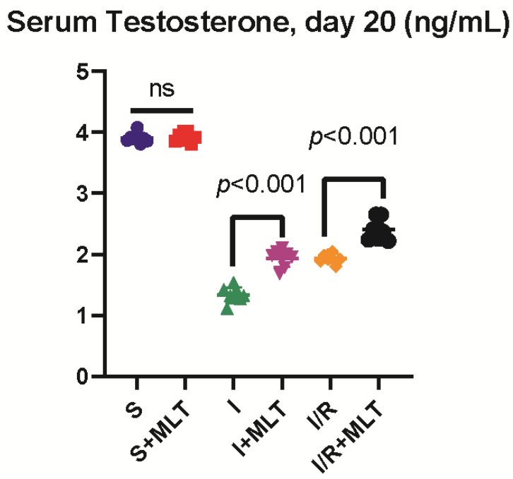
Figure 4.
In vitro fertilization outcomes of epididymal spermatozoa after sustained testicular ischemia (I) and ischemia/reperfusion (I/R) with or without melatonin. (A) Number of fertilized oocytes per male and (B) number of blastocysts per male. For each male, five MII oocytes from a common pool were inseminated; fertilization was assessed 16–18 h after insemination and blastocyst formation on day 5 of culture. Each symbol represents one male (n = 8 per group); horizontal lines indicate mean ± SD. Corresponding pooled rates: fertilization 55.0% (S), 52.5% (S+MLT), 10.0% (I), 30.0% (I+MLT), 15.0% (I/R) and 35.0% (I/R+MLT); blastocyst yield per inseminated oocyte 35.0%, 30.0%, 2.5%, 17.5%, 7.5% and 25.0%, respectively. Pairwise comparisons were made with the two-tailed Mann–Whitney U test after a significant Kruskal–Wallis test; p values are unadjusted. ns, not significant. S, sham; MLT, melatonin.
Figure 4.
In vitro fertilization outcomes of epididymal spermatozoa after sustained testicular ischemia (I) and ischemia/reperfusion (I/R) with or without melatonin. (A) Number of fertilized oocytes per male and (B) number of blastocysts per male. For each male, five MII oocytes from a common pool were inseminated; fertilization was assessed 16–18 h after insemination and blastocyst formation on day 5 of culture. Each symbol represents one male (n = 8 per group); horizontal lines indicate mean ± SD. Corresponding pooled rates: fertilization 55.0% (S), 52.5% (S+MLT), 10.0% (I), 30.0% (I+MLT), 15.0% (I/R) and 35.0% (I/R+MLT); blastocyst yield per inseminated oocyte 35.0%, 30.0%, 2.5%, 17.5%, 7.5% and 25.0%, respectively. Pairwise comparisons were made with the two-tailed Mann–Whitney U test after a significant Kruskal–Wallis test; p values are unadjusted. ns, not significant. S, sham; MLT, melatonin.
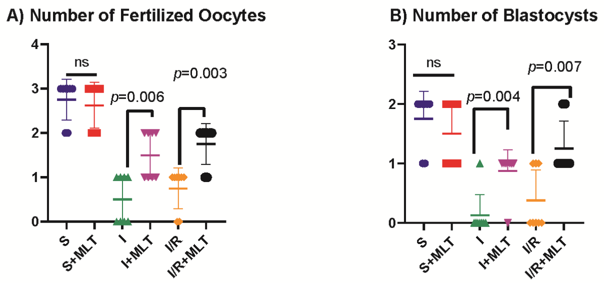
Figure 5.
Representative day-5 blastocysts obtained after in vitro fertilization with epididymal spermatozoa from the S, S+MLT, I, I+MLT, I/R and I/R+MLT groups. Embryos were classified as blastocysts by the presence of a discernible blastocoel cavity. The I group image shows the only blastocyst obtained in that group (1/40 inseminated oocytes). Blastocyst yields per inseminated oocyte were 35.0% (S), 30.0% (S+MLT), 2.5% (I), 17.5% (I+MLT), 7.5% (I/R) and 25.0% (I/R+MLT) The blastocyst obtained from group I was morphologically smaller than the other groups(see Figure 4B). Images were captured under an inverted microscope at original magnification [×200]. S, sham; MLT, melatonin; I, ischemia; I/R, ischemia/reperfusion.
Figure 5.
Representative day-5 blastocysts obtained after in vitro fertilization with epididymal spermatozoa from the S, S+MLT, I, I+MLT, I/R and I/R+MLT groups. Embryos were classified as blastocysts by the presence of a discernible blastocoel cavity. The I group image shows the only blastocyst obtained in that group (1/40 inseminated oocytes). Blastocyst yields per inseminated oocyte were 35.0% (S), 30.0% (S+MLT), 2.5% (I), 17.5% (I+MLT), 7.5% (I/R) and 25.0% (I/R+MLT) The blastocyst obtained from group I was morphologically smaller than the other groups(see Figure 4B). Images were captured under an inverted microscope at original magnification [×200]. S, sham; MLT, melatonin; I, ischemia; I/R, ischemia/reperfusion.
Disclaimer/Publisher’s Note: The statements, opinions and data contained in all publications are solely those of the individual author(s) and contributor(s) and not of MDPI and/or the editor(s). MDPI and/or the editor(s) disclaim responsibility for any injury to people or property resulting from any ideas, methods, instructions or products referred to in the content. |
© 2026 by the authors. Licensee MDPI, Basel, Switzerland. This article is an open access article distributed under the terms and conditions of the Creative Commons Attribution (CC BY) license (http://creativecommons.org/licenses/by/4.0/).
Copyright: This open access article is published under a Creative Commons CC BY 4.0 license, which permit the free download, distribution, and reuse, provided that the author and preprint are cited in any reuse.