

Submitted:
04 November 2026
Posted:
05 November 2026
You are already at the latest version
Abstract
Patient safety remains a major challenge in cardiac surgery, where cardiopulmonary bypass carries risks of residual structural lesions, hemodynamic instability, embolic events, and unrecognized technical complications. This review examines intraoperative transesophageal echocardiography as a patient-safety intervention across the pre-bypass, bypass, and post-bypass phases of adult cardiac surgery, drawing on observational studies, registry analyses, and professional society guidance. Before cardiopulmonary bypass, transesophageal echocardiography establishes baseline ventricular and valvular function, identifies previously unrecognized pathology, and may guide cannulation and operative planning. During bypass, it supports ventricular decompression, myocardial protection, cannula assessment, and the detection and clearance of intracardiac air. After bypass, it evaluates the adequacy of surgical repair and identifies residual valvular lesions, ventricular dysfunction, ischemia, intracardiac air, and aortic complications while immediate corrective intervention remains possible. Although associated changes in surgical or anesthetic management are consistently reported, their frequency varies according to procedure type, patient risk, institutional practice, and study design. Furthermore, management change is a process outcome and does not, by itself, establish improvement in patient-centered outcomes. Serious complications related to transesophageal echocardiography are uncommon, but safe use requires appropriate patient selection, contraindication screening, careful probe manipulation, standardized examination and communication, and operator competence. A phase-based approach may help surgical teams connect imaging findings with time-sensitive hazards and corrective actions. Further prospective evidence is needed to identify the procedures and patient-risk profiles in which routine rather than selective intraoperative transesophageal echocardiography provides clinically meaningful benefit.
Keywords:
transesophageal echocardiography
; intraoperative monitoring
; cardiac surgery
; patient safety
; cardiopulmonary bypass
; perioperative echocardiography
Introduction
Cardiac surgery remains one of the most complex and high-risk fields in modern medicine, commonly requiring cardiopulmonary bypass (CPB) and advanced technology, and multidisciplinary care [1]. Procedures involving CPB require precise surgical technique, careful myocardial protection, and vigilant hemodynamic monitoring, supported by close coordination among surgeons, anesthesiologists, perfusionists, and cardiologists [1,2]. Despite advances in extracorporeal technology and perioperative care, perioperative complications, such as residual valvular lesions, ventricular failure, air embolism, and unrecognized structural abnormalities, continue to contribute to morbidity and mortality [3,4].
From a patient safety perspective, intraoperative TEE is integral to contemporary cardiac surgical practice [5]. Since its introduction into clinical practice in the early 1980s, TEE has evolved from a primarily diagnostic modality into a real-time intraoperative monitor [5,6]. Beyond routine imaging, TEE refines diagnosis, informs surgical and CPB strategy, and enables early recognition of complications before the patient leaves the operating room [7,8].
The joint American Society of Echocardiography and Society of Cardiovascular Anesthesiologists (ASE/SCA) guidance published in 1999 standardized the comprehensive intraoperative TEE examination and endorsed its routine use in most open-heart and thoracic aortic procedures [9]. Advances in multiplane probe technology, Doppler modalities, and three-dimensional imaging have further expanded its role from a diagnostic aid to an intraoperative decision-support tool that guides cannulation, myocardial protection, and verification of repair [7,10]. Contemporary registry data underscore the increasing procedural complexity of adult cardiac surgery; the 2024 STS Adult Cardiac Surgery Database update includes more than 8.3 million operations across more than 1,000 institutions [11].
Multiple observational series have demonstrated that intraoperative TEE alters diagnosis and perioperative management in a substantial proportion of patients. In a large single-center cohort of more than 12,000 cardiac surgical procedures, pre-CPB TEE findings influenced surgical decision-making in approximately 7% of cases, with additional post-CPB modifications before chest closure [7]. Management changes have been reported in approximately 3-14% of procedures across most series, although higher rates of 25-33% have been reported in selected high-risk cohorts [7,12,13,14,15,16,17,18,19,20], as outlined in Table 1.
Registry-based analyses of isolated CABG have further shown that intraoperative TEE use is associated with modest but statistically significant reductions in operative mortality and increased detection of previously unrecognized valve pathology requiring intervention [21]. However, a change in management is a process outcome and does not necessarily reflect improved patient-centered clinical outcomes.
Recent scientific guidance has comprehensively summarized the indications, technical conduct, and procedure-specific applications of intraoperative TEE [8]. This review adopts a complementary, phase-based patient-safety perspective. It organizes the role of TEE according to when hazards arise before, during, and after CPB; links imaging findings to time-sensitive corrective actions; and distinguishes diagnostic yield and management change from demonstrated clinical benefit. Patient safety is therefore considered both in terms of the safe performance of TEE and its potential to prevent perioperative diagnostic, technical, and management errors.
Literature Search and Scope
Searches were conducted in PubMed and Scopus from January 1990 to August 2026, corresponding to the era of modern multiplane TEE. Studies published before 1990 and recent articles identified during manuscript preparation were included where relevant. Google Scholar was used to identify additional publications and related citations. Reference lists of major professional society guidelines and consensus statements were hand-searched, including the ASE/SCA TEE practice guidelines [5,9], the 2025 American Heart Association scientific statement on intraoperative TEE [8], the ESC/EACTS valvular heart disease guidelines [22], the ESC guidelines on aortic diseases [23], and relevant recommendations from anesthesia societies. Case reports and very small case series were excluded to focus on studies with broader clinical applicability.
Medical Subject Headings (MeSH) included “Echocardiography, Transesophageal,” “Cardiac Surgical Procedures,” and “Safety,” together with free-text terms such as “intraoperative TEE,” “perioperative TEE,” and “intraoperative monitoring.” Priority was given to English-language studies in adult cardiac surgery reporting safety outcomes, diagnostic yield, or effects on surgical or anesthetic management. Pediatric studies, nonoperative critical-care TEE applications, and purely technical imaging investigations were included selectively when relevant to patient-safety concepts. This was a narrative review; no protocol was registered, and formal PRISMA screening and meta-analysis were not performed.
Preoperative Safety and Contraindications
Patient safety in TEE begins before probe insertion. A targeted preoperative assessment aims to identify conditions that increase the risk of esophageal or airway injury and to plan appropriate mitigation strategies. TEE is considered a semi-invasive procedure with a low overall risk of serious complications. Preoperative screening for contraindications is an essential component of safe TEE practice. Although serious complications are uncommon, the risk of esophageal or airway injury may be increased in patients with underlying gastrointestinal pathology, prior esophageal surgery, cervical spine disease, or conditions associated with mucosal fragility. Current ASE/SCA recommendations identify several clinical scenarios in which intraoperative TEE is strongly indicated, particularly in complex cardiac surgical procedures, while emphasizing careful risk-benefit assessment in patients with relative contraindications. Major indications and contraindications for intraoperative TEE are summarized in Table 2.
Large observational cardiac surgery series indicate that major TEE-related complications occur in approximately 0.2% of examinations. Esophageal perforation is considerably rarer, with reported rates of approximately 0.01-0.04%, while mortality directly attributable to TEE is below 0.01% and is usually associated with esophageal perforation [26,28].
Airway and upper aerodigestive complications include endotracheal tube displacement, arytenoid or oropharyngeal trauma, and dental injury. TEE probe insertion may precipitate tube malposition or airway trauma, particularly in patients with difficult airway anatomy; in selected cases, video-laryngoscopic guidance may facilitate safer insertion [27,28]. Observational data from cardiac surgery and structural-intervention populations also suggest greater susceptibility to gastrointestinal injury among selected high-risk patients, including those with advanced liver disease, mucosal pathology, or exposure to antithrombotic therapy. These findings support individualized risk-benefit assessment and careful probe manipulation rather than automatic exclusion based on a relative contraindication alone [29].
Overall, safe use depends on systematic screening for contraindications, individualized risk-benefit assessment, gentle probe manipulation, avoidance of prolonged pressure or sustained probe flexion, and vigilance for early signs of esophageal or airway injury. Major complications associated with intraoperative TEE are summarized in Table 3.
Pre-Cardiopulmonary Bypass Phase
Before initiation of CPB, intraoperative TEE provides a comprehensive baseline assessment of cardiac anatomy and function. This phase frequently reveals previously unrecognized findings and therefore has the greatest potential to influence surgical strategy [5,7]. A structured overview of the perioperative TEE workflow is shown in Figure 1, and similar standardized checklists have been described [30,31]. The pre-CPB TEE examination is typically performed in a structured sequence that reflects cardiac anatomy; accordingly, the following sections are organized from intracardiac assessment to the evaluation of the aorta and related structures.
Baseline Ventricular and Valvular Assessment
Standardized multiplane TEE enables comprehensive evaluation of ventricular size and function, regional wall motion, and valvular anatomy. Baseline assessment should be performed under near-physiologic loading conditions prior to CPB, as anesthesia, vasoactive agents, and volume shifts may significantly alter hemodynamics and lead to misinterpretation of ventricular performance [5]. This is particularly important in patients undergoing CABG, where identification of pre-existing ventricular dysfunction and regional wall motion abnormalities guides intraoperative management and risk stratification [8].
Large observational cohorts demonstrate that intraoperative TEE influences surgical decision-making in approximately 5-12% of cases, with the greatest impact observed in combined or valvular procedures [7,32]. Pre-CPB TEE should therefore systematically evaluate for incidental findings that may alter the surgical approach or procedural safety, including intracardiac thrombi or masses, residual shunts, and significant aortic atheroma. Identification of such findings may prompt modifications in cannulation strategy, cardioplegia delivery, or the planned surgical intervention [8].
Left Atrial Appendage and Embolic Risk
Assessment of the left atrium and left atrial appendage (LAA) is an essential component of the pre-CPB examination, particularly in patients with atrial fibrillation or mitral valve disease, given the risk of thromboembolism and impact on surgical management [34]. Intraoperative TEE is the modality of choice for the detection of LAA thrombus in patients with atrial fibrillation. Reported sensitivities and specificities approach 100% and 99%, respectively [35], under optimal imaging conditions, although diagnostic performance may vary in clinical practice and depends on imaging technique and operator expertise [36]. TEE also identifies spontaneous echo contrast (SEC), a marker of blood stasis associated with increased thromboembolic risk and thrombus formation [37].
Ascending Aorta and Atheromatous Disease
Atherosclerotic disease of the ascending aorta and aortic arch is a major determinant of perioperative stroke in cardiac surgery, primarily due to embolization during aortic manipulation, with severe calcification (porcelain aorta) conferring particularly high risk [38,39].
Identification of significant aortic disease has direct implications for operative planning. Pre-CPB TEE may prompt modification of cannulation and cross-clamp sites, avoidance of heavily diseased aortic segments, and use of adjunctive epiaortic ultrasonography in patients at high risk for perioperative stroke [8,38]. Epiaortic ultrasound has been associated with reduced perioperative stroke in selected settings and is recommended by existing guidelines for patients with high-risk aortic atherosclerotic disease [40].
Acute Aortic Syndromes
TEE has high diagnostic accuracy in experienced hands for diagnosing aortic dissection and related acute aortic syndromes, and should be routinely used intraoperatively in patients undergoing repair of type A dissection [41]. Beyond diagnosis, TEE plays a critical role in guiding arterial cannulation strategy, enabling identification of the intimal flap, delineation of dissection extent, and differentiation of true and false lumens. Accurate localization of the true lumen is essential for safe arterial cannulation, and TEE provides real-time guidance during guidewire and cannula placement, helping to prevent malperfusion [42].
Coronary Ostial Assessment
Surgery involving the aortic valve or root carries a risk of iatrogenic coronary artery obstruction, a rare but potentially fatal complication. TEE enables visualization of the coronary ostia and assessment of coronary blood flow using color Doppler, while new regional wall-motion abnormalities or unexplained ventricular dysfunction may provide indirect evidence of impaired coronary perfusion following valve or aortic root procedures [43]. Prompt recognition permits corrective intervention, including return to CPB when necessary, before chest closure.
Cardiopulmonary Bypass Phase
During CPB, the heart is decompressed and largely motionless; nevertheless, TEE remains valuable for monitoring myocardial protection and identifying technical complications with direct safety implications [8,44]. While most reviews emphasize pre- and post-CPB assessment, the intra-CPB period represents an additional opportunity for real-time monitoring, including confirmation of cannula position, detection of intracardiac air, and recognition of left ventricular distention [8,44]. In addition, TEE may facilitate early recognition of issues such as inadequate venous drainage, persistent ventricular filling, or suboptimal cardioplegia delivery, which may compromise myocardial protection if uncorrected; for example, intraoperative echocardiographic studies have demonstrated that cardioplegia maldistribution and left ventricular distention may occur due to factors such as aortic regurgitation or intracardiac shunting [8,44,45,46].
Cannulation, Venting, and Myocardial Protection
When cardioplegia is delivered through the aortic root, TEE confirms effective valve closure, appropriate root distension, and absence of regurgitant leakage into the left ventricle [46]. Inadequate leaflet coaptation may allow cardioplegia to enter the left ventricle, increasing the risk of ventricular distension and subendocardial ischemia [46]. During retrograde cardioplegia, TEE verifies coronary sinus catheter position, balloon inflation, and adequate sinus distension, allowing prompt identification of malposition or coronary sinus injury [8]. TEE also provides real-time assessment of ventricular decompression and identifies cannula malposition, obstruction, or kinking during full or partial CPB [8].
Detection of Intracardiac Air
Deaeration is a critical safety step, as residual intracardiac air may embolize to the coronary or cerebral circulation. TEE is highly sensitive for detecting air within the left atrium, pulmonary veins, left ventricle, and left ventricular outflow tract [9,47]. High-frame-rate two-dimensional imaging, supplemented by targeted multiplane or biplane views, enhances visualization of macrobubbles and microbubbles, allowing immediate adjustment of venting or cardiac positioning until residual air is minimized [9,47]. Clinically significant air embolism may present as ventricular dysfunction or hemodynamic instability after separation from CPB. In such cases, TEE may demonstrate residual intracardiac air or associated regional ventricular dysfunction and guide further corrective measures before chest closure [9,44,47].
Post-Cardiopulmonary Bypass Phase
After separation from CPB, TEE becomes the primary modality for evaluating the adequacy of surgical repair, assessing hemodynamic status, and determining readiness for chest closure. At this stage, TEE detects residual valvular lesions, ventricular dysfunction, intracardiac air, and other complications that may require immediate correction before the patient leaves the operating room [9].
Assessment of Surgical Repair
TEE systematically reassesses valve repairs, replacements, and structural interventions by quantifying residual regurgitation or stenosis, evaluating leaflet or prosthetic motion, and detecting paravalvular leaks or prosthetic obstruction. Whenever feasible, valvular assessment should be performed after restoration of representative loading conditions, because reduced preload or afterload may underestimate residual regurgitation. In mitral repair cohorts, TEE-detected systolic anterior motion and dynamic left ventricular outflow tract obstruction prompt immediate hemodynamic optimization or surgical revision in selected patients [30,32]. Across valve-surgery series, clinically significant residual lesions identified by post-CPB TEE lead to immediate surgical revision in approximately 5-10% of cases, potentially preventing a significant residual lesion from persisting after surgery [7,13,16].
Ventricular Function and Ischemia Detection
Post-CPB TEE evaluates global and regional ventricular function, identifies new wall-motion abnormalities suggestive of ischemia, and assists in assessing ventricular filling and volume status [5,8]. When interpreted with operative, hemodynamic, and electrocardiographic findings, TEE helps differentiate myocardial stunning from inadequate revascularization, graft dysfunction, or coronary obstruction. Transgastric short-axis imaging is particularly sensitive for the early intraoperative detection of regional wall-motion abnormalities, which may precede electrocardiographic or hemodynamic evidence of ischemia [12,17]. In CABG populations, such findings may alter intraoperative management, particularly in elderly and high-risk patients [7,21].
In patients requiring peripheral venoarterial extracorporeal membrane oxygenation (VA-ECMO), TEE evaluates aortic valve opening, left ventricular distention, and intracavitary stasis within the left atrium, left ventricle, and aortic root. Absent or intermittent aortic valve opening increases the risk of ventricular distention and thrombus formation. Early recognition may prompt pharmacologic unloading, ventricular venting, or modification of the ECMO configuration [8,30].
Detection of Immediate Complications
TEE rapidly identifies post-CPB complications, including pericardial effusion or tamponade, right ventricular failure, dynamic left ventricular outflow tract obstruction, and aortic injury at cannulation or cross-clamp sites, thereby informing return to CPB or surgical revision when necessary [5,8].
In patients with newly implanted prosthetic valves, TEE identifies restricted leaflet or disc motion, thrombus, paravalvular regurgitation, and other causes of immediate prosthetic dysfunction [30,31]. Pannus is generally a late complication and is therefore not relevant to routine immediate post-CPB assessment.
Although off-pump CABG does not include a CPB phase, TEE provides continuous assessment of ventricular filling and function during cardiac displacement and may detect acute mitral regurgitation or hemodynamic compromise during heart positioning [12,17]. Final reassessment of regional wall motion and valvular function should be performed before chest closure.
Interpreting Management Changes and Clinical Outcomes
The frequent association between intraoperative TEE and changes in surgical or anesthetic management establishes its value as a decision-support modality but does not, by itself, demonstrate improved patient-centered outcomes. Most supporting evidence derives from observational cohorts and registry analyses that are vulnerable to confounding by procedure complexity, institutional expertise, and selective TEE use. Recent reviews have summarized the expanding clinical applications of intraoperative TEE and its influence on decision-making and outcomes [48,49]. However, the present review differs by organizing the evidence around the successive patient-safety hazards encountered before, during, and after CPB.
Recent evidence from isolated CABG further illustrates the distinction between process measures and clinical outcomes. A registry-based analysis found that associations between TEE use and operative outcomes varied according to institutional TEE volume, suggesting that implementation and operator experience may influence its effectiveness [50]. A subsequent randomized pilot trial demonstrated the feasibility of comparing routine with selective TEE during isolated CABG but was not designed to provide definitive estimates of mortality or major morbidity [51]. Thus, current evidence most strongly supports TEE’s ability to detect actionable abnormalities and alter intraoperative management, whereas its independent effect on mortality, stroke, and other patient-centered outcomes remains less certain.
Conclusion
Intraoperative TEE provides real-time information that can identify perioperative hazards, guide surgical and CPB strategy, and evaluate operative results while immediate corrective intervention remains possible. Its contribution to patient safety can be understood across the pre-CPB, CPB, and post-CPB phases, during which the relevant hazards and corrective actions differ. Although TEE-associated changes in diagnosis and management are well documented, direct evidence that routine use improves patient-centered outcomes remains predominantly observational. Safe and effective implementation requires appropriate patient selection, careful probe handling, standardized examination and communication, and validated operator competence. Further prospective studies should identify the procedures and patient-risk profiles in which routine TEE provides clinically meaningful benefit beyond selective use.
Author Contributions
Conceptualization: T.L. and A.A.; writing- original draft: A.A.; writing -review and editing: all authors; supervision: T.L. All authors reviewed and approved the final manuscript.
Funding
This research received no external funding.
Ethics statement
Ethical approval and informed consent were not required because this study is a narrative review based on previously published literature.
Acknowledgments
None.
Conflicts of Interest
The authors declare no commercial or financial relationships that could be construed as a potential conflict of interest.
AI-assisted writing disclosure
During manuscript preparation, the authors used OpenAI ChatGPT and Grammarly to assist with language editing, clarity, and organization of the text. The authors reviewed and revised the AI-assisted output and take full responsibility for the accuracy, integrity, and final content of the manuscript.
References
- Kirov, H.; Caldonazo, T.; Mukharyamov, M.; Toshmatov, S.; Fleckenstein, P.; Kyashif, T.; et al. Cardiac surgery 2024 reviewed. Thorac. Cardiovasc Surg. 2025, 73(5), 332–345. [Google Scholar] [CrossRef] [PubMed]
- Barry, A.E.; Chaney, M.A.; London, M.J. Anesthetic management during cardiopulmonary bypass: a systematic review. Anesth. Analg. 2015, 120(4), 749–69. [Google Scholar] [CrossRef] [PubMed]
- Crawford, T.C.; Magruder, J.T.; Grimm, J.C.; Suarez-Pierre, A.; Sciortino, C.M.; Mandal, K.; Zehr, K.J.; Conte, J.V.; Higgins, R.S.; Cameron, D.E.; Whitman, G.J. Complications After Cardiac Operations: All Are Not Created Equal. Ann. Thorac. Surg. 2017, 103(1), 32–40. [Google Scholar] [CrossRef] [PubMed]
- Seese, L.; Sultan, I.; Gleason, T.G.; Navid, F.; Wang, Y.; Thoma, F.; Kilic, A. The Impact of Major Postoperative Complications on Long-Term Survival After Cardiac Surgery. Ann. Thorac. Surg. 2020, 110(1), 128–135. [Google Scholar] [CrossRef] [PubMed]
- Hahn, R.T.; Abraham, T.; Adams, M.S.; Bruce, C.J.; Glas, K.E.; Lang, R.M.; et al. Guidelines for performing a comprehensive transesophageal echocardiographic examination: recommendations from the American Society of Echocardiography and the Society of Cardiovascular Anesthesiologists. J. Am. Soc. Echocardiogr. 2013, 26(9), 921–964. [Google Scholar] [CrossRef] [PubMed]
- Hisanaga, K.; Hisanaga, A.; Nagata, K.; Ichie, Y. Transesophageal cross-sectional echocardiography. Am. Heart J. 1980, 100(5), 605–609. [Google Scholar] [CrossRef] [PubMed]
- Eltzschig, H.K.; Rosenberger, P.; Löffler, M.; Fox, J.A.; Aranki, S.F.; Shernan, S.K. Impact of intraoperative transesophageal echocardiography on surgical decisions in 12,566 patients undergoing cardiac surgery. Ann. Thorac. Surg. 2008, 85(3), 845–52. [Google Scholar] [CrossRef] [PubMed]
- Rong, L.Q.; Shore-Lesserson, L.; Belani, K.; Faloye, A.; Garcia-Sayan, E.; Lawton, J.; Maus, T.; Miller-Hance, W.; Nicoara, A.; Sheu, R.; American Heart Association Council on Cardiovascular Surgery and Anesthesia; and Council on Basic Cardiovascular Sciences. Considerations of Intraoperative Transesophageal Echocardiography During Adult Cardiac Surgery: A Scientific Statement From the American Heart Association. Circulation 2025, 152(2), 129–145. [Google Scholar] [CrossRef] [PubMed]
- Reeves, S.T.; Finley, A.C.; Skubas, N.J.; Swaminathan, M.; Whitley, W.S.; Glas, K.E.; Hahn, R.T.; Shanewise, J.S.; Adams, M.S.; Shernan, S.K. Council on Perioperative Echocardiography of the American Society of Echocardiography; Society of Cardiovascular Anesthesiologists. Basic perioperative transesophageal echocardiography examination: a consensus statement of the American Society of Echocardiography and the Society of Cardiovascular Anesthesiologists. J. Am. Soc. Echocardiogr. 2013, 26(5), 443–56. [Google Scholar] [CrossRef] [PubMed]
- MacKay, E.J.; Zhang, B.; Augoustides, J.G.; Groeneveld, P.W.; Desai, N.D. Association of Intraoperative Transesophageal Echocardiography and Clinical Outcomes After Open Cardiac Valve or Proximal Aortic Surgery. JAMA Netw. Open 2022, 5(2), e2147820. [Google Scholar] [CrossRef] [PubMed]
- Iribarne, A.; Zwischenberger, B.; Hunter Mehaffey, J.; Kaneko, T.; Wyler von Ballmoos, M.C.; Jacobs, J.P.; Krohn, C.; Habib, R.H.; Parsons, N.; Badhwar, V.; Bowdish, M.E. The Society of Thoracic Surgeons Adult Cardiac Surgery Database: 2024 Update on National Trends and Outcomes. Ann. Thorac. Surg. 2025, 119(6), 1139–1150. [Google Scholar] [CrossRef] [PubMed]
- Savage, R.M.; Lytle, B.W.; Aronson, S.; Navia, J.L.; Licina, M.; Stewart, W.J.; Starr, N.J.; Loop, F.D. Intraoperative echocardiography is indicated in high-risk coronary artery bypass grafting. Ann. Thorac. Surg. 1997, 64(2), 368–374. [Google Scholar] [CrossRef]
- Couture, P.; Denault, A.Y.; McKenty, S.; Boudreault, D.; Plante, F.; Perron, R.; Babin, D.; Normandin, L.; Poirier, N. Impact of routine use of intraoperative transesophageal echocardiography during cardiac surgery. Can. J. Anaesth. 2000, 47(1), 20–6. [Google Scholar] [CrossRef] [PubMed]
- Shapira, Y.; Vaturi, M.; Weisenberg, D.E.; Raanani, E.; Sahar, G.; Snir, E.; Battler, A.; Vidne, B.A.; Sagie, A. Impact of intraoperative transesophageal echocardiography in patients undergoing valve replacement. Ann. Thorac. Surg. 2004, 78(2), 579–583. [Google Scholar] [CrossRef] [PubMed]
- Minhaj, M.; Patel, K.; Muzic, D.; Tung, A.; Jeevanandam, V.; Raman, J.; Chaney, M.A. The effect of routine intraoperative transesophageal echocardiography on surgical management. J. Cardiothorac. Vasc. Anesth. 2007, 21(6), 800–804. [Google Scholar] [CrossRef] [PubMed]
- Kihara, C.; Murata, K.; Wada, Y.; Kobayashi, T.; Hamano, K.; Matsuzaki, M. Impact of intraoperative transesophageal echocardiography in cardiac and thoracic aortic surgery: experience in 1011 cases. Am. Heart J. 2009, 158(2), 282–288. [Google Scholar] [CrossRef] [PubMed]
- Guarracino, F.; Cariello, C.; Tritapepe, L.; Doroni, L.; Baldassarri, R.; Danella, A.; Stefani, M. Transoesophageal echocardiography during coronary artery bypass procedures: impact on surgical planning. HSR Proc. Intensive Care Cardiovasc Anesth. 2010, 2(1), 43–49. [Google Scholar] [PubMed]
- Thorsgard, M.E.; Morrissette, G.J.; Sun, B.; Eales, F.; Kshettry, V.; Flavin, T.; Tyberg, R.; Garberich, R.F.; Harris, K.M. Impact of intraoperative transesophageal echocardiography on acute type-A aortic dissection. J. Cardiothorac. Vasc. Anesth. 2014, 28(5), 1203–1207. [Google Scholar] [CrossRef] [PubMed]
- Elsherbiny, M.; Abdelwahab, Y.; Nagy, K.; Mannaa, A.; Hassabelnaby, Y. Role of intraoperative transesophageal echocardiography in cardiac surgery: an observational study. Open Access Maced. J. Med. Sci. 2019, 7(15), 2480–2483. [Google Scholar] [CrossRef] [PubMed]
- Lomarat, N.; Suppasilp, C.; Sidfeldt, C.K. Impact of intraoperative transesophageal echocardiogram on changes in surgical management among patients undergoing cardiovascular surgery in Thailand. PLoS ONE 2026, 21(1), e0341156. [Google Scholar] [CrossRef] [PubMed]
- Metkus, T.S.; Thibault, D.; Grant, M.C.; Badhwar, V.; Jacobs, J.P.; Lawton, J.; O’Brien, S.M.; Thourani, V.; Wegermann, Z.K.; Zwischenberger, B.; Higgins, R. Transesophageal Echocardiography in Patients Undergoing Coronary Artery Bypass Graft Surgery. J. Am. Coll. Cardiol. 2021, 78(2), 112–122. [Google Scholar] [CrossRef] [PubMed]
- Praz, F.; Borger, M.A.; Lanz, J.; Marin-Cuartas, M.; Abreu, A.; Adamo, M.; Ajmone Marsan, N.; Barili, F.; Bonaros, N.; Cosyns, B.; De Paulis, R.; Gamra, H.; Jahangiri, M.; Jeppsson, A.; Klautz, R.J.M.; Mores, B.; Pérez-David, E.; Pöss, J.; Prendergast, B.D.; Rocca, B.; Rossello, X.; Suzuki, M.; Thiele, H.; Tribouilloy, C.M.; Wojakowski, W.; ESC/EACTS Scientific Document Group. 2025 ESC/EACTS Guidelines for the management of valvular heart disease. Eur. Heart J. 2025, 46(44), 4635–4736. [Google Scholar] [CrossRef] [PubMed]
- Mazzolai, L.; Teixido-Tura, G.; Lanzi, S.; Boc, V.; Bossone, E.; Brodmann, M.; Bura-Rivière, A.; De Backer, J.; Deglise, S.; Della Corte, A.; Heiss, C.; Kałużna-Oleksy, M.; Kurpas, D.; McEniery, C.M.; Mirault, T.; Pasquet, A.A.; Pitcher, A.; Schaubroeck, H.A.I.; Schlager, O.; Sirnes, P.A.; Sprynger, M.G.; Stabile, E.; Steinbach, F.; Thielmann, M.; van Kimmenade, R.R.J.; Venermo, M.; Rodriguez-Palomares, J.F.; ESC Scientific Document Group. 2024 ESC Guidelines for the management of peripheral arterial and aortic diseases. Eur. Heart J. 2024, 45(36), 3538–3700. [Google Scholar] [CrossRef] [PubMed]
- Freitas-Ferraz, A.B.; Bernier, M.; Vaillancourt, R.; Ugalde, P.A.; Nicodème, F.; Paradis, J.M.; Champagne, J.; O’Hara, G.; Junquera, L.; Del Val, D.; Muntané-Carol, G.; O’Connor, K.; Beaudoin, J.; Rodés-Cabau, J. Safety of Transesophageal Echocardiography to Guide Structural Cardiac Interventions. J. Am. Coll. Cardiol. 2020, 75(25), 3164–3173. [Google Scholar] [CrossRef] [PubMed]
- Gangwani, M.K.; Aziz, A.; Dahiya, D.S.; Awan, R.U.; Aziz, M.; Rani, A.; Sohail, A.H.; Hakmi, H.; Ali, H.; Hayat, U.; Lee-Smith, W.; Kamal, F.; Inamdar, S. Transesophageal echocardiography-associated gastrointestinal injuries: systematic review and pooled rates of gastrointestinal injuries. Proc (Bayl Univ Med Cent) 2023, 36(6), 729–733. [Google Scholar] [CrossRef] [PubMed]
- Kallmeyer, I.J.; Collard, C.D.; Fox, J.A.; Body, S.C.; Shernan, S.K. The safety of intraoperative transesophageal echocardiography: a case series of 7200 cardiac surgical patients. Anesth. Analg. 2001, 92(5), 1126–30. [Google Scholar] [CrossRef] [PubMed]
- Côté, G.; Denault, A. Transesophageal echocardiography-related complications. Can. J. Anesth. 2008, 55(9), 622–647. [Google Scholar] [CrossRef] [PubMed]
- Hilberath, J.N.; Oakes, D.A.; Shernan, S.K.; Bulwer, B.E.; D’Ambra, M.N.; Eltzschig, H.K. Safety of transesophageal echocardiography. J. Am. Soc. Echocardiogr. 2010, 23(11), 1115–1127. [Google Scholar] [CrossRef] [PubMed]
- Hasnie, A.A.; Parcha, V.; Hawi, R.; Trump, M.; Shetty, N.S.; Ahmed, M.I.; Booker, O.J.; Arora, P.; Arora, G. Complications Associated With Transesophageal Echocardiography in Transcatheter Structural Cardiac Interventions. J. Am. Soc. Echocardiogr. 2023, 36(4), 381–390. [Google Scholar] [CrossRef] [PubMed]
- Poddar, A.; Yang, H.S.; Padmanabhan, C.; Maalouf, J.; Chandrasekaran, K. Intra-operative trans-esophageal echocardiography in heart valve disease. Indian J. Thorac. Cardiovasc Surg. 2020, 36 (Suppl 1), 140–153. [Google Scholar] [CrossRef] [PubMed]
- Shanewise, J.S.; Cheung, A.T.; Aronson, S.; Stewart, W.J.; Weiss, R.L.; Mark, J.B.; Savage, R.M.; Sears-Rogan, P.; Mathew, J.P.; Quiñones, M.A.; Cahalan, M.K.; Savino, J.S. ASE/SCA guidelines for performing a comprehensive intraoperative multiplane transesophageal echocardiography examination: recommendations of the American Society of Echocardiography Council for Intraoperative Echocardiography and the Society of Cardiovascular Anesthesiologists Task Force for Certification in Perioperative Transesophageal Echocardiography. Anesth. Analg. 1999, 89(4), 870–84. [Google Scholar] [CrossRef] [PubMed]
- Michelena, H.I.; Abel, M.D.; Suri, R.M.; Freeman, W.K.; Click, R.L.; Sundt, T.M.; Schaff, H.V.; Enriquez-Sarano, M. Intraoperative echocardiography in valvular heart disease: an evidence-based appraisal. Mayo Clin. Proc. 2010, 85(7), 646–55. [Google Scholar] [CrossRef] [PubMed]
- Vahanian, A.; Beyersdorf, F.; Praz, F.; Milojevic, M.; Baldus, S.; Bauersachs, J.; Capodanno, D.; Conradi, L.; De Bonis, M.; De Paulis, R.; Delgado, V.; Freemantle, N.; Gilard, M.; Haugaa, K.H.; Jeppsson, A.; Jüni, P.; Pierard, L.; Prendergast, B.D.; Sádaba, J.R.; Tribouilloy, C.; Wojakowski, W.; ESC/EACTS Scientific Document Group. 2021 ESC/EACTS Guidelines for the management of valvular heart disease. Eur. Heart J. Erratum in: Eur Heart J. 2022 Jun 1;43(21):2022. doi: 10.1093/eurheartj/ehac051. 2022, 43(7), 561–632. [Google Scholar] [CrossRef] [PubMed]
- Otto, C.M.; Nishimura, R.A.; Bonow, R.O.; et al. 2020 ACC/AHA guideline for the management of patients with valvular heart disease: a report of the American College of Cardiology/American Heart Association Joint Committee on Clinical Practice Guidelines. J. Am. Coll. Cardiol. 2021, 77(4), e25–e197. [Google Scholar] [CrossRef] [PubMed]
- Manning, W.J.; Silverman, D.I.; Gordon, S.P.; Krumholz, H.M.; Douglas, P.S. Cardioversion from atrial fibrillation without prolonged anticoagulation with use of transesophageal echocardiography to exclude the presence of atrial thrombi. N Engl. J. Med. 1993, 328(11), 750–5. [Google Scholar] [CrossRef] [PubMed]
- Seward, J.B.; Khandheria, B.K.; Oh, J.K.; Freeman, W.K.; Tajik, A.J. Critical appraisal of transesophageal echocardiography: limitations, pitfalls, and complications. J. Am. Soc. Echocardiogr. 1992, 5(3), 288–305. [Google Scholar] [CrossRef] [PubMed]
- Fatkin, D.; Kuchar, D.L.; Thorburn, C.W.; Feneley, M.P. Transesophageal echocardiography before and during direct current cardioversion of atrial fibrillation: evidence for “atrial stunning” as a mechanism of thromboembolic complications. J. Am. Coll. Cardiol. 1994, 23(2), 307–16. [Google Scholar] [CrossRef] [PubMed]
- Knol, W.G.; Budde, R.P.J.; Mahtab, E.A.F.; Bekkers, J.A.; Bogers, A.J.J.C. Intimal aortic atherosclerosis in cardiac surgery: surgical strategies to prevent embolic stroke. Eur. J. Cardiothorac. Surg. 2021, 60(6), 1259–1267. [Google Scholar] [CrossRef] [PubMed]
- Osaka, S.; Tanaka, M. Strategy for Porcelain Ascending Aorta in Cardiac Surgery. Ann. Thorac. Cardiovasc Surg. 2018, 24(2), 57–64. [Google Scholar] [CrossRef] [PubMed]
- Ikram, A.; Mohiuddin, H.; Zia, A.; Siddiqui, H.U.; Javadikasgari, H.; Koprivanac, M.; Raza, S.; Zafar, A. Does epiaortic ultrasound screening reduce perioperative stroke in patients undergoing coronary surgery? A topical review. J. Clin. Neurosci. 2018, 50, 30–34. [Google Scholar] [CrossRef] [PubMed]
- Kelava, M.; Alfirevic, A.; Geube, M.; Bauer, A.; Skubas, N.J.; Zakaria, L.; Roselli, E.E.; Vargo, P.R.; Bakaeen, F.G.; Lou, X.; Svensson, L.G.; Koprivanac, M. Intraoperative Transesophageal Echocardiography in Acute Type A Aortic Dissection: Contemporary Approach. J. Am. Soc. Echocardiogr. 2025, 38(6), 498–508. [Google Scholar] [CrossRef] [PubMed]
- Ma, H.; Xiao, Z.; Shi, J.; Liu, L.; Qin, C.; Guo, Y. Aortic arch cannulation with the guidance of transesophageal echocardiography for Stanford type A aortic dissection. J. Cardiothorac. Surg. 2018, 13(1), 106. [Google Scholar] [CrossRef] [PubMed]
- Kondo, N.; Hirose, N.; Kihara, K.; Tashiro, M.; Miyashita, K.; Orihashi, K. Intraoperative Transesophageal Echocardiography for Coronary Artery Assessment. Circ. Rep. 2020, 2(9), 517–525. [Google Scholar] [CrossRef] [PubMed]
- Roberts, A.; Duncan, E.C.; Hargrave, P.; Kingery, D.R.; Barnes, J.; Horstemeyer, D.L.; Stahl, R.F. Complications of Cardiopulmonary Bypass From an Anesthesia Perspective: A Clinical Review. HCA Healthc. J. Med. 2023, 4(1), 13–21. [Google Scholar] [CrossRef] [PubMed]
- Voci, P.; Bilotta, F.; Caretta, Q.; Chiarotti, F.; Mercanti, C.; Marino, B. Mechanisms of incomplete cardioplegia distribution during coronary artery surgery. An intraoperative transesophageal contrast echocardiography study. Anesthesiology Erratum in: Anesthesiology 1994 Oct;81(4):1080. 1993, 79(5), 904–12. [Google Scholar] [CrossRef] [PubMed]
- Canty, D.J.; Joshi, P.; Royse, C.F.; McMillan, J.; Tayeh, S.; Smith, J.A. Transesophageal Echocardiography Guidance of Antegrade Cardioplegia Delivery for Cardiac Surgery. J. Cardiothorac. Vasc. Anesth. 2015, 29(6), 1498–503. [Google Scholar] [CrossRef] [PubMed]
- Oka, Y.; Inoue, T.; Hong, Y.; Sisto, D.A.; Strom, J.A.; Frater, R.W. Retained intracardiac air. Transesophageal echocardiography for definition of incidence and monitoring removal by improved techniques. J. Thorac. Cardiovasc Surg. 1986, 91(3), 329–38. [Google Scholar] [PubMed]
- Dimitroglou, Y.; Karanasos, A.; Katsaros, A.; et al. Intraoperative transesophageal echocardiographic guidance in cardiac surgery. J. Cardiovasc Dev. Dis. 2025, 12(3), 93. [Google Scholar] [CrossRef] [PubMed]
- Kalçik, M.; Bayam, E.; Özkan, M. Intraoperative Transesophageal Echocardiography in Cardiac Surgery: Clinical Applications, Decision-Making, and Impact on Outcomes. Cardiol. Rev. 2026. [Google Scholar] [CrossRef] [PubMed]
- MacKay, E.J.; Talham, C.J.; Szeto, W.Y.; Brown, C.R.; Augoustides, J.G.; Desai, N.D.; Groeneveld, P.W.; Zhang, B. Surgical Volume and Outcomes of Intraoperative Transesophageal Echocardiography in Coronary Artery Bypass Graft. JAMA Netw. Open 2025, 8(10), e2540559. [Google Scholar] [CrossRef] [PubMed]
- MacKay, E.J.; Zhang, B.; Konig, M.N.; Cook, K.N.; Mujib, W.A.; Kukafka, J.D.; et al. Randomized feasibility trial of routine versus selective transesophageal echocardiography during isolated coronary artery bypass grafting. JACC Adv. 2026, 5(8), 103030. [Google Scholar] [CrossRef] [PubMed]
Figure 1.
Role of intraoperative transesophageal echocardiography throughout the perioperative phases of cardiac surgery. Abbreviations: CPB, cardiopulmonary bypass; LV, left ventricle; RV, right ventricle; LAA, left atrial appendage.
Figure 1.
Role of intraoperative transesophageal echocardiography throughout the perioperative phases of cardiac surgery. Abbreviations: CPB, cardiopulmonary bypass; LV, left ventricle; RV, right ventricle; LAA, left atrial appendage.
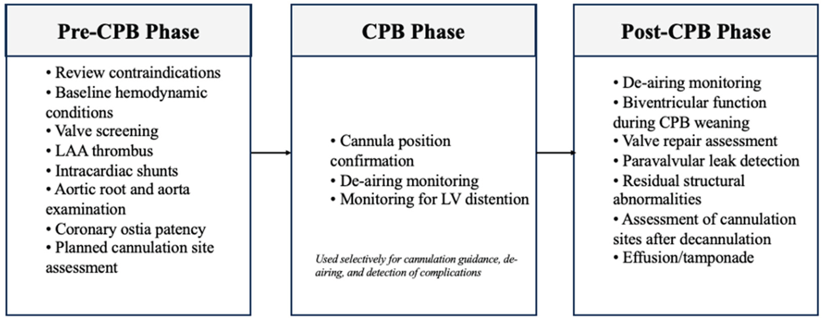

Table 1.
Impact of intraoperative transesophageal echocardiography on diagnosis and management.
| Study | Procedure type and population (n) | Key indication | Diagnostic impact | Management impact |
|---|---|---|---|---|
| Savage 1997 [12] | High-risk CABG (n=82) | Ventricular and valve assessment | Detection of unexpected pathology | Surgical management changed in 33% |
| Couture 2000 [13] | Mixed cardiac surgery (n=851) | Category-based TEE indications | Impactful findings in 14-28% | Surgical management modified in ~30% of impactful cases |
| Shapira 2004 [14] | Valve surgery (n=417) | Post-CPB valve assessment | Detection of prosthetic dysfunction | Immediate revision in 3.6% |
| Minhaj 2007 [15] | Mixed cardiac surgery (n=283) | Routine intraoperative TEE | New findings in 31% | Surgical plan altered in 25% |
| Eltzschig 2008 [7] | Mixed cardiac surgery (n=12,566) | Pre- and post-CPB TEE | Previously undiagnosed findings | Surgical decisions influenced in ≈9% |
| Kihara 2009 [16] | Cardiac and thoracic aortic surgery (n=1,011) | Detection of intraoperative abnormalities | Abnormal findings in 11.4% | Surgical decisions influenced in 5.8% |
| Guarracino 2010 [17] | CABG (n=521) | Detection of unexpected pathology | New findings in 17.2% | Surgical plan modified in 12.6% |
| Thorsgard 2014 [18] | Acute type A aortic dissection (n=64) | Assessment of dissection anatomy | Additional diagnostic data in 64% | Surgical plan changed in 39% |
| Elsherbiny 2019 [19] | Mixed cardiac surgery (n=100) | Routine TEE | New findings detected | Surgical management altered in 10% |
| Lomarat 2026 [20] | Mixed cardiac surgery (n=624) | Intraoperative diagnostic assessment | New findings detected | Surgical plan changed in 10.6% |
Abbreviations: CABG, coronary artery bypass grafting; CPB, cardiopulmonary bypass; TEE, transesophageal echocardiography. References are numbered according to first appearance in the manuscript.
Table 2.
Major indications and contraindications for intraoperative transesophageal echocardiography.
Table 2.
Major indications and contraindications for intraoperative transesophageal echocardiography.
| Major indications | Absolute contraindications | Relative contraindications |
|---|---|---|
| Hemodynamic instability of uncertain etiology | Esophageal perforation | Prior neck or mediastinal radiation |
| Valve repair or replacement surgery | Esophageal stricture | Esophageal diverticulum or varices |
| Congenital heart surgery requiring CPB | Obstructing esophageal neoplasm | Large hiatal hernia |
| Hypertrophic obstructive cardiomyopathy surgery | Active upper gastrointestinal bleeding | Recent esophageal or gastric surgery |
| Endocarditis with suspected perivalvular extension | Perforated upper gastrointestinal viscus | Dysphagia or odynophagia |
| Acute aortic syndrome requiring surgical repair | Severe coagulopathy or thrombocytopenia | |
| Cardiac tumor resection | Restricted neck mobility or instability | |
| Active esophagitis or peptic ulcer disease |
Adapted from ASE/SCA perioperative TEE recommendations and contemporary guidance. References 5, 8, and 9.
Table 3.
Reported complications and incidence.
| Complication type | Description | Estimated incidence | Key references |
|---|---|---|---|
| Mucosal injury (mild) | Erythema, petechiae, superficial tears | Up to 86% | Freitas-Ferraz et al., 2020 (n=50) [24] |
| GI injury (moderate-severe) | Bleeding, dysphagia and lacerations (overall) | 0.5-0.7% | Gangwani et al., 2023 (n=55,000) [25] |
| Esophageal perforation | 0.01-0.04% | Kallmeyer et al., 2001 (n=7,200) [26] | |
| Upper GI bleeding / hematoma | ~0.03% | Kallmeyer et al., 2001 (n=7,200) [26] | |
| Airway complications | Laryngospasm, accidental extubation, arytenoid trauma | 0.1-0.3% | Cote et al., 2008 [27]; Hilberath et al., 2010 [28] |
| Dental injury | Loose tooth, fracture | <0.03% | Kallmeyer et al., 2001 (n=7,200) [26] |
|
Hemodynamic arrhythmias |
Bradycardia, atrial arrhythmias, hypotension | Rare (reported, no large cohort data) | Hilberath et al., 2010 [28] |
| Thermal injury | Probe overheating causing mucosal burn | Extremely rare (case reports only) | Hilberath et al., 2010 [28] |
| Probe malfunction-related injury | Overheating or mechanical defect | Extremely rare (case reports only) | Hilberath et al., 2010 [28] |
| Mortality attributable to TEE | Typically, secondary to perforation | <0.01% | Kallmeyer et al., 2001 (n=7,200) [26] |
Disclaimer/Publisher’s Note: The statements, opinions and data contained in all publications are solely those of the individual author(s) and contributor(s) and not of MDPI and/or the editor(s). MDPI and/or the editor(s) disclaim responsibility for any injury to people or property resulting from any ideas, methods, instructions or products referred to in the content. |
© 2026 by the authors. Licensee MDPI, Basel, Switzerland. This article is an open access article distributed under the terms and conditions of the Creative Commons Attribution (CC BY) license (http://creativecommons.org/licenses/by/4.0/).
Copyright: This open access article is published under a Creative Commons CC BY 4.0 license, which permit the free download, distribution, and reuse, provided that the author and preprint are cited in any reuse.