

Submitted:
03 November 2026
Posted:
05 November 2026
You are already at the latest version
Abstract
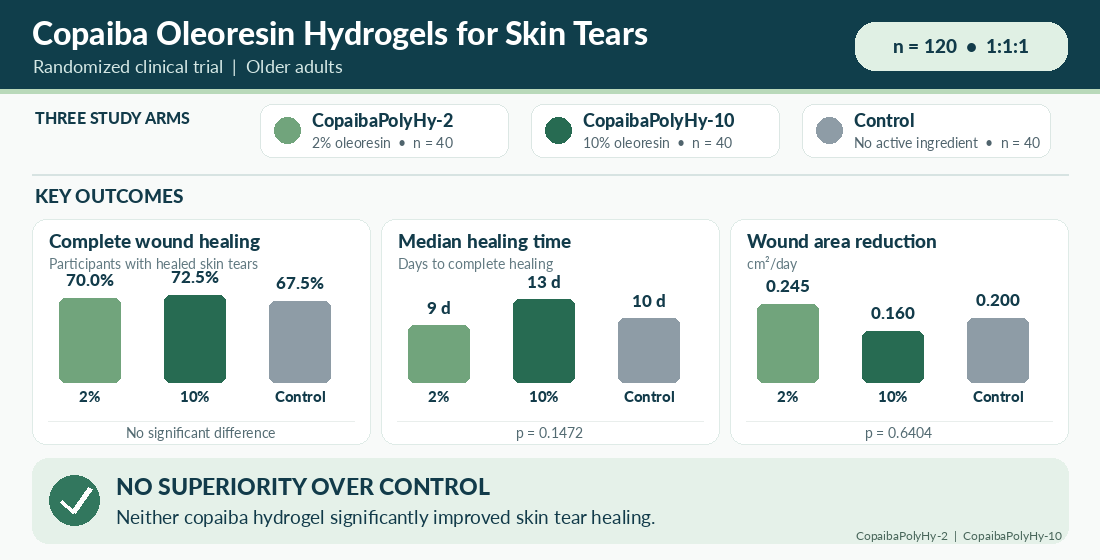
Skin tears are common acute wounds in older adults and are associated with morbidity, reduced quality of life, and increased healthcare burden. This randomized clinical trial evaluated two copaiba oleoresin polymeric hydrogels, CopaibaPolyHy-2 (2%) and Copai-baPolyHy-10 (10%), compared with a control hydrogel without active ingredient. A total of 120 participants were randomized equally among the three groups. Complete wound healing occurred in 70.0% of participants receiving CopaibaPolyHy-2, 72.5% receiving CopaibaPolyHy-10, and 67.5% receiving control treatment. No statistically significant dif-ferences were observed in complete healing rates for CopaibaPolyHy-2 versus control (RR 1.037; 95% CI 0.80–1.44) or CopaibaPolyHy-10 versus control (RR 1.070; 95% CI 0.81–1.43). Median healing times were 9, 13, and 10 days, respectively (p = 0.1472). Wound area reduction rates were 0.245, 0.160, and 0.200 cm²/day, respectively, with no significant be-tween-group difference (p = 0.6404). Although both copaiba formulations showed numer-ically favorable healing proportions, neither demonstrated superiority over the control hydrogel. These findings indicate that copaiba-based hydrogels were not associated with clinically or statistically significant improvements in skin tear healing in this population. Further studies should investigate optimized formulations, dosing strategies, and patient subgroups that may derive greater benefit. Larger multicenter trials may clarify potential clinical effects and therapeutic applicability.
Keywords:
skin tear
; copaiba oleoresin
; wound healing
; hydrogel
; clinical trial
; elderly patients
; Copaifera
1. Introduction
Skin tears are acute traumatic wounds caused by mechanical forces that separate the epidermis from the dermis or underlying tissues [1,2]. They predominantly affect older adults and represent a relevant burden in long-term, community, and acute care settings [3,4,5]. Population aging may further increase their incidence, reinforcing the need for effective prevention and treatment strategies [6].
Age-related skin changes are central to skin tear development. Reduced dermal thickness, collagen, elastin, moisture, and subcutaneous tissue decrease resistance to friction and shear [1,7]. Advanced age, frailty, female sex, photoaging, and dermal thickness below 0.80 mm have been associated with increased risk [8,9,10]. Additional factors include impaired mobility, sensory deficits, comorbidities, polypharmacy, corticosteroid or anticoagulant use, and nutritional impairment [1,2,3,7,8,11,12,13,14]. Previous skin tears strongly predict recurrence [14].
Standard management includes gentle cleansing, approximation of viable skin flaps, non-adherent dressings, and maintenance of a moist wound environment [2,11,15]. However, conventional dressings primarily provide passive protection and may not adequately address inflammation, infection risk, or impaired tissue repair. Frequent dressing changes may also cause additional trauma to fragile skin [16].
Copaiba oleoresin, obtained from Copaifera species, contains bioactive sesquiterpenes and diterpenes, including β-caryophyllene, α-humulene, copalic acid, and kaurenoic acid [17,18,19,20,21]. β-caryophyllene acts as a selective cannabinoid CB2 receptor agonist and contributes to the anti-inflammatory activity of copaiba oleoresin [22,23,24].
Preclinical studies have demonstrated anti-inflammatory, antimicrobial, and wound-healing effects of copaiba oleoresin. Reported mechanisms include modulation of NF-κB signaling, reduction of pro-inflammatory cytokines, increased IL-10, antimicrobial activity, collagen synthesis, angiogenesis, fibroblast proliferation, keratinocyte migration, and extracellular matrix remodeling [22,24,25,26,27,28,29,30,31,32,33,34,35,36]. Topical formulations have also improved re-epithelialization and wound contraction in experimental models, including diabetic animals [25,26,34,35,36].
Hydrogels are suitable vehicles for topical wound therapy because they maintain moisture, conform to wound surfaces, and support controlled delivery of active compounds [26,37,38,39,40]. Incorporating copaiba oleoresin into hydrogels may improve compound stability, release, bioavailability, and local therapeutic activity while minimizing additional trauma during wound care [26,38,39,40].
Despite encouraging preclinical findings, clinical evidence remains limited[30,40,41,42]. Most available studies are in vitro or animal investigations, and evidence in older adults with multiple comorbidities is particularly scarce [25,43,44]. This gap supports the need for controlled clinical evaluation of copaiba-based formulations for skin tears.
This randomized, controlled, parallel-group clinical trial evaluated the efficacy and safety of copaiba oleoresin hydrogels at 2% and 10% versus control hydrogel in older adults with skin tears. Outcomes included complete wound healing, healing time, and wound area reduction. We hypothesized that copaiba oleoresin would improve healing through its anti-inflammatory, antimicrobial, and tissue-regenerative properties.
2. Results and Discussion
The intention-to-treat (ITT) population comprised all randomized participants, regardless of treatment discontinuation, death, or incomplete adherence to the study protocol. This approach preserved the benefits of randomization and minimized selection bias. A total of 120 participants were included, with 40 allocated to each treatment arm: CopaibaPolyHy-2, CopaibaPolyHy-10, and control hydrogel.
Participants were predominantly older adults. Mean age was 68.3 ± 13.66 years in the CopaibaPolyHy-2 group, 65.6 ± 13.62 years in the CopaibaPolyHy-10 group, and 68.3 ± 14.77 years in the control group. Elderly participants represented 72.5%, 72.5%, and 82.5% of the groups, respectively (Table 1).
Female participants represented 47.5% of the CopaibaPolyHy-2 group, 37.5% of the CopaibaPolyHy-10 group, and 52.5% of the control group. No statistically significant difference was observed in sex distribution (p = 0.3907). Most participants were non-smokers, accounting for 87.5% of the CopaibaPolyHy-2 group, 85.0% of the CopaibaPolyHy-10 group, and 92.5% of the control group. Smoking status did not differ significantly among groups (p = 0.6790).
Regarding sociodemographic characteristics, married individuals predominated, and most participants had either incomplete or complete elementary education. Smoking prevalence was consistently low across all treatment groups.
Reduced or absent physical mobility was highly prevalent, affecting 75.0% of participants in the CopaibaPolyHy-2 group, 72.5% in CopaibaPolyHy-10, and 82.5% in the control group. Sensory impairment was also frequent, affecting 42.5%, 25.0%, and 42.5% of participants, respectively. These characteristics indicate a clinically complex population with multiple factors that may interfere with tissue repair. Hypertension was the most prevalent comorbidity, affecting 84 participants, followed by diabetes mellitus in 47 and chronic kidney disease in 21.
Overall, approximately 70% of participants had hypertension, 39% had diabetes mellitus, and 17.5% had chronic kidney disease. The most frequently used medications were omeprazole, insulin, losartan, acetylsalicylic acid, and simvastatin.
Baseline analyses showed no statistically significant differences among treatment groups regarding age, sex distribution, baseline wound area, baseline pain intensity, or smoking status. Baseline wound area showed p = 0.2838, while baseline Visual Analog Scale pain scores showed p = 0.1434.
These findings demonstrate adequate baseline comparability among the three treatment groups and support interpretation of subsequent outcomes within a balanced randomized population.
In the ITT population, complete wound healing occurred in 28 participants receiving CopaibaPolyHy-2, corresponding to 70.0% of the group. In the CopaibaPolyHy-10 group, 29 participants achieved complete healing, corresponding to 72.5%.
In the control group, complete healing occurred in 27 participants, corresponding to 67.5%. Treatment discontinuation occurred in four participants receiving CopaibaPolyHy-2, seven receiving CopaibaPolyHy-10, and one receiving the control hydrogel.
Eight deaths occurred in the CopaibaPolyHy-2 group, four in the CopaibaPolyHy-10 group, and 12 in the control group. These values corresponded to mortality rates of 20.0%, 10.0%, and 30.0%, respectively (Table 2).
The per-protocol (PP) population included participants who completed the study according to the protocol, correctly used the assigned intervention, and had complete primary outcome data available. Baseline demographic and clinical characteristics were comparable to those observed in the ITT analysis.
Mean age in the PP population ranged from 64.5 to 67.9 years across groups, with elderly individuals remaining the predominant age category. Hypertension, diabetes mellitus, and chronic kidney disease remained the most common comorbidities.
The medications most frequently used in the PP population were omeprazole, losartan, acetylsalicylic acid, furosemide, and simvastatin. The PP population comprised 84 participants who achieved complete wound healing. Among these participants, 28 were in the CopaibaPolyHy-2 group, 29 in the CopaibaPolyHy-10 group, and 27 in the control group.
For the primary outcome, complete healing occurred in 70.0% of participants receiving CopaibaPolyHy-2 and 67.5% of those receiving the control hydrogel. The relative risk was 1.037, with a 95% confidence interval of 0.80–1.44 and p = 1.00.
Although the healing rate was numerically higher with CopaibaPolyHy-2, the difference was not statistically significant. Under the conditions evaluated, the formulation did not demonstrate a clinically relevant therapeutic advantage over the control hydrogel.
Complete healing occurred in 72.5% of participants receiving CopaibaPolyHy-10 and 67.5% of those receiving the control hydrogel. The relative risk was 1.070, with a 95% confidence interval of 0.81–1.43 and p = 0.81.
Despite the numerically higher healing rate with CopaibaPolyHy-10, no statistically significant difference was observed. Therefore, the 10% formulation did not demonstrate a clinically relevant advantage over the control hydrogel.
Direct comparison of the two active formulations showed complete healing in 70.0% of participants receiving CopaibaPolyHy-2 and 72.5% receiving CopaibaPolyHy-10. The relative risk was 1.036, with a 95% confidence interval of 0.78–1.37 and p = 1.00 (Table 3).
Figure 1 illustrates the relative risks (RRs) and corresponding 95% confidence intervals for complete wound healing across the three pairwise treatment comparisons. Compared with the control group, CopaibaPolyHy-2 showed an RR of 1.037, whereas CopaibaPolyHy-10 showed an RR of 1.070. The direct comparison between the two active formulations yielded an RR of 1.036. In all comparisons, the 95% confidence intervals included the null value (RR = 1), indicating that no statistically significant differences in complete wound healing were detected between treatment groups.
Although the point estimates slightly favored the copaiba-based formulations, the confidence intervals indicate uncertainty regarding the magnitude and direction of the treatment effects. These findings reinforce the absence of demonstrated superiority of either copaiba-based hydrogel over the control formulation under the conditions evaluated.
Accordingly, no statistically significant difference in complete healing was detected between the 2% and 10% formulations.
Median time to complete wound healing was 9 days in the CopaibaPolyHy-2 group, with a 95% confidence interval of 7–13 days. In the CopaibaPolyHy-10 group, median healing time was 13 days, with a 95% confidence interval of 9–14 days. In the control group, median time to healing was 10 days, with a 95% confidence interval of 7–14 days. Levene’s test confirmed homogeneity of variances among groups (p = 0.4748).
However, Shapiro–Wilk tests demonstrated non-normal distribution of residuals across groups (p < 0.05). Therefore, comparisons were performed using the Kruskal–Wallis test.
No statistically significant difference in healing time was observed among treatment groups (p = 0.1472). Thus, neither copaiba formulation significantly shortened the time to complete wound healing compared with the control hydrogel.
For the secondary outcome, median wound area reduction was 0.245 cm2/day in the CopaibaPolyHy-2 group, with a 95% confidence interval of 0.160–0.300. In the CopaibaPolyHy-10 group, median wound area reduction was 0.160 cm2/day, with a 95% confidence interval of 0.114–0.350.
The control group showed a median wound area reduction of 0.200 cm2/day, with a 95% confidence interval of 0.133–0.300. Levene’s test indicated homogeneity of variances among groups (p = 0.9844). Residuals were not normally distributed according to the Shapiro–Wilk test, with p < 0.001 for all groups. Consequently, comparisons were conducted using the Kruskal–Wallis test.
No statistically significant difference in wound area reduction was identified among treatment groups (p = 0.6404).
Figure 2 presents the distribution of wound area reduction rates across the three treatment groups among participants who achieved complete wound healing. The boxplots provide a visual representation of the variability in healing rates, highlighting differences in dispersion and the presence of individual observations with relatively high or low wound area reduction rates.
This descriptive analysis complements the comparison of wound area reduction rates reported above. However, because the figure includes only participants who achieved complete healing, its interpretation is restricted to this subset and does not represent an intention-to-treat comparison. Consequently, the observed distributions should not be interpreted as evidence of differential treatment efficacy in the overall randomized population.
Taken together, the findings showed no statistically significant differences in complete healing at 21 days, time to complete healing, or wound area reduction. These findings are consistent with the pilot study reported by a Brazilian study. In that pilot study, copaiba oleoresin hydrogel also did not produce statistically significant differences in healing outcomes, although the investigation involved a smaller sample [28].
The absence of statistically significant differences should be interpreted in the context of extensive preclinical evidence supporting the anti-inflammatory, antimicrobial, and wound-healing properties of copaiba oleoresin.
Experimental studies have described mechanisms involving modulation of matrix metalloproteinases, stimulation of collagen synthesis, acceleration of re-epithelialization, reduction of inflammatory markers, and enhancement of angiogenesis [25,26,34,35,36].
In diabetic mice, which represent a model of impaired wound healing, copaiba oleoresin outperformed standard collagenase treatment in reducing wound area, decreasing pro-inflammatory cytokines, and improving histological outcomes [25]. Reported molecular mechanisms include β-caryophyllene-mediated CB2 receptor activation, NF-κB inhibition, and modulation of cytokine profiles [23,24,45].
The discrepancy between robust preclinical effects and the absence of clinical superiority in this trial may reflect the multifactorial nature of wound healing in complex clinical populations.
The characteristics of the study population represent a substantially more challenging healing environment than those commonly encountered in experimental animal models.
Mean age was approximately 66–68 years, and most participants were elderly. Aging is associated with skin changes that include reduced dermal thickness, decreased collagen content, and impaired vascular function [1,7].
The high burden of comorbidities also represented an important clinical factor. Diabetes mellitus, hypertension, and chronic kidney disease can impair wound healing through distinct mechanisms.
Diabetes mellitus is associated with impaired angiogenesis, neuropathy, chronic inflammation, immune dysfunction, and altered growth factor signaling [36,43]. Hypertension contributes to microvascular dysfunction and tissue hypoxia.
Chronic kidney disease may impair wound healing through uremic toxin accumulation, vessel calcification, vitamin and mineral deficiencies, immune dysregulation, oxidative stress, and anemia [30].
The combined presence of multiple comorbidities may exert synergistic negative effects on wound healing that exceed those associated with individual conditions [17,46].
Within this clinical context, complete healing rates ranging from 67.5% to 72.5% across all groups represent relevant outcomes considering the complexity of the study population.
The high prevalence of reduced or absent mobility, affecting approximately 72% to 82% of participants, represents an additional factor potentially associated with impaired healing.
Reduced mobility may limit circulation, increase pressure on vulnerable areas, and compromise adherence to wound care protocols [1,11]. Sensory impairment, affecting approximately 25% to 42% of participants, may further increase vulnerability. In patients with diabetic peripheral neuropathy, impaired sensation can reduce awareness of injury and delay treatment initiation [2,3].
These factors create an ongoing risk of trauma to healing wounds and may contribute to delayed healing independently of the topical treatment applied.
Consequently, targeting a single aspect of wound repair through topical treatment may be insufficient to produce measurable improvements when multiple systemic and local factors continue to impair healing.
Another relevant consideration is the effectiveness of standard wound care provided to all three groups. All participants received appropriate wound cleansing, approximation of wound edges when possible, and maintenance of a moist wound environment [2,11,47].
These measures are well-established components of optimal wound management. In addition, the control hydrogel itself provided moisture maintenance, protection from external contamination, and a cooling effect.
These properties may have improved patient comfort and adherence. Effective standard care may therefore have established a high baseline healing rate, limiting the ability to demonstrate incremental benefits from the active formulations.
Under these conditions, demonstrating superiority of experimental interventions may require larger sample sizes capable of detecting smaller effects or identification of subgroups in whom standard care is less effective.
Although not statistically significant, the numerical patterns observed deserve cautious consideration.
CopaibaPolyHy-2 showed a complete healing rate of 70.0%, compared with 67.5% in the control group, and a shorter median healing time of 9 days compared with 10 days in the control group.
CopaibaPolyHy-10 showed the highest complete healing rate, at 72.5%, but had a longer median healing time of 13 days.
A similar pattern was observed for wound area reduction. CopaibaPolyHy-2 showed the highest median reduction, at 0.245 cm2/day, compared with 0.200 cm2/day in the control group and 0.160 cm2/day with CopaibaPolyHy-10.
These patterns may suggest possible concentration-dependent effects that did not reach statistical significance because of sample size limitations or population heterogeneity.
The lower numerical performance of the 10% formulation for some outcomes may also reflect concentration-dependent effects. Experimental studies have shown that higher concentrations of natural products do not necessarily produce superior outcomes [48].
Alternatively, differences in formulation viscosity, release kinetics, or patient tolerance at higher concentrations may have influenced the results. These explanations remain hypothetical because they were not directly evaluated in the present trial.
Mortality also differed numerically among groups during the study period. Death occurred in 30% of participants in the control group, 20% in the CopaibaPolyHy-2 group, and 10% in the CopaibaPolyHy-10 group.
However, the trial was neither designed nor powered to evaluate mortality as an endpoint, and these differences did not reach statistical significance.
The study population consisted of elderly individuals with multiple comorbidities, including chronic kidney disease, diabetes mellitus, and cardiovascular disease, resulting in substantial frailty and clinical vulnerability.
In this population, skin tears may function as markers of overall frailty and declining health status rather than representing only isolated skin injuries [8,44].
The potential for copaiba oleoresin to reduce systemic inflammation through CB2 receptor-mediated mechanisms was considered as one possible explanation for the numerical mortality differences [24,45].
However, this interpretation is speculative. The observed mortality differences may also reflect chance variation, particularly given the sample size and absence of statistical significance.
Larger studies with longer follow-up periods would be required to determine whether copaiba oleoresin formulations influence mortality or other systemic outcomes in elderly patients with skin tears.
The findings can also be contextualized within the broader literature on natural product-based wound care interventions.
Waibel et al. evaluated copaiba oil incorporated into a silicone-based gel for scar reduction following laser treatment. No significant differences were observed compared with placebo regarding scar appearance, although the study was limited by its small sample size and high dropout rate [42].
Clinical trials evaluating other natural products for wound healing have produced mixed results. Some interventions have demonstrated benefits in specific populations or wound types, whereas others have failed to show superiority over standard care [49].
Recognized challenges in demonstrating the clinical efficacy of natural products include variability in product composition, differences between experimental and clinical populations, effectiveness of standard care, and the multifactorial nature of wound healing [30,40,41].
The present trial contributes controlled evidence in a well-defined population using standardized copaiba oleoresin formulations and validated outcome measures.
The randomized, controlled, parallel-group design represents an important methodological strength by minimizing bias and confounding.
Although clinically complex, the study population accurately reflects the real-world setting in which skin tears occur, thereby enhancing the external validity and generalizability of the findings [1,2].
The use of standardized formulations at two concentrations also permitted assessment of potential concentration-response relationships. The evaluation of complete healing, healing time, and wound area reduction provided a comprehensive assessment of treatment effects.
The ITT analysis included 120 participants, with 40 participants per group. According to the study, this sample provided adequate statistical power to detect clinically meaningful differences if they existed.
Documentation of comorbidities, mobility status, and sensory function further supported interpretation of the findings within the context of established risk factors for impaired healing [1,12].
Several limitations should nevertheless be acknowledged. The high burden of comorbidities and other risk factors for impaired wound healing may have reduced the ability to identify treatment effects [30,36].
The relatively short follow-up period focused on acute healing outcomes and did not assess long-term outcomes such as scar quality, recurrence rates, or quality of life [6].
Mechanistic assessments, including inflammatory markers, bacterial colonization, and histological analysis, were not performed. Such assessments could have provided information about biological effects even in the absence of demonstrable clinical superiority.
Although appropriate as a vehicle control, the control hydrogel provided active wound care benefits through moisture maintenance and local protection. These effects may have masked additional benefits associated with copaiba oleoresin [37].
The study was also conducted in a single healthcare setting, which may limit generalizability to settings with different populations or standard care practices.
Finally, lack of blinding, if applicable, may have introduced bias, although the use of objective outcomes such as complete healing and wound area may reduce this concern.
From a clinical perspective, the findings do not support routine use of copaiba oleoresin hydrogel formulations over standard hydrogel treatment for skin tears in elderly patients with multiple comorbidities. Neither formulation demonstrated statistically significant benefits for the clinical outcomes evaluated.
Nevertheless, the numerical trends observed, particularly with the 2% formulation, should be interpreted within the methodological and clinical limitations of the study.
The preclinical evidence and numerical differences in mortality were considered by the study as factors supporting further investigation, but they do not establish clinical efficacy.
The safety profile appeared acceptable, with no apparent increase in adverse events in the copaiba-treated groups. For clinicians managing skin tears in elderly patients, these findings reinforce the importance of comprehensive wound care, including appropriate cleansing, moisture management, and attention to underlying risk factors [11,47].
Management of comorbidities, nutritional support, and prevention of recurrent trauma should also be considered, particularly in elderly and clinically vulnerable patients.
Future research should focus on identifying patient subgroups most likely to benefit from copaiba oleoresin formulations. Subgroup analyses based on diabetes mellitus, chronic kidney disease, wound size, wound depth, infection status, or inflammatory markers may identify populations in which clearer benefits occur.
Optimization of formulation parameters also warrants investigation, including copaiba oleoresin concentration, hydrogel composition, release kinetics, and incorporation of additional bioactive components [20,37,38].
Mechanistic studies incorporating inflammatory markers, bacterial colonization assessments, and molecular analyses could clarify biological effects and help explain the discrepancy between preclinical and clinical findings.
Longer-term studies should evaluate scar quality, recurrence, quality of life, and cost-effectiveness to provide a broader assessment of clinical value.
Pragmatic trials conducted across diverse healthcare settings and standard care practices could enhance generalizability and identify clinical contexts in which copaiba oleoresin formulations provide greater benefit.
Finally, investigation of potential systemic effects, including anti-inflammatory and immunomodulatory actions, may clarify whether benefits extend beyond local wound healing, particularly in frail elderly populations [24,45].
Overall, CopaibaPolyHy-2 and CopaibaPolyHy-10 did not demonstrate statistically significant superiority over the control hydrogel regarding complete wound healing, time to healing, or wound area reduction.
These results should be interpreted considering the effectiveness of standard wound care, the clinical complexity of the population, and the multiple factors capable of influencing tissue repair.
Although numerical differences were observed, they do not establish a therapeutic benefit of the copaiba oleoresin formulations.
The findings support further investigation focused on optimized formulations, specific patient subgroups, and additional clinical and mechanistic outcomes.
3. Conclusions
This randomized controlled trial found no significant differences between copaiba oleoresin hydrogels and the control hydrogel in complete healing, healing time, or wound area reduction among older adults with skin tears and multiple comorbidities. Although CopaibaPolyHy-2 showed favorable numerical trends in healing outcomes, these differences were not statistically significant.
Overall, copaiba oleoresin hydrogels did not demonstrate superior clinical efficacy over the control treatment under the conditions evaluated. The findings highlight the complexity of wound healing in older adults with substantial comorbidity and functional impairment. Further studies should investigate optimized formulations, identify patient subgroups more likely to benefit, and evaluate longer-term outcomes to better define the potential role of copaiba oleoresin in skin tear management.
4. Materials and Methods
4.1. Study Design
This study was designed as a randomized, controlled clinical trial in accordance with CONSORT guidelines [50] evaluating the efficacy of two copaiba-based hydrogel formulations in the treatment of skin tears. Participants were allocated into three parallel treatment groups: CopaibaPolyHy-2 (2% copaiba-based hydrogel), CopaibaPolyHy-10 (10% copaiba-based hydrogel), and control (hydrogel without oleorresin).
The study was conducted in accordance with ethical principles for research involving human participants and followed the recommendations for randomized clinical trials. This study was registered in the National Genetic Heritage Management System (SISGEN)—A43A234.
4.2. Study Population
Eligible participants were adult patients presenting with skin tears and meeting the predefined eligibility criteria established by the study protocol. Participants were randomized into one of the three treatment groups.
Two analytical populations were defined:
- The intention-to-treat (ITT) population included all randomized participants regardless of treatment discontinuation, death, or incomplete adherence;
- The per-protocol (PP) population included only participants who completed the study protocol, correctly used the assigned intervention, and had complete primary outcome data available.
4.3. Interventions
The protocol established in the study consisted of cleaning the wound with 0.9% saline solution, drying the edges, applying one of the treatments according to randomized allocation (CopaibaPolyHy-2, CopaibaPolyHy-10 or control) and covering it with sterile gauze and fix it [51]. After that, the participant received an informative with all instructions to repeat the technique at home.
The products were applied according to the study protocol throughout the follow-up period.
4.4. Outcomes
4.4.1. Primary Outcomes
The primary outcomes evaluated in this study were:
- Complete wound healing;
- Time to complete wound healing.
Complete wound healing was operationally defined as complete closure and healing of the skin tear lesion.
For the analysis of complete healing, participants who discontinued treatment or died during follow-up were classified as non-events.
For the analysis of time to healing, the event was defined as complete healing, whereas discontinuation, death, or absence of healing by the end of follow-up were considered censored observations.
4.4.2. Secondary Outcomes
The secondary outcome analyzed was the wound healing velocity (cm2/day).
4.5. Baseline Variables
Baseline demographic and clinical variables included age, sex, smoking status, physical mobility, sensory status, pre-existing comorbidities, medication use, and baseline wound area.
4.6. Statistical Analysis
Statistical analyses were performed according to both the intention-to-treat (ITT) and per-protocol (PP) principles. The ITT population included all randomized participants regardless of adherence, discontinuation, or death, whereas the PP population included only participants who completed the study protocol and had complete primary outcome data.
Descriptive statistics were used to summarize demographic and clinical characteristics. Continuous variables were expressed as means and standard deviations or medians and 95% confidence intervals, according to data distribution. Categorical variables were presented as absolute and relative frequencies.
Baseline comparability among treatment groups was assessed using appropriate statistical tests according to the nature and distribution of the variables. Associations between categorical variables were evaluated using Pearson’s chi-square test or Fisher’s exact test when expected frequencies were small [52].
The primary outcome of complete wound healing was analyzed by calculating relative risks (RRs) and corresponding 95% confidence intervals (CIs) for pairwise comparisons between treatment groups [52].
For continuous secondary outcomes, including time to complete healing and wound area reduction, assumptions of normality and homogeneity of variances were evaluated using the Shapiro–Wilk and Levene tests, respectively. Since residuals did not meet normality assumptions, non-parametric analyses were performed using the Kruskal–Wallis test.
All statistical tests were two-tailed, and p-values < 0.05 were considered statistically significant.
Author Contributions
Conceptualization, C.C.C., E.P.d.S., J.T.G.P., K.C.D.P. and Z.M.F.d.F.; methodology, C.C.C., V.F.V.-J., J.T.G.P. and Z.M.F.d.F.; software, G.G.N.; investigation, C.C.C.; data curation, G.G.N.; writing—original draft, C.C.C., K.C.D.P. and Z.M.F.d.F.; writing—review and editing, V.F.V.-J. and B.G.R.B.d.O.; supervision, E.P.d.S. and Z.M.F.d.F. All authors have read and agreed to the published version of the manuscript.
Funding
This research received no external funding.
Institutional Review Board Statement
This study was conducted in accordance with the Declaration of Helsinki and approved by the Ethics Committee of Federal University of Rio de Janeiro (protocol code 4.225.729 on 20 August 2020).
Informed Consent Statement
Informed consent was obtained from all subjects involved in the study.
Data Availability Statement
The original contributions presented in this study are included in the article. Further inquiries can be directed to the corresponding authors.
Conflicts of Interest
The authors declare no conflicts of interest.
References
- Holloway, S.; Prentice, J.; Samuriwo, R. Preventing, Assessing and Treating Skin Tears. Nurs. Stand. 2023, 38, 37–45. [Google Scholar] [CrossRef]
- Ratliff, C.R.; Fletcher, K.R. Skin Tears: A Review of the Evidence to Support Prevention and Treatment. Ostomy Wound Manag. 2007, 53, 32–42. [Google Scholar]
- Hardie, C.; Wick, J.Y. Skin Tears in Older People. Sr. Care Pharm. 2020, 35, 379–387. [Google Scholar] [CrossRef]
- LeBlanc, K.; Woo, K.Y.; VanDenKerkhof, E.; Woodbury, M.G. Skin Tear Prevalence and Incidence in the Long-Term Care Population: A Prospective Study. J. Wound Care 2020, 29, S16–S22. [Google Scholar] [CrossRef]
- Yang, S.; Liang, X.; She, J.; Tian, J.; Wen, Z.; Tao, Y.; Wang, H.; Zhang, X. Prevalence and Incidence of Skin Tear in Older adults:A Systematic Review and Meta-Analysis. J. Tissue Viability 2024, 33, 1017–1024. [Google Scholar] [CrossRef]
- Sen, C.K. Human Wound and Its Burden: Updated 2020 Compendium of Estimates. Adv. Wound Care 2021, 10, 281–292. [Google Scholar] [CrossRef]
- Xu, X.; Lau, K.; Taira, B.R.; Singer, A.J. The Current Management of Skin Tears. Am. J. Emerg. Med. 2009, 27, 729–733. [Google Scholar] [CrossRef]
- Benbow, M. Assessment, Prevention and Management of Skin Tears. Nurs. Older People 2017, 29, 31–39. [Google Scholar] [CrossRef]
- Koyano, Y.; Nakagami, G.; Iizaka, S.; Minematsu, T.; Noguchi, H.; Tamai, N.; Mugita, Y.; Kitamura, A.; Tabata, K.; Abe, M.; et al. Exploring the Prevalence of Skin Tears and Skin Properties Related to Skin Tears in Elderly Patients at a Long-term Medical Facility in Japan. Int. Wound J. 2016, 13, 189–197. [Google Scholar] [CrossRef]
- Koyano, Y.; Nakagami, G.; Iizaka, S.; Sugama, J.; Sanada, H. Skin Property Can Predict the Development of Skin Tears among Elderly Patients: A Prospective Cohort Study. Int. Wound J. 2017, 14, 691–697. [Google Scholar] [CrossRef]
- McInulty, L. Prevention and Management of Skin Tears in Older People. Emerg. Nurse 2017, 25, 32–39. [Google Scholar] [CrossRef]
- Holmes, R.F.; Davidson, M.W.; Thompson, B.J.; Kelechi, T.J. Skin Tears: Care and Management of the Older Adult at Home. Home Healthc. Nurse 2013, 31, 90–101. [Google Scholar] [CrossRef]
- Serra, R.; Ielapi, N.; Barbetta, A.; De Franciscis, S. Skin Tears and Risk Factors Assessment: A Systematic Review on Evidence-based Medicine. Int. Wound J. 2018, 15, 38–42. [Google Scholar] [CrossRef]
- Lewin, G.F.; Newall, N.; Alan, J.J.; Carville, K.J.; Santamaria, N.M.; Roberts, P.A. Identification of Risk Factors Associated with the Development of Skin Tears in Hospitalised Older Persons: A Case–Control Study. Int. Wound J. 2016, 13, 1246–1251. [Google Scholar] [CrossRef]
- LeBlanc, K.; Campbell, K.E.; Beeckman, D.; Dunk, A.M.; Harley, C. Best Practice Recommendations for the Prevention and Management of Skin Tears in Aged Skin. In Wounds Int.; 2018. [Google Scholar]
- Peters, M.D.J.; Campbell, J.M. The Effectiveness of Treatments for Skin Tears in Older People: A Systematic Review Protocol. JBI Database Syst. Rev. Implement. Rep. 2014, 12, 127–140. [Google Scholar] [CrossRef]
- Carvalho, A.B.; Santiago, C.C.; Stanley, J.; Dias, K.D.R.; Araujo, L.E.D.; Cardoso, T.C. Propriedade Terapêutica Do Óleo de Copaiba (Copaifera Spp.) No Tratamento de Doenças de Pele. Obs. Econ. Latinoam. 2024, 22, e8144. [Google Scholar] [CrossRef]
- Lima, C.A.S.; Violante, I.M.P.; Alves, W.K.S.; Carvalho, F.T.; Dias, N.D.S.; Dos Anjos, T.R.; De Andrade, K.R.N.C.; Frasson, A.P.Z.; Dos Santos, M.D.; Carvalho, R.C.T. Atualizações Sobre as Propriedades Medicinais Do Óleo de Copaíba (Copaifera Spp.): Uma Revisão Bibliográfica. UNICIÊNCIAS 2021, 25, 100–106. [Google Scholar] [CrossRef]
- Pieri, F.A.; Mussi, M.C.; Moreira, M.A.S. Copaiba Oil (Copaifera Sp.): History, Extraction, Industrial Applications and Medicinal Properties. Rev. Bras. Plan. Med. 2009, 11, 465–472. [Google Scholar] [CrossRef]
- Pinheiro, J.; Tavares, E.; Silva, S.; Félix Silva, J.; Carvalho, Y.; Ferreira, M.; Araújo, A.; Barbosa, E.; Fernandes Pedrosa, M.; Soares, L.; et al. Inclusion Complexes of Copaiba (Copaifera Multijuga Hayne) Oleoresin and Cyclodextrins: Physicochemical Characterization and Anti-Inflammatory Activity. Int. J. Mol. Sci. 2017, 18, 2388. [Google Scholar] [CrossRef]
- Leandro, L.M.; De Sousa Vargas, F.; Barbosa, P.C.S.; Neves, J.K.O.; Da Silva, J.A.; Da Veiga-Junior, V.F. Chemistry and Biological Activities of Terpenoids from Copaiba (Copaifera Spp.) Oleoresins. Molecules 2012, 17, 3866–3889. [Google Scholar] [CrossRef]
- Gushiken, L.F.S.; Beserra, F.P.; Hussni, M.F.; Gonzaga, M.T.; Ribeiro, V.P.; de Souza, P.F.; Campos, J.C.L.; Massaro, T.N.C.; Hussni, C.A.; Takahira, R.K.; et al. Beta-Caryophyllene as an Antioxidant, Anti-Inflammatory and Re-Epithelialization Activities in a Rat Skin Wound Excision Model. Oxid. Med. Cell. Longev. 2022, 2022, 1–21. [Google Scholar] [CrossRef]
- Parisotto-Peterle, J.; Bidone, J.; Lucca, L.G.; Araújo, G.D.M.S.; Falkembach, M.C.; Da Silva Marques, M.; Horn, A.P.; Dos Santos, M.K.; Da Veiga, V.F.; Limberger, R.P.; et al. Healing Activity of Hydrogel Containing Nanoemulsified β-Caryophyllene. Eur. J. Pharm. Sci. 2020, 148, 105318. [Google Scholar] [CrossRef]
- Teixeira, F.B.; de Brito Silva, R.; Lameira, O.A.; Webber, L.P.; D’Almeida Couto, R.S.; Martins, M.D.; Lima, R.R. Copaiba Oil-Resin (Copaifera Reticulata Ducke) Modulates the Inflammation in a Model of Injury to Rats’ Tongues. BMC Complement. Altern. Med. 2017, 17, 313. [Google Scholar] [CrossRef]
- Amorim, J.L.; Figueiredo, J.D.B.; Amaral, A.C.F.; Barros, E.G.D.O.; Palmero, C.; MPalantinos, M.A.; Ramos, A.D.S.; Ferreira, J.L.P.; Silva, J.R.D.A.; Benjamim, C.F.; et al. Wound Healing Properties of Copaifera Paupera in Diabetic Mice. PLoS ONE 2017, 12, e0187380. [Google Scholar] [CrossRef]
- Gushiken, L.F.S.; Beserra, F.P.; Hussni, M.F.; Gonzaga, M.T.; Ribeiro, V.P.; De Souza, P.F.; Campos, J.C.L.; Massaro, T.N.C.; Hussni, C.A.; Takahira, R.K.; et al. Copaifera Langsdorffii Oleoresin-Loaded Nanostructured Lipid Carrier Emulgel Improves Cutaneous Healing by Anti-Inflammatory and Re-Epithelialization Mechanisms. Int. J. Mol. Sci. 2023, 24, 15882. [Google Scholar] [CrossRef]
- Basile, A.C.; SertiÉ, J.A.A.; Freitas, P.C.D.; Zanini, A.C. Anti-Inflammatory Activity of Oleoresin from Brazilian Copaifera. J. Ethnopharmacol. 1988, 22, 101–109. [Google Scholar] [CrossRef]
- Campanholi, K. da S.S.; Junior, R.C.D.S.; Braga, G.; De Morais, F.A.P.; Balbinot, R.B.; Caetano, W. Minireview about Medicinal Copaiba Oil in the Treatment of Skin Diseases. J. Dermatol. Skin. Sci. 2022, 4, 1–6. [Google Scholar] [CrossRef]
- Frazão, D.R.; Cruz, J.N.; Santana De Oliveira, M.; Baia-da-Silva, D.C.; Nazário, R.M.F.; Rodrigues, M.F.D.L.; Saito, M.T.; Souza-Rodrigues, R.D.; Lima, R.R. Evaluation of the Biological Activities of Copaiba (Copaifera Spp): A Comprehensive Review Based on Scientometric Analysis. Front. Pharmacol. 2023, 14, 1215437. [Google Scholar] [CrossRef]
- Sandepudi, K.; Shah, K.V.; Melnick, B.A.; Li, R.A.; Ho, K.; O’Connor, M.J.; Galiano, R.D. Pathophysiology of Wound Development and Chronicity in Renal Disease: A Narrative Review. Int. Wound J. 2025, 22, e70713. [Google Scholar] [CrossRef]
- Bardají, D.K.R.; Da Silva, J.J.M.; Bianchi, T.C.; De Souza Eugênio, D.; De Oliveira, P.F.; Leandro, L.F.; Rogez, H.L.G.; Venezianni, R.C.S.; Ambrosio, S.R.; Tavares, D.C.; et al. Copaifera Reticulata Oleoresin: Chemical Characterization and Antibacterial Properties against Oral Pathogens. Anaerobe 2016, 40, 18–27. [Google Scholar] [CrossRef]
- Masson, D.S.; Salvador, S.L.; Polizello, A.C.M.; Frade, M.A.C. Antimicrobial Activity of Copaíba (Copaifera Langsdorffii) Oleoresin on Bacteria of Clinical Significance in Cutaneous Wounds. Rev. Bras. Plan. Med. 2013, 15, 664–669. [Google Scholar] [CrossRef]
- Ziech, R.E.; Farias, L.D.; Balzan, C.; Ziech, M.F.; Heinzmann, B.M.V.; Lameira, O.A.; A. de V., C. Atividade Antimicrobiana Do Oleorresina de Copaíba (Copaifera Reticulata) Frente a Staphylococcus Coagulase Positiva Isolados de Casos de Otite Em Cães. Pesq. Vet. Bras. 2013, 33, 909–913. [Google Scholar] [CrossRef]
- Masson-Meyers, D.S.; Enwemeka, C.S.; Bumah, V.V.; Andrade, T.; Frade, M.A. Topical Treatment with Copaifera Langsdorffii Oleoresin Improves Wound Healing in Rats. Int. J. Phytomed. 2013, 5, 378–386. [Google Scholar]
- Gushiken, L.F.S.; Hussni, C.A.; Bastos, J.K.; Rozza, A.L.; Beserra, F.P.; Vieira, A.J.; Padovani, C.R.; Lemos, M.; Polizello Junior, M.; Silva, J.J.M.D.; et al. Skin Wound Healing Potential and Mechanisms of the Hydroalcoholic Extract of Leaves and Oleoresin of Copaifera Langsdorffii Desf. Kuntze in Rats. Evid. Based Complement. Altern. Med. 2017, 2017, 6589270. [Google Scholar] [CrossRef]
- Klein, K.C.; Guha, S.C. Cutaneous Wound Healing: Current Concepts and Advances in Wound Care. Indian J. Plast. Surg. 2014, 47, 303–317. [Google Scholar] [CrossRef]
- Koupaei, N. Polymeric Wound Dressings: From Classic Hydrogels to Self-Healing Systems. J. Adv. Mater. Process. 2024, 12, 21 pages. [Google Scholar] [CrossRef]
- Campanholi, K. da S.S.; da Silva Junior, R.C.; Gonçalves, R.S.; Bassi da Silva, J.; Pedroso de Morais, F.A.; Said Dos Santos, R.; Vilsinski, B.H.; de Oliveira, G.L.M.; Pozza, M.S.D.S.; Bruschi, M.L.; et al. Design and Optimization of a Natural Medicine from Copaifera Reticulata Ducke for Skin Wound Care. Polymers 2022, 14. [Google Scholar] [CrossRef]
- Garcia, M.H.; Dionísio, T.J.; Cestari, T.M.; Parisi, V.A.; Torres, E.A.; Santos, C.F. The Role of AT-1 Antagonist on Wound Healing in Rats with Hypertension and Diabetes. J. Mol. Histol. 2025, 56, 84. [Google Scholar] [CrossRef]
- Ferro, M.; Masso, S.; Souza, R.; Moreno, M.; Moreira, E. Meta-Analysis on Copaiba Oil: Its Functions in Metabolism and Its Properties as an Anti-Inflammatory Agent. J. Morphol. Sci. 2018, 35, 161–166. [Google Scholar] [CrossRef]
- Menezes, A.C.D.S.; Alves, L.D.B.; Goldemberg, D.C.; De Melo, A.C.; Antunes, H.S. Anti-Inflammatory and Wound Healing Effect of Copaiba Oleoresin on the Oral Cavity: A Systematic Review. Heliyon 2022, 8, e08993. [Google Scholar] [CrossRef]
- Waibel, J.; Patel, H.; Cull, E.; Sidhu, R.; Lupatini, R. Prospective, Randomized, Double-Blind, Placebo-Controlled Study on Efficacy of Copaiba Oil in Silicone-Based Gel to Reduce Scar Formation. Dermatol. Ther. 2021, 11, 2195–2205. [Google Scholar] [CrossRef]
- Ya’kub, A.; Kristianto, H.; Supriati, L. Analysis of Diabetic Foot Complication Factors in Type 2 Diabetes Mellitus Patients with Chronic Kidney Failure Complications: A Scoping Review. J. Foot Ankle Surg. Asia Pac. 2025, 13, 31–39. [Google Scholar] [CrossRef]
- Baranoski, S.; LeBlanc, K.; Gloeckner, M. CE: Preventing, Assessing, and Managing Skin Tears A Clinical Review. AJN Am. J. Nurs. 2016, 116, 24–30. [Google Scholar] [CrossRef]
- Gushiken, L.F.S.; Beserra, F.P.; Hussni, M.F.; Gonzaga, M.T.; Ribeiro, V.P.; De Souza, P.F.; Campos, J.C.L.; Massaro, T.N.C.; Hussni, C.A.; Takahira, R.K.; et al. Beta-caryophyllene as an Antioxidant, Anti-inflammatory and Re-epithelialization Activities in a Rat Skin Wound Excision Model. Oxid. Med. Cell. Longev. 2022, 2022, 9004014. [Google Scholar] [CrossRef]
- Montes, L.V.; Lis-Balchin, M.; Hart, S.; Amaral, M.T.; Frade, M.A. Evidências Para o Uso Da Óleo-Resina de Copaíba Na Cicatrização de Ferida - Uma Revisão Sistemática. Rev. Bras. Plan. Med. 2009, 11, 246–252. [Google Scholar]
- LeBlanc, K.; Campbell, K.E.; Wood, E.; Beeckman, D. Best Practice Recommendations for Prevention and Management of Skin Tears in Aged Skin: An Overview. J. Wound. Ostomy Cont. Nurs. 2018, 45, 540–542. [Google Scholar] [CrossRef]
- Tiah, A. Adjunctive Mesenchymal Stem Cell Secretome Therapy for a Chronic Hard to Heal Wound in a Patient with Multiple Comorbids: A Case Report. Med. Res. Arch. 2025, 13. [Google Scholar] [CrossRef]
- Moses, R.L.; Prescott, T.A.K.; Mas-Claret, E.; Steadman, R.; Moseley, R.; Sloan, A.J. Evidence for Natural Products as Alternative Wound-Healing Therapies. Biomolecules 2023, 13, 444. [Google Scholar] [CrossRef]
- Hopewell, S.; Chan, A.-W.; Collins, G.S.; Hróbjartsson, A.; Moher, D.; Schulz, K.F.; Tunn, R.; Aggarwal, R.; Berkwits, M.; Berlin, J.A.; et al. CONSORT 2025 Statement: Updated Guideline for Reporting Randomised Trials. Lancet 2025, S0140-6736((25)00672-5). [Google Scholar] [CrossRef]
- Cardinelli, C.C.; Passos, J.T.G.; Veiga-Junior, V.F.; de Oliveira, B.G.R.B.; dos Santos, E.P.; Neto, G.G.; Di Piero, K.C.; de Freitas, Z.M.F. Skin Tear Treatment with Copaifera Multijuga Hayne in Polymeric Hydrogel: A Randomized Clinical Trial. Pharmaceuticals 2024, 17, 1691. [Google Scholar] [CrossRef]
- Chow, S.-C.; Shao, J.; Wang, H. Sample Size Calculations in Clinical Research, 2nd ed.; Chapman & Hall/CRC: Boca Raton, 2008. [Google Scholar]
Figure 1.
Forest plot of relative risks (RRs) and corresponding 95% confidence intervals for complete wound healing in the intention-to-treat population. Squares represent point estimates, and horizontal lines indicate 95% confidence intervals. The dashed vertical line represents the null effect (RR = 1). Confidence intervals crossing this line indicate that the corresponding comparisons were not statistically significant.
Figure 1.
Forest plot of relative risks (RRs) and corresponding 95% confidence intervals for complete wound healing in the intention-to-treat population. Squares represent point estimates, and horizontal lines indicate 95% confidence intervals. The dashed vertical line represents the null effect (RR = 1). Confidence intervals crossing this line indicate that the corresponding comparisons were not statistically significant.
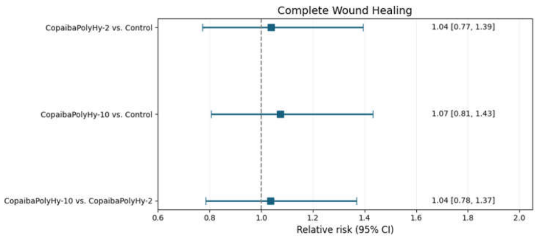
Figure 2.
Distribution of wound area reduction rates (cm2/day) among participants who achieved complete wound healing in the control, CopaibaPolyHy-2, and CopaibaPolyHy-10 groups. Boxes represent the interquartile range (25th–75th percentiles), horizontal lines indicate the median, and whiskers extend to the most extreme observations within 1.5 times the interquartile range. Individual observations are represented by dots. This analysis is restricted to participants with complete wound healing and should be interpreted as descriptive.
Figure 2.
Distribution of wound area reduction rates (cm2/day) among participants who achieved complete wound healing in the control, CopaibaPolyHy-2, and CopaibaPolyHy-10 groups. Boxes represent the interquartile range (25th–75th percentiles), horizontal lines indicate the median, and whiskers extend to the most extreme observations within 1.5 times the interquartile range. Individual observations are represented by dots. This analysis is restricted to participants with complete wound healing and should be interpreted as descriptive.
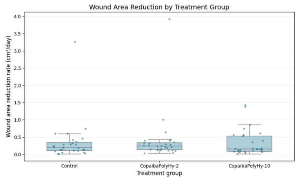

Table 1.
Baseline demographic and clinical characteristics of the intention-to-treat population.
| Variable | CopaibaPolyHy-2 (n = 40) | CopaibaPolyHy-10 (n = 40) |
Control (n = 40) |
p-Value |
|---|---|---|---|---|
| Age, mean ± SD1 (years) | 68.3 ± 13.66 | 65.6 ± 13.62 | 68.3 ± 14.77 | NS |
| Female sex, n (%) | 19 (47.5) | 15 (37.5) | 21 (52.5) | 0.3907 |
| Non-smoker, n (%) | 35 (87.5) | 34 (85.0) | 37 (92.5) | 0.6790 |
| Elderly participants, n (%) | 29 (72.5) | 29 (72.5) | 33 (82.5) | — |
| Reduced or absent mobility, n (%) |
30 (75.0) | 29 (72.5) | 33 (82.5) | — |
| Sensory impairment (absent or reduced), n (%) |
17 (42.5) | 10 (25.0) | 17 (42.5) | — |
1 Abbreviations: SD, standard deviation; NS, non-significant.
Table 2.
Primary outcome: complete wound healing in the intention-to-treat population.
| Outcome | CopaibaPolyHy-2 | CopaibaPolyHy-10 | Control |
|---|---|---|---|
| Complete healing, n (%) | 28 (70.0) | 29 (72.5) | 27 (67.5) |
| Discontinuation, n (%) | 4 (10.0) | 7 (17.5) | 1 (2.5) |
| Death, n (%) | 8 (20.0) | 4 (10.0) | 12 (30.0) |
Table 3.
Relative risk analyses for complete wound healing.
| Comparison | Relative Risk (RR) | 95% CI | p-Value |
|---|---|---|---|
| CopaibaPolyHy-2 vs. Control | 1.037 | 0.80-1.44 | 1.00 |
| CopaibaPolyHy-10 vs. Control | 1.070 | 0.81-1.43 | 0.81 |
| CopaibaPolyHy-10 vs. CopaibaPolyHy-2 | 1.036 | 0.78-1.37 | 1.00 |
Disclaimer/Publisher’s Note: The statements, opinions and data contained in all publications are solely those of the individual author(s) and contributor(s) and not of MDPI and/or the editor(s). MDPI and/or the editor(s) disclaim responsibility for any injury to people or property resulting from any ideas, methods, instructions or products referred to in the content. |
© 2026 by the authors. Licensee MDPI, Basel, Switzerland. This article is an open access article distributed under the terms and conditions of the Creative Commons Attribution (CC BY) license.
Copyright: This open access article is published under a Creative Commons CC BY 4.0 license, which permit the free download, distribution, and reuse, provided that the author and preprint are cited in any reuse.