

Submitted:
03 November 2026
Posted:
05 November 2026
You are already at the latest version
Abstract
De novo drug discovery requires over a decade of research and $1.5 billion to $2.5 billion in investment per approved molecule, yet more than 90% of candidates fail during clinical trials due to unforeseen toxicity or poor efficacy. In silico drug repurposing offers a faster, cost-effective alternative by finding new therapeutic uses for regulatory-approved or clinical-stage molecules. However, individual computational methods have clear blind spots. Network biology identifies disease-related genes but lacks atomic-level detail on how a drug physically binds to a receptor. Conversely, standalone molecular docking often produces false-positive hits and relies on an outdated single-target mindset. This review provides a practical blueprint for combining network pharmacology with structure-based virtual screening (SBVS) in preclinical drug repurposing. We first outline how disease genomics databases (DisGeNET, GeneCards) and protein–protein interaction (PPI) networks (STRING) pair with graph-theory tools in Cytoscape (cytoHubba, MCODE) to pinpoint central hub proteins. Next, we detail how to screen curated chemical libraries (DrugBank, ChEMBL) against these prioritized targets using AutoDock Vina and AlphaFold structures. We also explain why approved drugs still require indication-specific pharmacokinetic checks in SwissADME and ADMETlab 3.0, followed by 100–500 ns explicit-solvent molecular dynamics (MD) simulations in GROMACS and MM-PBSA/GBSA free-energy rescoring. Finally, we review recent preclinical applications across infectious, neurological, and oncological diseases and discuss how deep-learning tools like DiffDock and AlphaFold 3 are reshaping virtual screening.
Keywords:
drug repurposing
; network pharmacology
; structure-based virtual screening
; AutoDock vina
; protein–protein interaction network
; cytoscape
; SwissADME
; molecular dynamics simulation
; polypharmacology
1. Introduction
1.1. The Economic and Attrition Bottlenecks of De Novo Drug Discovery
Modern pharmaceutical research faces declining R&D efficiency despite major advances in high-throughput screening and multi-omics sequencing. Developing a single new molecular entity (NME) from initial target validation to market approval takes 10 to 15 years and costs between $1.5 billion and $2.5 billion (Pushpakom et al., 2018). Less than 10% of drug candidates that enter Phase I clinical trials ever reach the clinic. Most late-stage Phase II and Phase III failures stem from inadequate clinical efficacy, unexpected human cardiotoxicity or hepatotoxicity, and poor pharmacokinetic profiles (Pushpakom et al., 2018).
Drug repurposing, also called drug repositioning or therapeutic switching, offers a practical way around this bottleneck (Pushpakom et al., 2018). Repurposed candidates come from regulatory-approved libraries (such as the US FDA or EMA) or shelved clinical-stage compounds. Because these molecules already have established human Phase I safety profiles, preclinical toxicology data, and scalable manufacturing protocols, redirecting them to new diseases cuts both development time and pre-approval costs (Pushpakom et al., 2018; Ngernsombat et al., 2024). Open-access databases like DrugBank 6.0 (Knox et al., 2023) and ChEMBL (Zdrazil et al., 2023) provide ready-to-use 3D structures, bioactivity data, and clinical annotations for thousands of approved small molecules, making virtual screening accessible to academic labs worldwide.
1.2. Beyond Reductionism: The Shift Toward Systems-Level Polypharmacology
For decades, computer-aided drug design (CADD) followed the "magic bullet" model, designing selective ligands to hit a single protein target (Hopkins, 2008; Liu & Yu, 2025). That single-target approach works well for monogenic diseases or simple enzymatic pathways. It rarely succeeds in complex conditions like cancer, neurodegeneration, metabolic disorders, or severe viral hyperinflammation (Hopkins, 2008; Liu & Yu, 2025). Complex diseases rely on redundant biological networks with backup signaling loops. Blocking a single protein node often triggers compensatory pathways in the cell, which leads to drug resistance or clinical failure (Hopkins, 2008).
Network pharmacology overcomes this single-target bottleneck. It combines systems biology and graph theory to map how drugs, targets, and diseases interact across the human interactome (Hopkins, 2008). Researchers first pull disease-associated genes from genotype–phenotype repositories like DisGeNET (Piñero et al., 2016) and off-target predictions from SwissTargetPrediction (Daina et al., 2019). Mapping those genes onto protein–protein interaction (PPI) networks in STRING (Szklarczyk et al., 2022; Szklarczyk et al., 2025) reveals the broader disease module rather than a single isolated receptor. Graph-theory analysis in Cytoscape (Shannon et al., 2003) using the cytoHubba plugin (Chin et al., 2014) then identifies central "hub" and bottleneck nodes. Modulating several of these hub proteins at once can destabilize pathological signaling while leaving healthy cellular pathways intact (Zhang et al., 2024).
1.3. Bridging the Resolution Gap: Integrating Network Pharmacology with Structure-Based Virtual Screening
Network pharmacology is effective at prioritizing targets across whole pathways, but it operates only at a macro level. Edges in a PPI or drug–target network often come from text-mined literature mentions, mRNA co-expression patterns, or 2D chemical similarity. They tell us nothing about whether a drug can physically fit into a protein's 3D binding pocket. On the other hand, running structure-based virtual screening (SBVS) on an arbitrary protein without network validation often yields compounds that bind tightly in a test tube but fail to change the disease outcome in vivo (Ngernsombat et al., 2024).
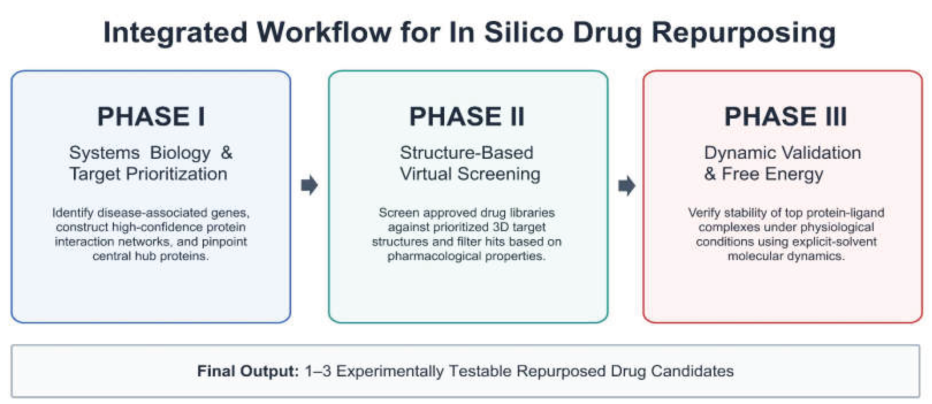
Pairing network pharmacology directly with SBVS solves both problems through a three-stage discovery funnel:
- Systems-Level Target Triaging (Network Pharmacology): Narrows thousands of disease-associated genes down to 3 to 10 druggable hub proteins that control key Kyoto Encyclopedia of Genes and Genomes (KEGG) and Gene Ontology (GO) pathways (Szklarczyk et al., 2022; Chin et al., 2014).
- Atomic-Scale Screening (SBVS & Molecular Docking): Screens FDA-approved drug libraries against these prioritized hub targets—using either experimental PDB crystal structures or high-confidence AlphaFold models (Jumper et al., 2021; Tunyasuvunakool et al., 2021; Abramson et al., 2024)—in docking engines like AutoDock Vina 1.2.0 (Eberhardt et al., 2021).
- Indication-Specific ADMET & Dynamic Validation: Checks top docking hits for blood–brain barrier (BBB) or gastrointestinal absorption in SwissADME (Daina et al., 2017), then runs explicit-solvent molecular dynamics (MD) simulations in GROMACS (Van Der Spoel et al., 2005; Páll et al., 2020) to confirm complex stability and binding free energy before wet-lab testing (Ngernsombat et al., 2024).
Figure 1.
Schematic workflow of the integrated three-phase in silico drug repurposing pipeline, progressing from macro-scale systems biology (Phase I) to atomic-resolution virtual screening (Phase II) and explicit-solvent molecular dynamics validation (Phase III).
Figure 1.
Schematic workflow of the integrated three-phase in silico drug repurposing pipeline, progressing from macro-scale systems biology (Phase I) to atomic-resolution virtual screening (Phase II) and explicit-solvent molecular dynamics validation (Phase III).
1.4. Scope and Objectives of This Review
Many recent studies combine network pharmacology with molecular docking, yet published workflows often suffer from avoidable technical errors. Common mistakes include using low-confidence PPI edges, running unvalidated blind docking grids, skipping pharmacokinetic checks for new target organs, and publishing static docking scores without MD simulation confirmation. This review establishes a step-by-step methodological standard for preclinical drug repurposing. Section 2 covers target mining, PPI network building, and hub identification. Section 3 details library preparation, active-site pocket mapping, docking validation, and pharmacokinetic re-triage. Section 4 focuses on post-docking MD simulations and MM-PBSA/GBSA free-energy calculations. Section 5 examines recent preclinical case studies, and Section 6 evaluates current limitations alongside new deep-learning docking and co-folding models.

Table 1.
Core Computational Toolkit of the Integrated Repurposing Pipeline.
| Stage | Tool / Database | Primary Function & Key Features | Reference |
| Library Curation | DrugBank 6.0 | >15,000 approved and investigational drug structures (SDF/SMILES), known targets, and clinical data. | (Knox et al., 2023) |
| Library Curation | ChEMBL | Curated bioactivity data (IC₅₀, Kᵢ, EC₅₀) and clinical candidate libraries from medicinal chemistry literature. | (Zdrazil et al., 2023) |
| Target Mining | DisGeNET | Human disease-gene associations scored via Gene–Disease Association (GDA) and specificity metrics. | (Piñero et al., 2016) |
| Target Mining | SwissTargetPrediction | Ligand-based reverse screening using 2D FP2 fingerprints and 3D ElectroShape vectors across 5 species. | (Daina et al., 2019) |
| PPI Network | STRING (v12.0) | Physical and functional PPI networks with combined confidence scores (0–1) and regulatory directionality. | (Szklarczyk et al., 2025) |
| Hub Analysis | Cytoscape & cytoHubba | Network visualization and topological hub ranking across 11 centrality algorithms (MCC, Degree, Betweenness). | (Shannon et al., 2003; Chin et al., 2014) |
| 3D Structure | AlphaFold 2/3 & CASTpFold | Deep-learning 3D protein structure prediction (pLDDT, PAE) and alpha-shape binding pocket topography. | (Abramson et al., 2024; Ye et al., 2024) |
| Docking | AutoDock Vina 1.2.0 & Vina-GPU 2.0 | High-throughput docking, binding affinity estimation (kcal/mol), Python scripting, and GPU acceleration. | (Eberhardt et al., 2021; Ding et al., 2023) |
| ADMET Triage | SwissADME & ADMETlab 3.0 | BOILED-Egg GI/BBB permeation, P-gp efflux, CYP inhibition, PAINS alerts, and DMPNN toxicity models. | (Daina et al., 2017; Fu et al., 2024) |
| MD & Energy | GROMACS & gmx_MMPBSA | Explicit-solvent MD simulations (RMSD, RMSF, Rg, SASA) and end-state MM-PBSA/GBSA free-energy scoring. | (Páll et al., 2020; Valdés-Tresanco et al., 2021) |
2. Phase I: Systems-Level Target Identification via Network Pharmacology
2.1. Disease Gene Mining and Reverse-Target Fishing
A reliable network pharmacology workflow starts by building two independent gene lists: the disease-associated gene module and the drug-target profile. Relying on a single database introduces annotation bias. Best practice requires cross-checking multiple orthogonal repositories to capture both inherited variants and polygenic disease drivers.
For disease target mining, GeneCards compiles transcriptomic, proteomic, genetic, and clinical data from over 150 sources, ranking genes with an Elasticsearch Relevance Score (Stelzer et al., 2016). DisGeNET complements this by combining expert-curated databases, GWAS catalogs, animal models, and peer-reviewed literature (Piñero et al., 2016). In DisGeNET, researchers can filter genes using a quantitative Gene–Disease Association (GDA) score (typically setting a cutoff of GDA ≥ 0.1 to 0.3) and a Disease Specificity Index (DSI) (Piñero et al., 2016). Additional targets are routinely pulled from Online Mendelian Inheritance in Man (OMIM) and differential gene expression (DGE) datasets in the NCBI Gene Expression Omnibus (GEO) using cutoffs of |log₂ Fold Change| > 1.0 and adjusted p < 0.05.
When screening approved drugs or bioactive compounds for repurposing, known physical targets from DrugBank 6.0 (Knox et al., 2023) and ChEMBL (Zdrazil et al., 2023) are expanded using reverse-screening (target fishing) tools to catch unannotated off-targets:
- SwissTargetPrediction estimates the probability of a small molecule binding to over 3,000 proteins across five species by combining 2D chemical fingerprint similarity (FP2) with 3D ElectroShape vector superpositions (Daina et al., 2019).
- PharmMapper runs inverse pharmacophore mapping, aligning 3D drug conformers against more than 53,000 receptor pharmacophore models from TargetBank, BindingDB, DrugBank, and PDTD, then ranks hits by normalized Fit Scores and statistical Z'-scores (Wang et al., 2017).
To clean up database noise before building the network, all gene symbols from both the disease and drug lists must be standardized to reviewed human (Homo sapiens) Swiss-Prot UniProtKB IDs. The two lists are then overlapped using a Venn diagram or UpSet plot. Only the shared "drug–disease intersection targets" move forward to PPI mapping.
2.2. Protein–Protein Interaction (PPI) Interactome Construction and Directionality
A flat list of target genes tells us very little until those proteins are placed into a cellular interaction network. The STRING database is the standard platform for building physical and functional PPI networks (Szklarczyk et al., 2022; Szklarczyk et al., 2025). STRING combines seven evidence channels—genomic neighborhood, gene fusion, phylogenetic co-occurrence, co-expression, wet-lab interaction assays (such as co-immunoprecipitation and yeast two-hybrid), curated pathway databases, and automated text mining—into a single Combined Score between 0 and 1 (Szklarczyk et al., 2022).
A common mistake in published network pharmacology papers is keeping STRING's default "medium confidence" setting (≥ 0.400). That threshold lets in substantial noise from unverified text-mining co-mentions. Rigorous drug repurposing workflows restrict PPI networks to high confidence (≥ 0.700) or highest confidence (≥ 0.900) and remove disconnected orphan nodes before exporting. The 2025 update of STRING (v12.0+) also added regulatory directionality annotations (Szklarczyk et al., 2025). Researchers can now see whether an interaction edge represents catalytic activation, transcriptional upregulation, or inhibition—a critical detail when deciding if a repurposed drug needs to act as an agonist or an antagonist (Szklarczyk et al., 2025).
2.3. Topological Interrogation, Hub Gene Triaging, and Functional Enrichment
After exporting the PPI network into Cytoscape (Shannon et al., 2003), the graph G = (V, E)—composed of protein nodes (V) and interaction edges (E)—is analyzed mathematically. Biological networks are scale-free: most proteins connect to only one or two partners, while a small set of densely connected hub proteins and high-traffic bottleneck nodes hold the signaling network together.
Two Cytoscape plugins handle target prioritization:
- cytoHubba (Centrality Node Ranking): Ranks node importance across 11 local and global graph algorithms (Chin et al., 2014). Relying only on Degree centrality (the raw count of direct edges attached to node v) often misses low-degree "bridge" proteins that link separate disease modules. A stronger approach intersects the top 10 nodes ranked by Maximal Clique Centrality (MCC)—which weights proteins inside dense 3- and 4-node subgraphs—with global path metrics like Betweenness Centrality and Closeness Centrality (Chin et al., 2014; Zhang et al., 2024).
- MCODE (Molecular Complex Detection): Instead of ranking single proteins, MCODE uses k-core neighborhood density weighting to extract tightly connected subnetworks (functional modules or multiprotein complexes) that act together in processes like inflammasome activation or cell-cycle regulation (Bader & Hogue, 2003).
The five mathematical formulas used to score hub targets in Cytoscape are defined in standard notation below:
- Degree Centrality (DC): Counts the direct physical or functional interaction partners in the immediate neighborhood N(v) of node v:DC(v) = |N(v)| = ∑ Aᵥᵤ (for all u ∈ V)
- Maximal Clique Centrality (MCC): Sums the factorial size of all maximal cliques C containing node v, giving higher weight to proteins embedded inside tightly co-regulated complexes (Chin et al., 2014):MCC(v) = ∑ (|C| − 1)! (for all C ∈ S(v))
- Betweenness Centrality (BC): Calculates the fraction of all shortest signaling paths (σₛₜ) between node pairs (s, t) that pass through node v, identifying bottleneck bridges between pathways:BC(v) = ∑ [σₛₜ(v) / σₛₜ] (for all s ≠ v ≠ t ∈ V)
- Closeness Centrality (CC): Measures the reciprocal of the sum of shortest path distances d(v, u) from node v to all other reachable nodes (|V| − 1):CC(v) = (|V| − 1) / ∑ d(v, u) (for all u ≠ v ∈ V)
- MCODE Vertex Weighting Score (W): Multiplies the local density (D_Ck) of node v's highest k-core neighborhood by the maximum core order (k_max) (Bader & Hogue, 2003):
W(v) = D_Ck × k_max
Table 2.
Graph-Theoretical Centrality Algorithms in cytoHubba and MCODE.
| Metric | Category | Formula | Role in Target Prioritization |
| Degree Centrality (DC) | Local | DC(v) = |N(v)| | Counts direct edges in neighborhood N(v). Finds major hubs, though blocking them indiscriminately can cause off-target toxicity. |
| Maximal Clique Centrality (MCC) | Local (Clique) | MCC(v) = ∑ (|C| − 1)! | Weights nodes inside dense 3- and 4-node cliques. High sensitivity for essential disease complexes (Chin et al., 2014). |
| Betweenness Centrality (BC) | Global (Path) | BC(v) = ∑ [σₛₜ(v) / σₛₜ] | Measures shortest paths crossing node v. Pinpoints bottleneck bridges linking separate disease cascades. |
| Closeness Centrality (CC) | Global (Distance) | CC(v) = (|V| − 1) / ∑ d(v, u) | Inverse shortest-path distance to all nodes. High-CC targets quickly spread drug effects across the module. |
| MCODE Node Score (W) | Subnetwork | W(v) = D_Ck × k_max | Multiplies k-core density by core order k_max to isolate functional multiprotein complexes (Bader & Hogue, 2003). |
To verify that the prioritized hub genes actually drive disease biology, researchers run functional enrichment in DAVID (Sherman et al., 2022) or clusterProfiler 4.0 (Wu et al., 2021). Hub targets are mapped onto Gene Ontology (GO) categories—Biological Process (BP), Cellular Component (CC), and Molecular Function (MF)—and KEGG pathways. P-values must be corrected for multiple testing using the Benjamini–Hochberg False Discovery Rate (FDR < 0.05) rather than raw nominal p-values, and redundant GO terms should be collapsed using semantic similarity filtering (simplify cutoff < 0.7 in clusterProfiler) (Wu et al., 2021).
3. Phase II: Structure-Based Virtual Screening (SBVS) and Pharmacokinetic Filtering
3.1. Target Structure Retrieval, AlphaFold Confidence Triage, and Active-Site Mapping
Moving from network nodes to structure-based screening requires accurate 3D protein coordinates. Experimental structures solved by X-ray crystallography or cry-EM are downloaded from the RCSB Protein Data Bank (PDB). Researchers should prioritize holo-structures co-crystallized with a ligand, atomic resolution ≤ 2.5 Å, low R_free values, and intact loop regions around the active site.
If an experimental structure for a human hub protein or viral variant has not been solved, AlphaFold 2 (Jumper et al., 2021; Tunyasuvunakool et al., 2021) and AlphaFold 3 (Abramson et al., 2024) offer proteome-wide structural models. However, docking directly into raw AlphaFold coordinates without checking model confidence is a major source of false positives. Two confidence metrics must be checked before setting up a docking grid:
- Per-residue Local Distance Difference Test (pLDDT): Residues lining the binding pocket need very high (pLDDT > 90) or confident (pLDDT > 70) scores. Segments with pLDDT < 50 are usually disordered loops that can artificially block the binding cleft; these low-confidence tails should be trimmed or remodeled before docking.
- Predicted Aligned Error (PAE): Checks positional certainty between domains. Low PAE values (< 5 Å) are essential when the binding site sits at the interface between two protein domains.
When working with apo-structures or AlphaFold models that lack a co-crystallized ligand to mark the active site, pocket-finding tools are used to locate druggable cavities:
- CASTpFold: Uses alpha-shape and Delaunay triangulation geometry across PDB and 183 million AlphaFold 2 structures to measure pocket solvent-accessible surface area (SASA), cavity volume, and mouth openings (Ye et al., 2024).
- PrankWeb 3 (P2Rank): Runs a random-forest classifier over points on the solvent-accessible protein surface, combining sequence conservation with AlphaFold pLDDT scores to rank binding sites (Jakubec et al., 2022).
- DoGSiteScorer: Calculates a Druggability Score (0 to 1) from pocket volume, enclosure, hydrophobicity, and hydrogen-bond donors/acceptors, where scores > 0.5 indicate a pocket suitable for small-molecule binding (Volkamer et al., 2012).
3.2. Chemical Library Standardization and Receptor Pre-Processing
Approved drug libraries (typically 2,500 to 3,500 compounds from DrugBank or ZINC) cannot be docked directly from raw 2D SMILES or SDF files. Cheminformatics preparation in Open Babel (O'Boyle et al., 2011) or RDKit is needed to strip counter-ions and salts, set protonation states to physiological pH 7.4, assign valid tautomers and stereocenters, and minimize 3D conformer energy using the Merck Molecular Force Field (MMFF94) or Universal Force Field (UFF). Prepared ligands are then saved in .pdbqt format with Gasteiger partial charges and defined rotatable bonds.
For receptor preparation, crystallographic water molecules are removed (unless a conserved water molecule bridges the catalytic triad), along with buffer additives like glycerol, PEG, or sulfate ions. Essential metal cofactors (Zn²⁺, Mg²⁺, heme groups) are kept with their proper formal charges, polar hydrogens are added, and Kollman united-atom charges are assigned.
3.3. High-Throughput Molecular Docking and Redocking Validation
AutoDock Vina (Trott & Olson, 2010) and AutoDock Vina 1.2.0 (Eberhardt et al., 2021) are the most widely used open-source engines for virtual screening because they combine fast empirical/knowledge-based scoring with Python bindings for batch screening. Vina estimates binding free energy (ΔG_bind in kcal/mol) from steric Gauss terms, hydrophobic contacts, directional hydrogen bonds, and an entropic penalty based on the number of active rotatable bonds (N_rot) using BFGS local gradient optimization:ΔG_bind = c_inter / (1 + w × N_rot)
Three practical checks keep virtual screening accurate:
- Grid Box Sizing vs. Blind Docking: Enclosing the entire protein in a massive grid ("blind docking") dilutes conformational sampling and increases false positives. Instead, the 3D grid box (usually 20 × 20 × 20 Å to 30 × 30 × 30 Å) should be centered tightly on the native ligand or top CASTpFold/PrankWeb pocket, with exhaustiveness set to ≥ 16–32 (rather than the default of 8).
- Redocking (Self-Docking) Validation: Before screening the drug library, the native co-crystallized ligand must be extracted, randomized, and redocked into the pocket. The docking setup is valid only if the heavy-atom Root Mean Square Deviation (RMSD) between the redocked pose and the crystal structure is ≤ 2.0 Å.
- GPU Acceleration: For large clinical and investigational libraries (>10,000 molecules) screened across multiple hub proteins, Vina-GPU 2.0 uses OpenCL parallelization to speed up docking more than 50-fold without losing pose accuracy (Ding et al., 2023).
3.4. Indication-Specific Pharmacokinetic and Toxicological Re-Triage
A common assumption in drug repurposing is that FDA-approved drugs can skip Absorption, Distribution, Metabolism, Excretion, and Toxicity (ADMET) screening because they already passed Phase I clinical trials. In practice, indication-specific pharmacokinetic checks are still required for three reasons:
- Compartmental Relocation and GI/BBB Thresholds: Repurposing a peripheral blood-pressure or antiparasitic drug for glioblastoma or ischemic stroke requires passive blood–brain barrier (BBB) penetration and freedom from P-glycoprotein (P-gp) efflux. Conversely, repurposing a CNS drug for peripheral cancer benefits from low BBB permeation to avoid central side effects.
- Polypharmacy and Cytochrome P450 (CYP) Inhibition: Complex diseases often require multi-drug regimens. Repurposed candidates must be checked against major liver enzymes (CYP1A2, CYP2C19, CYP2C9, CYP2D6, and CYP3A4) to avoid dangerous drug–drug interactions (DDIs).
- Assay Interference and Dose-Scaling Toxicity: A repurposed drug may require a higher plasma concentration (C_max) to inhibit its new target than its original indication, making it necessary to check hERG cardiotoxicity, drug-induced liver injury (DILI), and Pan-Assay Interference Compounds (PAINS) alerts.
SwissADME handles initial triage through its BOILED-Egg plot, which graphs Wildman–Crippen lipophilicity (WLOGP) against Topological Polar Surface Area (TPSA) (Daina & Zoete, 2016; Daina et al., 2017). Compounds inside the white ellipse (TPSA < 142 Ų and −2.3 < WLOGP < 6.8) show high passive human gastrointestinal absorption (HIA), while highly polar molecules with TPSA > 140 Ų cross cell membranes poorly. Compounds inside the yellow yolk (TPSA < 79 Ų and 0.4 < WLOGP < 6.0) are predicted to cross the BBB passively (Daina & Zoete, 2016).
For deeper toxicity checks, SwissADME is paired with pkCSM (Pires et al., 2015), ADMETlab 3.0—which uses a multi-task Directed Message Passing Neural Network (DMPNN) across 119 ADMET endpoints (Fu et al., 2024)—and ProTox 3.0, which predicts acute oral LD₅₀ toxicity classes, hepatotoxicity, neurotoxicity, and 15 Novartis safety-panel off-targets (Banerjee et al., 2024).
4. Phase III: Post-Docking Dynamic Validation and Free-Energy Rescoring
4.1. The Limitations of Static Docking and the Necessity of Molecular Dynamics
High-throughput docking quickly narrows thousands of drugs down to a shortlist, but static docking scores correlate poorly with experimental binding affinities (Kᵢ or IC₅₀). Standard docking treats the protein backbone as rigid, ignores explicit water bridges, and uses rough approximations for conformational entropy. As a result, many compounds that score well in a static grid drift out of the binding pocket under physiological temperature and motion.
All-atom Molecular Dynamics (MD) simulations test whether a docked complex actually holds together in solution. Using engines like GROMACS (Van Der Spoel et al., 2005; Páll et al., 2020) with CHARMM36m, AMBER ff19SB, or OPLS4 force fields, the protein–ligand complex is placed in an explicit water box (such as TIP3P or SPC/E water with 0.15 M NaCl) and integrated over 2 fs time steps. In practice, after running steepest-descent energy minimization and 100–500 ps of NVT (300 K) and NPT (1 bar via the Parrinello–Rahman barostat) equilibration, a production MD run of 100 ns to 500 ns is needed to verify that the repurposed drug stays anchored in the active site (Ngernsombat et al., 2024; Rajput et al., 2025).
4.2. Quantitative Trajectory Metrics: RMSD, RMSF, Rg, SASA, and PCA
Five trajectory metrics are used to judge whether a drug–target complex is dynamically stable:
- Root Mean Square Deviation (RMSD): Tracks the average distance (in Å) of the protein backbone (Cα atoms) and ligand heavy atoms relative to the starting docked frame (t = 0):RMSD(t) = √[ (1 / N) × ∑ mᵢ |rᵢ(t) − rᵢ(0)|² ]In a stable complex, the ligand RMSD levels off at a plateau between 1.5 Å and 3.0 Å. Steady upward drift or sudden jumps above 4.0 Å show that the ligand is slipping out of the pocket.
- Root Mean Square Fluctuation (RMSF): Measures time-averaged positional movement for each individual amino acid residue. When an inhibitor binds tightly, it typically lowers RMSF peaks across the catalytic loop residues that touch the drug compared to the apo-protein (Rajput et al., 2025).
- Radius of Gyration (Rg) and Solvent-Accessible Surface Area (SASA): Rg tracks overall protein compactness, while SASA measures how much of the protein surface is exposed to water. A stable complex keeps a flat Rg curve and often shows a slight drop in pocket SASA as the drug packs into the hydrophobic core.
- Hydrogen-Bond Persistence and Essential Dynamics (PCA): Measuring what percentage of simulation frames keep intermolecular hydrogen bonds (distance ≤ 3.5 Å, angle ≥ 120°) confirms stable polar anchoring. Principal Component Analysis (PCA) diagonalizes the atomic covariance matrix to project large-scale protein motions onto the first two eigenvectors (PC1 and PC2), allowing Free Energy Landscape (FEL) plots to verify that the complex settles into a deep, narrow energy minimum (Rajput et al., 2025).
4.3. End-State Thermodynamic Rescoring via MM-PBSA and MM-GBSA
To get more accurate binding energies than Vina scores without running expensive alchemical Free Energy Perturbation (FEP), researchers calculate Molecular Mechanics Poisson–Boltzmann Surface Area (MM-PBSA) or Generalized Born Surface Area (MM-GBSA) energies across equilibrium MD snapshots using gmx_MMPBSA (Valdés-Tresanco et al., 2021). Total binding free energy (ΔG_bind) in solution is broken down into gas-phase molecular mechanics energy (ΔE_MM), solvation free energy (ΔG_solv), and conformational entropy (−TΔS):
where:
and:
ΔG_bind = ΔE_MM + ΔG_solv − TΔS
ΔE_MM = ΔE_int + ΔE_vdW + ΔE_elec
ΔG_solv = ΔG_PB/GB + ΔG_SA
Here, ΔE_vdW and ΔE_elec are van der Waals and electrostatic interactions, ΔG_PB/GB is polar solvation energy from the Poisson–Boltzmann or Generalized Born equations, and ΔG_SA is nonpolar solvation energy scaled to surface area (ΔG_SA = γ × SASA + b) (Valdés-Tresanco et al., 2021). Instead of slow Normal Mode Analysis (NMODE), gmx_MMPBSA can compute the entropic penalty (−TΔS) directly from interaction energy fluctuations via the Interaction Entropy (IE) method, alongside per-residue energy decomposition to flag hotspot residues contributing ≤ −1.0 kcal/mol to binding (Valdés-Tresanco et al., 2021).
5. Preclinical Case Studies of the Integrated Repurposing Pipeline
5.1. Viral and Antimicrobial Pathogens (SARS-CoV-2, HMPV, and Multidrug-Resistant Bacteria)
Infectious disease outbreaks and antibiotic resistance have served as major testing grounds for integrated computational repurposing:
- SARS-CoV-2 Spike–ACE2 Interface and Variants: To block SARS-CoV-2 host entry, Hussein et al. (2024) screened approved drug libraries against the Spike receptor-binding domain (RBD)–human ACE2 interface using consensus docking and MD simulations. They identified 13 repurposed drugs—led by the tetracycline antibiotic lymecycline—that form salt bridges with His34 on ACE2 and π-stacking contacts with Tyr453 on the Spike RBD (Hussein et al., 2024). In parallel work on the SARS-CoV-2 Omicron variant, combining AutoDock virtual screening with SwissADME bioavailability profiling allowed rapid identification of cyclic scaffolds such as nigelladine A with strong target affinity and high predicted gastrointestinal absorption (Miraz et al., 2023).
- Human Metapneumovirus (HMPV) Fusion and Nucleoproteins: Rajput et al. (2025) screened the DrugBank FDA-approved library against the HMPV fusion (F) protein (PDB: 5WB0) and nucleoprotein (N) (PDB: 5FVD) using virtual screening, SwissADME filtering, 100 ns MD runs, PCA, and MM-GBSA rescoring. Static docking initially ranked rutin (−6.516 kcal/mol), carbetocin, and acarbose as top hits for the F protein. However, 100 ns MD trajectories and PCA showed that the acarbose complex suffered large conformational breathing motions that weakened binding, whereas rutin and carbetocin remained stable in the F pocket and lapatinib plus mobocertinib anchored tightly in the N protein (Rajput et al., 2025). This study illustrates why MD and MM-GBSA are needed to catch static docking false positives.
- Carbapenem-Resistant Acinetobacter baumannii (OXA-23 β-Lactamase): Combining network pharmacology with structure-based modeling, Sagini et al. (2026) mapped resistance pathways in multidrug-resistant A. baumannii and identified the FDA-approved anti-inflammatory drug sulfasalazine as a repurposed inhibitor of the OXA-23 carbapenemase active site, with explicit-solvent MD confirming stable catalytic pocket binding.
5.2. Oncology, Neurovascular Disorders, and Complex Chronic Diseases
The same systems-to-atomic workflow has accelerated lead discovery in neurology and oncology:
- Ischemic Stroke (LIMK1–Cofilin Signaling Axis): Alaqel et al. (2025) combined cheminformatics, QSAR, network pharmacology, and molecular docking to map stroke-related targets in STRING and Cytoscape (v3.9.1). Their network analysis isolated the LIM kinase 1 (LIMK1)–cofilin cytoskeletal pathway involved in blood–brain barrier breakdown and neuronal apoptosis. Screening repurposed and ChEMBL molecules against cofilin yielded hits reaching −6.68 kcal/mol that formed persistent hydrogen bonds with Asp98 and His133 (Alaqel et al., 2025).
- Nasopharyngeal Carcinoma (FZD10) and BRAF-Mutant Cancers: Targeting Frizzled-10 (FZD10) in the Wnt signaling pathway, Ngernsombat et al. (2024) paired SBVS of FDA-approved drugs with MD simulations, MM-PBSA calculations, and wet-lab cell assays to identify repurposed FZD10 antagonists that inhibit nasopharyngeal carcinoma growth. Similarly, Amini et al. (2026) used pharmacophore-based virtual screening, docking, and MD simulations across approved drugs to identify BRAF V600E kinase inhibitors, confirming their anticancer activity in vitro.
Table 3.
Benchmark Preclinical Case Studies Using Integrated Repurposing Workflows.
| Indication | Prioritized Target(s) | Top Repurposed Hit(s) & Key Finding | Reference |
| Carbapenem-Resistant A. baumannii | OXA-23 β-Lactamase | Sulfasalazine: Stable active-site binding across MD trajectory to counter β-lactam resistance. | (Sagini et al., 2026) |
| BRAF-Mutant Cancer | BRAF (V600E) Kinase | Approved Repurposed Hits: Confirmed kinase cleft stability in MD and in vitro cytotoxicity. | (Amini et al., 2026) |
| Ischemic Stroke | LIMK1–Cofilin (Asp98, His133) | CHEMBL3613624: Docking score −6.68 kcal/mol; targets cytoskeletal neuronal apoptosis. | (Alaqel et al., 2025) |
| Human Metapneumovirus | Fusion (5WB0) & Nucleoprotein (5FVD) | Rutin, Carbetocin, Lapatinib: 100 ns MD and PCA filtered out acarbose as a false positive. | (Rajput et al., 2025) |
| Nasopharyngeal Carcinoma | Frizzled-10 (FZD10 / Wnt) | FDA Repurposed Antagonists: Favorable MM-PBSA energy and verified in vitro tumor suppression. | (Ngernsombat et al., 2024) |
| SARS-CoV-2 Entry | Spike RBD–ACE2 Interface | Lymecycline (& 12 FDA drugs): Salt bridges and π-stacking with His34 and Tyr453 block RBD binding. | (Hussein et al., 2024) |
6. Current Bottlenecks, Methodological Pitfalls, and AI/ML Horizons
6.1. Intrinsic Bias and Reproducibility Traps in Current Workflows
Although combining network pharmacology with SBVS reduces false discoveries, researchers still need to watch out for three common methodological traps:
- Literature Bias ("Matthew Effect") in Network Databases: PPI databases like STRING and gene–disease platforms like DisGeNET are heavily weighted toward famous, well-funded proteins such as TP53, AKT1, TNF, EGFR, IL6, and VEGFA. Ranking networks by Degree centrality alone repeatedly pulls up these same generic hubs regardless of the disease being studied. Weighting nodes by transcriptomic fold-change, checking Disease Specificity Indices (DSI), and using clique-based algorithms (MCC) helps surface disease-specific targets.
- Molecular Weight Bias and PAINS Aggregators: Large molecules and polyphenols often produce artificially negative AutoDock Vina scores simply because they make more van der Waals contacts across a wide pocket. Normalizing docking scores by Ligand Efficiency (LE = −ΔG_bind / N_heavy_atoms, aiming for LE ≥ 0.30 kcal/mol per heavy atom) and filtering out PAINS aggregators and decoys (e.g., using DUD-E controls) prevents false leads.
- Rigid Receptor Assumptions: Standard docking keeps the protein backbone fixed and misses targets that undergo large conformational shifts upon binding, such as kinase DFG-in/DFG-out loop flips or GPCR helix movements. Ensemble docking across clustered MD frames helps account for pocket flexibility.
6.2. Next-Generation Horizons: Diffusion Docking and All-Atom Co-Folding
New deep-learning architectures are beginning to replace classical empirical scoring grids in virtual screening:
- Generative Diffusion Models for Blind Docking (DiffDock): DiffDock treats ligand docking as a generative probability problem over ligand translations, rotations, and torsion angles, using a reverse diffusion process to find binding poses without needing a predefined grid box (Corso et al., 2023). On the PDBBind blind-docking benchmark, DiffDock reached a 38% top-1 success rate (RMSD < 2.0 Å) compared to 23% for traditional search-based docking, while running 3 to 12 times faster on GPUs (Corso et al., 2023).
- All-Atom Biomolecular Co-Folding (AlphaFold 3): Rather than predicting static apo-proteins, AlphaFold 3 uses a Pairformer architecture and a diffusion network to jointly predict 3D coordinates for proteins, nucleic acids, ions, and small-molecule ligands directly from sequence and chemical inputs (Abramson et al., 2024). Because it folds the protein and ligand together, AlphaFold 3 captures induced-fit pocket rearrangements and outperforms classical docking engines on the PoseBusters benchmark (Abramson et al., 2024).
- Graph Neural Networks (GNNs) for Multi-Drug Synergy: Multimodal GNNs now combine transcriptomic perturbation data (such as LINCS L1000), single-cell RNA-seq, and 3D pocket graphs to predict synergistic drug pairs, allowing researchers to target two separate bottleneck nodes in a disease module at lower, safer doses.
Table 4.
Common Methodological Pitfalls and Best-Practice Standards.
| Stage | Common Pitfall | Best-Practice Standard |
| PPI Network (STRING) | Keeping default medium confidence (≥ 0.400), which adds text-mining noise. | Use high confidence (≥ 0.700 or ≥ 0.900), prune orphan nodes, and check edge directionality (Szklarczyk et al., 2025). |
| Hub Ranking (Cytoscape) | Picking hubs only by Degree centrality, which favors generic hubs like TP53 or AKT1. | Intersect MCC with Betweenness/Closeness and filter by Disease Specificity Index (DSI). |
| Pathway Enrichment | Reporting raw nominal p-values (< 0.05), inflating false-positive pathways. | Apply Benjamini–Hochberg FDR < 0.05 and collapse redundant GO terms (Wu et al., 2021). |
| AlphaFold Prep | Docking into raw AlphaFold models where disordered loops block the pocket. | Require pocket pLDDT > 70–90 and low inter-domain PAE (< 5 Å); trim pLDDT < 50 tails. |
| Molecular Docking | Running whole-protein blind docking without control redocking or size normalization. | Validate grid by redocking native ligand (RMSD ≤ 2.0 Å), set exhaustiveness ≥ 16–32, and check Ligand Efficiency (LE). |
| ADMET Triage | Skipping ADMET checks because the drug library is already FDA-approved. | Re-check BBB/GI permeation (BOILED-Egg), P-gp efflux, CYP450 inhibition, and PAINS for the new indication. |
| MD Validation | Publishing static docking scores without dynamic or free-energy validation. | Run ≥ 100 ns explicit-solvent MD (RMSD, RMSF, Rg, SASA, PCA) and rescore via MM-PBSA/GBSA. |
7. Conclusion
Combining systems-level network pharmacology with atomic-resolution structure-based virtual screening turns preclinical drug repurposing into a structured, reproducible workflow. Mapping disease genes across high-confidence PPI networks in STRING and Cytoscape allows researchers to isolate central bottleneck proteins rather than picking isolated targets by guesswork. Screening FDA-approved drug libraries against those prioritized hubs—using AlphaFold confidence checks, pocket mapping, and validated AutoDock Vina grids—quickly identifies candidate binders. Finally, filtering hits through SwissADME and ADMETlab 3.0, running 100–500 ns explicit-solvent MD simulations in GROMACS, and rescoring with MM-PBSA/GBSA weeds out static docking artifacts before wet-lab testing. As generative diffusion docking (DiffDock) and all-atom co-folding models (AlphaFold 3) become standard tools alongside multi-omics graphs, this integrated pipeline will continue to cut preclinical discovery costs and speed up the delivery of safe repurposed therapies.
Author Contributions
Conceptualization, A.A.N.; methodology, A.A.N. and S.T.I.T.; literature search and data curation, A.A.N., S.T.I.T., and S.D.; writing—original draft preparation, A.A.N. and S.T.I.T.; writing—review and editing, A.A.N., S.T.I.T., and S.D.; visualization, A.A.N. and S.D.; supervision and project administration, A.A.N. All authors have read and agreed to the published version of the manuscript.
Funding
This research received no external funding.
Data Availability Statement
No new experimental datasets were generated in this study. All databases and software tools discussed are publicly accessible through the cited references.
Conflicts of Interest
The authors declare no competing financial or non-financial interests.
References
- Abramson, J.; Adler, J.; Dunger, J.; et al. Accurate structure prediction of biomolecular interactions with AlphaFold 3. Nature 2024, 630, 493–500. [Google Scholar] [CrossRef] [PubMed]
- Alaqel, S. I.; Khan, A.; Alanazi, M. N.; Nayeem, N.; Ben Khaled, H.; Imran, M. Repurposing Cofilin-Targeting Compounds for Ischemic Stroke Through Cheminformatics and Network Pharmacology. Pharmaceuticals 2025, 18(9), 1323. [Google Scholar] [CrossRef] [PubMed]
- Amini, H.; Ebrahimi, N.; Mousavirahimi, S.; Zamani, M.; Bagherzadeh, K.; Montazeri, H.; Azizian, H. In silico repurposing of clinically approved drugs as potential BRAF inhibitors: a pharmacophore-based virtual screening and experimental investigation. In Cancer Cell International; 2026. [Google Scholar] [CrossRef] [PubMed]
- Bader, G. D.; Hogue, C. W. V. An automated method for finding molecular complexes in large protein interaction networks. BMC Bioinform. 2003, 4(1), 2. [Google Scholar] [CrossRef] [PubMed]
- Banerjee, P.; Kemmler, E.; Dunkel, M.; Preissner, R. ProTox 3.0: a webserver for the prediction of toxicity of chemicals. Nucleic Acids Res. 2024, 52(W1), W513–W520. [Google Scholar] [CrossRef] [PubMed]
- Chin, C.-H.; Chen, S.-H.; Wu, H.-H.; Ho, C.-W.; Ko, M.-T.; Lin, C.-Y. cytoHubba: identifying hub objects and sub-networks from complex interactome. BMC Syst. Biol. 2014, 8 (Suppl 4), S11. [Google Scholar] [CrossRef] [PubMed]
- Corso, G.; Stärk, H.; Jing, B.; Barzilay, R.; Jaakkola, T. DiffDock: Diffusion Steps, Twists, and Turns for Molecular Docking. International Conference on Learning Representations (ICLR), 2023. [Google Scholar] [CrossRef]
- Daina, A.; Zoete, V. A BOILED-Egg to predict gastrointestinal absorption and brain penetration of small molecules. ChemMedChem 2016, 11(11), 1117–1121. [Google Scholar] [CrossRef] [PubMed]
- Daina, A.; Michielin, O.; Zoete, V. SwissADME: a free web tool to evaluate pharmacokinetics, drug-likeness and medicinal chemistry friendliness of small molecules. Sci. Rep. 2017, 7, 42717. [Google Scholar] [CrossRef] [PubMed]
- Daina, A.; Michielin, O.; Zoete, V. SwissTargetPrediction: updated data and new features for efficient prediction of protein targets of small molecules. Nucleic Acids Res. 2019, 47(W1), W357–W364. [Google Scholar] [CrossRef] [PubMed]
- Ding, J.; Tang, S.; Mei, Z.; Wang, L.; Huang, Q.; Hu, H.; Ling, M.; Wu, J. Vina-GPU 2.0: Further accelerating AutoDock Vina and its derivatives with graphics processing units. J. Chem. Inf. Model. 2023, 63(7), 1982–1998. [Google Scholar] [CrossRef] [PubMed]
- Eberhardt, J.; Santos-Martins, D.; Tillack, A. F.; Forli, S. AutoDock Vina 1.2.0: New Docking Methods, Expanded Force Field, and Python Bindings. J. Chem. Inf. Model. 2021, 61(8), 3891–3898. [Google Scholar] [CrossRef] [PubMed]
- Fu, L.; Shi, S.; Yi, J.; et al. ADMETlab 3.0: an updated comprehensive online ADMET prediction platform enhanced with broader coverage, improved performance, API functionality and decision support. Nucleic Acids Res. 2024, 52(W1), W422–W431. [Google Scholar] [CrossRef] [PubMed]
- Hopkins, A. L. Network pharmacology: the next paradigm in drug discovery. Nat. Chem. Biol. 2008, 4(11), 682–690. [Google Scholar] [CrossRef] [PubMed]
- Hussein, D.; Almatrafi, A.; Gomaa, M.; Alhowsawi, A.; Almustafa, S.; Alsaihaty, H.; Alghamdi, M. In silico drug repurposing using molecular docking and dynamics to target the protein interaction between the SARS-CoV-2 S-glycoprotein and the ACE2 receptor. F1000Research 2024, 12, 1452. [Google Scholar] [CrossRef] [PubMed]
- Jakubec, D.; Skoda, P.; Krivak, R.; Novotny, M.; Hoksza, D. PrankWeb 3: accelerated ligand-binding site predictions for experimental and modelled protein structures. Nucleic Acids Res. 2022, 50(W1), W593–W597. [Google Scholar] [CrossRef] [PubMed]
- Jumper, J.; Evans, R.; Pritzel, A.; et al. Highly accurate protein structure prediction with AlphaFold. Nature 2021, 596, 583–589. [Google Scholar] [CrossRef] [PubMed]
- Knox, C.; Wilson, M.; Klinger, C. M.; et al. DrugBank 6.0: the DrugBank Knowledgebase for 2024. Nucleic Acids Res. 2023, 52(D1), D1265–D1275. [Google Scholar] [CrossRef] [PubMed]
- Liu, S.; Yu, Y.-W. Network pharmacology: Changes the treatment mode of "one disease-one target" in cancer treatment. World J. Gastrointest. Oncol. 2025, 17(1), 101581. [Google Scholar] [CrossRef] [PubMed]
- Miraz, M. M. H.; Ullah, M. A.; Nayem, A. A.; Chakrobortty, B.; Deb, S.; Laskar, A.; Tithi, N. U.; Saha, N.; Chowdhury, A. R.; Alam, K. M. K.; Wahed, T. B.; Alam, M. K.; Kundu, S. K. Nigelladine A among Selected Compounds from Nigella sativa Exhibits Propitious Interaction with Omicron Variant of SARS-CoV-2: An In Silico Study. Int. J. Clin. Pract. 2023, 2023, 9917306. [Google Scholar] [CrossRef] [PubMed]
- Ngernsombat, C.; Suriya, U.; Prattapong, P.; Verma, K.; Rungrotmongkol, T.; Soonkum, T.; et al. Repurposing FDA-approved drugs targeting FZD10 in nasopharyngeal carcinoma: insights from molecular dynamics simulations and experimental validation. Sci. Rep. 2024, 14, 30512. [Google Scholar] [CrossRef] [PubMed]
- O'Boyle, N. M.; Banck, M.; James, C. A.; Morley, C.; Vandermeersch, T.; Hutchison, G. R. Open Babel: An open chemical toolbox. J. Cheminformatics 2011, 3(1), 33. [Google Scholar] [CrossRef] [PubMed]
- Páll, S.; Zhmurov, A.; Bauer, P.; Abraham, M.; Lundborg, M.; Gray, A.; Hess, B.; Lindahl, E. Heterogeneous parallelization and acceleration of molecular dynamics simulations in GROMACS. J. Chem. Phys. 2020, 153(13), 134110. [Google Scholar] [CrossRef] [PubMed]
- Piñero, J.; Bravo, À.; Queralt-Rosinach, N.; et al. DisGeNET: a comprehensive platform integrating information on human disease-associated genes and variants. Nucleic Acids Res. 2016, 45(D1), D833–D839. [Google Scholar] [CrossRef] [PubMed]
- Pires, D. E. V.; Blundell, T. L.; Ascher, D. B. pkCSM: Predicting small-molecule pharmacokinetic and toxicity properties using graph-based signatures. J. Med. Chem. 2015, 58(9), 4066–4072. [Google Scholar] [CrossRef] [PubMed]
- Pushpakom, S.; Iorio, F.; Eyers, P. A.; et al. Drug repurposing: progress, challenges and recommendations. Nat. Rev. Drug Discov. 2018, 18(1), 41–58. [Google Scholar] [CrossRef] [PubMed]
- Rajput, H.; Choudhary, G.; Siddiqui, H.; Ghosh, A.; Prajapat, M.; Prakash, A.; Medhi, B. In silico drug repurposing targeting fusion and nucleoprotein of human metapneumovirus: A step toward pandemic preparedness. Indian J. Pharmacol. 2025, 57(5), 308–321. [Google Scholar] [CrossRef] [PubMed]
- Sagini, H. A.; et al. Network Pharmacology Guided Drug Repurposing and Molecular Modeling Identify Sulfasalazine as a Potential OXA-23 β-Lactamase Inhibitor in Carbapenem-Resistant Acinetobacter baumannii. Int. J. Mol. Sci. 2026, 27(14), 6390. [Google Scholar] [CrossRef] [PubMed]
- Shannon, P.; Markiel, A.; Ozier, O.; et al. Cytoscape: A Software Environment for Integrated Models of Biomolecular Interaction Networks. Genome Res. 2003, 13(11), 2498–2504. [Google Scholar] [CrossRef] [PubMed]
- Sherman, B. T.; Hao, M.; Qiu, J.; et al. DAVID: a web server for functional enrichment analysis and functional annotation of gene lists (2021 update). Nucleic Acids Res. 2022, 50(W1), W216–W221. [Google Scholar] [CrossRef] [PubMed]
- Stelzer, G.; Rosen, N.; Plaschkes, I.; et al. The GeneCards suite: From gene data mining to disease genome sequence analysis. Curr. Protoc. Bioinform. 2016, 54(1), 1.30.1–1.30.33. [Google Scholar] [CrossRef] [PubMed]
- Szklarczyk, D.; Kirsch, R.; Koutrouli, M.; et al. The STRING database in 2023: protein–protein association networks and functional enrichment analyses for any sequenced genome of interest. Nucleic Acids Res. 2022, 51(D1), D638–D646. [Google Scholar] [CrossRef] [PubMed]
- Szklarczyk, D.; Nastou, K.; Koutrouli, M.; et al. The STRING database in 2025: protein networks with directionality of regulation. Nucleic Acids Res. 2025, 53(D1), D730–D737. [Google Scholar] [CrossRef] [PubMed]
- Trott, O.; Olson, A. J. AutoDock Vina: improving the speed and accuracy of docking with a new scoring function, efficient optimization and multithreading. J. Comput. Chem. 2010, 31(2), 455–461. [Google Scholar] [CrossRef] [PubMed]
- Tunyasuvunakool, K.; Adler, J.; Wu, Z.; et al. Highly accurate protein structure prediction for the human proteome. Nature 2021, 596, 590–596. [Google Scholar] [CrossRef] [PubMed]
- Valdés-Tresanco, M. S.; Valdés-Tresanco, M. E.; Valiente, P. A.; Moreno, E. gmx_MMPBSA: A New Tool to Perform End-State Free Energy Calculations with GROMACS. J. Chem. Theory Comput. 2021, 17(10), 6281–6291. [Google Scholar] [CrossRef] [PubMed]
- Van Der Spoel, D.; Lindahl, E.; Hess, B.; Groenhof, G.; Mark, A. E.; Berendsen, H. J. C. GROMACS: Fast, flexible, and free. J. Comput. Chem. 2005, 26(16), 1701–1718. [Google Scholar] [CrossRef] [PubMed]
- Volkamer, A.; Kuhn, D.; Rippmann, F.; Rarey, M. DoGSiteScorer: a web server for automatic binding site prediction, analysis and druggability assessment. Bioinformatics 2012, 28(15), 2074–2075. [Google Scholar] [CrossRef] [PubMed]
- Wang, X.; Shen, Y.; Wang, S.; et al. PharmMapper 2017 update: a web server for potential drug target identification with a comprehensive target pharmacophore database. Nucleic Acids Res. 2017, 45(W1), W356–W360. [Google Scholar] [CrossRef] [PubMed]
- Wu, T.; Hu, E.; Xu, S.; et al. clusterProfiler 4.0: A universal enrichment tool for interpreting omics data. The Innovation 2021, 2(3), 100141. [Google Scholar] [CrossRef] [PubMed]
- Ye, B.; Tian, W.; Wang, B.; Liang, J. CASTpFold: Computed Atlas of Surface Topography of the universe of protein Folds. Nucleic Acids Res. 2024, 52(W1), W194–W199. [Google Scholar] [CrossRef] [PubMed]
- Zdrazil, B.; Felix, E.; Hunter, F.; et al. The ChEMBL Database in 2023: a drug discovery platform spanning multiple bioactivity data types and time periods. Nucleic Acids Res. 2023, 52(D1), D1180–D1192. [Google Scholar] [CrossRef] [PubMed]
- Zhang, X.; Liu, Y.; Guo, B.; et al. Identification of key mRNAs and signaling pathways in obsessive compulsive disorder based on weighted gene co-expression network analysis and cytoHubba plugin. Brain Behav. 2024, 14(4), e3412. [Google Scholar] [CrossRef] [PubMed]
Disclaimer/Publisher’s Note: The statements, opinions and data contained in all publications are solely those of the individual author(s) and contributor(s) and not of MDPI and/or the editor(s). MDPI and/or the editor(s) disclaim responsibility for any injury to people or property resulting from any ideas, methods, instructions or products referred to in the content. |
© 2026 by the authors. Licensee MDPI, Basel, Switzerland. This article is an open access article distributed under the terms and conditions of the Creative Commons Attribution (CC BY) license (http://creativecommons.org/licenses/by/4.0/).
Copyright: This open access article is published under a Creative Commons CC BY 4.0 license, which permit the free download, distribution, and reuse, provided that the author and preprint are cited in any reuse.