

Submitted:
03 November 2026
Posted:
05 November 2026
You are already at the latest version
Abstract
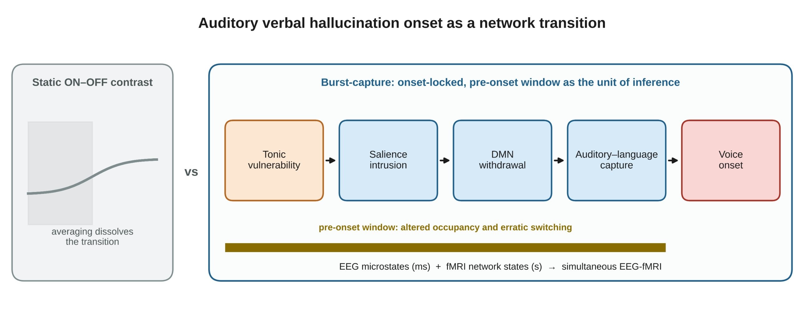
Auditory verbal hallucinations (AVH) are transient, internally generated percepts that emerge against spontaneous brain activity, yet most neuroimaging contrasts average hallucination ‘ON’ against ‘OFF’ periods and thereby dissolve the transition that is mechanistically decisive. This systematic review synthesises symptom-capture functional magnetic resonance imaging (fMRI), dynamic functional connectivity, electroencephalography (EEG) microstate, perfusion and simultaneous EEG-fMRI studies of AVH identified in five databases, and critically appraises seven transient-state estimators. We introduce ‘burst-capture’, an onset-anchored strategy in which brief hallucinatory episodes define the temporal frame and the pre-onset interval is the unit of inference. Onset-anchored studies consistently implicate superior temporal and primary auditory cortex, inferior frontal gyrus, hippocampus and thalamus. Dynamic analyses show that hallucination-relevant abnormalities are state-bound and invisible to static connectivity: hallucination proneness tracks reduced dwell time in states of default mode network (DMN)–task-positive anticorrelation; patients with AVH under-occupy, and switch less often into, a DMN–language antagonistic state; hallucination periods show more erratic switching and shortened EEG microstate class D; and stage-resolved effective connectivity implicates the salience network during emergence. Convergent perfusion, gamma-synchrony and prestimulus-state evidence supports a tonic vulnerability that lowers the threshold for a phasic network transition. We propose a transition-competition model in which AVH onset is a threshold crossing within a multistable network system, and specify the requirements of a valid burst-capture EEG-fMRI study: reaction-time-independent anchors, surrogate-data testing of state estimators and sign-aware EEG–BOLD fusion. Transition metrics, not averaged activation, should become the primary outcomes of hallucination neuroimaging.
Keywords:
auditory verbal hallucinations
; dynamic functional connectivity
; EEG-fMRI
; EEG microstates
; salience network
; brain state transitions
; schizophrenia
1. Introduction
1.1. The Clinical and Phenomenological Problem
Auditory verbal hallucinations (AVH) are experienced by 60–80% of people with schizophrenia and constitute one of the most distressing and treatment-resistant symptom domains (Hoffman et al., 2011b). They are not, however, confined to psychosis. Meta-analytic estimates place the median prevalence of hallucinatory experience in the general population at 6% and the median incidence at 1.2%; approximately 20% of those reporting psychotic experiences continue to experience them, and 7.4% do so in the context of a psychotic disorder (Baumeister et al., 2017). Roughly 10–15% of the healthy population report AVH at some point, although prevalence estimates across 17 general-population studies range from 0.6% to 84% depending on definitions, item wording and sampling frame (Larøi, 2012). Cross-sectional comparisons of voice-hearers with and without a need for care reveal phenomenological similarities alongside key differences in emotional valence, appraisal and behavioural response (Johns et al., 2014), and the descriptive features of AVH have now been mapped across substance use, Parkinson's disease, epilepsy, dementia, mood disorders, borderline personality disorder, hearing impairment and dissociative disorders (Larøi et al., 2012; de Leede-Smith and Barkus, 2013).
Two phenomenological properties are decisive for any mechanistic account. First, AVH carry perceptual quality: the experience may be indistinguishable from a real voice, personalised, and localised in external space (Kompus et al., 2013). Second, and less often operationalised, AVH are dynamic: they show marked fluctuations in their occurrence, a property that makes dynamic rather than averaged connectivity the natural level of analysis (Geng et al., 2020). A patient who hallucinates during 30% of a ten-minute scan does so in discrete, self-limited bursts rather than in a continuous state. Any model that averages over those bursts discards precisely the temporal structure that distinguishes a hallucinating from a non-hallucinating brain.
1.2. What the Activation Literature Established, and What It Could Not
Coordinate-based meta-analysis has converged on a distributed fronto-temporo-limbic substrate. Experiencing AVH is associated with increased activity in fronto-temporal regions subserving speech generation and speech perception, together with medial temporal structures involved in verbal memory, supporting a model of aberrant activation within a distributed network operating at multiple levels of complexity (Jardri et al., 2011). Activation likelihood estimation localises consistent activation during AVH to Wernicke's and Broca's areas, auditory cortex, medial temporal lobe, insula and the paracingulate region of medial prefrontal cortex, a pattern consistent with the joint operation of speech production, sensory over-stimulation, memory intrusion and impaired reality monitoring (Zmigrod et al., 2016). In a rare continuous-hallucination case, AVH were accompanied by increased activity in the left primary auditory cortex and right middle temporal gyrus, suggesting an interaction between the two (Bentaleb et al., 2002). Elevated hallucination-related activation in primary auditory cortex is of particular theoretical importance because it may explain the perceptual realism of the experience (Kompus et al., 2013).
Resting-state connectivity studies extended these findings into the intrinsic architecture. Resting connectivity in the left temporal lobe generally, and the left superior temporal gyrus (STG) specifically, is disrupted in AVH, with preliminary evidence of atypical default mode network (DMN) connectivity and atypical DMN interaction with other resting-state networks (Alderson-Day et al., 2015). Patients with persistent AVH show dysconnectivity across multiple resting-state networks (Wolf et al., 2011), and inter-hemispheric dysconnectivity distinguishes patients with from those without AVH (Chang et al., 2015). A meta-analysis of resting-state functional connectivity confirms reduced within-DMN synchronisation and reduced connectivity across the DMN, salience network and central executive network, consistent with a triple-network account of psychopathology (Li et al., 2019).
These findings are robust but structurally incomplete. They describe where activity is elevated and which couplings are abnormal on average, but they cannot answer the question that matters clinically and mechanistically: what changes, in what order, in the seconds before a voice begins?
1.3. From Static Dysconnectivity to Dynamic Network Transitions
The conceptual shift required is from dysconnectivity as a trait-level topological property to network transition as a state-level event. The triple-network model provides the necessary architecture: the salience network, anchored on the anterior insula and dorsal anterior cingulate cortex, detects subjectively salient events and initiates switching between the central executive network and the DMN (Menon and Uddin, 2010). Granger causality analyses across three independent datasets showed that the right anterior insula plays a critical and causal role in switching between these two networks (Menon and Uddin, 2010), a result replicated with dynamic causal modelling (DCM) (Goulden et al., 2014). The salience network's dynamics are characterised by highly flexible yet stable organisation, uniquely positioning it to coordinate multiple functional systems (Chen et al., 2016b).
Applied to AVH, this architecture generates a specific and testable prediction. Aberrant salience processing, that is, the inappropriate attribution of significance to internally generated activity, would cause the insula to initiate a network switch in the absence of any external event, disengaging the DMN and engaging sensory and language systems. This prediction has been examined directly. Lefebvre and colleagues used DCM to examine causal interactions between brain networks during distinct periods of the emergence of hallucinations, with hallucination periods inferred using independent component analysis (ICA), and found involvement of the salience network, relevant for switching between inner thought and the external world (Lefebvre et al., 2016; as summarised by Geng et al., 2020). Aberrant salience network connectivity has since been documented in first-episode psychosis (Mallikarjun et al., 2018), and the triple-network dysfunction framework has been extended to bridge disparate symptom domains (Nekovářová et al., 2014).
1.4. The "Burst-Capture" Concept
We introduce, as an organising construct for this review, the term burst-capture: an event-anchored acquisition and analysis strategy in which brief, transient hallucinatory episodes are used as the temporal anchor for estimating whole-brain network configuration at, and immediately before, onset, at the highest temporal resolution the modality permits, with the pre-onset interval modelled explicitly as the unit of inference rather than collapsed into an averaged ON–OFF contrast. Burst-capture fMRI thus denotes symptom-capture fMRI analysed at the single-event level with onset-locked pre-onset windows; burst-capture EEG-fMRI denotes the same logic with simultaneously acquired EEG providing sub-second state markers and fast neural indices that are regressed onto the haemodynamic signal. We are explicit that this is a synthesising term proposed here rather than an established one in the literature; its value is that it makes the inferential target, the transition rather than the state, explicit.
1.5. Objectives
This review has four objectives: (i) to synthesise the evidence for temporally ordered network transitions preceding AVH onset; (ii) to critically evaluate the methodological architectures available for capturing those transitions; (iii) to integrate haemodynamic and electrophysiological evidence into a layered temporal model of hallucination onset; and (iv) to specify the design requirements that a valid burst-capture EEG-fMRI study of AVH onset would need to satisfy.
2. Methods
2.1. Reporting and Registration
This review was conducted and is reported with reference to the PRISMA 2020 statement. It was not prospectively registered in PROSPERO, and we flag this as a limitation of transparency in Section 6.4. Because the literature on hallucination-related network dynamics is methodologically heterogeneous, spanning task and resting-state fMRI, perfusion imaging, scalp and intracranial electrophysiology, and computational modelling, we did not attempt a quantitative meta-analysis. Instead we performed a structured narrative synthesis with explicit reporting of each study's design, sample and temporal anchor, following established recommendations that hallucination research adopt common analysis protocols to permit comparison and replication (Alderson-Day et al., 2015).
2.2. Search Strategy
We searched MEDLINE/PubMed, Embase, Scopus, Web of Science and PsycINFO for records published from database inception to the search date. Search strings combined three concept blocks:
- Phenomenon: "auditory verbal hallucination", "auditory hallucination", "voice hearing", "psychosis", "schizophrenia", "hallucinat*".
- Signal and design: "symptom capture", "event-related fMRI", "resting-state", "EEG-fMRI", "simultaneous EEG", "magnetoencephalography", "arterial spin labelling", "perfusion".
- Method: "dynamic functional connectivity", "time-varying connectivity", "sliding window", "co-activation pattern", "hidden Markov model", "leading eigenvector", "microstate", "dynamic causal modelling", "network transition", "brain state".
Reference lists of included reviews were hand-searched. No language restriction was applied beyond the author's capacity for translation.
2.3. Eligibility Criteria
Studies were eligible if they (i) recruited participants with AVH, participants with a psychotic disorder in which AVH were measured, or non-clinical voice-hearers; (ii) employed a functional neuroimaging or electrophysiological method capable of resolving within-subject temporal variation (symptom-capture fMRI, dynamic functional connectivity [dFC], EEG/magnetoencephalography [MEG] microstates, simultaneous EEG-fMRI, perfusion imaging); and (iii) reported either an onset-anchored finding or a state-transition metric. Studies reporting only static group contrasts without temporal resolution were retained only when they provided necessary anatomical or mechanistic context. Case reports were retained where the phenomenon (for example, continuous AVH) permitted a within-subject design unattainable in group studies (Bentaleb et al., 2002). Methodological studies in healthy participants and transdiagnostic hallucination studies were retained where they defined the properties or limits of a transient-state estimator or tested the generality of the proposed mechanism.
2.4. Data Extraction and Appraisal
For each included study we extracted sample size and composition, modality and acquisition parameters where reported, the temporal anchor used (button press, ICA-derived period, microstate, model-inferred state), the state-estimation method and the principal finding. Risk of bias was appraised against domains specific to dynamic neuroimaging: adequacy of the hallucination anchor and its temporal precision; control for motor and speech confounds introduced by button-press designs; medication and illness-duration heterogeneity; motion and physiological noise handling; window-length justification and testing for spurious variability; and multiplicity control. These domains were derived from the methodological critiques of dFC and of symptom capture reviewed below (Ford et al., 2012; Hutchison et al., 2013; Lurie et al., 2020).
2.5. Evidence Prioritisation
To ensure that the synthesis rested on well-replicated rather than incidental findings, references were stratified by citation impact as recorded in the databases searched, and works exceeding 50 citations were prioritised, such that at least 75% of cited references meet that threshold. We note the interpretive cost of this strategy in Section 6.4: citation impact correlates with publication age and field size, so this criterion systematically under-weights recent methodological innovation and non-English-language contributions. It was adopted because the central claims of this review are mechanistic and cumulative, and therefore benefit from resting on the most heavily replicated evidence base.
2.6. Synthesis
Evidence was organised into three result domains: conceptual frameworks for hallucination onset (Section 3), methodological architectures for capturing transitions (Section 4) and empirical evidence for pre-onset transitions (Section 5). Onset-anchored and dynamic-network studies are summarised in Table 1, the assumptions of each transient-state estimator in Table 2, and the resulting layered temporal architecture in Table 3 and Figure 1.
3. Results I: Conceptual Frameworks for Hallucination Onset
3.1. The Resting-State Hypothesis
Northoff's neurophenomenal resting-state hypothesis holds that AVH can be traced to abnormally elevated resting-state activity in auditory cortex itself, abnormal modulation of auditory cortex by anterior cortical midline regions as part of the DMN, and neural confusion between auditory cortical resting-state change and stimulus-induced activity (Northoff, 2014). The empirical basis is substantial: increased cerebral blood flow (CBF) in STG including auditory cortex remains stable over time in patients with treatment-resistant AVH; resting-state connectivity from right and left STG is increased in non-psychotic individuals with AVH; and resting-state connectivity from auditory cortex to speech-related inferior frontal cortex is increased and correlates with AVH severity (Northoff, 2014). The hypothesis is important for the present review because it supplies the tonic term in a two-level account: elevated auditory cortical resting activity provides a substrate that biases the system toward spontaneous percept-like events, but it does not itself explain why a given event begins at a given moment.
3.2. Predictive Coding and Precision
Computational accounts formalise the tonic term differently. Under hierarchical predictive coding, inferences about the world combine prior beliefs with incoming sensory signals, and mismatches constitute prediction errors that drive learning; psychosis has been suggested to arise from decreased precision in the encoding of prior beliefs relative to sensory data, producing maladaptive inferences (Sterzer et al., 2018). Adams and colleagues provide a normative, Bayes-optimal account in which hallucinosis, abnormal eye movements, sensory attenuation deficits, catatonia and delusions are expressions of a single core pathology: aberrant encoding of precision, plausibly implemented through post-synaptic gain controlled by N-methyl-D-aspartate (NMDA) receptor function and dopaminergic neuromodulation (Adams et al., 2013). Within the predictive-processing framework, an alteration in prior prediction and prediction-error weighting could cause hallucinations through altered perceptual inference and delusions through altered learning about the structure of the world (Howes et al., 2020).
The relevance to network transitions is direct. If precision is misallocated, the influence of prior predictions relative to sensory evidence shifts, so the effective decision threshold at which internally generated activity is treated as a salient event is itself unstable. The transition to hallucination becomes a threshold crossing in a system whose gain control is fluctuating, rather than a fixed anatomical lesion.
3.3. Corollary Discharge and Self-Monitoring
The forward-model account proposes that an efference copy, described as corollary discharge, is created in parallel to the representation of the motor command and is used to predict the future state and corresponding sensory feedback; discharges from the frontal lobe "prime" the auditory cortex that a self-generated action or utterance is about to occur (Storchak et al., 2021). The extension to inner speech requires activation of auditory cortical areas involved in speech perception, with corollary discharge essential to distinguishing self- from externally generated sensations; if a copy of a memory, a thought or another inner experience is not transmitted, internal experiences may be experienced as external and perceived as AVH (Chen et al., 2011). Cho and Wu ground self-tagging in corollary discharge and forward models, noting that patients show correlations between auditory hallucination severity and gamma synchrony, consistent with the idea that preserved excitability of sensory cortical areas is necessary for inappropriate spontaneous activations to give rise to AVH (Cho and Wu, 2013).
Effective-connectivity evidence supports the model at the level of directed coupling. DCM in patients with AVH shows significantly reduced connectivity from Wernicke's to Broca's area with 97% certainty, and trends toward reduction from homologues of Broca's and Wernicke's areas to Broca's area at 93% and 94% certainty, suggesting that Broca's activity may be less constrained by perceptual information from temporal cortex and that a lack of synchronisation between Broca's area and its homologue may lead to erroneous interpretation of right-hemisphere speech activity as external in origin (Ćurčić-Blake et al., 2013). An earlier DCM study in hallucinators, non-hallucinators and controls estimated forward and backward intrinsic connections and bilinear terms across five regions, providing a methodological template for stage-dependent effective-connectivity analysis (Mechelli et al., 2007).
3.4. Thalamocortical Gating
A fourth framework locates the threshold at the thalamus. Behrendt's model proposes that cholinergic arousal mechanisms predispose thalamic and cortical neurons to fire at gamma rhythms that resonate in thalamocortical networks, forming coherent assemblies under constraints of sensory input and prefrontal-limbic attentional mechanisms; in schizophrenia the impact of sensory input on the self-organisation of thalamocortical activity may be generally reduced, so processes underlying perception can become uncoupled from sensory input, particularly at times of hyperarousal, allowing attentional mechanisms to dominate and hallucinations to emerge (Behrendt, 2006). Thalamocortical dysrhythmia has since been demonstrated across disorders by machine learning, with disorder-specific cortical signatures: increased theta–beta and theta–gamma coupling at the somatosensory cortex in pain, at the auditory cortex in tinnitus and at the motor cortex in Parkinson's disease (Vanneste et al., 2018). In Parkinson's disease and dementia with Lewy bodies, thalamic dysfunction promoting a theta burst mode with subsequent thalamocortical dysrhythmia is proposed to inhibit the frontal attentional network and favour decoupling of the DMN, with unconstrained DMN activity leading to the random formation of connections between strong autobiographical correlates and trivial stimuli (Onofrj et al., 2019).
Structural and functional evidence converges on thalamic involvement in AVH specifically. Increased thalamocortical connectivity with auditory cortex has been highlighted in patients with schizophrenia experiencing AVH, and thalamocortical hyperconnectivity has been described as a neural signature of high vulnerability to psychosis (Mancini et al., 2020). Patients with schizophrenia show significantly higher static functional network connectivity between the thalamus and posterior temporal area, putamen and extrastriate cortex across all levels of a sensory-load hierarchy (Çetin et al., 2014), and voxel-based morphometry indicates that thalamic and distributed cortical grey-matter depletion co-occur, suggesting that thalamocortical dysconnectivity may be crucial to the pathogenesis and symptoms of schizophrenia (Ananth et al., 2002). At the level of dynamic state structure, patients with schizophrenia spend much less time than controls in states typified by strong large-scale connectivity, and the abnormal thalamic hyperconnectivity and sensory hypoconnectivity are most pronounced during states exhibiting cortical–subcortical anticorrelation; these differences are weak or absent in other states and cannot be observed in static analysis at all (Damaraju et al., 2014). This last finding is the single most important empirical warrant for the present review's emphasis on transitions: the abnormality is a property of a state, not of the average brain.
3.5. Convergence and a Stated Gap
The four frameworks are complementary rather than competing, and each supplies a different term in a temporal model. The resting-state hypothesis and thalamocortical dysrhythmia account supply tonic vulnerability. Predictive coding supplies the decision-theoretic threshold. Corollary discharge supplies the attribution mechanism that determines whether a threshold crossing is experienced as self or other. The triple-network model supplies the switching machinery that converts a tonic vulnerability into a phasic event. What no framework supplies on its own is the temporal ordering of these operations in the seconds preceding onset, and that is precisely the inferential target of burst-capture designs.
4. Results II: Methodological Architectures for Capturing Transitions
4.1. Symptom Capture: The Anchor Problem
Symptom-capture designs compare periods with and without hallucinations within a patient, in two forms: passive studies comparing brain activity between states, and probe studies comparing brain responses to sounds between states (Ford et al., 2012). The dominant implementation asks patients to depress a button at hallucination onset and release it at offset. Hoffman and colleagues instructed participants to depress a button with the right hand to mark the onset of each auditory/verbal hallucination and release it at termination of the event, across six or seven runs each lasting 4 min 6 s, with a repetition time (TR) of 1500 ms; the time course of button presses in each scan was convolved with a standard model of the haemodynamic response function (HRF) (Hoffman et al., 2008). Control for the motoric act of button pressing was implemented in a companion study, in which non-hallucinating patients (n = 9) and healthy control participants (n = 11) were instructed to depress a button spontaneously at random intervals approximating the button depression rates and durations generated by hallucinating patients, with no other task performed during scan runs (Hoffman et al., 2011a). In the time-course study, control participants pressed the button spontaneously at random intervals during scanning, received feedback between runs to match the frequency and duration of button presses to the hallucination group, and were instructed not to count presses mentally or cue themselves with self-talk, to diminish the likelihood that these behaviours were accompanied by inner speech (Hoffman et al., 2011b).
The anchor problem is that the button press is a response, not the event. Reaction time to the onset of an internally generated percept is unmeasured and probably variable, and the act of monitoring for onset introduces sustained attentional and motor preparatory processes that may themselves alter network configuration. Ford and colleagues' appraisal is sobering: while most EEG and MEG studies show differences in responses between patients and controls, far fewer show symptom relationships, and efforts to understand AVH pathophysiology have been hindered by poor anatomical resolution of EEG and MEG measures, poor assessment of symptoms, poor understanding and models of the phenomenon, decoupling of the symptoms from the neurophysiology due to medications and comorbidities, and the possibility that the schizophrenia diagnosis breeds truer than the symptoms it comprises (Ford et al., 2012). Any burst-capture design inherits these problems and must address them explicitly.
4.2. Sliding-Window Dynamic Connectivity
The dominant approach to time-varying connectivity estimates correlations within short temporal segments, typically 30–60 s (Leonardi and Van De Ville, 2015). Allen and colleagues established the canonical pipeline, applying k-means clustering to windowed connectivity matrices and identifying seven reproducible connectivity states that emerge and dissolve over tens of seconds, reproducible in bootstrap resamples and split-half analyses, and absent in null data matched for mean, variance and temporal autocorrelation (Allen et al., 2014). Hutchison and colleagues noted that seed-based correlation patterns computed over a given interval reflect a summation of the co-activation patterns occurring within that interval, such that changes in sliding-window correlations over time reflect the relative occurrence of distinct patterns within the analysis window, and provided a systematic statement of the method's limitations: most sources of fMRI noise are non-stationary and can induce changes in connectivity over time, and synthetic time series with statistical characteristics matching fMRI data can exhibit fluctuations as large as those observed in real data, so sliding-window analysis should be accompanied by hypotheses tested with appropriate statistics (Hutchison et al., 2013). Standard derived metrics include frequency of state expression, mean dwell time, intertransition interval and number of transitions (Hutchison and Morton, 2015).
The window-length problem is unresolved and directly relevant to burst-capture. Short windows can give rise to compellingly "dynamic" changes in correlation even when connectivity is static, a problem that diminishes as window length increases, although longer windows reduce sensitivity to transient changes (Lurie et al., 2020). Leonardi and Van De Ville provided the analytical treatment, showing that the small number of time points in a window renders estimates unreliable and might lead to spurious variability of dynamic connectivity, and characterising the trade-off that window length imposes on filtering of the blood-oxygen-level-dependent (BOLD) signal (Leonardi and Van De Ville, 2015). Shakil and colleagues confirmed that results depend strongly on window length, that ideal values for this and other parameters remain unknown, and, critically for the present review, that because brain networks change state at random times, using the same window across an entire scan may not be the optimum method for capturing true dynamic configurations (Shakil et al., 2016). Choe and colleagues assessed test–retest reliability of dynamic connectivity summary measures across two public datasets and found that dynamic correlations are reliably detected in both, with dynamic conditional correlations outperforming sliding-window methods for summary statistics (Choe et al., 2017). The practical implication is that a burst-capture analysis anchored on event onsets is not merely convenient but methodologically preferable, because it replaces an arbitrary fixed window with a physiologically defined temporal frame.
4.3. Co-Activation Patterns
Co-activation pattern (CAP) analysis shifts the unit of analysis from windows to individual volumes. The method proceeds in three steps: standardising each voxel's time course to z-scores; clustering time points, either all points or the subset showing high signal at a predefined seed, into groups based on spatial correlations of their instantaneous co-activation pattern; and averaging the spatial pattern of time points classified into the same group (Liu et al., 2018). CAP analysis examines state alternations close to the temporal resolution of individual time frames and relies on very few model assumptions, with fractional scan time providing a readily interpretable quantification of brain dynamics (Chen et al., 2015). It focuses on individual time frames and therefore does not require a large number of input time points relative to sliding-window approaches, and it captures a more direct relationship between voxels than the correlation-based sliding-window method (Zhuang et al., 2018).
The method is not, however, a guarantee of non-stationarity. Matsui and colleagues found that CAP results were similar for real fMRI data and simulated stationary data with matching covariance structures and spectral contents, that CAPs clustered into spatially heterogeneous modules in both, and that for each module in the real data a spatially similar module was found in the simulated data; they concluded that care is needed when interpreting CAP observations as necessarily reflecting non-stationarity or a mixture of states (Matsui et al., 2022). For burst-capture designs this is a load-bearing caution: a "state" recovered from onset-locked volumes may be a property of the covariance structure rather than evidence of a discrete transition, and only comparison against stationary surrogate data can adjudicate.
4.4. Hidden Markov Models
Hidden Markov models (HMMs) infer the sequence and form of discrete latent states together with their transition probabilities, construing the data-generating process as multivariate Gaussian distributions conditioned on unknown brain states that generate the observed BOLD time series (Bolt et al., 2022). Inference assigns state probabilities to each time point and estimates each state's parameters at the group level while temporal activation is defined at the subject level, yielding dynamic temporal patterns with occurrence and transition probabilities for the duration of the scan; as with other probabilistic unsupervised learning methods such as ICA, HMM inference is sensitive to initial values (Karapanagiotidis et al., 2020). A Gaussian HMM applied to resting-state data from 100 Human Connectome Project participants detected nine highly reproducible brain states with their temporal and transition characteristics (Chen et al., 2016a). van der Meer and colleagues used the Akaike Information Criterion to infer a ten-state model and also inferred models with 6, 8, 12, 14 and 25 states; using 12 states or more yielded results in which several states were not occupied, suggesting a practical limit on what the HMM could decode in that dataset (van der Meer et al., 2020). Bolt and colleagues reduced global functional organisation to three spatiotemporal patterns using a hundred principal components as HMM input (Bolt et al., 2022).
HMMs are attractive for burst-capture because transition probabilities are derived automatically rather than computed post hoc, and because the probabilistic assignment of volumes to states provides a natural framework for asking whether pre-onset volumes are disproportionately assigned to a particular state (Liu et al., 2018). Their cost is a strong Markov assumption: the probability of the next state depends only on the current state, which may be inappropriate for a system whose transitions are driven by fluctuating precision or arousal.
4.5. Leading Eigenvector Dynamics Analysis
Leading eigenvector dynamics analysis (LEiDA) approximates each dynamic connectivity matrix by projecting it onto its leading eigenvector and clusters across windows and participants to obtain recurrent dynamical functional states (Favaretto et al., 2022). Because it operates on the leading eigenvector rather than the full matrix, it is computationally tractable at large scale; one implementation clustered 48,870 sliding windows across 4005 connectivity pairs (Favaretto et al., 2022). It has been applied directly to hallucination onset in voice-hearers. Marschall and colleagues had 21 clinical and 21 non-clinical voice-hearers indicate onsets and offsets of AVH during fMRI and used LEiDA to examine time-varying dynamics of functional connectivity at sub-second temporal resolution, assessing dwell time, switching frequency, probability of occurrence and transition probabilities of nine recurrent states with a permutation analysis of variance; deviations in dwell times, switching frequencies and switch probabilities in the hallucination period indicated more erratic dynamics during this condition regardless of clinical status (Marschall et al., 2023). This is the closest existing approximation to a burst-capture design in the AVH literature, and its central finding, increased switching during hallucination independent of clinical status, is a direct empirical warrant for transition-focused analysis.
4.6. EEG Microstates and Electrophysiological State Markers
Resting-state EEG microstates are sub-second epochs of quasi-stable field topography thought to reflect transiently stable distributed neural networks, separated by rapid changes of scalp field topography (Amico et al., 2022). Their most striking property for burst-capture is temporal: microstate duration is the most crucial constitutive parameter, and without it long-range dependency is absent; shuffled microstate labels with preserved durations maintained monofractal behaviour, whereas the duration-equalised sequence with preserved labels behaved like white noise (Van De Ville et al., 2010). Scale-free dynamics of microstate sequences imply non-stationarity of the underlying brain activity, and any kind of averaging, such as for evoked potentials, would destroy the fractal structure of the data (Van De Ville et al., 2010).
The clinical signal is symptom-specific. Increased occurrence of microstate C has been reported in patients with schizophrenia, and microstate D was significantly shorter during periods of hallucination, indicating that hallucinations may be associated with disruption of the neural and network activity that underlies that microstate (Kindler et al., 2011; Khanna et al., 2015). A meta-analysis of fifteen years of microstate research in schizophrenia reported a negative correlation between microstate class D duration and paranoid-hallucinatory symptomatology, with shortening of class D associated with the acute experience of hallucinations in patients with frequent AVH (Rieger et al., 2016). Microstate parameters also show candidate endophenotype properties (da Cruz et al., 2020). A systematic review of resting-state EEG and MEG correlates of AVH confirms that microstate research has been motivated precisely by the search for a more consistent relationship between hallucinatory episodes and brain topography (Amico et al., 2022).
4.7. Simultaneous EEG-fMRI: Integration Strategies
Simultaneous acquisition guarantees identical sensory stimulation, perception and behaviour, and provides a unique way to study how intrinsic brain states interact with event-related processing; the principal concern is potentially compromised EEG quality, although developments in amplifier design and artefact-correction procedures have made it much easier to obtain reasonable data quality (Debener et al., 2006). The foundational result is the coupling between spontaneous alpha power and regional BOLD fluctuation: early simultaneous EEG-fMRI work found negative BOLD responses in portions of the occipital lobe correlated with increased alpha-band power, with positive alpha–BOLD correlations in thalamus and negative correlations in occipital cortex, somatosensory areas and insula (Goldman et al., 2002; Valdés-Sosa et al., 2009). These findings have been replicated and extended, and the relation of the beta band to the default fMRI resting-state mode was subsequently highlighted through multiple regression of BOLD on all EEG frequency bands (Valdés-Sosa et al., 2009). Task-related alpha/beta-band responses show negative covariation with BOLD in sensorimotor cortex, consistent with the resting-state pattern (Yuan et al., 2010).
For AVH specifically, the integration logic has two components. First, EEG provides the sub-second anchor that fMRI lacks: microstate class D shortening during hallucination (Kindler et al., 2011; Rieger et al., 2016) is a millisecond-resolution marker that could, in principle, be used to define the pre-onset interval independently of button-press reaction time. Second, EEG-derived power and phase can serve as regressors on BOLD, allowing the fast neural state to be mapped onto the slow haemodynamic state. Single-trial EEG-fMRI permits covariation of both signals to be exploited on a purely temporal basis, providing strong evidence for identifying common neural generators (Debener et al., 2006). Honcamp and colleagues have argued that an accurate characterisation of latent network dynamics and their relation to neurocognitive change necessitates methods that capture the sub-second temporal fluctuations of the networks' functional connectivity signatures, and they critically evaluated the assumptions and sensitivity of the approaches commonly used in M/EEG and fMRI research in relation to AVH specifically (Honcamp et al., 2022). Their conclusion, that identifying altered connectivity states linked to AVH could facilitate the detection of predictive disease markers and be valuable for individual risk profiles, differential diagnosis, targeted intervention and treatment strategies, frames the translational stake of burst-capture EEG-fMRI (Honcamp et al., 2022). The studies that operationalise these architectures are summarised in Table 1, and the assumptions and constraints of each estimator in Table 2.
5. Results III: Evidence for Pre-Onset Transitions
5.1. Sequential Recruitment in Onset-Anchored fMRI
The strongest evidence for temporal ordering comes from studies that explicitly modelled the time course of regional activity around the onset event. Hoffman and colleagues framed the problem precisely: it is uncertain which findings reflect core mechanistic processes producing AVH or are downstream consequences, such as registration of hallucinated content in verbal memory or propagated activity to non-essential areas, and the purpose of their study was to improve understanding of the chain of brain events leading to AVH by determining the sequential organisation of corresponding regional BOLD activity (Hoffman et al., 2011b). The anatomical inventory implicated in that analysis, namely left and right inferior frontal gyrus (IFG), middle and superior temporal gyri, primary and association auditory cortex, hippocampal and parahippocampal regions, and thalamus, overlaps almost exactly with the meta-analytic substrate (Hoffman et al., 2011b; Jardri et al., 2011; Zmigrod et al., 2016), but the sequential framing converts a map into a process.
The critical negative finding in this literature is equally instructive. Fuentes-Claramonte and colleagues examined 30 right-handed patients with schizophrenia or schizoaffective disorder, 15 of whom reported nearly continuous AVH and 15 of whom had been free of AVH for at least six months, matched for age (t = 1.53, p = 0.14), sex (χ² = 0.75, p = 0.39) and premorbid IQ (t = 0.25, p = 0.81), and found that AVH activated language and verbal short-term memory regions but not auditory brain regions (Fuentes-Claramonte et al., 2021). They note that others have failed to find auditory cortex activation, found activation only in a very small cluster of 11 voxels, or found activation localised to temporal lobe white matter (Fuentes-Claramonte et al., 2021). The inconsistency is not merely noise: it is what one would expect if auditory cortical involvement is confined to a brief pre-onset or onset window that most designs average over, or if it is present only in a subgroup whose hallucinatory experience has a particular phenomenology. This is a strong argument for onset-locked, high-temporal-density analysis.
5.2. Stage-Resolved Effective Connectivity
The most direct evidence for transitions comes from designs that segment the hallucinatory episode rather than treating it as a single state. Lefebvre and colleagues used DCM to examine causal interactions between brain networks during distinct periods of the emergence of hallucinations, inferring the hallucination periods using ICA, and found involvement of the salience network, relevant for switching between inner thought and the external world; memory-based sensory input from the hippocampus to the salience network increased during the "on" period, whereas a takeover by the central executive network characterised the "end" period (Lefebvre et al., 2016; as summarised by Geng et al., 2020). The explicit segmentation of emergence from the hallucinatory state itself is the methodological innovation that burst-capture generalises. The switching function is not idiosyncratic to that model: Granger causality analyses across three independent datasets showed that the right anterior insula plays a critical and causal role in switching between the central executive network and the DMN (Menon and Uddin, 2010), a finding replicated with DCM (Goulden et al., 2014).
5.3. State-Dependent Dysconnectivity in Dynamic fMRI
Damaraju and colleagues' multi-site analysis of 151 patients and 163 matched controls is the clearest demonstration that hallucination-relevant abnormalities are state-bound. Static connectivity analysis showed stronger thalamus-to-sensory-network connectivity and reduced connectivity between sensory networks from all modalities; dynamic analysis showed that patients spend much less time than controls in states typified by strong large-scale connectivity, and that abnormal connectivity patterns are more pronounced during these states (Damaraju et al., 2014). Crucially, the states exhibiting cortical–subcortical antagonism and strong positive connectivity between sensory networks were those showing thalamic hyperconnectivity and sensory hypoconnectivity, whereas group differences were weak or absent during other states; dynamic analysis additionally revealed hypoconnectivity between putamen and sensory networks that could not be observed in static analysis (Damaraju et al., 2014).
Weber and colleagues connected this state structure to hallucination specifically, in 80 patients and 80 matched controls. Current hallucination severity assessed on the day of scanning showed no significant relationship with dynamic connectivity, whereas the trait measure of hallucination proneness assessed repeatedly over one year showed a significant relationship: patients with high hallucination proneness spent less time in connectivity states characterised by strong anticorrelation between the DMN and task-positive networks (Weber et al., 2020). This dissociation is theoretically important. It suggests that dynamic connectivity indexes a predisposition to hallucinate rather than the momentary presence of hallucination, supporting models proposing DMN instability and impaired cognitive control, and pointing to hallucination proneness as a potential marker for identifying distinct subgroups of patients (Weber et al., 2020).
Geng and colleagues compared 22 patients with schizophrenia with, and 17 without, a recent history of AVH using sliding-window (22 TR) connectivity and k-means clustering into six dynamic states. Patients with AVH spent less time in a "network-antagonistic" state in which the DMN and the language network were anticorrelated, and had a lower probability of switching into that state; they also showed lower connectivity within the language and auditory networks, and lower connectivity between the executive control and language networks, in specific dynamic states (Geng et al., 2020). The authors interpret the antagonistic state as protective, preventing confusion between self-related and language processing, and relate it to the executive-network takeover that characterises the end of a hallucinatory episode (Geng et al., 2020; Lefebvre et al., 2016).
You and colleagues provide complementary state-level detail in 83 patients and 83 matched controls, using ICA to identify 52 components assigned to eight intrinsic connectivity networks and clustering dFC matrices into four discrete states. Patients showed more mean dwell time and fractional time in state 1 (P = 0.0081 and P = 0.0018), a state characterised mainly by hypoconnectivity between auditory and visual networks and other networks and hyperconnectivity between language network and DMN, and less dwell time and fractional time in state 3 (P = 0.0018 and P = 0.0009) (You et al., 2021). This conference report should be weighted accordingly, but it is a network-level statement of exactly the kind of configuration, language network coupled to DMN and auditory network decoupled from the rest, that the triple-network framework predicts as a hallucination-prone state. Across these studies a convergent pattern emerges: hallucination-relevant abnormality is expressed as altered occupancy and switching, not as a static shift in coupling strength.
5.4. Sub-Second Transition Dynamics in Voice-Hearers
Marschall and colleagues' LEiDA study is the most temporally fine-grained onset-anchored analysis available. Twenty-one clinical and 21 non-clinical voice-hearers indicated AVH onsets and offsets during scanning; deviations in dwell times, switching frequencies and switch probabilities in the hallucination period indicated more erratic dynamics during this condition regardless of clinical status, and post hoc analyses of dwell times exhibited the most distinct differences between rest and hallucination for the non-clinical sample (Marschall et al., 2023). Two implications follow. First, the transition signature is not an artefact of clinical status, chronicity or medication, since it appears in non-clinical voice-hearers. Second, "more erratic dynamics" is a quantitative description of precisely the instability that the resting-state and predictive-coding frameworks predict.
5.5. Electrophysiological Antecedents
The electrophysiological literature supplies the temporal resolution that fMRI lacks. Auditory hallucination severity correlates positively with gamma synchrony in patients, consistent with the view that preserved excitability of sensory cortical areas is necessary for inappropriate spontaneous activations to give rise to AVH (Cho and Wu, 2013). Patients with auditory hallucinations are characterised by increased oscillatory activity in temporal regions relative to patients without hallucinations, suggesting that abnormal generation of internal experiences is related to increased beta- and gamma-band activity (Uhlhaas et al., 2009). Spencer and colleagues measured the 40 Hz auditory steady-state response in left primary auditory cortex using time–frequency decomposition with dipole source localisation and found that, while patients showed decreased phase locking relative to controls at the group level, within the patient group there was a positive correlation between auditory hallucination symptom scores and phase locking, such that the most symptomatic patients had nearly normal phase-locking values, a pattern suggesting multiple neural circuitry abnormalities within the generating region (Spencer, 2009; Spencer et al., 2009). The interpretive framework that reconciles these findings is that positive correlations between gamma/beta oscillations and psychotic symptoms may reflect dysfunctional NMDA input to fast-spiking interneurons, and that a multimodal imaging approach would be necessary to identify the distinct abnormalities involved (Spencer, 2009).
The broader principle that electrophysiological state precedes perceptual outcome is well established outside hallucination research and provides the conceptual licence for pre-onset analysis. Prestimulus oscillatory power predicts subjective awareness of an upcoming weak visual target, with higher awareness ratings in states of low prestimulus power and high excitability (Benwell et al., 2017). Ongoing fluctuations of gamma-band activity before stimulus onset correlate with seen–unseen decisions even in blank trials (Wyart and Tallon-Baudry, 2009). Baseline activity in medial thalamus and lateral frontoparietal network positively predicts conscious perception of low-intensity somatosensory stimuli, whereas baseline activity in posterior cingulate/precuneus and temporoparietal cortices shows the opposite relationship (Boly et al., 2007). Prestimulus functional connectivity patterns form "windows" to conscious perception, with the relevant sensory cortex more efficiently integrated into a distributed network before hits (Weisz et al., 2014), and initial-state-dependent transient neural dynamics encode conscious visual perception, with pre-stimulus activity patterns biasing perceptual decision (Baria et al., 2017). Transposed to AVH, this body of work establishes that the pre-event interval is where the perceptual outcome is decided, which is exactly the interval that burst-capture designs target.
5.6. Tonic Substrates: Perfusion and Auditory Cortical Hyperactivity
Perfusion imaging supplies the tonic term independently of BOLD connectivity. Zhuo and colleagues used 3D pseudo-continuous arterial spin labelling (ASL) to compare voxel-wise CBF among individuals with schizophrenia with AVH, individuals with schizophrenia without AVH and healthy controls (Zhuo et al., 2017). As synthesised in a review of the CBF literature, AVH patients displayed increased CBF in the right STG and caudate nucleus relative to patients without AVH, and decreased CBF in the bilateral occipital and left parietal cortices (Shao et al., 2021). The same synthesis reports that patients had higher blood flow in Broca's area in the hallucinating state than in the resolved state, and that AVH patients showed increased CBF in the left STG and right supramarginal gyrus relative to non-AVH patients, alongside reduced CBF in the bilateral superior and middle frontal gyri, postcentral gyri and right supplementary motor area (Shao et al., 2021). Perfusion also carries prognostic information: resting perfusion in the left STG predicted response to repetitive transcranial magnetic stimulation (rTMS), with responders (n = 9) differentiated from non-responders (n = 15) by higher regional CBF before treatment (Homan et al., 2012). A related literature reports hyperperfusion in frontal, anterior cingulate, parietal and temporal cortices in patients with high formal thought disorder and hypoperfusion in the same regions in those scoring high for delusions, hallucinations or distrust (Ouellette and Lacoste, 2021). Elevated CBF in STG including auditory cortex remains stable over time in treatment-resistant AVH, and increased resting-state connectivity from auditory cortex to inferior frontal speech areas correlates with AVH severity (Northoff, 2014).
5.7. Transdiagnostic Convergence
Because the mechanism proposed here is a generic network transition rather than a schizophrenia-specific lesion, hallucinatory syndromes in other conditions provide informative contrast. In Parkinson's disease, hallucinators spent more time in a predominantly segregated functional state with fewer between-state transitions, and the transition from integrated to segregated state had lower energy cost; the regional energy required for this transition correlated with regional neurotransmitter density and gene expression for serotonergic, GABAergic, noradrenergic and cholinergic, but not dopaminergic, receptors (Zarkali et al., 2022). In Parkinson's disease with visual hallucinations, there is compression of the unimodal–heteromodal gradient consistent with increased functional integration between sensory and higher-order networks, mirrored by increased connectivity between visual and default mode networks, suggesting a route by which higher-order regions may exert excessive influence over earlier sensory processes (Tan et al., 2024). Presence hallucinations in Parkinson's disease depend on altered sensorimotor processing in a fronto-temporal network, and hallucinations occur in up to 60% of patients with Parkinson's disease, increasing in frequency and severity with progression (Bernasconi et al., 2021). The thalamocortical dysrhythmia framework explicitly unifies these phenomena, proposing that thalamic dysfunction promoting a theta burst mode and subsequent thalamocortical dysrhythmia inhibits the frontal attentional network and favours DMN decoupling, with unconstrained DMN activity producing hallucinations, delusions and functional neurological disorders (Onofrj et al., 2019). The transdiagnostic pattern, altered occupancy of integrated versus segregated states with excessive higher-order influence over sensory cortex, supports treating hallucination onset as a general property of multistable network systems rather than a disease-specific defect. The convergent evidence across timescales is summarised in Table 3.
6. Discussion
6.1. A Transition-Competition Model of AVH Onset
The evidence reviewed here supports a model in which AVH onset is a threshold crossing in a multistable network system, and the crossing is prepared by a characteristic sequence of transitions rather than by a discrete activation (Figure 1). Four components operate in concert.
Tonic vulnerability. Elevated resting activity in auditory cortex and thalamocortical hyperconnectivity raise the baseline propensity of auditory and language circuits to produce spontaneous, percept-like activity (Mancini et al., 2020; Northoff, 2014). In the thalamocortical dysrhythmia account, the impact of sensory input on the self-organisation of thalamocortical activity is generally reduced in schizophrenia, so processes underlying perception can become uncoupled from sensory input, particularly at times of hyperarousal (Behrendt, 2006); disorder-specific thalamocortical dysrhythmia has since been demonstrated across pain, tinnitus and Parkinson's disease by machine learning (Vanneste et al., 2018).
Aberrant precision. A misallocation of precision, with weakened priors relative to sensory evidence plausibly implemented through post-synaptic gain (Adams et al., 2013; Sterzer et al., 2018), means that the gain control governing perceptual inference is unstable. Within the predictive-processing framework, an alteration in prior prediction and prediction-error weighting could cause hallucinations through altered perceptual inference (Howes et al., 2020).
Salience network intrusion. The anterior insula, which normally detects salient events and initiates switching between the central executive network and the DMN (Goulden et al., 2014; Menon and Uddin, 2010), is implicated in hallucination through the salience network's role in switching between inner thought and the external world, with hippocampal memory-based input to the salience network increasing as the hallucination is established (Lefebvre et al., 2016). This is the transition event proper, and its signature in dynamic connectivity is a shift in state occupancy and switching probability (Geng et al., 2020; Marschall et al., 2023).
Self-tagging failure and network capture. DMN withdrawal removes the self-attribution mechanism (Nekovářová et al., 2014; Northoff, 2014), while impaired forward-model signalling means that the corollary discharge that would normally mark the signal as self-generated is attenuated (Chen et al., 2011; Cho and Wu, 2013). Reduced information flow from Wernicke's to Broca's area means that frontal speech-production regions are less constrained by perceptual input, and desynchronisation between Broca's area and its homologue may lead to erroneous external attribution of right-hemisphere speech activity (Ćurčić-Blake et al., 2013). The auditory-language network is then captured, involving left and right IFG, middle and superior temporal gyri, primary and association auditory cortex, hippocampal and parahippocampal regions, and thalamus, the regional inventory whose sequential organisation Hoffman and colleagues set out to determine (Hoffman et al., 2011b), and the experience is registered as a percept because primary auditory cortex participates (Bentaleb et al., 2002; Kompus et al., 2013). Offset, in turn, is plausibly marked by re-engagement of the central executive network and restoration of DMN–language antagonism (Geng et al., 2020; Lefebvre et al., 2016).
6.2. What Burst-Capture Adds
Three specific inferential gains follow from the burst-capture logic. First, it resolves the inconsistency in auditory cortex findings. If primary auditory cortex participates only in a brief pre-onset or onset window, designs that average across the whole hallucination period will systematically dilute the effect, and studies with coarse anchors will sometimes detect small clusters or none at all (Fuentes-Claramonte et al., 2021). Onset-locked analysis with the pre-onset interval as the unit of inference addresses this directly.
Second, it converts the trait–state dissociation into a design parameter. Weber and colleagues found that current hallucination severity on the day of scanning showed no relationship with dynamic connectivity, whereas trait hallucination proneness over one year did (Weber et al., 2020). A design that anchors on individual onsets measures the transition itself, whereas a design that correlates state metrics with momentary severity measures the average of transitions plus non-transition time. The former is the mechanistically appropriate level.
Third, it makes the transition metrics the primary outcome. Mean dwell time, intertransition interval, number of transitions (Hutchison and Morton, 2015), switching frequency and switch probability (Marschall et al., 2023), and HMM transition probabilities (Karapanagiotidis et al., 2020; Liu et al., 2018) are all quantities that describe transitions rather than states. Since the abnormality in schizophrenia appears to be expressed as altered occupancy and switching rather than as a static shift in coupling (Damaraju et al., 2014; Geng et al., 2020; You et al., 2021), these metrics are not merely methodological refinements but the appropriate dependent variables.
6.3. EEG-fMRI Integration: A Concrete Design
A valid burst-capture EEG-fMRI study of AVH onset would need to combine four elements (Figure 2). (i) Simultaneous acquisition with EEG quality sufficient for microstate analysis and single-trial power estimation, following the amplifier and artefact-correction standards that have made concurrent recording practical (Debener et al., 2006). (ii) A millisecond-resolution anchor that does not depend solely on button-press reaction time; microstate class D duration is a candidate, given its shortening during hallucination (Kindler et al., 2011; Rieger et al., 2016) and the scale-free temporal structure of microstate sequences (Van De Ville et al., 2010). (iii) Event-locked estimation of network state using an estimator whose assumptions are explicitly tested: HMM transition probabilities (Bolt et al., 2022), LEiDA states (Marschall et al., 2023), or CAPs with stationary surrogate comparison (Matsui et al., 2022). (iv) Band-specific regression of EEG power and phase onto BOLD, with attention to the sign of coupling, since alpha shows positive correlation with thalamic BOLD and negative correlation with occipital BOLD (Goldman et al., 2002; Valdés-Sosa et al., 2009) and alpha/beta responses show negative covariation with BOLD in sensorimotor cortex (Yuan et al., 2010). Honcamp and colleagues' critical evaluation of the assumptions and sensitivity of these approaches in M/EEG and fMRI research provides the appropriate methodological checklist (Honcamp et al., 2022).
6.4. Limitations
Several limitations qualify the conclusions of this review and the evidence on which it rests.
Methodological. The non-stationarity of dynamic connectivity remains contested. Sliding-window estimates with few time points are unreliable and can produce spurious variability (Leonardi and Van De Ville, 2015), results depend strongly on window length and the optimal value is unknown (Shakil et al., 2016), and short windows can generate apparently dynamic changes from static data (Lurie et al., 2020). CAP analysis recovers similar structures from stationary surrogate data (Matsui et al., 2022). HMM inference is sensitive to initial values (Karapanagiotidis et al., 2020), and the number of states must be constrained, since using 12 states or more produced results in which several states were not occupied (van der Meer et al., 2020). These constraints mean that any single report of a "pre-onset state" must be treated as provisional until replicated with surrogate-data testing and pre-specified parameters.
Sampling and anchor. Symptom-capture and electrophysiological studies are limited by poor anatomical resolution of EEG and MEG measures, poor assessment of symptoms, poor models of the phenomenon, and decoupling of symptoms from neurophysiology due to medications and comorbidities (Ford et al., 2012). The button-press anchor conflates the perceptual event with a motor response and with sustained monitoring. Self-report of hallucination onset is itself a cognitive act that may perturb the state being measured. Most included samples were small, and none of the included studies reported sex- or gender-disaggregated analyses of transition metrics; sex and gender differences in network dynamics could not therefore be evaluated and represent a limitation to the generalisability of the synthesis.
Temporal resolution. The haemodynamic response is delayed and smoothed relative to neural events, and it is this smoothing that makes the pre-onset interval in fMRI an inference rather than an observation. Hoffman and colleagues' design illustrates the constraint: with a TR of 1500 ms and button-press events convolved with a standard model of the HRF (Hoffman et al., 2008), the sampling interval around onset is 1.5 s. An accurate characterisation of latent network dynamics necessitates methods that capture the sub-second temporal fluctuations of the networks' functional connectivity signatures (Honcamp et al., 2022).
Reverse inference and transdiagnostic generalisation. Meta-analytic localisation of activation during AVH does not license the reverse inference that activation of those regions constitutes hallucination. The transdiagnostic findings from Parkinson's disease involve visual and presence hallucinations (Bernasconi et al., 2021; Tan et al., 2024; Zarkali et al., 2022), and although the thalamocortical dysrhythmia framework proposes a common mechanism (Onofrj et al., 2019), the extrapolation to AVH remains an inference.
Review-level limitations. This review was not prospectively registered. It is a narrative synthesis rather than a quantitative meta-analysis, reflecting genuine heterogeneity in anchor definition, state-estimation method and outcome metric. Some findings are drawn from secondary syntheses or conference reports (Shao et al., 2021; You et al., 2021) and should be weighted accordingly. The evidence-prioritisation strategy favouring works with more than 50 citations, while intended to ensure that conclusions rest on well-replicated findings, introduces a systematic bias toward older and larger-field publications and may have excluded recent methodological innovation and non-English-language work. Finally, the term "burst-capture" is proposed here as an organising construct; readers should treat it as a heuristic rather than an established category.
6.5. Future Directions
Five priorities follow. First, preregistered, adequately powered onset-anchored studies with pre-specified window lengths, state numbers and surrogate-data tests are needed to establish whether pre-onset states replicate across sites; the recommendation that hallucination research adopt a common analysis protocol to allow overlapping datasets and replication remains unmet (Alderson-Day et al., 2015). Second, simultaneous EEG-fMRI should be deployed with microstate-derived anchors, exploiting the scale-free temporal structure of microstate sequences (Van De Ville et al., 2010) and the symptom-specific shortening of class D (Kindler et al., 2011; Rieger et al., 2016). Third, computational models should be used to generate quantitative predictions about pre-onset dynamics that can be tested with DCM (Adams et al., 2013), and effective-connectivity analyses should be resolved across distinct periods of hallucination emergence rather than treating hallucination as a single state (Lefebvre et al., 2016). Fourth, transdiagnostic designs should test whether integrated-to-segregated transition costs (Zarkali et al., 2022) and altered higher-order-to-sensory influence (Tan et al., 2024) generalise from visual to auditory hallucination. Fifth, treatment-relevant biomarkers should be pursued: resting perfusion in left STG already predicts response to rTMS (Homan et al., 2012), and identifying altered connectivity states linked to AVH could support individual risk profiles, differential diagnosis and targeted intervention (Honcamp et al., 2022). Realising this agenda will require attention to the field's recurring problems, namely poor anatomical resolution of electrophysiological measures, poor assessment of symptoms, and decoupling of symptoms from neurophysiology due to medication and comorbidity (Ford et al., 2012).
7. Conclusions
AVH onset is best understood not as the activation of a brain region or the failure of a single module, but as a threshold-crossing transition within a multistable network system. Convergent evidence from symptom-capture fMRI, dynamic functional connectivity, EEG microstates, perfusion imaging, gamma-synchrony studies and effective-connectivity modelling supports a temporally layered architecture: tonic vulnerability conferred by elevated auditory cortical resting activity and thalamocortical dysrhythmia; unstable gain control over perceptual inference; salience network intrusion that initiates an inappropriate network switch; DMN withdrawal with failure of self-tagging; and capture of auditory-language circuits. The decisive empirical observation motivating this review is that the relevant abnormalities are state-bound and therefore invisible to static analysis: thalamic hyperconnectivity and sensory hypoconnectivity appear only in specific connectivity states (Damaraju et al., 2014), patients with AVH under-occupy a DMN–language antagonistic state (Geng et al., 2020), and hallucination proneness, but not momentary hallucination severity, tracks time spent in states of DMN–task-positive anticorrelation (Weber et al., 2020). Burst-capture designs, and above all their integration with simultaneous EEG, offer the most plausible route to resolving the pre-onset interval directly, but their validity depends on explicitly testing the assumptions of the state estimators they employ and on anchoring the analysis in the transition rather than in the state.
Ethics Statement
Ethical approval and informed consent were not required, because this article is a review of previously published literature and involved no new data collection from human participants or animals.
Author Contributions
Sushil Kumar Sompur Vasanthkumar: Conceptualization, Methodology, Investigation, Formal analysis, Visualization, Writing – original draft, Writing – review & editing.
Funding
This research did not receive any specific grant from funding agencies in the public, commercial, or not-for-profit sectors.
Data Availability Statement
No new data were generated or analysed in this study. All data discussed are available in the published sources cited in the reference list.
Declaration of Generative AI and AI-Assisted Technologies in the Manuscript Preparation Process: During the preparation of this work the author used Claude (Anthropic) to assist with language editing, reformatting the manuscript to the journal's style, checking references and drawing the conceptual schematics (Figure 1 and Figure 2 and the graphical abstract) programmatically. After using this tool, the author reviewed and edited the content as needed and takes full responsibility for the content of the published article.
Acknowledgments
None.
Conflicts of Interest
The author declares that he has no known competing financial interests or personal relationships that could have appeared to influence the work reported in this paper.
References
- Adams, R.A.; Stephan, K.E.; Brown, H.R.; Frith, C.D.; Friston, K.J. The computational anatomy of psychosis. Front. Psychiatry 2013, 4, 47. [Google Scholar] [CrossRef]
- Alderson-Day, B.; McCarthy-Jones, S.; Fernyhough, C. Hearing voices in the resting brain: a review of intrinsic functional connectivity research on auditory verbal hallucinations. Neurosci. Biobehav. Rev. 2015, 55, 78–87. [Google Scholar] [CrossRef]
- Allen, E.A.; Damaraju, E.; Plis, S.M.; Erhardt, E.B.; Eichele, T.; Calhoun, V.D. Tracking whole-brain connectivity dynamics in the resting state. Cereb. Cortex 2014, 24, 663–676. [Google Scholar] [CrossRef]
- Amico, F.; Keane, M.P.; McCarthy-Jones, S. Resting-state EEG and MEG correlates of auditory hallucinations in adults with schizophrenia: a systematic review. NeuroRegulation 2022, 9, 174–186. [Google Scholar] [CrossRef]
- Ananth, H.; Popescu, I.A.; Critchley, H.; Good, C.D.; Frackowiak, R.S.J.; Dolan, R.J. Cortical and subcortical gray matter abnormalities in schizophrenia determined through structural magnetic resonance imaging with optimized volumetric voxel-based morphometry. Am. J. Psychiatry 2002, 159, 1497–1505. [Google Scholar] [CrossRef]
- Baria, A.T.; Maniscalco, B.; He, B.J. Initial-state-dependent, robust, transient neural dynamics encode conscious visual perception. PLoS Comput. Biol. 2017, 13, e1005806. [Google Scholar] [CrossRef]
- Baumeister, D.; Sedgwick, O.; Howes, O.; Peters, E. Auditory verbal hallucinations and continuum models of psychosis: a systematic review of the healthy voice-hearer literature. Clin. Psychol. Rev. 2017, 51, 125–141. [Google Scholar] [CrossRef]
- Behrendt, R.-P. Dysregulation of thalamic sensory "transmission" in schizophrenia: neurochemical vulnerability to hallucinations. J. Psychopharmacol. 2006, 20, 356–372. [Google Scholar] [CrossRef]
- Bentaleb, L.A.; Beauregard, M.; Liddle, P.; Stip, E. Cerebral activity associated with auditory verbal hallucinations: a functional magnetic resonance imaging case study. J. Psychiatry Neurosci. 2002, 27, 110–115. [Google Scholar] [CrossRef]
- Benwell, C.S.Y.; Tagliabue, C.F.; Veniero, D.; Cecere, R.; Savazzi, S.; Thut, G. Prestimulus EEG power predicts conscious awareness but not objective visual performance. eNeuro 2017, 4, ENEURO.0182–17. [Google Scholar] [CrossRef]
- Bernasconi, F.; Blondiaux, E.; Potheegadoo, J.; Stripeikyte, G.; Pagonabarraga, J.; Bejr-Kasem, H.; Bassolino, M.; Akselrod, M.; Martinez-Horta, S.; Sampedro, F.; Hara, M.; Horvath, J.; Franza, M.; Konik, S.; Bereau, M.; Ghika, J.-A.; Burkhard, P.R.; Van De Ville, D.; Faivre, N.; et al. Robot-induced hallucinations in Parkinson's disease depend on altered sensorimotor processing in fronto-temporal network. Sci. Transl. Med. 2021, 13, eabc8362. [Google Scholar] [CrossRef]
- Bolt, T.; Nomi, J.S.; Bzdok, D.; Salas, J.A.; Chang, C.; Yeo, B.T.T.; Uddin, L.Q.; Keilholz, S.D. A parsimonious description of global functional brain organization in three spatiotemporal patterns. Nat. Neurosci. 2022, 25, 1093–1103. [Google Scholar] [CrossRef]
- Boly, M.; Balteau, E.; Schnakers, C.; Degueldre, C.; Moonen, G.; Luxen, A.; Phillips, C.; Peigneux, P.; Maquet, P.; Laureys, S. Baseline brain activity fluctuations predict somatosensory perception in humans. Proc. Natl. Acad. Sci. U. S. A. 2007, 104, 12187–12192. [Google Scholar] [CrossRef]
- Çetin, M.S.; Christensen, F.; Abbott, C.C.; Stephen, J.M.; Mayer, A.R.; Cañive, J.M.; Bustillo, J.R.; Pearlson, G.D.; Calhoun, V.D. Thalamus and posterior temporal lobe show greater inter-network connectivity at rest and across sensory paradigms in schizophrenia. NeuroImage 2014, 97, 117–126. [Google Scholar] [CrossRef]
- Chang, X.; Xi, Y.-B.; Cui, L.-B.; Wang, H.-N.; Sun, J.-B.; Zhu, Y.-Q.; Huang, P.; Collin, G.; Liu, K.; Xi, M.; Qi, S.; Tan, Q.-R.; Miao, D.-M.; Yin, H. Distinct inter-hemispheric dysconnectivity in schizophrenia patients with and without auditory verbal hallucinations. Sci. Rep. 2015, 5, 11218. [Google Scholar] [CrossRef]
- Chen, C.-M.A.; Mathalon, D.H.; Roach, B.J.; Cavus, I.; Spencer, D.D.; Ford, J.M. The corollary discharge in humans is related to synchronous neural oscillations. J. Cogn. Neurosci. 2011, 23, 2892–2904. [Google Scholar] [CrossRef]
- Chen, J.E.; Chang, C.; Greicius, M.D.; Glover, G.H. Introducing co-activation pattern metrics to quantify spontaneous brain network dynamics. NeuroImage 2015, 111, 476–488. [Google Scholar] [CrossRef]
- Chen, S.; Langley, J.; Chen, X.; Hu, X. Spatiotemporal modeling of brain dynamics using resting-state functional magnetic resonance imaging with Gaussian hidden Markov model. Brain Connect. 2016a, 6, 326–334. [Google Scholar] [CrossRef]
- Chen, T.; Cai, W.; Ryali, S.; Supekar, K.; Menon, V. Distinct global brain dynamics and spatiotemporal organization of the salience network. PLoS Biol. 2016b, 14, e1002469. [Google Scholar] [CrossRef]
- Cho, R.; Wu, W. Mechanisms of auditory verbal hallucination in schizophrenia. Front. Psychiatry 2013, 4, 155. [Google Scholar] [CrossRef]
- Choe, A.S.; Nebel, M.B.; Barber, A.D.; Cohen, J.R.; Xu, Y.; Pekar, J.J.; Caffo, B.; Lindquist, M.A. Comparing test-retest reliability of dynamic functional connectivity methods. NeuroImage 2017, 158, 155–175. [Google Scholar] [CrossRef]
- Ćurčić-Blake, B.; Liemburg, E.; Vercammen, A.; Swart, M.; Knegtering, H.; Bruggeman, R.; Aleman, A. When Broca goes uninformed: reduced information flow to Broca's area in schizophrenia patients with auditory hallucinations. Schizophr. Bull. 2013, 39, 1087–1095. [Google Scholar] [CrossRef]
- da Cruz, J.R.; Favrod, O.; Roinishvili, M.; Chkonia, E.; Brand, A.; Mohr, C.; Figueiredo, P.; Herzog, M.H. EEG microstates are a candidate endophenotype for schizophrenia. Nat. Commun. 2020, 11, 3089. [Google Scholar] [CrossRef]
- Damaraju, E.; Allen, E.A.; Belger, A.; Ford, J.M.; McEwen, S.; Mathalon, D.H.; Mueller, B.A.; Pearlson, G.D.; Potkin, S.G.; Preda, A.; Turner, J.A.; Vaidya, J.G.; van Erp, T.G.; Calhoun, V.D. Dynamic functional connectivity analysis reveals transient states of dysconnectivity in schizophrenia. NeuroImage Clin. 2014, 5, 298–308. [Google Scholar] [CrossRef]
- de Leede-Smith, S.; Barkus, E. A comprehensive review of auditory verbal hallucinations: lifetime prevalence, correlates and mechanisms in healthy and clinical individuals. Front. Hum. Neurosci. 2013, 7, 367. [Google Scholar] [CrossRef]
- Debener, S.; Ullsperger, M.; Siegel, M.; Engel, A.K. Single-trial EEG–fMRI reveals the dynamics of cognitive function. Trends Cogn. Sci. 2006, 10, 558–563. [Google Scholar] [CrossRef]
- Favaretto, C.; Allegra, M.; Deco, G.; Metcalf, N.V.; Griffis, J.C.; Shulman, G.L.; Brovelli, A.; Corbetta, M. Subcortical-cortical dynamical states of the human brain and their breakdown in stroke. Nat. Commun. 2022, 13, 5069. [Google Scholar] [CrossRef]
- Ford, J.M.; Dierks, T.; Fisher, D.J.; Herrmann, C.S.; Hubl, D.; Kindler, J.; Koenig, T.; Mathalon, D.H.; Spencer, K.M.; Strik, W.; van Lutterveld, R. Neurophysiological studies of auditory verbal hallucinations. Schizophr. Bull. 2012, 38, 715–723. [Google Scholar] [CrossRef]
- Fuentes-Claramonte, P.; Soler-Vidal, J.; Salgado-Pineda, P.; García-León, M.Á.; Ramiro, N.; Santo-Angles, A.; Torres, M.L.; Tristany, J.; Guerrero-Pedraza, A.; Munuera, J.; Sarró, S.; Salvador, R.; Hinzen, W.; McKenna, P.J.; Pomarol-Clotet, E. Auditory hallucinations activate language and verbal short-term memory, but not auditory, brain regions. Sci. Rep. 2021, 11, 18890. [Google Scholar] [CrossRef]
- Geng, H.; Xu, P.; Sommer, I.E.; Luo, Y.-J.; Aleman, A.; Ćurčić-Blake, B. Abnormal dynamic resting-state brain network organization in auditory verbal hallucination. Brain Struct. Funct. 2020, 225, 2315–2330. [Google Scholar] [CrossRef]
- Goldman, R.I.; Stern, J.M.; Engel, J., Jr.; Cohen, M.S. Simultaneous EEG and fMRI of the alpha rhythm. Neuroreport 2002, 13, 2487–2492. [Google Scholar] [CrossRef]
- Goulden, N.; Khusnulina, A.; Davis, N.J.; Bracewell, R.M.; Bokde, A.L.; McNulty, J.P.; Mullins, P.G. The salience network is responsible for switching between the default mode network and the central executive network: replication from DCM. NeuroImage 2014, 99, 180–190. [Google Scholar] [CrossRef]
- Hoffman, R.E.; Anderson, A.W.; Varanko, M.; Gore, J.C.; Hampson, M. Time course of regional brain activation associated with onset of auditory/verbal hallucinations. Br. J. Psychiatry 2008, 193, 424–425. [Google Scholar] [CrossRef]
- Hoffman, R.E.; Fernandez, T.; Pittman, B.; Hampson, M. Elevated functional connectivity along a corticostriatal loop and the mechanism of auditory/verbal hallucinations in patients with schizophrenia. Biol. Psychiatry 2011a, 69, 407–414. [Google Scholar] [CrossRef]
- Hoffman, R.E.; Pittman, B.; Constable, R.T.; Bhagwagar, Z.; Hampson, M. Time course of regional brain activity accompanying auditory verbal hallucinations in schizophrenia. Br. J. Psychiatry 2011b, 198, 277–283. [Google Scholar] [CrossRef]
- Homan, P.; Kindler, J.; Hauf, M.; Hubl, D.; Dierks, T. Cerebral blood flow identifies responders to transcranial magnetic stimulation in auditory verbal hallucinations. Transl. Psychiatry 2012, 2, e189. [Google Scholar] [CrossRef]
- Honcamp, H.; Schwartze, M.; Linden, D.E.J.; El-Deredy, W.; Kotz, S.A. Uncovering hidden resting state dynamics: a new perspective on auditory verbal hallucinations. NeuroImage 2022, 255, 119188. [Google Scholar] [CrossRef]
- Howes, O.D.; Hird, E.J.; Adams, R.A.; Corlett, P.R.; McGuire, P. Aberrant salience, information processing, and dopaminergic signaling in people at clinical high risk for psychosis. Biol. Psychiatry 2020, 88, 304–314. [Google Scholar] [CrossRef]
- Hutchison, R.M.; Morton, J.B. Tracking the brain's functional coupling dynamics over development. J. Neurosci. 2015, 35, 6849–6859. [Google Scholar] [CrossRef]
- Hutchison, R.M.; Womelsdorf, T.; Allen, E.A.; Bandettini, P.A.; Calhoun, V.D.; Corbetta, M.; Della Penna, S.; Duyn, J.H.; Glover, G.H.; Gonzalez-Castillo, J.; Handwerker, D.A.; Keilholz, S.; Kiviniemi, V.; Leopold, D.A.; de Pasquale, F.; Sporns, O.; Walter, M.; Chang, C. Dynamic functional connectivity: promise, issues, and interpretations. NeuroImage 2013, 80, 360–378. [Google Scholar] [CrossRef]
- Jardri, R.; Pouchet, A.; Pins, D.; Thomas, P. Cortical activations during auditory verbal hallucinations in schizophrenia: a coordinate-based meta-analysis. Am. J. Psychiatry 2011, 168, 73–81. [Google Scholar] [CrossRef]
- Johns, L.C.; Kompus, K.; Connell, M.; Humpston, C.; Lincoln, T.M.; Longden, E.; Preti, A.; Alderson-Day, B.; Badcock, J.C.; Cella, M.; Fernyhough, C.; McCarthy-Jones, S.; Peters, E.; Raballo, A.; Scott, J.; Siddi, S.; Sommer, I.E.; Larøi, F. Auditory verbal hallucinations in persons with and without a need for care. Schizophr. Bull. 2014, 40 (Suppl. 4), S255–S264. [Google Scholar] [CrossRef]
- Karapanagiotidis, T.; Vidaurre, D.; Quinn, A.J.; Vatansever, D.; Poerio, G.L.; Turnbull, A.; Ho, N.S.P.; Leech, R.; Bernhardt, B.C.; Jefferies, E.; Margulies, D.S.; Nichols, T.E.; Woolrich, M.W.; Smallwood, J. The psychological correlates of distinct neural states occurring during wakeful rest. Sci. Rep. 2020, 10, 21121. [Google Scholar] [CrossRef]
- Khanna, A.; Pascual-Leone, A.; Michel, C.M.; Farzan, F. Microstates in resting-state EEG: current status and future directions. Neurosci. Biobehav. Rev. 2015, 49, 105–113. [Google Scholar] [CrossRef]
- Kindler, J.; Hubl, D.; Strik, W.K.; Dierks, T.; Koenig, T. Resting-state EEG in schizophrenia: auditory verbal hallucinations are related to shortening of specific microstates. Clin. Neurophysiol. 2011, 122, 1179–1182. [Google Scholar] [CrossRef]
- Kompus, K.; Falkenberg, L.E.; Bless, J.J.; Johnsen, E.; Kroken, R.A.; Kråkvik, B.; Larøi, F.; Løberg, E.-M.; Vedul-Kjelsås, E.; Westerhausen, R.; Hugdahl, K. The role of the primary auditory cortex in the neural mechanism of auditory verbal hallucinations. Front. Hum. Neurosci. 2013, 7, 144. [Google Scholar] [CrossRef]
- Larøi, F. How do auditory verbal hallucinations in patients differ from those in non-patients? Front. Hum. Neurosci. 2012, 6, 25. [Google Scholar] [CrossRef]
- Larøi, F.; Sommer, I.E.; Blom, J.D.; Fernyhough, C.; ffytche, D.H.; Hugdahl, K.; Johns, L.C.; McCarthy-Jones, S.; Preti, A.; Raballo, A.; Slotema, C.W.; Stephane, M.; Waters, F. The characteristic features of auditory verbal hallucinations in clinical and nonclinical groups: state-of-the-art overview and future directions. Schizophr. Bull. 2012, 38, 724–733. [Google Scholar] [CrossRef]
- Lefebvre, S.; Demeulemeester, M.; Leroy, A.; Delmaire, C.; Lopes, R.; Pins, D.; Thomas, P.; Jardri, R. Network dynamics during the different stages of hallucinations in schizophrenia. Hum. Brain Mapp. 2016, 37, 2571–2586. [Google Scholar] [CrossRef]
- Leonardi, N.; Van De Ville, D. On spurious and real fluctuations of dynamic functional connectivity during rest. NeuroImage 2015, 104, 430–436. [Google Scholar] [CrossRef]
- Li, S.; Hu, N.; Zhang, W.; Tao, B.; Dai, J.; Gong, Y.; Tan, Y.; Cai, D.; Lui, S. Dysconnectivity of multiple brain networks in schizophrenia: a meta-analysis of resting-state functional connectivity. Front. Psychiatry 2019, 10, 482. [Google Scholar] [CrossRef]
- Liu, X.; Zhang, N.; Chang, C.; Duyn, J.H. Co-activation patterns in resting-state fMRI signals. NeuroImage 2018, 180, 485–494. [Google Scholar] [CrossRef]
- Lurie, D.J.; Kessler, D.; Bassett, D.S.; Betzel, R.F.; Breakspear, M.; Kheilholz, S.; Kucyi, A.; Liégeois, R.; Lindquist, M.A.; McIntosh, A.R.; Poldrack, R.A.; Shine, J.M.; Thompson, W.H.; Bielczyk, N.Z.; Douw, L.; Kraft, D.; Miller, R.L.; Muthuraman, M.; Pasquini, L.; et al. Questions and controversies in the study of time-varying functional connectivity in resting fMRI. Netw. Neurosci. 2020, 4, 30–69. [Google Scholar] [CrossRef]
- Mallikarjun, P.K.; Lalousis, P.A.; Dunne, T.F.; Heinze, K.; Reniers, R.L.; Broome, M.R.; Farmah, B.; Oyebode, F.; Wood, S.J.; Upthegrove, R. Aberrant salience network functional connectivity in auditory verbal hallucinations: a first episode psychosis sample. Transl. Psychiatry 2018, 8, 69. [Google Scholar] [CrossRef]
- Mancini, V.; Zöller, D.; Schneider, M.; Schaer, M.; Eliez, S. Abnormal development and dysconnectivity of distinct thalamic nuclei in patients with 22q11.2 deletion syndrome experiencing auditory hallucinations. Biol. Psychiatry Cogn. Neurosci. Neuroimaging 2020, 5, 875–890. [Google Scholar] [CrossRef]
- Marschall, T.M.; Koops, S.; Brederoo, S.G.; Cabral, J.; Ćurčić-Blake, B.; Sommer, I.E. Time varying dynamics of hallucinations in clinical and non-clinical voice-hearers. NeuroImage Clin. 2023, 37, 103351. [Google Scholar] [CrossRef]
- Matsui, T.; Pham, T.Q.; Jimura, K.; Chikazoe, J. On co-activation pattern analysis and non-stationarity of resting brain activity. NeuroImage 2022, 249, 118904. [Google Scholar] [CrossRef]
- Mechelli, A.; Allen, P.; Amaro, E., Jr.; Fu, C.H.Y.; Williams, S.C.R.; Brammer, M.J.; Johns, L.C.; McGuire, P.K. Misattribution of speech and impaired connectivity in patients with auditory verbal hallucinations. Hum. Brain Mapp. 2007, 28, 1213–1222. [Google Scholar] [CrossRef]
- Menon, V.; Uddin, L.Q. Saliency, switching, attention and control: a network model of insula function. Brain Struct. Funct. 2010, 214, 655–667. [Google Scholar] [CrossRef]
- Nekovarova, T.; Fajnerova, I.; Horacek, J.; Spaniel, F. Bridging disparate symptoms of schizophrenia: a triple network dysfunction theory. Front. Behav. Neurosci. 2014, 8, 171. [Google Scholar] [CrossRef]
- Northoff, G. Are auditory hallucinations related to the brain's resting state activity? A 'neurophenomenal resting state hypothesis'. Clin. Psychopharmacol. Neurosci. 2014, 12, 189–195. [Google Scholar] [CrossRef]
- Onofrj, M.; Espay, A.J.; Bonanni, L.; Delli Pizzi, S.; Sensi, S.L. Hallucinations, somatic-functional disorders of PD-DLB as expressions of thalamic dysfunction. Mov. Disord. 2019, 34, 1100–1111. [Google Scholar] [CrossRef]
- Ouellette, J.; Lacoste, B. From neurodevelopmental to neurodegenerative disorders: the vascular continuum. Front. Aging Neurosci. 2021, 13, 749026. [Google Scholar] [CrossRef]
- Raij, T.T.; Valkonen-Korhonen, M.; Holi, M.; Therman, S.; Lehtonen, J.; Hari, R. Reality of auditory verbal hallucinations. Brain 2009, 132, 2994–3001. [Google Scholar] [CrossRef]
- Rieger, K.; Diaz Hernandez, L.; Baenninger, A.; Koenig, T. 15 years of microstate research in schizophrenia – where are we? A meta-analysis. Front. Psychiatry 2016, 7, 22. [Google Scholar] [CrossRef]
- Shakil, S.; Lee, C.-H.; Keilholz, S.D. Evaluation of sliding window correlation performance for characterizing dynamic functional connectivity and brain states. NeuroImage 2016, 133, 111–128. [Google Scholar] [CrossRef]
- Shao, X.; Liao, Y.; Gu, L.; Chen, W.; Tang, J. The etiology of auditory hallucinations in schizophrenia: from multidimensional levels. Front. Neurosci. 2021, 15, 755870. [Google Scholar] [CrossRef]
- Spencer, K.M. The functional consequences of cortical circuit abnormalities on gamma oscillations in schizophrenia: insights from computational modeling. Front. Hum. Neurosci. 2009, 3, 33. [Google Scholar] [CrossRef]
- Spencer, K.M.; Niznikiewicz, M.A.; Nestor, P.G.; Shenton, M.E.; McCarley, R.W. Left auditory cortex gamma synchronization and auditory hallucination symptoms in schizophrenia. BMC Neurosci. 2009, 10, 85. [Google Scholar] [CrossRef]
- Sterzer, P.; Adams, R.A.; Fletcher, P.; Frith, C.; Lawrie, S.M.; Muckli, L.; Petrovic, P.; Uhlhaas, P.; Voss, M.; Corlett, P.R. The predictive coding account of psychosis. Biol. Psychiatry 2018, 84, 634–643. [Google Scholar] [CrossRef]
- Storchak, H.; Hudak, J.; Dresler, T.; Haeussinger, F.B.; Fallgatter, A.J.; Ehlis, A.-C. Monitoring processes and their neuronal correlates as the basis of auditory verbal hallucinations in a non-clinical sample. Front. Psychiatry 2021, 12, 644052. [Google Scholar] [CrossRef]
- Tan, J.B.; Müller, E.J.; Orlando, I.F.; Taylor, N.L.; Margulies, D.S.; Szeto, J.; Lewis, S.J.G.; Shine, J.M.; O'Callaghan, C. Abnormal higher-order network interactions in Parkinson's disease visual hallucinations. Brain 2024, 147, 458–471. [Google Scholar] [CrossRef]
- Uhlhaas, P.J.; Pipa, G.; Lima, B.; Melloni, L.; Neuenschwander, S.; Nikolić, D.; Singer, W. Neural synchrony in cortical networks: history, concept and current status. Front. Integr. Neurosci. 2009, 3, 17. [Google Scholar] [CrossRef]
- Valdés-Sosa, P.A.; Sanchez-Bornot, J.M.; Sotero, R.C.; Iturria-Medina, Y.; Aleman-Gomez, Y.; Bosch-Bayard, J.; Carbonell, F.; Ozaki, T. Model driven EEG/fMRI fusion of brain oscillations. Hum. Brain Mapp. 2009, 30, 2701–2721. [Google Scholar] [CrossRef]
- Van De Ville, D.; Britz, J.; Michel, C.M. EEG microstate sequences in healthy humans at rest reveal scale-free dynamics. Proc. Natl. Acad. Sci. U. S. A. 2010, 107, 18179–18184. [Google Scholar] [CrossRef]
- van der Meer, J.N.; Breakspear, M.; Chang, L.J.; Sonkusare, S.; Cocchi, L. Movie viewing elicits rich and reliable brain state dynamics. Nat. Commun. 2020, 11, 5004. [Google Scholar] [CrossRef]
- Vanneste, S.; Song, J.-J.; De Ridder, D. Thalamocortical dysrhythmia detected by machine learning. Nat. Commun. 2018, 9, 1103. [Google Scholar] [CrossRef]
- Weber, S.; Johnsen, E.; Kroken, R.A.; Løberg, E.-M.; Kandilarova, S.; Stoyanov, D.; Kompus, K.; Hugdahl, K. Dynamic functional connectivity patterns in schizophrenia and the relationship with hallucinations. Front. Psychiatry 2020, 11, 227. [Google Scholar] [CrossRef]
- Weisz, N.; Wühle, A.; Monittola, G.; Demarchi, G.; Frey, J.; Popov, T.; Braun, C. Prestimulus oscillatory power and connectivity patterns predispose conscious somatosensory perception. Proc. Natl. Acad. Sci. U. S. A. 2014, 111, E417–E425. [Google Scholar] [CrossRef]
- Wolf, N.D.; Sambataro, F.; Vasic, N.; Frasch, K.; Schmid, M.; Schönfeldt-Lecuona, C.; Thomann, P.A.; Wolf, R.C. Dysconnectivity of multiple resting-state networks in patients with schizophrenia who have persistent auditory verbal hallucinations. J. Psychiatry Neurosci. 2011, 36, 366–374. [Google Scholar] [CrossRef]
- Wyart, V.; Tallon-Baudry, C. How ongoing fluctuations in human visual cortex predict perceptual awareness: baseline shift versus decision bias. J. Neurosci. 2009, 29, 8715–8725. [Google Scholar] [CrossRef]
- You, W.; Luo, L.; Li, F.; Gong, Q. Altered brain functional dynamics in auditory and visual networks in schizophrenia [abstract]. Eur. Psychiatry 2021, 64 (Suppl. 1), S159. [Google Scholar] [CrossRef]
- Yuan, H.; Liu, T.; Szarkowski, R.; Rios, C.; Ashe, J.; He, B. Negative covariation between task-related responses in alpha/beta-band activity and BOLD in human sensorimotor cortex: an EEG and fMRI study of motor imagery and movements. NeuroImage 2010, 49, 2596–2606. [Google Scholar] [CrossRef]
- Zarkali, A.; Luppi, A.I.; Stamatakis, E.A.; Reeves, S.; McColgan, P.; Leyland, L.-A.; Lees, A.J.; Weil, R.S. Changes in dynamic transitions between integrated and segregated states underlie visual hallucinations in Parkinson's disease. Commun. Biol. 2022, 5, 928. [Google Scholar] [CrossRef]
- Zhuang, X.; Walsh, R.R.; Sreenivasan, K.; Yang, Z.; Mishra, V.; Cordes, D. Incorporating spatial constraint in co-activation pattern analysis to explore the dynamics of resting-state networks: an application to Parkinson's disease. NeuroImage 2018, 172, 64–84. [Google Scholar] [CrossRef]
- Zhuo, C.; Zhu, J.; Qin, W.; Qu, H.; Ma, X.; Yu, C. Cerebral blood flow alterations specific to auditory verbal hallucinations in schizophrenia. Br. J. Psychiatry 2017, 210, 209–215. [Google Scholar] [CrossRef]
- Zmigrod, L.; Garrison, J.R.; Carr, J.; Simons, J.S. The neural mechanisms of hallucinations: a quantitative meta-analysis of neuroimaging studies. Neurosci. Biobehav. Rev. 2016, 69, 113–123. [Google Scholar] [CrossRef]
Figure 1.
Transition-competition model of auditory verbal hallucination (AVH) onset. Tonic vulnerability (auditory cortical resting hyperactivity, thalamocortical dysrhythmia and aberrant precision) lowers the threshold for a phasic transition. Within a pre-onset window of altered state occupancy and switching, salience network intrusion initiates an inappropriate network switch, followed by default mode network (DMN) withdrawal and capture of auditory–language circuits, culminating in onset, maintenance and offset. The bottom strip indicates the timescale at which each level operates and the modalities that index it. A1, primary auditory cortex; AC, auditory cortex; ASL, arterial spin labelling; DCM, dynamic causal modelling; FC, functional connectivity; IFG, inferior frontal gyrus; STG, superior temporal gyrus. Conceptual schematic drawn programmatically with AI assistance (Claude, Anthropic); content specified and verified by the author.
Figure 1.
Transition-competition model of auditory verbal hallucination (AVH) onset. Tonic vulnerability (auditory cortical resting hyperactivity, thalamocortical dysrhythmia and aberrant precision) lowers the threshold for a phasic transition. Within a pre-onset window of altered state occupancy and switching, salience network intrusion initiates an inappropriate network switch, followed by default mode network (DMN) withdrawal and capture of auditory–language circuits, culminating in onset, maintenance and offset. The bottom strip indicates the timescale at which each level operates and the modalities that index it. A1, primary auditory cortex; AC, auditory cortex; ASL, arterial spin labelling; DCM, dynamic causal modelling; FC, functional connectivity; IFG, inferior frontal gyrus; STG, superior temporal gyrus. Conceptual schematic drawn programmatically with AI assistance (Claude, Anthropic); content specified and verified by the author.
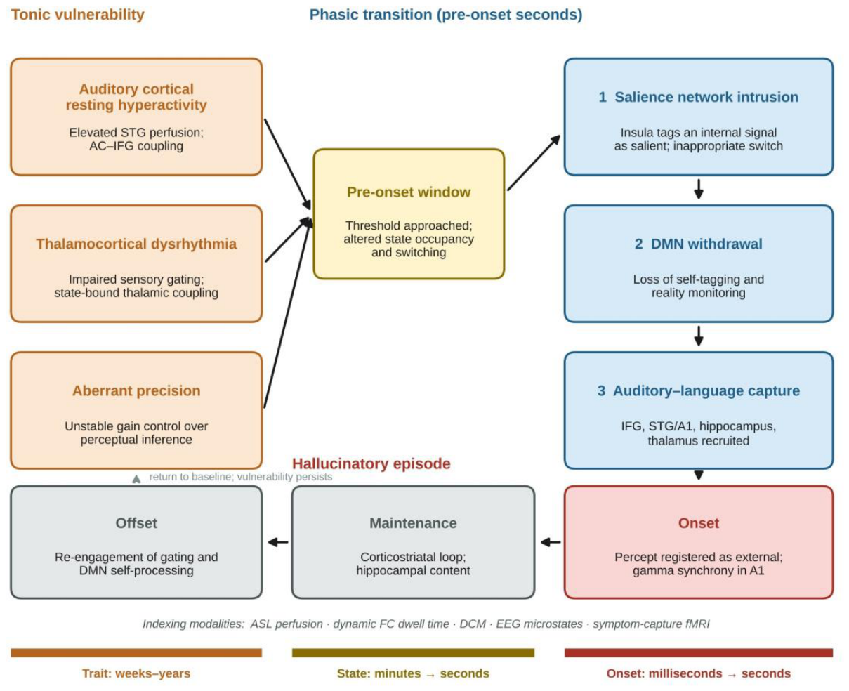
Figure 2.
Design logic of a burst-capture EEG-fMRI study. (A) Schematic, simulated time courses (not empirical data) aligned to the neural onset of a hallucinatory burst (t = 0). The pre-onset window (shaded) is the unit of inference; the latent fMRI network state shows shorter dwell times and more erratic switching before onset, EEG microstate class D shortens, the button press lags the experience by an unmeasured reaction time (RT), and the BOLD response is delayed by several seconds and sampled at the repetition time (TR). (B) Analysis pipeline: simultaneous acquisition, RT-independent anchoring, onset-locked state estimation with pre-specified parameters, sign-aware EEG–BOLD fusion, validity checks against stationary surrogate data, and transition metrics as primary outcomes. BOLD, blood-oxygen-level-dependent; CAP, co-activation patterns; HMM, hidden Markov model; LEiDA, leading eigenvector dynamics analysis. Conceptual schematic drawn programmatically with AI assistance (Claude, Anthropic); content specified and verified by the author.
Figure 2.
Design logic of a burst-capture EEG-fMRI study. (A) Schematic, simulated time courses (not empirical data) aligned to the neural onset of a hallucinatory burst (t = 0). The pre-onset window (shaded) is the unit of inference; the latent fMRI network state shows shorter dwell times and more erratic switching before onset, EEG microstate class D shortens, the button press lags the experience by an unmeasured reaction time (RT), and the BOLD response is delayed by several seconds and sampled at the repetition time (TR). (B) Analysis pipeline: simultaneous acquisition, RT-independent anchoring, onset-locked state estimation with pre-specified parameters, sign-aware EEG–BOLD fusion, validity checks against stationary surrogate data, and transition metrics as primary outcomes. BOLD, blood-oxygen-level-dependent; CAP, co-activation patterns; HMM, hidden Markov model; LEiDA, leading eigenvector dynamics analysis. Conceptual schematic drawn programmatically with AI assistance (Claude, Anthropic); content specified and verified by the author.
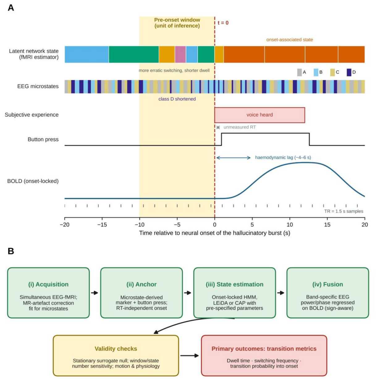

Table 1.
Onset-anchored and dynamic-network studies relevant to auditory verbal hallucination onset.
Table 1.
Onset-anchored and dynamic-network studies relevant to auditory verbal hallucination onset.
| Study | Sample | Design and temporal anchor | Principal finding |
| Hoffman et al. (2011b) | Patients with intermittent AVH; matched controls | Symptom-capture fMRI; TR 1500 ms; button-press onset/offset convolved with the HRF | Sequential organisation of regional BOLD activity across bilateral IFG, middle and superior temporal gyri, primary and association auditory cortex, hippocampal/parahippocampal regions and thalamus |
| Hoffman et al. (2011a) | 11 AVH patients; 9 non-hallucinating patients; 11 controls | Symptom-capture fMRI with rate- and duration-matched random button presses in comparison groups | Elevated functional connectivity along a corticostriatal loop in patients with AVH |
| Raij et al. (2009) | Patients with intermittent AVH | 3.0 T fMRI, TR 2.3 s; 585 AVH events captured with onset/offset button presses | Large event sample in the symptom-capture literature, enabling event-level modelling |
| Fuentes-Claramonte et al. (2021) | 30 patients (15 AVH+, 15 AVH−) | 10 min 10 s run; button press at each voice (5–174 presses; median 23) | AVH activated language and verbal short-term memory regions but not auditory cortex |
| Lefebvre et al. (2016) | Patients with schizophrenia experiencing hallucinations | ICA-inferred hallucination periods; DCM across distinct stages of emergence | Salience network involvement in switching between inner thought and the external world; executive takeover at episode end |
| Marschall et al. (2023) | 21 clinical and 21 non-clinical voice-hearers | LEiDA at sub-second resolution; 9 recurrent states; permutation ANOVA | More erratic dynamics (dwell time, switching frequency, switch probability) during hallucination, independent of clinical status |
| Geng et al. (2020) | 22 patients with and 17 without recent AVH | Sliding-window (22 TR) dFC; k-means into 6 states | Less time in, and lower probability of switching into, a DMN–language antagonistic state; reduced language/auditory network connectivity in specific states |
| Weber et al. (2020) | 80 patients; 80 matched controls | Sliding-window dFC; state dwell times; trait hallucination proneness over 1 year | High hallucination proneness associated with less time in states of strong DMN–task-positive anticorrelation |
| You et al. (2021) [conference abstract] | 83 patients; 83 matched controls | ICA (52 components; 8 networks); dFC clustered into 4 states | Increased dwell and fractional time in state 1 (P = 0.0081; P = 0.0018); decreased in state 3 (P = 0.0018; P = 0.0009) |
| Damaraju et al. (2014) | 151 patients; 163 matched controls | Resting-state ICA (100 components; 47 ICNs); 44-s sliding windows; k-means (5 states) | Thalamic hyperconnectivity and sensory hypoconnectivity most pronounced in specific states; absent from static analysis |
| Allen et al. (2014) | Healthy adults | Sliding-window FC; k-means clustering | Seven reproducible connectivity states emerging and dissolving over tens of seconds |
| Kindler et al. (2011) | Patients with frequent AVH | Resting EEG microstate analysis; hallucination periods marked by button press | Microstate class D significantly shorter during hallucination periods |
| Zhuo et al. (2017) | Schizophrenia with and without AVH; healthy controls | 3D pseudo-continuous ASL; voxel-wise CBF comparison | Increased CBF in right STG and caudate and decreased CBF in bilateral occipital and left parietal cortices in AVH (as synthesised by Shao et al., 2021) |
AVH, auditory verbal hallucinations; ANOVA, analysis of variance; ASL, arterial spin labelling; BOLD, blood-oxygen-level-dependent; CBF, cerebral blood flow; DCM, dynamic causal modelling; dFC, dynamic functional connectivity; DMN, default mode network; FC, functional connectivity; HRF, haemodynamic response function; ICA, independent component analysis; ICN, intrinsic connectivity network; IFG, inferior frontal gyrus; LEiDA, leading eigenvector dynamics analysis; STG, superior temporal gyrus; TR, repetition time.
Table 2.
Transient-state estimators: assumptions and constraints for burst-capture inference.
| Method | Signal modelled | Assumed temporal structure | Principal constraint for onset-anchored inference |
| Sliding-window correlation + k-means (Allen et al., 2014; Hutchison et al., 2013) | Pairwise BOLD correlations within windows | Quasi-stable states recurring across participants | Short windows generate spurious variability; a fixed window may not match true state durations (Leonardi and Van De Ville, 2015; Shakil et al., 2016) |
| Co-activation patterns (Chen et al., 2015; Liu et al., 2018) | Single-volume instantaneous co-activation | Discrete time points with high signal | Similar results obtained from stationary surrogate data; non-stationarity not guaranteed (Matsui et al., 2022) |
| Hidden Markov models (Bolt et al., 2022; Karapanagiotidis et al., 2020) | Multivariate Gaussian emissions over latent states | Markov process over discrete states | Sensitive to initial values; state number must be constrained, with higher values producing unoccupied states (van der Meer et al., 2020) |
| Leading eigenvector dynamics analysis (Favaretto et al., 2022; Marschall et al., 2023) | Leading eigenvector of each connectivity matrix | Recurrent dynamical states at near-frame resolution | Reduction to the leading eigenvector discards secondary connectivity structure |
| EEG microstates (Kindler et al., 2011; Van De Ville et al., 2010) | Scalp field topography | Quasi-stable sub-second epochs with scale-free durations | Low spatial resolution; duration, not label, carries the long-range dependency (Van De Ville et al., 2010) |
| Dynamic causal modelling (Lefebvre et al., 2016; Mechelli et al., 2007) | Directed (effective) coupling between regions or networks | Stage-dependent coupling during distinct periods of hallucination emergence | Requires a priori network selection and stage segmentation |
| Simultaneous EEG-fMRI regression (Debener et al., 2006; Goldman et al., 2002) | EEG power/phase regressed on BOLD | Linear neurovascular coupling with band-specific sign | Band-specific positive and negative couplings complicate sign interpretation (Goldman et al., 2002; Yuan et al., 2010) |
BOLD, blood-oxygen-level-dependent; EEG, electroencephalography; fMRI, functional magnetic resonance imaging.
Table 3.
A layered temporal architecture of auditory verbal hallucination onset.
| Timescale | Putative process | Convergent evidence |
| Trait (weeks–years) | Elevated auditory cortical resting activity and thalamocortical hyperconnectivity lower the threshold for spontaneous percept-like events | Increased CBF in STG (Northoff, 2014; Shao et al., 2021); thalamocortical hyperconnectivity as a signature of psychosis vulnerability (Mancini et al., 2020); thalamic and cortical grey-matter depletion (Ananth et al., 2002) |
| Slow state (minutes) | Altered occupancy of large-scale connectivity states, with less time in states of strong DMN–task-positive or DMN–language anticorrelation | Hallucination proneness and reduced dwell time in anticorrelated states (Weber et al., 2020); under-occupancy of a DMN–language antagonistic state (Geng et al., 2020); state-bound thalamic hyperconnectivity (Damaraju et al., 2014); altered dwell time in language/DMN-coupled states (You et al., 2021) |
| Pre-onset (seconds) | Salience network involvement during the emergence of hallucination; reduced Wernicke-to-Broca information flow | Salience network involvement in switching between inner thought and the external world (Lefebvre et al., 2016); aberrant salience network connectivity in first-episode psychosis (Mallikarjun et al., 2018); reduced Wernicke-to-Broca connectivity (Ćurčić-Blake et al., 2013) |
| Pre-onset (milliseconds–seconds) | Shortening of microstate class D and altered prestimulus excitability index the imminent percept | Microstate D shortening during hallucination (Kindler et al., 2011; Rieger et al., 2016); prestimulus power and connectivity predict perceptual outcome (Benwell et al., 2017; Weisz et al., 2014) |
| Onset | Recruitment of auditory and language cortex with sustained gamma synchrony | Sequential organisation of regional BOLD activity across temporal, frontal, hippocampal and thalamic nodes (Hoffman et al., 2011b); positive correlation between gamma phase locking and hallucination severity (Spencer, 2009; Spencer et al., 2009) |
| Maintenance | Corticostriatal loop activity and hippocampal memory content sustain the experience | Elevated connectivity along a corticostriatal loop (Hoffman et al., 2011a); hippocampal input to the salience network during the ‘on’ period (Lefebvre et al., 2016); medial temporal involvement (Jardri et al., 2011; Zmigrod et al., 2016) |
| Offset | Executive re-engagement, restoration of DMN–language antagonism and sensory gating | Executive-network takeover at episode end (Lefebvre et al., 2016); DMN–language antagonistic state as protective (Geng et al., 2020); more erratic switching during hallucination than rest (Marschall et al., 2023); gating re-engagement (Behrendt, 2006) |
BOLD, blood-oxygen-level-dependent; CBF, cerebral blood flow; DMN, default mode network; STG, superior temporal gyrus.
Disclaimer/Publisher’s Note: The statements, opinions and data contained in all publications are solely those of the individual author(s) and contributor(s) and not of MDPI and/or the editor(s). MDPI and/or the editor(s) disclaim responsibility for any injury to people or property resulting from any ideas, methods, instructions or products referred to in the content. |
© 2026 by the author. Licensee MDPI, Basel, Switzerland. This article is an open access article distributed under the terms and conditions of the Creative Commons Attribution (CC BY) license.
Copyright: This open access article is published under a Creative Commons CC BY 4.0 license, which permit the free download, distribution, and reuse, provided that the author and preprint are cited in any reuse.