

Submitted:
01 November 2026
Posted:
05 November 2026
You are already at the latest version
Abstract
Background: Traditional animal models in preclinical pharmacology show limited predictive accuracy for human outcomes due to interspecies differences, contributing to high drug attrition rates and ethical concerns. Recent regulatory changes have pushed the development of alternative, human-centered methods.Objective: To comprehensively investigate and evaluate novel non-animal approaches for establishing pharmacological safety and efficacy, such as organ-on-a-chip technologies, in vitro systems, and in silico models.Methods: A narrative evaluation of existing literature was conducted, concentrating on the technological principles, pharmacological applications, predictive performance, and regulatory acceptance of alternative testing strategies.Results: Compared with traditional 2D cultures, advanced in vitro models such as 3D organoids and iPSC-derived cells exhibit greater physiological relevance. Rapid, economical, and high-throughput predictions are made possible by in silico methods such as QSAR, PBPK modeling, and AI-based technologies. With improved translational fidelity, organ-on-a-chip technologies mimic the dynamic microenvironments of human organs. Strong, mechanism-based safety assessment frameworks are produced by integrated testing procedures that combine these methods. But there are still issues with scalability, validation, and standards.Conclusion: With growing regulatory approval for their future application, the incorporation of alternative approaches signifies a paradigm change toward more ethical, human-centered, and predictive drug development.
Keywords:
Organ-on-chip
; In silico
; QSAR
; 3D organoids
; iPSC
; PBPK
; New Approach Methodologies
; 3Rs
; Microphysiological systems
; Drug safety
; Alternative methods
; AI toxicology
1. Introduction
Since the late nineteenth century, preclinical pharmaceutical research has relied heavily on animal experiments. The 1950s–1960s thalidomide tragedy, which resulted in severe limb malformations in over 10,000 infants worldwide, served as the impetus for the 1962 Kefauver–Harris Drug Amendments in the United States and similar laws around the world, which mandated preclinical animal safety testing(Franks et al., 2004; J. H. Kim & Scialli, 2011) Despite this entrenched regulatory reliance, accumulating evidence across six decades has comprehensively demonstrated that animal models are poor predictors of human pharmacological outcomes. Over 90 medications that cleared all preclinical animal safety testing between 1991 and 2000 went on to cause major side effects in human clinical trials, suc h as rofecoxib's cardiovascular toxicity and troglitazone's deadly hepatotoxicity (Hartung, 2009; Olson et al., 2000)
Biological relevance is the main obstacle. Responses in rodents, dogs, or non-human primates often do not translate to humans due to profound interspecies differences in metabolic enzyme expression, receptor pharmacology, immune system architecture, and drug transporter function. About 75% of pharmaceuticals on the market are metabolized by the cytochrome P450 enzyme system, which varies significantly between species in terms of substrate specificities, expression patterns, and induction profiles (Chauhan et al., 2015; Martignoni et al., 2006). The translational failure rate is quantitatively alarming. Of compounds entering Phase I clinical trials, fewer than 10% reach market approval. For central nervous system drugs and oncology agents, success rates fall below 7% and 5%, respectively. A seminal analysis by Olson et al. (2000) found that standard preclinical animal toxicity studies predicted only 71% of human adverse effects for high-severity toxicities and as few as 43% across all organ systems. (Harrison, 2016; Kola & Landis, 2004; Olson et al., 2000)Animal testing poses serious ethical issues that go beyond scientific constraints. Ten to fifteen percent of the 192 million vertebrate animals used in research worldwide each year are thought to be utilized for pharmaceutical safety testing. The associated suffering and the recognition of animal sentience and capacity for pain have generated substantial societal and legislative pressure to reduce and eliminate animal use. (Balls, 2022; Taylor et al., 2008)
In their groundbreaking 1959 monograph, The Principles of Humane Experimental Technique, Russell and Burch outlined the fundamental principles for the moral and scientific reform of animal research. Since then, the three Rs—Replacement (replacing animal studies with non-animal alternatives), Reduction (reducing the number of animals used), and Refinement (improving procedures to minimize suffering) have been incorporated into the laws of more than 40 countries as well as the policies of all significant international regulatory bodies. (Festing & Wilkinson, 2007; Tannenbaum & Bennett, 2015) Significant changes in scientific technique, regulations, and policy have been prompted by the 3Rs. The European Union Directive 2010/63/EU, which regulates animal experimentation in 27 member states, specifically requires the 3Rs to be applied and encourages the creation and approval of substitute techniques. The legislative framework for this shift in the US is provided by the Animal Welfare Act and the FDA's new New Approach Methodologies (NAMs) program (Deckers, 2012; Lilienblum et al., 2008) Since the 1962 Kefauver-Harris amendments, the regulatory framework guiding preclinical drug testing has undergone its most significant change. Enacted in December 2022, the U.S. FDA Modernization Act 2.0 officially authorized the use of cell-based assays, organ chips, computer modeling, and other human-relevant NAMs as scientifically equivalent substitutes for animal data in Investigational New Drug (IND) applications(Commissioner, 2026; Stewart et al., 2023) The FDA's shift to nonclinical testing frameworks was further institutionalized in December 2025 with the Senate's unanimous approval of the FDA Modernization Act 3.0. Simultaneously, in April 2025, the FDA released a formal policy framework with specific instructions to prioritize MPS data and AI-driven toxicity modeling in IND and NDA submissions to decrease reliance on animal studies, improve their quality, and eventually replace them. A significant policy change with worldwide ramifications occurred when the National Institutes of Health (NIH) announced in July 2025 that research applications that relied solely on animal evidence would no longer be eligible for agency funding. (Commissioner, 2025; Pound, 2025).
Figure 1.
Integrated in silico drug discovery workflow combining molecular docking, dynamics simulation, PBPK modeling, and AI/ML approaches for predictive drug development.
Figure 1.
Integrated in silico drug discovery workflow combining molecular docking, dynamics simulation, PBPK modeling, and AI/ML approaches for predictive drug development.
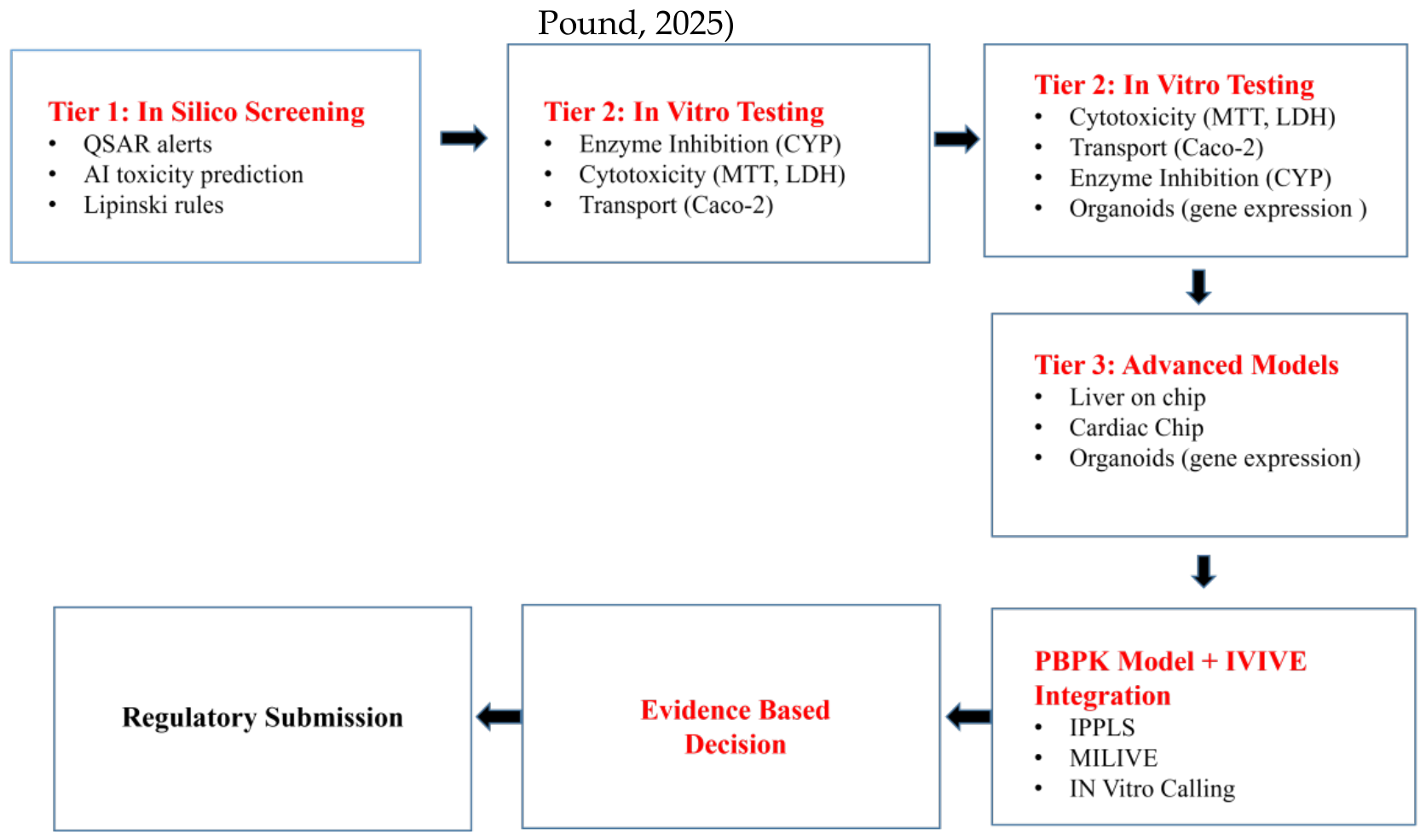
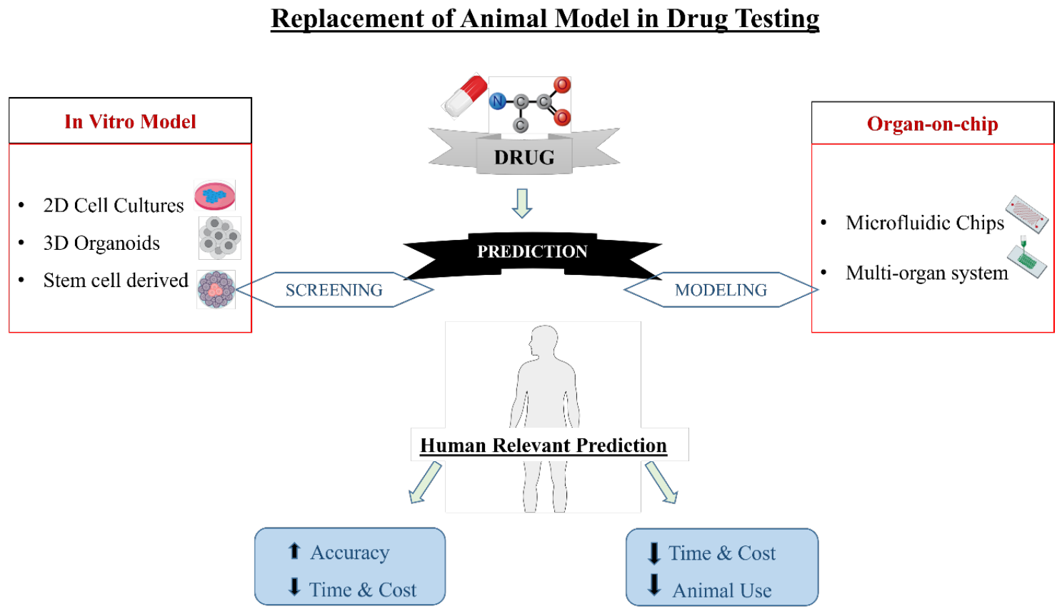
This review comprehensively evaluates the three major classes of emerging alternatives to animal experimentation for assessing pharmacological safety and efficacy: in vitro cell-based and tissue models, in silico computational approaches, and organ-on-chip microphysiological systems. For each category, the review examines underlying principles and technologies, pharmacological applications and validated endpoints, predictive performance and limitations, and current regulatory acceptance status. The review further addresses integrated testing strategies, future directions, and the path toward a fully human-relevant preclinical evaluation paradigm.
Figure 2.
human-relevant prediction system for translational drug evaluation that combines sophisticated in vitro, in silico, and organ-on-a-chip models with real-world data mapping.
Figure 2.
human-relevant prediction system for translational drug evaluation that combines sophisticated in vitro, in silico, and organ-on-a-chip models with real-world data mapping.
2. In Vitro Techniques: Tissue and Cell-Based Models
From basic monolayer cell cultures to complex three-dimensional organotypic constructs, in vitro pharmacological testing encompasses a wide range of experimental systems conducted in controlled laboratory settings, separate from living organisms. These systems offer inherent advantages over animal models: they employ human-derived biological material, can be implemented at high throughput, and permit precise control of experimental variables. Their progressive sophistication over the past two decades has substantially narrowed the biological relevance gap with in vivo systems. (Astashkina et al., 2012; Pampaloni et al., 2007)
3. Two-Dimensional Cell Culture Models
In vitro pharmacological research has historically been based on two-dimensional (2D) monolayer cell cultures, where cells are grown as a flat sheet on stiff plastic or glass substrates. HepG2 and HepaRG (hepatocellular), Caco-2 (intestinal epithelial), HEK293 (human embryonic kidney), MCF-7 (breast cancer), SH-SY5Y (neuronal), and HUVEC (vascular endothelial) are examples of well-known human cell lines that have undergone extensive characterization and validation for specific pharmacological endpoints (Guillouzo et al., 2007; LeCluyse et al., 2012)Cytotoxicity screening (MTT, LDH, and resazurin assays), receptor-binding studies, enzyme-inhibition profiling (IC50 determination), membrane permeability evaluation (PAMPA, Caco-2 monolayers), and transporter interaction studies (P-glycoprotein, BCRP, OAT, and OCT families) all make extensive use of 2D cultures. Their main drawback is the lack of multicellular communication, extracellular matrix interactions, and three-dimensional tissue architecture, all of which are essential for understanding how drugs behave in real tissue (Berridge et al., 2005; Dunn et al., 1989)
4. Three-Dimensional Cell Culture Models
1. Multicellular Spheroids
Three-dimensional spheroid cultures, formed by self-aggregation of cells under non-adherent or suspension conditions, recapitulate critical in vivo tissue features, including proliferating outer zones, quiescent mid-layers, and necrotic cores, features absent in 2D monolayers but characteristic of solid tumours and metabolically active organs. Multicellular tumor spheroids (MCTS) faithfully recapitulate drug penetration gradients, hypoxic signaling, and drug resistance mechanisms relevant to oncology pharmacology. (Hirschhaeuser et al., 2010; Zanoni et al., 2016)
2. Organoids
Organoids are three-dimensional, self-organizing miniature organ structures made from pluripotent or adult tissue stem cells. Through mechanisms that mimic developmental biology, they acquire organ-specific architecture, cellular diversity, and functional traits. Liver organoids for hepatotoxicity, intestinal organoids for absorption and barrier function, kidney organoids for nephrotoxicity, brain/cerebral organoids for CNS pharmacology, cardiac organoids for cardiotoxicity, and pancreatic organoids for metabolic drug evaluation are examples of established organoid systems currently employed in pharmacological research (Clevers, 2016; Kaur et al., 2015; Lancaster et al., 2013a)A pivotal pharmacological application of liver organoids has been the prediction of patient-derived hepatotoxicity. It has been demonstrated that organoids derived from individuals with polymorphisms in CYP2D6, CYP2C9, and DPYD mimic clinically observed variable drug sensitivities, providing a proof of concept for precision pharmacology without using animals (Peng et al., 2018; Schutgens et al., 2019)
4. Stem Cell-Based Models
The field of in vitro pharmacological testing has completely changed with Takahashi and Yamanaka's 2006 discovery of human induced pluripotent stem cell (iPSC) technology, which was awarded the Nobel Prize in Physiology or Medicine in 2012. iPSCs are an endless, patient-specific, human-relevant biological substrate that may differentiate into almost any form of human cell. They are produced by epigenetically reprogramming adult somatic cells using specific transcription factors. (Takahashi et al., 2007; Yu et al., 2007)
The predominant in vitro platform for preclinical evaluation of cardiotoxicity in the pharmaceutical industry is now iPSC-derived cardiomyocytes (iPSC-CMs). They enable assessment of the main safety pharmacology endpoints listed in the ICH S7B guidelines, including action potential shape, calcium transients, contractile function, mitochondrial integrity, and hERG channel activity. They enable assessment of the main safety pharmacology endpoints listed in the ICH S7B guidelines, including action potential shape, calcium transients, contractile function, mitochondrial integrity, and hERG channel activity. Clinical correlation studies have demonstrated that iPSC-CM-based assays predict the risk of QT prolongation with sensitivity and specificity exceeding those of traditional animal cardiac studies. (Fermini et al., 2016a; Liang et al., 2013; Moretti et al., 2010)
Hepatocytes produced from iPSCs fill a vital gap in DILI prediction, which has historically been the main reason for medicine withdrawals from the market. Progressive differentiation protocols using 3D culture conditions, hepatocyte growth factor gradients, and co-culture with non-parenchymal cells have significantly increased CYP enzyme activity, albumin secretion, and phase II conjugation fidelity in iPSC-hepatocytes when compared to HepG2 cells, even though maturation limitations still exist (Baxter et al., 2015; Si-Tayeb et al., 2010; Szkolnicka et al., 2014).
5. High-Throughput Screening Platforms
Pharmacological profiling of tens of thousands of compounds in a matter of days is made possible by automated high-throughput screening (HTS) devices that combine robotic liquid handling, smaller microplate formats (384-well, 1536-well), high-content imaging systems, and multimodal detection technologies. The combination of HTS with phenotypic readouts, including cell morphology, organelle integrity, mitochondrial membrane potential, nuclear size, and cytoskeletal architecture, assessed via high-content analysis (HCA), provides mechanistically rich safety information far beyond simple cell viability endpoints. (L. M. Mayr & Fuerst, 2008; Zanella et al., 2010)Using a panel of 60 cell-based assays across 1536-well platforms, the US government's Tox21 program has screened over 10,000 environmental chemicals and drug candidates, creating the largest publicly available in vitro toxicology dataset and proving the viability of high-throughput mechanistic toxicology at a national scale (Kavlock et al., 2012; Tice et al., 2013)
6. Tissue Models in Ex Vivo
Precision-cut tissue slices (PCTS), thin sections (100–250 µm) of fresh liver, lung, kidney, or intestinal tissue maintained in organ culture, preserve the intact multicellular architecture, extracellular matrix composition, and intercellular communication of the parent organ while substantially reducing animal use compared with in vivo studies. Liver PCTS (PCLS) maintain CYP enzyme activity, hepatocyte polarity, Kupffer cell function, and biliary transport for 24–72 hours of culture, enabling mechanistic investigations of hepatotoxicity at the organ-cellular interface. (De Graaf et al., 2010; Vickers, 2004) Isolated perfused organ systems, particularly isolated perfused rat kidney (IPK), isolated perfused rat liver (IPL), and isolated perfused rat heart (Langendorff preparation) enable pharmacokinetic and pharmacodynamic studies at the organ level with controlled vascular perfusion, providing mechanistically superior data on renal clearance, hepatic extraction ratio, and cardiac function compared with in vitro cell culture, while substantially reducing whole-animal usage. (Bessems et al., 2006; Masereeuw et al., 1999)
Figure 3.
In vitro models have evolved from 2D cell culture to 3D spheroids, organoids, and organ-on-chip systems, thereby increasing physiological relevance and predictive potential.
Figure 3.
In vitro models have evolved from 2D cell culture to 3D spheroids, organoids, and organ-on-chip systems, thereby increasing physiological relevance and predictive potential.
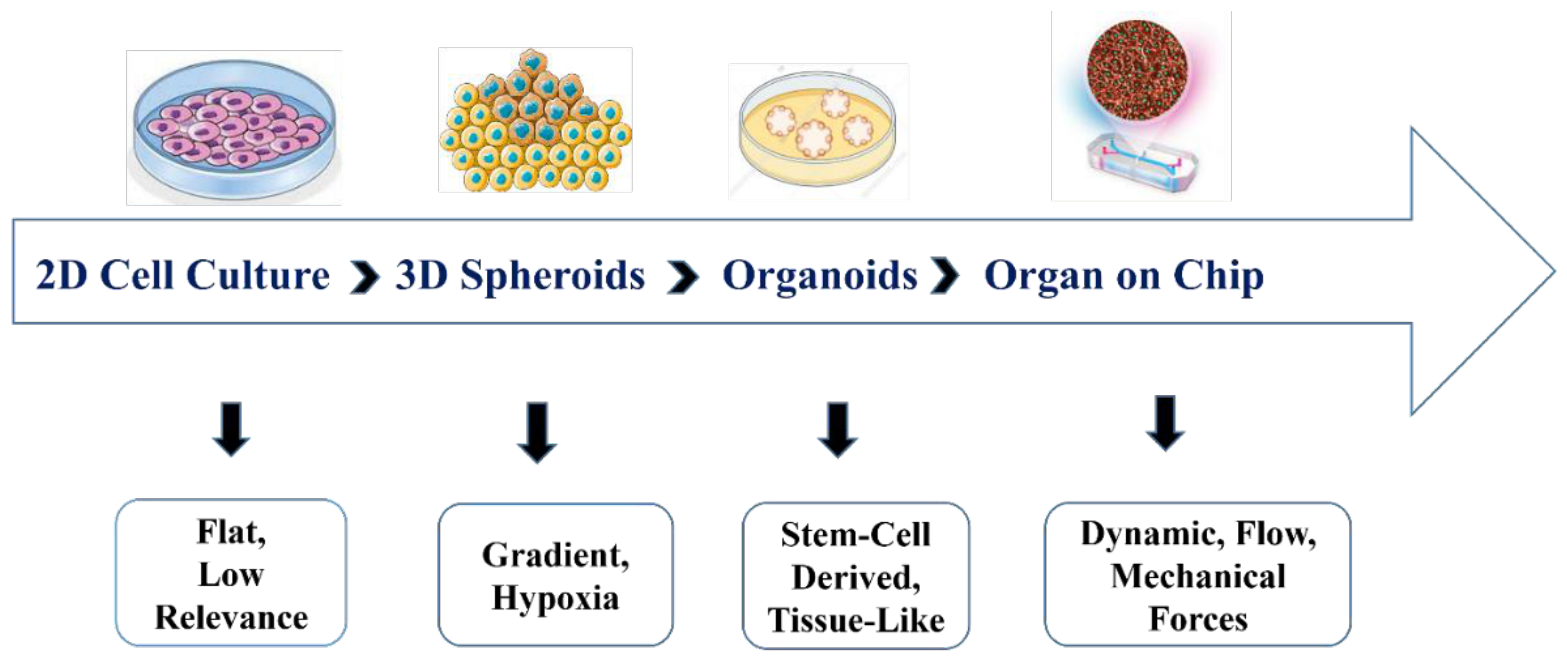

Table 1.
Comparison of in vitro alternative methods for pharmacological safety and efficacy evaluation.
Table 1.
Comparison of in vitro alternative methods for pharmacological safety and efficacy evaluation.
| Model | Representative Platforms | Principal Applications | Validated Pharmacological Endpoints | Key Limitations | APA Citation |
| 2D Cell Lines | HepG2, Caco-2, HEK293, SH-SY5Y, HUVEC | Cytotoxicity, receptor binding, enzyme inhibition, permeability | IC50, CC50, hERG IC50, Caco-2 Papp, CYP inhibition | No ECM/3D architecture; Loss of polarity; Low in vivo relevance | (Dunn et al., 1989; LeCluyse et al., 2012) |
| Multicellular Spheroids / MCTS | Ultra-low attachment plates, hanging drop, spinner flask | Oncology efficacy, solid tumor penetration, and drug resistance | GI50, proliferation index, necrotic core formation, O2 gradient modeling | Batch variability; Limited vascularisation; Non-standard protocols | (Hirschhaeuser et al., 2010; Zanoni et al., 2016) |
| Organoids | Liver, gut, kidney, brain, cardiac organoids (Matrigel) | DILI, GI toxicity, nephrotoxicity, neuroPharm, precision oncology | Transaminase release, barrier integrity, gene expression fidelity, and drug sensitivity | High cost; Batch heterogeneity; Lack of vascularisation; Maturation gaps | (Clevers, 2016; Kaur et al., 2015) |
| iPSC-Derived Models | iPSC-CMs, iPSC-hepatocytes, iPSC-neurons, iPSC-podocytes | Cardiotoxicity, DILI, neurotoxicity, nephrotoxicity, and personalized medicine | APD, QTc prediction, albumin, CYP activity, calcium transients | Fetal-like maturation phenotype; High production cost; Differentiation variability | (Liang et al., 2013; Takahashi et al., 2007) |
| High-Throughput Screening (HTS) | 384/1536-well platforms; High-content imaging (HCA); Tox21 panel | Primary toxicity screening, lead identification, and mechanism of action | Phenotypic endpoints (morphology, mitochondrial potential, nuclear area); Multi-parametric profiles | Phenotypic complexity limited; Artifact-prone at high compound conc. | (Tice et al., 2013; Zanella et al., 2010) |
| Precision-Cut Tissue Slices (PCTS) | PCLS (liver, lung); PCKS (kidney) | Organ-level toxicity, metabolic studies, fibrosis/inflammation | CYP activity, viability, Ki67 proliferation, cytokine release | Short culture viability (24–72 h); Requires fresh tissue; Animal use not eliminated | (De Graaf et al., 2010; Vickers, 2004) |
| Isolated Perfused Organ Systems | Langendorff heart; IPK; IPL | PK/PD at the organ level; cardiac electrophysiology; hepatic extraction ratio | Hepatic clearance, GFR, contractility, AP morphology | Brief viability window; High technical expertise; Requires animal sacrifice | (Bessems et al., 2006; Masereeuw et al., 1999) |
Note: Ipsc: induced pluripotent stem cell; CM: cardiomyocyte; DILI: drug-induced liver injury; APD: action potential duration; CYP: cytochrome P450; GFR: glomerular filtration rate; PCLS: precisely cut liver slices; AP: action potential, HCA: high-content analysis, and PCKS: precisely cut kidney slices.
5. In Silico Methods: Computational and AI-Driven Approaches
Without the need for actual experiments, in silico pharmacological methods comprise a wide and quickly growing set of computational techniques that model pharmacokinetic and toxicological consequences, forecast drug activity, and mimic biological systems. In silico methods create virtual representations of human pharmacology at molecular, cellular, organ, and whole-body sizes by combining structural chemistry, genomics, proteomics, systems biology, and machine learning. They are essential parts of contemporary drug development pipelines due to their basic benefits, speed, affordability, capacity for huge compound throughput, and ability to incorporate multiscale biological data (Lounnas et al., 2013; Sliwoski et al., 2014).
- Quantitative Structural Activity Relationship (QSAR) Modelling
The physicochemical and structural characteristics of chemical substances and their biological activities or toxicological outcomes are quantitatively and mathematically related by QSAR models. First formalized by Hansch and Fujita in 1964, QSAR has evolved from simple linear regression models to sophisticated ensemble machine-learning approaches that incorporate thousands of molecular descriptors derived from two- and three-dimensional molecular representations. (Cherkasov et al., 2014; Hansch & Fujita, 1964)
A wide range of toxicological endpoints, such as mutagenicity and genotoxicity (Ames test surrogate), cardiac toxicity through hERG potassium channel inhibition, skin sensitization potential, acute aquatic toxicity, carcinogenicity, and reproductive toxicity, are now covered by regulatory-accepted QSAR applications. The OECD's Principles for the Validation of QSAR Models (2004, revised 2014) established a five-criterion validation framework that has been adopted by the FDA, EMA, and ECHA as the basis for regulatory acceptance of QSAR predictions instead of experimental data. (Barber et al., 2024; Worth & Balls, 2001)
The US EPA's TEST (Toxicity Estimation Software Tool), which uses multiple QSAR methods with consensus averaging, the EU-funded VEGA platform, which offers transparently validated models for REACH compliance, TOPKAT (Accelrys) for regulatory toxicity endpoint prediction, and DEREK Nexus (Lhasa Limited) for structural alert-based toxicity flagging are some of the major validated QSAR platforms currently in use. Ensemble QSAR techniques significantly lower uncertainty in individual models and increase overall forecast reliability by combining predictions from several independently produced models (Fjodorova et al., 2010; Roncaglioni et al., 2013)
5.1. Molecular Docking and Dynamic Simulations
Molecular docking enables logical, structure-based drug design and a mechanistic understanding of drug–target interactions by computationally predicting the preferred binding orientation, conformation, and affinity of small-molecule ligands inside specified protein binding sites. In order to score candidate poses with high prediction fidelity, modern docking algorithms such as AutoDock Vina, Schrödinger Glide, GOLD, and rDock use complex scoring systems that include electrostatic interactions, hydrogen bonding, hydrophobic contacts, and entropic contributions.. (Friesner et al., 2006; Trott & Olson, 2010)By explicitly simulating the temporal evolution of molecular systems, including protein flexibility, solvent effects, membrane contacts, and conformational sampling on nanosecond to microsecond timescales, molecular dynamics (MD) simulations go beyond static docking. More mechanistic information than rigid-body docking alone can be obtained using MD simulations with platforms such as GROMACS, AMBER, NAMD, and CHARMM to predict drug-binding kinetics (kon/koff), allosteric mechanisms, membrane permeability, and protein–protein interaction disruption. (Abraham et al., 2015a; Case et al., 2005a)
- 2.
- Physiologically Based Pharmacokinetic Modelling
Human anatomical and physiological data are used to parameterize physiologically based pharmacokinetic (PBPK) models, which are mechanistic, multi-compartmental mathematical frameworks that simulate the absorption, distribution, metabolism, and excretion (ADME) of drugs across interconnected physiological compartments. With organ-specific blood flow rates, volumes, and partitioning coefficients derived from in vitro data or literature, each compartment represents a distinct organ or tissue (liver, kidney, lung, adipose, brain, etc.) (Jones & Rowland-Yeo, 2013; Rowland et al., 2011a).
Regulatory approval for PBPK modeling has significantly improved. Guidance documents that specify the evidence standards and reporting formats needed for PBPK data to support regulatory submissions have been released by the European Medicines Agency (EMA, 2018 guideline on the reporting of PBPK modeling and simulation) and the U.S. FDA (2018 guidance on PBPK analyses). First-in-human dose prediction, dietary effect prediction, dose optimization in particular populations (pediatrics, pregnant women, hepatic and renal impairment), and drug–drug interaction (DDI) prediction to replace in vivo crossover studies are among the approved applications (Jamei, 2016; Paul et al., 2025a; Rowland et al., 2011a)
- 3.
- Artificial Intelligence and Machine Learning
The availability of extensive curated toxicity and bioactivity databases (Tox21, ToxCast, ChEMBL, PubChem BioAssay), advancements in deep learning architectures, and growing computational resources have all contributed to the significant acceleration of the application of artificial intelligence (AI) and machine learning (ML) to preclinical pharmacological evaluation over the past ten years. AI/ML approaches now enable toxicity prediction, off-target effect identification, metabolic liability flagging, and polypharmacology profiling at scales impossible with experimental methods. (H. Chen et al., 2018; A. Mayr et al., 2016a)
On molecular property prediction benchmarks, such as the MoleculeNet suite, graph neural networks (GNNs), which depict molecules as mathematical graphs with atoms as nodes and bonds as edges, have shown state-of-the-art performance. For forecasting hERG toxicity, hepatotoxicity, and mutagenicity on held-out test sets, deep learning models, such as DeepTox (winner of the Tox21 challenge), ChemProp, and AttentiveFP, have outperformed conventional QSAR methods with AUC values greater than 0.85 (Duvenaud et al., 2015; Wu et al., 2018; Yang et al., 2019)By offering structural models for thousands of previously undruggable or structurally uncharacterized targets, DeepMind's 2021 release of AlphaFold2, which achieved near-experimental accuracy in protein structure prediction across the entire human proteome, transformed structure-based virtual screening. AlphaFold2 structures are now routinely integrated into docking pipelines, enabling target-based drug discovery for the full druggable genome. (Jumper et al., 2021a; Varadi et al., 2022)
- 4.
- In Vitro–In Vivo Extrapolation
The quantitative technique known as "in vitro–in vivo extrapolation" (IVIVE) uses physiological scaling factors and, increasingly, full PBPK model integration to mathematically scale pharmacokinetic parameters measured in vitro, such as hepatic intrinsic clearance (CLint) from microsomal or hepatocyte assays, plasma protein binding (fu,p), blood-to-plasma ratio, and Caco-2 permeability (Papp), to predict in vivo human pharmacokinetic behavior. IVIVE is now a regulatory expectation for predicting hepatic clearance and renal secretion in the absence of animal data. (Hallifax & Houston, 2012; Obach, 1999; Poulin & Theil, 2002)
5.2. Digital Twins and Virtual Patient Modelling
Digital twin technology, originally developed in aerospace and manufacturing engineering, is being progressively translated to clinical pharmacology. A patient digital twin is a dynamic, continuously updated computational model that integrates genomic data, proteomic biomarkers, medical imaging, electronic health record data, and, increasingly, organ-on-chip measurements to simulate individual patients' drug responses in silico. Pharmaceutical applications include personalised dose optimisation for oncology regimens, prediction of individual adverse event risk before clinical exposure, and simulation of virtual patient cohorts for virtual clinical trial design. (Corral-Acero et al., 2020a; on behalf of the Swedish Digital Twin Consortium et al., 2020)
Figure 4.
Integrated drug discovery workflow combining USAR/QSAR, molecular docking, molecular dynamics, PBPK simulation, in vitro validation, and AI/ML-driven digital twin modeling for enhanced prediction.
Figure 4.
Integrated drug discovery workflow combining USAR/QSAR, molecular docking, molecular dynamics, PBPK simulation, in vitro validation, and AI/ML-driven digital twin modeling for enhanced prediction.
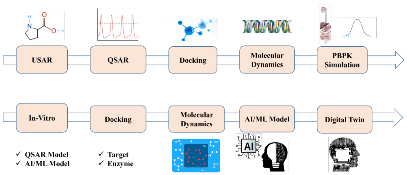
Table 2.
Comparison of in silico alternative methods for pharmacological prediction and toxicity assessment.
Table 2.
Comparison of in silico alternative methods for pharmacological prediction and toxicity assessment.
| Method | Key Tools / Platforms | Applications | Regulatory Status | Strengths | Limitations | Key Citation |
| QSAR Modelling | DEREK Nexus, TOPKAT, TEST (EPA), VEGA | Mutagenicity, hERG, skin sensitisation, carcinogenicity | OECD TG-validated; ECHA/FDA/EMA accepted for REACH and IND | High throughput; No animals; Interpretable alerts | Applicability domain; Sparse structural classes; Single endpoint models | (Barber et al., 2024; Cherkasov et al., 2014) |
| Molecular Docking | AutoDock Vina, Schrödinger Glide, GOLD, rDock | Target binding, Virtual screening, Lead optimisation | Used in IND and patent filings; Not formally qualified as standalone | Rapid, cost-free ligand pose prediction; Structure-based | Rigid protein approximation; Scoring function errors; False positives | (Friesner et al., 2006; Trott & Olson, 2010) |
| Molecular Dynamics | GROMACS, AMBER, NAMD, CHARMM | Binding kinetics, protein flexibility, membrane permeability, allosteric | Supporting data for mechanism-based submissions | Mechanistic depth; Kinetic parameters; Conformational sampling | Computationally expensive; Limited timescale; Force field accuracy | (Abraham et al., 2015b; Case et al., 2005b) |
| PBPK Modelling | SimCyp, PK-Sim, GastroPlus, Matlab | DDI, special populations, first-in-human dose, food effects | FDA 2018 guidance; EMA 2018 guideline; Formal IND/NDA use | Mechanistic; Integrates in vitro data; Replaces clinical DDI studies | Extensive parameterisation; Uncertainty propagation; Model over-fitting risk | (Rowland et al., 2011b) |
| AI / Deep Learning | DeepTox, ChemProp, AttentiveFP, GNNs | Multi-endpoint toxicity, hERG, DILI, AMES, metabolic liability | Tox21 outcomes accepted; AI-assisted submissions under FDA AI/ML framework | Ultra-high throughput; Multi-endpoint; Improves with data size | Black-box interpretability; Data quality dependent; Generalisation to new scaffolds | (Jumper et al., 2021b; A. Mayr et al., 2016b) |
| IVIVE | Retromer scaling, PBPK integration, fugacity models | Hepatic CL prediction, renal secretion, Papp to Fa, fu scaling | FDA-accepted for CLhep and DDI predictions in IND/NDA submissions | Eliminates some PK animal studies; Mechanistically grounded | CLint measurement variability; In vitro-in vivo disconnect for some CL | (Hallifax & Houston, 2012; Obach, 1999) |
| Digital Twins | Modelling platforms + EHR + omics data + MPS integration | Personalised dosing, virtual clinical trials, adverse event prediction | Early regulatory engagement; No formal qualification to date | Integrative; Personalised; Continuously updatable | Requires extensive multi-modal data; Clinical validation limited | (Björnsson et al., 2020; Corral-Acero et al., 2020a) |
Take note. QSAR = quantitative structure-activity relationship; PBPK = physiologically based pharmacokinetic; DDI = drug-drug interaction; DILI = drug-induced liver injury; CLhep = hepatic clearance; GNN = graph neural network; IVIVE = in vitro-in vivo extrapolation; EHR = electronic health record; and MPS = microphysiological system.
6. Organ-on-Chip and Microphysiological Systems
Microphysiological systems (MPS), also known as organ-on-chip (OoC) devices, are microfluidic platforms that replicate the essential mechanical, structural, and functional milieu of human organs at the microscale. By integrating human primary cells or iPSC-derived cells within precisely engineered microchannels that deliver physiologically relevant fluid flow, shear stress, mechanical strain, and chemical gradients, OoC systems replicate dynamic organ-level biology in ways that static in vitro models and interspecies animal models fundamentally cannot. (Bhatia & Ingber, 2014; Huh et al., 2010)
The field was sparked by Huh et al.'s groundbreaking paper in Science (2010), which showed that a microfluidic chip seeded with human pulmonary epithelial and endothelial cells could replicate the mechanical and biological complexity of the human alveolar-capillary interface, including cyclic mechanical breathing motions and the air–liquid interface. This lung-on-a-chip replicated medication absorption, nanoparticle translocation, and inflammatory cytokine responses with a level of accuracy not possible with traditional cell culture (Huh et al., 2010, 2012).
7. Design Principles and Fabrication
OoC devices are made of glass, polydimethylsiloxane (PDMS), or increasingly thermoplastic polymers like polymethylmethacrylate (PMMA) and cyclic olefin copolymer (COC) utilizing injection molding, photolithography, or soft lithography. Mechanical actuation systems that provide cyclic stretch mimicking organ biomechanics (breathing, peristalsis, cardiac contraction), parallel or coaxial microfluidic channels divided by thin porous membranes that allow co-culture of two or more cell compartments (such as epithelial and endothelial) under controlled fluidic conditions, and integrated biosensors such as transepithelial/endothelial electrical resistance (TEER) electrodes, impedance spectroscopy, electrochemical oxygen and metabolite sensors, and optical windows for confocal microscopy are important design elements (Leung et al., 2022; Sackmann et al., 2014).
8. Liver-on-Chip for Hepatotoxicity and DILI Prediction
Acute liver failure, post-market medication withdrawal, and late-phase clinical trial failure are all most commonly caused by drug-induced liver injury (DILI). Primary human hepatocyte monolayers only function for 24 to 48 hours, which is insufficient to capture chronic DILI causes, whereas conventional HepG2 2D culture predicts DILI with a sensitivity below 50%. By using human primary hepatocytes or iPSC-derived hepatocytes co-cultured with sinusoidal endothelial cells, Kupffer cells (resident macrophages), and hepatic stellate cells under physiological fluid shear stress that simulates sinusoidal perfusion, liver-on-chip platforms fill these crucial gaps (Ewart et al., 2022; Godoy et al., 2013).
Compared with HepaRG 2D culture and primary hepatocyte spheroids, the Emulate liver chip showed higher sensitivity and specificity for DILI prediction, earning the first official FDA ISTAND qualification for an OoC system in September 2024. In a 27-drug validation set encompassing known DILI-positive and DILI-negative compounds, the liver chip achieved 87% sensitivity and 100% specificity, compared with 47% sensitivity for 2D HepG2 culture. (Ewart et al., 2022, p. 2024)
9. Heart-on-Chip for Cardiotoxicity Screening
28% of drug withdrawals from the pharmaceutical market are due to cardiac toxicity, specifically proarrhythmia caused by hERG potassium channel blockage and QT interval prolongation. Preclinical cardiac safety evaluations are required by ICH S7B recommendations and have traditionally needed both in vitro hERG patch-clamp and in vivo telemetry studies. The organized sarcomeric architecture, calcium cycling, electrical conduction, and force generation of the human heart are replicated by heart-on-chip devices that use aligned iPSC-derived cardiomyocytes under cyclic mechanical strain and physiological flow (Lian et al., 2012; Mathur et al., 2015).
Field potential duration (FPD), a surrogate for the QT interval, conduction velocity, beat rate, and arrhythmia morphology may all be recorded in real time across drug-exposure time courses thanks to multi-electrode array (MEA) systems built into cardiac chips. An AUROC of 0.89 was shown in studies comparing cardiac OoC predictions with clinical QT prolongation data from 28 reference drugs, significantly outperforming the predictive performance of isolated hERG patch-clamp alone (AUROC 0.72) (Colatsky et al., 2016; Doherty et al., 2013).
10. Kidney-on-Chip for Nephrotoxicity
Up to 25% of hospitalized patients' acute kidney injury cases are caused by medication-induced nephrotoxicity, which also plays a major role in the failure of otherwise promising therapeutic candidates in clinical trials. Because proximal tubule cells are sensitive to cisplatin, aminoglycosides, and contrast agents and participate in active drug secretion via OAT and OCT transporters, they are the primary cause of nephrotoxic injury. Human proximal tubule epithelial cells (PTECs) have been shown to restore megalin-cubilin endocytic receptor expression, primary cilia formation, and transporter-mediated secretion activity under physiological shear stress, all of which are lacking in static cultures on kidney-on-chip platforms (Weber et al., 2016; Wilmer et al., 2016).
A comparative study by Jang et al. (2013) demonstrated that kidney-on-chip accurately predicted the differential nephrotoxicity of cephaloridine and cephaloglycin, a distinction that standard static PTEC culture failed to distinguish by recapitulating the flow-dependent transporter-mediated accumulation mechanism responsible for cephaloridine's selective tubular toxicity in vivo(Jang et al., 2013; S. Kim et al., 2016).
11. Blood–Brain Barrier and Brain-on-Chip
At more than 95%, the field of central nervous system (CNS) medication development has the highest attrition rate of any therapeutic area. This is partially due to efflux by blood–brain barrier (BBB) transporters such as P-glycoprotein (ABCB1) and BCRP (ABCG2), which prevents most medication candidates from reaching therapeutic concentrations in brain tissue. BBB-on-chip platforms co-culture brain microvascular endothelial cells (BMECs) with astrocytes and pericytes separated by a thin ECM membrane under physiological flow, restoring tight junction expression (ZO-1, claudin-5, occludin), TEER values of 200–1500 Ω·cm2 (approaching in vivo values of ~2000 Ω·cm2), and functional P-gp efflux activity. (Booth & Kim, 2012; Wang et al., 2016)
12. Gut-on-Chip for Absorption and Microbiome Interactions
By applying cyclic mechanical strain to flexible PDMS membranes seeded with Caco-2 or human intestinal organoid-derived cells, the gut-on-chip created by Kim et al. (2012) replicated intestinal peristalsis. This led to the formation of villus-like three-dimensional folds, the restoration of mucus production, improved tight junction formation, and significantly increased drug-absorption fidelity compared with static Caco-2 monolayers. The platform has been extended to incorporate living gut microbiome communities (including anaerobic bacterial consortia) that coexist with human intestinal epithelial cells, enabling, for the first time, the study of host–microbiome pharmacological interactions (drug biotransformation, microbiome-mediated modulation of drug efficacy) in a human-relevant system. (Jalili-Firoozinezhad et al., 2019; H. J. Kim et al., 2012)
13. Multi-Organ and Body-on-Chip Platforms
Connecting many organ modules into integrated multi-organ micro-physiological systems (multi-MPS) that replicate how medications are absorbed, transported, digested, and eliminated in the body is the next stage after single-organ chips. For instance, innovative multi-organ platforms have linked gut–liver–brain chips for neurological drug PK, liver and intestine chips for first-pass metabolism research, and liver and kidney chips for systemic clearance profiles. For medications like irinotecan, naphthalene, and methotrexate, comparisons between multi-organ chip PK predictions and human clinical pharmacokinetic data have revealed consistency within 1.5–2-fold (Edington et al., 2018; Herland et al., 2020).
Table 3.
Organ-on-a-chip microphysiological systems and its pharmaceutical uses have been verified.
| Organ Chip | Cell Types Used | Pharmacological Application | Key Validated Endpoint | Comparative Advantage Over 2D | Regulatory Status | Key Citation |
| Liver-on-Chip | Primary hepatocytes, Kupffer cells, LSECs, iPSC-hepatocytes | DILI prediction, CYP-mediated metabolism, cholestasis | ALT/AST release, CYP3A4 activity, bile acid transport, steatosis markers | 87% sensitivity vs 47% HepG2 (Ewart et al., 2022) | FDA ISTAND qualified (September 2024) | (Ewart et al., 2022; Godoy et al., 2013) |
| Heart-on-Chip | iPSC-cardiomyocytes, cardiac fibroblasts, endothelial cells | QT prolongation, cardiotoxicity, arrhythmia risk | Field potential duration, conduction velocity, calcium transients | AUROC 0.89 vs 0.72 hERG alone | ICH S7B supportive data; Not yet formally qualified | (Colatsky et al., 2016; Mathur et al., 2015) |
| Kidney-on-Chip | Human proximal tubule ECs (PTECs), glomerular podocytes | Nephrotoxicity, transporter-mediated drug interactions | OAT/OCT transport, KIM-1, NGAL biomarkers, filtration barrier function | Restores active secretion absent in static PTEC culture | Regulatory qualification in progress | (Jang et al., 2013; Wilmer et al., 2016) |
| Kidney-on-Chip | Primary Renal Proximal Tubule Epithelial Cells (PTEC), Peritubular Endothelial cells | Nephrotoxicity screening, Drug clearance/reabsorption, Proteinuria | Creatinine clearance, Glucose transport, KIM-1(Injury marker) | Fluid shear stress induces primary cilia and 10x higher transporter expression | Part of EMA/FDA collaborative 3R guidelines | (Musah et al., 2017) |
| BBB-on-Chip | BMECs, astrocytes, pericytes (NVU model) | CNS drug penetration, neuroinflammation, P-gp efflux screening | TEER (200–1500 Ω·cm2), Papp brain, P-gp inhibition | Achieves physiological TEER; Functional efflux transporters | Regulatory qualification in development | (Booth & Kim, 2012; Wang et al., 2016) |
| Gut-on-Chip | Caco-2, intestinal organoid-derived cells, living microbiome | Drug absorption, microbiome–drug interactions, GI toxicity | Papp, villus formation, mucus thickness, tight junction proteins (ZO-1) | Restores villous morphology; Enables anaerobic microbiome co-culture | Regulatory engagement ongoing | (Jalili-Firoozinezhad et al., 2019; H. J. Kim et al., 2012) |
| Lung-on-Chip | Alveolar epithelial (A549/primary), microvascular endothelial | Pulmonary drug delivery, inhalation toxicity, infection pharmacology | Barrier integrity, surfactant production, cytokine release, drug permeability | Cyclic breathing motions; Air–liquid interface | Used in regulatory supporting packages | (Huh et al., 2010, 2012) |
| Alveolar epithelial cells, Microvascular endothelial cells, Neutrophils | Aerosol drug delivery, Pulmonary edema , Viral infection (e.g.,SARS-CoV-2) | TEER, Inflammatory cytokines, Surfactant production | Recapitulates the “Breathing” mechanical strain (cyclic stretch) | FDA-supported for inhaled drug toxicity | (Benam et al., 2016; Huh et al., 2010) | |
| Tumor-on-Chip | Patient-derived tumor cells + CAFs + immune cells + vasculature | Anticancer drug efficacy, TME interactions, immuno-oncology | Tumor growth inhibition, cytotoxic T-cell infiltration, and angiogenesis markers | Models TME resistance; Patient-derived personalised response testing | Research stage; Early regulatory interest | (Bersini et al., 2014; Oushyani Roudsari et al., 2024) |
| Multi-Organ (Body-on-Chip) | Coupled liver + gut + kidney +/- brain chips | Systemic ADME, DDI, organ–organ interactions, PK/PD | Multi-organ clearance, metabolite profiling, plasma PK curves | PK prediction within 1.5–2-fold of human clinical data | Early regulatory dialogue; FDA MPS qualification pathway active | (Edington et al., 2018; Herland et al., 2020) |
Note. DILI: drug-induced liver injury, LSECs: liver sinusoidal endothelial cells, iPSC: induced pluripotent stem cell, TEER: transepithelial/transendothelial electrical resistance, TME: tumor microenvironment, CAFs: cancer-associated fibroblasts, BBB: blood–brain barrier, BMECs: brain microvascular endothelial cells, NVU: neurovascular unit, P-gp: P-glycoprotein, AUROC: area under the receiver operating characteristic curve, ADME: absorption, distribution, metabolism, excretion, DDI: drug–drug interaction, KIM-1: kidney injury molecule-1, and NGAL: neutrophil gelatinase-associated lipocalin.
14. Integrated Methods and Integrated Testing Techniques
No single non-animal technique, whether in vitro, in silico, or OoC, can independently replace the thorough safety and efficacy data produced by an extensive in vivo preclinical program. This fundamental idea is now widely acknowledged within the alternative technique’s community. Every platform has intrinsic technical and biological scope limitations. Complementary techniques should be systematically and mechanistically integrated into tiered Integrated Testing Strategies (ITS) to create a safety argument strong enough to support regulatory decision-making (Hartung, 2013; Leist et al., 2017).
15. Integrated Testing Strategies and IATA
The Integrated Approaches to Testing and Assessment (IATA) framework from the OECD offers organized, scientifically supported methods for compiling data from various non-animal data streams, existing data, in vitro results, in silico predictions, and in chemico assays into a logical, defendable regulatory submission package. IATA frameworks have been formally adopted for specific endpoints, including skin sensitisation (the 2019 OECD Guidance Document on Skin Sensitisation), eye irritation and corrosion, acute oral toxicity, repeated-dose systemic toxicity, and genotoxicity. Each IATA specifies the logic of evidence assembly, applicable methods, confidence scoring, and the conditions under which a conclusion can be drawn with high confidence(Rovida, 2015).
16. PBPK-OoC Integration for IVIVE
The most mechanistically powerful integrated approach currently under advanced development couples OoC-derived kinetic data with PBPK modelling to enable quantitative in vitro-in vivo extrapolation (IVIVE) without animal experimentation. Multi-organ chip platforms generating comprehensive ADME datasets, intestinal absorption rate (Papp), hepatic intrinsic clearance (CLint), renal secretion clearance (CLrenal), plasma protein binding (fu,p), and blood–brain barrier permeability (Papp, brain) can be used to parameterise full human PBPK models, enabling first-in-human plasma concentration–time curve prediction. A study by Herland et al. (2020) demonstrated that a coupled fluidic OoC system, which generated liver, gut, and kidney data inputs to a PBPK model, predicted the human pharmacokinetics of nine tested drugs within a 2-fold accuracy threshold for peak plasma concentration (Cmax) and area under the curve (AUC) (Herland et al., 2020; Wikswo et al., 2013).
17. Multi-Omics Integration with AI
When combined with AI-driven pathway enrichment analysis and adverse outcome pathway (AOP) modeling, OoC platforms that generate transcriptomic, proteomic, and metabolomic datasets (multi-omics) from their effluents and cellular lysates enable mechanistically anchored safety predictions that go well beyond straightforward phenotypic endpoints. This multi-omics–AI–AOP framework transforms OoC from a phenotypic screening tool into a mechanism-resolving investigative platform. Integration of AOP networks formally documented in the OECD's AOP-Wiki with OoC multi-omics provides a scientifically defensible bridge between molecular initiating events and adverse outcomes that regulators can assess within weight-of-evidence frameworks (Nyman et al., 2016; Sturla et al., 2014).
Case Studies in Integrated Approaches
- 1)
- Drug-Induced Liver Injury (DILI):
QSAR structural alerts (DEREK Nexus), high-content 2D hepatocyte imaging (mitochondrial toxicity, lipid accumulation, phospholipidosis), liver organoid viability assays, liver-on-chip functional outputs (transaminases, bile acid secretion, fibrosis markers), PBPK-predicted hepatic exposure, and AI-based integration of the evidence weight are all combined in the most sophisticated integrated DILI prediction approach. This tiered strategy has been prospectively validated in a 61-compound Mechanism-based Integrated Testing Strategy (MechITS) study, achieving 87% positive predictive value for clinical DILI outcomes. (M. Chen et al., 2014; Proctor et al., 2017)
- 2)
- Cardiotoxicity:
The Integrated Testing Strategy (ITS) for cardiac safety is demonstrated by the Comprehensive In Vitro Proarrhythmia Assay (CiPA) effort, a collaborative regulatory research program among the FDA, HESI, and CSRC. CiPA integrates in vitro ion channel pharmacology (hERG, NaV1.5, CaV1.2, Kv4.3/KChIP2 assays), a mathematical AP simulation model (ORd model), and iPSC-CM MEA validation to generate a mechanistic cardiac safety assessment that replaces traditional in vivo telemetry studies. CiPA data packages are now accepted by the FDA, EMA, PMDA, and Health Canada as primary cardiac safety evidence in IND submissions. (Fermini et al., 2016b; Gintant et al., 2016)
Figure 5.
Schematic representation of the transition from initial drug screening to advanced predictive modelling using multi-organ systems for improved translational outcomes.
Figure 5.
Schematic representation of the transition from initial drug screening to advanced predictive modelling using multi-organ systems for improved translational outcomes.
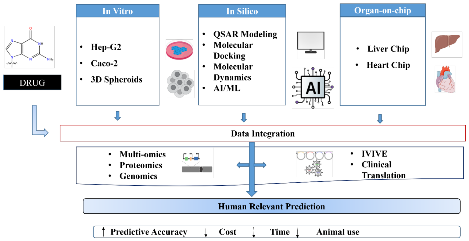
Table 4.
Integrated testing strategy (ITS) frameworks combining alternative methods for pharmacological safety assessment.
Table 4.
Integrated testing strategy (ITS) frameworks combining alternative methods for pharmacological safety assessment.
| Safety Endpoint | Tier 1 (In Silico) | Tier 2 (In Vitro 2D/HTS) | Tier 3 (Advanced In Vitro / OoC) | Regulatory Framework | Validation Status | Key References |
| DILI | QSAR structural alerts (DEREK Nexus, TOPKAT) | HepG2/HepaRG HCS; mitochondrial toxicity; lipid accumulation | Liver organoids; Liver-on-chip (Emulate, Mimetas) | HESI DILI MPS consortium; FDA ISTAND | 87% PPV in 61-drug prospective study | (Ewart et al., 2022; Proctor et al., 2017) |
| Cardiotoxicity (Proarrhythmia) | hERG QSAR; CiPA ORd AP model; Silico-pharmacology | hERG patch-clamp; NaV/CaV channel assays | iPSC-CM MEA; Heart-on-chip (Emulate, TissUse) | ICH S7B revised guideline; CiPA Initiative (FDA/HESI) | FDA accepted in IND submissions (2022+) | (Fermini et al., 2016b; Gintant et al., 2016) |
| Nephrotoxicity | QSAR OAT/OCT transport; known nephrotoxicant alerts | HK-2 cells; Primary PTECs; LDH, KIM-1, NGAL | Kidney-on-chip (Harvard Wyss); Kidney organoids | MPS Qualification pathway (FDA/EMA) | Ongoing validation (14-drug study) | (Jang et al., 2013; Wilmer et al., 2016) |
| BBB Penetration & CNS Safety | Lipinski Ro5; AD–ME QSAR; MDCK-MDR1 Papp in silico | PAMPA-BBB; MDCK-MDR1; P-gp substrate assay | BBB-on-chip (NVU model); Cerebral organoids | Exploratory regulatory use; Not yet qualified | Predictive for P-gp substrates confirmed | (Booth & Kim, 2012; Wang et al., 2016) |
| Skin Sensitisation | QSAR (DEREK, TOPKAT); DANN, TIMES SS | DPRA (in chemico); KeratinoSens (ARE) | h-CLAT; U-SENS; LuSens assay | OECD TG 442C/D/E; EU cosmetics ban on animal tests | Fully validated; Globally accepted in regulatory submissions | (Ewart et al., 2022) |
| Genotoxicity | QSAR mutagenicity (Ames, MN prediction) | In vitro Ames (TA98/100); Micronucleus (OECD 487) | 3D reconstructed skin genotoxicity (EpiDermGTL) | ICH S2(R1); OECD TG 487, 489 | Ames and MN in vitro globally accepted | (Andre et al., 2010; Kirkland et al., 2007) |
Note. DILI = drug-induced liver injury; PPV = positive predictive value; CiPA = Comprehensive In Vitro Proarrhythmia Assay; MEA = multi-electrode array; PTEC = proximal tubule epithelial cell; KIM-1 = kidney injury molecule-1; NGAL = neutrophil gelatinase-associated lipocalin; DPRA = direct peptide reactivity assay; ARE = antioxidant response element; h-CLAT = human cell line activation test; MN = micronucleus.
18. Regulatory Landscape and Acceptance of Alternative Methods
18.1. United States: FDA and Legislative Framework
The United States is the most progressive regulatory jurisdiction in formally accepting non-animal pharmacological testing methods. The pivotal FDA Modernization Act 2.0 (enacted December 2022 as part of Public Law 117-328) removed the longstanding statutory requirement that IND applications include data from animal studies, formally replacing “preclinical tests (including tests on animals)" with the broader term "nonclinical tests,” explicitly encompassing cell-based assays, organ chips, computer modelling, and other NAMs(Stewart et al., 2023).
The FDA's Innovative Science and Technology Approaches for New Drugs (ISTAND) pilot program provides a formal regulatory pathway for the qualification of novel methodologies, including MPS. A Drug Development Tool (DDT) submitted through ISTAND must demonstrate biological plausibility, technical feasibility, and clinical relevance of use, followed by formal qualification through a structured evidentiary review. The first ISTAND submission for an OoC system, the Emulate liver chip for DILI assessment, received a qualification decision in September 2024, establishing the precedent for chip-based clinical decision tools(Cook et al., 2014).
18.2. European Union: EMA and ECVAM
The European Medicines Agency (EMA) provides specific guidance on the qualification of Novel Methodologies for Human Medicines Development (EMA/CHMP/SAWP/72894/2008), a pathway enabling sponsors to request formal scientific advice and qualification opinions on novel biomarkers, methodologies, and surrogate endpoints. EMA has issued positive qualification opinions for PBPK modelling (2018), several in vitro genotoxicity assays (micronucleus, Ames), and is actively evaluating MPS qualification packages submitted through the ECVAM–EURL partnership(European Commission. Joint Research Centre., 2018; Paul et al., 2025b).
The European Union Reference Laboratory for Alternatives to Animal Testing (EURL ECVAM) coordinates validation and regulatory acceptance of alternative methods within the EU framework. ECVAM's scientific advisory committee (ESAC) provides independent validation opinions that, when endorsed, lead to the development of OECD Test Guidelines. Validated methods include the KeratinoSens assay (OECD TG 442D), the Human Cell Line Activation Test (h-CLAT, OECD TG 442E), and multiple in vitro genotoxicity assays. (Ashikaga et al., 2010; Leist, 2014; Leist et al., 2017)
18.3. International Harmonisation: ICH and OECD
The International Council for Harmonisation of Technical Requirements for Pharmaceuticals for Human Use (ICH) provides globally harmonised safety guidelines that govern preclinical testing requirements across the US, EU, Japan, Canada, China, and Australia. Relevant ICH guidelines include S7A and S7B (safety pharmacology, cardiac risk), M3(R2) (nonclinical safety for clinical trials), S2(R1) (genotoxicity), and S5(R3) (reproductive toxicity). While several ICH guidelines retain traditional animal study requirements, ongoing revision processes (S7B R2 incorporating CiPA, M3 revision accommodating NAMs) are progressively creating regulatory space for alternatives. (Food and Drug Administration, HHS, 2010; Friedrichs et al., 2005) Table 5.
19. Challenges and Current Limitations
19.1. Technical and Biological Challenges
Despite rapid advances, each alternative technology platform retains significant technical limitations that constrain its pharmacological utility and regulatory acceptance. OoC systems face persistent challenges in standardising fabrication protocols, cell seeding conditions, and flow parameters across different laboratories and chip designs, resulting in substantial interlaboratory variability that undermines cross-study comparison and regulatory confidence. The absence of innervation, adaptive immune responses (only select immune cells can be incorporated), and systemic hormonal regulation in all current OoC configurations represents a fundamental biological limitation that precludes modelling of immune-mediated hypersensitivity reactions, hormone-dependent drug effects, and neuroendocrine pharmacology(Bhise et al., 2016; Ewart et al., 2022).
iPSC-derived organ models retain fetal-like maturation phenotypes that substantially affect pharmacological relevance. iPSC-derived cardiomyocytes exhibit neonatal-like electrophysiology with slower conduction velocity, higher automaticity, and distinct calcium-handling proteins compared with adult human cardiomyocytes, potentially misrepresenting the effects of drugs that specifically target adult cardiac phenotypes. iPSC-hepatocytes express foetal-predominant CYP3A7 rather than adult CYP3A4 and demonstrate lower albumin secretion and phase II conjugation activity than primary human hepatocytes. (Ogawa et al., 2013; Ronaldson-Bouchard & Vunjak-Novakovic, 2018)
19.2. Standardisation and Reproducibility
Standardisation represents the single most critical unresolved challenge for the regulatory adoption of OoC technologies. A 2018 inter-laboratory comparison study involving five independent research groups using nominally identical liver chip designs found coefficients of variation exceeding 40% for key hepatotoxicity endpoints, including interleukin-6 secretion, CYP3A4 activity, and cellular ATP content, with variability that substantially exceeds the tolerance of regulatory data quality standards(Bale et al., 2014; Ewart et al., 2022).
International standards bodies, including ISO (Technical Committee TC276 on Biotechnology) and ASTM International (Committee E56 on Nanotechnology), have initiated working groups to develop standardised characterisation protocols for MPS, covering cell seeding procedures, flow parameter specification, biosensor calibration, and data reporting formats. ASTM standard WK85009 (Standard Guide for Characterization of Microphysiological Systems) is currently in active development, with regulatory agency input from FDA and EMA.
19.3. Economic and Infrastructure Barriers
The capital investment required for OoC technology adoption, including chip fabrication equipment (cleanroom access or contract manufacturing), specialised microfluidic pumping systems, integrated biosensor apparatus, and high-content imaging infrastructure, represents a substantial barrier for small and medium-sized pharmaceutical enterprises and academic institutions, particularly in low- and middle-income countries. Estimates suggest that a fully equipped MPS laboratory requires an initial investment of USD 500,000–1,500,000, with recurring consumable costs of USD 50,000–200,000 per year for active research programmes(Mastrangeli et al., 2019).
19.4. Data and Translational Gaps
A fundamental challenge that limits regulatory confidence in alternative method predictions is the paucity of prospectively collected human clinical outcome datasets for validation. Most historical clinical safety databases (adverse drug reaction reports, Phase I/II trial results, post-market pharmacovigilance data) do not include paired in vitro OoC or in silico data for the same compounds collected under standardised conditions, thereby limiting retrospective validation studies by selection bias and data heterogeneity. (Hartung, 2013; Walker et al., 2020)
Table 6.
Key challenges and limitations across alternative method categories for pharmacological evaluation.
Table 6.
Key challenges and limitations across alternative method categories for pharmacological evaluation.
| Challenge Category | In Vitro Models | In Silico Methods | Organ-on-Chip / MPS | Potential Solutions / Status |
| Biological Relevance | Fetal-like iPSC maturation; No immune/endocrine; Loss of tissue architecture | Applicability domain; Out-of-distribution scaffolds; Single-species training data | No innervation; No adaptive immune system; No hormonal regulation | Maturation protocols; Co-culture strategies; Immune-competent chips |
| Standardisation | Variable cell lots; Passage number effects; Media formulation variability | Model version changes; Descript. set differences; Validation dataset heterogeneity | Chip-to-chip variability; Inter-lab >40% CV; Non-standard protocols | ASTM WK85009; ISO TC276; Reference standards cells |
| Throughput | Organoids: low throughput; Expensive per well; Manual scoring required | High throughput achieved; Bottleneck: data quality; Model training cost | Low throughput per chip; High manual operation; Limited parallelization | Automated OoC platforms; Intelligent sampling; Microplate-format chips |
| Regulatory Acceptance | Organoids: not formally qualified; Accepted as supportive data | QSAR/PBPK: mature acceptance; AI: emerging; no formal TG for deep learning | Only liver chip formally qualified (US, 2024); Other organs in process | ISTAND submissions; EMA qualification pathway; Consortium validation studies |
| Data Validation | Limited prospective human outcome datasets; Selection bias in validation | Curated database gaps; Public data quality issues; Chemical space coverage | Insufficient prospective clinical correlation studies; Small compound sets tested | Consortium data sharing; Open pharmacovigilance databases; NIH/FDA grants |
| Economic Barriers | iPSC differentiation cost; Organoid culture cost; Specialised reagents | Relatively low cost; Main barrier: expertise and software licensing | High capital investment (USD 500K–1.5M); Consumable costs | Open-source platforms; 3D bioprinted chips; Public-private consortia |
| Ethical / Legal | Human donor tissue access; Stem cell regulation; Data privacy (patient iPSCs) | IP around AI models; Data ownership; GDPR compliance for patient data | IP fragmentation across chip suppliers; Cell IP; Multi-stakeholder ownership | Standardised material transfer agreements; Open-source chip designs |
Note. iPSC = induced pluripotent stem cell; CV = coefficient of variation; QSAR = quantitative structure–activity relationship; PBPK = physiologically based pharmacokinetic; TG = test guideline; MPS = microphysiological system; ASTM = American Society for Testing and Materials; ISO = International Organization for Standardization; IP = intellectual property; GDPR = General Data Protection Regulation.
20. Future Directions
20.1. Vascularised, Innervated, and Immune-Competent Chips
The next generation of OoC platforms will overcome the current biological completeness gap by integrating vascularisation (perfused capillary networks formed by endothelial self-assembly or bioprinting), innervation (co-culture with iPSC-derived peripheral neurons or dorsal root ganglia explants), and adaptive immune components (T-cells, B-cells, dendritic cells from matched donors). Vascularised brain organoids incorporating functional cortical networks and iPSC-derived microvascular endothelial cells have already been demonstrated to exhibit improved maturation and drug sensitivity profiles compared with avascular organoids (Lancaster et al., 2013b; Pham et al., 2018).
20.2. AI-Powered Autonomous Drug Discovery
The convergence of AI-driven molecular design, automated laboratory systems (lab-on-a-robot), and OoC testing is producing the first fully autonomous closed-loop drug discovery pipelines. Systems, including those developed by Insilico Medicine, Recursion Pharmaceuticals, and Exscientia, can generate novel molecular structures through generative adversarial networks or reinforcement learning, route them to automated synthesis, screen pharmacological activity using HTS platforms, and iteratively optimise structures based on SAR and ADMET feedback without human intervention at each cycle. The first AI-designed drug candidate (INS018_055 for idiopathic pulmonary fibrosis, Insilico Medicine) entered Phase II clinical trials in 2023, validating the translational potential of autonomous AI drug discovery (Schneider, 2018; Zhavoronkov et al., 2019).
20.3. Digital Twins for Virtual Clinical Trials
As OoC platforms generate increasingly comprehensive human pharmacological datasets, their integration with patient genomic data, electronic health records, and medical imaging through digital twin frameworks will enable the simulation of virtual patient cohorts for in silico clinical trial design. Regulatory agencies have expressed interest in accepting virtual control arms in which historical patient data or digital twin simulations replace placebo control groups to reduce clinical trial size and patient burden, particularly in rare disease indications where recruitment is inherently limited(Corral-Acero et al., 2020b).
20.4. Democratisation of Alternative Technologies
Ensuring global equity in access to alternative testing technologies is a critical challenge for the international research community. Progressive reduction in OoC fabrication costs through advances in 3D bioprinting, tape-based microfluidics, and injection-molded thermoplastic chip manufacturing is making these platforms progressively accessible to academic institutions and pharmaceutical enterprises in low- and middle-income countries, including India, Brazil, and South Africa, where pharmaceutical manufacturing capacity is substantial but advanced preclinical capabilities remain limited(Luni et al., 2014).
20.5. Regulatory Science Investment
The full realisation of the alternative methods paradigm shift requires sustained, coordinated investment in regulatory science, the discipline of developing and validating the tools, standards, and approaches that advance regulatory decision-making. Current regulatory science initiatives including the FDA’s National Center for Toxicological Research (NCTR) MPS programme, the NIH’s National Center for Advancing Translational Sciences (NCATS) Tissue Chip programme, and the EU’s HORIZON Europe PARC (Partnership for the Assessment of Risks from Chemicals) consortium represent the largest coordinated investments in alternative method development in history, collectively allocating over USD 500 million to developing and qualifying NAMs through 2030(Herzler et al., 2025; Modi et al., 2025).
21. Conclusions
In conclusion, preclinical pharmacological evaluation is rapidly transitioning from traditional animal models to human-relevant alternative approaches. In vitro systems, including 3D organoids and iPSC-derived models, provide improved physiological relevance, while in silico tools such as QSAR, PBPK modelling, and artificial intelligence enable rapid and cost-effective prediction of drug behaviour. Organ-on-chip technologies further enhance translational accuracy by mimicking the dynamic environments of human organs. However, no single method can fully replace animal studies; thus, integrated testing strategies that combine multiple approaches offer the most reliable framework for assessing safety and efficacy. Despite challenges related to standardisation, biological complexity, and validation, recent regulatory advancements highlight growing acceptance of these methodologies. Overall, the convergence of advanced experimental and computational tools marks a significant step toward a more ethical, predictive, and human-centric drug development paradigm, aligning with the principles of the 3Rs and improving translational success.
Authors contributions
Writing–original draft: J.H.P.; Conceptualisation: J.H.P.; Investigation: J.H.P., R.A.; Writing–review & editing: J.H.P., R.A., C.C., S.W., O.M.S., A.B.; Visualisation: S.W.; O.M.S.; R.A.; All authors have read and agreed to the published version of the manuscript.
Ethics statement
No direct interactions with human or animal subjects were involved. Therefore, ethical approval and informed consent were not required.
Funding
No funding or sponsorship was received for this study.
Data Availability statement
Not Applicable.
Acknowledgments
The authors would like to express their gratitude to Ojaswi Scientific Research Services for their invaluable advice and assistance during the creation of this publication.
Conflicts of Interest
Authors declare no conflict of interest.
Consent for publication: Not Applicable.
References
- Abraham, M. J.; Murtola, T.; Schulz, R.; Páll, S.; Smith, J. C.; Hess, B.; Lindahl, E. GROMACS: High-performance molecular simulations through multi-level parallelism from laptops to supercomputers. SoftwareX 2015a, 1–2, 19–25. [Google Scholar] [CrossRef]
- Abraham, M. J.; Murtola, T.; Schulz, R.; Páll, S.; Smith, J. C.; Hess, B.; Lindahl, E. GROMACS: High performance molecular simulations through multi-level parallelism from laptops to supercomputers. SoftwareX 2015b, 1–2, 19–25. [Google Scholar] [CrossRef]
- Andre, C.; Larondelle, Y.; Evers, D. Dietary Antioxidants and Oxidative Stress from a Human and Plant Perspective: A Review. Curr. Nutr. Food Sci. 2010, 6(1), 2–12. [Google Scholar] [CrossRef]
- Ashikaga, T.; Sakaguchi, H.; Sono, S.; Kosaka, N.; Ishikawa, M.; Nukada, Y.; Miyazawa, M.; Ito, Y.; Nishiyama, N.; Itagaki, H. A Comparative Evaluation of In Vitro Skin Sensitisation Tests: The Human Cell-line Activation Test (h-CLAT) versus the Local Lymph Node Assay (LLNA). Altern. To Lab. Anim. 2010, 38(4), 275–284. [Google Scholar] [CrossRef] [PubMed]
- Astashkina, A.; Mann, B.; Grainger, D. W. A critical evaluation of in vitro cell culture models for high-throughput drug screening and toxicity. Pharmacol. Ther. 2012, 134(1), 82–106. [Google Scholar] [CrossRef] [PubMed]
- Bale, S. S.; Vernetti, L.; Senutovitch, N.; Jindal, R.; Hegde, M.; Gough, A.; McCarty, W. J.; Bakan, A.; Bhushan, A.; Shun, T. Y.; Golberg, I.; DeBiasio, R.; Usta, O. B.; Taylor, D. L.; Yarmush, M. L. In vitro platforms for evaluating liver toxicity. Exp. Biol. Med. 2014, 239(9), 1180–1191. [Google Scholar] [CrossRef] [PubMed]
- Balls, M. Alternatives to Laboratory Animals: Trends in Replacement and the Three Rs. Altern. To Lab. Anim. 2022, 50(1), 10–26. [Google Scholar] [CrossRef] [PubMed]
- Barber, C.; Heghes, C.; Johnston, L. A framework to support the application of the OECD guidance documents on (Q)SAR model validation and prediction assessment for regulatory decisions. Comput. Toxicol. 2024, 30, 100305. [Google Scholar] [CrossRef]
- Baxter, M.; Withey, S.; Harrison, S.; Segeritz, C.-P.; Zhang, F.; Atkinson-Dell, R.; Rowe, C.; Gerrard, D. T.; Sison-Young, R.; Jenkins, R.; Henry, J.; Berry, A. A.; Mohamet, L.; Best, M.; Fenwick, S. W.; Malik, H.; Kitteringham, N. R.; Goldring, C. E.; Piper Hanley, K.; …; Hanley, N. A. Phenotypic and functional analyses show stem cell-derived hepatocyte-like cells better mimic fetal rather than adult hepatocytes. J. Hepatol. 2015, 62(3), 581–589. [Google Scholar] [CrossRef] [PubMed]
- Benam, K. H.; Villenave, R.; Lucchesi, C.; Varone, A.; Hubeau, C.; Lee, H.-H.; Alves, S. E.; Salmon, M.; Ferrante, T. C.; Weaver, J. C.; Bahinski, A.; Hamilton, G. A.; Ingber, D. E. Small airway-on-a-chip enables analysis of human lung inflammation and drug responses in vitro. Nat. Methods 2016, 13(2), 151–157. [Google Scholar] [CrossRef] [PubMed]
- Berridge, M. V.; Herst, P. M.; Tan, A. S. Tetrazolium dyes as tools in cell biology: New insights into their cellular reduction. In Biotechnology Annual Review; Elsevier, 2005; Vol. 11, pp. 127–152. [Google Scholar] [CrossRef] [PubMed]
- Bersini, S.; Jeon, J. S.; Dubini, G.; Arrigoni, C.; Chung, S.; Charest, J. L.; Moretti, M.; Kamm, R. D. A microfluidic 3D in vitro model for specificity of breast cancer metastasis to bone. Biomaterials 2014, 35(8), 2454–2461. [Google Scholar] [CrossRef] [PubMed]
- Bessems, M.; T Hart, N. A.; Tolba, R.; Doorschodt, B. M.; Leuvenink, H. G. D.; Ploeg, R. J.; Minor, T.; Van Gulik, T. M. The isolated perfused rat liver: Standardization of a time-honoured model. Lab. Anim. 2006, 40(3), 236–246. [Google Scholar] [CrossRef] [PubMed]
- Bhatia, S. N.; Ingber, D. E. Microfluidic organs-on-chips. Nat. Biotechnol. 2014, 32(8), 760–772. [Google Scholar] [CrossRef] [PubMed]
- Bhise, N. S.; Manoharan, V.; Massa, S.; Tamayol, A.; Ghaderi, M.; Miscuglio, M.; Lang, Q.; Shrike Zhang, Y.; Shin, S. R.; Calzone, G.; Annabi, N.; Shupe, T. D.; Bishop, C. E.; Atala, A.; Dokmeci, M. R.; Khademhosseini, A. A liver-on-a-chip platform with bioprinted hepatic spheroids. Biofabrication 2016, 8(1), 014101. [Google Scholar] [CrossRef] [PubMed]
- Björnsson, B.; Borrebaeck, C.; Elander, N.; Gasslander, T.; Gawel, D. R.; Gustafsson, M.; Jörnsten, R.; Lee, E. J.; Li, X.; Lilja, S.; Martínez-Enguita, D.; Matussek, A.; Sandström, P.; Schäfer, S.; Stenmarker, M.; Sun, X. F.; Sysoev, O.; Zhang, H.; Benson, M. Digital twins to personalize medicine. Genome Med. 2020, 12(1), 4. [Google Scholar] [CrossRef] [PubMed]
- Booth, R.; Kim, H. Characterization of a microfluidic in vitro model of the blood-brain barrier (μBBB). Lab A Chip 2012, 12(10), 1784. [Google Scholar] [CrossRef] [PubMed]
- Case, D. A.; Cheatham, T. E.; Darden, T.; Gohlke, H.; Luo, R.; Merz, K. M.; Onufriev, A.; Simmerling, C.; Wang, B.; Woods, R. J. The Amber biomolecular simulation programs. J. Comput. Chem. 2005a, 26(16), 1668–1688. [Google Scholar] [CrossRef] [PubMed]
- Case, D. A.; Cheatham, T. E.; Darden, T.; Gohlke, H.; Luo, R.; Merz, K. M.; Onufriev, A.; Simmerling, C.; Wang, B.; Woods, R. J. The Amber biomolecular simulation programs. J. Comput. Chem. 2005b, 26(16), 1668–1688. [Google Scholar] [CrossRef] [PubMed]
- Chauhan, A.; Semwal, D.; Mishra, S.; Semwal, R. Ayurvedic research and methodology: Present status and future strategies. AYU 2015, 36(4), 364. [Google Scholar] [CrossRef] [PubMed]
- Chen, H.; Engkvist, O.; Wang, Y.; Olivecrona, M.; Blaschke, T. The rise of deep learning in drug discovery. Drug Discov. Today 2018, 23(6), 1241–1250. [Google Scholar] [CrossRef] [PubMed]
- Chen, M.; Bisgin, H.; Tong, L.; Hong, H.; Fang, H.; Borlak, J.; Tong, W. Toward Predictive Models for Drug-Induced Liver Injury in Humans: Are we There Yet? Biomark. Med. 2014, 8(2), 201–213. [Google Scholar] [CrossRef] [PubMed]
- Cherkasov, A.; Muratov, E. N.; Fourches, D.; Varnek, A.; Baskin, I. I.; Cronin, M.; Dearden, J.; Gramatica, P.; Martin, Y. C.; Todeschini, R.; Consonni, V.; Kuz’min, V. E.; Cramer, R.; Benigni, R.; Yang, C.; Rathman, J.; Terfloth, L.; Gasteiger, J.; Richard, A.; Tropsha, A. QSAR Modeling: Where Have You Been? Where Are You Going To? J. Med. Chem. 2014, 57(12), 4977–5010. [Google Scholar] [CrossRef] [PubMed]
- Clevers, H. Modeling Development and Disease with Organoids. Cell 2016, 165(7), 1586–1597. [Google Scholar] [CrossRef] [PubMed]
- Colatsky, T.; Fermini, B.; Gintant, G.; Pierson, J. B.; Sager, P.; Sekino, Y.; Strauss, D. G.; Stockbridge, N. The Comprehensive in Vitro Proarrhythmia Assay (CiPA) initiative—Update on progress. J. Pharmacol. Toxicol. Methods 2016, 81, 15–20. [Google Scholar] [CrossRef] [PubMed]
- Commissioner; O. of the. FDA Announces Plan to Phase Out Animal Testing Requirement for Monoclonal Antibodies and Other Drugs. FDA. May 2025. Available online: https://www.fda.gov/news-events/press-announcements/fda-announces-plan-phase-out-animal-testing-requirement-monoclonal-antibodies-and-other-drugs.
- Commissioner, O. (Ed.) Modernization of Cosmetics Regulation Act of 2022 (MoCRA); FDA, March 2026; Available online: https://www.fda.gov/cosmetics/cosmetics-laws-regulations/modernization-cosmetics-regulation-act-2022-mocra.
- Cook, D.; Brown, D.; Alexander, R.; March, R.; Morgan, P.; Satterthwaite, G.; Pangalos, M. N. Lessons learned from the fate of AstraZeneca’s drug pipeline: A five-dimensional framework. Nat. Rev. Drug Discov. 2014, 13(6), 419–431. [Google Scholar] [CrossRef] [PubMed]
- Corral-Acero, J.; Margara, F.; Marciniak, M.; Rodero, C.; Loncaric, F.; Feng, Y.; Gilbert, A.; Fernandes, J. F.; Bukhari, H. A.; Wajdan, A.; Martinez, M. V.; Santos, M. S.; Shamohammdi, M.; Luo, H.; Westphal, P.; Leeson, P.; DiAchille, P.; Gurev, V.; Mayr, M.; …; Lamata, P. The ‘Digital Twin’ to enable the vision of precision cardiology. Eur. Heart J. 2020a, 41(48), 4556–4564. [Google Scholar] [CrossRef] [PubMed]
- Corral-Acero, J.; Margara, F.; Marciniak, M.; Rodero, C.; Loncaric, F.; Feng, Y.; Gilbert, A.; Fernandes, J. F.; Bukhari, H. A.; Wajdan, A.; Martinez, M. V.; Santos, M. S.; Shamohammdi, M.; Luo, H.; Westphal, P.; Leeson, P.; DiAchille, P.; Gurev, V.; Mayr, M.; …; Lamata, P. The ‘Digital Twin’ to enable the vision of precision cardiology. Eur. Heart J. 2020b, 41(48), 4556–4564. [Google Scholar] [CrossRef] [PubMed]
- De Graaf, I. A. M.; Olinga, P.; De Jager, M. H.; Merema, M. T.; De Kanter, R.; Van De Kerkhof, E. G.; Groothuis, G. M. M. Preparation and incubation of precision-cut liver and intestinal slices for application in drug metabolism and toxicity studies. Nat. Protoc. 2010, 5(9), 1540–1551. [Google Scholar] [CrossRef] [PubMed]
- Deckers, J. The New EU Directive on the Use of Animals for Research and the Value of Moral Consistency. J. Bioethical Inq. 2012, 9(4), 377–379. [Google Scholar] [CrossRef] [PubMed]
- Doherty, K. R.; Wappel, R. L.; Talbert, D. R.; Trusk, P. B.; Moran, D. M.; Kramer, J. W.; Brown, A. M.; Shell, S. A.; Bacus, S. Multi-parameter in vitro toxicity testing of crizotinib, sunitinib, erlotinib, and nilotinib in human cardiomyocytes. Toxicol. Appl. Pharmacol. 2013, 272(1), 245–255. [Google Scholar] [CrossRef] [PubMed]
- Dunn, J. C. Y.; Yarmush, M. L.; Koebe, H. G.; Tompkins, R. G. Hepatocyte function and extracellular matrix geometry: Long-term culture in a sandwich configuration. FASEB J. 1989, 3(2), 174–177. [Google Scholar] [CrossRef] [PubMed]
- Duvenaud, D.; Maclaurin, D.; Aguilera-Iparraguirre, J.; Gómez-Bombarelli, R.; Hirzel, T.; Aspuru-Guzik, A.; Adams, R. P. Convolutional Networks on Graphs for Learning Molecular Fingerprints; arXiv, 2015. [Google Scholar] [CrossRef]
- Edington, C. D.; Chen, W. L. K.; Geishecker, E.; Kassis, T.; Soenksen, L. R.; Bhushan, B. M.; Freake, D.; Kirschner, J.; Maass, C.; Tsamandouras, N.; Valdez, J.; Cook, C. D.; Parent, T.; Snyder, S.; Yu, J.; Suter, E.; Shockley, M.; Velazquez, J.; Velazquez, J. J.; …; Griffith, L. G. Interconnected Microphysiological Systems for Quantitative Biology and Pharmacology Studies. Sci. Rep. 2018, 8(1), 4530. [Google Scholar] [CrossRef] [PubMed]
- European Commission. Joint Research Centre. EURL ECVAM status report on the development, validation and regulatory acceptance of alternative methods and approaches; Publications Office, 2018. [Google Scholar] [CrossRef]
- Ewart, L.; Apostolou, A.; Briggs, S. A.; Carman, C. V.; Chaff, J. T.; Heng, A. R.; Jadalannagari, S.; Janardhanan, J.; Jang, K.-J.; Joshipura, S. R.; Kadam, M. M.; Kanellias, M.; Kujala, V. J.; Kulkarni, G.; Le, C. Y.; Lucchesi, C.; Manatakis, D. V.; Maniar, K. K.; Quinn, M. E.; …; Levner, D. Performance assessment and economic analysis of a human Liver-Chip for predictive toxicology. Commun. Med. 2022, 2(1), 154. [Google Scholar] [CrossRef] [PubMed]
- Fermini, B.; Hancox, J. C.; Abi-Gerges, N.; Bridgland-Taylor, M.; Chaudhary, K. W.; Colatsky, T.; Correll, K.; Crumb, W.; Damiano, B.; Erdemli, G.; Gintant, G.; Imredy, J.; Koerner, J.; Kramer, J.; Levesque, P.; Li, Z.; Lindqvist, A.; Obejero-Paz, C. A.; Rampe, D.; …; Vandenberg, J. I. A New Perspective in the Field of Cardiac Safety Testing through the Comprehensive In Vitro Proarrhythmia Assay Paradigm. SLAS Discov. 2016a, 21(1), 1–11. [Google Scholar] [CrossRef] [PubMed]
- Fermini, B.; Hancox, J. C.; Abi-Gerges, N.; Bridgland-Taylor, M.; Chaudhary, K. W.; Colatsky, T.; Correll, K.; Crumb, W.; Damiano, B.; Erdemli, G.; Gintant, G.; Imredy, J.; Koerner, J.; Kramer, J.; Levesque, P.; Li, Z.; Lindqvist, A.; Obejero-Paz, C. A.; Rampe, D.; …; Vandenberg, J. I. A New Perspective in the Field of Cardiac Safety Testing through the Comprehensive In Vitro Proarrhythmia Assay Paradigm. SLAS Discov. 2016b, 21(1), 1–11. [Google Scholar] [CrossRef]
- Festing, S.; Wilkinson, R. The ethics of animal research: Talking Point on the use of animals in scientific research. EMBO Rep. 2007, 8(6), 526–530. [Google Scholar] [CrossRef] [PubMed]
- Fjodorova, N.; Vračko, M.; Tušar, M.; Jezierska, A.; Novič, M.; Kühne, R.; Schüürmann, G. Quantitative and qualitative models for carcinogenicity prediction for non-congeneric chemicals using CP ANN method for regulatory uses. Mol. Divers. 2010, 14(3), 581–594. [Google Scholar] [CrossRef] [PubMed]
- Food and Drug Administration. International Conference on Harmonisation; Guidance on M3(R2) Nonclinical Safety Studies for the Conduct of Human Clinical Trials and Marketing Authorization for Pharmaceuticals; availability. Not. Fed. Regist. 2010, 75(13), 3471–3472. [Google Scholar]
- Franks, M. E.; Macpherson, G. R.; Figg, W. D. Thalidomide. The Lancet 2004, 363(9423), 1802–1811. [Google Scholar] [CrossRef]
- Friedrichs, G. S.; Patmore, L.; Bass, A. Non-clinical evaluation of ventricular repolarization (ICH S7B): Results of an interim survey of international pharmaceutical companies. J. Pharmacol. Toxicol. Methods 2005, 52(1), 6–11. [Google Scholar] [CrossRef] [PubMed]
- Friesner, R. A.; Murphy, R. B.; Repasky, M. P.; Frye, L. L.; Greenwood, J. R.; Halgren, T. A.; Sanschagrin, P. C.; Mainz, D. T. Extra precision glide: Docking and scoring incorporating a model of hydrophobic enclosure for protein-ligand complexes. J. Med. Chem. 2006, 49(21), 6177–6196. [Google Scholar] [CrossRef] [PubMed]
- Gintant, G.; Sager, P. T.; Stockbridge, N. Evolution of strategies to improve preclinical cardiac safety testing. Nat. Rev. Drug Discov. 2016, 15(7), 457–471. [Google Scholar] [CrossRef] [PubMed]
- Godoy, P.; Hewitt, N. J.; Albrecht, U.; Andersen, M. E.; Ansari, N.; Bhattacharya, S.; Bode, J. G.; Bolleyn, J.; Borner, C.; Böttger, J.; Braeuning, A.; Budinsky, R. A.; Burkhardt, B.; Cameron, N. R.; Camussi, G.; Cho, C.-S.; Choi, Y.-J.; Craig Rowlands, J.; Dahmen, U.; …; Hengstler, J. G. Recent advances in 2D and 3D in vitro systems using primary hepatocytes, alternative hepatocyte sources and non-parenchymal liver cells and their use in investigating mechanisms of hepatotoxicity, cell signaling and ADME. Arch. Toxicol. 2013, 87(8), 1315–1530. [Google Scholar] [CrossRef] [PubMed]
- Guillouzo, A.; Corlu, A.; Aninat, C.; Glaise, D.; Morel, F.; Guguen-Guillouzo, C. The human hepatoma HepaRG cells: A highly differentiated model for studies of liver metabolism and toxicity of xenobiotics. Chem.-Biol. Interact. 2007, 168(1), 66–73. [Google Scholar] [CrossRef] [PubMed]
- Hallifax, D.; Houston, J. B. Evaluation of Hepatic Clearance Prediction Using In Vitro Data: Emphasis on Fraction Unbound in Plasma and Drug Ionisation Using a Database of 107 Drugs. J. Pharm. Sci. 2012, 101(8), 2645–2652. [Google Scholar] [CrossRef] [PubMed]
- Hansch, Corwin.; Fujita, Toshio. p -σ-π Analysis. A Method for the Correlation of Biological Activity and Chemical Structure. J. Am. Chem. Soc. 1964, 86(8), 1616–1626. [Google Scholar] [CrossRef]
- Harrison, R. K. Phase II and phase III failures: 2013–2015. Nat. Rev. Drug Discov. 2016, 15(12), 817–818. [Google Scholar] [CrossRef] [PubMed]
- Hartung, T. Toxicology for the twenty-first century. Nature 2009, 460(7252), 208–212. [Google Scholar] [CrossRef] [PubMed]
- Hartung, T. Look back in anger – what clinical studies tell us about preclinical work. ALTEX 2013, 30(3), 275–291. [Google Scholar] [CrossRef] [PubMed]
- Herland, A.; Maoz, B. M.; Das, D.; Somayaji, M. R.; Prantil-Baun, R.; Novak, R.; Cronce, M.; Huffstater, T.; Jeanty, S. S. F.; Ingram, M.; Chalkiadaki, A.; Benson Chou, D.; Marquez, S.; Delahanty, A.; Jalili-Firoozinezhad, S.; Milton, Y.; Sontheimer-Phelps, A.; Swenor, B.; Levy, O.; …; Ingber, D. E. Quantitative prediction of human pharmacokinetic responses to drugs via fluidically coupled vascularized organ chips. Nat. Biomed. Eng. 2020, 4(4), 421–436. [Google Scholar] [CrossRef] [PubMed]
- Herzler, M.; Luijten, M.; Marx-Stoelting, P.; Rivière, G. The European Partnership PARC’s role in actively promoting the uptake of new approach methodologies and next-generation risk assessment into regulatory risk assessment practice. Curr. Opin. Toxicol. 2025, 42, 100517. [Google Scholar] [CrossRef]
- Hirschhaeuser, F.; Menne, H.; Dittfeld, C.; West, J.; Mueller-Klieser, W.; Kunz-Schughart, L. A. Multicellular tumor spheroids: An underestimated tool is catching up again. J. Biotechnol. 2010, 148(1), 3–15. [Google Scholar] [CrossRef] [PubMed]
- Huh, D.; Leslie, D. C.; Matthews, B. D.; Fraser, J. P.; Jurek, S.; Hamilton, G. A.; Thorneloe, K. S.; McAlexander, M. A.; Ingber, D. E. A Human Disease Model of Drug Toxicity–Induced Pulmonary Edema in a Lung-on-a-Chip Microdevice. Sci. Transl. Med. 2012, 4(159). [Google Scholar] [CrossRef] [PubMed]
- Huh, D.; Matthews, B. D.; Mammoto, A.; Montoya-Zavala, M.; Hsin, H. Y.; Ingber, D. E. Reconstituting Organ-Level Lung Functions on a Chip. Science 2010, 328(5986), 1662–1668. [Google Scholar] [CrossRef] [PubMed]
- Jalili-Firoozinezhad, S.; Gazzaniga, F. S.; Calamari, E. L.; Camacho, D. M.; Fadel, C. W.; Bein, A.; Swenor, B.; Nestor, B.; Cronce, M. J.; Tovaglieri, A.; Levy, O.; Gregory, K. E.; Breault, D. T.; Cabral, J. M. S.; Kasper, D. L.; Novak, R.; Ingber, D. E. A complex human gut microbiome cultured in an anaerobic intestine-on-a-chip. Nat. Biomed. Eng. 2019, 3(7), 520–531. [Google Scholar] [CrossRef] [PubMed]
- Jamei, M. Recent Advances in Development and Application of Physiologically-Based Pharmacokinetic (PBPK) Models: A Transition from Academic Curiosity to Regulatory Acceptance. Curr. Pharmacol. Rep. 2016, 2(3), 161–169. [Google Scholar] [CrossRef] [PubMed]
- Jang, K.-J.; Mehr, A. P.; Hamilton, G. A.; McPartlin, L. A.; Chung, S.; Suh, K.-Y.; Ingber, D. E. Human kidney proximal tubule-on-a-chip for drug transport and nephrotoxicity assessment. Integr. Biol. 2013, 5(9), 1119–1129. [Google Scholar] [CrossRef] [PubMed]
- Jones, H.; Rowland-Yeo, K. Basic Concepts in Physiologically Based Pharmacokinetic Modeling in Drug Discovery and Development. CPT Pharmacomet. Syst. Pharmacol. 2013, 2(8), 1–12. [Google Scholar] [CrossRef] [PubMed]
- Jumper, J.; Evans, R.; Pritzel, A.; Green, T.; Figurnov, M.; Ronneberger, O.; Tunyasuvunakool, K.; Bates, R.; Žídek, A.; Potapenko, A.; Bridgland, A.; Meyer, C.; Kohl, S. A. A.; Ballard, A. J.; Cowie, A.; Romera-Paredes, B.; Nikolov, S.; Jain, R.; Adler, J.; …; Hassabis, D. Highly accurate protein structure prediction with AlphaFold. Nature 2021a, 596(7873), 583–589. [Google Scholar] [CrossRef] [PubMed]
- Jumper, J.; Evans, R.; Pritzel, A.; Green, T.; Figurnov, M.; Ronneberger, O.; Tunyasuvunakool, K.; Bates, R.; Žídek, A.; Potapenko, A.; Bridgland, A.; Meyer, C.; Kohl, S. A. A.; Ballard, A. J.; Cowie, A.; Romera-Paredes, B.; Nikolov, S.; Jain, R.; Adler, J.; …; Hassabis, D. Highly accurate protein structure prediction with AlphaFold. Nature 2021b, 596(7873), 583–589. [Google Scholar] [CrossRef] [PubMed]
- Kaur, S.; Siddiqui, H.; Bhat, M. H. Hepatic Progenitor Cells in Action. Am. J. Pathol. 2015, 185(9), 2342–2350. [Google Scholar] [CrossRef] [PubMed]
- Kavlock, R.; Chandler, K.; Houck, K.; Hunter, S.; Judson, R.; Kleinstreuer, N.; Knudsen, T.; Martin, M.; Padilla, S.; Reif, D.; Richard, A.; Rotroff, D.; Sipes, N.; Dix, D. Update on EPA’s ToxCast Program: Providing High Throughput Decision Support Tools for Chemical Risk Management. Chem. Res. Toxicol. 2012, 25(7), 1287–1302. [Google Scholar] [CrossRef] [PubMed]
- Kim, H. J.; Huh, D.; Hamilton, G.; Ingber, D. E. Human gut-on-a-chip inhabited by microbial flora that experiences intestinal peristalsis-like motions and flow. Lab A Chip 2012, 12(12), 2165. [Google Scholar] [CrossRef] [PubMed]
- Kim, J. H.; Scialli, A. R. Thalidomide: The Tragedy of Birth Defects and the Effective Treatment of Disease. Toxicol. Sci. 2011, 122(1), 1–6. [Google Scholar] [CrossRef] [PubMed]
- Kim, S.; LesherPerez, S. C.; Kim, B. C. C.; Yamanishi, C.; Labuz, J. M.; Leung, B.; Takayama, S. Pharmacokinetic profile that reduces nephrotoxicity of gentamicin in a perfused kidney-on-a-chip. Biofabrication 2016, 8(1), 015021. [Google Scholar] [CrossRef] [PubMed]
- Kirkland, D.; Pfuhler, S.; Tweats, D.; Aardema, M.; Corvi, R.; Darroudi, F.; Elhajouji, A.; Glatt, H.; Hastwell, P.; Hayashi, M.; Kasper, P.; Kirchner, S.; Lynch, A.; Marzin, D.; Maurici, D.; Meunier, J.-R.; Müller, L.; Nohynek, G.; Parry, J.; …; White, P. How to reduce false positive results when undertaking in vitro genotoxicity testing and thus avoid unnecessary follow-up animal tests: Report of an ECVAM Workshop. Mutat. Res. Toxicol. Environ. Mutagen. 2007, 628(1), 31–55. [Google Scholar] [CrossRef] [PubMed]
- Kola, I.; Landis, J. Can the pharmaceutical industry reduce attrition rates? Nat. Rev. Drug Discov. 2004, 3(8), 711–716. [Google Scholar] [CrossRef] [PubMed]
- Lancaster, M. A.; Renner, M.; Martin, C.-A.; Wenzel, D.; Bicknell, L. S.; Hurles, M. E.; Homfray, T.; Penninger, J. M.; Jackson, A. P.; Knoblich, J. A. Cerebral organoids model human brain development and microcephaly. Nature 2013a, 501(7467), 373–379. [Google Scholar] [CrossRef] [PubMed]
- Lancaster, M. A.; Renner, M.; Martin, C.-A.; Wenzel, D.; Bicknell, L. S.; Hurles, M. E.; Homfray, T.; Penninger, J. M.; Jackson, A. P.; Knoblich, J. A. Cerebral organoids model human brain development and microcephaly. Nature 2013b, 501(7467), 373–379. [Google Scholar] [CrossRef] [PubMed]
- LeCluyse, E. L.; Witek, R. P.; Andersen, M. E.; Powers, M. J. Organotypic liver culture models: Meeting current challenges in toxicity testing. Crit. Rev. Toxicol. 2012, 42(6), 501–548. [Google Scholar] [CrossRef] [PubMed]
- Leist, M. Consensus report on the future of animal-free systemic toxicity testing; ALTEX, 2014; pp. 341–356. [Google Scholar] [CrossRef] [PubMed]
- Leist, M.; Ghallab, A.; Graepel, R.; Marchan, R.; Hassan, R.; Bennekou, S. H.; Limonciel, A.; Vinken, M.; Schildknecht, S.; Waldmann, T.; Danen, E.; Van Ravenzwaay, B.; Kamp, H.; Gardner, I.; Godoy, P.; Bois, F. Y.; Braeuning, A.; Reif, R.; Oesch, F.; …; Hengstler, J. G. Adverse outcome pathways: Opportunities, limitations and open questions. Arch. Toxicol. 2017, 91(11), 3477–3505. [Google Scholar] [CrossRef] [PubMed]
- Leung, C. M.; De Haan, P.; Ronaldson-Bouchard, K.; Kim, G.-A.; Ko, J.; Rho, H. S.; Chen, Z.; Habibovic, P.; Jeon, N. L.; Takayama, S.; Shuler, M. L.; Vunjak-Novakovic, G.; Frey, O.; Verpoorte, E.; Toh, Y.-C. A guide to the organ-on-a-chip. Nat. Rev. Methods Primers 2022, 2(1), 33. [Google Scholar] [CrossRef]
- Lian, X.; Hsiao, C.; Wilson, G.; Zhu, K.; Hazeltine, L. B.; Azarin, S. M.; Raval, K. K.; Zhang, J.; Kamp, T. J.; Palecek, S. P. Robust cardiomyocyte differentiation from human pluripotent stem cells via temporal modulation of canonical Wnt signaling. Proc. Natl. Acad. Sci. 2012, 109(27). [Google Scholar] [CrossRef] [PubMed]
- Liang, P.; Lan, F.; Lee, A. S.; Gong, T.; Sanchez-Freire, V.; Wang, Y.; Diecke, S.; Sallam, K.; Knowles, J. W.; Wang, P. J.; Nguyen, P. K.; Bers, D. M.; Robbins, R. C.; Wu, J. C. Drug Screening Using a Library of Human Induced Pluripotent Stem Cell–Derived Cardiomyocytes Reveals Disease-Specific Patterns of Cardiotoxicity. Circulation 2013, 127(16), 1677–1691. [Google Scholar] [CrossRef] [PubMed]
- Lilienblum, W.; Dekant, W.; Foth, H.; Gebel, T.; Hengstler, J. G.; Kahl, R.; Kramer, P.-J.; Schweinfurth, H.; Wollin, K.-M. Alternative methods to safety studies in experimental animals: Role in the risk assessment of chemicals under the new European Chemicals Legislation (REACH). Arch. Toxicol. 2008, 82(4), 211–236. [Google Scholar] [CrossRef] [PubMed]
- Lounnas, V.; Ritschel, T.; Kelder, J.; McGuire, R.; Bywater, R. P.; Foloppe, N. CURRENT PROGRESS IN STRUCTURE-BASED RATIONAL DRUG DESIGN MARKS A NEW MINDSET IN DRUG DISCOVERY. Comput. Struct. Biotechnol. J. 2013, 5(6), e201302011. [Google Scholar] [CrossRef] [PubMed]
- Luni, C.; Serena, E.; Elvassore, N. Human-on-chip for therapy development and fundamental science. Curr. Opin. Biotechnol. 2014, 25, 45–50. [Google Scholar] [CrossRef] [PubMed]
- Martignoni, M.; Groothuis, G. M. M.; De Kanter, R. Species differences between mouse, rat, dog, monkey and human CYP-mediated drug metabolism, inhibition and induction. Expert Opin. Drug Metab. Toxicol. 2006, 2(6), 875–894. [Google Scholar] [CrossRef] [PubMed]
- Masereeuw, R.; Moons, M. M.; Toomey, B. H.; Russel, F. G. M.; Miller, D. S. Active Lucifer Yellow Secretion in Renal Proximal Tubule: Evidence for Organic Anion Transport System Crossover. J. Pharmacol. Exp. Ther. 1999, 289(2), 1104–1111. [Google Scholar] [CrossRef]
- Mastrangeli, M.; Millet, S.; Van Den Eijnden-van Raaij, J. Organ-on-chip in development: Towards a roadmap for organs-on-chip; ALTEX, 2019; pp. 650–668. [Google Scholar] [CrossRef] [PubMed]
- Mathur, A.; Loskill, P.; Shao, K.; Huebsch, N.; Hong, S.; Marcus, S. G.; Marks, N.; Mandegar, M.; Conklin, B. R.; Lee, L. P.; Healy, K. E. Human iPSC-based Cardiac Microphysiological System For Drug Screening Applications. Sci. Rep. 2015, 5(1), 8883. [Google Scholar] [CrossRef] [PubMed]
- Mayr, A.; Klambauer, G.; Unterthiner, T.; Hochreiter, S. DeepTox: Toxicity Prediction using Deep Learning. In Frontiers in Environmental Science; 2016a. [Google Scholar] [CrossRef]
- Mayr, A.; Klambauer, G.; Unterthiner, T.; Hochreiter, S. DeepTox: Toxicity Prediction using Deep Learning. In Frontiers in Environmental Science; 2016b. [Google Scholar] [CrossRef]
- Mayr, L. M.; Fuerst, P. The Future of High-Throughput Screening. SLAS Discov. 2008, 13(6), 443–448. [Google Scholar] [CrossRef] [PubMed]
- Modi, P. S.; Singh, A.; Chaturvedi, A.; Agarwal, S.; Dutta, R.; Nayak, R.; Singh, A. K. Tissue chips as headway model and incitement technology. Synth. Syst. Biotechnol. 2025, 10(1), 86–101. [Google Scholar] [CrossRef] [PubMed]
- Moretti, A.; Bellin, M.; Welling, A.; Jung, C. B.; Lam, J. T.; Bott-Flügel, L.; Dorn, T.; Goedel, A.; Höhnke, C.; Hofmann, F.; Seyfarth, M.; Sinnecker, D.; Schömig, A.; Laugwitz, K.-L. Patient-Specific Induced Pluripotent Stem-Cell Models for Long-QT Syndrome. N. Engl. J. Med. 2010, 363(15), 1397–1409. [Google Scholar] [CrossRef] [PubMed]
- Musah, S.; Mammoto, A.; Ferrante, T. C.; Jeanty, S. S. F.; Hirano-Kobayashi, M.; Mammoto, T.; Roberts, K.; Chung, S.; Novak, R.; Ingram, M.; Fatanat-Didar, T.; Koshy, S.; Weaver, J. C.; Church, G. M.; Ingber, D. E. Mature induced-pluripotent-stem-cell-derived human podocytes reconstitute kidney glomerular-capillary-wall function on a chip. Nat. Biomed. Eng. 2017, 1(5), 0069. [Google Scholar] [CrossRef] [PubMed]
- Nyman, E.; Rozendaal, Y. J. W.; Helmlinger, G.; Hamrén, B.; Kjellsson, M. C.; Strålfors, P.; Van Riel, N. A. W.; Gennemark, P.; Cedersund, G. Requirements for multi-level systems pharmacology models to reach end-usage: The case of type 2 diabetes. Interface Focus 2016, 6(2), 20150075. [Google Scholar] [CrossRef] [PubMed]
- Obach, R. S. Prediction of Human Clearance of Twenty-Nine Drugs from Hepatic Microsomal Intrinsic Clearance Data: An Examination of In Vitro Half-Life Approach and Nonspecific Binding to Microsomes. Drug Metab. Dispos. 1999, 27(11), 1350–1359. [Google Scholar] [CrossRef]
- Ogawa, S.; Surapisitchat, J.; Virtanen, C.; Ogawa, M.; Niapour, M.; Sugamori, K. S.; Wang, S.; Tamblyn, L.; Guillemette, C.; Hoffmann, E.; Zhao, B.; Strom, S.; Laposa, R. R.; Tyndale, R. F.; Grant, D. M.; Keller, G. Three-dimensional culture and cAMP signaling promote the maturation of human pluripotent stem cell-derived hepatocytes. Development 2013, 140(15), 3285–3296. [Google Scholar] [CrossRef] [PubMed]
- Olson, H.; Betton, G.; Robinson, D.; Thomas, K.; Monro, A.; Kolaja, G.; Lilly, P.; Sanders, J.; Sipes, G.; Bracken, W.; Dorato, M.; Van Deun, K.; Smith, P.; Berger, B.; Heller, A. Concordance of the Toxicity of Pharmaceuticals in Humans and in Animals. Regul. Toxicol. Pharmacol. 2000, 32(1), 56–67. [Google Scholar] [CrossRef] [PubMed]
- on behalf of the Swedish Digital Twin Consortium; Björnsson, B.; Borrebaeck, C.; Elander, N.; Gasslander, T.; Gawel, D. R.; Gustafsson, M.; Jörnsten, R.; Lee, E. J.; Li, X.; Lilja, S.; Martínez-Enguita, D.; Matussek, A.; Sandström, P.; Schäfer, S.; Stenmarker, M.; Sun, X. F.; Sysoev, O.; Zhang, H.; Benson, M. Digital twins to personalize medicine. Genome Med. 2020, 12(1), 4. [Google Scholar] [CrossRef] [PubMed]
- Oushyani Roudsari, Z.; Esmaeili, Z.; Nasirzadeh, N.; Heidari Keshel, S.; Sefat, F.; Bakhtyari, H.; Nadri, S. Microfluidics as a promising technology for personalized medicine. BioImpacts 2024, 1. [Google Scholar] [CrossRef] [PubMed]
- Pampaloni, F.; Reynaud, E. G.; Stelzer, E. H. K. The third dimension bridges the gap between cell culture and live tissue. Nat. Rev. Mol. Cell Biol. 2007, 8(10), 839–845. [Google Scholar] [CrossRef] [PubMed]
- Paul, P.; Colin, P. J.; Musuamba Tshinanu, F.; Versantvoort, C.; Manolis, E.; Blake, K. Current Use of Physiologically Based Pharmacokinetic modeling in New Medicinal Product Approvals at EMA. Clin. Pharmacol. Ther. 2025a, 117(3), 808–817. [Google Scholar] [CrossRef] [PubMed]
- Paul, P.; Colin, P. J.; Musuamba Tshinanu, F.; Versantvoort, C.; Manolis, E.; Blake, K. Current Use of Physiologically Based Pharmacokinetic modeling in New Medicinal Product Approvals at EMA. Clin. Pharmacol. Ther. 2025b, 117(3), 808–817. [Google Scholar] [CrossRef] [PubMed]
- Peng, W. C.; Logan, C. Y.; Fish, M.; Anbarchian, T.; Aguisanda, F.; Álvarez-Varela, A.; Wu, P.; Jin, Y.; Zhu, J.; Li, B.; Grompe, M.; Wang, B.; Nusse, R. Inflammatory Cytokine TNFα Promotes the Long-Term Expansion of Primary Hepatocytes in 3D Culture. Cell 2018, 175(6), 1607–1619.e15. [Google Scholar] [CrossRef] [PubMed]
- Pham, M. T.; Pollock, K. M.; Rose, M. D.; Cary, W. A.; Stewart, H. R.; Zhou, P.; Nolta, J. A.; Waldau, B. Generation of human vascularized brain organoids. NeuroReport 2018, 29(7), 588–593. [Google Scholar] [CrossRef] [PubMed]
- Poulin, P.; Theil, F.-P. Prediction of Pharmacokinetics Prior to In Vivo Studies. II. Generic Physiologically Based Pharmacokinetic Models of Drug Disposition. J. Pharm. Sci. 2002, 91(5), 1358–1370. [Google Scholar] [CrossRef] [PubMed]
- Pound, P. A sociological perspective on the challenges of displacing animal research within academia: The contribution of Bourdieu. NAM J. 2025, 1, 100057. [Google Scholar] [CrossRef] [PubMed]
- Proctor, W. R.; Foster, A. J.; Vogt, J.; Summers, C.; Middleton, B.; Pilling, M. A.; Shienson, D.; Kijanska, M.; Ströbel, S.; Kelm, J. M.; Morgan, P.; Messner, S.; Williams, D. Utility of spherical human liver microtissues for prediction of clinical drug-induced liver injury. Arch. Toxicol. 2017, 91(8), 2849–2863. [Google Scholar] [CrossRef] [PubMed]
- Ronaldson-Bouchard, K.; Vunjak-Novakovic, G. Organs-on-a-Chip: A Fast Track for Engineered Human Tissues in Drug Development. Cell Stem Cell 2018, 22(3), 310–324. [Google Scholar] [CrossRef] [PubMed]
- Roncaglioni, A.; Toropov, A. A.; Toropova, A. P.; Benfenati, E. In silico methods to predict drug toxicity. Curr. Opin. Pharmacol. 2013, 13(5), 802–806. [Google Scholar] [CrossRef] [PubMed]
- Rovida, C. Toxicity testing in the 21st century beyond environmental chemicals. ALTEX 2015, 32(3), 171–181. [Google Scholar] [CrossRef] [PubMed]
- Rowland, M.; Peck, C.; Tucker, G. Physiologically-Based Pharmacokinetics in Drug Development and Regulatory Science. Annu. Rev. Pharmacol. Toxicol. 2011a, 51(1), 45–73. [Google Scholar] [CrossRef] [PubMed]
- Rowland, M.; Peck, C.; Tucker, G. Physiologically-Based Pharmacokinetics in Drug Development and Regulatory Science. Annu. Rev. Pharmacol. Toxicol. 2011b, 51(1), 45–73. [Google Scholar] [CrossRef] [PubMed]
- Sackmann, E. K.; Fulton, A. L.; Beebe, D. J. The present and future role of microfluidics in biomedical research. Nature 2014, 507(7491), 181–189. [Google Scholar] [CrossRef] [PubMed]
- Schneider, G. Automating drug discovery. Nat. Rev. Drug Discov. 2018, 17(2), 97–113. [Google Scholar] [CrossRef] [PubMed]
- Schutgens, F.; Rookmaaker, M. B.; Margaritis, T.; Rios, A.; Ammerlaan, C.; Jansen, J.; Gijzen, L.; Vormann, M.; Vonk, A.; Viveen, M.; Yengej, F. Y.; Derakhshan, S.; De Winter-de Groot, K. M.; Artegiani, B.; Van Boxtel, R.; Cuppen, E.; Hendrickx, A. P. A.; Van Den Heuvel-Eibrink, M. M.; Heitzer, E.; …; Clevers, H. Tubuloids derived from human adult kidney and urine for personalized disease modeling. Nat. Biotechnol. 2019, 37(3), 303–313. [Google Scholar] [CrossRef] [PubMed]
- Si-Tayeb, K.; Noto, F. K.; Nagaoka, M.; Li, J.; Battle, M. A.; Duris, C.; North, P. E.; Dalton, S.; Duncan, S. A. Highly Efficient Generation of Human Hepatocyte–Like Cells From Induced Pluripotent Stem Cells. Hepatology 2010, 51(1), 297–305. [Google Scholar] [CrossRef] [PubMed]
- Sliwoski, G.; Kothiwale, S.; Meiler, J.; Lowe, E. W. Computational Methods in Drug Discovery. Pharmacol. Rev. 2014, 66(1), 334–395. [Google Scholar] [CrossRef] [PubMed]
- Stewart, A.; Denoyer, D.; Gao, X.; Toh, Y.-C. The FDA modernisation act 2.0: Bringing non-animal technologies to the regulatory table. Drug Discov. Today 2023, 28(4), 103496. [Google Scholar] [CrossRef] [PubMed]
- Sturla, S. J.; Boobis, A. R.; FitzGerald, R. E.; Hoeng, J.; Kavlock, R. J.; Schirmer, K.; Whelan, M.; Wilks, M. F.; Peitsch, M. C. Systems Toxicology: From Basic Research to Risk Assessment. Chem. Res. Toxicol. 2014, 27(3), 314–329. [Google Scholar] [CrossRef] [PubMed]
- Szkolnicka, D.; Farnworth, S. L.; Lucendo-Villarin, B.; Hay, D. C. Deriving Functional Hepatocytes from Pluripotent Stem Cells. Curr. Protoc. Stem Cell Biol. 2014, 30(1). [Google Scholar] [CrossRef] [PubMed]
- Takahashi, K.; Tanabe, K.; Ohnuki, M.; Narita, M.; Ichisaka, T.; Tomoda, K.; Yamanaka, S. Induction of Pluripotent Stem Cells from Adult Human Fibroblasts by Defined Factors. Cell 2007, 131(5), 861–872. [Google Scholar] [CrossRef] [PubMed]
- Tannenbaum, J.; Bennett, B. T. Russell and Burch’s 3Rs Then and Now: The Need for Clarity in Definition and Purpose. J. Am. Assoc. Lab. Anim. Sci. JAALAS 2015, 54(2), 120–132. Available online: https://www.ncbi.nlm.nih.gov/pmc/articles/PMC4382615/. [PubMed]
- Taylor, K.; Gordon, N.; Langley, G.; Higgins, W. Estimates for Worldwide Laboratory Animal Use in 2005. Altern. To Lab. Anim. 2008, 36(3), 327–342. [Google Scholar] [CrossRef] [PubMed]
- Tice, R. R.; Austin, C. P.; Kavlock, R. J.; Bucher, J. R. Improving the Human Hazard Characterization of Chemicals: A Tox21 Update. Environ. Health Perspect. 2013, 121(7), 756–765. [Google Scholar] [CrossRef] [PubMed]
- Trott, O.; Olson, A. J. AutoDock Vina: Improving the speed and accuracy of docking with a new scoring function, efficient optimization, and multithreading. J. Comput. Chem. 2010, 31(2), 455–461. [Google Scholar] [CrossRef] [PubMed]
- Varadi, M.; Anyango, S.; Deshpande, M.; Nair, S.; Natassia, C.; Yordanova, G.; Yuan, D.; Stroe, O.; Wood, G.; Laydon, A.; Žídek, A.; Green, T.; Tunyasuvunakool, K.; Petersen, S.; Jumper, J.; Clancy, E.; Green, R.; Vora, A.; Lutfi, M.; …; Velankar, S. AlphaFold Protein Structure Database: Massively expanding the structural coverage of protein-sequence space with high-accuracy models. Nucleic Acids Res. 2022, 50(D1), D439–D444. [Google Scholar] [CrossRef] [PubMed]
- Vickers, A. E. M. Organ Slice Viability Extended for Pathway Characterization: An in Vitro Model to Investigate Fibrosis. Toxicol. Sci. 2004, 82(2), 534–544. [Google Scholar] [CrossRef] [PubMed]
- Walker, P. A.; Ryder, S.; Lavado, A.; Dilworth, C.; Riley, R. J. The evolution of strategies to minimise the risk of human drug-induced liver injury (DILI) in drug discovery and development. Arch. Toxicol. 2020, 94(8), 2559–2585. [Google Scholar] [CrossRef] [PubMed]
- Wang, J. D.; Khafagy, E.-S.; Khanafer, K.; Takayama, S.; ElSayed, M. E. H. Organization of Endothelial Cells, Pericytes, and Astrocytes into a 3D Microfluidic in Vitro Model of the Blood–Brain Barrier. Mol. Pharm. 2016, 13(3), 895–906. [Google Scholar] [CrossRef] [PubMed]
- Weber, E. J.; Chapron, A.; Chapron, B. D.; Voellinger, J. L.; Lidberg, K. A.; Yeung, C. K.; Wang, Z.; Yamaura, Y.; Hailey, D. W.; Neumann, T.; Shen, D. D.; Thummel, K. E.; Muczynski, K. A.; Himmelfarb, J.; Kelly, E. J. Development of a microphysiological model of human kidney proximal tubule function. Kidney Int. 2016, 90(3), 627–637. [Google Scholar] [CrossRef] [PubMed]
- Wikswo, J. P.; Curtis, E. L.; Eagleton, Z. E.; Evans, B. C.; Kole, A.; Hofmeister, L. H.; Matloff, W. J. Scaling and systems biology for integrating multiple organs-on-a-chip. Lab A Chip 2013, 13(18), 3496. [Google Scholar] [CrossRef] [PubMed]
- Wilmer, M. J.; Ng, C. P.; Lanz, H. L.; Vulto, P.; Suter-Dick, L.; Masereeuw, R. Kidney-on-a-Chip Technology for Drug-Induced Nephrotoxicity Screening. Trends Biotechnol. 2016, 34(2), 156–170. [Google Scholar] [CrossRef] [PubMed]
- Worth, A. P.; Balls, M. The Importance of the Prediction Model in the Validation of Alternative Tests. Altern. To Lab. Anim. 2001, 29(2), 135–143. [Google Scholar] [CrossRef] [PubMed]
- Wu, Z.; Ramsundar, B.; Feinberg, E. N.; Gomes, J.; Geniesse, C.; Pappu, A. S.; Leswing, K.; Pande, V. MoleculeNet: A benchmark for molecular machine learning. Chem. Sci. 2018, 9(2), 513–530. [Google Scholar] [CrossRef] [PubMed]
- Yang, K.; Swanson, K.; Jin, W.; Coley, C.; Eiden, P.; Gao, H.; Guzman-Perez, A.; Hopper, T.; Kelley, B.; Mathea, M.; Palmer, A.; Settels, V.; Jaakkola, T.; Jensen, K.; Barzilay, R. Analyzing Learned Molecular Representations for Property Prediction. J. Chem. Inf. Model. 2019, 59(8), 3370–3388. [Google Scholar] [CrossRef] [PubMed]
- Yu, J.; Vodyanik, M. A.; Smuga-Otto, K.; Antosiewicz-Bourget, J.; Frane, J. L.; Tian, S.; Nie, J.; Jonsdottir, G. A.; Ruotti, V.; Stewart, R.; Slukvin, I. I.; Thomson, J. A. Induced Pluripotent Stem Cell Lines Derived from Human Somatic Cells. Science 2007, 318(5858), 1917–1920. [Google Scholar] [CrossRef] [PubMed]
- Zanella, F.; Lorens, J. B.; Link, W. High content screening: Seeing is believing. Trends Biotechnol. 2010, 28(5), 237–245. [Google Scholar] [CrossRef] [PubMed]
- Zanoni, M.; Piccinini, F.; Arienti, C.; Zamagni, A.; Santi, S.; Polico, R.; Bevilacqua, A.; Tesei, A. 3D tumor spheroid models for in vitro therapeutic screening: A systematic approach to enhance the biological relevance of data obtained. Sci. Rep. 2016, 6(1), 19103. [Google Scholar] [CrossRef] [PubMed]
- Zhavoronkov, A.; Ivanenkov, Y. A.; Aliper, A.; Veselov, M. S.; Aladinskiy, V. A.; Aladinskaya, A. V.; Terentiev, V. A.; Polykovskiy, D. A.; Kuznetsov, M. D.; Asadulaev, A.; Volkov, Y.; Zholus, A.; Shayakhmetov, R. R.; Zhebrak, A.; Minaeva, L. I.; Zagribelnyy, B. A.; Lee, L. H.; Soll, R.; Madge, D.; …; Aspuru-Guzik, A. Deep learning enables rapid identification of potent DDR1 kinase inhibitors. Nat. Biotechnol. 2019, 37(9), 1038–1040. [Google Scholar] [CrossRef] [PubMed]
Table 5.
Global regulatory acceptance status of key alternative methods for pharmacological safety and efficacy assessment.
Table 5.
Global regulatory acceptance status of key alternative methods for pharmacological safety and efficacy assessment.
| Alternative Method | US FDA | European EMA / ECHA | ICH Guideline | OECD Test Guideline | India CDSCO | Status Summary |
| QSAR (toxicity prediction) | Accepted for genotoxicity, ECOTOX, REACH; supported in IND submissions | ECHA mandatory for REACH endpoint gaps; EMA accepted in dossiers | S2(R1) permits QSAR mutagenicity data | OECD TG series; OECD guidance on QSAR validation | Accepted per Schedule Y guidance | Globally accepted for specified endpoints |
| In vitro genotoxicity (Ames, MN TG 487) | Fully accepted; replaces in vivo MN in many contexts | Fully accepted under REACH and EMA guidelines | ICH S2(R1) permits in vitro battery as primary assessment | OECD TG 471, 473, 476, 487, 490 | Accepted under ICH S2(R1) adoption | Globally accepted; standard regulatory practice |
| PBPK Modelling | FDA 2018 guidance; formal IND/NDA use for DDI, special pops | EMA 2018 guideline; qualification opinion pathway available | Not formally embedded; supporting data permitted in dossiers | No OECD TG; but used in regulatory submissions globally | Accepted in specific contexts; growing adoption | Mature acceptance for DDI, special populations |
| iPSC-CM cardiac safety (CiPA) | Accepted as primary evidence in IND (2022 update) | ICH S7B R2 revision incorporates CiPA approach (2024) | ICH S7B revised (2024) incorporates in vitro AP modelling | No dedicated TG; CiPA data accepted as supplementary | Emerging; ICH S7B adoption expected 2025–2026 | Rapidly advancing; replacing some telemetry animal studies |
| Liver MPS / OoC (DILI) | ISTAND qualified (September 2024, Emulate liver chip) | EMA qualification evaluation ongoing; IMPACT programme | Not yet embedded; regulatory letters of support issued | No dedicated OECD TG for OoC; working group established | Not yet formally qualified in India | Landmark qualification in US; global adoption anticipated |
| 3D skin models (irritation / corrosion) | Accepted via ICH; OECD TG compliance accepted in submissions | EU cosmetics Directive bans animal testing; 3D skin mandated | ICH S8 references local tolerance; in vitro accepted | OECD TG 431, 439 (skin corrosion / irritation) | Accepted under OECD TG compliance route | Mature; standard practice in cosmetics and pharmaceuticals |
| AI / Deep Learning toxicity prediction | AI/ML framework (2021); case-by-case NAM submission | Accepted as supportive evidence; qualification opinions pending | No specific guideline; weight-of-evidence use permitted | No OECD TG; data quality guidance under development | Pilot adoption; regulatory guidance expected 2025+ | Rapidly evolving; increasing regulatory engagement globally |
Note. QSAR: quantitative structure–activity relationship; MPS: microphysiological system; OoC: organ-on-chip; CiPA: Comprehensive In Vitro Proarrhythmia Assay; DILI : drug-induced liver injury; PBPK: physiologically based pharmacokinetic; DDI : drug–drug interaction; ISTAND: Innovative Science and Technology Approaches for New Drugs; IMPACT : Innovative Medicines Platform; AI/ML : artificial intelligence/machine learning; ICH : International Council for Harmonisation; OECD TG : OECD Test Guideline; ECHA : European Chemicals Agency; CDSCO : Central Drugs Standard Control Organisation.
Disclaimer/Publisher’s Note: The statements, opinions and data contained in all publications are solely those of the individual author(s) and contributor(s) and not of MDPI and/or the editor(s). MDPI and/or the editor(s) disclaim responsibility for any injury to people or property resulting from any ideas, methods, instructions or products referred to in the content. |
© 2026 by the authors. Licensee MDPI, Basel, Switzerland. This article is an open access article distributed under the terms and conditions of the Creative Commons Attribution (CC BY) license (http://creativecommons.org/licenses/by/4.0/).
Copyright: This open access article is published under a Creative Commons CC BY 4.0 license, which permit the free download, distribution, and reuse, provided that the author and preprint are cited in any reuse.