
Submitted:
02 November 2026
Posted:
04 November 2026
You are already at the latest version
Abstract
Idiopathic pulmonary fibrosis (IPF) is a chronic and progressive fibrotic lung disease associated with mitochondrial dysfunction. Mitochondrial transplantation (MTx) has recently emerged as a promising regenerative medicine strategy for diseases associated with mitochondrial dysfunction. Growing evidence supports the therapeutic potential of MTx in pulmonary fibrosis. Preclinical studies have demonstrated that the delivery of healthy mitochondria can improve mitochondrial function, reduce fibrotic responses, and restore cellular homeostasis in experimental models of lung fibrosis. However, the available evidence is largely limited to in vitro and animal studies, each of which captures only select features of IPF and fails to fully reproduce the complexity of human disease. Consequently, the translation of these promising findings into clinical therapies remains challenging. To better understand the opportunities and limitations of MTx in IPF, this paper examines the experimental platforms currently used in the field. The physiological relevance, translational value, and ability to bridge the gap between mechanistic discovery and clinical application are given. The selection of appropriate experimental models is critical for advancing the field. Each platform offers distinct advantages and limitations. Combining complementary platforms may provide the most effective strategy for evaluating mitochondrial-based therapies and accelerating clinical translation.
Keywords:
idiopathic pulmonary fibrosis
; end-stage lung disease
; mitochondrial transplantation
; translational medicine
; cell culture models
1. Introduction: The Need for New Therapeutic Strategies in IPF
Idiopathic pulmonary fibrosis (IPF) is a chronic, progressive fibrotic lung disease that affects approximately five million people worldwide and is associated with a poor prognosis, with a median survival of only 2-5 years following diagnosis [1,2] . IPF is one of the most common and severe forms of interstitial lung disease (ILD), a diverse group of more than 200 disorders characterized by varying degrees of inflammation and fibrosis within the lung parenchyma [3] . Although the cause of IPF remains unknown, both genetic and environmental factors are thought to contribute to abnormal healing and increased susceptibility of the alveolar epithelium to injury [4] . Current models of IPF pathogenesis propose that repeated epithelial injury and aberrant activation of alveolar epithelial cells promote the migration and proliferation of fibroblasts and their differentiation into myofibroblasts. The persistence of these activated myofibroblasts leads to the formation of fibroblastic foci and excessive deposition of extracellular matrix (ECM) [5] . Over time, the accumulation of ECM remodels normal lung architecture, leading to irreversible fibrosis, honeycomb cyst formation, loss of pulmonary function and, ultimately, respiratory failure [6] .
Despite advances in research, there is currently no cure for IPF. Pharmacological management primarily consists of antifibrotic agents, including pirfenidone, nintedanib, and, more recently, the FDA-approved phosphodiesterase 4B inhibitor—nerandomilast [6] . These therapies can slow down the decline in lung function but cannot reverse established fibrosis or recover lung function. Furthermore, treatment-associated adverse effects often limit long-term tolerability and adherence to these drugs.
For patients with advanced IPF, lung transplantation (LTx) remains the only intervention capable of significantly extending survival [7] . However, LTx is available to only a proportion of patients because of the limited supply of donor organs, strict eligibility criteria, and the risk of post-transplant complications [7,8] . Outcomes also remain a concern, as patients transplanted for IPF have been reported to experience poorer post-transplant outcomes than recipients transplanted for other chronic respiratory diseases, including chronic obstructive pulmonary disease and cystic fibrosis [9] .
Together, these limitations highlight the urgent need for innovative therapeutic approaches that go beyond slowing disease progression to target the fundamental mechanisms driving fibrosis. One emerging approach is mitochondrial transplantation (MTx), which has been investigated for its potential to restore cellular bioenergetics, enhance tissue repair, and improve regenerative capacity in the injured lung [10] .
2. Mitochondrial Dysfunction in the Pathogenesis of IPF
IPF develops through the dysregulation of multiple interconnected cellular and molecular pathways, many of which overlap with biological processes associated with aging. Aging is one of the strongest risk factors for IPF. Many of the hallmarks of aging including cellular senescence, impaired proteostasis, and mitochondrial dysfunction, are prominent features of the disease. Among these, mitochondrial dysfunction has emerged as a contributor to IPF pathogenesis and progression [11] . Increasing evidence suggests that disruptions in mitochondrial quality control mechanisms, including mitochondrial biogenesis, mitochondrial dynamics (fusion and fission), and mitophagy, have roles in the development of fibrotic lung remodeling [11,12,13,14] .
Mitochondria are often referred to as the “energy powerhouse of the cell”; however, these organelles have further functions beyond producing about 90% of cellular ATP through oxidative phosphorylation. Mitochondria regulate several processes, including ionic homeostasis, innate immune signalling, and apoptosis, many of which are relevant to pulmonary fibrosis [15,16,17] . When mitochondria are unable to effectively carry out their essential functions, defects in oxidative phosphorylation, loss of mitochondrial membrane potential, defective respiratory chain activity, and excessive generation of mitochondrial reactive oxygen species (mtROS) can promote cellular stress, senescence, and apoptosis [11] . Dysfunction of these processes contributes to abnormal tissue repair and the persistence of fibrogenic signalling within the injured lung.
Evidence of mitochondrial dysfunction has been identified in several cell populations involved in IPF. Alveolar epithelial type II (AECII) cells, which play an important role in maintaining and repairing the alveolar epithelium, contain enlarged and structurally abnormal mitochondria in IPF lungs and show reduced mitochondrial abundance compared with healthy controls [12] . Reduced expression of PTEN-induced kinase 1 (PINK1), a key regulator of mitophagy, has been associated with defective clearance of damaged mitochondria and the accumulation of mitochondrial stress in AECII cells [12] . Similarly, dysregulation of mitofusins, which regulate mitochondrial fusion, have been linked to disrupted mitochondrial dynamics and the development of pulmonary fibrosis [14] . In lung fibroblasts, reduced expression of peroxisome proliferator-activated receptor gamma coactivator 1-alpha (PGC-1α), a master regulator of mitochondrial biogenesis, has been associated with persistent fibroblast activation and enhanced ECM deposition [13] . Together, these findings suggest that defects in mitochondrial quality control may contribute to IPF through effects on both epithelial dysfunction and fibroblast-driven fibrosis (Figure 1).
Taken together, this evidence supports the concept that mitochondrial dysfunction is not only a consequence of fibrosis, but also an active driver of disease initiation and progression [16,18] . Importantly, mitochondrial dysfunction in IPF involves several interconnected processes, raising the possibility that therapeutic approaches targeting a single signalling pathway may have limited efficacy. This has generated considerable interest in mitochondrial replenishment strategies, including mitochondrial transplantation, which aim to restore functional mitochondria and re-establish cellular bioenergetic homeostasis. By simultaneously improving energy production, reducing oxidative stress, and potentially correcting several interconnected mitochondrial defects, mitochondrial transplantation has emerged as a promising therapeutic approach to mitigate fibrotic remodelling [19] .
3. Mitochondrial Transplantation (MTx)
Mitochondrial transplantation therapy has recently emerged as a promising regenerative medicine strategy for diseases associated with mitochondrial dysfunction [19] . It has shown therapeutic benefits in preclinical models of several human diseases, including ischemia-reperfusion injury in multiple organs [10,20,21,22] , neurodegenerative disorders [23] , and pulmonary diseases [10] .
The approach relies on the isolation and delivery of functional mitochondria from autologous, allogeneic, or xenogeneic cell or tissue sources [24,25] . The isolated mitochondria can be administered through a variety of routes, including co-incubation in cell culture systems [26] , direct tissue injection [27] , intravenous infusion [28] , intratracheal delivery [29] , and nebulization [21] . Following administration, recipient cells rapidly internalize exogenous mitochondria through endocytic mechanisms, allowing their integration into the recipient’s mitochondrial network [19,30,31] .
Advances in mitochondrial labelling techniques such as mitochondrial probes and fluorescently tagged mitochondria have improved the ability to track mitochondrial uptake and intracellular fate [32] . Biomaterial-based approaches involving nanoparticles and biopolymers have also been developed to enhance mitochondrial stability and delivery efficiency [26,29,33] . Collectively, these findings support MTx as a therapeutic strategy capable of targeting multiple aspects of mitochondrial dysfunction simultaneously.
4. Current Evidence Supporting MTx in Pulmonary Fibrosis
Growing evidence supports the therapeutic potential of MTx in pulmonary fibrosis. Preclinical studies have demonstrated that the delivery of healthy mitochondria can improve mitochondrial function, reduce fibrotic responses, and restore cellular homeostasis in experimental models of lung injury and fibrosis [10] .
However, the available evidence is largely limited to in vitro and animal (non-human) studies, each of which captures only selected features of IPF and fails to fully reproduce the complexity of the human disease [34,35] . Consequently, the translation of these promising findings into clinical therapies remains challenging.
To better understand the opportunities and limitations of MTx in IPF, the following sections examine the experimental platforms currently used in the field, with particular emphasis on their physiological relevance, translational value, and ability to bridge the gap between mechanistic discovery and clinical application.
5. Experimental Platforms for Studying MTx in IPF
5.1. Animal Models
Animal models of pulmonary fibrosis have played an important role in the preclinical evaluation of novel therapeutic strategies [34] , including MTx [28,29] . Among these, the bleomycin-induced mouse model remains the most widely used and extensively characterized. These models have been invaluable for investigating mechanisms of fibrogenesis and assessing the biodistribution, pharmacodynamics, safety, and therapeutic efficacy of emerging interventions in vivo. However, despite decades of promising preclinical findings, relatively few therapies that demonstrate efficacy in animal models have successfully translated into effective treatments for patients with IPF [34] .
This limited translation reflects the inherent challenges of modelling IPF in animals. Unlike human IPF, which is a heterogeneous, progressive disease of unknown etiology, bleomycin-induced fibrosis results from a defined acute injury and does not fully recapitulate the histopathological features of usual interstitial pneumonia or the chronic disease course observed in patients. Nevertheless, animal models remain essential for evaluating therapeutic efficacy within the context of systemic physiology and for investigating treatment-related effects that cannot be assessed in vitro. Adherence to the American Thoracic Society’s recommendations for preclinical pulmonary fibrosis studies may improve the translational value and rigour of these investigations [34] .
At least two studies have demonstrated the feasibility of MTx in animal models of pulmonary fibrosis using different delivery routes. Lee et al. showed that mitochondria isolated from human umbilical cord mesenchymal stem cells attenuated bleomycin-induced lung fibrosis and accelerated post-injury weight recovery following intravenous administration [28] . Similarly, Wang et al. reported that intratracheally delivered mitophagy-enhanced engineered mitochondria (Mito-MEN) restored mitochondrial function and reduced fibrosis-associated phenotypes in bleomycin-treated mice [29] . These experimental findings in mice support the therapeutic potential of MTx in vivo and provide important proof-of-concept evidence for its application in pulmonary fibrosis.
Despite these encouraging results, whether the benefits observed in animal models will translate to patients remains uncertain. Consequently, animal studies should be viewed as a step in the preclinical development pipeline rather than definitive predictors of clinical efficacy given their limited ability to recapitulate the complexity and progression of human IPF.
5.2. 2D Cell Culture
Two-dimensional (2D) cell culture systems are the most widely used in vitro models for studying IPF and typically employ cell types involved in disease pathogenesis, including fibroblasts, alveolar epithelial cells, endothelial cells, and macrophages derived from healthy or diseased human or animal tissues [35] . These models provide a highly controlled platform for investigating disease mechanisms and evaluating novel therapeutic strategies. Their simplicity, reproducibility, and scalability make them particularly attractive for studying MTx.
Fibroblast-based models are commonly used because fibroblast activation, myofibroblast differentiation, and excessive ECM deposition are central features of IPF. Primary fibroblasts isolated from healthy donors or IPF lungs, as well as immortalized cell lines, are frequently stimulated with transforming growth factor-β (TGF-β) to induce a profibrotic phenotype and to model key aspects of fibrosis in vitro [26,35] .
Baudo et al. treated TGF-β-stimulated fibroblasts with a functionalized mitochondrion for 24 hours. MTx partially restored cellular bioenergetics and reduced myofibroblast differentiation and ECM production, providing proof of concept for the antifibrotic potential of MTx [26] . However, these findings require validation in primary IPF cells and more physiologically relevant models.
Epithelial cell models are also widely used because epithelial injury and dysfunction are recognized drivers of fibrosis [35] . Lee et al. investigated MTx in TGF-β-treated fibroblasts and bronchial epithelial cells using mitochondria isolated from human umbilical cord mesenchymal stem cells. Mitochondrial treatment reduced mitochondrial injury, attenuated epithelial-to-mesenchymal transition, and decreased ECM accumulation following co-incubation [28] .
More complex 2D systems have also been developed to model epithelial-fibroblast interactions. These include conditioned media approaches and transwell co-culture systems, in which epithelial cells and fibroblasts communicate via soluble factors while remaining physically separated [35,36,37,38,39] . Such models provide additional insight into intercellular signalling and may represent useful platforms for evaluating mitochondrial-based therapies.
Overall, 2D cell culture models are valuable for defining cell-specific mechanisms, confirming mitochondrial uptake, and assessing the bioenergetic and antifibrotic effects of MTx. However, their major limitation is the inability to reproduce the multicellular complexity, ECM architecture, and mechanical properties of the fibrotic lung. Consequently, findings obtained in 2D systems should be considered as an important first step that requires validation in more complex experimental platforms [35] .
5.3. Organoid Models
Three-dimensional (3D) in vitro models provide a more physiologically relevant platform for studying IPF than conventional 2D cultures [35] . These models include hydrogel-based constructs, spheroids, and organoids that better recapitulate the architecture and cellular interactions of lung tissue. Spheroids are self-assembled 3D structures composed of lung fibroblasts, epithelial cells, macrophages, or combinations thereof, derived from either cell lines or primary healthy and diseased tissues [35,39,40,41] .
Among these platforms, lung organoids represent a major advance in disease modelling. Derived from stem cells, organoids contain multiple lung cell populations organized into complex 3D structures that mimic key features of human lung development and tissue organization [35] . As a result, they provide a valuable system for investigating fibrotic mechanisms and evaluating MTx in a controlled yet biologically relevant environment. Compared with 2D cultures, organoids better capture cell-cell interactions and tissue-level responses, making them particularly attractive for studying therapeutic efficacy [35,42] .
However, several challenges limit their application in MTx studies. The dense ECM and increased structural complexity of organoids may impair the penetration, distribution, and uptake of exogenous mitochondria. In addition, the absence of a functional vascular network can result in oxygen and nutrient gradients, leading to hypoxia and reduced viability in the organoid core [24,42,43] . These limitations should be carefully considered when designing and interpreting MTx experiments.
5.4. Lung-on-a-Chip Models
Lung-on-a-chip devices are microfluidic platforms that recreate key structural and functional features of the lung by co-culturing multiple lung-relevant cell types on opposite sides of a thin, porous membrane that mimics the alveolar-capillary interface [35] . Unlike conventional 2D and 3D culture systems, these platforms incorporate controlled fluid flow and/or mechanical stimuli, such as cyclic stretch to simulate breathing, thereby providing a more physiologically relevant environment.
The integration of microfluidics represents a major advantage of lung-on-a-chip technology, as it enables the study of cell-cell interactions, vascular transport, and tissue responses under dynamic conditions that more closely resemble the alveolar units. Several lung fibrosis-on-a-chip models have been successfully developed and applied to disease modelling and drug discovery [35,44] . These features make lung-on-a-chip systems particularly attractive for investigating mitochondrial-based therapies, as they may permit the evaluation of mitochondrial delivery, uptake, biodistribution, and therapeutic efficacy within a biomimetic setting [46] .
Despite these advantages, lung-on-a-chip platforms remain technically demanding, costly, and relatively low throughput compared with conventional cell culture systems. Furthermore, most current models do not fully reproduce the cellular diversity and long-term remodelling observed in IPF. Nevertheless, by incorporating physiological architecture, mechanical forces, and vascular-like flow, lung-on-a-chip technology represents a promising intermediate platform that may help bridge the gap between simplified in vitro studies and in vivo models [35,45] .
5.5. Precision-Cut Lung Slices (PCLS)
Precision-cut lung slices are a valuable ex vivo platform for studying pulmonary diseases and accelerating drug discovery. PCLS are generated from human or animal lung tissue, including donor lungs, explanted lungs, or surgical specimens, which are inflated with low-melting-point agarose and sectioned into thin slices that can be maintained in culture for several days to weeks [46,47] .
A major advantage of PCLS is the preservation of the native three-dimensional lung architecture, including resident cell populations, ECM composition, and endogenous cell-cell interactions. To model pulmonary fibrosis, PCLS can be generated directly from fibrotic lungs or derived from healthy tissue and stimulated with profibrotic mediators to induce fibrosis-like features [47] . This flexibility enables the evaluation of therapeutic strategies in human disease within a complex tissue environment that more closely resembles the diseased human lung than conventional cell culture systems [35] .
PCLS may be particularly well suited for studying MTx, as they allow simultaneous assessment of mitochondrial uptake, tissue bioenergetics, inflammatory responses, and fibrotic remodelling within an intact multicellular microenvironment. However, several limitations should be considered. Similarly to organoids, PCLS lack a functional vascular circulation, which may influence mitochondrial distribution and long-term tissue viability. Tissue thickness, agarose content, and culture duration may also affect mitochondrial penetration, uptake efficiency, and experimental outcomes. Furthermore, access to human lung tissue is limited to specialized centers with donor or explanted lungs [35,47,48] .
Overall, PCLS may represent one of the most powerful pre-clinical platforms to evaluate mitochondrial related treatment. By preserving native lung architecture while maintaining experimental control, they offer a highly translational system for evaluating mitochondrial-based therapies in pulmonary fibrosis [47,48] .
5.6. Explanted Human IPF Lungs and Ex Vivo Lung Perfusion
Ex vivo lung perfusion (EVLP) is an advanced platform that enables the assessment, preservation, and treatment of intact lungs outside the body under normothermic ventilated and perfused conditions [49] . While EVLP was originally developed to evaluate and optimize donor lungs for transplantation, it has also emerged as a powerful tool for translational lung research and development of therapeutics. Donor lungs deemed unsuitable for transplantation can be maintained on EVLP and utilized for experimental studies, offering a unique opportunity to evaluate novel therapies for human lung disease in actual human lungs.
In addition to donor lungs, explanted diseased lungs obtained from patients undergoing lung transplantation can be studied ex vivo, allowing the investigation of therapies directly within human tissue with the disease. By preserving physiological ventilation, perfusion, and tissue architecture, EVLP provides a highly relevant platform for assessing therapeutic efficacy, biodistribution, and safety. For mitochondrial transplantation studies, both intravenous and intratracheal routes of delivery can be explored in a controlled and clinically representative environment.
Although EVLP has already been used to investigate MTx in models of ischemia-reperfusion injury [50] , its application to pulmonary fibrosis remains largely unexplored. Nevertheless, the ability to evaluate therapeutic interventions in intact human lungs makes EVLP one of the most important translational platforms available for preclinical testing of mitochondrial-based therapies.
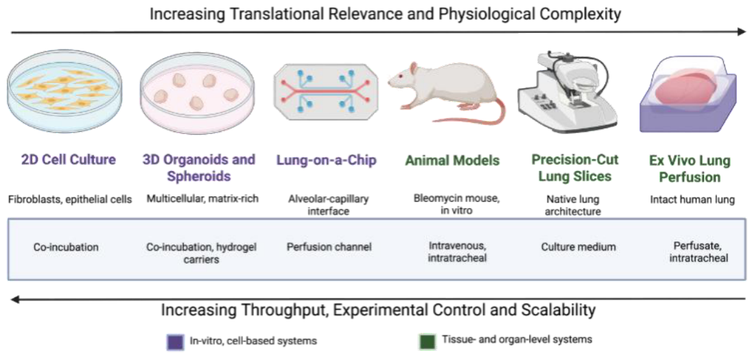
The major limitations of EVLP include high cost, technical complexity, relatively short experimental duration and limited availability; as specialized equipment, expertise, and access to donor or explanted lungs are typically restricted to lung transplant centers. Despite these challenges, EVLP represents a critical bridge between conventional preclinical models and clinical implementation (Figure 2).
6. Conclusions
Mitochondrial transplantation is an emerging therapeutic strategy for IPF that aims to address a driver of disease progression: mitochondrial dysfunction. While promising results have been reported in preclinical studies, significant challenges remain before this approach can be translated into clinical practice.
The selection of appropriate experimental models is critical for advancing the field. Each platform offers distinct advantages and limitations, ranging from the mechanistic simplicity and high throughput of 2D cultures to the high translational relevance of EVLP and human lung tissue models. Combining complementary platforms may provide the most effective strategy for evaluating mitochondrial-based therapies and accelerating clinical translation. Finally, greater emphasis on methodological studies and on reporting both positive and negative findings will improve reproducibility, refine experimental approaches, and help move mitochondrial transplantation closer to becoming a viable therapy for pulmonary fibrosis.
Author Contributions
A.C. De Avila, Y. Fine, and M. Liu contributed to the conception and design of the study. A.C. De Avila and Y. Fine prepared the original draft of the manuscript. M. Liu and S. Keshavjee provided critical revisions and intellectual input. All authors reviewed and approved the final manuscript.
Funding
This work is supported by Canadian Institutes of Health Research (PJT-173392 to S.K. and M.L.). New Frontiers in Research Fund (NFRFT-2020-00787 to S.K., and M.L.). Mingyao Liu is James and Mary Davie Chair in Lung Injury, Repair and Regeneration.
Acknowledgments
The authors would like to sincerely thank Dr. Jessica Kozlowski for her guidance, support, and insightful comments that helped strengthen this manuscript. The authors declare that Microsoft 365 Copilot (Enterprise Copilot), was used to assist with language refinement and generation of Figure 1. All AI-generated content was critically reviewed, edited, and approved by the authors, who assume full responsibility for the final content of the manuscript.
Conflicts of Interest
The authors declare no conflicts of interest.
Abbreviations
The following abbreviations are used in this manuscript:
| α-SMA | Alpha-smooth muscle actin |
| AECII | Alveolar epithelial type II cell |
| ECM | Extracellular matrix |
| EVLP | Ex vivo lung perfusion |
| IPF | Idiopathic pulmonary fibrosis |
| LTx | Lung Transplantation |
| mtROS | Mitochondrial reactive oxygen species |
| MTx | Mitochondrial Transplantation |
| PCLS | Precision-cut lung slices |
| PGC-1α | Peroxisome proliferator-activated receptor gamma coactivator 1-alpha |
| PINK1 | PTEN-induced kinase 1 |
| TGF-β | Transforming growth factor-β |
| α-SMA | Alpha-smooth muscle actin |
References
- Nathan, S.D.; Shlobin, O.A.; Weir, N.; Ahmad, S.; Kaldjob, J.M.; Battle, E.; Sheridan, M.J.; du Bois, R.M. Long-Term Course and Prognosis of Idiopathic Pulmonary Fibrosis in the New Millennium. Chest 2011, 140, 221–229. [Google Scholar] [CrossRef]
- Nava, S.; Prediletto, I. Idiopathic Pulmonary Fibrosis Management: Caring Is Sparing. J. Thorac. Dis. 2020, 12, 5265. [Google Scholar] [CrossRef]
- Althobiani, M.A.; Russell, A.M.; Jacob, J.; Ranjan, Y.; Folarin, A.A.; Hurst, J.R.; Porter, J.C. Interstitial Lung Disease: A Review of Classification, Etiology, Epidemiology, Clinical Diagnosis, Pharmacological and Non-Pharmacological Treatment. Front. Med. 2024, 11, 1296890. [Google Scholar] [CrossRef]
- Bellaye, P.-S.; Kolb, M. Why Do Patients Get Idiopathic Pulmonary Fibrosis? Current Concepts in the Pathogenesis of Pulmonary Fibrosis. BMC Med. 2015, 13, 176. [Google Scholar] [CrossRef]
- Selman, M.; Pardo, A. Revealing the Pathogenic and Aging-Related Mechanisms of the Enigmatic Idiopathic Pulmonary Fibrosis. An Integral Model. Am. J. Respir. Crit. Care Med. 2014, 189, 1161–1172. [Google Scholar] [CrossRef]
- Senhaji, L.; Senhaji, N.; Abbassi, M.; Karhate, M.; Serraj, M.; El Biaze, M.; Benjelloun, M.C.; Ouldim, K.; Bouguenouch, L.; Amara, B. Idiopathic Pulmonary Fibrosis: A Comprehensive Review of Risk Factors, Genetics, Diagnosis, and Therapeutic Approaches. Biomedicines 2026, 14, 90. [Google Scholar] [CrossRef]
- George, P.M.; Patterson, C.M.; Reed, A.K.; Thillai, M. Lung Transplantation for Idiopathic Pulmonary Fibrosis. Lancet Respir. Med. 2019, 7, 271–282. [Google Scholar] [CrossRef]
- Meyers, B.F.; Lynch, J.; Trulock, E.P.; Guthrie, T.J.; Cooper, J.D.; Patterson, G.A. Lung Transplantation: A Decade of Experience. Ann. Surg. 1999, 230, 362–371. [Google Scholar] [CrossRef]
- Balestro, E.; Cocconcelli, E.; Tinè, M.; Biondini, D.; Faccioli, E.; Saetta, M.; Rea, F. Idiopathic Pulmonary Fibrosis and Lung Transplantation: When It Is Feasible. Medicina 2019, 55, 702. [Google Scholar] [CrossRef]
- Wang, L.-J.; Guo, P.-F.; Zhang, S.; Wang, S.; Chen, Y.-Z.; Yan, H.-W.; Zhang, X.-L. Mitochondrial Transfer/Transplantation in Lung Injury: Mechanism, Therapeutic Potential, and Clinical Application. Front. Immunol. 2025, 16, 1668281. [Google Scholar] [CrossRef]
- Moss, B.J.; Ryter, S.W.; Rosas, I.O. Pathogenic Mechanisms Underlying Idiopathic Pulmonary Fibrosis. Annu. Rev. Pathol. Mech. Dis. 2022, 17, 515–546. [Google Scholar] [CrossRef]
- Bueno, M.; Lai, Y.-C.; Romero, Y.; Brands, J.; St Croix, C.M.; Kamga, C.; Corey, C.; Herazo-Maya, J.D.; Sembrat, J.; Lee, J.S.; et al. PINK1 Deficiency Impairs Mitochondrial Homeostasis and Promotes Lung Fibrosis. J. Clin. Invest. 2015, 125, 521–538. [Google Scholar] [CrossRef]
- Caporarello, N.; Meridew, J.A.; Jones, D.L.; Tan, Q.; Haak, A.J.; Choi, K.M.; Manlove, L.J.; Prakash, Y.S.; Tschumperlin, D.J.; Ligresti, G. PGC1α Repression in IPF Fibroblasts Drives a Pathologic Metabolic, Secretory and Fibrogenic State. Thorax 2019, 74, 749–760. [Google Scholar] [CrossRef]
- Chung, K.-P.; Hsu, C.-L.; Fan, L.-C.; Huang, Z.; Bhatia, D.; Chen, Y.-J.; Hisata, S.; Cho, S.J.; Nakahira, K.; Imamura, M.; et al. Mitofusins Regulate Lipid Metabolism to Mediate the Development of Lung Fibrosis. Nat. Commun. 2019, 10, 3390. [Google Scholar] [CrossRef]
- Cala-Garcia, J.D.; Medina-Rincon, G.J.; Sierra-Salas, P.A.; Rojano, J.; Romero, F. The Role of Mitochondrial Dysfunction in Idiopathic Pulmonary Fibrosis: New Perspectives for a Challenging Disease. Biology 2023, 12, 1237. [Google Scholar] [CrossRef]
- Bueno, M.; Calyeca, J.; Rojas, M.; Mora, A.L. Mitochondria Dysfunction and Metabolic Reprogramming as Drivers of Idiopathic Pulmonary Fibrosis. Redox Biol. 2020, 33, 101509. [Google Scholar] [CrossRef]
- Shin, H.R.; Lee, G.; Kim, K.H. Mitochondrial Transplantation Therapy: A Novel Approach in the Era of Organelle-Centered Regenerative Medicine. Front. Biosci. (Landmark Ed.) 2025, 30, 37006. [Google Scholar] [CrossRef]
- Li, X.; Zhang, W.; Cao, Q.; Wang, Z.; Zhao, M.; Xu, L.; Zhuang, Q. Mitochondrial Dysfunction in Fibrotic Diseases. Cell Death Discov. 2020, 6, 80. [Google Scholar] [CrossRef]
- McCully, J.D.; del Nido, P.J.; Emani, S.M. Mitochondrial Transplantation: The Advance to Therapeutic Application and Molecular Modulation. Front. Cardiovasc. Med. 2023, 10, 1268814. [Google Scholar] [CrossRef]
- McCully, J.D.; del Nido, P.J.; Emani, S.M. Mitochondrial Transplantation for Organ Rescue. Mitochondrion 2022, 64, 27–33. [Google Scholar] [CrossRef]
- Moskowitzova, K.; Orfany, A.; Liu, K.; Ramirez-Barbieri, G.; Thedsanamoorthy, J.K.; Yao, R.; Guariento, A.; Doulamis, I.P.; Blitzer, D.; Shin, B.; et al. Mitochondrial Transplantation Enhances Murine Lung Viability and Recovery after Ischemia-Reperfusion Injury. Am. J. Physiol. Lung Cell. Mol. Physiol. 2020, 318, L78–L88. [Google Scholar] [CrossRef]
- Mukkala, A.N.; David, B.A.; Ailenberg, M.; Liang, J.; Vaswani, C.M.; Karakas, D.; Goldfarb, R.; Barbour, W.; Gasner, A.; Wu, R.S.; et al. Mitochondrial Transplantation: A Novel Therapy for Liver Ischemia/Reperfusion Injury. Ann. Surg. 2025, 281, 1032–1047. [Google Scholar] [CrossRef]
- Eo, H.; Yu, S.-H.; Choi, Y.; Kim, Y.; Kang, Y.C.; Lee, H.; Kim, J.H.; Han, K.; Lee, H.K.; Chang, M.-Y.; et al. Mitochondrial Transplantation Exhibits Neuroprotective Effects and Improves Behavioral Deficits in an Animal Model of Parkinson’s Disease. Neurotherapeutics 2024, 21, e00355. [Google Scholar] [CrossRef]
- Bodenstein, D.F.; Powlowski, P.; Zachos, K.A.; El Soufi El Sabbagh, D.; Jeong, H.; Attisano, L.; Edgar, L.; Wallace, D.C.; Andreazza, A.C. Optimization of Differential Filtration-Based Mitochondrial Isolation for Mitochondrial Transplant to Cerebral Organoids. Stem Cell Res. Ther. 2023, 14, 202. [Google Scholar] [CrossRef]
- Preble, J.M.; Pacak, C.A.; Kondo, H.; MacKay, A.A.; Cowan, D.B.; McCully, J.D. Rapid Isolation and Purification of Mitochondria for Transplantation by Tissue Dissociation and Differential Filtration. J. Vis. Exp. 2014, e51682. [Google Scholar] [CrossRef]
- Baudo, G.; Wu, S.; Massaro, M.; Liu, H.; Lee, H.; Zhang, A.; Hamilton, D.J.; Blanco, E. Polymer-Functionalized Mitochondrial Transplantation to Fibroblasts Counteracts a Pro-Fibrotic Phenotype. Int. J. Mol. Sci. 2023, 24, 10913. [Google Scholar] [CrossRef]
- Guariento, A.; Piekarski, B.L.; Doulamis, I.P.; Blitzer, D.; Ferraro, A.M.; Harrild, D.M.; Zurakowski, D.; del Nido, P.J.; McCully, J.D.; Emani, S.M. Autologous Mitochondrial Transplantation for Cardiogenic Shock in Pediatric Patients Following Ischemia-Reperfusion Injury. J. Thorac. Cardiovasc. Surg. 2021, 162, 992–1001. [Google Scholar] [CrossRef]
- Lee, S.E.; Yu, S.H.; Kim, I.H.; Kang, Y.C.; Kim, Y.; Yeo, J.S.; Lim, J.H.; Kwon, I.; Kim, J.H.; Park, S.W.; et al. Mitochondrial Transplantation Ameliorates Pulmonary Fibrosis by Suppressing Myofibroblast Activation. Int. J. Mol. Sci. 2024, 25, 12783. [Google Scholar] [CrossRef]
- Wang, Y.; Hu, L.F.; Liu, N.H.; Yang, J.S.; Xing, L.; Jeong, J.H.; Li, L.; Jiang, H.L. Mitophagy-Enhanced Nanoparticle-Engineered Mitochondria Restore Homeostasis of Mitochondrial Pool for Alleviating Pulmonary Fibrosis. ACS Nano 2024, 18, 32705–32722. [Google Scholar] [CrossRef]
- Cowan, D.B.; Yao, R.; Thedsanamoorthy, J.K.; Zurakowski, D.; del Nido, P.J.; McCully, J.D. Transit and Integration of Extracellular Mitochondria in Human Heart Cells. Sci. Rep. 2017, 7, 17450. [Google Scholar] [CrossRef]
- Kitani, T.; Kami, D.; Matoba, S.; Gojo, S. Internalization of Isolated Functional Mitochondria: Involvement of Macropinocytosis. J. Cell. Mol. Med. 2014, 18, 1694–1703. [Google Scholar] [CrossRef]
- Taiko, I.; Takano, C.; Nomoto, M.; Hayashida, S.; Kanemaru, K.; Miki, T. Selection of Red Fluorescent Protein for Genetic Labeling of Mitochondria and Intercellular Transfer of Viable Mitochondria. Sci. Rep. 2022, 12, 19841. [Google Scholar] [CrossRef]
- Deng, S.; Ren, Y.; Zhang, Q.; Liu, Q.; Long, J.; Picard, K.; Martin, M.; Miller, T.; Yuan, C.; He, Y.; et al. Nanoengineered Mitochondria for Mitochondrial Dysfunction and Anti-Aging Interventions. Front. Aging 2025, 6, 1688482. [Google Scholar] [CrossRef]
- Jenkins, R.G.; Moore, B.B.; Chambers, R.C.; Eickelberg, O.; Königshoff, M.; Kolb, M.; Laurent, G.J.; Nanthakumar, C.B.; Olman, M.A.; Pardo, A.; et al. An Official American Thoracic Society Workshop Report: Use of Animal Models for the Preclinical Assessment of Potential Therapies for Pulmonary Fibrosis. Am. J. Respir. Cell Mol. Biol. 2017, 56, 667–679. [Google Scholar] [CrossRef]
- Kolanko, E.; Cargnoni, A.; Papait, A.; Silini, A.R.; Czekaj, P.; Parolini, O. The Evolution of in Vitro Models of Lung Fibrosis: Promising Prospects for Drug Discovery. Eur. Respir. Rev. 2024, 33, 230127. [Google Scholar] [CrossRef]
- Osei, E.T.; Brandsma, C.-A.; Noordhoek, J.A.; Timens, W.; Postma, D.; Heijink, I. Crosstalk between Epithelium and Fibroblasts; Implications for COPD. Eur. Respir. J. 2014, 44 (Suppl. 58), P3899. [Google Scholar] [CrossRef]
- Brussow, J.; Feng, K.; Thiam, F.; Phogat, S.; Osei, E.T. Epithelial-Fibroblast Interactions in IPF: Lessons from in Vitro Co-Culture Studies. Differentiation 2023, 134, 11–19. [Google Scholar] [CrossRef]
- Osei, E.T.; Booth, S.; Hackett, T.-L. What Have In Vitro Co-Culture Models Taught Us about the Contribution of Epithelial-Mesenchymal Interactions to Airway Inflammation and Remodeling in Asthma? Cells 2020, 9, 1694. [Google Scholar] [CrossRef]
- Dsouza, K.G.; Surolia, R.; Kulkarni, T.; Li, F.J.; Singh, P.; Zeng, H.; Stephens, C.; Kumar, A.; Wang, Z.; Antony, V.B. Use of a Pulmosphere Model to Evaluate Drug Antifibrotic Responses in Interstitial Lung Diseases. Respir. Res. 2023, 24, 96. [Google Scholar] [CrossRef]
- Asmani, M.; Velumani, S.; Li, Y.; Wawrzyniak, N.; Hsia, I.; Chen, Z.; Hinz, B.; Zhao, R. Fibrotic Microtissue Array to Predict Anti-Fibrosis Drug Efficacy. Nat. Commun. 2018, 9, 2066. [Google Scholar] [CrossRef]
- Kabadi, P.K.; Rodd, A.L.; Simmons, A.E.; Messier, N.J.; Hurt, R.H.; Kane, A.B. A Novel Human 3D Lung Microtissue Model for Nanoparticle-Induced Cell-Matrix Alterations. Part. Fibre Toxicol. 2019, 16, 15. [Google Scholar] [CrossRef]
- Eş, I.; Ulger, O. Organoids as 3D Models for Studying Exogenous Mitochondrial Transplantation. In Cell Biology and Translational Medicine, Volume 26; Advances in Experimental Medicine and Biology; Turksen, K., Ed.; Springer: Cham, Switzerland, 2025; Volume 1488. [Google Scholar] [CrossRef]
- Huang, T.; Lin, R.; Su, Y.; Sun, H.; Zheng, X.; Zhang, J.; Lu, X.; Zhao, B.; Jiang, X.; Huang, L.; et al. Efficient Intervention for Pulmonary Fibrosis via Mitochondrial Transfer Promoted by Mitochondrial Biogenesis. Nat. Commun. 2023, 14, 5781. [Google Scholar] [CrossRef]
- Zhang, Y.; Wang, X.; Yang, Y.; Yan, J.; Xiong, Y.; Wang, W.; Lei, J.; Jiang, T. Recapitulating Essential Pathophysiological Characteristics in Lung-on-a-Chip for Disease Studies. Front. Immunol. 2023, 14, 1093460. [Google Scholar] [CrossRef]
- Bodenstein, D.F.; Siebiger, G.; Zhao, Y.; Clasky, A.J.; Mukkala, A.N.; Beroncal, E.L.; Banh, L.; Aslostovar, L.; Brijbassi, S.; Hogan, S.E.; et al. Bridging the Gap between in Vitro and in Vivo Models: A Way Forward to Clinical Translation of Mitochondrial Transplantation in Acute Disease States. Stem Cell Res. Ther. 2024, 15, 157. [Google Scholar] [CrossRef]
- Preuß, E.B.; Schubert, S.; Werlein, C.; Stark, H.; Braubach, P.; Höfer, A.; Plucinski, E.K.J.; Shah, H.R.; Geffers, R.; Sewald, K.; et al. The Challenge of Long-Term Cultivation of Human Precision-Cut Lung Slices. Am. J. Pathol. 2022, 192, 239–253. [Google Scholar] [CrossRef]
- Königshoff, M.; Lehmann, M.; Rojas, M.; Bourke, J.; Anderson, C.; Banovich, N.E.; Behrsing, H.P.; Dean, C.H.; Gosens, R.; Haak, A.; et al. Precision-Cut Lung Slices: Emerging Tools for Preclinical and Translational Lung Research. An Official American Thoracic Society Workshop Report. Am. J. Respir. Cell Mol. Biol. 2025, 72, 16–31. [Google Scholar] [CrossRef]
- Koziol-White, C.; Gebski, E.; Cao, G.; Panettieri, R.A. Precision Cut Lung Slices: An Integrated Ex Vivo Model for Studying Lung Physiology, Pharmacology, Disease Pathogenesis and Drug Discovery. Respir. Res. 2024, 25, 231. [Google Scholar] [CrossRef]
- Cypel, M.; Yeung, J.C.; Hirayama, S.; Rubacha, M.; Fischer, S.; Anraku, M.; Sato, M.; Harwood, S.; Pierre, A.; Waddell, T.K.; et al. Technique for Prolonged Normothermic Ex Vivo Lung Perfusion. J. Heart Lung Transplant. 2008, 27, 1319–1325. [Google Scholar] [CrossRef]
- Cloer, C.M.; Givens, C.S.; Buie, L.K.; Rochelle, L.K.; Lin, Y.-T.; Popa, S.; Shelton, R.V.M.; Zhan, J.; Zimmerman, T.R.; Jones, B.G.; et al. Mitochondrial Transplant after Ischemia Reperfusion Promotes Cellular Salvage and Improves Lung Function during Ex-Vivo Lung Perfusion. J. Heart Lung Transplant. 2023, 42, 575–584. [Google Scholar] [CrossRef]
Figure 1.
Mitochondrial dysfunction in AECII cells promotes fibrogenesis in IPF and can potentially be ameliorated by MTx. In the fibrotic alveolus (left), AECII cells exhibit mitochondrial dysfunction characterized by structurally abnormal mitochondria, resulting in increased mtROS production and the release of inflammatory cytokines and profibrotic mediators. These signals promote upregulation of transforming growth factor-β (TGF-β), that drives fibroblast activation and differentiation into myofibroblasts. Activated myofibroblasts (α-SMA+) produce excessive ECM components, including collagen and fibronectin, leading to alveolar wall thickening and progressive fibrotic remodeling. Mitochondrial replenishment therapy (center) aims to delivers healthy donor mitochondria to injured AECII cells with the goal to restore mitochondrial function. Restoration of mitochondrial homeostasis reduces mtROS generation, inflammatory cytokine release, and TGF-β signaling (right), potentially resulting in decreased fibroblast activation, reduced myofibroblast differentiation, diminished ECM deposition, and attenuation of fibrotic remodeling. Abbreviations: AECII, alveolar epithelial type II cell; ECM, extracellular matrix; mtROS, mitochondrial reactive oxygen species; TGF-β, transforming growth factor-β; α-SMA, alpha-smooth muscle actin. Generated with Microsoft Copilot.
Figure 1.
Mitochondrial dysfunction in AECII cells promotes fibrogenesis in IPF and can potentially be ameliorated by MTx. In the fibrotic alveolus (left), AECII cells exhibit mitochondrial dysfunction characterized by structurally abnormal mitochondria, resulting in increased mtROS production and the release of inflammatory cytokines and profibrotic mediators. These signals promote upregulation of transforming growth factor-β (TGF-β), that drives fibroblast activation and differentiation into myofibroblasts. Activated myofibroblasts (α-SMA+) produce excessive ECM components, including collagen and fibronectin, leading to alveolar wall thickening and progressive fibrotic remodeling. Mitochondrial replenishment therapy (center) aims to delivers healthy donor mitochondria to injured AECII cells with the goal to restore mitochondrial function. Restoration of mitochondrial homeostasis reduces mtROS generation, inflammatory cytokine release, and TGF-β signaling (right), potentially resulting in decreased fibroblast activation, reduced myofibroblast differentiation, diminished ECM deposition, and attenuation of fibrotic remodeling. Abbreviations: AECII, alveolar epithelial type II cell; ECM, extracellular matrix; mtROS, mitochondrial reactive oxygen species; TGF-β, transforming growth factor-β; α-SMA, alpha-smooth muscle actin. Generated with Microsoft Copilot.
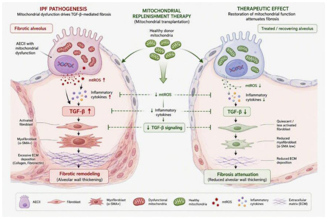
Figure 2.
Experimental platforms used to evaluate mitochondrial transplantation in pulmonary fibrosis, arranged according to increasing physiological complexity and translational relevance (left to right). Models range from 2D cell cultures and 3D organoids/spheroids to lung-on-a-chip, animal models, PCLS, and EVLP. Representative routes of exogenous mitochondrial delivery for each platform are shown. While translational relevance increases across the spectrum, throughput decreases and experimental complexity generally increases, highlighting the complementary role of these models in the preclinical development of mitochondrial therapies for lung disease. Generated with BioRender.
Figure 2.
Experimental platforms used to evaluate mitochondrial transplantation in pulmonary fibrosis, arranged according to increasing physiological complexity and translational relevance (left to right). Models range from 2D cell cultures and 3D organoids/spheroids to lung-on-a-chip, animal models, PCLS, and EVLP. Representative routes of exogenous mitochondrial delivery for each platform are shown. While translational relevance increases across the spectrum, throughput decreases and experimental complexity generally increases, highlighting the complementary role of these models in the preclinical development of mitochondrial therapies for lung disease. Generated with BioRender.
Disclaimer/Publisher’s Note: The statements, opinions and data contained in all publications are solely those of the individual author(s) and contributor(s) and not of MDPI and/or the editor(s). MDPI and/or the editor(s) disclaim responsibility for any injury to people or property resulting from any ideas, methods, instructions or products referred to in the content. |
© 2026 by the authors. Licensee MDPI, Basel, Switzerland. This article is an open access article distributed under the terms and conditions of the Creative Commons Attribution (CC BY) license.
Copyright: This open access article is published under a Creative Commons CC BY 4.0 license, which permit the free download, distribution, and reuse, provided that the author and preprint are cited in any reuse.