
Submitted:
02 November 2026
Posted:
04 November 2026
You are already at the latest version
Abstract
Median sternotomy remains the gold standard for surgical access in the majority of open-heart procedures, providing reliable exposure for complex cardiac operations. Despite advances in surgical technique and perioperative care, it continues to be associated with significant complications, including bleeding, fragile or calcified tissue and sternal instability, and surgical site infections (SSIs). These complications are not isolated but are deeply interconnected, often progressing in a vicious cycle that leads to increased morbidity, mortality, prolonged hospitalization, and substantial healthcare costs. Although the individual challenges of median sternotomy have been extensively studied, a comprehensive synthesis that conceptualizes an integrated framework for optimizing outcomes is lacking. This review proposes a novel three-pillar Comprehensive Sternotomy Management Bundle that simultaneously addresses (1) perioperative bleeding management, (2) handling of fragile or calcified tissue with robust sternal stabilization, and (3) multimodal prevention of SSI. By integrating evidence-based strategies across the preoperative, intraoperative, and postoperative phases, the bundle aims to disrupt the cycle of complications, promote secure sternal healing, reduce transfusion requirements, and accelerate recovery. This multimodal care bundle aligns with the Quintuple Aim by improving clinical outcomes, enhancing the patient and clinician experience, and reducing overall healthcare costs in an increasingly older and comorbid cardiac surgical population.
Keywords:
sternotomy
; outcomes
; complications
; bleeding
; sternal instability
; surgical site infection
; quality of care
1. Introduction
Median sternotomy remains the gold standard for surgical access in most open-heart procedures, offering direct and reliable exposure of the cardiac structures. Despite the growing appeal for minimally invasive approaches, sternotomy remains indispensable, particularly in complex or multi-procedural surgery [1]. Cardiac surgical procedures requiring median sternotomy are performed in hundreds of thousands of individuals in the United States (US) [2] and in millions of individuals worldwide [3] each year. However, even with ongoing refinements in surgical technique and perioperative care, sternotomy-related complications continue to impose significant morbidity, mortality, prolonged hospitalization, and substantial healthcare costs [4,5].
Bleeding, fragile or calcified tissue and sternal instability (particularly in patients with fragile or calcified bone), and surgical site infections (SSIs) including deep sternal wound infection (DSWI) and mediastinitis constitute the most clinically relevant challenges of median sternotomy [6,7]. These complications are not isolated events but rather are deeply interconnected, often progressing in a vicious cycle where one complication predisposes to and exacerbates the others [8]. Excessive bleeding can lead to hematoma formation and subsequent infection, while poor sternal stability in osteoporotic or heavily calcified sternums predisposes to dehiscence, impaired wound healing, and secondary infection [6,7,9,10]. Reported incidences of sternal wound complications range from 0.5% to 5%, with associated mortality rates reaching up to 20–40% in cases of deep infection [9].
Although substantial literature has addressed the challenges of median sternotomy in isolation, a comprehensive synthesis that conceptualizes an integrated framework—or “pillars”—for optimizing median sternotomy outcomes in a cohesive manner is currently lacking. Existing reviews tend to treat the challenges of median sternotomy separately or within broader perioperative discussions [6,9,11,12,13], limiting actionable, surgeon-focused guidance that highlights their interdependence. Simultaneously targeting bleeding, fragile or calcified tissue and sternal instability, and SSI offers the greatest potential to disrupt the cycle of complications, improve sternal healing, and meaningfully enhance outcomes. With an aging cardiac surgical population increasingly presenting with comorbidities such as osteoporosis, aortic calcification, frailty, and colonization with multidrug-resistant organism [14,15,16], there is a pressing need for a unified, evidence-based approach. This review proposes an evidence-based novel three-pillar Comprehensive Sternotomy Management Bundle to support prompt recovery after sternotomy across preoperative, intraoperative, and postoperative phases (Figure 1). The proposed evidence-based framework is aligned with the Institute for Healthcare Improvement (IHI)’s Triple Aim goals of better care, improved population health, and lower per capita costs and the Quadruple and Quintuple Aim’s goals of higher quality clinician experiences and health equity, respectively [17].
2. The 3 Pillars for Optimizing Sternotomy Outcomes
2.1. Pillar 1: Managing Bleeding Challenges
Bleeding is a common complication of cardiac surgery that contributes to perioperative morbidity, the need for allogeneic blood transfusions, intensive care unit (ICU) stay, total length of hospital stay, and in some cases a return to the operating room (OR) [18,19]. Post-sternotomy bleeding remains a significant and multifactorial challenge, arising from sources such as sternal edges, vascular anastomoses, needle puncture sites, and diffuse capillary oozing [9,20]. This persistent issue imposes a substantial economic burden on healthcare systems [21,22]. Unaddressed disruptive bleeding has been shown to be associated with a 21% increase in hospital costs among patients with coronary artery bypass graft (CABG) surgery [22].
2.1.1. Pre-Operative Phase: Risk Stratification and Optimization
Patient bleeding history should be evaluated during the pre-operative assessment, if available, as it is a strong predictor of intra- and post-operative bleeding risk [23,24]. Coagulation testing can determine hemostatic defects in high-risk patients, who may have inherited coagulopathies and underlying conditions [23,24]. However, it may not be possible to properly predict bleeding risk in high-risk patients [25,26].
Preoperative optimization of modifiable risk factors is essential [27]. In a US hospital database study of over 300,000 patients, 30% had three or more comorbidities, which put them at greater risk of disruptive bleeding [21]. Screening for and correction of anemia should be performed in all elective cases, using iron supplementation with or without erythropoietin-stimulating agents according to established Patient Blood Management (PBM) protocols [27]. Uncontrolled diabetes disrupts normal hemostasis and wound healing by inducing a prothrombotic state while impairing tissue repair processes, thereby contributing to increased perioperative complications [28]. Chronic kidney disease (CKD) is also associated with risk for postoperative bleeding due to platelet dysfunction, anemia, and accumulation of uremic toxins that hinder the normal coagulation cascade [29,30].
Chronic medication use represents one of the most important and modifiable bleeding risk factors. Approximately one in four surgical patients takes a prescribed anticoagulant or antiplatelet therapy that can impair hemostasis by modulating different clotting pathways, including platelet function and coagulation, which increases bleeding risk [31,32,33]. In cardiac surgery patients, careful perioperative management of these agents – guided by multidisciplinary discussion and institutional protocols – is critical to balance bleeding and thrombotic risks [31,34]. Validated bleeding risk scores (e.g., TRACK or STS risk models) can further help identify patients at elevated risk and guide individualized planning.
Overall, a structured PBM approach incorporating anemia correction, comorbidity optimization, and individualized antithrombotic strategies should be implemented in all patients undergoing sternotomy.
2.1.2. Intraoperative Phase: Surgical Technique, Blood Conservation, and Hemostatic Tools
Meticulous surgical technique remains the foundation of effective intraoperative bleeding control. Careful handling of sternal edges with limited or selective use of bone wax (or preferably alternatives to preserve sternal vascularity), precise internal mammary artery (IMA) harvest with skeletonization when possible to preserve sternal perfusion, and rigorous control of periosteal, pericardial, and soft-tissue bleeding are essential [35]. Excessive use of electrocautery should be minimized to reduce thermal injury to bone and surrounding tissues [35].
Cardiopulmonary bypass (CPB) strategies play a central role in blood conservation. Retrograde autologous priming (RAP), minimized circuit volume, and acute normovolemic hemodilution (ANH) reduce hemodilution and help preserve hematocrit [27,36]. Precise heparin and protamine management, combined with point-of-care viscoelastic testing (thromboelastography [TEG] or rotational thromboelastometry [ROTEM]), allows goal-directed transfusion and coagulation management rather than empiric product administration [27].
Routine administration of antifibrinolytic agents is strongly recommended (Class I, Level A). Tranexamic acid (TXA) or ε-aminocaproic acid (EACA) significantly reduces bleeding and transfusion requirements [27].
Intraoperative cell salvage (autotransfusion of washed shed blood using centrifugation) should be used routinely in sternotomy cases, as it is effective in recovering red cells and further decreases allogeneic transfusion exposure (Class I, Level A) [27].
Topical hemostatic agents serve as important adjuncts when standard surgical techniques are insufficient. These agents are particularly valuable for controlling diffuse oozing from sternal edges, raw surfaces, or vascular anastomoses. Application before sternal closure is recommended to achieve complete hemostasis prior to chest closure. Table 1 summarizes key hemostatic tools available for intraoperative use during sternotomy.
2.1.3. Postoperative Phase: Monitoring, Transfusion Stewardship, and Re-Exploration
Effective postoperative bleeding management is critical to interrupt the vicious cycle linking hemorrhage, fragile or calcified tissue and sternal instability, and infection [27]. Continuous surveillance in the ICU forms the cornerstone of care. Serial assessment of chest tube output, hemodynamic parameters, lactate levels, and viscoelastic testing (e.g., TEG or ROTEM) enables early detection of ongoing bleeding and coagulopathy [47].
A restrictive transfusion strategy is recommended [27]. Current guidelines advocate red blood cell transfusion only when hemoglobin falls below 7–8 g/dL in stable patients, or higher thresholds in those with active bleeding, hemodynamic instability, or significant comorbidities [48]. Point-of-care viscoelastic testing-guided transfusion has been shown to significantly reduce allogeneic blood product use compared with conventional laboratory-based algorithms [49].
When bleeding persists, targeted correction of coagulopathy is essential [47]. This may include fibrinogen concentrate, prothrombin complex concentrate (PCC), platelets, or cryoprecipitate, guided by real-time viscoelastic results rather than empiric therapy [27]. Normothermia, correction of acidosis and hypocalcemia, and minimization of iatrogenic blood loss (e.g., reduced phlebotomy) remain important adjuncts [50].
Re-exploration of bleeding after cardiac surgery is associated with significant morbidity and mortality [51]. Every effort should be exerted to avoid postoperative bleeding and to manage it properly if it happens [51]. Low rate of re-exploration for bleeding can be achieved by strict preoperative preparation, intraoperative checklist for hemostasis and adopting an algorithm for management [51]. If re-exploration is needed, meticulous hemostasis of sternal edges, suture lines, and raw surfaces should be achieved, often with liberal use of topical hemostatic agents [27].
2.2. Pillar 2: Handling of Fragile or Calcified Tissue and Achieving and Maintaining Sternal Stability
The increasing complexity of cardiac surgical patients, characterized by advanced age, diabetes, obesity, and prior sternotomy, has significantly heightened the technical demands of tissue handling during and after sternotomy. Patients often present with fragile vascular structures or calcified tissues, both of which pose substantial challenges during anastomosis, hemostasis, and sternal closure [52].
Tissue fragility also affects patients with delicate pulmonary or thoracic structures, often due to age, smoking, or other factors, where even minor disruptions during closure can cause air leaks or bleeding. Conversely, vascular calcification, common in those with diabetes or kidney disease, stiffens vessels and resists needle penetration, which makes closure more difficult, contributing to poor outcomes [53]. Inadequate handling may result in tearing, bleeding, sternal instability, or infection. Sternal instability and dehiscence represent major complications after median sternotomy, contributing to pain, impaired wound healing, sternal wound infection, mediastinitis, prolonged hospitalization, and increased mortality [8]. Therefore, specific precautions and advanced closure techniques are essential to ensure secure anastomotic lines, reduce tissue trauma, and optimize recovery.
2.2.1. Preoperative Phase: Risk Identification and Optimization
Identification of patients at high risk for fragile or calcified tissue and sternal instability begins preoperatively. Key risk factors include advanced age, female sex, osteoporosis or poor bone quality, obesity (BMI >30), diabetes mellitus, chronic obstructive pulmonary disease (COPD), smoking, chronic steroid use, redo sternotomy, and off-midline sternotomy [8]. Validated risk prediction models can help stratify patients and guide decision-making regarding closure technique.
Optimization strategies include smoking cessation, glycemic control, nutritional support, and preoperative bone health assessment in selected high-risk patients. In patients with known severe osteoporosis, consideration should be given to alternative or hybrid closure strategies from the outset.
2.2.2. Intraoperative Phase: Optimal Tissue Handling and Closure Techniques
Anastomosis quality, both in direct and axillary approaches, is very important and can prove to be technically challenging in high-risk patients with calcified or thin-walled vessels (e.g., diabetic patients) [54,55,56,57]. High-strength tungsten-rhenium alloy cardiovascular needles engineered to reduce needle hole bleeding can improve outcomes in patients with advanced stages of coronary disease and calcified tissue by reducing blood loss at the anastomosis suture line [58,59]. In patients with calcified vessels, frequently encountered in those with diabetes, CKD, or severe atherosclerosis, standard surgical needles often fail to penetrate without trauma. Tungsten–rhenium alloy needles have demonstrated superior performance over conventional stainless-steel alternatives. These high-performance needles exhibit up to 40% increased strength, 100% higher stiffness, and improved resistance to deformation, allowing for precise multiple-pass penetration in heavily calcified tissues (p<0.01) [58]. Their mechanical reliability minimizes the risk of vessel rupture, enhances suture placement accuracy, and may improve outcomes in CABG and valve surgery in high-risk populations [58].
To further support secure closure in compromised tissues, antibacterial barbed sutures offer knotless, continuous wound approximation with improved tension distribution [60]. This is particularly beneficial in friable tissues, where conventional knotting may create focal stress points and elevate the risk of tearing. A retrospective cohort study demonstrated that antibacterial barbed sutures were a suitable and reliable method for sternotomy closure in pediatric cardiovascular surgery cases [61]. Antibacterial barbed sutures also integrate antibacterial properties-Triclosan), which may contribute to SSI reduction [61]. The use of antibacterial sutures is endorsed by National Institute for Health and Care Excellence (NICE) guidelines for infection prevention [62], and they have demonstrated superior outcomes in terms of closure time and tensile strength, especially in re-operations or complex closures involving fragile tissues [60].
2.2.3. Postoperative Phase: Protection, Monitoring, and Management of Fragile or Calcified Tissue
Postoperative care is crucial for patients with fragile or calcified tissues, as sternal healing can be delayed and mechanical stress may lead to dehiscence or chronic pain [63]. External sternal support devices (e.g., thoracic braces or Posthorax vest) may be considered in high-risk patients to provide anteroposterior stabilization, reduce pain during coughing or movement, and minimize shear forces across the closure site [64].
Activity modification remains important during the first 6–8 weeks [65,66,67]. While overly restrictive traditional sternal precautions have been relaxed in favor of guided early mobilization under Enhanced Recovery After Surgery (ERAS) protocols, patients should still avoid heavy lifting (>10 lbs), bilateral arm extension beyond shoulder level, and forceful coughing without splinting [65,66,67]. Close clinical monitoring for signs of instability (clicking, palpable motion, disproportionate pain, or wound drainage) is essential, with prompt imaging (chest CT if needed) and early re-intervention (rewiring or rigid fixation) when dehiscence is suspected [6,11,67].
Multimodal analgesia, including regional techniques (e.g., parasternal blocks) and opioid-sparing regimens, helps reduce stress on the healing sternum while facilitating early mobilization [68,69,70]. In patients with significant tissue fragility, prolonged use of supportive devices and delayed return to full activity may be required to promote solid bony union and prevent secondary complications such as non-union or chronic post-sternotomy pain [6,71].
2.3. Pillar 3: Minimizing Surgical Site Infection (SSI) Risk
Surgical site infections (SSIs) can affect up to 26.8% of cardiac procedures and are associated with poor prognosis, increased hospital stay, substantial medical costs, and high mortality [72]. The incidence of superficial SSI after cardiac surgery typically ranges between 0.5% to 8%, whereas the incidence of deep infection ranges between 0.5% and 5.6% [72]. The reported incidence of DSWI in large cardiac surgery centers typically ranges between 1% and 2% per year [72]. Post-sternotomy mediastinitis incidence rates range from 1–5%, with a rate >2% being an indicator of poor quality care in the surgery center [72]. SSIs in cardiac surgery patients are one of the most problematic hospital acquired infections due to the immediate proximity of the surgical site to vital organs [72].
2.3.1. Preoperative Phase: Risk Identification and Optimization
Preventative care begins with the preoperative identification of both modifiable and nonmodifiable SSI risks by health care providers. Assessment tools can be used to assist in identifying patients at a high risk of SSI [73]. Patient-related comorbidities that significantly increase the risk of SSI in cardiac surgery include nasal colonization with Staphylococcus aureus, diabetes mellitus or perioperative hyperglycemia, obesity, macromastia, peripheral artery disease, congestive heart failure with low ejection fraction, COPD, and end-stage renal disease requiring dialysis [73,74]. Additional contributing factors are tobacco use, a history of prior cardiac surgery, and inappropriate hair removal techniques [12,73,75].
Nasal decolonization using intranasal mupirocin combined with chlorhexidine body washes for several days prior to surgery has been shown to reduce Staphylococcus aureus colonization and subsequent wound infections and ICU respiratory disease [76,77]. Preoperative skin antisepsis is critical, and the use of chlorhexidine alcohol has shown superior efficacy compared to povidone-iodine [76,77].
Among all comorbidities, diabetes mellitus and obesity are the most influential risk factors for SSI [78,79]. Diabetes promotes a pro-inflammatory milieu, impairs leukocyte function, and inhibits neovascularization, all of which hinder effective wound healing [80]. Hyperglycemia reduces collagen synthesis and increases susceptibility to infection [81]. Furthermore, diabetes is known to deteriorate sternal bone quality and accelerate bone resorption, predisposing patients to deep sternal wound complications [82]. Obesity, similarly, contributes to chronic systemic inflammation, increased shear stress on wound edges, impaired perfusion, and a greater risk of seroma formation and dead space. A study by Alebrahim et al. demonstrated that while the incidence of deep sternal wound infection in patients with BMI <30 kg/m² is approximately 0.8–1.5%, this rate rises to nearly 8% in obese individuals [83]. Glycemic control is paramount, with a recommended perioperative blood glucose target of <180 mg/dL and preoperative hemoglobin A1c <7% when feasible, given the strong association between hyperglycemia and impaired immune function, neovascularization, and wound healing [84]. For obese patients, preoperative weight reduction is encouraged to minimize shear forces on the sternal wound, reduce dead space, and improve soft tissue perfusion; all factors that influence SSI risk. In nonemergent cases, preoperative weight loss with a target BMI of <30 may be considered to enhance outcomes and reduce the risk of SSIs [72,83].
Lifestyle optimization, including smoking cessation, nutritional support, and prehabilitation, enhances immune competence and tissue recovery capacity [85]. Lastly, if hair removal is necessary, it must be performed only on the day of surgery using electric clippers, as razors are known to cause micro abrasions that compromise the skin barrier and increase bacterial entry [86].
2.3.2. Intraoperative Phase: Optimal Surgical and Closure Techniques
Intraoperative risk factors for SSIs include emergency surgery, prolonged operative times (greater than five hours), extended cardiopulmonary bypass (CPB) and aortic cross-clamp durations, as well as inadequate sternal approximation, such as asymmetric closure or residual separation [72]. Bone ischemia, especially due to excessive electrocautery or bilateral internal thoracic artery harvesting or harvesting in a non-skeletonized fashion, compromises local perfusion and healing [12,87]. Immunosuppressive conditions, whether iatrogenic (blood transfusions or transplant status) or CPB-induced, also impair host defense mechanisms [72,73,88,89].
Intraoperative strategies form a critical pillar in the prevention of SSIs following median sternotomy. Mitigating SSI is in line with Society of Thoracic Surgeons (STS)/ERAS principles (tight glycemic control, CHG–alcohol prep, skeletonized ITA harvest in high-risk patients) and should be anticipated, especially in high-risk patient populations [11,66,73,90]. Maintaining strict intraoperative glycemic control is essential to minimize the effects of diabetes on SSIs [86]. The foundation of this phase is a gentle, atraumatic surgical technique, prioritizing meticulous tissue handling and precise hemostasis to minimize both mechanical trauma and thermal injury. Preservation of sternal perfusion is equally important and requires the avoidance of maneuvers that compromise local blood flow, such as excessive electrocautery and use of bone wax which can induce focal bone ischemia [12,91]. When bilateral internal thoracic artery harvesting is indicated, particularly in high-risk patients such as those with diabetes or obesity, the skeletonized technique is preferred, as it preserves sternal vascularization and reduces the risk of wound complications [92,93,94].
To protect tissues during prolonged retraction, wound edge protectors can reduce mechanical pressure on soft tissues and act as a barrier to contamination. In procedures exceeding three to four hours, intraoperative antibiotic redosing is essential to maintain therapeutic antimicrobial levels in both serum and tissues, preventing bacterial proliferation during the most vulnerable phases of the operation [76,95].
Advanced wound closure systems have emerged as important adjuncts to enhance healing and reduce postoperative complications . Closure technique using antibacterial barbed sutures can help mitigate wound complications and minimize the incidence of incisional SSIs [96,97,98]. A meta-analysis of 31 RCTs including 17 968 participants found that use of triclosan-coated sutures was associated with fewer SSIs compared with sutures without triclosan (relative risk [RR], 0.75; 95% CI, 0.65-0.86) [99]. Antibacterial barbed sutures with unidirectional symmetrical anchor design also allow tension to be maintained on each pass and are effective in high tension areas, achieving a watertight closure [98,100]. This minimizes localized stress points, reduces dead space, and has shown particular benefit in obese or diabetic patients, where the risk of seroma and infection is higher [101,102,103,104,105].
In addition, topical skin adhesives such as 2-octyl cyanoacrylate plus polymer mesh tape provide a flexible microbial barrier with 99% protection in vitro for 72 hours against organisms commonly responsible for SSIs [106]. In a real-world study, 2-octyl cyanoacrylate plus polymer mesh tape was associated with a 53% reduction in SSI incidence compared to standard dressing in cardiac surgery [107] and was associated with 25% lower incidence of wound dehiscence compared to conventional sutures and waterproof dressing [108]. Skin closure with 2-octyl cyanoacrylate plus polymer mesh tape reduced length of stay and costs when compared to conventional absorbable sutures after median sternotomy for cardiac surgery [109]. This system limits shear forces across the wound and also enables early patient mobilization from postoperative Day 0, aligning with contemporary ERAS protocols [107].
2.3.3. Postoperative Phase
Implementation of a protocol to maintain mean blood glucose level less than 200 mg/dL in the immediate postoperative period has been shown to reduce the incidence of deep wound infection in diabetic patients [110]. Postoperative infection risk also increases in the presence of multiple drains or invasive catheters, return to the operating room (commonly for bleeding control), prolonged mechanical ventilation due to respiratory failure, and extended ICU stays [74]. Poor glycemic control and suboptimal wound care, such as frequent wound manipulation and inappropriate dressing, further exacerbate vulnerability to SSI, especially in patients with diabetes or obesity, where impaired immune function and altered wound healing are well documented [73,104,105,110,111].
3. Discussion
This narrative review introduces a novel Comprehensive Sternotomy Management Bundle built on three interconnected pillars for enhancing outcomes after sternotomy: (1) meticulous perioperative bleeding management, (2) tailored handling of fragile or calcified tissue with robust sternal stabilization, and (3) multimodal SSI prevention. By integrating evidence-based strategies across preoperative, intraoperative, and postoperative phases, this framework addresses the vicious cycle in which bleeding, tissue and sternal instability, and infection reinforce one another, leading to increased morbidity and healthcare utilization.
The strength of this three-pillar approach lies in its recognition of the interdependence of these complications. Effective bleeding control reduces hematoma formation and subsequent infection risk, while secure sternal stabilization in fragile or calcified bone minimizes mechanical stress that can precipitate dehiscence and wound breakdown. Concurrently, rigorous infection prevention strategies preserve tissue integrity and support bony union. When implemented together, these pillars produce synergistic rather than merely additive benefits.
This bundle aligns closely with contemporary Society of Thoracic Surgeons (STS) and Enhanced Recovery After Surgery (ERAS) Cardiac Society recommendations. Key elements such as restrictive transfusion strategies, goal-directed hemostasis using viscoelastic testing, skeletonized internal thoracic artery harvest in high-risk patients, rigid or hybrid sternal closure techniques, multimodal opioid-sparing analgesia, and supported early mobilization are all reinforced within the framework. Institutions adopting similar multimodal bundles have reported substantial reductions in deep sternal wound infection rates, often approaching zero, underscoring the transformative potential of systematic, team-based care [112,113].
From a broader healthcare perspective, this integrated strategy directly supports the Quintuple Aim. By reducing transfusion requirements, re-exploration rates, ICU and hospital length of stay, and sternal wound complications, the bundle improves clinical outcomes, enhances both patient and clinician experience, advances health equity in an increasingly comorbid population, and lowers per capita costs. Economic modeling suggests that even modest reductions in sternal complications and bleeding-related events could generate significant annual savings for cardiac surgery programs [112,114].
Successful implementation will require multidisciplinary collaboration, standardized protocols, and ongoing quality improvement using Plan-Do-Study-Act (PDSA) cycles [115,116]. Preoperative risk stratification, intraoperative checklists, and postoperative surveillance protocols should be embedded into existing workflows. While upfront investment in specialized tools (e.g., advanced hemostatic agents, rigid fixation plates, external support devices, and antibacterial sutures) is necessary, these costs are typically offset by downstream reductions in complications.
Nevertheless, several limitations warrant acknowledgment. High-quality prospective studies evaluating complete multimodal bundles versus standard care remain limited. Patient-specific factors such as extreme frailty, emergency surgery, or severe comorbidities may necessitate individualized adaptation of the bundle. Additionally, variability in institutional resources and surgical expertise may influence real-world effectiveness.
In summary, the proposed Comprehensive Sternotomy Management Bundle represents a pragmatic, evidence-based framework for optimizing outcomes after median sternotomy. Moving forward, transitioning from isolated interventions to fully integrated care bundles will be essential to meet the challenges of an aging and increasingly complex cardiac surgical population. Other quality improvement efforts have demonstrated the value of implementing bundles in hospital intensive care [117,118,119].
4. Conclusions
The continued aging of the population has led to older and more complex patients requiring cardiac surgical interventions for management of cardiovascular disease. Advanced end-to-end solutions – temporary hemodynamic support, hemostasis, tissue handling, wound closure, and wound healing are critical for optimizing surgical outcomes in this challenging group of patients. When applied across the preoperative, intraoperative, and postoperative phases, these strategies form a Comprehensive Sternotomy Management Bundle that can help achieve Quintuple Aim goals by synergistically reducing risks associated with bleeding, fragile or calcified tissue and sternal instability, and SSIs.
5. Future Directions
Future research should prioritize prospective, multicenter studies evaluating the complete Comprehensive Sternotomy Management Bundle versus standard care, with standardized endpoints including composite complication rates, sternal healing (assessed by CT at 3–6 months), quality of life, and cost-effectiveness. Attention may be needed for high-risk subgroups such as patients with severe osteoporosis, extreme obesity (BMI >40), revision sternotomy, and those on chronic steroids or immunosuppressants. Long-term studies are also needed to evaluate durability of sternal healing beyond 12 months and the impact of these strategies on chronic post-sternotomy pain and quality of life. Ultimately, transitioning from siloed interventions to fully integrated, team-based bundles will be essential to further reduce complications in an increasingly frail and comorbid cardiac surgical population.
Author Contributions
Conceptualization, ZG; Methodology, ZG; Investigation, ZG; Writing – Original Draft Preparation, ZG; Writing – Review & Editing, ZG.
Funding
This study was funded by Johnson & Johnson MedTech.
Data Availability Statement
The original contributions presented in this study are included in the article. Further inquiries can be directed to the corresponding author.
Acknowledgments
The author thanks Natalie Edwards of Health Services Consulting Corporation.
Conflicts of Interest
The author is a speaker for Johnson & Johnson MedTech.
References
- Senst, B.; Kumar, A.; Diaz, R.R. Cardiac Surgery. In StatPearls; StatPearls Publishing Copyright © 2026; StatPearls Publishing LLC: Treasure Island (FL), 2026. [Google Scholar]
- Iribarne, A.; Zwischenberger, B.; Hunter Mehaffey, J.; Kaneko, T.; Wyler von Ballmoos, M.C.; Jacobs, J.P.; Krohn, C.; Habib, R.H.; Parsons, N.; Badhwar, V.; et al. The Society of Thoracic Surgeons Adult Cardiac Surgery Database: 2024 Update on National Trends and Outcomes. Ann. Thorac. Surg. 2025, 119, 1139–1150. [Google Scholar] [CrossRef] [PubMed]
- Vervoort, D.; Lee, G.; Ghandour, H.; Guetter, C.R.; Adreak, N.; Till, B.M.; Lin, Y. Global Cardiac Surgical Volume and Gaps: Trends, Targets, and Way Forward. Ann. Thorac. Surg. Short. Rep. 2024, 2, 320–324. [Google Scholar] [CrossRef] [PubMed]
- Morrell Scott, N.; Lotto, R.R.; Spencer, E.; Grant, M.J.; Penson, P.; Jones, I.D. Risk factors for post sternotomy wound complications across the patient journey: A systematised review of the literature. Heart Lung 2022, 55, 89–101. [Google Scholar] [CrossRef] [PubMed]
- Losanoff, J.E.; Richman, B.W.; Jones, J.W. Disruption and infection of median sternotomy: a comprehensive review. Eur. J. Cardio-Thorac. Surg. 2002, 21, 831–839. [Google Scholar] [CrossRef] [PubMed]
- Hota, P.; Dass, C.; Erkmen, C.; Donuru, A.; Kumaran, M. Poststernotomy Complications: A Multimodal Review of Normal and Abnormal Postoperative Imaging Findings. Am. J. Roentgenol. 2018, 211, 1194–1205. [Google Scholar] [CrossRef] [PubMed]
- Robichek F, M.J. Complications of Midline Sternotomy; Society of Thoracic Surgeons, Ed.; Pearson's General Thoracic: Chicago, 2026. [Google Scholar]
- Balachandran, S.; Lee, A.; Denehy, L.; Lin, K.-Y.; Royse, A.; Royse, C.; El-Ansary, D. Risk Factors for Sternal Complications After Cardiac Operations: A Systematic Review. Ann. Thorac. Surg. 2016, 102, 2109–2117. [Google Scholar] [CrossRef] [PubMed]
- Jęczmyk, A.; Krych, S.; Jekiełek, M.; Jurkiewicz, M.; Kowalczyk, P.; Kramkowski, K.; Hrapkowicz, T. Wound Healing Complications After Sternotomy-Causes, Prevention, and Treatment-A New Look at an Old Problem. J. Clin. Med. 2024, 13. [Google Scholar] [CrossRef] [PubMed]
- Yellin, A.; Refaely, Y.; Paley, M.; Simansky, D. Major bleeding complicating deep sternal infection after cardiac surgery. J. Thorac. Cardiovasc Surg. 2003, 125, 554–558. [Google Scholar] [CrossRef] [PubMed]
- Lazar, H.L.; Salm, T.V.; Engelman, R.; Orgill, D.; Gordon, S. Prevention and management of sternal wound infections. J. Thorac. Cardiovasc Surg. 2016, 152, 962–972. [Google Scholar] [CrossRef] [PubMed]
- Song, Y.; Chu, W.; Sun, J.; Liu, X.; Zhu, H.; Yu, H.; Shen, C. Review on risk factors, classification, and treatment of sternal wound infection. J. Cardiothorac. Surg. 2023, 18, 184. [Google Scholar] [CrossRef] [PubMed]
- Alhalawani, A.M.; Towler, M.R. A review of sternal closure techniques. J. Biomater. Appl. 2013, 28, 483–497. [Google Scholar] [CrossRef] [PubMed]
- Graham, A.; Brown, C.H.t. Frailty, Aging, and Cardiovascular Surgery. Anesth. Analg. 2017, 124, 1053–1060. [Google Scholar] [CrossRef] [PubMed]
- Sarmadi, S.; Sanaie, N.; Zare-Kaseb, A. Prevalence and risk factors of frailty among cardiac surgery patients: a systematic review and meta-analysis. Ann. Med. 2025, 57, 2576638. [Google Scholar] [CrossRef] [PubMed]
- Wong, C.W.Y.; Li, P.W.C.; Yu, D.S.F.; Ho, B.M.H.; Chan, B.S. Estimated prevalence of frailty and prefrailty in patients undergoing coronary artery or valvular surgeries/procedures: A systematic review and proportional meta-analysis. Ageing Res. Rev. 2024, 96, 102266. [Google Scholar] [CrossRef] [PubMed]
- Nundy, S.; Cooper, L.A.; Mate, K.S. The Quintuple Aim for Health Care Improvement: A New Imperative to Advance Health Equity. Jama 2022, 327, 521–522. [Google Scholar] [CrossRef] [PubMed]
- Corral, M.; Ferko, N.; Hollmann, S.; Broder, M.S.; Chang, E. Health and economic outcomes associated with uncontrolled surgical bleeding: a retrospective analysis of the Premier Perspectives Database. Clin. Outcomes Res. 2015, 7, 409–421. [Google Scholar] [CrossRef] [PubMed]
- Özcan, Z.S.; Arslanhan, G.; Senay, S.; Koçyiğit, M.; Alhan, C. Simple Tool for Bleeding Control During Minimally Invasive and Robotic Cardiac Surgery. Innovations (Phila) 2023, 18, 503–505. [Google Scholar] [CrossRef] [PubMed]
- Bouboulis, N.; Rivas, L.F.; Kuo, J.; Dougenis, D.; Dark, J.H.; Holden, M.P. Packing the chest: a useful technique for intractable bleeding after open heart operation. Ann. Thorac. Surg. 1994, 57, 856-860; discussion 860-851. [Google Scholar] [CrossRef] [PubMed]
- Afolabi, M.; Johnston, S.S.; Tewari, P.; Danker, W.A. Increasing Incremental Burden of Surgical Bleeding Associated with Multiple Comorbidities as Measured by the Elixhauser Comorbidity Index: A Retrospective Database Analysis. Med. Devices (Auckl) 2023, 16, 237–249. [Google Scholar] [CrossRef] [PubMed]
- Johnston, S.S.; Afolabi, M.; Tewari, P.; Danker, W. Clinical and Economic Burden Associated with Disruptive Surgical Bleeding: A Retrospective Database Analysis. Clin. Outcomes Res. 2023, 15, 535–547. [Google Scholar] [CrossRef] [PubMed]
- Curnow, J.; Pasalic, L.; Favaloro, E.J. Why Do Patients Bleed? Surg. J. (N Y) 2016, 2, e29–e43. [Google Scholar] [CrossRef] [PubMed]
- Lester, W.; Bent, C.; Alikhan, R.; Roberts, L.; Gordon-Walker, T.; Trenfield, S.; White, R.; Forde, C.; Arachchillage, D.J. A British Society for Haematology guideline on the assessment and management of bleeding risk prior to invasive procedures. Br. J. Haematol. 2024, 204, 1697–1713. [Google Scholar] [CrossRef] [PubMed]
- Petrou, A.; Tzimas, P.; Siminelakis, S. Massive bleeding in cardiac surgery. Definitions, predictors and challenges. Hippokratia 2016, 20, 179–186. [Google Scholar] [PubMed]
- Greiff, G.; Pleym, H.; Stenseth, R.; Berg, K.S.; Wahba, A.; Videm, V. Prediction of bleeding after cardiac surgery: comparison of model performances: a prospective observational study. J. Cardiothorac. Vasc. Anesth. 2015, 29, 311–319. [Google Scholar] [CrossRef] [PubMed]
- Tibi, P.; McClure, R.S.; Huang, J.; Baker, R.A.; Fitzgerald, D.; Mazer, C.D.; Stone, M.; Chu, D.; Stammers, A.H.; Dickinson, T.; et al. STS/SCA/AmSECT/SABM Update to the Clinical Practice Guidelines on Patient Blood Management. J. Extra Corpor. Technol. 2021, 53, 97–124. [Google Scholar] [CrossRef] [PubMed]
- Li, X.; Weber, N.C.; Cohn, D.M.; Hollmann, M.W.; DeVries, J.H.; Hermanides, J.; Preckel, B. Effects of Hyperglycemia and Diabetes Mellitus on Coagulation and Hemostasis. J. Clin. Med. 2021, 10. [Google Scholar] [CrossRef] [PubMed]
- Heras, M.; Hernández, R.; Fernández-Reyes, M.; Díez, A. Management of perioperative bleeding in renal patients. Nefrol. Publicación Of. De La Soc. Española Nefrol. 2008, 28, 593–596. [Google Scholar]
- Acedillo, R.R.; Shah, M.; Devereaux, P.J.; Li, L.; Iansavichus, A.V.; Walsh, M.; Garg, A.X. The risk of perioperative bleeding in patients with chronic kidney disease: a systematic review and meta-analysis. Ann. Surg. 2013, 258, 901–913. [Google Scholar] [CrossRef] [PubMed]
- Douketis, J.D.; Spyropoulos, A.C.; Murad, M.H.; Arcelus, J.I.; Dager, W.E.; Dunn, A.S.; Fargo, R.A.; Levy, J.H.; Samama, C.M.; Shah, S.H.; et al. Perioperative Management of Antithrombotic Therapy: An American College of Chest Physicians Clinical Practice Guideline. CHEST 2022, 162, e207–e243. [Google Scholar] [CrossRef] [PubMed]
- Hornor, M.; Duane, T.; Ehlers, A.; Jensen, E.; Brown, P.; Pohl, D.; da Costa, P.; Ko, C.; Laronga, C. American College of Surgeons’ Guidelines for the Perioperative Management of Antithrombotic Medication. J. Am. Coll. Surg. 2018, 227. [Google Scholar] [CrossRef] [PubMed]
- Hassett, C.E.; Cho, S.M.; Hasan, S.; Rice, C.J.; Migdady, I.; Starling, R.C.; Soltesz, E.; Uchino, K. Ischemic Stroke and Intracranial Hemorrhages During Impella Cardiac Support. ASAIO J. 2020, 66, e105–e109. [Google Scholar] [CrossRef] [PubMed]
- Group, E.E.E.S.D.; Casselman, F.P.A.; Lance, M.D.; Ahmed, A.; Ascari, A.; Blanco-Morillo, J.; Bolliger, D.; Eid, M.; Erdoes, G.; Haumann, R.G.; et al. 2024 EACTS/EACTAIC Guidelines on Patient Blood Management in Adult Cardiac Surgery in Collaboration With EBCP. J. Cardiothorac. Vasc. Anesth. 2025, 39, 1964–2018. [Google Scholar] [CrossRef] [PubMed]
- Sajja, L.R. Strategies to reduce deep sternal wound infection after bilateral internal mammary artery grafting. Int. J. Surg. 2015, 16, 171–178. [Google Scholar] [CrossRef] [PubMed]
- Gupta, S.; McEwen, C.; Basha, A.; Panchal, P.; Eqbal, A.; Wu, N.; Belley-Cote, E.P.; Whitlock, R. Retrograde autologous priming in cardiac surgery: a systematic review and meta-analysis. Eur. J. Cardiothorac. Surg. 2021, 60, 1245–1256. [Google Scholar] [CrossRef] [PubMed]
- Danker, W., Iii; DeAnglis, A.; Ferko, N.; Garcia, D.; Hogan, A. Comparison of fibrin sealants in peripheral vascular surgery: A systematic review and network meta-analysis. Ann. Med. Surg. (Lond) 2021, 61, 161–168. [Google Scholar] [CrossRef] [PubMed]
- Bjelović, M.; Ayguasanosa, J.; Kim, R.D.; Stojanović, M.; Vereczkei, A.; Nikolić, S.; Winslow, E.; Emre, S.; Xiao, G.; Navarro-Puerto, J.; et al. A Prospective, Randomized, Phase III Study to Evaluate the Efficacy and Safety of Fibrin Sealant Grifols as an Adjunct to Hemostasis as Compared to Cellulose Sheets in Hepatic Surgery Resections. J. Gastrointest. Surg. 2018, 22, 1939–1949. [Google Scholar] [CrossRef] [PubMed]
- Nenezić, D.; Ayguasanosa, J.; Menyhei, G.; Tamás, H.; Mátyás, L.; Muluk, S.; Courtney, K.; Ibáñez, J.; Chen, J. A prospective, single-blind, randomized, phase III study to evaluate the safety and efficacy of Fibrin Sealant Grifols as an adjunct to hemostasis compared with manual compression in vascular surgery. J. Vasc. Surg. 2019, 70, 1642–1651. [Google Scholar] [CrossRef] [PubMed]
- Spotnitz, W.D. Fibrin Sealant: The Only Approved Hemostat, Sealant, and Adhesive-a Laboratory and Clinical Perspective. ISRN Surg. 2014, 2014, 203943. [Google Scholar] [CrossRef] [PubMed]
- Tanaka, Y.; Tane, S.; Hokka, D.; Ogawa, H.; Maniwa, Y. The Use of Oxidized Regenerated Cellulose for Video-Assisted Thoracic Surgery. Ann. Thorac. Surg. 2016, 101, 786–788. [Google Scholar] [CrossRef] [PubMed]
- Johnson; Johnson MedTech. Surgicel Family of Absorbable Hemostats. Available online: https://www.jnjmedtech.com/en-US/campaign/surgicel-family-absorbable-hemostats (accessed on January 13).
- MacDonald, M.H.; Wang, A.Y.; Clymer, J.W.; Hutchinson, R.W.; Kocharian, R. An in vivo comparison of the efficacy of hemostatic powders, using two porcine bleeding models. Med. Devices (Auckl) 2017, 10, 273–279. [Google Scholar] [CrossRef] [PubMed]
- Spangler, D.; Rothenburger, S.; Nguyen, K.; Jampani, H.; Weiss, S.; Bhende, S. In vitro antimicrobial activity of oxidized regenerated cellulose against antibiotic-resistant microorganisms. Surg. Infect. (Larchmt) 2003, 4, 255–262. [Google Scholar] [CrossRef] [PubMed]
- Dineen, P. The effect of oxidized regenerated cellulose on experimental infected splenotomies. J. Surg. Res. 1977, 23, 114–125. [Google Scholar] [CrossRef] [PubMed]
- Palleja, M.; Castillo, R.; Soto, J.R.; Gay, J.; Zanela, O.; Cabra, H.; Sosa, C.; Sanchez, D. SYSTEMATIC REVISION AND META-ANALYSIS OF GELATIN-THROMBIN HEMOSTATIC MATRICES FOR BLEEDING CONTROL. Value Health 2016, 19, A311. [Google Scholar] [CrossRef]
- Raphael, J.; Mazer, C.D.; Subramani, S.; Schroeder, A.; Abdalla, M.; Ferreira, R.; Roman, P.E.; Patel, N.; Welsby, I.; Greilich, P.E.; et al. Society of Cardiovascular Anesthesiologists Clinical Practice Improvement Advisory for Management of Perioperative Bleeding and Hemostasis in Cardiac Surgery Patients. Anesth. Analg. 2019, 129, 1209–1221. [Google Scholar] [CrossRef] [PubMed]
- Carson, J.L.; Stanworth, S.J.; Guyatt, G.; Valentine, S.; Dennis, J.; Bakhtary, S.; Cohn, C.S.; Dubon, A.; Grossman, B.J.; Gupta, G.K.; et al. Red Blood Cell Transfusion: 2023 AABB International Guidelines. Jama 2023, 330, 1892–1902. [Google Scholar] [CrossRef] [PubMed]
- Wikkelsø, A.; Wetterslev, J.; Møller, A.M.; Afshari, A. Thromboelastography (TEG) or thromboelastometry (ROTEM) to monitor haemostatic treatment versus usual care in adults or children with bleeding. Cochrane Database Syst. Rev. 2016, 2016, Cd007871. [Google Scholar] [CrossRef] [PubMed]
- Ferraris, V.A.; Brown, J.R.; Despotis, G.J.; Hammon, J.W.; Reece, T.B.; Saha, S.P.; Song, H.K.; Clough, E.R.; Shore-Lesserson, L.J.; Goodnough, L.T.; et al. 2011 update to the Society of Thoracic Surgeons and the Society of Cardiovascular Anesthesiologists blood conservation clinical practice guidelines. Ann. Thorac. Surg. 2011, 91, 944–982. [Google Scholar] [CrossRef] [PubMed]
- Elassal, A.A.; Al-Ebrahim, K.E.; Debis, R.S.; Ragab, E.S.; Faden, M.S.; Fatani, M.A.; Allam, A.R.; Abdulla, A.H.; Bukhary, A.M.; Noaman, N.A.; et al. Re-exploration for bleeding after cardiac surgery: revaluation of urgency and factors promoting low rate. J. Cardiothorac. Surg. 2021, 16, 166. [Google Scholar] [CrossRef] [PubMed]
- Ertelt, K.; Généreux, P.; Mintz, G.S.; Reiss, G.R.; Kirtane, A.J.; Madhavan, M.V.; Fahy, M.; Williams, M.R.; Brener, S.J.; Mehran, R.; et al. Impact of the severity of coronary artery calcification on clinical events in patients undergoing coronary artery bypass grafting (from the Acute Catheterization and Urgent Intervention Triage Strategy Trial). Am. J. Cardiol. 2013, 112, 1730–1737. [Google Scholar] [CrossRef] [PubMed]
- Phan, O.; Joki, N. Vascular Calcification in Diabetic Kidney Disease. Kidney Dial. 2022, 2, 595–606. [Google Scholar] [CrossRef]
- Kayatta, M.O.; Halkos, M.E.; Narayan, P. Minimally invasive coronary artery bypass grafting. Indian J. Thorac. Cardiovasc Surg. 2018, 34, 302–309. [Google Scholar] [CrossRef] [PubMed]
- Gul, F.; Janzer, S.F. Peripheral Vascular Disease. In StatPearls; StatPearls Publishing Copyright © 2025; StatPearls Publishing LLC: Treasure Island (FL), 2025. [Google Scholar]
- Lee, J.A.; Yanagawa, B.; An, K.R.; Arora, R.C.; Verma, S.; Friedrich, J.O. Frailty and pre-frailty in cardiac surgery: a systematic review and meta-analysis of 66,448 patients. J. Cardiothorac. Surg. 2021, 16, 184. [Google Scholar] [CrossRef] [PubMed]
- Orbach, A.; Halon, D.A.; Jaffe, R.; Rubinshtein, R.; Karkabi, B.; Flugelman, M.Y.; Zafrir, B. Impact of diabetes and early revascularization on the need for late and repeat procedures. Cardiovasc Diabetol. 2018, 17, 25. [Google Scholar] [CrossRef] [PubMed]
- Cichocki, F.R., Jr.; Maurer, R.E.; Bar, S.N. Tungsten-rhenium suture needles with improved properties for coronary artery bypass graft surgery. J. BioMed Mater. Res. B Appl. Biomater. 2010, 94, 493–500. [Google Scholar] [CrossRef] [PubMed]
- Miller, C.M.; Sangiolo, P.; Jacobson, J.H., 2nd. Reduced anastomotic bleeding using new sutures with a needle-suture diameter ratio of one. Surgery 1987, 101, 156–160. [Google Scholar] [PubMed]
- Haenen, F.; Rodrigus, I. Knotless Closure of the Cardiac Venous Cannulation Site Using Barbed Suture: A First Step in Including Barbed Sutures in our Cardiac Surgery Practice. Surg. Case Rep. 2021. [Google Scholar] [CrossRef]
- Heinisch, P.P.; Nucera, M.; Bartkevics, M.; Erdoes, G.; Hutter, D.; Gloeckler, M.; Kadner, A. Early Experience With a Novel Suture Device for Sternal Closure in Pediatric Cardiac Surgery. Ann. Thorac. Surg. 2022, 114, 1804–1809. [Google Scholar] [CrossRef] [PubMed]
- National Institute for Health and Care Excellence (NICE). Plus Sutures for preventing surgical site infection. Available online: https://www.nice.org.uk/guidance/mtg59/resources/plus-suturesfor-preventingsurgical-site-infection-pdf-64372124642245 (accessed on May 19).
- Chandiramani, A.; Ali, J.M. Frailty in Cardiac Surgery-Assessment Tools, Impact on Outcomes, and Optimisation Strategies: A Narrative Review. J. Cardiovasc Dev. Dis. 2025, 12. [Google Scholar] [CrossRef] [PubMed]
- Gorlitzer, M.; Wagner, F.; Pfeiffer, S.; Folkmann, S.; Meinhart, J.; Fischlein, T.; Reichenspurner, H.; Grabenwoeger, M. Prevention of sternal wound complications after sternotomy: results of a large prospective randomized multicentre trial. Interact. Cardiovasc Thorac. Surg. 2013, 17, 515–522. [Google Scholar] [CrossRef] [PubMed]
- Cahalin, L.P.; Lapier, T.K.; Shaw, D.K. Sternal Precautions: Is It Time for Change? Precautions versus Restrictions - A Review of Literature and Recommendations for Revision. Cardiopulm. Phys. Ther. J. 2011, 22, 5–15. [Google Scholar] [CrossRef]
- Grant, M.C.; Crisafi, C.; Alvarez, A.; Arora, R.C.; Brindle, M.E.; Chatterjee, S.; Ender, J.; Fletcher, N.; Gregory, A.J.; Gunaydin, S.; et al. Perioperative Care in Cardiac Surgery: A Joint Consensus Statement by the Enhanced Recovery After Surgery (ERAS) Cardiac Society, ERAS International Society, and The Society of Thoracic Surgeons (STS). Ann. Thorac. Surg. 2024, 117, 669–689. [Google Scholar] [CrossRef] [PubMed]
- El-Ansary, D.; LaPier, T.K.; Adams, J.; Gach, R.; Triano, S.; Katijjahbe, M.A.; Hirschhorn, A.D.; Mungovan, S.F.; Lotshaw, A.; Cahalin, L.P. An Evidence-Based Perspective on Movement and Activity Following Median Sternotomy. Phys. Ther. 2019, 99, 1587–1601. [Google Scholar] [CrossRef] [PubMed]
- Darras, M.; Schneider, C.; Marguerite, S.; Saadé, S.; Maechel, A.-L.; Oulehri, W.; Collange, O.; Mazzucotelli, J.-P.; Mertes, P.-M.; Kindo, M. Multimodal analgesia with parasternal plane block protocol within an enhanced recovery after cardiac surgery program decreases opioid use. JTCVS Open 2024, 22, 25–35. [Google Scholar] [CrossRef] [PubMed]
- PeriOperative Quality, I.; the Enhanced Recovery After Surgery Cardiac Society; W.; Grant, M.C.; Chappell, D.; Gan, T.J.; Manning, M.W.; Miller, T.E.; Brodt, J.L.; Shaw, A.D.; Engelman, D.; et al. Pain management and opioid stewardship in adult cardiac surgery: Joint consensus report of the PeriOperative Quality Initiative and the Enhanced Recovery After Surgery Cardiac Society. J. Thorac. Cardiovasc. Surg. 2023, 166, 1695–1706.e1692. [Google Scholar] [CrossRef] [PubMed]
- Fernandes, R.M.; Pontes, J.P.J.; Rezende Borges, C.E.; de Brito Neto, D.R.; Pereira, A.d.J.; Carvalho, V.P.; Gomes, L.G.; Prado Silva, F.C.d. Multimodal Analgesia Strategies for Cardiac Surgery: A Literature Review. Hearts 2024, 5, 349–364. [Google Scholar] [CrossRef]
- Chepla, K.J.; Salgado, C.J.; Tang, C.J.; Mardini, S.; Evans, K.K. Late complications of chest wall reconstruction: management of painful sternal nonunion. Semin Plast. Surg. 2011, 25, 98–106. [Google Scholar] [CrossRef] [PubMed]
- Zukowska, A.; Zukowski, M. Surgical Site Infection in Cardiac Surgery. J. Clin. Med. 2022, 11. [Google Scholar] [CrossRef] [PubMed]
- Group, E.C.S.W.; Crisafi, C.; Grant, M.C.; Rea, A.; Morton-Bailey, V.; Gregory, A.J.; Arora, R.C.; Chatterjee, S.; Lother, S.A.; Cangut, B.; et al. Enhanced Recovery After Surgery Cardiac Society turnkey order set for surgical-site infection prevention: Proceedings from the American Association for Thoracic Surgery ERAS Conclave 2023. J. Thorac. Cardiovasc. Surg. 2024, 168, 1500–1509. [Google Scholar] [CrossRef] [PubMed]
- Kamensek, T.; Kalisnik, J.M.; Ledwon, M.; Santarpino, G.; Fittkau, M.; Vogt, F.A.; Zibert, J. Improved early risk stratification of deep sternal wound infection risk after coronary artery bypass grafting. J. Cardiothorac. Surg. 2024, 19, 93. [Google Scholar] [CrossRef] [PubMed]
- Adisa, A.O.; Lawal, O.O.; Adejuyigbe, O. Evaluation of two methods of preoperative hair removal and their relationship to postoperative wound infection. J. Infect. Dev. Ctries. 2011, 5, 717–722. [Google Scholar] [CrossRef] [PubMed]
- Jeppsson, A.; Rocca, B.; Hansson, E.C.; Gudbjartsson, T.; James, S.; Kaski, J.C.; Landmesser, U.; Landoni, G.; Magro, P.; Pan, E.; et al. 2024 EACTS Guidelines on perioperative medication in adult cardiac surgery. Eur. J. Cardio-Thorac. Surg. 2025, 67, ezae355. [Google Scholar] [CrossRef] [PubMed]
- Darouiche, R.O.; Wall, M.J., Jr.; Itani, K.M.; Otterson, M.F.; Webb, A.L.; Carrick, M.M.; Miller, H.J.; Awad, S.S.; Crosby, C.T.; Mosier, M.C.; et al. Chlorhexidine-Alcohol versus Povidone-Iodine for Surgical-Site Antisepsis. N Engl. J. Med. 2010, 362, 18–26. [Google Scholar] [CrossRef] [PubMed]
- Wilson, S.J.; Sexton, D.J. Elevated preoperative fasting serum glucose levels increase the risk of postoperative mediastinitis in patients undergoing open heart surgery. Infect. Control Hosp. Epidemiol. 2003, 24, 776–778. [Google Scholar] [CrossRef] [PubMed]
- Chen, D.; Zhang, J.; Wang, Y.; Jiang, W.; Xu, Y.; Xiong, C.; Feng, Z.; Han, Y.; Chen, Y. Risk factors for sternal wound infection after open-heart operations: A systematic review and meta-analysis. Int. Wound J. 2024, 21, e14457. [Google Scholar] [CrossRef]
- Lin, S.; Wang, Q.; Huang, X.; Feng, J.; Wang, Y.; Shao, T.; Deng, X.; Cao, Y.; Chen, X.; Zhou, M.; et al. Wounds under diabetic milieu: The role of immune cellar components and signaling pathways. BioMed Pharmacother. 2023, 157, 114052. [Google Scholar] [CrossRef] [PubMed]
- Msheik, L.; Kazan, Z.; Saifan, A.; Mroueh, F.; Afyouni, A.; Hamideh, K.; Dagher, J. Surgical Site Infections in Diabetic Patients: literature review. J. Intern. Med. Health Aff. 2023, 2. [Google Scholar] [CrossRef]
- Sundararaghavan, V.; Mazur, M.M.; Evans, B.; Liu, J.; Ebraheim, N.A. Diabetes and bone health: latest evidence and clinical implications. Ther. Adv. Musculoskelet. Dis. 2017, 9, 67–74. [Google Scholar] [CrossRef] [PubMed]
- Alebrahim, K.; Al-Ebrahim, E. Prevention, Classification and Management Review of Deep Sternal Wound Infection. Heart Surg. Forum 2020, 23, E652–e657. [Google Scholar] [CrossRef] [PubMed]
- Végh, D.; Bencze, B.; Banyai, D.; Vegh, A.; Rózsa, N.; Nagy Dobó, C.; Biczo, Z.; Kammerhofer, G.; Ujpal, M.; Díaz Agurto, L.; et al. Preoperative HbA1c and Blood Glucose Measurements in Diabetes Mellitus before Oral Surgery and Implantology Treatments. Int. J. Environ. Res. Public Health 2023, 20. [Google Scholar] [CrossRef] [PubMed]
- Gregory, A.J.; Noss, C.D.; Chun, R.; Gysel, M.; Prusinkiewicz, C.; Webb, N.; Raymond, M.; Cogan, J.; Rousseau-Saine, N.; Lam, W.; et al. Perioperative Optimization of the Cardiac Surgical Patient. Can. J. Cardiol. 2023, 39, 497–514. [Google Scholar] [CrossRef] [PubMed]
- Engelman, D.T.; Ben Ali, W.; Williams, J.B.; Perrault, L.P.; Reddy, V.S.; Arora, R.C.; Roselli, E.E.; Khoynezhad, A.; Gerdisch, M.; Levy, J.H.; et al. Guidelines for Perioperative Care in Cardiac Surgery: Enhanced Recovery After Surgery Society Recommendations. JAMA Surg. 2019, 154, 755–766. [Google Scholar] [CrossRef] [PubMed]
- Rubens, F.D.; Chen, L.; Bourke, M. Assessment of the Association of Bilateral Internal Thoracic Artery Skeletonization and Sternal Wound Infection After Coronary Artery Bypass Grafting. Ann. Thorac. Surg. 2016, 101, 1677–1682. [Google Scholar] [CrossRef] [PubMed]
- Rodríguez-López, J.M.; Iglesias-González, J.L.; Lozano-Sánchez, F.S.; Palomero-Rodríguez, M.; Sánchez-Conde, P. Inflammatory Response, Immunosuppression and Arginase Activity after Cardiac Surgery Using Cardiopulmonary Bypass. J. Clin. Med. 2022, 11. [Google Scholar] [CrossRef]
- Bhaskar, B.; Dulhunty, J.; Mullany, D.V.; Fraser, J.F. Impact of blood product transfusion on short and long-term survival after cardiac surgery: more evidence. Ann. Thorac. Surg. 2012, 94, 460–467. [Google Scholar] [CrossRef] [PubMed]
- Lazar, H.L. Commentary: Preventing postoperative surgical-site infections in cardiac surgery: Just follow the guidelines. JTCVS Open 2021, 8, 489–490. [Google Scholar] [CrossRef]
- Vestergaard, R.F.; Jensen, H.; Vind-Kezunovic, S.; Jakobsen, T.; Søballe, K.; Hasenkam, J.M. Bone healing after median sternotomy: a comparison of two hemostatic devices. J. Cardiothorac. Surg. 2010, 5, 117. [Google Scholar] [CrossRef] [PubMed]
- Lytle, B.W. Bilateral internal thoracic artery grafting. Ann. Cardiothorac. Surg. 2013, 2, 485–492. [Google Scholar] [CrossRef] [PubMed]
- Aldea, G.S.; Bakaeen, F.G.; Pal, J.; Fremes, S.; Head, S.J.; Sabik, J.; Rosengart, T.; Kappetein, A.P.; Thourani, V.H.; Firestone, S.; et al. The Society of Thoracic Surgeons Clinical Practice Guidelines on Arterial Conduits for Coronary Artery Bypass Grafting. Ann. Thorac. Surg. 2016, 101, 801–809. [Google Scholar] [CrossRef] [PubMed]
- Dai, C.; Lu, Z.; Zhu, H.; Xue, S.; Lian, F. Bilateral internal mammary artery grafting and risk of sternal wound infection: evidence from observational studies. Ann. Thorac. Surg. 2013, 95, 1938–1945. [Google Scholar] [CrossRef] [PubMed]
- Hanai, Y.; Hirai, J.; Kobayashi, M.; Matsuo, K.; Kouzu, K.; Shinkawa, H.; Shinji, S.; Kobayashi, M.; Kitagawa, Y.; Yamashita, C.; et al. Intraoperative redosing of antibiotics for prevention of surgical site infections: A systematic review and meta-analysis. Ann. Gastroenterol. Surg. 2025, 9, 369–378. [Google Scholar] [CrossRef]
- Bhende, S.; Burkley, D.; Nawrocki, J. In Vivo and In Vitro Anti-Bacterial Efficacy of Absorbable Barbed Polydioxanone Monofilament Tissue Control Device with Triclosan(). Surg. Infect. (Larchmt) 2018, 19, 430–437. [Google Scholar] [CrossRef] [PubMed]
- Berrevoet, F.; van Cauteren, L.; Gunja, N.; Danker, W.A., 3rd; Schmitz, N.D.; Tomaszewski, J.; Stern, L.; Chandak, A. Clinical outcomes of triclosan-coated barbed suture in open hernia repair: a retrospective cohort study. Hernia 2024, 28, 1239–1247. [Google Scholar] [CrossRef] [PubMed]
- Johnson; Johnson MedTech. STRATAFIX™ Symmetric PDS™ Plus Knotless Tissue Control Device. Available online: https://www.jnjmedtech.com/en-US/product/stratafix-symmetric-pds-plus-knotless-tissue-control-device.
- Jalalzadeh, H.; Timmer, A.S.; Buis, D.R.; Dreissen, Y.E.M.; Goosen, J.H.M.; Graveland, H.; Griekspoor, M.; FFA, I.J.; van der Laan, M.J.; Schaad, R.R.; et al. Triclosan-Containing Sutures for the Prevention of Surgical Site Infection: A Systematic Review and Meta-Analysis. JAMA Netw. Open 2025, 8, e250306. [Google Scholar] [CrossRef] [PubMed]
- Johnson; MedTech, Johnson. Time Zero Tissue Holding-Competitive Claims Comparisons for STRATAFIX Knotless Tissue Control Devices vs. Various Products 2015. [CrossRef]
- Paul, M.D. Bidirectional barbed sutures for wound closure: evolution and applications. J. Am. Col. Certif. Wound Spec. 2009, 1, 51–57. [Google Scholar] [CrossRef] [PubMed]
- Nambi Gowri, K.; King, M.W. A Review of Barbed Sutures-Evolution, Applications and Clinical Significance. Bioengineering 2023, 10. [Google Scholar] [CrossRef] [PubMed]
- Isaac, K.V.; Lista, F.; McIsaac, M.P.; Ahmad, J. Drainless Abdominoplasty Using Barbed Progressive Tension Sutures. Aesthet. Surg. J. 2017, 37, 428–429. [Google Scholar] [CrossRef] [PubMed]
- stephen-Haynes, J.; Bielby, A.; Searle, R. Putting patients first: Reducing the human and economic costs of wounds. 2011. [Google Scholar] [PubMed]
- Lindholm, C.; Searle, R. Wound management for the 21st century: combining effectiveness and efficiency. Int. Wound J. 2016, 13 Suppl 2, 5–15. [Google Scholar] [CrossRef] [PubMed]
- Shapiro, A.J.; Dinsmore, R.C.; North, J.H., Jr. Tensile strength of wound closure with cyanoacrylate glue. Am. Surg. 2001, 67, 1113–1115. [Google Scholar] [CrossRef]
- Gunga, Z.; Marchese, M.V.; Pfister, R.; Dulgorov, F.; Nowacka, A.; Rancati, V.; Ltaief, Z.; Niclaus, L.; Pretre, R.; Kirsch, M. Topical skin adhesive PRINEO as the ideal wound closure system in cardiac surgery to limit surgical site infection. J. Wound Care 2023, 32, S24–s30. [Google Scholar] [CrossRef] [PubMed]
- Johnston, S.; Gunja, N.; Jha, A.; Tomaszewski, J.; Danker, W. Economic and clinical outcomes of cesarean deliveries with skin closure using 2-octyl cyanoacrylate plus polymer mesh tape versus conventional smooth sutures plus waterproof wound dressings. J. Matern.-Fetal Neonatal Med. 2025, 38. [Google Scholar] [CrossRef] [PubMed]
- Kiaii, B.; Johnston, S.S.; Jang, S.R.; Elangovanraaj, N.; Tewari, P.; Chen, B.P. Clinical and economic outcomes after sternotomy for cardiac surgery with skin closure through 2-octyl cyanoacrylate plus polymer mesh tape versus absorbable sutures plus waterproof wound dressings: a retrospective cohort study. J. Cardiothorac. Surg. 2022, 17, 212. [Google Scholar] [CrossRef] [PubMed]
- Zerr, K.J.; Furnary, A.P.; Grunkemeier, G.L.; Bookin, S.; Kanhere, V.; Starr, A. Glucose control lowers the risk of wound infection in diabetics after open heart operations. Ann. Thorac. Surg. 1997, 63, 356–361. [Google Scholar] [CrossRef] [PubMed]
- Vitale, M.G.; Riedel, M.D.; Glotzbecker, M.P.; Matsumoto, H.; Roye, D.P.; Akbarnia, B.A.; Anderson, R.C.; Brockmeyer, D.L.; Emans, J.B.; Erickson, M.; et al. Building consensus: development of a Best Practice Guideline (BPG) for surgical site infection (SSI) prevention in high-risk pediatric spine surgery. J. Pediatr. Orthop. 2013, 33, 471–478. [Google Scholar] [CrossRef] [PubMed]
- Downing, M.; Modrow, M.; Thompson-Brazill, K.A.; Ledford, J.E.; Harr, C.D.; Williams, J.B. Eliminating sternal wound infections: Why every cardiac surgery program needs an I hate infections team. JTCVS Tech 2023, 19, 93–103. [Google Scholar] [CrossRef] [PubMed]
- Miyahara, K.; Matsuura, A.; Takemura, H.; Mizutani, S.; Saito, S.; Toyama, M. Implementation of bundled interventions greatly decreases deep sternal wound infection following cardiovascular surgery. J. Thorac. Cardiovasc Surg. 2014, 148, 2381–2388. [Google Scholar] [CrossRef] [PubMed]
- Brandt, D.; Blüher, M.; Lankiewicz, J.; Mallow, P.J.; Saunders, R. Sternal-Wound Infections following Coronary Artery Bypass Graft: Could Implementing Value-Based Purchasing be Beneficial? J. Health Econ. Outcomes Res. 2020, 7, 130–138. [Google Scholar] [CrossRef] [PubMed]
- Institute for Healthcare Improvement (IHI). IHI Innovation System. Available online: https://www.ihi.org/library/white-papers/ihi-innovation-system (accessed on May 21).
- Brown, J.C.; Ding, L.; Querubin, J.A.; Peden, C.J.; Barr, J.; Cobb, J.P. Lessons Learned From a Systematic, Hospital-Wide Implementation of the ABCDEF Bundle: A Survey Evaluation. Crit. Care Explor 2023, 5, e1007. [Google Scholar] [CrossRef] [PubMed]
- Resar, R.; Pronovost, P.; Haraden, C.; Simmonds, T.; Rainey, T.; Nolan, T. Using a bundle approach to improve ventilator care processes and reduce ventilator-associated pneumonia. Jt. Comm. J. Qual. Patient Saf. 2005, 31, 243–248. [Google Scholar] [CrossRef] [PubMed]
- Marra, A.; Ely, E.W.; Pandharipande, P.P.; Patel, M.B. The ABCDEF Bundle in Critical Care. Crit. Care Clin. 2017, 33, 225–243. [Google Scholar] [CrossRef] [PubMed]
- Horner, D.L.; Bellamy, M.C. Care bundles in intensive care. Contin. Educ. Anaesth. Crit. Care Pain 2012, 12, 199–202. [Google Scholar] [CrossRef]
Figure 1.
Three Pillar Comprehensive Sternotomy Management Bundle.
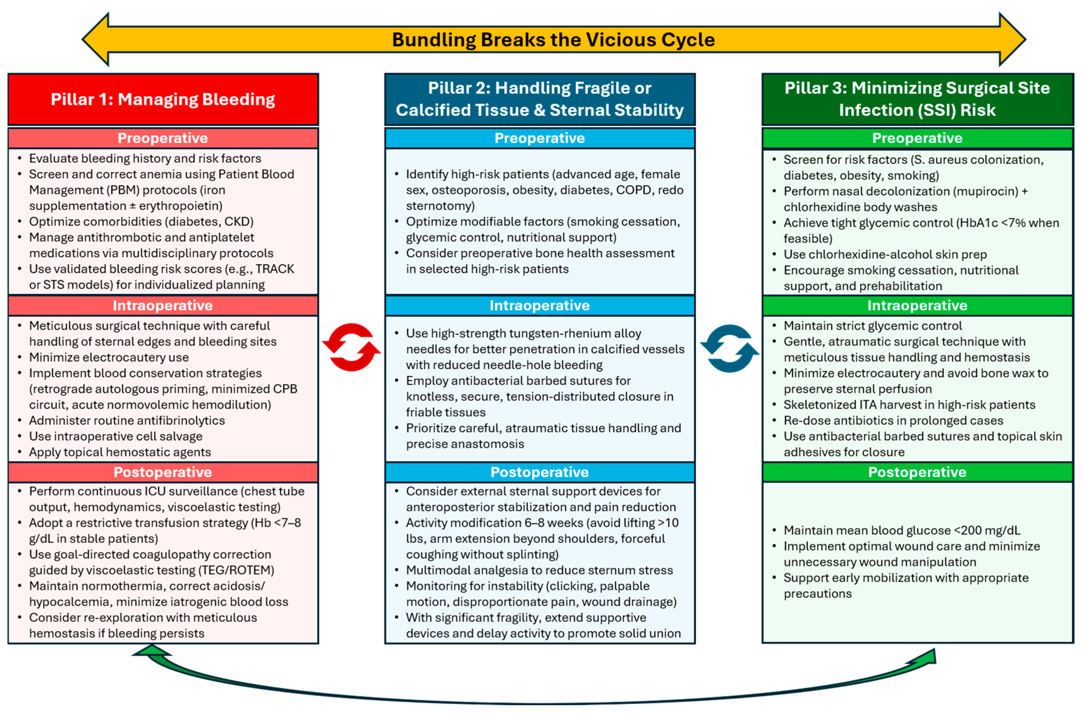

Table 1.
Hemostatic tools intraoperative management of sternotomy bleeding.
| Hemostatic Tool | Mechanism of Action | Key Advantages and Indications | Clinical Benefits |
| Fibrin Sealants (Human) | Mimics the final stage of the natural clotting cascade by combining fibrinogen and thrombin to form a stable fibrin clot. |
Works independently of the patient’s own coagulation profile [37]. Particularly effective in coagulopathic or high-bleeding-risk patients [37]. Due to its dual-syringe, flexible design that facilitates application in confined spaces or on friable tissues. Its effectiveness in reducing localized bleeding and improving hemostasis efficiency makes it suitable in minimally accessible or delicate areas [38,39,40]. | Rapid, adherent, durable clot formation. Reduces postoperative complications and transfusion requirements. |
| Oxidized Regenerated Cellulose (ORC) |
Absorbs fluid (7–10× its weight), activates platelets, and forms a gelatinous matrix that promotes clot formation [41]. | Excellent for capillary, venous, and small arterial oozing [42,43]. Conformable and easy to apply. |
In vitro bactericidal activity against common pathogens (MRSA, MRSE, VRE, E. coli, etc.) [42,44,45]. Fully absorbable. |
| Flowable Hemostatic Matrix |
Combination of gelatin matrix and thrombin that conforms to irregular surfaces and fills bleeding sites. | Ideal for diffuse, broad-surface, or hard-to-access bleeding sites where conventional methods are ineffective [46]. | Effective in complex sternotomy fields with multiple bleeding points. |
| Polypropylene Sutures | Specialized vascular sutures with a 1:1 needle-to-suture ratio that creates a tighter tissue seal at each puncture site. | Specifically designed to reduce suture-hole bleeding from vascular anastomoses, especially in fragile or calcified vessels. | Prevents oozing from needle holes in high-pressure vascular repairs (e.g., aortic or graft anastomoses) . |
Disclaimer/Publisher’s Note: The statements, opinions and data contained in all publications are solely those of the individual author(s) and contributor(s) and not of MDPI and/or the editor(s). MDPI and/or the editor(s) disclaim responsibility for any injury to people or property resulting from any ideas, methods, instructions or products referred to in the content. |
© 2026 by the authors. Licensee MDPI, Basel, Switzerland. This article is an open access article distributed under the terms and conditions of the Creative Commons Attribution (CC BY) license (http://creativecommons.org/licenses/by/4.0/).
Copyright: This open access article is published under a Creative Commons CC BY 4.0 license, which permit the free download, distribution, and reuse, provided that the author and preprint are cited in any reuse.