
Submitted:
03 November 2026
Posted:
04 November 2026
You are already at the latest version
Abstract
RARγ is oncogenic in many cancers including some cases of acute myeloid leukemia, cholangiocarcinoma, and colorectal, gastric, head and neck, hepatocellular, ovarian, pancreatic, prostate, renal, and thyroid cancers (reviewed in [1]). From immuno-histological studies, high level expression was seen for tissue samples from patients with cholangiocarcinoma with lymph node metastases, stage III–IV ovarian cancer, pancreatic cancer, and high-grade prostate cancer. High expression of RARγ in colorectal cancer correlated with the expression of multidrug resistance 1 and a reduced sensitivity to chemotherapeutics. For thyroid cancer, high expression has been linked to the promotion of tumor invasiveness and metastasis. Examination of the survival curves for patients regarding the level of expression of RARγ has shown that high ex-pression is a predictor of poor overall survival outcomes for patients with AML, cholangiocarcinoma, and gastric, ovarian and pancreatic cancers. Active RARγ enhances cell proliferation and plays a role in the maintenance of stem cells. Both these roles are commensurate with RARγhaving a role in aggressive cancer.
Keywords:
retinoic acid receptors
; aggressive cancer
; prognosis
; cell proliferation
; cancer stem cells
1. Introduction
Cancer relapse occurs after initial successful treatment whereby during cancer remission the neoplasm returns. Such occurs for both solid tumors and hematological malignancies. There are various reasons for cancer relapse, such as uncomplete removal of the tumor by surgery, or undetected micro-metastases, but currently cancer stem cells (CSCs) are believed to be the most common cause of relapse [2].
The concept of CSCs was first evidenced by the group of John Dick, as demonstrated when only CD34+CD38−, but neither CD34−, nor CD34+CD38+ cells, from human acute myeloid leukemia (AML) patients, were able to initiate leukemia in severe combined immune-deficient (SCID) mice. These cells, termed leukemia stem cells (LSCs), were scarce in human AML constituting only one in 250,000 of the blast cells [3]. Interestingly, LSCs reconstituted both LSCs and the bulk blast cells in mice, in proportions like the initial AML [4]. Later studies revealed the presence of mutations typical for AML in the hematopoietic stem cells (HSCs) of healthy people. These mutations may occur as early as in utero or in infancy, and they accumulate later in life, leading to hematologic malignancies in some individuals. Specifically, external stressors, such as chemotherapy, bone marrow transplants or HIV infection increase the risk of developing the disease [5].
The CSCs were later identified in solid tumors, such as human breast cancer, whereby 100 CD44+CD24−/low lineage− cells were enough to give rise to tumors in nonobese diabetic/severe combined immunodeficient (NOD/SCID) mice, while much greater numbers of cells bearing other phenotypes failed to form tumors [6]. For human brain tumors, only CD133+ cells were capable of tumor initiation in NOD-SCID mice [7], and the same cell surface molecule, CD133, was identified as CSC marker in human colon cancer [8]. However, cell surface molecules are not always the most important identifiers of CSCs. For example, in cervical [9], prostate [10], colorectal [11], lung [12] and ovarian [13] cancers high aldehyde dehydrogenase (ALDH) enzymatic activity defines CSCs, while SOX2, NANOG, and OCT4 transcription factors are additional markers for lung CSCs [14].
Like normal stem cells, CSCs can maintain their undifferentiated state through self-renewal via asymmetric cell division. One of the daughter cells renews the stem cell pool, while the other gives rise to a progeny of differentiated tumor cells. The CSCs are not susceptible to cell cycle-dependent chemo- or radiotherapy, because of their quiescent state [15]. Moreover, CSCs show high plasticity in response to various stressors, such as inflammation, hypoxia, chemo- or radiotherapy. An example of adaptation is overexpression of detoxifying enzymes such as ALDH, as mentioned above. One of the roles of ALDH is to oxidize retinaldehyde into retinoic acid (ATRA) which is the highly active natural ligand for the retinoic acid receptors (RARs) [16].
RARs belong to the superfamily of nuclear receptors, which are ligand-activated transcription factors. To activate or inhibit gene transcription RARs dimerize with one of the retinoid X receptors (RXRs). The dimers bind to their DNA response elements in promoter regions of the target genes, providing platforms to the complexes of corepressors or coactivators of transcription [17]. The activation of the complex occurs upon binding of the high affinity ligand ATRA to RAR. Additionally, the high affinity ligand to RXR, 9-cis RA may modulate the transcriptional activity, however this step is dispensable [18].
There are three RARs, RARα, RARβ and RARγ, encoded by RARA, RARB and RARG genes, respectively. The three receptors are indispensable for proper embryonic development [19]. Postnatally, RARα and RARβ are ubiquitously distributed among tissues, while RARγ was found in skin [20], in digestive tract, in endocrine tissues, as well as in the bone marrow and lymphoid tissues (https://www.proteinatlas.org/ENSG00000172819-RARG/tissue).
The ligand binding domains (LBDs) of all nuclear receptors are composed of 12 α-helices, which are repositioned upon binding of the respective ligand. The LBDs of the three RARs differ in only three residues, but these differences allow for design of receptor-specific synthetic agonists or antagonists. The difference between agonist and antagonist lies in variable positioning of the helix 12 of LBD. When the receptor is bound to an agonist, helix 12 exposes the surface to which co-activators of transcription can bind, but when bound to an antagonist, the above surface is not exposed, and the receptor remains bound to co-repressors [21]. Due to the known crystal structures of RAR LBDs, it was possible to design subtype-selective synthetic agonists and antagonists [22]. Selective ligands may have also better stability and bioavailability than natural retinoids and thus may be used as drugs. One example is tazarotene, a prodrug of tazarotenic acid, which selectively activates RARβ and RARγ [23]. This drug is topically used against psoriasis, acne and photoaging [24]. Many other selective RAR agonists and antagonists are available for preclinical testing with a view to their use to treat various diseases.
RARγ is oncogenic in many cancers and there is good evidence to support the view that RARγ is expressed by CSCs [25]. This review examines the expression of RARγ in acute myeloid leukemia and solid cancers bringing to attention, from findings for survival curves and other studies, that high expression is linked to aggressive disease and poor prognosis.
2. Acute Myeloid Leukemia (AML)
RARγ is selectively expressed by HSCs and primitive progenitor cells. These cells are within the lineage−, c-kit+, and Sca-1+ (LKS+) compartment of the bone marrow cells of the adult mouse and they express mRNA for RARγ1 [26]. A substantial number of human leukemia cell lines have been derived to date, but they do not typify HSCs, regarding pluripotency, nor primitive progenitor cells. Examination of a database (https://www.proteinatlas.org/ENSG00000172819-RARG/cell+line) of 93 leukemia cell lines for the expression of RARγ mRNA revealed that just the one cell line HNT-34 expressed a good level. This cell line, derived from an AML patient that had a prior myelodysplastic syndrome with 3q21q26 syndrome, is a rare model for EVI1 and P190 BCR/ABL (Philadelphia chromosome) transformation. Morphologically, the cells are undifferentiated blasts that are positive for CD4, CD13, CD33 and CD34, negative for CD41a and CD42b, and negative for myeloperoxidase [27].
AML is genetically and clinically a heterogeneous group of leukemias rather than a single uniform disease. Accordingly, in 1976 and based on morphological criteria, the French-American-British (FAB) classification system divided AML into eight subtypes (M0 – M7) and set aside the myelodysplastic syndromes [28]. The subtypes of AML are undifferentiated (M0), minimal maturation of blasts (M1), with maturation of blasts (M2), acute promyelocytic leukemia with the PML-RARA gene fusion (APL; M3), myelomonocytic (M4), monocytic (M5), erythroid (M6), and megakaryocytic (M7). This subtyping is still largely used regarding the examination of the molecular features of AML.
AML is generally a very aggressive disease whereby the cells grow rapidly. The elderly account for most cases, and prognosis remains poor despite advances in treatment [29]. Prognosis post treatment varies according to the M0 to M7 subtypes. M0, M6, and M7 patients generally have poor prognosis, though a diagnosis of M6 per se does not impart a worse prognosis [30,31,32]. The outlook for patients with APL (M3) is highly favorable because of the combined use of all-trans retinoic acid (ATRA), to target PML-RARα fusion oncoprotein and arsenic trioxide to induce its degradation. This differentiation therapy has transformed APL from being a fatal disease to one that is highly curable because of the direct targeting of the PML-RARα fusion oncoprotein and that APL is associated with few other cytogenetic defects (see also below) [33,34]. Predicting risk has become a more precise and complex matter with the advance of technologies that can be used to readily identify genetic mutations. Stratification of AML into favorable, intermediate I and II, and adverse risks has been related to subsets of genetic mutations [35].
The blood cells of AML patients from The Cancer Genome Atlas project for Acute Myeloid Leukemia (TCGA-LAML) expressed RARA and RARG as seen from analysis of a transcriptomic data set. Figure 1 and Table 1 show the levels of mRNAs for RARα and RARγ as related to the FAB classification of AML. The data for M6 and M7 are not shown because the numbers of patients in the dataset were 2 and 3, respectively. Blast cells from patients with undifferentiated (M0), minimal maturation (M1), and with maturation (M2) AML expressed RARα mRNA at similar levels. Blast cells from patients with acute promyelocytic leukemia (M3), myelomonocytic (M4), and monocytic leukemia (M5) expressed RARα mRNA at higher levels that were similar. APL cells are abnormal promyelocytes as seen from micro and macro azurophilic granulation and folded nuclei [36,37]. The appearance of myeloblasts, monoblasts, and monocytes is a feature of myelomonocytic leukemia, and the cells can present myelomonocytic antigens [38]. The cells vary from monoblasts to promonocytes and abnormal monocytes in monocytic leukemia [39]. It is well known that RARα activity drives myeloid cell differentiation [40]. Hence, the higher levels of expression of RARα mRNA in M3, M4, and M5 AML appear to reflect the increased maturation status of patients’ cells.
RARγ mRNA was expressed by all AML subtypes. The AML M0, M1, M4, and M5 subtypes showed a significant increase in expression as compared to the M3 subtype. This finding is in keeping with the promyelocytic nature of patients’ cells. Intriguingly, the lower level of expression of RARγ mRNA was not seen from the M4 and M5 subtypes of AML whereby the presence of myeloblasts, monoblasts, and monocytes is a feature. The cell of origin, which gives rise to the leukemia stem cells that sustain a patients’ AML cells, is relevant to whether RARγ mRNA is seen to be present at a high or low level or absent. AML can originate from the transformation of normal HSCs, multipotent progenitors or committed progenitors [41]. However, there is some uncertainty regarding this matter. For example, a long-held view is that a committed myeloid progenitor cell is the cell of origin that sustains APL because the bulk cells are promyelocytes [42]. However, the hallmark of APL is the t(15;17) translocation, which generates the PML/RARα fusion protein, and the translocation has been observed to be present in CD34+CD38- cells from patients indicating the involvement of a much early hematopoietic progenitor cell [43].
Does the level of expression of RARγ mRNA provide an indication of a patient’s prognosis? The TCGA-LAML data set was used to answer this question by categorizing whether the expressions of the RARA and RARG genes were low or high in all AML patients’ cells and relating this information to survival. The median value of gene's expression across all samples was used as the threshold to generate the 2 groups of high and low expression. Figure 2 shows the results of the analysis of data for 161 patients. The survival curves for patients with high and low levels of expression of RARα mRNA are almost identical (p = 0.7). By contrast, the survival curves for patients with high and low levels of expression of RARγ mRNA are very different (p = 0.0087). Hence, expression of RARγ mRNA at a high level can be considered as an indicator of poor prognosis.
To examine the relationship between high expression of RARγ mRNA and poor prognosis, the TCGA-LAML database patients were also grouped into AML M0 with undifferentiated leukemia, versus M1 to M7, patients with partially differentiated leukemia. As shown in Figure 3, the survival curves for AML M0 patients with high and low levels of expression of RARγ mRNA are very different (p = 0.029). There is less difference in prognosis for AML M1 to M7 patients providing support to the view that high expression of RARγ mRNA is linked to poor prognosis regarding the aggressive and undifferentiated form of AML.
To further investigate the impact of high RARγ mRNA expression, patients from the TCGA-LAML cohort were also grouped into three cytogenetic risk categories: favorable, intermediate/normal and poor. As shown in Figure 4, the patients from the poor risk category have a significantly higher RARG gene expression compared to the favorable and intermediate/normal risk categories. The correlation between high RARG gene expression and poor prognosis supports a role for RARγ in AML aggressiveness.
As considered above, the t(15;17) translocation generates the PML/RARα fusion protein which, in turn, is the master driver of APL. From in vitro studies that made use of artificial PML-RAR fusion proteins that were generated for each of the RARα, β, and γ isoforms, the fusion proteins were seen to have oncogenic potential [44]. Indeed, for some patients with AML that resembled APL, the RARG gene was seen to be rearranged rather than the RARA gene. The rearrangements give rise to the PML/RARγ fusion protein and fusion proteins from other RARG translocations. They include between RARG and NUP98, which encodes a protein that can act as a transcription activator; between RARG and CPSF6, which encodes a subunit of cleavage factor 1 which, in turn, is involved in the polyadenylation process; between RARG and NPMI, which is the nucleophosmin 1 gene, and between RARG and HNRNPC, which encodes heterogenous nuclear ribonucleoprotein C (reviewed in [45,46]). AML patients with RARG gene rearrangements were insensitive to ATRA and arsenic trioxide (the standard therapy for APL) and had a poor prognosis with a mortality rate of over 60% providing support to the view that RARγ plays a role in aggressive AML.
The modes of action of the RARγ fusion proteins have been investigated. Vectors were used to express the NUP98-RARG, NMI-RARG, and HNRNPC-RARG gene fusion proteins in human CD34+ cells which led to substantial disruption to normal myelopoiesis. Expression increased the ability of the CD34+ cells to self-renew and proliferate and reduced myeloid differentiation [47]. CPSF6-RARγ is the most prevalent fusion protein identified to date. Expression in human CD34+ cells led to the cells exhibiting intensive proliferative capabilities and impaired myeloid maturation. The cells were largely immature myeloid cells (CD34+ CD33+) with fewer mature cells. CPSF6-RARγ interacted with histone deacetylase 3 to suppress myeloid differentiation associated gene expression including the myeloid transcription factor PU.1. RAS mutations are commonly seen alongside CPSF-6-RARγ fusion proteins. The investigators examined their interaction by expressing oncogenic RAS in CPSF-6-RARG mouse haematopoietic stem and progenitor cells. Sub-lethally irradiated mice died from induced disease at around 150 days post transplantation of NRAS HSPCs. The co-presence of CPSF6-RARγ significantly accelerated leukemic progression with the mice dying within 110 days. Similar results were obtained for KRAS. From these findings, synergy between CPSF6-RARγ and RAS mutations was proposed to drive more aggressive AML with RARγ enhancing AML aggressiveness [48].
3. Solid Cancer
3.1. Cholangiocarcinoma
Cholangiocarcinoma is a rare and aggressive bile duct epithelial tumour that spreads fast and is hard to cure. The prognosis is poor [49]. From immuno-histological analysis of a set of 42 tumor tissues, their adjacent non-cancerous tissues, 27 cholangitis samples and 13 normal tissues, RARγ protein was frequently overexpressed in human cholangiocarcinoma specimens. Overexpression was associated with poor differentiation and lymph node metastases. Examination of the postoperative survival rate in patients showed that patients with a high level of RARG gene expression had a median survival time of 9.2 months compared to 27.6 months for patients with a low level of expression. The investigators concluded from findings that the level of RARG gene expression is clinically significant and valuable to the prognosis of patients with cholangiocarcinoma [50].
3.2. Colorectal Cancer
RARγ mRNA and protein levels were found to be upregulated in 23 colorectal cancer (CRC) specimens compared to their adjacent non-tumorous tissue [51]. The level of RARγ protein was also upregulated in the CRC cell lines HT29, HCT116, KKO, and SW480 compared to the normal colon epithelial cells HCoEpiC. The investigators showed that RARγ knockdown in three CRC cell lines enhanced their sensitivity to chemotherapeutic drugs. They showed that HT29, HCT116, and RKO shRARγ cells treated with oxaliplatin, fluorouracil or vincristine had a decreased cell viability compared to the shCtrl cells. Furthermore, the multidrug resistance 1 (MDR1) protein was downregulated in shRARγ CRC cells. The correlation between RARγ and MDR1 expression was confirmed across 90 tumor tissue microarrays. These results highlight the role of RARγ in CRC progression and drug resistance through the activation of the Wnt/β-catenin pathway by upregulating MDR1 [51].
3.3. Gastric Cancer
Gastric cancer (GC) is associated with poor outcomes mainly due to heterogeneous tumor microenvironments and genetic events. In the GSE183904 database, RARG gene expression was enriched in the epithelial cells within tumors compared to normal tissues [52]. Tissue microarray analysis confirmed these results in 76 gastric cancer specimens compared to their paired normal tissues. Moreover, the analysis of the TCGA database and data from GC patients shows that high expression of RARγ mRNA was associated with poor overall survival. RARγ activated Prostate Transmembrane Protein, Androgen Induced 1 (PMEPA1) in gastric cancer cells. Knock out of RARγ expression decreased PMEPA1 expression, as well as the cell proliferation and invasiveness of MKN-25 cells. CUT&Tag sequencing analysis of HGC-27 cells overexpressing RARγ-Flag followed by visualization using the Integrative Genomics Viewer (IGV), showed that RARγ binds to the PMEPA1 promoter. Further analysis demonstrated that RARγ activated PMEPA1 via binding to its promoter region site 2. They also showed that RARγ activates the Wnt/β-catenin signaling pathway. This activation was due to the direct binding of RARg to β-catenin to facilitate its nuclear translocation. The inhibition of tumor cells proliferation in the murine CDX model inoculated with MKN-45 cells knockout for RARγ indicated its role in gastric cancer progression. The investigators identified a dual mechanism involving the RARγ-PMEPA1 axis and the activation Wnt/β-catenin signaling pathway as contributing to gastric cancer progression [52].
3.4. Ovarian Cancer
The levels of mRNA for RARγ within 88 normal ovarian samples and tissue from 378 patients with ovarian cancer were compared by making use of information downloaded from the Cancer Genome Atlas (TCGA) and the Genotype Tissue Expression (GTEx) databases. RARγ mRNA was significantly over expressed in ovarian cancer cells compared to expression in normal ovarian samples. Analysis of subgroups revealed that expression in cells from patients with Tumor, Node, and Metastasis (TNM) stage III-IV was higher than that seen in the cells from patients with stage I-II ovarian cancer (p < 0.05). One and a half percent of patients had RARG gene alterations including amplifications and mutations [53].
Advanced-stage ovarian cancer is an aggressive disease with a median overall survival for patients treated with chemotherapy alone ranging from 7.8 to 19.5 months [54]. High RARG gene expression was an independent predictor of poor overall survival outcomes. Three hundred and seventy-five patients were divided into patients with a high (187 patients) and low level (188 patients) of expression of RARγ mRNA. The patients with high level expression of RARγ mRNA had poorer prognosis (p < 0.0001) than the rest. Survival curves for subgroups of patients revealed that those with high RARγ mRNA in the age <60, age ≥60, histological grade 1–2, histological grade 3, and TNM stage III–IV ovarian cancer had a poor prognosis. Hence, high expression of RARγ mRNA led to poor prognosis in most clinical subgroups. The investigators concluded from GSEA enrichment analysis that RARG gene expression participates in multiple pathways and can act as an oncogene to promote ovarian cancer progression [53].
3.5. Pancreatic Cancer
Pancreatic cancer is an aggressive and lethal disease with a medium survival of ~4 months [55]. Examination of the TCGA and GEO database showed RARγ mRNA was commonly overexpressed in human pancreatic cancer. RARγ protein was likely to be highly expressed because 11 of 12 tumor samples showed a staining quantity of 75% in the immuno-histochemical analysis of pancreatic tumor tissues. Expression of RARγ mRNA was an independent diagnostic marker predicting the poor prognosis of pancreatic cancer patients as seen from comparisons of the survival of groups of patients that expressed RARγ mRNA at high and low levels. Patient survival was not associated with the levels of expression of mRNAs for RARα, RARβ, RXRα, RXRβ, and RXRγ [56]. Other workers have reported that overexpression of RARγ in pancreatic cancer and that this was associated with poor patient prognosis. Protein expression was examined by immunostaining of surgical specimens of pancreatic ductal adenocarcinoma, precancerous lesions, and adjacent normal pancreatic ductal epithelium. The immunohistology scores for RARγ expression for pancreatic ductal adenocarcinoma, high-grade precancerous lesions, and low-grade precancerous lesions were significantly higher than those obtained for normal pancreatic ductal epithelium [57].
3.6. Prostate Cancer
RARγ is an oncogene in prostate cancer [58] and RARβ is viewed as a tumour suppressor as expression is often silenced in prostate cancer [59]. Immuno-histological studies examined the levels of expression of RAR proteins for normal prostate epithelium, benign hyperplasia, prostatic intra-epithelial neoplasia, and various grade cancerous glands [60]. The levels of RARβ and RARγ are of particular interest. RARγ was present in cells throughout normal epithelium but with little nuclear accumulation. RARβ was present only in the nuclei of basal epithelial cells. Expression of RARγ was seen in each of the pathological conditions. This included strong positivity in the nuclei of basal cells and the nuclei and cytoplasm of luminal cells in benign prostate hyperplasia, strong positivity in the nuclei of basal cells in intra-epithelial neoplasia, an increased expression in some glands regarding low- and intermediate-grade prostate cancer with strong positive staining in the cytoplasm and nucleus of some cells, and strong positive nuclear staining in high grade prostate cancer. The increased RARγ staining seen in the basal epithelial cells of benign hyperplasia and intra-epithelial neoplasia may indicate that these cells are contributing to the pathological conditions. An increased expression of RARγ in some low-and intermediate-grade cells may reflect cells undergoing further tumorigenic change whereby strong staining was seen for high grade cancer cells. The expression level of RARβ was increased in the nuclei of basal cells and nuclei and cytoplasm of luminal epithelial cells for glands that exhibited benign prostatic hyperplasia and levels persisted in the nuclei of the basal epithelial cells for glands that exhibited prostatic intra-epithelial neoplasia. RARβ expression was absent in low-, intermediate-, and high-grade prostate cancer in keeping with RARβ expression is often absent. The above findings support the view that the levels of expression RARγ and RARβ appear to play roles in the development and/or nature of aggressive prostate cancer.
3.7. Thyroid Cancer
Thyroid carcinoma is an endocrine carcinoma with a favorable prognosis, but metastasis occurs for a subset of patients [61]. RARγ was upregulated in thyroid carcinoma [62] and RARγ mRNA level was increased in thyroid carcinoma tissues from the TCGA database, compared to normal thyroid tissues. These results were confirmed at the mRNA and protein levels using RT-qPCR and Western blot analysis of 14 thyroid carcinoma samples compared to 13 normal thyroid samples. The overexpression of RARγ increases the expression of CD24 and the matrix metalloproteinases MMP2 and MMP9, through the activation of the JAK-STAT3 pathway. The MMPs degrade the extracellular matrix and promote tumor invasion and metastasis [63]. Moreover, the protein expression level of STAT3, p-STAT3 and CD24 was higher for seven thyroid carcinoma samples as compared to a patient’s normal thyroid tissue [62]. The investigators concluded that RARγ promotes proliferation, invasion and migration of thyroid carcinoma cells. Furthermore, RARγ may reduce the phagocytic capacity of macrophages to respond to thyroid carcinoma cells by upregulating CD24.
4. RARγ Drives Cell Proliferation
In addition to the correlation clinical studies presented above, mechanistic studies have been undertaken using cell lines. The involvements of RARγ in the progression of cholangiocarcinoma, pancreatic, ovarian, and prostate cancers have been attributed to RARγ-mediated enhancement of cell proliferation. RAR signaling modulates pathways that play a key role in oncogenesis to influence cell proliferation and tumor progression. They include the MAPK, PI3K/Akt, Wnt/β-catenin, and TGFβ pathways [64,65,66].
Endogenous levels of RARγ were reduced in the three cholangiocarcinoma cell lines QBC939, SK-ChA-1, and M2-ChA-1 by siRARγ transfection which inhibited cell proliferation and cell cycle progression with cells accumulating in the G1 phase of cell cycle. Transfection of siRARγ also significantly impaired the capacity of the three cell lines to give rise to colonies in a plate assay. Knockdown of expression of RARγ inhibited the migration of QBC939 cells in wound-healing and trans-well assays and enhanced the sensitivity of the cell line cells to 5-fluorouracil. SiRARγ and siControl QBC939 cells were grafted into mice which were then treated with 5-fluorouracil and a higher rate of growth inhibition was seen for siRARγ tumors (56.7%) as compared to the control tumor group of mice (5.6%) [50]. The oncogenic action of RARγ was attributed to activation of the Akt/NF-κB and Wnt/β-catenin pathways whereby both pathways drive cell proliferation.
Expression of RARγ was required for the proliferation of pancreatic cancer cells [56]. RARγ mRNA and protein vanished from CRISPR-Cas9 knockout of RARG in the PANC1 pancreatic cell line cells with such leading to cell cycle arrest at the S phase of the cell cycle followed by apoptosis. Knockdown of expression of RARγ significantly reduced the proliferation of the pancreatic cancer cell lines BXPC3 and SW1990. Expression levels of the oncogenes MYC, SLC2A1, and STAT3 were downregulated in the RARG knockdown PANC1 cells and chip-qPCR analysis showed that RARγ bound directly to the promoters of the oncogenic MYC and STAT3 genes. As compared to wild type PANC1 cells when injected into immunodeficient NOD/SCID mice, the growth of RARγ-deficient PANC1 cells was substantially reduced. Other workers have made use of siRNA knockdown to investigate the role of RARγ in the PANC1 and PK-1 cell line cells showing that loss of RARγ signalling suppressed cell proliferation [57].
The proliferation and the colony forming capacity of the A2780 and SVOV3 human ovarian cancer cell lines in vitro were suppressed by siRNA down regulation of RARγ expression. The growth of tumors by A2780 cells in nude mice was decreased by sh-RARG knockdown of RARγ and reduced levels of Ki-67 and the proliferation cell nuclear antigen were observed [53]. RARγ plays a role in the proliferation of prostate cancer cells because the treatment of the prostate cancer cell lines LNCaP, PC-3, and DU145 with the RARγ agonist AGN205327 or a low dose of ATRA (10−11 to 10−9 M), which was just sufficient to activate only RARγ, enhanced the proliferation of the three cell lines cells. The RARα-selective agonists AM580 and AGN195183 failed to promote cell growth and co-treatment of LNCaP cells with ATRA and the RARα-selective antagonist AGN196996 promoted proliferation of LNCaP cells, likely due to the activity of ATRA on RARγ.
For the above cancers, RARγ expression contributed to enhanced cell proliferation. An important feature is that there are interactions between RARγ and oncogenes that are the frequent cornerstones to malignancies and that strongly enhance cell proliferation. As considered above, synergy between RARγ and mutated RAS was seen in aggressive AML. RARγ bound directly to the promotor of the MYC gene in aggressive pancreatic cancer and MYC gene expression was downregulated in RARγ knockdown cells. MMTV-Myc is considered as an aggressive and metastatic disease model of breast cancer [67]. From studies of this model of tumorigenesis, the prevalence of expression of RARγ over that of RARα/β accompanied by MYC gene overexpression or amplification have been linked to the progression of tumor growth [68]. Some cancer cells, including breast, ovarian, pancreatic, and prostate cancer cells, reside in a low ATRA environment and their survival and growth may be dependent on RARγ as activated by sub nM ATRA (reviewed in [65]).
5. RARγ Expression and Stem Cell Stemness
RARγ also plays a role in the maintenance of stem cells. Activity of RARγ was required for the maintenance of HSCs in the adult mouse. They were profoundly decreased in number within the bone marrow of femurs of RARγ−/− mice and there was a corresponding increase in differentiated progenitor cells [26]. Similarly, mature myeloid cells increased in conditional RARγ−/− mice, and when wild type mice were treated with the pan-RAR antagonist AGN194310. Primitive hematopoietic precursors that had been transduced to overexpress RARγ1 exhibited an undifferentiated phenotype [69]. Further support to the importance of RARγ to the maintenance of stem cells is that agonism of RARγ enhanced the generation of induced pluripotent stem cells from somatic cells [70,71,72].
Expression of RARγ mRNA was restricted to stem cells and primitive progenitor cells during mouse and zebrafish embryogenesis and mouse gastruloid development [73,74,75,76]. Gastruloids were generated by aggregating E14Tg2a embryonic stem cells into spheroids and treating them with the Wnt/β-catenin agonist CHIR9901. The spheroids recapitulated many aspects of early development. Activation of RARγ by the selective agonist AGN205728 blocked the developmental progression of stem/primitive progenitor cells as seen for zebrafish embryos [77] and mouse gastruloids [76]. The RARγ agonist mediated blocks to pectoral and dorsal fin development were reversed by washout of the agonist or the subsequent addition of an RARγ antagonist and the block to gastruloid development was partially reversible by the subsequent addition of antagonist. Cartilaginous nodule formation was blocked when micro-mass cultures of mesenchymal embryo limb cells obtained from day 11.5 were treated with the RARγ agonist NRX20467. This agonist also blocked ectopic bone mass formation when cultured mesenchymal cells were treated with the RARγ agonist and then injected into nude mice and heterotopic ossification in a transgenic model of fibrodysplasia ossificans progressiva [78]. From all the above findings it appears that RARγ acts as a gatekeeper to stem/progenitor cell development whereby development is blocked when ATRA agonized and permissive when ligand is not bound [1].
6. Targeting the Activity of RARγ
As considered above, for proliferative diseases the need is to antagonize RARγ because its activity enhances cell proliferation. Antagonists include AGN205728 [79], MM11253 [80], and LYS2955303 [81] for the selective antagonism of RARγ and AGN194310 [79] and AGN193109 [82] for all RARs.
Antagonism of all RARs was effective in reducing the abundance of LSCs in the context of AML with overexpression of EVI1 which characterizes aggressive AML with a poor prognosis [83]. The cancer cells in the MLL-AF9-driven mouse model of AML express the transcription factor Evi1 and this model was used to examine the effect of antagonizing all RARs [84]. In vitro treatment of bone marrow cells from mice that were terminally ill from MLL-AF9-driven leukemia with 1 μM of the pan-RAR antagonist AGN193109 decreased LSC abundance and quiescence. Treatment of mice with MLL-AF9-driven leukemia with AGN193109 reduced LSC abundance, quiescence, and delayed leukemogenesis. LSCs in the spleen were reduced and overall survival of mice was improved as compared to the control group. AGN193109 also increased myeloid differentiation (CD11b+ GR1+ cells) and decreased cell stemness. For three out of four EVI1high AML patient samples, the AGN193109 decreased the clonogenic capacity of cells, but had a minimal effect on EVI1low AML patient samples [84]. The investigators concluded that antagonizing all RARs provides a means to target LSCs in EVI1high AML.
The pan-RAR antagonist AGN194310 and the RARγ-selective antagonist AGN205728 reduced the colony-forming capacity of the LNCaP, PC-3 and DU145 prostate cancer cell line cells and were effective against primary patients’ cells [58,85,86]. Both antagonists induced growth arrest followed by cell death of flask cultured LNCaP, PC-3 and DU145 cells. Treatment of LNCaP cells with 1µM of AGN194310 or AGN205728 increased the proportion of cells in the sub-G1 phase of cell cycle (at 48 hours) and ~35% (AGN194310) and ~40% (AGN205728) of cells underwent caspase-independent cell death via necroptosis (at 60 hours). Antagonism of RARγ was sufficient to induce growth arrest and cell death of prostate cancer cells. Combination treatment of LNCaP cells with the chemotherapeutic agents (etoposide, docetaxel or fluorouracil) and the RARγ antagonist AGN205728 significantly reduced cell viability as compared to treatment with each chemotherapeutic agent alone.
Regarding other solid cancers, treatment of pancreatic cancer cell lines and established patient-derived organoids (KYK070, KYK002, KYK023, KYK090 and KYK093) with the RARγ antagonists LY2955303 and MM11225, suppressed cell proliferation with the cells arresting in G1 of cell cycle and dying by apoptosis [57]. The pan-RAR antagonist AGN194310 ablated neurosphere formation by the CSCs within primary cell cultures that were obtained from two paediatric patients with primitive neuroectodermal tumours, and from a patient with an astrocytoma [25]. The pan-RAR antagonist AGN193109 reduced the expression of the RAR target genes RARB and POLQ in the ovarian cancer OVCAR3 and PEO1 cell line cells [87]. In addition, inhibitors of ATRA metabolism have been used to target the activity of RARs within ovarian cancer. To sensitize these cells to cisplatin and paclitaxel, chemo resistant A2780CP70 cells were treated with Solanum incanum extract (SR-T100) which contains solomargine. Solomargine downregulated ALDH1 expression, which contributes to the synthesis of ATRA. ALDH1 is considered to be a CSCs marker and involved in the maintenance of cell stemness in ovarian cancer [16]. The natural flavonoid acacetin, which displaces ATRA from RARγ, induced apoptosis of hepatocellular cancer cell line cells [88]. Overall, findings to date highlight the therapeutic potential of RARs and RARγ as druggable targets to decrease tumor growth and eliminate CSCs.
7. Concluding Remarks
RARγ is oncogenic in many cancers by virtue of overexpression in solid cancers and the presence of RARγ fusion proteins in AML. Findings from histological and mechanistic studies and examination of patient survival curves have linked overexpression of RARγ within AML and cholangiocarcinoma and colorectal, gastric, ovarian, pancreatic and thyroid cancers to aggressive disease, metastases, and poor patient outcomes. RARγ activity enhanced the proliferation of cancer cells whereby agonism enhanced the growth of prostate cancer cells and downregulation suppressed the growth of cholangiocarcinoma, pancreatic and ovarian cancer cells. From developmental studies and of the generation of induced pluripotent stem cells from somatic cells, RARγ plays a role to block stem cell development. It facilitated cell stemness including the maintenance of HSCs and the generation of induced pluripotent stem cells. For the future, there is the need to resolve the extents to which RARγ-driven enhancement of cell proliferation and/or stem cell maintenance/stemness are pivotal to the aggressive nature of cancer. Nevertheless, RARγ playing a role in aggressive cancer leads to the possible use of an antagonist as an adjunct to the treatment of aggressive disease.
Author Contributions
Performed the analyses, M.R.; wrote the article, G.B., E.M. and M.R. All authors have read and agreed to the published version of the manuscript.
Funding
G.B. and E.M. are partners to the EU funded program eRaDicate. G.B. is funded by UK Research and Innovation (UKRI) under the UK government’s Horizon Europe funding guarantee EP/Y030818/1. M.R. is a Marie Curie Fellow to eRaDicate project funded by Horizon Europe under the grant agreement No 101119427.
Institutional Review Board Statement
Not applicable.
Informed Consent Statement
Not applicable.
Data Availability Statement
No new data were created or analyzed in this study. Data sharing is not applicable to this article.
Conflicts of Interest
The authors declare no conflict of interest.
References
- Johnson, W. E. B.; McQueen, C.; Brown, G. Retinoic Acid Receptor γ Is a Ligand-Activated Gatekeeper to Stem Cell Developmental Progression. Int. J. Mol. Sci. 2026, 27((16)), 7160. [Google Scholar] [CrossRef] [PubMed]
- Marzagalli, M.; Fontana, F.; Raimondi, M.; Limonta, P. Cancer Stem Cells—Key Players in Tumor Relapse. Cancers 2021, 13((3)), 376. [Google Scholar] [CrossRef] [PubMed]
- Lapidot, T.; Sirard, C.; Vormoor, J.; Murdoch, B.; Hoang, T.; Caceres-Cortes, J.; Minden, M.; Paterson, B.; Caligiuri, M. A.; Dick, J. E. A cell initiating human acute myeloid leukaemia after transplantation into SCID mice. Nature 1994, 367((6464)), 645–648. [Google Scholar] [CrossRef] [PubMed]
- Bonnet, D.; Dick, J. Human acute myeloid leukemia is organized as a hierarchy that originates from a primitive hematopoietic cell. Nat. Med. 1997, 3((7)), 730–7. [Google Scholar] [CrossRef] [PubMed]
- Köhnke, T.; Majeti, R. Clonal Hematopoiesis: From Mechanisms to Clinical Intervention. Cancer Discov. 2021, 11((12)), 2987–2997. [Google Scholar] [CrossRef] [PubMed]
- Al-Hajj, M.; Wicha, M. S.; Benito-Hernandez, A.; Morrison, S. J.; Clarke, M. F. Prospective identification of tumorigenic breast cancer cells. Proc. Natl. Acad. Sci. U S A 2003, 100((7)), 3983–8. [Google Scholar] [CrossRef] [PubMed]
- Singh, S. K.; Hawkins, C.; Clarke, I. D.; Squire, J. A.; Bayani, J.; Hide, T.; Henkelman, R. M.; Cusimano, M. D.; Dirks, P. B. Identification of human brain tumour initiating cells. Nature 2004, 432((7015)), 396–401. [Google Scholar] [CrossRef] [PubMed]
- O'Brien, C. A.; Pollett, A.; Gallinger, S.; Dick, J. E. A human colon cancer cell capable of initiating tumour growth in immunodeficient mice. Nature 2007, 445((7123)), 106–10. [Google Scholar] [CrossRef] [PubMed]
- Sarabia-Sánchez, M. Á.; Alvarado-Ortiz, E.; Toledo-Guzman, M. E.; García-Carrancá, A.; Ortiz-Sánchez, E. ALDHHIGH Population Is Regulated by the AKT/β-Catenin Pathway in a Cervical Cancer Model. Front. Oncol. 2020, 10. [Google Scholar] [CrossRef] [PubMed]
- Li, T.; Su, Y.; Mei, Y.; Leng, Q.; Leng, B.; Liu, Z.; Stass, S. A.; Jiang, F. ALDH1A1 is a marker for malignant prostate stem cells and predictor of prostate cancer patients' outcome. Lab Invest 2010, 90((2)), 234–44. [Google Scholar] [CrossRef] [PubMed]
- Singh, S.; Arcaroli, J.; Chen, Y.; Thompson, D. C.; Messersmith, W.; Jimeno, A.; Vasiliou, V. ALDH1B1 Is Crucial for Colon Tumorigenesis by Modulating Wnt/β-Catenin, Notch and PI3K/Akt Signaling Pathways. PLoS ONE 2015, 10((5)), e0121648. [Google Scholar] [CrossRef] [PubMed]
- Liu, J.; Xiao, Z.; Wong, S. K.-M.; Tin, V. P.-C.; Ho, K.-Y.; Wang, J.; Sham, M.-H.; Wong, M. P. Lung cancer tumorigenicity and drug resistance are maintained through ALDHhi CD44hi tumor initiating cells. Oncotarget 2013, 4, 10. [Google Scholar] [CrossRef] [PubMed]
- Kuroda, T.; Hirohashi, Y.; Torigoe, T.; Yasuda, K.; Takahashi, A.; Asanuma, H.; Morita, R.; Mariya, T.; Asano, T.; Mizuuchi, M.; Saito, T.; Sato, N. ALDH1-high ovarian cancer stem-like cells can be isolated from serous and clear cell adenocarcinoma cells, and ALDH1 high expression is associated with poor prognosis. PLoS ONE 2013, 8((6)), e65158. [Google Scholar] [CrossRef] [PubMed]
- Zheng, Y.; Wang, L.; Yin, L.; Yao, Z.; Tong, R.; Xue, J.; Lu, Y. Lung Cancer Stem Cell Markers as Therapeutic Targets: An Update on Signaling Pathways and Therapies. Front Oncol. 2022, 12, 873994. [Google Scholar] [CrossRef] [PubMed]
- De Angelis, M. L.; Francescangeli, F.; La Torre, F.; Zeuner, A. Stem Cell Plasticity and Dormancy in the Development of Cancer Therapy Resistance. Front Oncol. 2019, 9, 626. [Google Scholar] [CrossRef] [PubMed]
- Labrecque, J.; Dumas, F.; Lacroix, A.; Bhat, P. V. A novel isoenzyme of aldehyde dehydrogenase specifically involved in the biosynthesis of 9-cis and all-trans retinoic acid. Biochem J. 1995, 305 ( Pt 2) (Pt 2), 681–4. [Google Scholar] [CrossRef] [PubMed]
- Rastinejad, F. Retinoic acid receptor structures: the journey from single domains to full-length complex. J. Mol. Endocrinol. 2022, 69((4)), T25–t36. [Google Scholar] [CrossRef] [PubMed]
- Mic, F. A.; Molotkov, A.; Benbrook, D. M.; Duester, G. Retinoid activation of retinoic acid receptor but not retinoid X receptor is sufficient to rescue lethal defect in retinoic acid synthesis. Proc. Natl. Acad. Sci. U S A 2003, 100((12)), 7135–40. [Google Scholar] [CrossRef] [PubMed]
- Mark, M.; Ghyselinck, N. B.; Chambon, P. Function of retinoic acid receptors during embryonic development. Nucl. Recept Signal 2009, 7, e002. [Google Scholar] [CrossRef] [PubMed]
- Zelent, A.; Krust, A.; Petkovich, M.; Kastner, P.; Chambon, P. Cloning of murine α and β retinoic acid receptors and a novel receptor γ predominantly expressed in skin. Nature 1989, 339((6227)), 714–717. [Google Scholar] [CrossRef] [PubMed]
- Géhin, M.; Vivat, V.; Wurtz, J. M.; Losson, R.; Chambon, P.; Moras, D.; Gronemeyer, H. Structural basis for engineering of retinoic acid receptor isotype-selective agonists and antagonists. Chem. Biol. 1999, 6((8)), 519–29. [Google Scholar] [CrossRef] [PubMed]
- Tambones, I.; Sagar, A.; Vankova, P.; Loginov, D.; Carivenc, C.; Rochel, N.; Bourguet, W.; Man, P.; Bernadó, P.; le Maire, A. New structural insights into the control of the retinoic acid receptors RAR/RXR by DNA, ligands, and transcriptional coregulators. Nucleic Acids Res. 2025, 53, 18. [Google Scholar] [CrossRef] [PubMed]
- Chandraratna, R. A. S. Tazarotene—first of a new generation of receptor-selective retinoids. Br. J. Dermatol. 1996, 135((s49)), 18–25. [Google Scholar] [CrossRef] [PubMed]
- Roeder, A.; Schaller, M.; Schäfer-Korting, M.; Korting, H. C. Tazarotene: therapeutic strategies in the treatment of psoriasis, acne and photoaging. Skin. Pharmacol. Physiol. 2004, 17((3)), 111–8. [Google Scholar] [CrossRef] [PubMed]
- Brown, G. The Influences of RARγ on the Behavior of Normal and Cancer Stem Cells. Int. J. Mol. Sci. 2026, 27((3)), 1291. [Google Scholar] [CrossRef] [PubMed]
- Purton, L. E.; Dworkin, S.; Olsen, G. H.; Walkley, C. R.; Fabb, S. A.; Collins, S. J.; Chambon, P. RARgamma is critical for maintaining a balance between hematopoietic stem cell self-renewal and differentiation. J. Exp. Med. 2006, 203((5)), 1283–93. [Google Scholar] [CrossRef] [PubMed]
- Hamaguchi, H.; Nagata, K.; Yamamoto, K.; Fujikawa, I.; Kobayashi, M.; Eguchi, M. Establishment of a novel human myeloid leukaemia cell line (FKH-1) with t(6;9)(p23;q34) and the expression of dek-can chimaeric transcript. Br. J. Haematol. 1998, 102((5)), 1249–56. [Google Scholar] [CrossRef] [PubMed]
- Bennett, J.; Catovsky, D.; Daniel, M.; Flandrin, G.; Galton, D.; Gralnick, H.; Sultan, C. Proposals for the classification of the acute leukaemias. French-American-British (FAB) co-operative group. Br. J. Haematol. 1976, 33, 451–8. [Google Scholar] [CrossRef] [PubMed]
- De Kouchkovsky, I.; Abdul-Hay, M. Acute myeloid leukemia: a comprehensive review and 2016 update. Blood Cancer J. 2016, 6((7)), e441. [Google Scholar] [CrossRef] [PubMed]
- Gougounon, A.; Abahssain, H.; Rigollet, L.; Elhamri, M.; Tigaud, I.; Chelghoum, Y.; Plesa, A.; Dumontet, C.; Michallet, M.; Thomas, X. Minimally differentiated acute myeloid leukemia (FAB AML-M0): Prognostic factors and treatment effects on survival—A retrospective study of 42 adult cases. Leuk. Res. 2011, 35((8)), 1027–1031. [Google Scholar] [CrossRef] [PubMed]
- Zuo, Z.; Medeiros, L. J.; Chen, Z.; Liu, D.; Bueso-Ramos, C. E.; Luthra, R.; Wang, S. A. Acute myeloid leukemia (AML) with erythroid predominance exhibits clinical and molecular characteristics that differ from other types of AML. PLoS ONE 2012, 7((7)), e41485. [Google Scholar] [CrossRef] [PubMed]
- Oki, Y.; Kantarjian, H. M.; Zhou, X.; Cortes, J.; Faderl, S.; Verstovsek, S.; O'Brien, S.; Koller, C.; Beran, M.; Bekele, B. N.; Pierce, S.; Thomas, D.; Ravandi, F.; Wierda, W. G.; Giles, F.; Ferrajoli, A.; Jabbour, E.; Keating, M. J.; Bueso-Ramos, C. E.; Estey, E.; Garcia-Manero, G. Adult acute megakaryocytic leukemia: an analysis of 37 patients treated at M.D. Anderson Cancer Center. Blood 2006, 107((3)), 880–4. [Google Scholar] [CrossRef] [PubMed]
- de Thé, H.; Pandolfi, P. P.; Chen, Z. Acute Promyelocytic Leukemia: A Paradigm for Oncoprotein-Targeted Cure. Cancer Cell 2017, 32((5)), 552–560. [Google Scholar] [CrossRef] [PubMed]
- Wang, Z.; Chen, Z. Acute promyelocytic leukemia: from highly fatal to highly curable. Blood 2008, 111, 2505–15. [Google Scholar] [CrossRef] [PubMed]
- Saultz, J. N.; Garzon, R. Acute Myeloid Leukemia: A Concise Review. J. Clin. Med. 2016, 5((3)), 33. [Google Scholar] [CrossRef] [PubMed]
- Neame, P. B.; Soamboonsrup, P.; Leber, B.; Carter, R. F.; Sunisloe, L.; Patterson, W.; Orzel, A.; Bates, S.; McBride, J. A. Morphology of acute promyelocytic leukemia with cytogenetic or molecular evidence for the diagnosis: characterization of additional microgranular variants. Am. J. Hematol. 1997, 56((3)), 131–42. [Google Scholar] [CrossRef]
- Yoshii, M.; Ishida, M.; Yoshida, T.; Okuno, H.; Nakanishi, R.; Horinouchi, A.; Hodohara, K.; Okabe, H. Clinicopathological features of acute promyelocytic leukemia: an experience in one institute emphasizing the morphological and immunophenotypic changes at the time of relapse. Int. J. Clin. Exp. Pathol. 2013, 6((10)), 2192–8. [Google Scholar] [PubMed]
- Santos, I. M.; Franzon, C. M.; Koga, A. H. Laboratory diagnosis of chronic myelomonocytic leukemia and progression to acute leukemia in association with chronic lymphocytic leukemia: morphological features and immunophenotypic profile. Rev. Bras. Hematol. Hemoter. 2012, 34((3)), 242–4. [Google Scholar] [CrossRef] [PubMed]
- Varotto, E.; Munaretto, E.; Stefanachi, F.; Della Torre, F.; Buldini, B. Diagnostic challenges in acute monoblastic/monocytic leukemia in children. Front Pediatr. 2022, 10, 911093. [Google Scholar] [CrossRef] [PubMed]
- Kastner, P.; Chan, S. Function of RARalpha during the maturation of neutrophils. Oncogene 2001, 20, 7178–85. [Google Scholar] [CrossRef] [PubMed]
- Long, N. A.; Golla, U.; Sharma, A.; Claxton, D. F. Acute Myeloid Leukemia Stem Cells: Origin, Characteristics, and Clinical Implications. Stem Cell Rev. Rep. 2022, 18((4)), 1211–1226. [Google Scholar] [CrossRef] [PubMed]
- Grimwade, D.; Hills, R.; Moorman, A.; Walker, H.; Chatters, S.; Goldstone, A.; Wheatley, K.; Harrison, C.; Burnett, A. National Cancer Research Institute Adult Leukaemia Working Group, Refinement of cytogenetic classification in acute myeloid leukemia: determination of prognostic significance of rare recurring chromosomal abnormalities among 5876 younger adult patients treated in the United Kingdom Medical Research Council trials. Blood 2010, 116((3)), 354–65. [Google Scholar] [PubMed]
- Edwards, R. H.; Wasik, M. A.; Finan, J.; Rodriguez, R.; Moore, J.; Kamoun, M.; Rennert, H.; Bird, J.; Nowell, P. C.; Salhany, K. E. Evidence for early hematopoietic progenitor cell involvement in acute promyelocytic leukemia. Am. J. Clin. Pathol. 1999, 112((6)), 819–27. [Google Scholar] [CrossRef] [PubMed]
- Marinelli, A.; Bossi, D.; Pelicci, P. G.; Minucci, S. A redundant oncogenic potential of the retinoic receptor (RAR) α, β and γ isoforms in acute promyelocytic leukemia. Leukemia 2007, 21((4)), 647–650. [Google Scholar] [CrossRef] [PubMed]
- Conserva, M. R.; Redavid, I.; Anelli, L.; Zagaria, A.; Specchia, G.; Albano, F. RARG Gene Dysregulation in Acute Myeloid Leukemia. Front Mol. Biosci. 2019, 6, 114. [Google Scholar] [CrossRef] [PubMed]
- Su, Z.; Liu, X.; Xu, Y.; Hu, W.; Zhao, C.; Zhao, H.; Feng, X.; Zhang, S.; Yang, J.; Shi, X.; Peng, J. Novel reciprocal fusion genes involving HNRNPC and RARG in acute promyelocytic leukemia lacking RARA rearrangement. Haematologica 2020, 105((7)), e376–e378. [Google Scholar] [CrossRef] [PubMed]
- Wang, F.; Zhao, L.; Tan, Y.; Cen, X.; Gao, H.; Jiang, H.; Liu, Y.; Li, Y.; Zhang, T.; Zhao, C.; Shi, T.; Xu, G.; Wang, C.; Hu, J.; Li, X.; Qin, Y.-Z.; Wang, K.; Zhu, H.-H.; Li, K. Oncogenic role of RARG rearrangements in acute myeloid leukemia resembling acute promyelocytic leukemia. Nat. Commun. 2025, 16((1)), 617. [Google Scholar] [CrossRef] [PubMed]
- Liu, T.; Wang, T.; Qi, L.; Liu, Y.; Shan, M.; Wang, F.; Fang, Y.; Liu, S.; Wen, L.; Chen, S.; Wu, D.; Xu, Y. CPSF6-RARγ interacts with histone deacetylase 3 to promote myeloid transformation in RARG-fusion acute myeloid leukemia. Nat. Commun. 2025, 16((1)), 616. [Google Scholar] [CrossRef] [PubMed]
- Braconi, C.; Patel, T. Cholangiocarcinoma: new insights into disease pathogenesis and biology. Infect. Dis. Clin. North Am. 2010, 24((4)), 871–84, vii. [Google Scholar] [CrossRef] [PubMed]
- Huang, G.-L.; Luo, Q.; Rui, G.; Zhang, W.; Zhang, Q.-Y.; Chen, Q.-X.; Shen, D.-Y. Oncogenic Activity of Retinoic Acid Receptor γ Is Exhibited through Activation of the Akt/NF-κB and Wnt/β-Catenin Pathways in Cholangiocarcinoma. Mol. Cell. Biol. 2013, 33((17)), 3416–3425. [Google Scholar] [CrossRef] [PubMed]
- Huang, G. L.; Song, W.; Zhou, P.; Fu, Q. R.; Lin, C. L.; Chen, Q. X.; Shen, D. Y. Oncogenic retinoic acid receptor γ knockdown reverses multi-drug resistance of human colorectal cancer via Wnt/β-catenin pathway. Cell Cycle 2017, 16((7)), 685–692. [Google Scholar] [CrossRef] [PubMed]
- Qin, L.; Wen, F.; Guo, S.; Zhang, W.; Cai, W.; Ma, R.; Liu, C.; Ren, W.; Ye, H.; Jiao, Z. RARG promotes gastric cancer progression by activating PMEPA1 transcription and Wnt/β-catenin signalling. Cell. Signal. 2026, 144, 112552. [Google Scholar] [CrossRef] [PubMed]
- Xiu, L.; Zhao, Y.; Li, N.; Zeng, J.; Liu, J.; Fu, Y.; Gao, Q.; Wu, L. High expression of RARG accelerates ovarian cancer progression by regulating cell proliferation. Front Oncol. 2022, 12, 1063031. [Google Scholar] [CrossRef] [PubMed]
- Garkhail, P.; de Bruin, P. C.; Boere, I. A.; van Doorn, H. C.; van Beekhuizen, H. J.; Nieuwenhuyzen-de Boer, G. M. Survival in advanced-staged ovarian cancer treated without cytoreductive surgery—a systematic review and exploratory meta-analysis. Int. J. Gynecol. Cancer 2026, 36((7)), 104503. [Google Scholar] [CrossRef] [PubMed]
- Stoop, T. F.; Javed, A. A.; Oba, A.; Koerkamp, B. G.; Seufferlein, T.; Wilmink, J. W.; Besselink, M. G. Pancreatic cancer. Lancet 2025, 405, 1182–1202. [Google Scholar] [PubMed]
- Ji, K.; Dou, W.; Zhang, N.; Wen, B.; Zhong, M.; Zhang, Q.; Xu, S.; Zhou, J.; Liu, J. Retinoic acid receptor gamma is required for proliferation of pancreatic cancer cells. Cell Biol. Int. 2023, 47((1)), 144–155. [Google Scholar] [CrossRef] [PubMed]
- Yamakawa, K.; Koyanagi-Aoi, M.; Machinaga, A.; Kakiuchi, N.; Hirano, T.; Kodama, Y.; Aoi, T. Blockage of retinoic acid signaling via RARγ suppressed the proliferation of pancreatic cancer cells by arresting the cell cycle progression of the G1-S phase. Cancer Cell Int. 2023, 23((1)), 94. [Google Scholar] [CrossRef] [PubMed]
- Petrie, K.; Urban-Wójciuk, Z.; Sbirkov, Y.; Graham, A.; Hamann, A.; Brown, G. Retinoic acid receptor γ is a therapeutically targetable driver of growth and survival in prostate cancer. Cancer Rep. 2020, 3((6)), e1284. [Google Scholar] [CrossRef] [PubMed]
- Moison, C.; Assemat, F.; Daunay, A.; Tost, J.; Guieysse-Peugeot, A.-L.; Arimondo, P. B. Synergistic chromatin repression of the tumor suppressor gene RARB in human prostate cancers. Epigenetics 2014, 9((4)), 477–482. [Google Scholar] [CrossRef] [PubMed]
- Richter, F.; Joyce, A.; Fromowitz, F.; Wang, S.; Watson, J.; Watson, R.; Jr, R. J. I.; Huang, H. F. S. Immunohistochemical Localization of the Retinoic Acid Receptors in Human Prostate. J. Androl. 2002, 23((6)), 830–838. [Google Scholar] [CrossRef]
- Regalbuto, C.; Frasca, F.; Pellegriti, G.; Malandrino, P.; Marturano, I.; Di Carlo, I.; Pezzino, V. Update on Thyroid Cancer Treatment. Future Oncol. 2012, 8((10)), 1331–1348. [Google Scholar] [CrossRef] [PubMed]
- Zhang, F. X.; Xu, P.; Zhang, L. J.; Fan, R.; Zhang, H. X.; Liu, D. H.; Liu, K.; Shen, D. Y. RARγ promotes the invasion and metastasis of thyroid carcinoma by activating the JAK1-STAT3-CD24/MMPs axis. Int. Immunopharmacol. 2023, 125 Pt A, 111129. [Google Scholar] [CrossRef] [PubMed]
- Stamenkovic, I. Matrix metalloproteinases in tumor invasion and metastasis. Semin Cancer Biol. 2000, 10((6)), 415–33. [Google Scholar] [CrossRef] [PubMed]
- Lee, S. H.; Shin, J. H.; Shin, M. H.; Kim, Y. S.; Chung, K. S.; Song, J. H.; Kim, S. Y.; Kim, E. Y.; Jung, J. Y.; Kang, Y. A.; Chang, J.; Park, M. S. The Effects of Retinoic Acid and MAPK Inhibitors on Phosphorylation of Smad2/3 Induced by Transforming Growth Factor β1. Tuberc. Respir. Dis. 2019, 82((1)), 42–52. [Google Scholar] [CrossRef] [PubMed]
- Brown, G. Deregulation of All-Trans Retinoic Acid Signaling and Development in Cancer. Int. J. Mol. Sci. 2023, 24((15)), 12089. [Google Scholar] [CrossRef] [PubMed]
- Bahar, M. E.; Kim, H. J.; Kim, D. R. Targeting the RAS/RAF/MAPK pathway for cancer therapy: from mechanism to clinical studies. Signal Transduct. Target Ther. 2023, 8((1)), 455. [Google Scholar] [CrossRef] [PubMed]
- Khalid, H. F.; Bibi, S.; Ali, A.; Fawad, N.; Shams, M. U.; Idrees, W.; Waqas, M.; Rafique, S.; Khan, A.; Khan, F.; Almajhdi, F. N. Decoding the mystery of MMTV-like virus and its relationship with breast cancer metastasis. J. Infect. Public Health 2023, 16((9)), 1396–1402. [Google Scholar] [CrossRef] [PubMed]
- Bosch, A.; Bertran, S. P.; Lu, Y.; Garcia, A.; Jones, A. M.; Dawson, M. I.; Farias, E. F. Reversal by RARα agonist Am580 of c-Myc-induced imbalance in RARα/RARγ expression during MMTV-Myc tumorigenesis. Breast Cancer Res. 2012, 14((4)), R121. [Google Scholar] [CrossRef] [PubMed]
- Chee, L. C. Y.; Hendy, J.; Purton, L. E.; McArthur, G. A. The granulocyte-colony stimulating factor receptor (G-CSFR) interacts with retinoic acid receptors (RARs) in the regulation of myeloid differentiation. J. Leukoc. Biol. 2013, 93((2)), 235–43. [Google Scholar] [CrossRef] [PubMed]
- Yang, J.; Wang, W.; Ooi, J.; Campos, L. S.; Lu, L.; Liu, P. Signalling Through Retinoic Acid Receptors is Required for Reprogramming of Both Mouse Embryonic Fibroblast Cells and Epiblast Stem Cells to Induced Pluripotent Stem Cells. Stem Cells 2015, 33((5)), 1390–404. [Google Scholar] [CrossRef] [PubMed]
- Wang, W.; Yang, J.; Liu, H.; Lu, D.; Chen, X.; Zenonos, Z.; Campos, L. S.; Rad, R.; Guo, G.; Zhang, S.; Bradley, A.; Liu, P. Rapid and efficient reprogramming of somatic cells to induced pluripotent stem cells by retinoic acid receptor gamma and liver receptor homolog 1. Proc. Natl. Acad. Sci. 2011, 108((45)), 18283–18288. [Google Scholar] [CrossRef] [PubMed]
- Taei, A.; Kiani, T.; Taghizadeh, Z.; Moradi, S.; Samadian, A.; Mollamohammadi, S.; Sharifi-Zarchi, A.; Guenther, S.; Akhlaghpour, A.; Asgari Abibeiglou, B.; Najar-Asl, M.; Karamzadeh, R.; Khalooghi, K.; Braun, T.; Hassani, S. N.; Baharvand, H. Temporal activation of LRH-1 and RAR-γ in human pluripotent stem cells induces a functional naïve-like state. EMBO Rep. 2020, 21((10)), EMBR201847533. [Google Scholar] [CrossRef] [PubMed]
- Ruberte, E.; Dolle, P.; Krust, A.; Zelent, A.; Morriss-Kay, G.; Chambon, P. Specific spatial and temporal distribution of retinoic acid receptor gamma transcripts during mouse embryogenesis. Development 1990, 108((2)), 213–22. [Google Scholar] [CrossRef] [PubMed]
- Hale, L. A.; Tallafuss, A.; Yan, Y.-L.; Dudley, L.; Eisen, J. S.; Postlethwait, J. H. Characterization of the retinoic acid receptor genes raraa, rarab and rarg during zebrafish development. Gene Expr. Patterns 2006, 6((5)), 546–555. [Google Scholar] [CrossRef] [PubMed]
- Waxman, J. S.; Yelon, D. Comparison of the expression patterns of newly identified zebrafish retinoic acid and retinoid X receptors. Dev. Dyn. 2007, 236((2)), 587–95. [Google Scholar] [CrossRef] [PubMed]
- Olanipekun, J. T.; Edginton-White, B.; McQueen, C.; Brown, G.; Johnson, W. E. B. Retinoic Acid Receptor γ Activity Plays a Critical Role in Regulating Early Mouse Gastruloid Development. Int. J. Mol. Sci. 2026, 27((9)), 3995. [Google Scholar] [CrossRef] [PubMed]
- Wai, H.; Kawakami, K.; Wada, H.; Müller, F.; Vernallis, A.; Brown, G.; Johnson, W. The Development and Growth of Tissues Derived from Cranial Neural Crest and Primitive Mesoderm Is Dependent on the Ligation Status of Retinoic Acid Receptor γ: Evidence That Retinoic Acid Receptor γ Functions to Maintain Stem/Progenitor Cells in the Absence of Retinoic Acid. Stem Cells Dev. 2015, 24((4)), 507–19. [Google Scholar] [CrossRef] [PubMed]
- Shimono, K.; Tung, W. E.; Macolino, C.; Chi, A. H.; Didizian, J. H.; Mundy, C.; Chandraratna, R. A.; Mishina, Y.; Enomoto-Iwamoto, M.; Pacifici, M.; Iwamoto, M. Potent inhibition of heterotopic ossification by nuclear retinoic acid receptor-γ agonists. Nat. Med. 2011, 17((4)), 454–60. [Google Scholar] [CrossRef] [PubMed]
- Hughes, P. J.; Zhao, Y.; Chandraratna, R. A.; Brown, G. Retinoid-mediated stimulation of steroid sulfatase activity in myeloid leukemic cell lines requires RARalpha and RXR and involves the phosphoinositide 3-kinase and ERK-MAP kinase pathways. J. Cell. Biochem. 2006, 97((2)), 327–50. [Google Scholar] [CrossRef] [PubMed]
- Le, Q.; Dawson, M. I.; Soprano, D. R.; Soprano, K. J. Modulation of retinoic acid receptor function alters the growth inhibitory response of oral SCC cells to retinoids. Oncogene 2000, 19((11)), 1457–1465. [Google Scholar] [CrossRef] [PubMed]
- Hughes, N. E.; Bleisch, T. J.; Jones, S. A.; Richardson, T. I.; Doti, R. A.; Wang, Y.; Stout, S. L.; Durst, G. L.; Chambers, M. G.; Oskins, J. L.; Lin, C.; Adams, L. A.; Page, T. J.; Barr, R. J.; Zink, R. W.; Osborne, H.; Montrose-Rafizadeh, C.; Norman, B. H. Identification of potent and selective retinoic acid receptor gamma (RARγ) antagonists for the treatment of osteoarthritis pain using structure based drug design. Bioorganic Med. Chem. Lett. 2016, 26((14)), 3274–3277. [Google Scholar] [CrossRef] [PubMed]
- Johnson, A. T.; Wang, L.; Gillett, S. J.; Chandraratna, R. A. S. High affinity retinoic acid receptor antagonists: Analogs of AGN 193109. Bioorganic Med. Chem. Lett. 1999, 9((4)), 573–576. [Google Scholar] [CrossRef] [PubMed]
- Lugthart, S.; van Drunen, E.; van Norden, Y.; van Hoven, A.; Erpelinck, C. A.; Valk, P. J.; Beverloo, H. B.; Löwenberg, B.; Delwel, R. High EVI1 levels predict adverse outcome in acute myeloid leukemia: prevalence of EVI1 overexpression and chromosome 3q26 abnormalities underestimated. Blood 2008, 111((8)), 4329–37. [Google Scholar] [CrossRef] [PubMed]
- Nguyen, C. H.; Bauer, K.; Hackl, H.; Schlerka, A.; Koller, E.; Hladik, A.; Stoiber, D.; Zuber, J.; Staber, P. B.; Hoelbl-Kovacic, A.; Purton, L. E.; Grebien, F.; Wieser, R. All-trans retinoic acid enhances, and a pan-RAR antagonist counteracts, the stem cell promoting activity of EVI1 in acute myeloid leukemia. Cell Death Dis. 2019, 10((12)), 944. [Google Scholar] [CrossRef] [PubMed]
- Keedwell, R.; Zhao, Y.; Hammond, L.; Wen, K.; Qin, S.; Atangan, L.; Shurland, D.; Wallace, D.; Bird, R.; Reitmair, A.; Chandraratna, R.; Brown, G. An antagonist of retinoic acid receptors more effectively inhibits growth of human prostate cancer cells than normal prostate epithelium. Br. J. Cancer 2004, 91((3)), 580–8. [Google Scholar] [CrossRef] [PubMed]
- Keedwell, R. G.; Zhao, Y.; Hammond, L. A.; Qin, S.; Tsang, K.-Y.; Reitmair, A.; Molina, Y.; Okawa, Y.; Atangan, L. I.; Shurland, D.-L.; Wen, K.; Wallace, D. M. A.; Bird, R.; Chandraratna, R. A. S.; Brown, G. A Retinoid-Related Molecule that Does Not Bind to Classical Retinoid Receptors Potently Induces Apoptosis in Human Prostate Cancer Cells through Rapid Caspase Activation. Cancer Res. 2004, 64((9)), 3302–3312. [Google Scholar] [CrossRef] [PubMed]
- Lavudi, K.; Banerjee, A.; Li, N.; Yang, Y.; Cai, S.; Bai, X.; Zhang, X.; Li, A.; Wani, E.; Yang, S.-M.; Zhang, J.; Rai, G.; Backes, F.; Patnaik, S.; Guo, P.; Wang, Q.-E. ALDH1A1 promotes PARP inhibitor resistance by enhancing retinoic acid receptor-mediated DNA polymerase θ expression. npj Precis. Oncol. 2023, 7((1)), 66. [Google Scholar] [CrossRef] [PubMed]
- Zeng, L.; Yan, J.; Luo, L.; Ma, M.; Zhu, H. Preparation and characterization of (−)-Epigallocatechin-3-gallate (EGCG)-loaded nanoparticles and their inhibitory effects on Human breast cancer MCF-7 cells. Sci. Rep. 2017, 7((1)), 45521. [Google Scholar] [CrossRef] [PubMed]
Figure 1.
The levels of RARA and RARG gene expression in relation to the FAB subtype of AML. The TCGA-LAML gene expression data (IlluminaHiSeq) and clinical data were obtained from UCSC Xena TCGA Hub. Differences in RARA (left) and RARG (right) gene expression among FAB subtypes were assessed using the Kruskal–Wallis test.
Figure 1.
The levels of RARA and RARG gene expression in relation to the FAB subtype of AML. The TCGA-LAML gene expression data (IlluminaHiSeq) and clinical data were obtained from UCSC Xena TCGA Hub. Differences in RARA (left) and RARG (right) gene expression among FAB subtypes were assessed using the Kruskal–Wallis test.
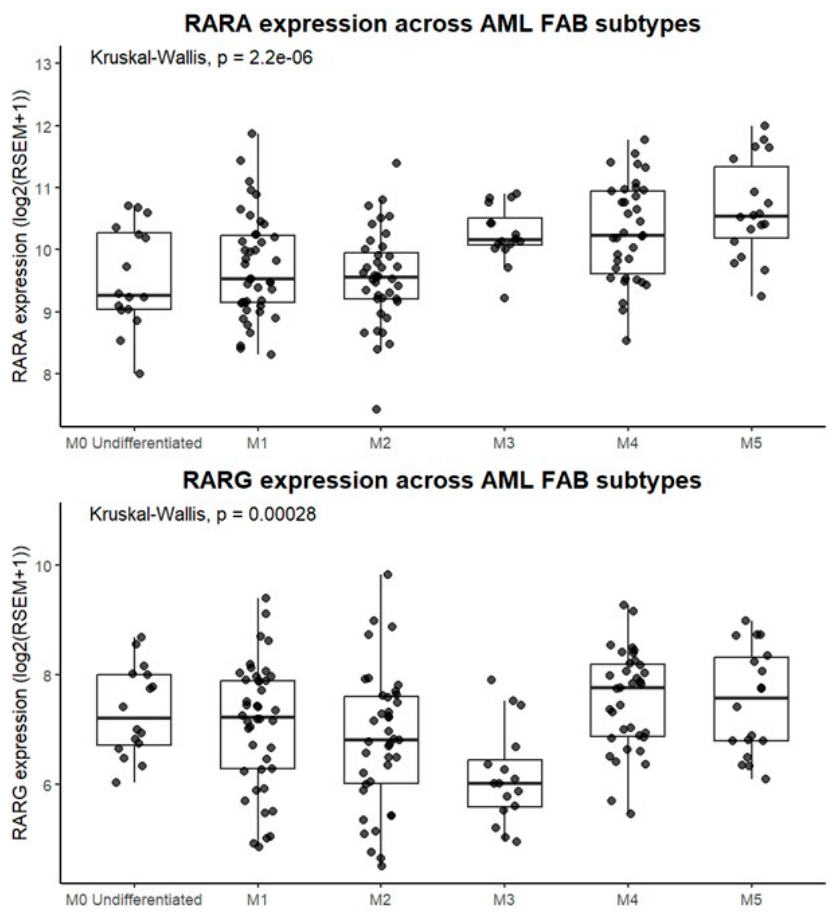
Figure 2.
Overall survival probabilities for AML patients with high and low expression of the RARA and RARG genes. The TCGA-LAML gene expression data (IlluminaHiSeq) and clinical data were obtained from UCSC Xena TCGA Hub. The AML patients were stratified into high and low gene expression using the median gene expression value as a threshold. Kaplan-Meier survival curves were generated using R version 4.5.1 with survival and survivor packages.
Figure 2.
Overall survival probabilities for AML patients with high and low expression of the RARA and RARG genes. The TCGA-LAML gene expression data (IlluminaHiSeq) and clinical data were obtained from UCSC Xena TCGA Hub. The AML patients were stratified into high and low gene expression using the median gene expression value as a threshold. Kaplan-Meier survival curves were generated using R version 4.5.1 with survival and survivor packages.
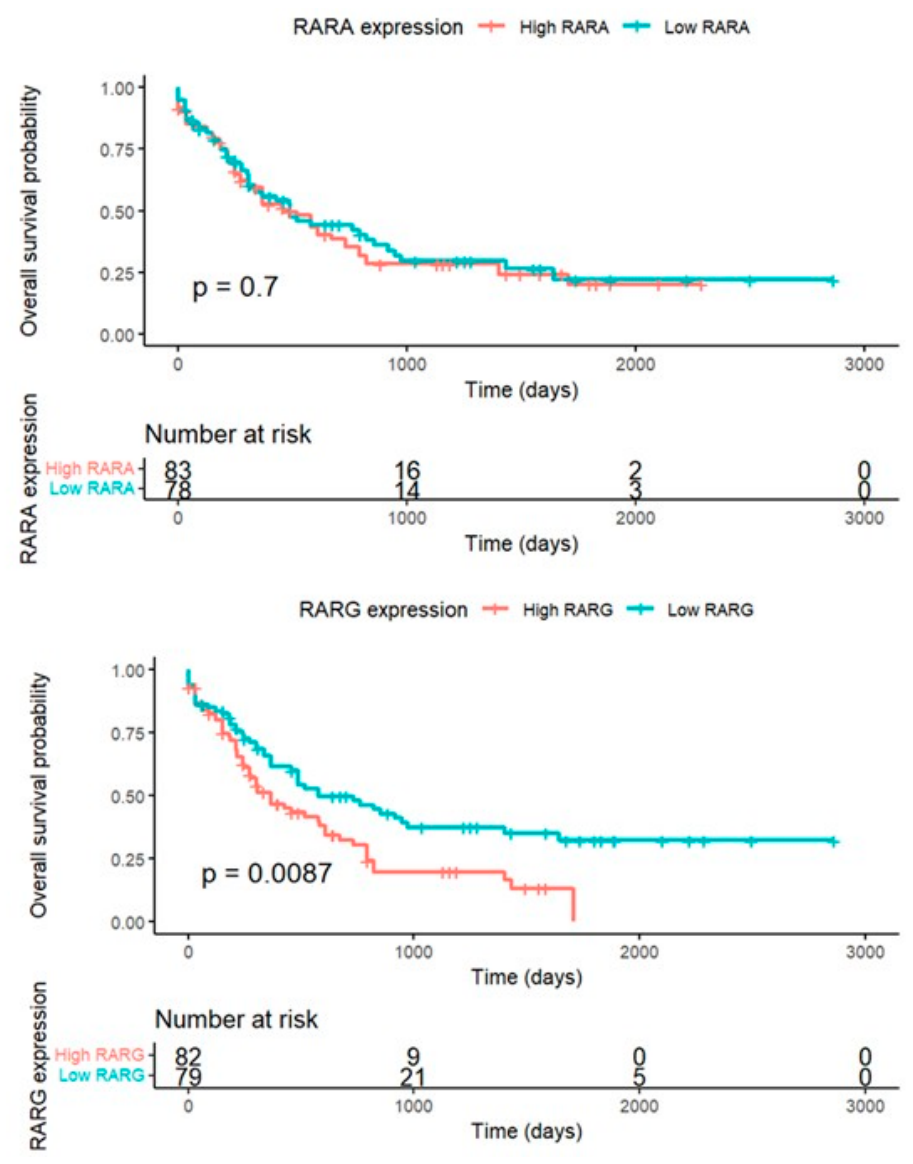
Figure 3.
The relationships between high and low levels of RARG gene expression to survival for AML M0 (top) versus AML M1 to M7 (bottom). The analysis is of the TCGA-LAML dataset (https://portal.gdc.cancer.gov/projects/TCGA-LAML).
Figure 3.
The relationships between high and low levels of RARG gene expression to survival for AML M0 (top) versus AML M1 to M7 (bottom). The analysis is of the TCGA-LAML dataset (https://portal.gdc.cancer.gov/projects/TCGA-LAML).
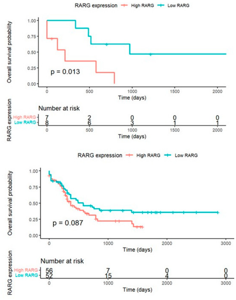
Figure 4.
RARG gene expression level across AML cytogenetic risk categories. The TCGA-LAML gene expression data (IlluminaHiSeq) and clinical data were obtained from UCSC Xena TCGA Hub. Kruskal–Wallis test followed by pairwise Wilcoxon tests with Benjamini–Hochberg correction was performed using R version 4.5.1. Statistical significance was defined as p < 0.05 (*), p < 0.01 (**).
Figure 4.
RARG gene expression level across AML cytogenetic risk categories. The TCGA-LAML gene expression data (IlluminaHiSeq) and clinical data were obtained from UCSC Xena TCGA Hub. Kruskal–Wallis test followed by pairwise Wilcoxon tests with Benjamini–Hochberg correction was performed using R version 4.5.1. Statistical significance was defined as p < 0.05 (*), p < 0.01 (**).
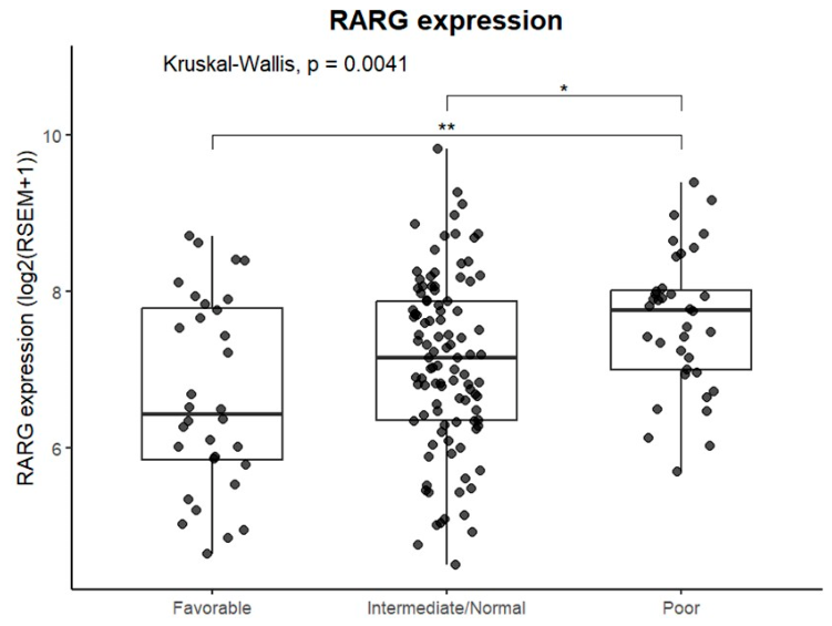

Table 1.
Statistical comparison of RARA and RARG gene expressions levels for the FAB subtypes. Adjusted p-values were obtained from pairwise Wilcoxon tests with Benjamini–Hochberg correction for multiple comparisons.
Table 1.
Statistical comparison of RARA and RARG gene expressions levels for the FAB subtypes. Adjusted p-values were obtained from pairwise Wilcoxon tests with Benjamini–Hochberg correction for multiple comparisons.
| RARA expression | RARG expression | ||
|---|---|---|---|
| Comparison | Adjusted p-value | Comparison | Adjusted p-value |
| M0 vs M3 | 0.0433 | M0 vs M3 | 0.0016 |
| M0 vs M4 | 0.0109 | M1 vs M3 | 0.0200 |
| M0 vs M5 | 0.0031 | M2 vs M4 | 0.0104 |
| M1 vs M3 | 0.0162 | M3 vs M4 | 0.0002 |
| M1 vs M4 | 0.0055 | M3 vs M5 | 0.0008 |
| M1 vs M5 | 0.0009 | ||
| M2 vs M3 | 0.0019 | ||
| M2 vs M4 | 0.0009 | ||
| M2 vs M5 | 0.0001 | ||
Disclaimer/Publisher’s Note: The statements, opinions and data contained in all publications are solely those of the individual author(s) and contributor(s) and not of MDPI and/or the editor(s). MDPI and/or the editor(s) disclaim responsibility for any injury to people or property resulting from any ideas, methods, instructions or products referred to in the content. |
© 2026 by the authors. Licensee MDPI, Basel, Switzerland. This article is an open access article distributed under the terms and conditions of the Creative Commons Attribution (CC BY) license.
Copyright: This open access article is published under a Creative Commons CC BY 4.0 license, which permit the free download, distribution, and reuse, provided that the author and preprint are cited in any reuse.