

Submitted:
02 November 2026
Posted:
04 November 2026
You are already at the latest version
Abstract
Neurotransmitters, including acetylcholine (ACh) and dopamine, play critical roles in regulating immune cell function and inflammatory responses across various pathologies. Beyond its classical neuronal role, ACh is actively secreted by diverse immune populations, including T cells, B cells, macrophages, dendritic cells, and natural killer cells. Upon binding to nicotinic (nAChRs) and muscarinic (mAChRs) acetylcholine receptors expressed on neutrophils, T cells, B cells, and monocytes/macrophages, ACh triggers intracellular signaling pathways that modulate immune cell proliferation, polarization, and cytokine secretion. Similarly, dopamine receptors (D1R–D5R) are widely expressed across these identical immune cell subsets. Dopamine signaling dynamically orchestrates immune cell proliferation, differentiation, activation, migration, polarization, and cytokine profiles. Consequently, these neurotransmitter-mediated pathways play pivotal roles in the pathogenesis of neurodegenerative, autoimmune, and systemic inflammatory diseases, including Parkinson’s disease, multiple sclerosis, inflammatory bowel disease, acute pancreatitis, chronic obstructive pulmonary disease, acute respiratory distress syndrome, infective endocarditis, and rheumatic heart disease.
Keywords:
neuroimmune axis
; neurotransmitters
; acetylcholine
; dopamine
; cholinergic anti-inflammatory pathway
; immune modulation
; inflammation
; neurodegenerative diseases
Neurotransmitters are chemical substances that transmit signals between neurons. In the nervous system, they convey information to form functional afferent and efferent neural pathways that adjust and maintain physiological activities in response to internal and external changes [1,2]. Recently, accumulating evidence has suggested that neurotransmitters also play a regulatory role in the immune system and inflammatory response [3]. This review summarizes current knowledge on the function of acetylcholine (ACh) and dopamine in regulating immune cells and inflammatory response in diseases.
1. Acetylcholine (ACh)
ACh is a critical neurotransmitter in the nervous system, secreted primarily by the spinal cord bulbar motor neurons, autonomic preganglionic neurons, cholinergic (parasympathetic) postganglionic neurons, and neurons in basal ganglia and cerebral motor cortex. ACh functions by binding to the muscarinic and nicotinic cholinergic receptors [4]. In the nervous system and neuromuscular junctions, muscarinic receptors (mAChRs) are distributed on the postsynaptic membranes of parasympathetic postganglionic neurons, in the substantia nigra of the central nervous system, and in smooth muscles of gallbladder, gastrointestinal tract, bronchi, pupillary sphincter, blood vessels and sweat glands [5]. Nicotinic receptors (nAChRs) link directly to ion channels transmitting signals at the neuromuscular junction to control muscle contraction and mediate transmission within the sympathetic and parasympathetic nervous systems. Accumulating evidence demonstrates that ACh and its receptors play critical roles in the immune system.
Since ACh is, on the one hand, inherently unstable and, on the other hand, easily cleaved by the ubiquitous acetylcholinesterase (AChE), its half-life in vivo is short [6]. Because cholinergic neurons are usually not in close proximity to the immune cells, it is reasonable that the ACh towards regulating the immune system originates from non-neuronal cells. The enzyme choline acetyltransferase (ChAT) catalyzes the conversion of choline and acetyl-CoA to acetylcholine, therefore, it is considered a marker of ACh-producing cells [7].
1.1. ACh Secretion from Immune Cells
T lymphocytes have been proven to produce ACh. Some researchers even believe that T lymphocytes are the primary source of ACh in blood [8,9]. By employing a ChAT-GFP BacIn reporter mouse, Tracey and colleagues found that memory T cells express ChAT after infection with lymphocytic choriomeningitis virus (LCMV-Arm) and this expression is induced by IL-21 [7,10]. Furthermore, ChAT-expressing T cells facilitate local vasodilation, blood pressure reduction, and migration of immune cells into infected tissue [10].
T cells are not the only immune cells that express ChAT and produce ACh. In ChAT-GFP BacIn reporter mice, Reardon C. et al. proved that splenic macrophages, dendritic cells (DCs), and mature recirculating B cells express ChAT and generate ACh [11].
ChAT expression in B cells is induced by postnatal microbial colonization in a MyD88-dependent manner [11]. MyD88 is an adaptor protein that couples with almost all Toll-like receptors (TLRs) to transduce signals and activate innate immune cells [12]. ChAT+ B cells restrict neutrophil recruitment in sterile sepsis [11], and ACh from B cells plays a critical role in skin inflammation [13]. In the skin, ACh is mainly produced by B cells; insufficient production of ACh leads to skin inflammation, characterized by increased inflammatory cytokines, hyperproliferation of keratinocytes, and thickening of the epidermis [13].
ACh has also been proven to be produced by NK cells [14]. Jiang W. et al. found that in experimental autoimmune encephalomyelitis (EAE) mouse model, splenic NK cells express ChAT, and this expression is upregulated by LPS stimulation in vitro [14]. They showed that ChAT+ NK cells in spleen and blood increased and peaked at 7 days post-infection (dpi), migrated to the central nervous system (CNS) in the following days, and reached a peak in the CNS at 16 dpi. The recruited ChAT+ NK cells were demonstrated to inhibit demyelination by restricting the recruitment of monocytes/macrophages and reducing the levels of IL-6, IL-1β, IL-12, and TNF-α in the CNS [14]. This regulatory function depends on α7 nicotinic acetylcholine receptors (α7nAChRs) on the surfaces of monocytes and the NKG2A–Qa-1 pathway [14].
1.2. ACh Receptors on Immune Cells
ACh regulates immune cells through nicotinic acetylcholine receptors (nAChRs) and muscarinic acetylcholine receptors(mAChRs). Studies have revealed that nAChRs are expressed on mature and immature T and B cells, but not on most granulocytes [15]. On the contrary, mAChRs have been detected on lymphocytes and neutrophils in humans [16,17].
Research emphasizes the critical function of nAChRs in immune cells. Different nAChR isoforms are assembled from five individual subunits, which are encoded by 16 nicotinic genes in humans, including CHRNA1–7, CHRNA9, CHRNA10, CHRNB1–4, CHRNG, CHRND, and CHRNE [18]. These genes encode 11 neuronal subunits (α2–7, α9, α10, β2–4) and 5 muscular subunits (α1, β1, γ, δ, and ε) [18]. Lymphocytes, granulocytes, and monocytes/macrophages have been shown to express α7, α9, and/or α10 nAChRs [19]. Another study showed that nAChR subunits (α3–α7, β2–β4) are distributed on lymphocytes, although the distribution varies between individuals and may depend on infection or physiological stress [16].
α7 nAChR
Monocytes, macrophages, T cells, and B cells have been shown to express α7 nAChR [20,21,22,23]. Macrophages and monocytes express α7 nAChR, which plays a critical role in cholinergic anti-inflammatory pathway (CAP) [20,21]. ACh from the vagus nerve or T lymphocytes binds to α7 nAChR on macrophages to inhibit local inflammation [20,24]. This inhibitory function is mediated by the downregulation of nuclear translocation of nuclear factor kappa B (NF-κB), the activation of Janus kinase/signal transducer and activator of transcription (JAK/STAT) pathway, and the suppression of proinflammatory cytokine release, including TNF-α, IL-1β, IL-6, and IL-18 [25,26,27]. T cells also express α7 nAChR. Nizri E. et al. showed that CD4(+) T cells express α7 nAChR and this expression is upregulated upon activation [22]. Furthermore, B lymphocyte-derived cell lines and mouse B lymphocytes express both α7 and α4β2 nAChRs [23,28].
Numerous studies have shown that, in cases of uncontrolled inflammation, direct vagus nerve stimulation (VNS) to induce the release of ACh or the peripheral administration of α7 nAChR agonists (e.g., nicotine or GTS-21) significantly lowers tissue and serum levels of TNF-α and IL-1β, mitigating tissue damage and preserving organ function [24]. Furthermore, this protective effect is nullified by α7 nAChR antagonists [24]. The inflammatory regulatory pathway involving the vagus nerve system and α7 nAChR is known as the cholinergic anti-inflammatory pathway (CAP) [29]. The CAP consists of the afferent vagus nerve sensing and transmitting inflammatory signals, the medullary visceral zone integrating these signals, and the efferent vagus nerve regulating the inflammatory response [29].
Based on the effective anti-inflammatory function of α7 nAChR, many agonist compounds have been investigated in numerous models of inflammation [19]. For instance, PNU-282987 has been demonstrated to inhibit edema and macrophage infiltration in a rat model of loose ligation-induced peripheral mononeuropathy [30]. In a postoperative ileus mouse model, Kimura H. et al. revealed that activation of α7 nAChR by PNU-282987 (PNU) and AR-R17779 (AR-R) significantly inhibited the recruitment of macrophages, but not neutrophils, after intestinal manipulation [17]. Pinheiro N. et al. demonstrated that in a mouse model of LPS intratracheal instillation-induced acute lung injury, PNU-282987 treatment inhibits neutrophil recruitment to the lung and suppresses levels of TNF-α, IL-1β, IL-6, and IL-10 in bronchoalveolar lavage fluid [31]. Additionally, activation of α7 nAChR shifts the lung macrophage profile by decreasing pro-inflammatory M1 macrophages and increasing M2 macrophages, which promotes inflammation resolution and tissue repair [31]. Ren et al. found that intracerebroventricular administration of α7nAChR agonists activates neuronal α7nAChR, reversing inflammation-induced peripheral immune suppression and reducing damage to vital organs [32,33,34].
The downstream intracellular molecular events of α7 nAChR stimulation and the mechanisms of its inhibitory function in inflammation have been elucidated [35,36,37,38]. Activation of α7 nAChR enhances the JAK/STAT signaling pathway within immune cells while suppressing the NF-κB pathway [35,36]. This reduces the production of pro-inflammatory cytokines such as TNF-α, high mobility group box 1 (HMGB1), IL-1β, and IL-6, while elevating serum levels of anti-inflammatory cytokines like IL-10 [35,36]. Pharmacological blockade or genetic knockout of α7 nAChR abrogates the anti-inflammatory effects of the CAP [35,36]. At the gene expression level, Sun et al. proposed that activated α7 nAChRs mediate intracellular anti-inflammatory effects via miR-124, which inhibits IL-6 and TNF-α production by targeting STAT3 and TACE (TNF-α converting enzyme); notably, nicotine upregulates miR-124 expression in macrophages [37,38].
α9, α10 nAChRs
The expression of α9 and α10 nAChRs has been demonstrated on CD3+, CD4+, CD8+ T lymphocytes, CD19+ and CD80+ B lymphocytes, and monocytes [19,21,39]. In vitro, ACh inhibits LPS-primed, BzATP-stimulated monocytes from releasing IL-1β but not IL-6 or TNFα, and α9/α10 nAChRs are involved in this regulation [21]. The nAChR agonists lysophosphatidylcholine (LPC) and glycerophosphocholine (G-PC) are metabolites of phosphatidylcholines [40]. LPC and G-PC effectively suppress IL-1β release from monocytes, and this inhibitory function depends mainly on α9 and α10 nAChRs, with only a weak dependence on the α7 nAChR [40].
Research indicates that α9 and α10 nAChRs constitute a crucial cholinergic anti-inflammatory pathway by regulating NLRP3 inflammasome activity to inhibit IL-1β release from monocytes [41]. Specifically, C-reactive protein (CRP) binds phosphatidylcholine (PC), activating α9 and α10 nAChRs on monocytes. This significantly inhibits ATP-induced NLRP3 inflammasome assembly, caspase-1 activation, and the secretion of mature IL-1β [41]. The α9 and α10 nAChRs play a crucial signaling role in this process: they mediate the metabolic signaling of the CRP-PC complex, effectively inhibiting ATP-mediated activation of P2X7 receptors and the resulting ionic influx. Simultaneously, they prevent ASC speck aggregation within the inflammasome and downregulate the caspase-1-mediated cleavage of pro-IL-1β, ultimately reducing the release of mature IL-1β [41].
Electrophysiological experiments have further confirmed that CRP-PC complex completely inhibits ATP-induced ionic currents in monocytes via α9/ α10 nAChRs. This is an effect blocked by the specific antagonist RgIA4, highlighting the direct regulation of inflammatory signaling at the plasma membrane level by α9 and α10 nAChRs. Moreover, phospholipid metabolites such as LPC and G-PC similarly inhibit IL-1β secretion via α9/α10 nAChR-dependent pathways, an action that is independent of α7 nAChRs [21]. Notably, this inhibitory effect is specific, affecting only IL-1β release without impacting the production of other inflammatory mediators such as IL-6 and TNF-α [41]. Collectively, the α9/ α10 nAChRs–NLRP3–caspase-1–IL-1β pathway plays a crucial role in regulating immune responses and controlling inflammatory damage [21,41].
mAChRs
The function of mAChRs in immune system has been well-established. These receptors belong to G protein-coupled receptor (GPCR) family and include 5 subtypes, M1-M5, which are encoded by the CHRM1-5 genes [42]. Receptors M1, M3, and M5 signal through the Gq/11 family to activate phospholipase C and catalyze the hydrolysis of phosphatidylinositides [42,43]. In contrast, M2 and M4 couple with Gi/o proteins [44]. Beyond their distribution in the nervous system and neuromuscular junctions, mAChRs are also expressed in immune cells.
Sato K. et al. demonstrated that M4 and M5 receptors are robustly expressed in human mononuclear leukocytes (MNLs, including T cells, B cells, and monocytes), as well as in various T cell lines (CEM, HPB-ALL, HUT-78, Jurkat, MOLT-3) and B cell lines (BALL-1, Daudi, and NALM-6). In contrast, the expression of M1-M3 varies between individuals [16]. In murine immune cells, MNLs, dendritic cells (DCs) and macrophages have been shown to express the mRNA for all five mAChR subtypes (M1–M5) [45].
Stimulation of M3 on T cells has been shown to induce Ca2+ mobilization [46]. Using the human helper T lymphoma cell line Jurkat, Kaneda T. et al. demonstrated that these cells express the M3 mAChR. Activation of this receptor triggers phospholipase C, leading to the formation of inositol trisphosphate and a subsequent increase in intracellular Ca²⁺ concentration [46]. Table 1 summarizes the regulatory function of ACh and its receptors in immune cells and diseases.
1.3. ACh Regulates Immune Cells in Inflammatory Responses
Neutrophils
ACh is proven to regulate neutrophil recruitment during inflammation. In a model of intraperitoneal injection of LPS-induced peritonitis, B cells produce ACh, which subsequently suppresses the recruitment of neutrophils into the peritoneal cavity without affecting their activation [11]. Furthermore, ACh derived from group 2 innate lymphoid cells (ILC2) is also shown to limit neutrophil counts and the levels of chemoattractants CXCL1 and CXCL2 in the airways of mice exposed to allergenic fungal extracts [47].
ACh appears to inhibit the formation and release of neutrophil extracellular traps (NETs). In vitro, ACh incubation significantly attenuated fMLP-induced NETosis and decreased the release of neutrophil elastase [48].
T Cells
ACh regulates T cell recruitment [49]. In a facial nerve axotomy model, the ACh pathway limits T cell recruitment into the axotomized facial motor nucleus in an α7 nAChR-dependent manner [49]. Accordingly, α7 nAChR knockout (nAChRα7−/−) mice exhibit significantly higher T cell counts in the lesioned facial nucleus compared to wild-type mice [49].
B Cells
B cells are regulated by ACh primarily through α7 and α4β2 nAChRs [28]. Specifically, α7 nAChRs modulate B cell activation and proliferation via the CD40-mediated pathway, while α4β2 nAChRs function in cooperation with BCR/FcɛRII [28,51]. Nicotinic receptor deficiency results in a reduction of IgG-secreting cells compared to WT mice [28]. Studies also indicate that B cell proliferation and survival in the bone marrow are influenced by both α7 and α4β2 nAChRs [52], while proliferation in the spleen is regulated solely by α7 nAChRs [53]. Furthermore, deficiency in α7 and β2 nAChRs leads to increased apoptosis in the bone marrow [28], and fewer IgG-producing cells in the blood [52]. These effects are likely mediated by both neuronal and cell-originating ACh [28].
Macrophages and Monocytes
As noted, ACh inhibits the production of proinflammatory cytokines, including TNF-α, IL-1β, IL-6, and IL-18, by monocytes and macrophages [20,24,25,26,27]. By binding to α7 nAChRs, ACh suppresses NF-κB nuclear translocation and JAK/STAT pathway activation [20,24,25,26,27].
Research also demonstrates that ACh regulates macrophage polarization [47]. In allergic airway inflammation, ACh derived from group 2 innate lymphoid cells (ILC2s) promotes M2 macrophage upregulation and M1 macrophage downregulation, since depletion of ACh via intranasal administration of acetylcholinesterase decreased M2 and increased M1 macrophages [47]. Furthermore, an outstanding study showed that during liver regeneration after damage, ACh from B cells polarizes Kupffer cells towards an immune-promoting phenotype, which subsequently produces more IL-6 to facilitate liver repair. The regulation of ACh on Kupffer cells depends on α7 nAChR and plays a critical role in liver regeneration [50]. Figure 1 summarizes the regulatory Effects of ACh on immune cells.
2. Dopamine
Dopamine is a critical catecholamine neurotransmitter, accounting for approximately 80% of all catecholamines in the brain [54]. In the central nervous system, dopamine and its receptors are involved in three major pathways: a) the nigrostriatal pathway, in which it regulates the motor control and integration of sensory information; b) the mesolimbic/mesocortical pathway, in which it is involved in the regulation of emotion, cognition, and reward and reinforcement; c) the tuberoinfundibular pathway, where it acts as a neurotransmitter within the hypothalamic–pituitary axis to regulate neuroendocrine system [55]. Additionally, dopamine regulates peripheral vascular tone and sodium homeostasis through receptors located in the kidney and vasculature [56].
Dopamine receptors (D1R-D5R) belong to G protein-coupled receptor (GPCR) family [57]. Their affinities for dopamine vary significantly, following the order: D3R>D5R>D4R>D2R>D1R [58]. Based on their structural and biochemical properties, these receptors are categorized into two groups, D1-like receptors (D1 and D5) and D2-like receptors (D2, D3, and D4) [58,59]. D1-like receptors typically couple to the stimulatory G proteins (Gs) and stimulate the activation of adenylate cyclase (AC) to produce cAMP [59]. In contrast, D2-like receptors couple with inhibitory G protein subtypes (Gi and Go), and their activation inhibits AC. Additionally, both D1- and D2-like receptors can activate other non-cAMP-related cascades through G-protein-independent pathways [60].
Beyond its critical roles in the nervous system, dopamine and its receptors have also been proven to play important roles in regulating inflammation [59,60,61]. This is initially supported by the diverse expression of dopamine receptors (DRs) across various immune cell populations [60,61]. For instance, B cells exhibit strong and consistent expression of D2, D3, and D5, and NK cells of D2, D3, D4, and D5 [61]. Neutrophils and eosinophils show moderate expression of D3 and D5 receptors [61]. T lymphocytes and monocytes express D2, D3, D4, and D5 at lower levels [61]. Notably, the D1 receptor is generally not detected in these immune cell types [61]. The following sections detail our current understanding of how dopamine modulates immune cell function. Table 2 summarizes the regulatory function of dopamine and its receptors in the immune system.
2.1. Dopamine and Immune Cells
T Lymphocytes
Current reviews provide a comprehensive summary of the relationship between T cells and dopamine [62,63]. Research indicates that T cells synthesize and maintain intracellular dopamine stores, with measured concentrations ranging from 7 × 10^-20 to 2 × 10^-18 moles per cell [64].
Numerous studies have demonstrated the expression of dopamine receptors on T cells [65,66,67,68,69,70,71,72]. It is generally established that T cells express D2-D5 receptors [61,65,66,67,68,69], whereas the expression of D1 receptor remains a subject of debate [61,69,70]. Expression profiles appear to vary depending on the T cell subtype and activation state [69,71,72]. For instance, Watanabe et al. showed that naive CD8+ T cells express high levels of D3R mRNA but low levels of D4R mRNA, while resting CD4+ T cells generate low levels of D2R and D3R mRNA [71]. Furthermore, D2 receptor expression was upregulated in activated CD4+ T cells, simultaneously with decreased expression of D3 receptor. Meanwhile, expression of D3 and D4 receptors was proven to be reduced dramatically in activated CD8+ T cells [69,71]. This variability underscores the role of dopamine in lymphocyte maturation, proliferation, and differentiation [72,73].
Several studies have shown that dopamine inhibits T cell proliferation [73]. Ghosh M.C. et al. al demonstrated that dopamine at 1–5 ng/ml inhibits the proliferation of T cells [73]. Saha B. et al. showed that dopamine plasma level in individuals with oral malignancies (uncoping stress group) was 46.6±3.9 pg/ml, significantly higher than the level in normal volunteers (10±2.7 pg/ml) [74]. The increased dopamine significantly suppressed the proliferation of CD4+ and CD8+ T cells by inducing intracellular cAMP accumulation [74].
Dopamine has also been demonstrated to play critical roles in T cell differentiation. For instance, dopamine promotes naïve CD4+ T cells to differentiate into Th2 cells [75]. By binding to D1 receptors on CD4+ T cells, dopamine increases intracellular cAMP and shifts naïve CD4+ T cells toward T helper 2 (Th2) cells [75]. Furthermore, this dopamine might be produced by dendritic cells (DCs) [75]. Th2 cell differentiation can also be facilitated by binding to D4 receptors [76]. Dopamine facilitates naïve CD4+ T cells polarizing to Th2 cells, which produce more IL-4, IL-5, and IL-13 [76]. This function signals through the D4 dopamine receptor and subsequently decreases the level of cAMP, induces IL-2 activation, and promotes Th2 differentiation [76]. This signaling pathway is associated with asthma development in young children [76]. Naïve T cells are accelerated to differentiate to T follicular helper (Tfh) cells by dopamine through D1-like receptors [77]. This process is mediated by the PI3K/AKT/FOXO1/Klf2 signaling pathway and is related to the pathogenesis of systemic lupus erythematosus(SLE) [77].
Chen Y. et al. demonstrated that dopamine facilitates the differentiation of CD8+ T cells to CD103+ tissue-resident memory T cells (TRM) through a D5 receptor-dependent mechanism [78]. As long-lived memory cells, TRM cells produce IFN-γ and TNF, playing a vital role in tumor immunosurveillance [78].
Besides proliferation and differentiation, dopamine plays a critical role in the immunological reaction of T cells. For example, dopamine promotes T cell adhesion to fibronectin within the extracellular matrix (ECM) [79]. By binding to D2 and D3 receptors on T cells, dopamine activates the β1 integrins VLA-4 and VLA-5, which mediate T cell adhesion and extravasation [79]. Furthermore, Watanabe et al. demonstrated that dopamine is essential for the homing of naïve CD8+ T cells, since blocking D3 receptor significantly reduced their recruitment into secondary lymphoid tissues [71]. Specifically, dopamine facilitates this process by activating integrins VLA-4 and VLA-5, which subsequently bind to fibronectin and ICAM-1 [71].
Dopamine also influences cytokine secretion in T cells. Huang Y. et al. showed that stimulation of D2-like receptors upregulated the production of IL-4 while downregulating IFN-γ [70]. Furthermore, dopamine promotes the differentiation of naïve CD4+ T cells into Th2 cells, increasing the production of IL-4, IL-5, and IL-13 [76]. Conversely, Ghosh et al. demonstrated that dopamine, at concentrations of 1–5 ng/ml, inhibits the release of IL-2, IFN-γ, and IL-4 through a mechanism independent of D2 and D3 receptors [73]. Additionally, D1-like receptor agonist Fenoldopam has been shown to inhibit T cell activation, migration, and secretion of inflammatory cytokines, such as TNFα, IL-2, IL-4, IL-6, and IL-1β [80].
B Lymphocytes
Evidence indicates that B cells consistently express dopamine receptors D2, D3, D4, and D5, while the expression of the D1 receptor appears limited to particular B cell subsets [61,65,81,82,83,84].
For example, McKenna et al. utilized flow cytometry to show that B cells in peripheral venous blood express dopamine receptors D2–D5, but notably lack the D1 receptor [61]. However, expression patterns appear to be subset-specific; for instance, regulatory B cells (Bregs) have been shown to express all five receptor types [82]. In another study, B cells can express D1-like receptors, with levels significantly upregulated on activated B cells in patients with systemic lupus erythematosus (SLE) [81]. Similarly, Santambrogio L. demonstrated the presence of D2 and D4 receptors on B cells [65], noting that D2 expression increases under inflammatory conditions such as rheumatoid arthritis (RA) [83]. Additionally, Wu et al. confirmed high levels of D5 receptor expression in both murine and human B cells using RT-PCR and immunofluorescent staining [84].
Through binding to its receptors, dopamine modulates key B cell functions, including proliferation, differentiation, activation, migration, and cytokine secretion [65,81,83,84,85,86].
Kouassi E. et al. revealed that dopamine upregulates the concentration of cAMP in lipopolysaccharide (LPS) stimulated mouse B cells, subsequently inhibiting B cell proliferation, maturation, immunoglobulin (Ig) synthesis, and secretion in vitro [86]. The inhibitory function could not be blocked by antagonists of D1, D2, or α-adrenoceptors or β-adrenoceptors, leading the authors to suggest that the effect may instead originate from dopamine’s interference with lysosomal function [86]. In contrast, D1-like receptor agonists have been shown to facilitate B cell activation and differentiation into plasma cells (PCs) [81]. This regulation involves the upregulation of PTGS2 and its downstream targets, CCL17 and PRDM1 [81]. In RA patients, the expression of D2 receptor on B cells is upregulated and positively correlates with the plasma concentration of TNF-α [83]. Furthermore, dopamine induces the downregulation of cAMP in B cells through binding to D2 and D3 receptors [65]. Through the D5 receptor, dopamine enhances the proliferation of B cells and subsequently facilitates CD8+ T cell activation and the secretion of TNF-α and IFN-γ [84]. This regulatory mechanism functions via the JAK-STAT signaling pathway and contributes to antitumor immunity [84]. In addition, dopamine promotes B cell CNS-tropism via the D3 receptor without affecting antigen presentation functions [85].
Monocytes and Macrophages
Peripheral monocytes have been shown to express dopamine receptors of D2, D3, D4, and D5, but the expression of D1 remains a point of contention [61,82]. While Josefsson E. et al. failed to detect D1 receptors on monocytes using flow cytometry [61], Arce-Sillas A. reported the expression of all five receptor subtypes, including D1, on intermediate monocytes [82]. In contrast, macrophages are revealed to express all five subtypes of dopamine receptors, with D1 and D5 levels significantly exceeding those of the D2-like receptors [87]. Furthermore, Gaskill P. J. uncovered that in vitro, primary human monocyte-derived macrophages (MDM) encode mRNA of all five types of dopamine receptors, and specifically express D3 and D4 receptors on the plasma membrane [88]. Macrophages themselves have been shown to produce dopamine and contain dopamine at an average concentration of 7 × 10^-17 and 2 × 10^-17 moles per cell, respectively [64].
Dopamine is proven to regulate macrophage polarization and cytokine release in a concentration-dependent manner. For instance, a low concentration (2 mg/mL) of dopamine is revealed to promote macrophage polarization to M2 type and suppress inflammation; conversely, a higher concentration (10 mg/mL) exerts pro-inflammatory effects [89]. This modulation involves the NF-κB signaling pathway [89].
Dopamine is demonstrated to enhance cytokine production under both resting and inflammatory conditions [88]. Specifically, in resting macrophages, dopamine upregulates the secretion of IL-6 and CCL2, and in LPS- stimulated macrophages, it increases the production of IL-6, IL-10, CCL2, and CXCL8, while simultaneously suppressing TNF-α release [88]. This modulation is involved in drug abuse-induced neurological diseases [88]. Nolan R. A. revealed that dopamine induces macrophages to secrete inflammatory cytokines, including CCL2, CXCL8, CXCL9, CXCL10, IL-1β, IL-6, and IL-18 [90]. This modulation of cytokines is associated with the expression of D5 receptors on macrophages and may contribute to the development of HIV-associated neuropathology [90]. Additionally, Tarazona R. demonstrated that combined injection of chlorpromazine (CPZ) and LPS, or CPZ and staphylococcal enterotoxin B (SEB) , significantly raised the concentration of IL-10 in BALB/c mice, and the elevated IL-10 originated from macrophages rather than T cells [91]. Furthermore, D1 but not D2 receptors are associated with the effect of CPZ on macrophages, as the selective blockade D1 receptors eliminates its impact on IL-10 production [91]. The ability of dopamine to stimulate macrophage IL-10 production was also demonstrated in the study by Haskó G. et al. . Using the J774.1 macrophage cell line and mouse peritoneal macrophages, their study revealed that dopamine upregulates IL-10 production while concurrently downregulating IL-12 p40 [92]. Notably, the suppression of IL-12 p40 and part of the upregulation of IL-10 depended on beta-adrenoceptors but not dopamine receptors [92].
Dopamine also modulates macrophage phagocytic function in a concentration-dependent manner. Roy B. showed that catecholamines, including dopamine, norepinephrine, and epinephrine, enhance macrophage phagocytosis at lower concentrations from 10-15 to 10-11 M, whereas they suppress this activity at higher concentrations of 10-7 to 10-5 M [93]. Furthermore, Sternberg E. M. showed that dopamine specifically enhanced IFN-γ-induced phagocytosis of macrophages [94].
Macrophages express Fcγ receptors (FcγR), which bind to IgG and facilitate clearance of IgG-coated pathogens and cells [95]. Gomez F. et al. demonstrated that dopaminergic agonists increase expression of FcγR on macrophages, thereby upregulating the clearance of IgG-sensitized red blood cells [96]. Both D1 and D2 dopamine receptors have been shown to be involved in this upregulation of FcγR expression on macrophages [96]. Ali R. A. showed that at concentrations of 0.1 and 0.25 μg/mL, dopamine, as well as norepinephrine and epinephrine, increases Fc receptor expression on chicken macrophages and enhances their phagocytosis of E. coli and sheep red blood cells [97].
Different pathways have been implicated in the dopaminergic regulation of macrophages. For instance, dopamine is demonstrated to decrease cAMP levels in tumor-associated macrophages in pancreatic cancer [98]. The regulation signals through D4 receptors on macrophages and subsequently suppresses the activation of the PKA/p38 pathway [98]. This modulation ultimately restricts macrophage-derived inflammation within the tumor tissue [98]. In another study, dopamine was shown to stimulate PKC phosphorylation and Ca2+ release from the endoplasmic reticulum of macrophages. These effects depend on Gq/11, which is proven to couple with D5 receptor [87].
Neutrophils
Neutrophils exhibit robust expression of D3 and D5 receptors, whereas the presence of D2 and D4 receptors varies by individual [61,99,100]. D1 receptor has also been shown to be expressed on neutrophils [99,100,101]. Stimulation with fMLP induces an upregulation of D1 receptor expression, alongside downregulation of all D2-like receptors and the D5 receptor [100]. Additionally, neutrophils have been shown to secrete dopamine, and their activation significantly increases intracellular dopamine concentrations [100].
Dopamine, as well as dobutamine, enhanced premature neonate neutrophil apoptosis in vitro at the concentration of 10-4 M [102]. Similarly, in adults, dopamine at concentrations of 10 and 100 µmol/L was proven to facilitate apoptosis of neutrophils isolated from both healthy volunteers and patients with systemic inflammatory response syndrome (SIRS). This effect was found to be dependent on the D1 receptor [103].
Furthermore, dopamine regulates neutrophil phagocytosis [99]. Dopamine is shown to restore neutrophil phagocytic function, which is often suppressed in patients with SIRS compared to healthy individuals [103]. Antipsychotic drugs (APDs), such as clozapine, haloperidol, and risperidone, are clinically available dopamine D2 receptor blockers, although their effects may also involve binding to other receptors, such as 5-HT receptors [99]. Chen M. L. et al. demonstrated that clozapine and haloperidol enhance neutrophil phagocytosis of Escherichia coli in vitro [99]. Simultaneously, these agents enhance IL-8 secretion and myeloperoxidase activity in neutrophils, and facilitate the intracellular activation of the AKT signaling pathway [99].
Neutrophil respiratory burst and reactive oxygen species (ROS) generation may also be regulated by dopamine. Marino F. showed that dopamine, as well as a D1-receptor agonist, dramatically suppressed ROS production in fMLP-stimulated neutrophils, though this effect was not observed in resting neutrophils [100]. In contrast, a study by Sookhai, S. found that the respiratory burst of neutrophils was not altered by dopamine [103].
Moreover, dopamine has been shown to downregulate neutrophil adhesion and transmigration under inflammatory conditions. At concentrations of 10 and 100 µmol/L, dopamine has been shown to inhibit the expression of CD11b and CD18 on neutrophils isolated from SIRS patients [103]. CD11b and CD18 are integrin subunits on neutrophils that compose integrin Mac-1 (CD11b/CD18) and LFA-1 (CD11a/CD18), both of which play central roles in neutrophil adhesion [101]. Consistent with these findings, Marino F. demonstrated that dopamine pretreatment significantly inhibits fMLP-induced neutrophil transmigration but does not influence transmigration of resting neutrophils [100]. The inhibitory function of dopamine is dependent on D1-like receptors but not D2-like receptors [100].
In another study, ropinirole, a D2-like receptor agonist, was also shown to inhibit neutrophil infiltration and alveolar bone destruction in periodontitis [104]. Similarly, the D2 receptor agonist pramipexole exhibited a comparable inhibitory effect on neutrophil infiltration in carrageenan- or formalin-induced paw inflammation in rats [105]. In line with this, D2 dopamine receptor blockers haloperidol and clozapine have been shown to upregulate expression of integrin Mac-1 on neutrophils, which is critical for neutrophil adhesion and transmigration [99]. However, blocking D1-like receptors with an antagonist has been reported to inhibit neutrophil infiltration in ovalbumin antigen-induced airway inflammation in mice [106]. Thus, the dopaminergic regulation of neutrophil adhesion and recruitment remains complex and requires further research for full clarification. Figure 2 and Figure 3 summarize the regulatory effect of dopamine on immune cells via D1- and D2-like receptors, respectively.
2.2. Dopamine and Neurological Inflammatory Diseases
Parkinson’s Disease (PD)
Dopamine is at the core of the pathogenesis of Parkinson’s disease, a neurodegenerative disorder characterized by the selective loss of dopaminergic neurons in the substantia nigra. In addition to motor dysfunction, dopamine deficiency regulates the neuroinflammatory network, where the depletion of dopamine triggers the excessive activation of microglia [107,108]. These activated microglia release inflammatory cytokines, such as TNF-α and IL-1β, thereby exacerbating neurodegeneration in the substantia nigra [107,108]. This vicious cycle has been confirmed in postmortem studies of Parkinson’s disease brains, which show an increase in microglial markers and their aggregation within dopamine-exhausted regions [109,110]. Motor function can be restored by replacing exogenous dopamine with exogenous levodopa, but its immunomodulatory properties are equally significant. Pramipexole and other D2R agonists can inhibit the production of IL-6 and nitric oxide by LPS-induced microglia in vitro. Furthermore, in 6-OHDA-diseased mice, these agonists have been shown to reduce microglial infiltration and α-synuclein aggregation [111,112].
Genetic studies have further revealed that dopamine-derived quinones, formed through oxidative metabolism, can modify α—synuclein and promote its aggregation. Conversely, α -synuclein aggregates can disrupt the vesicular storage of dopamine in surviving neurons, leading to dopamine overflow from the cell membrane and thereby expanding oxidative stress [113,114]. This interaction was reproduced in a transgenic mouse model expressing human α—synuclein, in which drug-induced dopamine depletion accelerates microglial activation and neuroinflammation [110]. Clinically, Parkinson’s disease patients with higher levels of dopamine metabolites in the cerebrospinal fluid exhibit slower disease progression, indicating that dopamine plays a dual role in both motor and inflammatory pathways [113,114].
Multiple Sclerosis (MS)
Dopamine regulates immune cell polarization in multiple sclerosis (MS), an autoimmune demyelinating disease. Peripheral blood mononuclear cells from MS patients show decreased expression of D1R on Th1 cells, weakening the inhibitory effect of dopamine on IFN-γ production. This defect can be reversed by interferon-β treatment [115,116]. In the experimental autoimmune encephalomyelitis (EAE) model, the administration of dopamine at disease onset biases naive CD4+ T cells toward Th2 and regulatory T cell (Treg) phenotypes, thereby reducing the clinical score by 40% [117]. Mechanistically, dopamine activates D1 receptor (D1R) on Th17 cells, inhibiting STAT3 phosphorylation, and reducing IL-17 secretion. This effect has been verified in vitro. The D1R antagonist SCH-23390 has been shown to eliminate the inhibitory effect of dopamine on Th17 differentiation [118]. Regulatory T cells (Tregs) in MS patients exhibit a paradoxical upregulation of D1R, which impairs their suppressive function via cAMP-PKA pathway hyperactivation [63,119]. This finding is mirrored in EAE mice lacking D1R on Tregs, which exhibit reduced disease severity compared to wild-type controls [9]. Clinically, dopamine precursor levodopa has shown trends toward reduced relapse rates in small MS cohorts, though large trials are required to confirm its efficacy [116]. The dopamine-immune axis in MS is further supported by post-mortem studies demonstrating D2R expression on perivascular lymphocytes within MS lesions [115].
2.3. Dopamine and Gastrointestinal Inflammatory Diseases
Inflammatory Bowel Disease (IBD)
Dopamine dysregulation is a hallmark of IBD, with mucosal dopamine levels significantly reduced in patients with active ulcerative colitis compared to healthy controls [120]. This deficit stems from enteroendocrine cell dysfunction and altered gut microbiota metabolism. Gut microbiota can influence dopamine production via specific metabolic pathways, while enteroendocrine cells, including enterochromaffin cells, sense microbial signals to regulate mucosal dopamine levels in IBD [121,122]. Dopamine exerts dual protective effects: on macrophages, D2R activation inhibits NF-κB translocation, reducing the release of TNF α and IL-6; meanwhile, in Caco-2 cell monolayers, dopamine enhances tight junction integrity via D1R-mediated phosphorylation of ZO-1, decreasing paracellular flux [123,124].
In the mouse model of DSS-induced colitis, the dopamine receptor agonist ropinirole can significantly alleviate histological damage, which is reflected in the decreased activity of mucosal myeloperoxidase (MPO) and the recovery of the expression of the tight junction protein occludin [104,125]. This protective effect is consistent with systematic evidence indicating that D2R-mediated signaling inhibits intestinal inflammation by suppressing the activation of pro-inflammatory cells (such as by reducing MPO activity) and enhancing the structural integrity of the epithelial barrier by up-regulating tight junction proteins [60]. Mechanistically, ropinirole, as a D2 receptor agonist, may be involved in signaling pathways (such as PI3K/Akt) to alleviate mucosal damage, which is consistent with the results of preclinical studies, namely that dopamine receptor activation can improve barrier function in inflammatory bowel disease models [122,126]. The gut microbiota-dopamine axis is particularly notable: The probiotic Bifidobacterium longum increases intracavitary dopamine through tyrosine decarboxylation, and thereby inhibits Escherichia coli’s adhesion to intestinal cells by down-regulating the bacterial ciliary gene. Clinically, the increase in dopamine levels in the feces of IBD patients treated with probiotics is associated with clinical remission [127,128,129]. The protective effect of dopamine was further verified in in vitro human colon cultures. Supplementation of dopamine could reduce IL-1β -induced epithelial permeability [124].
Acute Pancreatitis (AP)
Dopamine exhibits dichotomous effects in AP, dependent on dosage and disease stage. In a cat model of hemorrhagic AP, low-dose dopamine (5 μg/kg/min) administered 6 hours post-induction reduces pancreatic edema and neutrophil infiltration by 35%, via D1R-mediated vasodilation of pancreatic arterioles and β2-adrenergic inhibition of leukocyte-endothelium adhesion [130]. By contrast, high-dose dopamine (50 μg/kg/min) in rats exacerbates acinar cell necrosis by promoting TNF-α release and disrupting endothelial nitric oxide synthase activity [130].
Mechanistically, these cellular responses are highly regulated by the D2R signal within pancreatic acinar cells, which helps maintain cellular homeostasis and inhibit the activation of terminal caspase-3 [131,132]; while the accumulation of dopamine within the cells may trigger oxidative stress through auto-oxidation [133,134]. In vivo randomized controlled studies have demonstrated the therapeutic efficacy of dopaminergic intervention in acute pancreatitis (AP) models. Systemic administration of low-dose dopamine (5μg/kg/min) significantly alleviates pancreatic histopathologic damage and dampens systemic inflammation, as evidenced by a marked reduction in serum levels of key inflammatory cytokines, including IL-6 and TNF-α [135]. This protective effect is primarily attributed to the capacity of low-dose dopamine to enhance pancreatic microcirculatory blood flow and concurrently reduce microvascular permeability, thereby preventing the transition from mild tissue edema to severe pancreatic necrosis [130]. However, in severe AP cases with multiorgan failure, dopamine failed to improve survival in a meta-analysis of 6 trials [136]. Emerging evidence highlights dopamine’s nuanced role in pancreatic microcirculation during acute pancreatitis (AP). In vivo intravital fluorescence microscopy has demonstrated that targeted dopaminergic intervention preserves functional pancreatic capillary perfusion and functional capillary density (FCD) in the early stages of AP. However, this microvascular protection exhibits a strict dose-dependent profile and is entirely lost at high doses, where alpha-adrenergic-mediated vasoconstriction supervenes, leading to visceral ischemia and further exacerbation of pancreatic hypoperfusion[130,137].
2.4. Dopamine and Respiratory Inflammatory Diseases
Chronic Obstructive Pulmonary Disease (COPD)
Plasma dopamine levels correlate with COPD severity, increasing during exacerbations due to sympathetic activation, as supported by studies in animal models and human patients [138,139]. In the pathogenesis of chronic obstructive pulmonary disease (COPD), alveolar macrophages serve as a primary source for the secretion of pro-inflammatory cytokines like interleukin-8 (IL-8) [140]. Peripheral dopamine has been shown to exert profound anti-inflammatory effects on these immune cells [141], wherein it can inhibit downstream cellular activation and inflammatory responses by signaling through dopamine receptors, including the D2R, and subsequently modulating the phosphorylation of downstream signaling cascades such as extracellular signal-regulated kinase 1/2 (ERK1/2) [60]. However, among smokers, this effect is weakened due to the decreased expression of D2R [142,143]. In bronchial smooth muscle cells, chronic β₂-agonist exposure may impair the dopamine D2 receptor (D2R)-mediated heterologous sensitization of adenylyl cyclase, a mechanism that normally facilitates and enhances airway relaxation [144]. Ex vivo studies demonstrate dopamine relaxes human bronchial rings via D2R-mediated activation of inward-rectifying potassium channels, a mechanism complementary to β2-adrenergic bronchodilation [145,146,147].
Smoking-induced downregulation of bronchial D2R is mechanistically linked to histone deacetylase 2 (HDAC2) inhibition: nicotine reduces HDAC2 activity, leading to hypermethylation of the DRD2 promoter. This is validated in a rat model of cigarette smoke exposure, where D2R agonist quinpirole restores bronchial responsiveness and reduces airway eosinophilia [148,149]. Clinically, COPD patients with higher baseline dopamine levels have better forced expiratory volume in 1 second (FEV1) response to bronchodilators, suggesting dopamine status may predict treatment efficacy [150].
Acute Respiratory Distress Syndrome (ARDS)
The hemodynamic benefits and pro-inflammatory effects of dopamine prominently demonstrate its dual role in ARDS. Dopamine exhibits a significant dose-dependent dual effect on pulmonary function and microcirculation in acute respiratory distress syndrome (ARDS) and acute lung injury models induced by endotoxin (LPS)[151,152,153]. At low doses (typically ≤ 3 μg/(kg·min)), dopamine mainly stimulates dopamine receptors and mild β₁-adrenergic receptors; by optimizing systemic hemodynamics and increasing cardiac output, it effectively reverses the decline in lung compliance and reduces vascular resistance, thereby exerting a protective effect and improving oxygenation index[151,152] Conversely, at high doses (typically ≥ 10 μg/(kg·min)), dopamine mainly activates potent α₁-adrenergic receptors, causing severe systemic and pulmonary vasoconstriction. In the highly inflammatory environment of LPS-induced ARDS, this elevated microvascular pressure and mechanical stress promote the retention, marginal migration, and isolation of activated neutrophils within the pulmonary capillaries. This occlusion subsequently leads to the release of reactive oxygen species and proteases, exacerbating the disruption of the alveolar-capillary barrier and further worsening the degree of lung injury[152,153]ung endothelial cells exposed to dopamine showed increased permeability through D1R-mediated cAMP-activated myosin light chain kinase, which was blocked by the D1R antagonist SCH-23390[142,154,155]rsely, D2R agonists like cabergoline protect against ventilator-induced lung injury in mice by suppressing NLRP3 inflammasome activation and downregulating alveolar IL-1β secretion [141,156]. In clinical settings, multicenter randomized controlled trials have demonstrated that while low-dose dopamine may help preserve renal perfusion and urine output in critically ill patients, it does not worsen acute lung injury, although it fails to decrease 28-day mortality compared to placebo [157]. Recent proteomic analysis of ARDS bronchoalveolar lavage fluid shows dopamine modulates surfactant protein expression, with D2R activation enhancing SP-A and SP-D secretion, critical for alveolar stability [139,142,154].
2.5. Dopamine and Cardiovascular Inflammatory Diseases
Infective Endocarditis (IE)
Dopamine released during sepsis influences IE pathogenesis by modulating both valvular destruction and host defense. Cardiac fibroblasts exposed to dopamine upregulate matrix metalloproteinase-9 (MMP-9) via D1R-cAMP-PKA signaling, promoting valvular extracellular matrix degradation. This is confirmed in a rabbit model of streptococcal IE, where dopamine administration increases mitral valve MMP-9 activity and leaflet perforation [158].
Studies have shown that in vitro, excess dopamine disrupts neutrophil chemotaxis. It inhibits the PI3K-AKT signaling pathway via D1-like receptors, thereby suppressing downstream cytoskeletal reorganization and significantly reducing neutrophil migration to fMLP [100]. In a mouse model of infective endocarditis (IE), haloperidol blockade of dopamine receptors increased survival by 60%. This may be related to the restoration of neutrophil recruitment in cardiac vegetations, which has important implications for understanding the role of dopamine in infectious diseases and potential therapeutic strategies [100]. Clinical data show IE patients with septic shock have plasma dopamine levels >200 pg/mL, which correlate with increased valvular surgery need and 30-day mortality [158].
Rheumatic Heart Disease (RHD)
How an autoimmune response triggered by molecular mimicry between streptococcal M proteins and cardiac tissue proteins leads to cardiac lesions in rheumatic heart disease [159,160] remains a central focus of investigation. Emerging evidence suggests a novel neuroimmune dimension to this pathology: specifically, the identification of dopamine-producing cells by tyrosine hydroxylase expression in rheumatic heart disease tissues has been found to be located in the vicinity of valve infiltrates [60,161]. This structural proximity implies that cross-reactive autoantibodies may not only attack structural tissue, but also target local dopamine pathways, fueling chronic inflammation and lesions at the lesion sites [162,163]. Recent studies have shown that the density of these cells correlates with autoantibody titers. This observation suggests a potential role for the dopaminergic system in the immunopathogenesis of rheumatic heart disease [164].
Dopamine regulates cardiac fibroblasts by activating the D1R, thereby mitigating valvular fibrosis [165]. In vitro experiments have demonstrated that D1R activation effectively suppresses the TGF-β1/Smad signaling pathway, leading to a down-regulation of TGF-β1 secretion and subsequent Smad phosphorylation, which ultimately decreases type I collagen deposition [165,166,167]. This protective pathway has been substantiated in vivo; conversely, blocking this pathway with the D1R antagonist SCH-23390 eliminates these beneficial effects, thereby accelerating collagen accumulation and aggravating valvular thickening [168]. Clinical studies have demonstrated that low-dose haloperidol (1 mg/d) is highly effective in alleviating Sydenham’s chorea symptoms in patients with acute rheumatic fever, though it has no direct regulatory effect on anticardiac antibody levels [169,170]. Although direct clinical trials are limited, studies in related autoimmune diseases suggest that dopamine D2 receptor antagonists (e.g., via haloperidol) may modulate autoantibody production. For example, in rheumatoid arthritis, low doses of haloperidol were associated with reduced inflammatory markers and autoantibody titers, with no significant extrapyramidal effects [171].
In summary, acetylcholine and dopamine, as neurotransmitters, are crucial in regulating immune cells and inflammatory responses, and play important regulatory roles in inflammatory diseases.
Conflict-of-Interest Disclosure
The authors declare no competing financial interests.
Funding
This work was supported by HaiYa Young Scientist Foundation of Shenzhen University General Hospital, No. 2024-HY009 (to LT); and Shenzhen University Teaching Reform Research Project 2026, No. JG2026114 (to LT); and Stable Support Project of Shenzhen, No. 20231120124911001 (to ZS).
Data Availability Statement
The original contributions presented in this study are included in the article/supplementary material. Further inquiries can be directed to the corresponding author(s).
References
- Sousa, A.M.M.; et al. Evol. Hum. Nerv. Syst. Funct. Struct. Dev. Cell 2017, 170(2), 226–247.
- Goodyear, R.J.; Richardson, G.P. Structure, Function, and Development of the Tectorial Membrane: An Extracellular Matrix Essential for Hearing Curr Top Dev Biol 2018, 130, 217–244.
- Tan, E.K.; et al. Parkinson disease and the immune system—associations, mechanisms and therapeutics Nat Rev Neurol 2020, 16(6), 303–318.
- Sam, C.; Bordoni, B. Physiology, Acetylcholine. In StatPearls [Internet]; StatPearls Publishing: Treasure Island (FL), 10 Apr 2023; Available online: https://www.ncbi.nlm.nih.gov/books/NBK557825/.
- Kudlak, M.; Tadi, P. Physiology, Muscarinic Receptor, in StatPearls; Treasure Island (FL), 2025. [Google Scholar]
- Cox, M.A.; et al. Beyond Neurotransmission Acetylcholine Immun. Inflamm. J. Intern Med. 2020, 287(2), 120–133.
- Tallini, Y.N.; et al. BAC transgenic mice express enhanced green fluorescent protein in central and peripheral cholinergic neurons Physiol Genomics 2006, 27(3), 391–7.
- Kawashima, K.; et al. Acetylcholine synthesis and muscarinic receptor subtype mRNA expression in T-lymphocytes Life Sci 1998, 62(17-18), 1701–5.
- Fujii, T. Constitutive expression of mRNA for the same choline acetyltransferase as that in the nervous system, an acetylcholine-synthesizing enzyme, in human leukemic T-cell lines Neurosci Lett 1999, 259(2), 71–4.
- Cox, M.A.; et al. Choline acetyltransferase-expressing T cells are required to control chronic viral infection Science 2019, 363(6427), 639–644.
- Reardon, C.; et al. Lymphocyte-derived ACh regulates local innate but not adaptive immunity Proc Natl Acad Sci U S A 2013, 110(4), 1410–5.
- Deguine, J.; Barton, G.M. MyD88 A Cent. Play. Innate Immune Signal. F1000Prime Rep. 2014, 6, 97.
- Foffi, E. B cell-derived acetylcholine mitigates skin inflammation in mice through alpha9 nicotinic acetylcholine receptor-mediated signaling Proc Natl Acad Sci U S A 2025, 122(17), e2501960122.
- Jiang, W.; et al. Acetylcholine-producing NK cells attenuate CNS inflammation via modulation of infiltrating monocytes/macrophages Proc Natl Acad Sci U S A 2017, 114(30), E6202–E6211.
- Toyabe, S.; et al. Identification of nicotinic acetylcholine receptors on lymphocytes in the periphery as well as thymus in mice Immunology 1997, 92(2), 201–5.
- Sato, K.Z.; et al. Diversity of mRNA expression for muscarinic acetylcholine receptor subtypes and neuronal nicotinic acetylcholine receptor subunits in human mononuclear leukocytes and leukemic cell lines Neurosci Lett 1999, 266(1), 17–20.
- Kimura, H.; et al. Neural anti-inflammatory action mediated by two types of acetylcholine receptors in the small intestine Sci Rep 2019, 9(1), 5887. [CrossRef]
- Schaaf, C.P. Nicotinic Acetylcholine Recept. Hum. Genet. Dis. Genet Med. 2014, 16(9), 649–56.
- Hone, A.J.; McIntosh, J.M. Nicotinic acetylcholine receptors: Therapeutic targets for novel ligands to treat pain and inflammation Pharmacol Res 2023, 190, 106715. [CrossRef]
- Wang, H.; et al. Nicotinic acetylcholine receptor alpha7 subunit is an essential regulator of inflammation Nature 2003, 421(6921), 384–8.
- Hecker, A.; et al. Phosphocholine-Modified Macromolecules and Canonical Nicotinic Agonists Inhibit ATP-Induced IL-1beta Release J Immunol 2015, 195(5), 2325–34.
- Nizri, E.; et al. Activation of the cholinergic anti-inflammatory system by nicotine attenuates neuroinflammation via suppression of Th1 and Th17 responses J Immunol 2009, 183(10), 6681–8.
- Skok, M.V.; et al. Functional nicotinic acetylcholine receptors are expressed in B lymphocyte-derived cell lines Mol Pharmacol 2003, 64(4), 885–9.
- Benfante, R.; et al. Acetylcholinesterase inhibitors targeting the cholinergic anti-inflammatory pathway: a new therapeutic perspective in aging-related disorders Aging Clin Exp Res 2021, 33(4), 823–834.
- Borovikova, L.V.; et al. Vagus nerve stimulation attenuates the systemic inflammatory response to endotoxin Nature 2000, 405(6785), 458–62.
- de Jonge, W.J.; et al. Stimulation of the vagus nerve attenuates macrophage activation by activating the Jak2-STAT3 signaling pathway Nat Immunol 2005, 6(8), 844–51.
- Parrish, W.R.; et al. Modulation of TNF release by choline requires alpha7 subunit nicotinic acetylcholine receptor-mediated signaling Mol Med 2008, 14(9-10), 567–74.
- Skok, M.; Grailhe, R.; Changeux, J.P. Nicotinic receptors regulate B lymphocyte activation and immune response Eur J Pharmacol 2005, 517(3), 246–51.
- Huston, J.M. The vagus nerve and the inflammatory reflex: wandering on a new treatment paradigm for systemic inflammation and sepsis Surg Infect (Larchmt) 2012, 13(4), 187–93.
- Pacini, A.; et al. Protective effect of alpha7 nAChR: behavioural and morphological features on neuropathy Pain 2010, 150(3), 542–549.
- Pinheiro, N.M. Acute lung injury is reduced by the alpha7nAChR agonist PNU-282987 through changes in the macrophage profile FASEB J 2017, 31(1), 320–332.
- Xue, R. Pyridostigmine attenuated high-fat-diet induced liver injury by the reduction of mitochondrial damage and oxidative stress via α7nAChR and M3AChR J Biochem Mol Toxicol 2024, 38(3), e23671.
- Serafini, M.A.; Paz, A.H.; Nunes, N.S. Cholinergic Immunomodulation Inflamm. Bowel Dis. Brain Behav. Immun. Health 2022, 19, 100401.
- Yu, J.; et al. Liang-Ge-San protects against viral infection-induced acute lung injury through inhibiting α7nAChR-mediated mitophagy Phytomedicine 2024, 135, 156231.
- Hu, S.; Wang, Y.; Li, H. The Regulation Effect of α7nAChRs and M1AChRs on Inflammation and Immunity in Sepsis Mediators Inflamm 2021, 9059601.
- Liu, H.; et al. α7 Nicotinic acetylcholine receptor: a key receptor in the cholinergic anti-inflammatory pathway exerting an antidepressant effect J Neuroinflammation 2023, 20(1), 84.
- Ren, C.; et al. Activation of Central Alpha 7 Nicotinic Acetylcholine Receptor Reverses Suppressed Immune Function of T Lymphocytes and Protects Against Sepsis Lethality Int J Biol Sci 2018, 14(7), 748–759.
- Liu, F.; Qiu, F.; Chen, H. miR-124-3p Ameliorates Isoflurane-Induced Learning and Memory Impairment via Targeting STAT3 and Inhibiting Neuroinflammation Neuroimmunomodulation 2021, 28(4), 248–254.
- Peng, H.; et al. Charact. Hum. Nicotinic Acetylcholine Recept. Subunit Alpha (alpha) 9 (CHRNA9) and alpha (alpha) 10 (CHRNA10) In Lymph. Life Sci. 2004, 76(3), 263–80.
- Zakrzewicz, A.; et al. Canonical and Novel Non-Canonical Cholinergic Agonists Inhibit ATP-Induced Release of Monocytic Interleukin-1beta via Different Combinations of Nicotinic Acetylcholine Receptor Subunits alpha7, alpha9 and alpha10 Front Cell Neurosci 2017, 11, 189.
- Richter, K.; et al. C-Reactive Protein Stimulates Nicotinic Acetylcholine Receptors to Control ATP-Mediated Monocytic Inflammasome Activation Front Immunol 2018, 9, 1604.
- Fredriksson, R.; et al. The G-protein-coupled receptors in the human genome form five main families. Phylogenetic analysis, paralogon groups, and fingerprints Mol Pharmacol 2003, 63(6), 1256–72.
- Burford, N.T.; Tobin, A.B.; Nahorski, S.R. Differential coupling of m1, m2 and m3 muscarinic receptor subtypes to inositol 1,4,5-trisphosphate and adenosine 3′,5′-cyclic monophosphate accumulation in Chinese hamster ovary cells J Pharmacol Exp Ther 1995, 274(1), 134–42.
- Hulme, E.C.; Birdsall, N.J.; Buckley, N.J. Muscarinic Recept. Subtypes Annu Rev. Pharmacol. Toxicol. 1990, 30, 633–73.
- Kawashima, K.; et al. Expression and function of genes encoding cholinergic components in murine immune cells Life Sci 2007, 80(24-25), 2314–9.
- Kaneda, T.; Kitamura, Y.; Nomura, Y. Presence of m3 subtype muscarinic acetylcholine receptors and receptor-mediated increases in the cytoplasmic concentration of Ca2+ in Jurkat, a human leukemic helper T lymphocyte line Mol Pharmacol 1993, 43(3), 356–64.
- Roberts, L.B.; et al. Differ. Regul. Allerg. Airw. Inflamm. By Acetylcholine Front Immunol. 2022, 13, 893844.
- Vogelgesang, A.; et al. Ischaemic stroke and the recanalization drug tissue plasminogen activator interfere with antibacterial phagocyte function J Neuroinflammation 2017, 14(1), 140.
- Nicolussi, E.M.; et al. The cholinergic anti-inflammatory system limits T cell infiltration into the neurodegenerative CNS, but cannot counteract complex CNS inflammation Neurobiol Dis 2009, 35(1), 24–31.
- Modares, N.F.; et al. B cell-derived acetylcholine promotes liver regeneration by regulating Kupffer cell and hepatic CD8 (+) T Cell Funct. Immun. 2025, 58(5), 1201–1216 e7.
- Koval, L.; et al. Differential involvement of alpha4beta2, alpha7 and alpha9alpha10 nicotinic acetylcholine receptors in B lymphocyte activation in vitro. Int. J. Biochem Cell Biol. 2011, 43(4), 516–24. [Google Scholar]
- Skok, M.; et al. Role Nicotinic Acetylcholine Recept. Lymph. Dev. J. Neuroimmunol. 2006, 171(1-2), 86–98.
- Skok, M.V.; et al. The role of nicotinic receptors in B-lymphocyte development and activation Life Sci 2007, 80(24-25), 2334–6.
- Kraboth, Z.; et al. Involv. Catech. Pathw. Glioblastoma Dev. Cells 2021, 10(3).
- Michael-Titus, Adina; Revest, Patricia; Shortland, Peter. The Nervous System (Second Edition); Churchill Livingstone, 2010; pp. Pages 181–197. ISBN 9780702033735. [Google Scholar] [CrossRef]
- Missale, C.; et al. Dopamine Recept. From Struct. To Funct. Physiol. Rev. 1998, 78(1), 189–225.
- Xu, P.; et al. Struct. Genom. Hum. Dopamine Recept. Syst. Cell Res. 2023, 33(8), 604–616.
- Van Tol, H.H.; et al. Cloning of the gene for a human dopamine D4 receptor with high affinity for the antipsychotic clozapine Nature 1991, 350(6319), 610–4.
- Beaulieu, J.M.; Gainetdinov, R.R. Physiol. Signal. Pharmacol. Dopamine Recept. Pharmacol. Rev. 2011, 63(1), 182–217.
- Channer, B.; et al. Dopamine Immun. Dis. Pharmacol. Rev. 2023, 75(1), 62–158.
- McKenna, F.; et al. Dopamine receptor expression on human T-and B-lymphocytes, monocytes, neutrophils, eosinophils and NK cells: a flow cytometric study J Neuroimmunol 2002, 132(1-2), 34–40.
- Levite, M. Dopamine and T cells: dopamine receptors and potent effects on T cells, dopamine production in T cells, and abnormalities in the dopaminergic system in T cells in autoimmune, neurological and psychiatric diseases Acta Physiol (Oxf) 2016, 216(1), 42–89.
- Levite, M.; Marino, F.; Cosentino, M. Dopamine T Cells Mult. Scler. 2017, 124(5), 525–542.
- Josefsson, E. Catecholamines are synthesized by mouse lymphocytes and regulate function of these cells by induction of apoptosis Immunology 1996, 88(1), 140–6.
- Santambrogio, L.; et al. Dopamine Recept. Hum. T-B-Lymph. J. Neuroimmunol. 1993, 45(1-2), 113–9.
- Basu, S.; Dasgupta, P.S. Dopamine A Neurotransm. Influ. Immune Syst. J. Neuroimmunol. 2000, 102(2), 113–24.
- Ilani, T.; Strous, R.D.; Fuchs, S. Dopaminergic regulation of immune cells via D3 dopamine receptor: a pathway mediated by activated T cells FASEB J 2004, 18(13), 1600–2.
- Strell, C.; et al. Divergent effects of norepinephrine, dopamine and substance P on the activation, differentiation and effector functions of human cytotoxic T lymphocytes BMC Immunol 2009, 10, 62.
- Kustrimovic, N. Expression of dopaminergic receptors on human CD4+ T lymphocytes: flow cytometric analysis of naive and memory subsets and relevance for the neuroimmunology of neurodegenerative disease J Neuroimmune Pharmacol 2014, 9(3), 302–12.
- Huang, Y.; et al. Roles of dopamine receptor subtypes in mediating modulation of T lymphocyte function Neuro Endocrinol Lett 2010, 31(6), 782–91. [CrossRef]
- Watanabe, Y.; et al. Dopamine selectively induces migration and homing of naive CD8+ T cells via dopamine receptor D3 J Immunol 2006, 176(2), 848–56.
- Mignini, F.; et al. T-cell subpopulations express a different pattern of dopaminergic markers in intra-and extra-thymic compartments J Biol Regul Homeost Agents 2013, 27(2), 463–75.
- Ghosh, M.C.; et al. Dopamine inhibits cytokine release and expression of tyrosine kinases, Lck and Fyn in activated T cells Int Immunopharmacol 2003, 3(7), 1019–26.
- Saha, B.; et al. Physiological concentrations of dopamine inhibit the proliferation and cytotoxicity of human CD4+ and CD8+ T cells in vitro: a receptor-mediated mechanism. Neuroimmunomodulation 2001, 9(1), 23–33. [Google Scholar] [CrossRef]
- Nakano, K.; et al. Dopamine released by dendritic cells polarizes Th2 differentiation Int Immunol 2009, 21(6), 645–54.
- Wang, W.; et al. Age-Related Dopaminergic Innervation Augments T Helper 2-Type Allergic Inflammation in the Postnatal Lung Immunity 2019, 51(6), 1102–1118 e7.
- Wu, F. Abnorm. High Expr. D1-like Dopamine Recept. Lupus CD4 (+) T cells promotes Tfh cell differentiation Lupus Sci Med 2023, 10(2).
- Chen, Y.; et al. Dopamine Signaling Promotes Tissue-Resident Memory Differentiation of CD8+ T Cells and Antitumor Immunity Cancer Res 2022, 82(17), 3130–3142.
- Levite, M.; et al. Dopamine interacts directly with its D3 and D2 receptors on normal human T cells, and activates beta1 integrin function Eur J Immunol 2001, 31(12), 3504–12.
- Keren, A. Instantaneous depolarization of T cells via dopamine receptors, and inhibition of activated T cells of Psoriasis patients and inflamed human skin, by D1-like receptor agonist: Fenoldopam Immunology 2019, 158(3), 171–193.
- Xiang, Z.; et al. D1-like dopamine receptors promote B-cell differentiation in systemic lupus erythematosus Cell Commun Signal 2024, 22(1), 502.
- Arce-Sillas, A.; et al. Expr. Dopamine Recept. Immune Regul. Cells Neuroimmunomodulation 2019, 26(3), 159–166.
- Wei, L.; et al. The effects of dopamine receptor 2 expression on B cells on bone metabolism and TNF-alpha levels in rheumatoid arthritis BMC Musculoskelet Disord 2016, 17, 352.
- Wu, Y.; et al. DA-DRD5 Signal. Reprograms B Cells To Promot. CD8 (+) T Cell-Mediat. Antitumor Immun. Cell Rep. 2025, 44(3), 115364.
- Prado, C.; et al. Dopaminergic stimulation leads B-cell infiltration into the central nervous system upon autoimmunity J Neuroinflammation 2021, 18(1), 292.
- Kouassi, E.; et al. Opposite effects of the catecholamines dopamine and norepinephrine on murine polyclonal B-cell activation Immunopharmacology 1988, 16(3), 125–37.
- Nickoloff-Bybel, E.A.; et al. Dopamine increases HIV entry into macrophages by increasing calcium release via an alternative signaling pathway Brain Behav Immun 2019, 82, 239–252.
- Gaskill, P.J. Characterization and function of the human macrophage dopaminergic system: implications for CNS disease and drug abuse J Neuroinflammation 2012, 9, 203.
- Pei, D. Modulation of macrophage polarization by secondary cross-linked hyaluronan-dopamine hydrogels Int J Biol Macromol 2024, 270 Pt 2, 132417.
- Nolan, R.A.; et al. Role of Macrophage Dopamine Receptors in Mediating Cytokine Production: Implications for Neuroinflammation in the Context of HIV-Associated Neurocognitive Disorders J Neuroimmune Pharmacol 2019, 14(1), 134–156.
- Tarazona, R.; et al. Chlorpromazine amplifies macrophage-dependent IL-10 production in vivo. J. Immunol. 1995, 154(2), 861–70. [Google Scholar] [CrossRef]
- Hasko, G. Dopamine suppresses IL-12 p40 production by lipopolysaccharide-stimulated macrophages via a beta-adrenoceptor-mediated mechanism J Neuroimmunol 2002, 122(1-2), 34–9.
- Roy, B.; Rai, U. Dual mode of catecholamine action on splenic macrophage phagocytosis in wall lizard, Hemidactylus flaviviridis Gen Comp Endocrinol 2004, 136(2), 180–91.
- Sternberg, E.M. Eff. Serotonin (5-HT) and other monoamines on murine macrophages: modulation of interferon-gamma induced phagocytosis J Immunol 1987, 138(12), 4360–5.
- Guilliams, M.; et al. The function of Fcgamma receptors in dendritic cells and macrophages Nat Rev Immunol 2014, 14(2), 94–108.
- Gomez, F.; et al. Macrophage Fcgamma receptors expression is altered by treatment with dopaminergic drugs Clin Immunol 1999, 90(3), 375–87.
- Ali, R.A.; Qureshi, M.A.; McCorkle, F.M. Profile of chicken macrophage functions after exposure to catecholamines in vitro. Immunopharmacol. Immunotoxicol. 1994, 16(4), 611–25. [Google Scholar] [CrossRef]
- Liu, Q.; et al. Dopamine improves chemotherapeutic efficacy for pancreatic cancer by regulating macrophage-derived inflammations Cancer Immunol Immunother 2021, 70(8), 2165–2177.
- Chen, M.L.; et al. Regulation of neutrophil phagocytosis of Escherichia coli by antipsychotic drugs Int Immunopharmacol 2014, 23(2), 550–7.
- Marino, F.; et al. Dopaminergic inhibition of human neutrophils is exerted through D1-like receptors and affected by bacterial infection Immunology 2022, 167(4), 508–527.
- Silva, L.M.; et al. Fibrin is a critical regulator of neutrophil effector function at the oral mucosal barrier Science 2021, 374(6575), eabl5450.
- Aslan, Y.; et al. Apopitotic effects of dopamine and dobutamine on neutrophils of premature neonates J Matern Fetal Neonatal Med 2011, 24(9), 1155–8.
- Sookhai, S.; et al. Dopamine induces neutrophil apoptosis through a dopamine D-1 receptor-independent mechanism Surgery 1999, 126(2), 314–22.
- Isozaki, Y. Ropinirole inhibits inflammatory cytokine production in gingival epithelial cells and suppresses alveolar bone loss in an experimental rat model of periodontitis Exp Ther Med 2023, 25(2), 78.
- Sadeghi, H.; et al. Pramipexole reduces inflammation in the experimental animal models of inflammation Immunopharmacol Immunotoxicol 2017, 39(2), 80–86.
- Nakagome, K.; et al. Dopamine D1-like receptor antagonist attenuates Th17-mediated immune response and ovalbumin antigen-induced neutrophilic airway inflammation J Immunol 2011, 186(10), 5975–82.
- Pike, A.F. Dopamine signaling modulates microglial NLRP3 inflammasome activation: implications for Parkinson’s disease J Neuroinflammation 2022, 19(1), 50.
- Le, W.; et al. Microglial activation and dopaminergic cell injury: an in vitro model relevant to Parkinson’s disease. J. Neurosci. 2001, 21(21), 8447–55. [Google Scholar] [CrossRef]
- Fearnley, J.M.; Lees, A.J. Ageing Park. Dis. Subst. Nigra Reg. Sel. Brain 1991, 114 Pt 5, 2283–301.
- Block, M.L.; Zecca, L.; Hong, J.S. Microglia-Mediat. Neurotox. Uncovering Mol. Mech. Nat. Rev. Neurosci. 2007, 8(1), 57–69.
- Dong, A.Q. Pramipexole inhibits astrocytic NLRP3 inflammasome activation via Drd3-dependent autophagy in a mouse model of Parkinson’s disease Acta Pharmacol Sin 2023, 44(1), 32–43.
- Soper, K.; Di Meglio, G. Accessibility of emergency contraception for adolescents in Quebec community pharmacies Paediatr Child Health 2020, 25(6), 385–388.
- Naoi, M. Toxic interactions between dopamine, α-synuclein, monoamine oxidase, and genes in mitochondria of Parkinson’s disease J Neural Transm 2024, 131(6), 639–661.
- Mor, D.E.; Daniels, M.J.; Ischiropoulos, H. The usual suspects, dopamine and alpha-synuclein, conspire to cause neurodegeneration Mov Disord 2019, 34(2), 167–179.
- Nakano, K.; et al. Antagonizing dopamine D1-like receptor inhibits Th17 cell differentiation: preventive and therapeutic effects on experimental autoimmune encephalomyelitis Biochem Biophys Res Commun 2008, 373(2), 286–91.
- Sospedra, M.; Martin, R. Immunol. Mult. Scler. Annu Rev. Immunol. 2005, 23, 683–747. [CrossRef]
- Riskind, P.N.; et al. The role of prolactin in autoimmune demyelination: suppression of experimental allergic encephalomyelitis by bromocriptine Ann Neurol 1991, 29(5), 542–7.
- Nakano, K. Dopamine induces IL-6-dependent IL-17 production via D1-like receptor on CD4 naive T cells and D1-like receptor antagonist SCH-23390 inhibits cartilage destruction in a human rheumatoid arthritis/SCID mouse chimera model J Immunol 2011, 186(6), 3745–52.
- Çetin, E.; et al. Evaluation of the Effect of Fixation Angle between Polyaxial Pedicle Screw Head and Rod on the Failure of Screw-Rod Connection Appl Bionics Biomech 2015, 2015, 150649.
- Ugalde, V.; et al. Dopaminergic signalling limits suppressive activity and gut homing of regulatory T cells upon intestinal inflammation Mucosal Immunol 2021, 14(3), 652–666.
- Yu, Y.; et al. Enteroendocrine Cells: Sensing Gut Microbiota and Regulating Inflammatory Bowel Diseases Inflamm Bowel Dis 2020, 26(1), 11–20.
- Kurnik-Łucka, M.; et al. Gastrointestinal Dopamine in Inflammatory Bowel Diseases: A Systematic Review Int J Mol Sci 2021, 22(23). [CrossRef]
- Zhang, Y.; et al. Activation of Dopamine D2 Receptor Suppresses Neuroinflammation Through αB-Crystalline by Inhibition of NF-κB Nuclear Translocation in Experimental ICH Mice Model Stroke 2015, 46(9), 2637–46.
- Lu, X. Research progress on the roles of dopamine and dopamine receptors in digestive system diseases J Cell Mol Med 2024, 28(7), e18154.
- Kawano, M.; et al. Tannic acid acts as an agonist of the dopamine D2L receptor, regulates immune responses, and ameliorates experimentally induced colitis in mice Brain Behav Immun Health 2020, 100071.
- Serio, R.; Zizzo, M.G. The multiple roles of dopamine receptor activation in the modulation of gastrointestinal motility and mucosal function Auton Neurosci 2023, 244, 103041.
- Cirstea, M.S.; et al. A Nov. Pathw. Levodopa Metab. By Commensal Bifidobact. Sci. Rep. 2023, 13(1), 19155.
- Candela, M.; et al. Interaction of probiotic Lactobacillus and Bifidobacterium strains with human intestinal epithelial cells: adhesion properties, competition against enteropathogens and modulation of IL-8 production Int J Food Microbiol 2008, 125(3), 286–92.
- Lorentz, A.; Müller, L. Probiotics in the Treatment of Inflammatory Bowel Disease in Adulthood: A Systematic Review J Gastrointestin Liver Dis 2022, 31(1), 74–84.
- Karanjia, N.D.; et al. Low dose dopamine protects against hemorrhagic pancreatitis in cats J Surg Res 1990, 48(5), 440–3.
- Han, X.; et al. Dopamine D. (2) receptor signalling controls inflammation in acute pancreatitis via a PP2A-dependent Akt/NF-κB signalling pathway Br J Pharmacol 2017, 174(24), 4751–4770.
- Wu, Z.; et al. Dopamine D2 Receptor Signaling Attenuates Acinar Cell Necroptosis in Acute Pancreatitis through the Cathepsin B/TFAM/ROS Pathway Oxid Med Cell Longev 2022, 4499219.
- Miyazaki, I.; Asanuma, M. Dopaminergic neuron-specific oxidative stress caused by dopamine itself Acta Med Okayama 2008, 62(3), 141–50.
- Schulz, H.U.; Niederau, C. Oxidative stress-induced changes in pancreatic acinar cells: insights from in vitro studies. Hepatogastroenterology 1994, 41(4), 309–12. [Google Scholar]
- Zhang, S.; et al. Low-dose dopamine reduces inflammatory factors of acute pancreatitis in rats Hepatobiliary Pancreat Dis Int 2007, 6(6), 646–9.
- Tenner, S.; et al. American College of Gastroenterology Guidelines: Management of Acute Pancreatitis Am J Gastroenterol 2024, 119(3), 419–437.
- Sonne, J.; Goyal, A.; Patel, P. Dopamine. In StatPearls [Internet]; StatPearls Publishing: Treasure Island (FL), 9 Apr 2026; Available online: https://www.ncbi.nlm.nih.gov/books/NBK535451/.
- Tashkin, D.P.; et al. Association of dopamine-related gene alleles, smoking behavior and decline in FEV1 in subjects with COPD: findings from the lung health study Copd 2012, 9(6), 620–8.
- Liu, X. Dopamine relieves inflammatory responses through the D2 receptor after electroacupuncture at ST36 in a mouse model of chronic obstructive pulmonary disease Acupunct Med 2023, 41(3), 163–174.
- Wang, C.; et al. Progress in the mechanism and targeted drug therapy for COPD Signal Transduct Target Ther 2020, 5(1), 248.
- Moore, S.C. Anti-Inflamm. Eff. Peripher. Dopamine Int. J. Mol. Sci. 2023, 24(18).
- Meng, L. Dopamine D1 receptor agonist alleviates acute lung injury via modulating inflammatory responses in macrophages and barrier function in airway epithelial cells Free Radic Biol Med 2023, 202, 2–16.
- Kawano, M.; et al. Dopamine regulates cytokine secretion during innate and adaptive immune responses Int Immunol 2018, 30(12), 591–606.
- Mizuta, K.; et al. Dopamine D. (2) receptor is expressed and sensitizes adenylyl cyclase activity in airway smooth muscle Am J Physiol Lung Cell Mol Physiol 2012, 302(3), L316–24.
- Dougall, I.G.; et al. Dual dopamine D2 receptor and beta2-adrenoceptor agonists for the treatment of chronic obstructive pulmonary disease: the pre-clinical rationale Respir Med 2003, 97 Suppl A, S3–7.
- Cabezas, G.A.; Israili, Z.H.; Velasco, M. Actions Dopamine Airw. Am. J. Ther. 2003, 10(6), 477–86. [CrossRef]
- Cabezas, G.A.; Lezama, Y.; Velasco, M. Dopaminergic Modul. Hum. Bronchial Tone Arch. Med. Res. 2001, 32(2), 143–7.
- Xia, M.; et al. The role of HDAC2 in cigarette smoke-induced airway inflammation in a murine model of asthma and the effect of intervention with roxithromycin J Asthma 2018, 55(4), 337–344.
- Ito, K. Cigarette smoking reduces histone deacetylase 2 expression, enhances cytokine expression, and inhibits glucocorticoid actions in alveolar macrophages Faseb j 2001, 15(6), 1110–2.
- Barnes, P.J. Inflammatory mechanisms in patients with chronic obstructive pulmonary disease J Allergy Clin Immunol 2016, 138(1), 16–27.
- Peták, F.; et al. Dopamine Reverses Lung Function Deterioration After Cardiopulmonary Bypass Without Affecting Gas Exchange J Cardiothorac Vasc Anesth 2022, 36(4), 1047–1055.
- Park, C.; et al. Early management of adult sepsis and septic shock: Korean clinical practice guidelines Acute Crit Care 2024, 39(4), 445–472.
- Ricard, J.D.; Dreyfuss, D.; Saumon, G. Vent.-Induc. Lung Inj. Curr. Opin. Crit. Care 2002, 8(1), 12–20.
- Vohra, P.K.; et al. Dopamine inhibits pulmonary edema through the VEGF-VEGFR2 axis in a murine model of acute lung injury Am J Physiol Lung Cell Mol Physiol 2012, 302(2), L185–92.
- Gaskill, P.J.; Khoshbouei, H. Dopamine and norepinephrine are embracing their immune side and so should we Curr Opin Neurobiol 2022, 77, 102626.
- Yan, Y.; et al. Dopamine Control. Syst. Inflamm. Through Inhib. NLRP3 Inflammasome Cell 2015, 160(1-2), 62–73.
- Bellomo, R.; et al. Low-dose dopamine in patients with early renal dysfunction: a placebo-controlled randomised trial Australian and New Zealand Intensive Care Society (ANZICS) Clin. Trials Group. Lancet 2000, 356(9248), 2139–43.
- Thuny, F.; et al. Manag. Infect. Endocarditis Chall. Perspect. Lancet 2012, 379(9819), 965–975.
- Faé, K.C.; et al. How an autoimmune reaction triggered by molecular mimicry between streptococcal M protein and cardiac tissue proteins leads to heart lesions in rheumatic heart disease J Autoimmun 2005, 24(2), 101–9.
- Capellino, S. Dopaminergic Agents Rheum. Arthritis J. Neuroimmune Pharmacol. 2020, 15(1), 48–56.
- Marron, K.; et al. Inn. Hum. Atrioventricular Arter. Valves Circ. 1996, 94(3), 368–75.
- Kirvan, C.A. IgG2 rules: N-acetyl-β-D-glucosamine-specific IgG2 and Th17/Th1 cooperation may promote the pathogenesis of acute rheumatic heart disease and be a biomarker of the autoimmune sequelae of Streptococcus pyogenes Front Cardiovasc Med 2022, 919700.
- Janani, G.; Nguyen, K.L.; Butcher, J.T. Active endothelial and neural regulation of valve biology, health, and disease Front Cardiovasc Med 2026, 1768642.
- Pacheco, R.; Contreras, F.; Zouali, M. Dopaminergic Syst. Autoimmune Dis. Front Immunol. 2014, 5, 117.
- Liao, Z.; et al. Dopamine Recept. Organ Fibros. Biochem Biophys. Rep. 2025, 41, 101910.
- Gao, A.Y.; et al. Dopamine Receptor D1 Is Exempt from Transforming Growth Factor β-Mediated Antifibrotic G Protein-Coupled Receptor Landscape Tampering in Lung Fibroblasts J Pharmacol Exp Ther 2023, 386(3), 277–287.
- Yamaguchi, T.; et al. Cardiac dopamine D1 receptor triggers ventricular arrhythmia in chronic heart failure Nat Commun 2020, 11(1), 4364.
- Chen, J.H.; et al. β-catenin mediates mechanically regulated, transforming growth factor-β1-induced myofibroblast differentiation of aortic valve interstitial cells Arterioscler Thromb Vasc Biol 2011, 31(3), 590–7.
- de Carvalho, J.F.; Churilov, L.P. Sydenham’s Chorea as the First Manifestation of Rheumatic Fever in Two Boys Mediterr J Rheumatol 2021, 32(4), 369–372.
- Cunningham, M.W. Streptococcus Rheum. Fever Curr. Opin. Rheumatol. 2012, 24(4), 408–16.
- Grimaldi, M.G. Long-term low dose haloperidol treatment in rheumatoid patients: effects on serum sulphydryl levels, technetium index, ESR, and clinical response Br J Clin Pharmacol 1981, 12(4), 579–81.
Figure 1.
Regulatory Effects of Acetylcholine (ACh) on Immune Cells.Acetylcholine (ACh) is released from neurons and immune cells (e.g., lymphocytes, NK cells) and transmits regulatory signals to target immune cells via nicotinic acetylcholine receptors (nAChRs) and muscarinic acetylcholine receptors (mAChRs). α7nAChR, as the core of the cholinergic anti-inflammatory pathway (CAP), exerts its effects by inhibiting NF-κB, modulating JAK/STAT signaling, and upregulating miR-124, thereby suppressing proinflammatory factors (TNF-α, IL-1β), promoting IL-10 release, inducing M2 macrophage polarization, regulating B cell activation, proliferation, and IgG secretion, and controlling T cell recruitment and activation. α9α10nAChR precisely regulates IL-1β secretion by inhibiting NLRP3 inflammasome activation and caspase-1 cleavage. M3 mAChR mediates the Gq/11-PLC-IP3 pathway, through which ACh induces intracellular calcium elevation in T cells. M2/M4 mAChRs, when activated by ACh, couple with Gi/o proteins to inhibit adenylyl cyclase, reduce cAMP levels and PKA activity, and ultimately generate an inhibitory signal. Abbreviations: AC, adenylyl cyclase; ACh, acetylcholine; cAMP, cyclic adenosine monophosphate; CAP, cholinergic anti-inflammatory pathway; Gi/o, inhibitory G protein; Gq/11, Gq alpha subunit; IL, interleukin; IP₃, inositol 1,4,5-trisphosphate; JAK/STAT, Janus kinase/signal transducer and activator of transcription; mAChR, muscarinic acetylcholine receptor; miR-124, microRNA-124; nAChR, nicotinic acetylcholine receptor; NF-κB, nuclear factor kappa B; NK cell, natural killer cell; NLRP3, NLR family pyrin domain containing 3; PKA, protein kinase A; PLC, phospholipase C; STAT3, signal transducer and activator of transcription 3; TNF-α, tumor necrosis factor alpha.
Figure 1.
Regulatory Effects of Acetylcholine (ACh) on Immune Cells.Acetylcholine (ACh) is released from neurons and immune cells (e.g., lymphocytes, NK cells) and transmits regulatory signals to target immune cells via nicotinic acetylcholine receptors (nAChRs) and muscarinic acetylcholine receptors (mAChRs). α7nAChR, as the core of the cholinergic anti-inflammatory pathway (CAP), exerts its effects by inhibiting NF-κB, modulating JAK/STAT signaling, and upregulating miR-124, thereby suppressing proinflammatory factors (TNF-α, IL-1β), promoting IL-10 release, inducing M2 macrophage polarization, regulating B cell activation, proliferation, and IgG secretion, and controlling T cell recruitment and activation. α9α10nAChR precisely regulates IL-1β secretion by inhibiting NLRP3 inflammasome activation and caspase-1 cleavage. M3 mAChR mediates the Gq/11-PLC-IP3 pathway, through which ACh induces intracellular calcium elevation in T cells. M2/M4 mAChRs, when activated by ACh, couple with Gi/o proteins to inhibit adenylyl cyclase, reduce cAMP levels and PKA activity, and ultimately generate an inhibitory signal. Abbreviations: AC, adenylyl cyclase; ACh, acetylcholine; cAMP, cyclic adenosine monophosphate; CAP, cholinergic anti-inflammatory pathway; Gi/o, inhibitory G protein; Gq/11, Gq alpha subunit; IL, interleukin; IP₃, inositol 1,4,5-trisphosphate; JAK/STAT, Janus kinase/signal transducer and activator of transcription; mAChR, muscarinic acetylcholine receptor; miR-124, microRNA-124; nAChR, nicotinic acetylcholine receptor; NF-κB, nuclear factor kappa B; NK cell, natural killer cell; NLRP3, NLR family pyrin domain containing 3; PKA, protein kinase A; PLC, phospholipase C; STAT3, signal transducer and activator of transcription 3; TNF-α, tumor necrosis factor alpha.
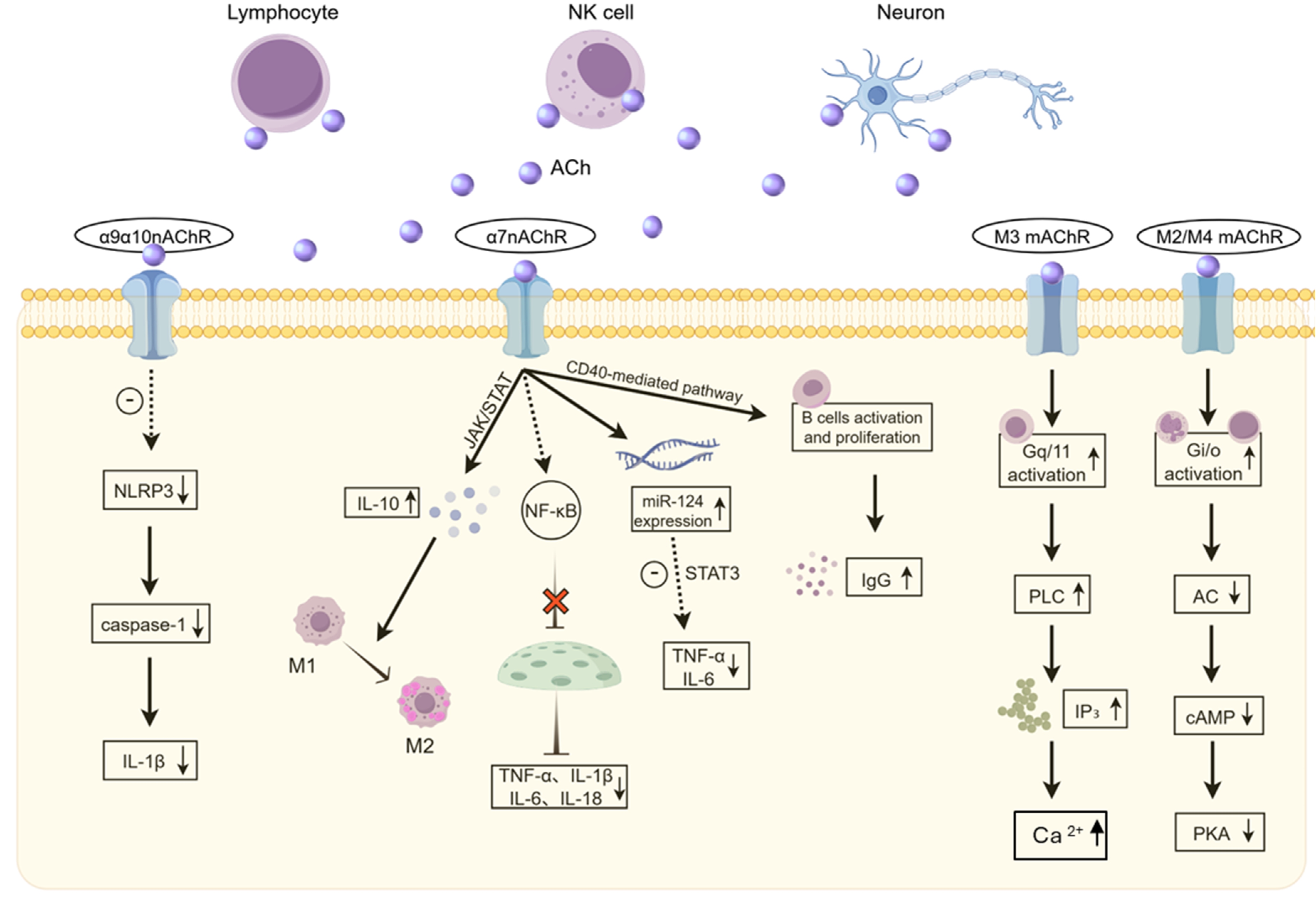
Figure 2.
Dopamine Regulation of Immune Cells via D1-like Receptors (D1R/D5R). Dopamine (DA) is released by neurons and immune cells such as macrophages, lymphocytes, and neutrophils, and transmits regulatory signals through D1-like receptors (D1R/D5R). D1-like receptors regulate cell differentiation through the PI3K/AKT/FOXO1/Klf2 pathway and influence intracellular cAMP levels via the Gs/cAMP/PKA pathway. They also drive cell activation and differentiation by upregulating PTGS2 and its downstream molecules CCL17 and PRDM1. D5 receptors promote cell proliferation and enhance cytokine secretion by activating the JAK-STAT signaling pathway. Additionally, D1-like receptor-mediated signaling regulates CD11b/CD18 integrin expression, thereby influencing cell adhesion and migration functions. Abbreviations: cAMP, cyclic adenosine monophosphate; CCL17, CC chemokine ligand 17; DA, dopamine; D1likeR, dopamine D1-like receptor; D5R, dopamine D5 receptor; FOXO1, forkhead box O1; Gs/cAMP/PKA, stimulatory G protein/cyclic adenosine monophosphate/protein kinase A; IFN-γ, interferon-gamma; IL, interleukin; JAK-STAT, Janus kinase-signal transducer and activator of transcription; Klf2, Kruppel-like factor 2; PCs, plasma cells; PI3K/AKT, phosphoinositide 3-kinase/protein kinase B; PRDM1, PR domain zinc finger protein 1; PTGS2, prostaglandin-endoperoxide synthase 2 (COX-2); Tfh, T follicular helper cell; Th2 cell, T helper type 2 cell; TNF-α, tumor necrosis factor alpha.
Figure 2.
Dopamine Regulation of Immune Cells via D1-like Receptors (D1R/D5R). Dopamine (DA) is released by neurons and immune cells such as macrophages, lymphocytes, and neutrophils, and transmits regulatory signals through D1-like receptors (D1R/D5R). D1-like receptors regulate cell differentiation through the PI3K/AKT/FOXO1/Klf2 pathway and influence intracellular cAMP levels via the Gs/cAMP/PKA pathway. They also drive cell activation and differentiation by upregulating PTGS2 and its downstream molecules CCL17 and PRDM1. D5 receptors promote cell proliferation and enhance cytokine secretion by activating the JAK-STAT signaling pathway. Additionally, D1-like receptor-mediated signaling regulates CD11b/CD18 integrin expression, thereby influencing cell adhesion and migration functions. Abbreviations: cAMP, cyclic adenosine monophosphate; CCL17, CC chemokine ligand 17; DA, dopamine; D1likeR, dopamine D1-like receptor; D5R, dopamine D5 receptor; FOXO1, forkhead box O1; Gs/cAMP/PKA, stimulatory G protein/cyclic adenosine monophosphate/protein kinase A; IFN-γ, interferon-gamma; IL, interleukin; JAK-STAT, Janus kinase-signal transducer and activator of transcription; Klf2, Kruppel-like factor 2; PCs, plasma cells; PI3K/AKT, phosphoinositide 3-kinase/protein kinase B; PRDM1, PR domain zinc finger protein 1; PTGS2, prostaglandin-endoperoxide synthase 2 (COX-2); Tfh, T follicular helper cell; Th2 cell, T helper type 2 cell; TNF-α, tumor necrosis factor alpha.
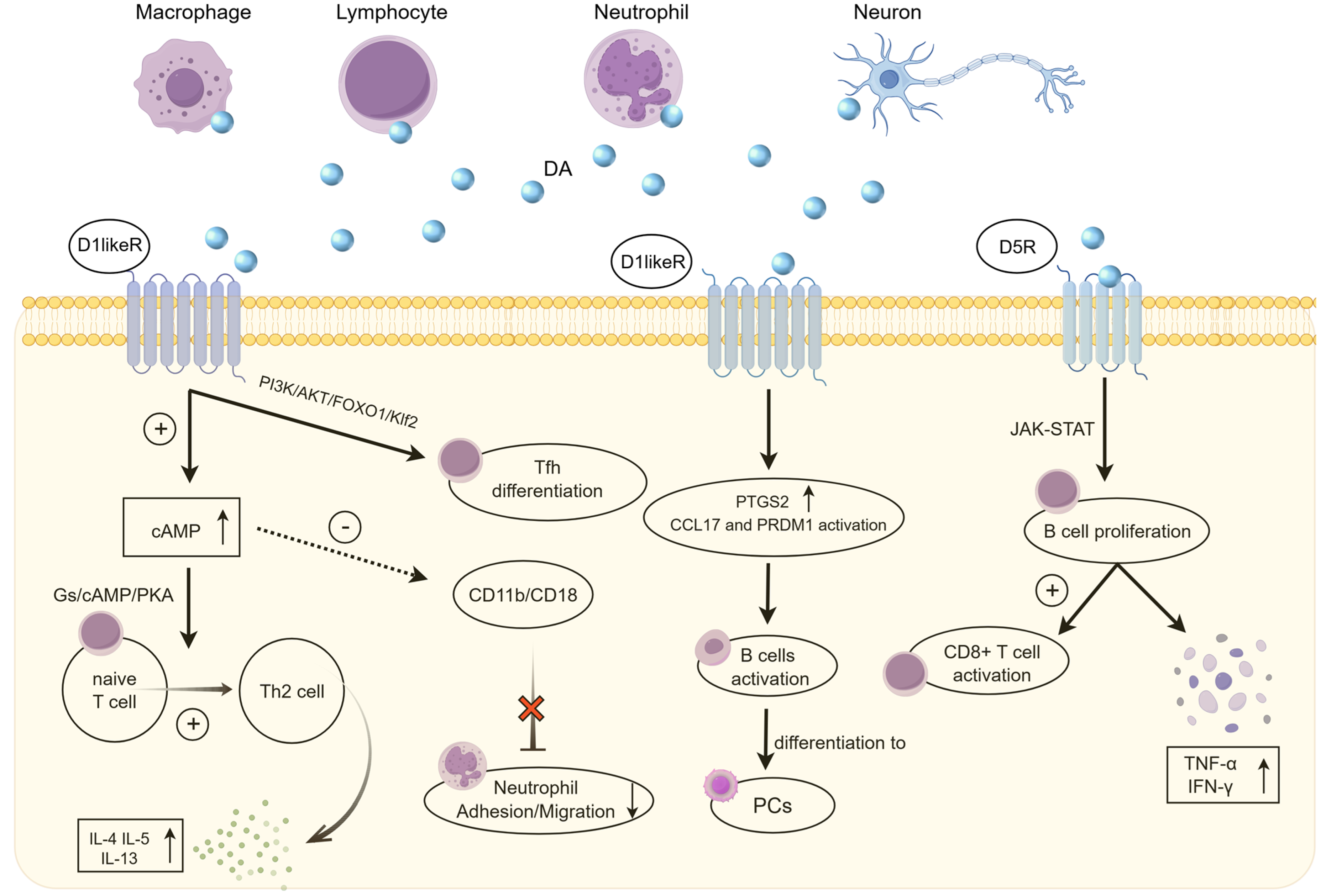
Figure 3.
Dopamine Regulation of Immune Cells via D2-like Receptors (D2R/D3R/D4R).Dopamine transmits regulatory signals through D2-like receptors (D2R/D3R/D4R): D2-like receptors inhibit T cell proliferation and the release of IL-2, IFN-γ, and IL-4 by suppressing cAMP levels, and promote Th2 differentiation by inhibiting cAMP-induced IL-2 activation. Meanwhile, D2-like receptors inhibit PKA/p38 signaling to alleviate inflammation in tumor-associated macrophages, and reduce IL-8 secretion by alveolar macrophages via suppressing ERK1/2 phosphorylation. On macrophages, D2R inhibits NF-κB translocation to reduce TNF-α and IL-6 release. Furthermore, D2-like receptors activate integrins VLA-4/VLA-5 to mediate T cell adhesion to fibronectin. Abbreviations: AC, adenylyl cyclase; cAMP, cyclic adenosine monophosphate; DA, dopamine; D2likeR, dopamine D2-like receptor; ERK1/2, extracellular signal-regulated kinase 1/2; Gi/o, inhibitory G protein; IFN-γ, interferon-gamma; IL,interleukin; NF-κB, nuclear factor kappa B; PKA/p38, protein kinase A/p38 mitogen-activated protein kinase; Th2, T helper type 2 cell; TNF-α, tumor necrosis factor alpha; VLA-4/VLA-5, very late antigen-4/5 (integrins α4β1/α5β1).
Figure 3.
Dopamine Regulation of Immune Cells via D2-like Receptors (D2R/D3R/D4R).Dopamine transmits regulatory signals through D2-like receptors (D2R/D3R/D4R): D2-like receptors inhibit T cell proliferation and the release of IL-2, IFN-γ, and IL-4 by suppressing cAMP levels, and promote Th2 differentiation by inhibiting cAMP-induced IL-2 activation. Meanwhile, D2-like receptors inhibit PKA/p38 signaling to alleviate inflammation in tumor-associated macrophages, and reduce IL-8 secretion by alveolar macrophages via suppressing ERK1/2 phosphorylation. On macrophages, D2R inhibits NF-κB translocation to reduce TNF-α and IL-6 release. Furthermore, D2-like receptors activate integrins VLA-4/VLA-5 to mediate T cell adhesion to fibronectin. Abbreviations: AC, adenylyl cyclase; cAMP, cyclic adenosine monophosphate; DA, dopamine; D2likeR, dopamine D2-like receptor; ERK1/2, extracellular signal-regulated kinase 1/2; Gi/o, inhibitory G protein; IFN-γ, interferon-gamma; IL,interleukin; NF-κB, nuclear factor kappa B; PKA/p38, protein kinase A/p38 mitogen-activated protein kinase; Th2, T helper type 2 cell; TNF-α, tumor necrosis factor alpha; VLA-4/VLA-5, very late antigen-4/5 (integrins α4β1/α5β1).
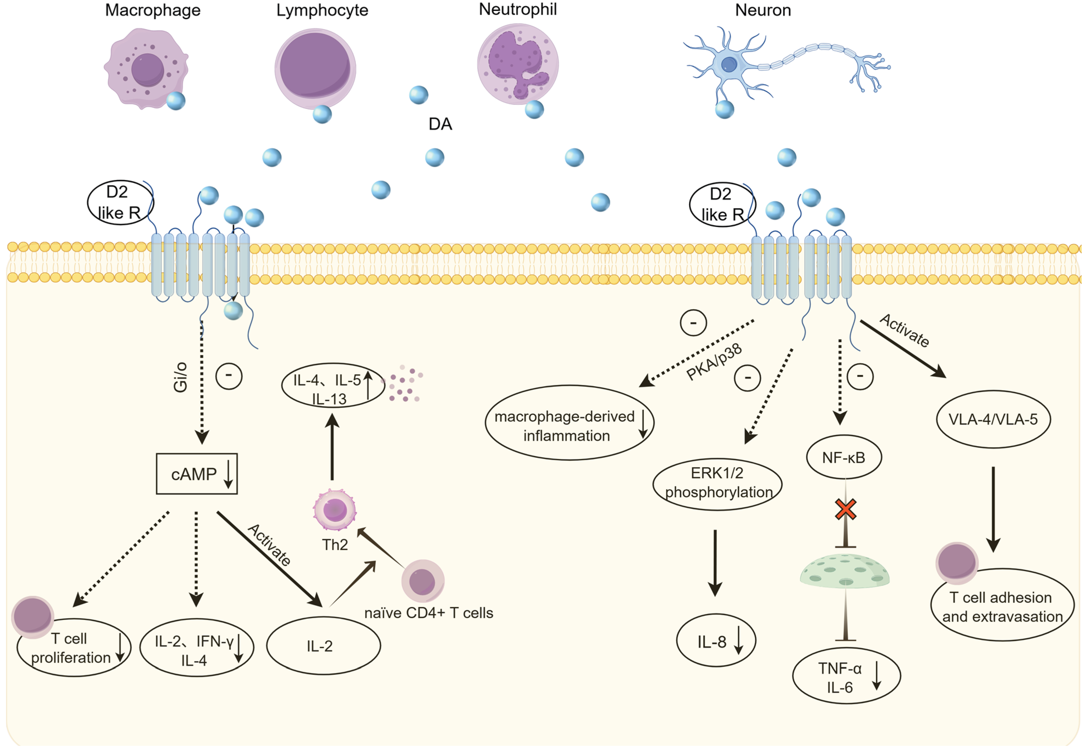

Table 1.
Function of Acetylcholine(ACh) and its Receptors in Immune Regulation.
| Receptor | Core Pathway | Target Cells | Functional Direction | Related Diseases/Models | Intervention Strategy |
| α7 nAChR | Suppression of NF-κB; activation of JAK/STAT; upregulation of miR-124 targeting STAT3 and TNF-α converting enzyme | Macrophages, monocytes, T cells, B cells | Anti-inflammatory, pro-repair, inhibits infiltration | Sepsis, EAE, postoperative ileus, acute lung injury, liver regeneration, facial nerve axotomy | α7 agonists, vagus nerve stimulation |
| α9/α10 nAChR |
Inhibition of NLRP3 inflammasome activation and caspase-1-mediated cleavage, blocking IL-1β release | Monocytes, T cells, B cells | Anti-inflammatory (IL-1β-specific) | Sterile inflammation, skin inflammation, endotoxemia models | LPC, G-PC (endogenous agonists) |
| α4β2 nAChR |
Co-stimulation with BCR/FcεRII to enhance proliferation cascades | B cells | Enhancement of B cell proliferation and survival | B cell development, humoral antibody responses | Nicotine analogs |
| M3 mAChR | Gq/PLC signaling causing IP₃ production and subsequent mobilization of intracellular Ca²⁺ | T cells (e.g., Jurkat), monocytes | Induction of intracellular Ca²⁺ mobilization and potential immune activation | T cell activation models | Receptor modulators |
| M2/M4 mAChR |
Gi-protein coupling and downstream inhibition of cAMP accumulation | Monocytes, T/B cells, macrophages, DCs | Unresolved direct function (inferred immune modulation) | Not clearly established | Under investigation |
| CAP Pathway | Efferent vagus nerve fibers releasing ACh to activate macrophage α7 nAChR, suppressing NF-κB | Systemic immune cells (esp. macrophages) | Systemic anti-inflammation and resolution of hyperinflammation | Systemic inflammation, sepsis, multi-organ hyperinflammatory protection | Vagus nerve stimulation, α7 agonists |
| Non-Receptor Action | Direct ACh release from ChAT+ immune populations modulating microenvironment | Local vasculature, neighboring immune cells | Local vasodilation and selective immune cell recruitment | LCMV infection, skin inflammation, allergic airway inflammation | AChE administration, ChAT induction |
Abbreviations: ACh, acetylcholine; AChE, acetylcholinesterase; BCR, B-cell receptor; CAP, cholinergic anti-inflammatory pathway; Ca²⁺, calcium ion; cAMP, cyclic adenosine monophosphate; ChAT, choline acetyltransferase; EAE, experimental autoimmune encephalomyelitis; G-PC, glycerophosphocholine; IL-1β, interleukin-1 beta; IP₃, inositol 1,4,5-trisphosphate; JAK/STAT, Janus kinase/signal transducer and activator of transcription; LCMV, lymphocytic choriomeningitis virus; LPC, lysophosphatidylcholine; mAChR, muscarinic acetylcholine receptor; nAChR, nicotinic acetylcholine receptor; NF-κB, nuclear factor kappa B; NLRP3, NLR family pyrin domain containing 3; PLC, phospholipase C; TNF-α, tumor necrosis factor alpha.
Table 2.
Function of Dopamineand its Receptors in Immune Regulation.
| Receptor | Core Pathway | Target Cells | Functional Direction | Related Diseases/Models | Intervention Strategy | |
| D1/D5 (D1-like) | Gs coupling leading to upregulation of cAMP and activation of PKA | T cells, B cells, epithelial cells, fibroblasts | Promotion of Th2/Tfh polarization, enhancement of epithelial barrier, anti-migration | MS, SLE, IBD, RHD | D1-like agonists (IBD and RHD) | |
| D5 | Activation of the JAK-STAT signaling cascade | B cells, CD8⁺ T cells | Upregulation of B cell proliferation and CD8⁺ T cell activation | Antitumor immunity and tumor immunosurveillance | Target D5 for immunotherapy | |
| D2/D3/D4 (D2-like) | Gi coupling leading to suppression of cAMP and downregulation of PKA | Macrophages, T cells, neutrophils, smooth muscle cells | Anti-inflammatory, M2 Polarization, Bronchodilation | PD, COPD/ARDS, RA | D2-like agonists (e.g., pramipexole) | |
| D2/D3 | Gi-mediated suppression of cAMP leading to downstream transcriptional regulation | CD4⁺ T cells | Induction of Th2 bias (upregulation of IL-4, downregulation of IFN-γ) | Th1/Th2 Imbalance | D2/D3 agonists | |
| D3 | Inside-out signaling leading to activation of VLA-4 and VLA-5 integrins | CD8⁺ T cells | Facilitation of lymphocyte homing and peripheral trafficking | Lymphocyte Trafficking | D3 modulators | |
| Concentration-dependent biphasic effects | Concentration-dependent differential receptor and adrenergic system activation | Macrophages, neutrophils, vasculature | Low dose: Promotes phagocytosis and M2 polarization; High dose: Drives pro-inflammatory cascades and tissue ischemia |
SIRS, Pancreatitis, ARDS | Dose-controlled infusion | |
Abbreviations: ARDS, acute respiratory distress syndrome; cAMP, cyclic adenosine monophosphate; COPD, chronic obstructive pulmonary disease; IBD, inflammatory bowel disease; IFN-γ, interferon-gamma; IL-4, interleukin-4; JAK/STAT, Janus kinase/signal transducer and activator of transcription; PKA, protein kinase A; RHD, rheumatic heart disease; SIRS, systemic inflammatory response syndrome; Tfh, T follicular helper cell; Th1/Th2, T helper type 1/2 cells; VLA-4/5, very late antigen-4/5.
Disclaimer/Publisher’s Note: The statements, opinions and data contained in all publications are solely those of the individual author(s) and contributor(s) and not of MDPI and/or the editor(s). MDPI and/or the editor(s) disclaim responsibility for any injury to people or property resulting from any ideas, methods, instructions or products referred to in the content. |
© 2026 by the authors. Licensee MDPI, Basel, Switzerland. This article is an open access article distributed under the terms and conditions of the Creative Commons Attribution (CC BY) license (http://creativecommons.org/licenses/by/4.0/).
Copyright: This open access article is published under a Creative Commons CC BY 4.0 license, which permit the free download, distribution, and reuse, provided that the author and preprint are cited in any reuse.