
Submitted:
02 November 2026
Posted:
04 November 2026
You are already at the latest version
Abstract
Background: Primary thyroid lymphoma (PTL) is a rare extranodal malignancy accounting for 1%–5% of all thyroid neoplasms, with diffuse large B-cell lymphoma (DLBCL) being the most common and aggressive subtype. Due to its low incidence, clinical data remain limited. This study aims to describe the clinical features, diagnostic approaches, treatment and outcomes of primary thyroid DLBCL in a single-center case series. Methods: We retrospectively enrolled four consecutive patients with histopathologically verified primary thyroid DLBCL who received treatment at our institution between 2021 and 2026. All relevant clinical data were extracted from archived electronic medical records. No statistical comparative analyses were performed given the limited sample size. Results: The cohort comprised four females, with a mean age of 73.5 years (range, 50–88 years). All patients presented with a rapidly enlarging painless neck mass, and two (50%) exhibited compressive symptoms. Elevated thyroid autoantibodies (TPO-Ab and/or TG-Ab) were detected in three patients (75%), indicating underlying Hashimoto’s thyroiditis. Diagnostic procedures varied among individuals: fine-needle aspiration cytology (FNAC) and/or core-needle biopsy (CNB), partial thyroidectomy were applied for pathological confirmation. All cases were classified as DLBCL. One patient received R-CHOP immunochemotherapy, one received Pola-R-CHP, and two opted for observation management. Conclusion: This case series provides descriptive real-world observations of primary thyroid DLBCL among elderly Chinese with underlying Hashimoto’s thyroiditis. Findings from this small cohort cannot be generalized. Immunochemotherapy regimens including R-CHOP and Pola-R-CHP may be considered within individualized multidisciplinary strategies; further large-scale studies are required to validate therapeutic performance.
Keywords:
primary thyroid lymphoma
; diffuse large B-cell lymphoma
; hashimoto’s thyroiditis
; treatment
; extranodal involvement
1. Introduction
Diffuse large B-cell lymphoma (DLBCL) is the most common subtype of non-Hodgkin’s lymphoma with distinct clinical and molecular heterogeneity. DLBCL that arises in extranodal organs is particularly linked to poor prognosis. Primary thyroid lymphoma (PTL) is a rare neoplasm originating from lymphoid tissue within the thyroid gland, accounting for 1-5% of all thyroid malignancies and less than 2% of all extranodal lymphomas [1].
PTL demonstrates remarkable histopathological heterogeneity. Non-Hodgkin lymphomas account for 98% of PTL cases, with diffuse large B-cell lymphoma being the most frequent subtype, representing 50% to 70% of cases, followed by mucosa-associated lymphoid tissue (MALT) lymphoma [2]. These two predominant subtypes differ substantially in clinical aggressiveness, treatment response and long-term prognosis: MALT lymphoma generally confers favorable survival outcomes, while primary thyroid DLBCL carries higher risk of disease progression and requires timely systemic immunochemotherapy [27]. Accurate pathological subtyping is therefore critical to guide clinical management.
PTL is strongly associated with Hashimoto’s thyroiditis (HT), with studies suggesting that up to 80% of patients with PTL have an underlying background of HT [1]. The disease predominantly affects middle-aged to elderly women, with a female-to-male ratio of approximately 3:1 [3]. The risk of developing PTL is estimated to be 40-80 times higher in patients with chronic lymphocytic thyroiditis compared to the general population [1].
Clinically, PTL typically presents as a rapidly enlarging, painless neck mass, often accompanied by compressive symptoms such as dysphagia, dysphonia, and dyspnea [4]. B symptoms (fever, night sweats, weight loss) occur in approximately 10–20% of cases [4]. The clinical presentation of PTL can closely mimic anaplastic thyroid carcinoma, making accurate diagnosis critical as management strategies and prognoses differ substantially [5]. Unlike other thyroid malignancies, PTL is highly responsive to nonsurgical treatments, with immunochemotherapy serving as the cornerstone of management [6].
Due to the low incidence of PTL, large-scale prospective cohorts are difficult to implement, most existing clinical knowledge relies heavily on retrospective case series and institutional reviews [1,3,5,6]. Single-center real-world case reports remain valuable for summarizing diagnostic and therapeutic experience, especially for elderly Chinese patients. Therefore, in this report, we present a case series of four patients with pathologically confirmed primary thyroid DLBCL. We aimed to retrospectively summarize their clinicopathological features, diagnostic pathways, treatment strategies and clinical outcomes together with relevant literature review, so as to enrich real-world evidence for this rare disease entity.
2. Materials and Methods
2.1. Study Design and Setting
This is a single-center retrospective observational study conducted at Beijing Tsinghua Changgung Hospital (BTCH), a tertiary-level comprehensive teaching hospital located in Beijing, China. Cases were retrieved from the hospital’s electronic medical record system. The study period spanned from January 1, 2021 to June 30, 2026; the data cutoff date for clinical and outcome analysis was July 31, 2026. Then four consecutive patients with histopathologically verified primary thyroid DLBCL were identified in our center. No additional patients with primary thyroid lymphoma or secondary thyroid-involving lymphoma were retrieved from our medical record system within this time frame. All four eligible patients were included in the present study. A simplified PRISMA-compliant case-selection flowchart was provided as Figure 1.
2.2. Data Collection
Clinical data, including age, gender, medical and surgical history, imaging findings, diagnostic methods, histopathological results, treatment approaches, laboratory indicators (thyroid function, thyroid autoantibodies, LDH), and presenting symptoms (compressive symptoms [dysphagia, dyspnea, voice changes] and B symptoms [fever, night sweats, weight loss]), were retrospectively extracted from archived electronic medical records by two independent reviewers. Discrepancies were resolved by discussion.
2.3. Inclusion and Exclusion Criteria
Inclusion criteria: 1) Patients with pathologically confirmed primary thyroid DLBCL according to the diagnostic criteria for primary thyroid lymphoma: thyroid as the primary site of lymphoma, with lymphoma lesion predominantly confined to thyroid at initial diagnosis; 2) Initial diagnosis and pathological specimen acquisition performed at Beijing Tsinghua Changgung Hospital; 3) Histopathological and immunohistochemical diagnosis consistent with DLBCL; 4) Sufficient retrievable core clinical data for case analysis. Enrolled patients did not need to complete the full course of antitumor treatment within our institution; observation-group patients who chose non-active treatment were also eligible for inclusion.
Exclusion criteria: 1) Secondary thyroid involvement by systemic non-Hodgkin lymphoma; 2) Patients whose definitive diagnosis was established in an external hospital; 3) Critical clinical, pathological or imaging information irreparably missing.
2.4. Pathological Review, Staging and Therapeutic Response Evaluation
All biopsy or surgical specimens were reviewed by hospital-attending pathologists specialized in hematopathology. Immunohistochemistry markers including CD20, CD3, Ki-67, CD10, BCL-6, MUM1 were applied to subtype DLBCL into germinal-center B-cell-like (GCB) or non-GCB subtype. Disease staging was performed using the Ann Arbor staging system for extranodal lymphoma. Treatment response was assessed according to Lugano 2014 lymphoma response criteria.
2.5. Ethical Considerations
Ethical approval was obtained from the Research Ethics Committee of BTCH. Owing to its retrospective nature, the requirement for individual written informed consent was waived by the ethics committee; all patient-identifiable information was de-identified during data extraction. This retrospective study inevitably carries risks of selection bias and information bias originating from incomplete medical records and non-standardized follow-up. We tried to reduce bias by double-checking archived medical records.
3. Results
Four patients were diagnosed with PTL between 2021 and 2026. The cohort included four females (8100%), with a mean age of 73.5 years (range, 50-88 years). The clinical characteristics of patients and are presented in Table 1. All the patients (4, 100%) presented with a rapidly growing mass in the neck, and 2 (50%) patients exhibited the symptoms of local compression, such as dysphagia, dyspnea, dysphonia. Lactate dehydrogenase (LDH) levels were increased in one patient (25%).
Thyroid function tests showed that two patients (50%) had raised TSH level (hypothyroid) and three patients (75%) had elevated TPO-Ab and/or TG-Ab (HT) (Table 2). Thyroid ultrasound (US) was utilized as the initial imaging tool for all patients. US showed bilaterally heterogeneous and hyperplastic thyroid gland in all patients. According to previous literature, the sonographic features of PTL can be categorized into three principal patterns: diffuse, nodular, and mixed types [7,8,9]. In this case cohort, there were 3 cases of nodular type (75%), and 1 case of diffuse type (25%). Additionally, 2 (50%) patients were observed multiple enlarged cervical lymph nodes. Furthermore, three patients underwent thyroid mass biopsy via FNAB or/and CNB, and one underwent surgical excision (partial thyroidectomy) (Table 2).
The pathological examinations of all patients were reported as primary thyroid lymphoma, and in all cases it was DLBCL (Table 3). Patients underwent computed tomography (CT) imaging of the neck, chest and abdomen, or positron emission tomography/CT (PET/CT), and bone marrow aspiration for systemic evaluation and staging. According to the Ann Arbor Staging System,one patients were stage ⅡE, one patient was stage ⅣE and two cases were not available. One patients completed treatment with R-CHOP (rituximab, cyclophosphamide, doxorubicin, vincristine and prednisone), one case received treatment with Pola-R-CHP (polatuzumab, rituximab, cyclophosphamide, doxorubicin, and prednisone), and two patients chose observation. Among the treated patients, one achieved complete remission (CR). One patient had just initiated treatment and had not yet reached the time-point for response assessment.
In summary, all four enrolled patients were elderly females presenting with rapidly enlarging painless neck masses, consistent with classic clinical phenotypes of primary thyroid DLBCL reported in prior literature. Three patients had positive thyroid autoantibodies suggesting underlying Hashimoto’s thyroiditis. Diagnostic pathways varied across individuals: three patients received fine-needle aspiration cytology as initial diagnostic test, while one patient obtained pathological diagnosis via partial thyroidectomy due to severe compressive symptoms. Nodular ultrasonic pattern predominated in this cohort. Systemic staging assessment was not completed in two elderly patients owing to patient refusal. Therapeutic strategies were highly individualized: two patients received standard immuno-chemotherapy (R-CHOP or Pola-R-CHP), and two highly-elderly patients opted for close observation without antitumor therapy considering advanced age and performance status. For the two actively treated patients, one achieved complete remission without major adverse events, whereas the other had just initiated therapy with response assessment pending. No recurrence or death events were documented during available follow-up. Given the small sample size, these observations should be interpreted as descriptive real-world experience rather than generalizable evidence.
4. Discussion
Primary thyroid lymphoma (PTL) is a rare extranodal lymphoma accounting for only 1%–5% of all thyroid malignancies, with diffuse large B-cell lymphoma (DLBCL) as the most aggressive and predominant subtype [1,2], which poses prominent diagnostic and therapeutic challenges due to its low incidence and overlapping manifestations with other thyroid lesions. This single-center retrospective case series summarized four consecutive patients with pathologically confirmed primary thyroid DLBCL treated from 2021 to 2026. In the present cohort, 100% of patients were female with a mean age of 73.5 years, consistent with the well-documented female predominance and elderly onset characteristic of PTL-DLBCL in previous studies[1,10,11]. All patients presented with a rapidly progressive painless neck mass as the chief complaint, which is the hallmark clinical manifestation distinguishing PTL from differentiated thyroid carcinoma with slow-growing nodules [5].
A strong pathogenetic link between chronic autoimmune thyroiditis and PTL was verified in our series:75% of patients showed elevated TPO-Ab/TG-Ab. Chronic lymphocytic infiltration in HT forms a persistent antigen-stimulated microenvironment that drives clonal expansion of intrathyroidal B lymphocytes, conferring a 40-80-fold higher lymphoma risk compared with the general population [1,10]. Clinicians should maintain high vigilance for PTL when HT patients develop rapidly enlarging thyroid nodules, rather than simply attributing gland enlargement to inflammatory exacerbation of autoimmune thyroiditis [3].
The diagnosis of PTL is built upon two complementary pillars: imaging and pathology. Ultrasound, the first-line imaging investigation for patients with suspected thyroid cancer, provides the preliminary clues based on characteristic morphological features [10,12]. According to previous literature, the sonographic features of PTL can be categorized into three principal patterns: diffuse, nodular, and mixed types [7,8,9]. Among these, the nodular type typically manifests as a homogeneous hypoechoic nodule, which may closely resemble anaplastic thyroid carcinoma (ATC) on imaging. However, a critical differentiating feature is that PTL nodules generally exhibit rich internal vascularity and an absence of calcifications in contrast to ATC [7]. The diffuse type, on the other hand, is characterized by markedly hypoechoic areas interspersed with cord-like or reticulate hyperechoic septations, with poorly defined borders against the adjacent normal thyroid parenchyma—a presentation that can be easily confused with HT. Of note, severe HT may also present with hypoechogenicity due to post-follicular destruction and subsequent fibrosis. Conversely, in diffuse PTL, the densely packed lymphocytic infiltration and paucity of fibrous stroma result in reduced acoustic impedance, leading to a paradoxical but characteristic posterior acoustic enhancement, which serves as a valuable sonographic marker to distinguish PTL from HT [7,8,9]. In alignment with the above classifications, the ultrasound findings of this cohort were 3 cases of nodular type (75%), and 1 case of diffuse type (25%).
However, these radiological findings are not conclusive; the diagnosis must be solidified by histopathological examination, which is achieved through the analysis of samples procured via biopsy or FNAC [13]. FNAC is generally the first-line diagnostic tool for thyroid nodules. According to published meta-analysis and large-cohort studies, ultrasound-guided CNB yields higher diagnostic sensitivity for PTL compared with FNAC and is recommended when PTL is clinically suspected [12,25]. In our small cohort, diagnostic modalities varied across individual patients.
According to the Ann Arbor staging system, previous studies have shown that most patients of PTL were diagnosed in the early stage [10,14]. In our study, one patients were stage ⅡE (25%), and one was stage ⅣE (25%), while staging data of two elderly patients were unavailable due to refusal of complete systemic staging examinations.
The management of PTL has transitioned from surgery-dominated approaches to immunochemotherapy-based comprehensive strategies, with or without involved site radiation therapy (ISRT) [15]. However, a standardized treatment protocol for PTL has yet to be established due to its rarity, and therapeutic decisions continue to be individualized based on pathological subtype, disease stage, and patient-specific factors. In localized (ⅠE-ⅡE) PTL-DLBCL, combined chemoradiotherapy is preferred, supported by retrospective data demonstrating 5-year OS and PFS rates of 81.2% and 77.8%, significantly higher than chemotherapy or surgery alone [16]. For advanced (ⅢE-ⅣE) PTL-DLBCL, R-CHOP-based immunochemotherapy is the established standard [17]. Notably, the POLARIX trial has recently shown Pola-R-CHP to be superior to R-CHOP in treatment-naïve DLBCL, especially in IPI score ≥2 patients, suggesting a promising avenue for future exploration in PTL [4]. Surgery is often reserved for diagnosis and to relief air way compression [18,19]. In the present cohort, one patients received the R-CHOP regimen, one patient was treated with the Pola-R-CHP regimen, and two cases opted for observation without active antitumor therapy. Despite the absence of consolidative radiation, the patient treated with R-CHOP achieved a complete remission, indicating promising preliminary efficacy of systemic immunochemotherapy in this setting.
In general, the prognosis of PTL is cautiously optimistic; however, it is profoundly influenced by a constellation of interrelated factors, including histopathological subtype, clinical stage, molecular biological characteristics, and the selected therapeutic regimen. Notably, the prognostic landscape varies considerably across distinct pathological entities. Indolent subtypes, such as mucosa-associated lymphoid tissue (MALT) lymphoma and follicular lymphoma, generally confer a favorable outcome. In contrast, highly aggressive variants, including DLBCL and Burkitt lymphoma, are consistently associated with an inferior prognosis [14,20,21]. Given this considerable prognostic heterogeneity, the clinical management of PTL patients mandates a comprehensive, individualized, and multidisciplinary therapeutic strategy, aiming to optimize both long-term survival and health-related quality of life.
PTL remains a diagnostically and therapeutically challenging malignancy, owing to its rarity, heterogeneous histopathological subtypes, and the frequent absence of pathognomonic clinical features.
This study has several limitations. First, it was a retrospective, single-center study with a relatively small sample size of four patients. The small sample directly reflects the extreme rarity of primary thyroid DLBCL; no additional primary thyroid lymphoma cases were seen in our hospital across the 5-year study period. Some patients refused complete systemic staging examinations, resulting in incomplete staging information and lack of long-term survival data. Although limited in statistical power, this case series still provides real-world observational evidence in elderly primary thyroid DLBCL. In recent years, a growing body of large-scale retrospective, multicenter, and population-based studies have substantially expanded our understanding of the disease spectrum, refined prognostic stratification, and informed evidence-based therapeutic algorithms. In the following sections, we synthesized the key findings from these contemporary investigations and discuss their implications for clinical practice (Table 4) [22,23,24,25,26,27]. Second, selection bias and unavoidable missing clinical data may exist due to the retrospective nature of data collection. Third, Follow-up durations were inconsistent across subjects; complete progression-free survival and overall survival endpoints were not available for all patients. Fourth, this small sample size precludes any formal statistical comparison of diagnostic performance between FNAC and CNB, as well as robust inference of treatment efficacy. Systemic staging was incomplete in two elderly patients due to patient refusal. Follow-up durations were inconsistent across subjects; complete progression-free survival and overall survival endpoints were not available for all patients. Our observations are supplementary real-world experience rather than confirmatory clinical evidence, and should not be generalized to broader PTL-DLBCL populations. Our observations are supplementary real-world experience rather than confirmatory clinical evidence, and should not be generalized to broader PTL-DLBCL populations.
Despite these limitations, this single-center retrospective study provides detailed clinical, ultrasonographic, pathological features, and therapeutic information of primary thyroid diffuse large B-cell lymphoma (PTL-DLBCL) in elderly Chinese patients and enrich real-world evidence for this rare thyroid malignancy. Within the limitations of this small case series, our work adds real-world descriptive observations for elderly Chinese patients with PTL-DLBCL. We documented sonographic patterns that may assist differential diagnosis against Hashimoto’s thyroiditis and anaplastic thyroid carcinoma. We also report preliminary real-world experience of Pola-R-CHP application in this rare subtype. These observations cannot serve as confirmatory evidence and should be interpreted cautiously. Further large-sample, multicenter, and long-term follow-up studies are warranted to validate our findings, clarify the long-term prognostic value of novel immunochemotherapy regimens, and establish standardized individualized diagnostic and therapeutic strategies for PTL-DLBCL.
5. Conclusion
PTL-DLBCL represents a rare and clinically challenging malignancy predominantly seen in elderly females with underlying Hashimoto’s thyroiditis. This single-center retrospective case series delivers descriptive clinical, ultrasound, pathological and therapeutic observations from four consecutive patients. Owing to limited sample size, incomplete staging, and inconsistent follow-up, no robust conclusions on diagnostic yield or treatment efficacy can be drawn from our local data. Individualized multidisciplinary management, including immunochemotherapy (R-CHOP or Pola-R-CHP) with or without involved-site radiation therapy, should be guided by existing large-scale literature. Further multicenter long-term studies are needed to establish optimal diagnostic and therapeutic strategies for PTL-DLBCL.
Ethics statement
The studies involving humans were approved by the Institutional Review Board of Beijing Tsinghua Changgung Hospital, Beijing, China (No.:27061-6-01). The studies were conducted in accordance with the local legislation and institutional requirements. Written informed consent was obtained from the individual(s) for the publication of any potentially identifiable images or data included in this article.
Author Contributions
Jiao Li: Conceptualization, Methodology, Writing – Original Draft. Yanying Wang: Data curation, Formal analysis, Validation. Jingjing Yao: Investigation, Visualization. Fan Yu: Resources, Software. Xuehan Mao: Writing – Review & Editing. Yuehua Huang: Supervision, Project administration.
Funding
The author(s) declared that financial support was not received for this work and/or its publication.
Data Availability Statement
The datasets presented in this article are not readily available because of ethical and privacy restrictions. Requests to access the datasets should be directed to the corresponding author.
Acknowledgments
We thank the Department of Ultrasound, Beijing Tsinghua Chang Gung Hospital, School of Clinical Medicine, Tsinghua University, for their assistance with imaging diagnosis.
Conflicts of Interest
The author(s) declared that this work was conducted in the absence of any commercial or financial relationships that could be construed as a potential conflict of interest.
References
- Alalawi, Y.; Alrashidi, T.N.; Shafie, S.; Moharram, L.; Asiri, A. Primary Thyroid Lymphoma: A Case Series. Cureus 2025, 17(8), e89197. [Google Scholar] [CrossRef]
- Watanabe, N.; Narimatsu, H.; Noh, J.Y.; et al. Long-Term Outcomes of 107 Cases of Primary Thyroid Mucosa-Associated Lymphoid Tissue Lymphoma at a Single Medical Institution in Japan. J. Clin. Endocrinol. Metab. 2018, 103(2), 732–739. [Google Scholar] [CrossRef]
- Harahap, A.S.; Santoso, I.; Charles, S.; et al. Variability in primary thyroid lymphoma: A clinicopathological exploration of diffuse large B-cell, marginal zone, and follicular lymphoma. Ann. Diagn. Pathol. 2025, 75, 152444. [Google Scholar] [CrossRef]
- Sharma, R.; Orogian, A.; Garcia-Pacheco, R.; et al. Primary Thyroid Lymphoma: A Rare Diagnosis Treated With Polatuzumab Vedotin. J. Investig. Med. High Impact Case Rep. 2024, 12, 23247096241299566. [Google Scholar] [CrossRef]
- Boeckermann, L.M.; Bellamkonda, N.; McCrary, H.; et al. Diffuse large B-cell lymphoma - an unusual cause of a rapidly enlarging thyroid mass: A large case series. Otolaryngol. Case Rep. 2025, 36, 100686. [Google Scholar] [CrossRef]
- Antunes, C.M.; Pinheiro, S.L.; Leite, V.; et al. Primary thyroid lymphoma: a tertiary-center experience. Endokrynol. Pol. 2025, 76(6), 659–664. [Google Scholar] [CrossRef]
- Ota, H.; Ito, Y.; Matsuzuka, F.; et al. Usefulness of ultrasonography for diagnosis of malignant lymphoma of the thyroid. Thyroid 2006, 16(10), 983–987. [Google Scholar] [CrossRef]
- Sharma, A.; Jasim, S.; Reading, C.C.; et al. Clinical presentation and diagnostic challenges of thyroid lymphoma: a cohort study. Thyroid 2016, 26(8), 1061–1067. [Google Scholar] [CrossRef]
- Yang, L.; Wang, A.; Zhang, Y.; et al. 12 cases of primary thyroid lymphoma in China. J. Endocrinol. Invest 2015, 38(7), 739–744. [Google Scholar] [CrossRef]
- Sakhri, S.; Zemni, I.; Ayadi, M.A.; et al. Primary thyroid lymphoma: a case series. J. Med. Case Rep. 2024, 18(1), 108. [Google Scholar] [CrossRef]
- Vander Poorten, V.; Goedseels, N.; Triantafyllou, A.; et al. Effectiveness of core needle biopsy in the diagnosis of thyroid lymphoma and anaplastic thyroid carcinoma: A systematic review and meta-analysis. Front Endocrinol. 2022, 13, 971249. [Google Scholar] [CrossRef]
- Wang, Y.; Chen, M.; Chen, P.; et al. Diagnostic performance of ultrasound and computed tomography in parallel for the diagnosis of lymph node metastasis in patients with thyroid cancer: a systematic review and meta-analysis. Gland Surg. 2022, 11(7), 1212–1223. [Google Scholar] [CrossRef]
- Walsh, S.; Lowery, A.J.; Evoy, D.; et al. Thyroid lymphoma: recent advances in diagnosis and optimal management strategies. Oncologist 2013, 18(9), 994–1003. [Google Scholar] [CrossRef]
- Vardell Noble, V.; Ermann, D.A.; Griffin, E.K.; et al. Primary Thyroid Lymphoma: An Analysis of the National Cancer Database. Cureus 2019, 11(2), e4088. [Google Scholar] [CrossRef]
- Zhao, J.L.; Zhao, J.H.; Yang, X.Y.; et al. Comprehensive analysis of primary thyroid lymphoma: a case report and review of diagnostic and treatment protocols. AME Case Rep. 2026, 10: 26. [Google Scholar] [CrossRef]
- Yi, J.; Yi, P.; Wang, W.; et al. A Multicenter Retrospective Study of 58 Patients With Primary Thyroid Diffuse Large B Cell Lymphoma. Front Endocrinol. 2020, 11, 542. [Google Scholar] [CrossRef]
- Sato, S.; Sawazaki, E.; Tsunoda, S.; et al. Dose-Attenuated-Polatuzumab Vedotine plus CHP vs. Dose-Attenuated -R-CHOP in Patients Aged ≥ 80 Years with Diffuse Large B-Cell Lymphoma: A Retrospective, Propensity Score-Matched Analysis Stratified by Simplified Frailty Score. Mediterr. J. Hematol. Infect. Dis. 2025, 17(1), e2025070. [Google Scholar] [CrossRef]
- Stein, S.A.; Wartofsky, L. Primary thyroid lymphoma: a clinical review. J. Clin. Endocrinol. Metab. 2013, 98(8), 3131–3138. [Google Scholar] [CrossRef]
- Agarwaf, N.; Wangnoo, S.K.; Sidiqqi, A.; et al. Primary thyroid lymphoma: a series of two cases and review of literature. J. Assoc. Physicians India 2013, 61(7), 496–498. [Google Scholar]
- Zhang, K.; Peng, X.; Wei, T.; et al. Prognostic nomogram and competing risk analysis of death for primary thyroid lymphoma: a long-term survival study of 1638 patients. Ann. Surg. Open 2022, 3(4), e226. [Google Scholar]
- Florindez, J.A.; Alderuccio, J.P.; Reis, I.M.; et al. Primary thyroid lymphoma: survival analysis of SEER database (1995-2016). Leuk. Lymphoma 2021, 62(11), 2796–2799. [Google Scholar] [CrossRef]
- Gao, Y.; Wang, J.; Tao, W.; et al. Developing a prognostic nomogram for primary thyroid lymphoma: insights from a large retrospective study. Discov. oncol. 2025, 16(1), 472. [Google Scholar] [CrossRef]
- Toloza Bonilla, F.; Gubbi, S.; Klubo-Gwiezdzinska. SAT-331 Survival Trends and Prognostic Factors in Primary Thyroid Lymphoma: Insights from SEER Database Analysis. J. Endocr. Soc. 2025, 9 (Suppl 1), bvaf149.2249. [Google Scholar] [CrossRef]
- Su, M.W.; Beck, T.N.; Knepprath, J.; et al. Primary thyroid lymphoma: A multi-center retrospective review. Am. J. Surg. 2024, 237, 115927. [Google Scholar] [CrossRef]
- Sanjaya, N.P.; Marcevianto, K.V.; Sanjaya, I.S.; et al. Validity of core needle biopsy in diagnosing primary thyroid lymphoma: a systematic-review and meta-analysis. Univ. Med. 2026, 45, 153–165. [Google Scholar] [CrossRef]
- Saito, Y.; Watanabe, N.; Inoue, K.; et al. Survival Outcomes and Treatment Strategies in Primary Thyroid Lymphoma: A Population-Based Study of 401 Cases from the Tokyo Cancer Registry. Thyroid 2025, 35(12), 1422–1429. [Google Scholar] [CrossRef]
- Chai, Y.J.; Hong, J.H.; Koo do, H.; et al. Clinicopathological characteristics and treatment outcomes of 38 cases of primary thyroid lymphoma: a multicenter study. Ann. Surg. Treat. Res. 2015, 89(6), 295–9. [Google Scholar] [CrossRef]
Figure 1.
Flowchart of patient inclusion. Four consecutive patients diagnosed with primary thyroid DLBCL were identified in our center from January 1, 2021 to June 30, 2026, and all patients fulfilled the inclusion criteria and were enrolled in this study. No additional lymphoma cases involving the thyroid gland were identified during the study period.
Figure 1.
Flowchart of patient inclusion. Four consecutive patients diagnosed with primary thyroid DLBCL were identified in our center from January 1, 2021 to June 30, 2026, and all patients fulfilled the inclusion criteria and were enrolled in this study. No additional lymphoma cases involving the thyroid gland were identified during the study period.
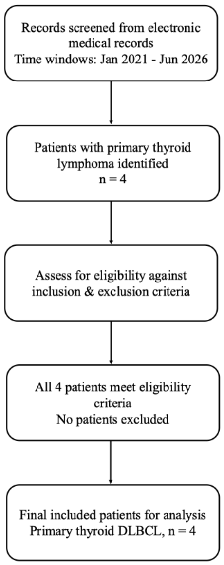

Table 1.
Clinical characteristics of patients.
| Case | Age | Gender | Symptoms | Latency period | B symptoms | Compression symptoms | Medical history | LDH |
| 1 | 50 | Female | Rapidly enlarging neck mass | 4 years | Weight loss | None | None | Normal |
| 2 | 85 | Female | Rapidly enlarging neck mass | 4 months | None | Dyspnea, dysphonia | HTN | Normal |
| 3 | 88 | Female | Rapidly enlarging neck mass | 5 years | None | Dyspnea | None | 3*Normal |
| 4 | 71 | Female | Rapidly enlarging neck mass | 1 week | None | None | HTN | Normal |
HTN: hypertension;.
Table 2.
Thyroid function tests, ultrasound findings, and diagnosis methods.
| Case |
TPO-Ab (KIU/L) |
TG-Ab (KIU/L) |
TSH μIU/mL |
FT3 pmol/L |
FT4 pmol/L |
Thyroid ultrasound | Cervical lymph nodes | Diagnosis methods |
| 1 | 12.30 | 339.00↑ | 3.480 | 4.32 | 16.90 | Nodular type, hypoechoic nodule in left lobe, 1.5*1.6 cm | None | FNAC |
| 2 | 41.3 | 229.4↑ | 23.89↑ | 3.17 | 11.67 | Nodular type, mutinodular, 5.7*3.2 cm in left lobe, 6.2*3.8 cm in right lobe | None | PT |
| 3 | <28.0 | 41.8 | 8.387↑ | 4.27 | 11.81 | Nodular type, nodule in left lobe, 6.3*4.0 cm | Multiple enlarged lymph node in left neck | FNAC |
| 4 | >600↑ | 144↑ | 0.502 | 3.27 | 17.1 | Diffuse type, hypoechoic area (4.7*3.3*1.7 cm) in the thyroid isthmus | Multiple enlarged lymph node in both neck | FNAC |
TG-Ab: Thyroglobulin antibody; TPO-Ab: Thyroid peroxidase antibody; TSH: thyroid stimulating hormone; FT3: Free Triiodothyronine; FT4: Free thyroxine; FNAC: fine-needle aspiration cytology; CNB: core needle biopsy; PT: partial thyroidectomy.
Table 3.
Histology, treatment, and prognosis of patients.
| Case | Histological type | Ki67 | Clinical stage | IPI score | Treatment | Prognosis/mortality |
| 1 | DLBCL, non-GCB | 50% | ⅡE | 0 | R-CHOP | CR, alive |
| 2 | DLBCL, GCB | 80% | NA | NA | Observation | NA |
| 3 | DLBCL, GCB | 70% | NA | NA | Observation | NA |
| 4 | DLBCL, non-GCB | 80%+ | IVE | 3 | Pola-R-CHP | PR, alive |
DLBCL: diffuse large B-cell lymphoma; GCB: germinal center B-cell like; non-GCB: non- germinal center B-cell like; R-CHOP: rituximab, cyclophosphamide, doxorubicin, vincristine, and prednisone; Pola-R-CHP (polatuzumab, rituximab, cyclophosphamide, doxorubicin, and prednisone); CR: complete response; NA: not available; PR: partial response.
Table 4.
Overview of large-sample studies on PTL.
| No. | Author / Year | Journal | Study type | Sample size | Main histological subtypes | Key results / Conclusions |
| 1 | Gao Y, et al, 2025 [22] | Discov Oncol | Retrospective and prognostic nomogram | 1469 | PTL (general) | The nomogram incorporating age, stage, multifocality, marital status, income, surgery, and radiotherapy effectively stratified PTL patients into three prognostic tiers (P < 0.0001) for 5-, 10-, and 15-year overall survival prediction. |
| 2 | Toloza Bonilla F, et al, 2025 [23] | J Endocr Soc | Retrospective cohort (SEER database) | 2,465 | DLBCL (62.5%) | PTL prognosis has improved in recent decades with 10-year disease-specific survival (DSS) 80.5%. Combined chemo-radiotherapy best survival; age ≥70 and advanced stage predict worse outcomes. |
| 3 | Su MW, et al, 2024 [24] | Am J Surg | Multicenter retrospective review | 31 | DLBCL, MALT | Diagnosis challenging; only 13% diagnosed preoperatively. Needle biopsy may facilitate earlier diagnosis and treatment. |
| 4 | Sanjaya NP, et al, 2026 [25] | Universa Medicina | Systematic rev iew; meta-analysis | 1091 | PTL (general) | CNB shows high diagnostic sensitivity (pooled 91.6%) and favorable safety profile, supporting its role when FNAC is inconclusive. |
| 5 | Saito Y, Inoue K, et al, 2025 [26] | Thyroid | Population-based registry study | 401 | DLBCL, others | Mortality highest in first 2 years, especially first 6 months. Age ≥80 and DLBCL subtype predict poorer survival. |
| 6 | Chai YJ, et al, 2016 [27] | Ann Surg Treat Res | Multicenter retrospective | 38 | MALT (42%), DLBCL (42%) | Early-stage PTL has excellent prognosis. 5-year OS: 100% for MALT, 87.5% for DLBCL. Underlying HT is a key clinical clue. |
Disclaimer/Publisher’s Note: The statements, opinions and data contained in all publications are solely those of the individual author(s) and contributor(s) and not of MDPI and/or the editor(s). MDPI and/or the editor(s) disclaim responsibility for any injury to people or property resulting from any ideas, methods, instructions or products referred to in the content. |
© 2026 by the authors. Licensee MDPI, Basel, Switzerland. This article is an open access article distributed under the terms and conditions of the Creative Commons Attribution (CC BY) license.
Copyright: This open access article is published under a Creative Commons CC BY 4.0 license, which permit the free download, distribution, and reuse, provided that the author and preprint are cited in any reuse.