

Submitted:
31 October 2026
Posted:
03 November 2026
You are already at the latest version
Abstract
Background/Objectives: After extraction, the crest may close before the socket has mineralised, leaving a covered socket residuum that may enclose the tissue lining the socket. We examined the socket wall histologically at the moment of extraction and compared it with the periapical region and the socket walls on preoperative cone-beam computed tomography (CBCT). Methods: Retrospective single-centre study of teeth extracted by one surgeon for a clinical indication in routine care. Before decortication according to the ARENA-Protocol® (Alveolar Ridge Epithelialization after Non-viable Bone Debridement and Augmentation), a trephine core was taken along the long axis of the socket wall; soft tissue was submitted when present. Each specimen received a leading diagnosis from a predefined hierarchy. Results: Sixty-six specimens from 41 sockets in 23 patients were examined. Thirty-eight sockets (92.7%) were altered. Bone showed mainly chronic inflammation and marrow fibrosis, soft tissue mainly florid inflammation. The radiographic lesion lay periapically and was visible on CBCT and its three-dimensional visualisation, whereas the lateral wall from which the core was taken showed no distinct change on either and was nevertheless altered, also in all three sockets without a periapical finding. All 14 root-filled and 24 of 27 non-root-filled sockets were altered. Conclusions: In this cohort, the socket wall was altered at extraction in almost every case, and the alteration along the long axis of the socket was detected only histologically. If the crest closes over such a wall, a residuum may enclose altered tissue. Controlled studies are needed to test this.
Keywords:
tooth extraction
; alveolar socket
; histopathology
; cone-beam computed tomography
; covered socket residuum
; apical periodontitis
; osteomyelitis
; root canal treatment
1. Introduction
Tooth extraction is one of the most frequent procedures in dentistry, and the healing of the socket is still usually described as an orderly sequence of clot, granulation tissue, woven bone and finally lamellar bone [1,2]. Part of this description is that the bundle bone lining the socket, the lamina dura, is resorbed as healing proceeds [3]. A clinical assumption has grown out of it. If the wall of the socket disappears by itself, there seems little reason to treat it, and the socket is left to the clot.
Histomorphometric and volumetric studies of human sockets have qualified the first part of this picture [4,5]. Months after extraction only part of the socket is mineralised, and the ridge loses a considerable part of its width, most of it within the first three to six months [6,7,8,9]. Three-dimensional analyses from our group have added a further observation. In unassisted upper premolar sockets the buccal wall collapsed by about half within six months, and new bone reached through the whole defect in only a minority of sockets [10]. In third molar sockets treated with platelet-rich fibrin alone, a non-mineralised residuum beneath a closed crest was the usual finding after six months [11], and in premolar and molar sockets platelet-rich fibrin did not measurably change mineralisation at three months [12]. We have described this enclosed space as the covered socket residuum (CSR), a non-mineralised area within the former socket that is surrounded by a mineralised crestal layer [13].
Our working explanation for why the centre is left behind draws on the way open wounds close. Myofibroblasts pull the edges of a wound together [14]. In the socket this pull acts on the walls, which move inward, the thin buccal wall most, and it brings the crest together first, because the crestal edges lie closest to each other and the mucosa contracts over them. Once the edges have met and the tension in the tissue falls, myofibroblasts lose their contractile activity and are removed by apoptosis [15,16]. From then on nothing draws the deeper walls together, and whatever space is still open beneath the closed crest remains. The larger the socket, the more space is left. Read this way, the residuum is not a failure of an otherwise complete repair but the expected end point of contraction in a defect too large to be closed by it. It may also explain an older clinical observation. Part of what implant surgeons have classified by touch as soft D3 or D4 bone [17] may have been such residual spaces, at a time when imaging could not show them.
This changes how the socket wall should be regarded. If the walls are drawn inward and closed over, the tissue that lined the socket at extraction is not necessarily replaced; it may be enclosed. The resorption of bundle bone after extraction [3] concerns the lamina dura and says nothing about the marrow behind it. Inflammation of endodontic origin extends from the root canal into the surrounding marrow [18,19,20], and a tooth that has to be removed has usually carried such a process for a long time. It might be argued that the wall needs no attention because the lamina dura is lost anyway. An alteration that has reached the marrow beyond the lamina dura, however, would not be lost with it.
From this follows a hypothesis. The inner surface of a covered socket residuum may be the former socket wall itself, carrying whatever condition it had when the crest closed over it. Two routes could lead there. The wall may already be altered when the tooth is removed, and closure of the crest conserves that state. Alternatively, a socket that was unremarkable at extraction may become affected during the weeks in which it is open, and that state is enclosed in the same way. Either route would place altered tissue inside a residuum that looks inconspicuous from the outside. Our own material points in this direction. At former third molar sites with a covered socket residuum, three of five bone sections were histopathologically altered years after the extraction, with marrow fibrosis and granulocytic or lymphoplasmacytic inflammation as the main findings and non-vital bone in a smaller part [21], and residua at the former sites of teeth other than third molars showed the same kinds of alteration, among them avital bone, osteonecrosis and osteomyelitis (unpublished data). The finding is therefore not confined to the wisdom tooth, where follicle remnants or impaction might otherwise explain it. Biopsies from clinically and radiographically unremarkable edentulous ridges have shown chronic inflammation, fibrosis and necrosis [22]. None of these observations, however, shows when the alteration arose, whether at the extraction or later, while the socket was open or after the crest had closed. This question led to the present study.
The first route can be examined directly at the moment of extraction. This has rarely been done, for a practical reason. Bone is not routinely sampled during an extraction, so there is almost no histology of the socket wall at that point. In our practice, the socket wall is decorticated at extraction as part of the ARENA-Protocol® (Alveolar Ridge Epithelialization after Non-viable Bone Debridement and Augmentation), and before decortication a trephine core is taken from the same wall and submitted for histology. The core consists only of bone that the decortication would remove anyway. These specimens are archived together with the operative reports that document their origin, and they allow the socket wall to be examined at the moment of extraction.
Two terms need to be kept apart. Tooth-related bony lesions (TRBL) are hypodense, hyperdense or mixed alterations of the bone around a tooth that is still in place. The covered socket residuum is a non-mineralised area inside a former extraction socket, often found years after the extraction. The hypothesis above connects the two, because the wall examined at extraction is the wall that would line a residuum if the crest closed over it. The present study examines only the first. It asks what the socket wall shows histopathologically at the time the tooth is removed, and whether the preoperative CBCT, in the periapical region or along the socket wall, predicts it. Whether an altered wall becomes the lining of a residuum, or affects healing, cannot be answered by this design and is addressed in the Discussion.
2. Materials and Methods
2.1. Study Design and Setting
This was a retrospective observational study at a single university department of oral and maxillofacial surgery. No tooth was removed and no specimen was obtained for the purposes of this investigation. All biopsies had been taken and submitted as part of routine care and were analysed retrospectively from the existing reports. Operations took place between April 2024 and June 2026. All teeth were extracted by one of the authors (S.G.), and every tooth had a clinical indication for extraction of its own, being either non-vital or tender to percussion. Some patients were also treated for covered socket residua at other sites, others were not. The indication for extraction did not depend on this treatment, and the teeth would have required removal in either case. Many patients were systemically healthy.
2.2. Case Identification and Inclusion Criteria
All pathology reports of the department from the study period were retrieved together with the corresponding operative reports and read specimen by specimen. Each specimen carries its own roman numeral, its own material designation and, in most reports, its own diagnosis. A specimen was included when both of the following applied. (i) the pathology report designated the material as socket wall, socket rim, socket content, tooth socket, interradicular septum or soft tissue from the socket, or the operative report stated unambiguously that the specimen was taken from the socket of a tooth removed in the same operation; and (ii) the tooth in question was removed during that operation, as documented in the operative report.
Specimens were excluded when they came from decortication or curettage of a former extraction site without tooth removal; from a bone lid, cortical plate, ascending ramus, mandibular angle or periosteum; from peri-implant tissue, the maxillary sinus or the nasal cavity; from a cyst lining; from mucosa without a bony or connective-tissue component of the socket; or when the specimen was the tooth root itself.
Three hundred and nine specimens were read and 66 met both criteria. One hundred and eighteen came from decortication or curettage of former extraction sites without tooth removal, 34 from other sites, 11 were socket material consisting of cyst lining, tooth root or mucosa alone, and 80 could not be assigned because the corresponding operative report was not retrievable (Figure 1). The excluded specimens are listed with the reason for exclusion in Table S1.
The diagnoses themselves are those of the reporting pathologists and were made in routine care. For this study, the extraction of the report wording and its coding were prepared with the assistance of a large language model (Section 2.9). The pathologist among the authors (R.W.) then read all pathology reports of the included specimens and checked every coded finding and every leading diagnosis against the original wording. Where her reading differed, the coding was corrected, and the dataset was used for the analysis only after her approval.
2.3. Surgical Procedure and Specimen Retrieval
Extractions were performed under local or general anaesthesia, depending on their extent. The ARENA-Protocol® treats the extraction as an operation in bone. After the tooth has been removed, diseased soft tissue is cleared from the socket and the socket wall is decorticated with a round bur until vital, bleeding bone is exposed on all sides. The socket is then filled, covered with a membrane and closed with sutures, and the ridge is left to epithelialise over the membrane without advancing or transferring a flap. Before the decortication, a trephine core was taken at each extraction along the long axis of the socket, from the crest at the interface between tooth and bone to the apical base, giving an elongated core of about 5 to 10 mm. Depending on the tooth, the core came from the mesial, distal, buccal, lingual or palatal wall or from the interradicular septum. It therefore consisted of bone that the subsequent decortication would have removed, and no additional tissue was taken for the purpose of sampling. Where the operative report names a piezosurgical instrument, this refers to sectioning of the tooth for a less traumatic removal; the biopsy itself was always taken with the trephine. Soft tissue was submitted in addition whenever it was present in the socket. The pathology request forms named osteomyelitis as the working question, to direct attention to inflammatory and necrotic changes of the bone, which are not routinely sought in extraction material.
2.4. Histopathological Assessment
Bone-containing specimens, partially accompanied by soft tissue, were fixed in 4% buffered formalin. Following assessment of tissue hardness, specimens requiring decalcification were selectively decalcified using either ethylenediaminetetraacetic acid (EDTA) or trichloroacetic acid (TCA), according to standard institutional protocols. After routine tissue processing, specimens were sectioned and stained with haematoxylin and eosin (HE). Periodic acid–Schiff (PAS) staining and additional histochemical stains were performed where indicated, in accordance with institutional procedures at the Dr. Senckenberg Institute of Pathology, Neuropathology and Human Genetics, University Hospital Frankfurt am Main, Germany. Most specimens were evaluated by one board-certified pathologist (R.W.) with nine years of professional experience; the remaining reports were issued by other board-certified pathologists of the institute.
Histopathological findings were documented using a standardised electronic reporting template with predefined response categories. Tissue components (epithelium, cystic components, connective tissue, adipose tissue and bone marrow) were recorded as present or absent, and bone vitality was classified as vital or avital. Bone remodelling and haemorrhage were recorded as present or absent. Granulation tissue, lymphocytes, plasma cells, granulocytes, multinucleated giant cells, epithelioid cells, necrotising granulomas and non-necrotising granulomas were each graded as absent, few or numerous. Bacterial and fungal colonisation were recorded as present or absent and, when present, further categorised by extent. Evidence of neoplasia was recorded as present or absent. For this analysis the reports were coded from their wording; the sections were not re-examined.
2.5. Radiographic Assessment
A cone-beam computed tomogram had been acquired before extraction as part of routine planning, partly at the University Hospital Frankfurt and partly by external practices, from which the scans were sent to the department before the operation. Devices and acquisition parameters therefore varied. All scans were anonymised before transfer and visualised by Visiogenics GmbH (Frankfurt am Main, Germany) in accordance with the applicable data protection regulations. As part of this three-dimensional visualisation, the visualisation team classified the tooth-related bony lesion at each site as hypodense, hyperdense, both, or neither. The assessment covered both the periapical region and the lateral socket walls along the long axis of each tooth. Because the visualisation was carried out before the operation and the histology only afterwards, the classification was made without knowledge of the histological findings. It was taken over unchanged and not repeated for this analysis. Hypodense and hyperdense changes were found periapically, whereas the lateral walls, from which the trephine core was taken, showed no distinct change on CBCT or its three-dimensional visualisation. Sockets without a periapical finding are referred to as radiographically inconspicuous. The comparison between image and histology is therefore one between the periapical finding and the state of the wall along its whole length.
2.6. Coding and Leading Diagnosis
A pathology report often names several findings in one sentence; a bone fragment may show marrow fibrosis, chronic inflammation and a florid component at the same time. Counting each finding separately is informative, but the totals then exceed the number of specimens. To allow tables in which every specimen is counted once, each specimen was additionally assigned a single leading diagnosis according to a fixed hierarchy. In descending order, the ranks were (1) osteonecrosis or non-vital bone, (2) osteomyelitis where so named in the report, (3) florid inflammation, (4) chronic inflammation, (5) granulation tissue, (6) fibrosis without inflammation, in bone marrow fibrosis, and (7) no detectable alteration. Where more than one finding applied, the higher-ranking one decided.
Negations in the report text, such as “no florid inflammation”, “without relevant inflammatory reaction” or “free of inflammation”, were evaluated clause by clause so that a negated finding was not counted. A site was classified by the leading diagnosis of its most severely affected specimen, separately for bone and for soft tissue.
2.7. Soft-Tissue Groups
Soft-tissue specimens were assigned to one of two groups by the wording of the material designation before the histology was read. The intra-alveolar and periapical group comprised specimens designated as soft tissue from the socket, socket content or tissue apical to the socket; this is the tissue that occupied the space the radiograph shows as hypodense, and it forms the primary soft-tissue group. The peri-coronal group comprised specimens designated by region only, without reference to the socket, typically granulation tissue excised from around the crown before the tooth was delivered. This group is reported separately. A specimen in the second group is not thereby shown to have come from outside the socket; the report simply does not say.
2.8. Statistical Analysis
The analysis is descriptive. Counts and proportions are given at specimen and at site level; the site is the unit of interest, because several specimens may come from one socket. No inferential testing was performed, because the cohort is a single-surgeon series without a comparison group, and p values would suggest a precision the design cannot provide.
2.9. Use of Generative Artificial Intelligence
A large language model (Claude, Anthropic) was used to assist in extracting and coding the wording of the pathology and operative reports according to the predefined criteria described above, in preparing the figures, and in drafting and language editing of the manuscript. For the analysis reported here, the reports were pseudonymised before they were processed with the language model, with names, dates of birth and addresses replaced by case numbers. The model did not make diagnoses. It organised the wording of reports written by pathologists, and its output was checked in full by a pathologist (R.W.) as described in Section 2.2. Coding rules, borderline assignments and the final text were decided by the authors.
3. Results
3.1. Cohort
Sixty-six specimens from 41 sockets in 23 patients met the inclusion criteria, 47 bone and 19 soft-tissue specimens. Sixteen patients were women and seven were men; the median age at operation was 61 years (range 24–81). Thirteen patients contributed more than one socket. Twenty-four sockets were in the maxilla and 17 in the mandible; by tooth type there were 16 molars, 10 third molars, 10 premolars, four incisors and one canine. Bone was sampled at 34 sockets and soft tissue at 16; both tissues were sampled at nine sockets, bone only at 25 and soft tissue only at seven.
3.2. The Socket Wall at the Time of Extraction
Thirty-eight of the 41 sockets (92.7%) showed a histopathological alteration of the socket wall or its soft tissue at the time the tooth was removed. Three sockets showed none. One bony specimen was reported as bone and connective tissue without further findings, one socket content consisted predominantly of fatty connective tissue with isolated vital bone fragments, and one soft-tissue specimen showed nerve fibres and reactive stromal change only.
Within the bone specimens the dominant picture was chronic. Marrow fibrosis was named in 22 of the 47 bone specimens and inflammation in 24, with a florid component in seven. Osteonecrosis or non-vital bone was found in four specimens from three sockets, and osteomyelitis was named in four specimens. At site level both led in three bone sites each (Table 1), because two of the osteonecrotic specimens came from the same socket and one specimen with osteomyelitis came from a socket in which osteonecrosis ranked higher. In nine bone sites marrow fibrosis was the only finding, that is, a socket wall in which the marrow space had been replaced by fibrous tissue without an inflammatory infiltrate. Representative examples of these alterations are shown in Figure 2.
3.3. Bone and Soft Tissue
Histopathological findings differed between bone and soft-tissue specimens (Table 1, Figure 3). Among bone sites the leading diagnosis was chronic inflammation in 13 and marrow fibrosis without inflammation in nine, with a florid component leading in only four. Among the 16 soft-tissue sites florid inflammation led in 11. Marrow fibrosis and fatty marrow were found only in bone. In all nine sockets with paired bone and soft-tissue samples, both tissue types showed histopathological alterations, with no discordance between the two.
3.4. Soft-Tissue Groups
Nine of the 16 soft-tissue sites belonged to the primary group. Six were designated as taken from the socket and three as apical to the socket. All nine were altered, six with florid inflammation as the leading diagnosis, two with chronic inflammation and one with fibrosis without inflammation. None of the specimens from inside or apical to the socket was unremarkable. The remaining seven sites were peri-coronal tissue designated by region only; six of these were altered, five with a florid picture, and one showed no detectable change. Both groups showed a similar trend; however, only the first could be directly related to the anatomical space depicted in the image.
3.5. Radiographic Pattern and Histology
In the periapical region, a hypodense pattern was the most frequent finding (18 sites), followed by a combined hypodense and hyperdense pattern (13 sites); a purely hyperdense pattern did not occur (Table 2, Figure 4). Alteration was found in 17 of 18 hypodense and in all 13 combined sites. The three sockets without a periapical finding were all histologically altered. Two showed chronic inflammation and one osteomyelitis, and all three carried a root-filled tooth. Of the nine sockets whose soft tissue came from inside or apical to the socket, that is, from the tissue occupying the radiolucent space, five had been classified as combined, three as hypodense and one was not classified, and all nine were altered.
The radiographic and the histological findings came from different parts of the socket. The hypodense and hyperdense changes lay at the apex and were visible on the image, whereas the lateral wall sampled by the trephine showed no distinct change (Section 2.5). This wall was nevertheless histologically altered in 38 of 41 sockets, in the sockets with a periapical lesion as well as in the three radiographically inconspicuous sockets.
3.6. Endodontic Status
Fourteen sockets held a root-canal-treated tooth and 27 a tooth that had not been root-filled (Table 3). Alteration was present in 14 of 14 root-filled and in 24 of 27 non-root-filled sockets. The two groups differed in severity rather than in frequency. A leading diagnosis from the three most severe ranks (osteonecrosis, osteomyelitis or florid inflammation) was present in 11 of 14 root-filled sockets (79%) but in only 9 of 27 non-root-filled sockets (33%). Non-root-filled sockets more often led with chronic inflammation or marrow fibrosis alone. The numbers are small, and this is reported as an observation, not as an effect.
The periapical pattern did not separate the two groups either (Table 4). Among root-filled sockets, seven were hypodense, four combined and three radiographically inconspicuous; among non-root-filled sockets, 11 were hypodense, nine combined and seven not classified, and none was inconspicuous. Endodontic status therefore did not explain whether a socket was altered.
3.7. Cystic Lesions of the Socket
Six further specimens came from sockets of teeth removed in the same operation but consisted of cyst lining, namely a radicular cyst in one socket, odontogenic cysts in four sockets of one patient and one cyst sac. They are not part of the main cohort, because cyst lining is not socket wall and pooling them would combine different entities. Both cystic sites for which a periapical classification existed were hypodense.
3.8. Bacterial Colonisation
Bacterial colonisation was reported at five sockets. At three of them the bacteria were identified as actinomycetes, in one with druse formation, and at the other two, which were reported together, the colonisation was focal and sparse. Fungal colonisation was reported in none. Culture and sequencing were not performed, and the term infection is therefore not used for the cohort as a whole.
4. Discussion
4.1. The Socket Wall Is Rarely Unremarkable
In almost every socket examined, the wall was already altered when the tooth was removed. This is not the picture of a healthy alveolus waiting for a clot, but of a bone compartment that has been reacting to a stimulus for a long time. The obvious objection is that these are the sockets of teeth that had to be removed, so some inflammation is to be expected. The objection carries less weight than it first seems. The alteration was present regardless of endodontic status, it reached beyond the apical region into the socket wall and the interradicular septum, and in nine bone sites it consisted of fibrosis without any inflammatory infiltrate, a state that follows inflammation rather than accompanying it.
Bone and soft tissue told different parts of the same story. The soft tissue carried the acute component and the bone the chronic one (Table 1, Figure 3), and where both had been sampled, they agreed in every case. To our knowledge this difference has not been described in this form before, and it was the most consistent observation in the dataset. We therefore read the two tissues not as independent findings but as two stages of one process, sampled at the same moment. This has a practical side. A study that samples only soft tissue will overestimate the acute component, and one that samples only bone will overestimate the chronic one.
4.2. What the Image Shows and What It Does Not
The central radiographic observation of this study concerns the place of the alteration. The image captured the periapical lesion well, but the lateral wall of the socket showed no distinct change on the image, and it was altered in almost every socket. The part of the socket that the extraction leaves behind, and that would later line a residuum, was therefore altered in a way that only histology revealed. That imaging understates what histology shows is not new in itself. Periapical radiography misses a substantial share of histologically confirmed apical periodontitis, and CBCT narrows the gap without closing it [23,24,25]. A lesion becomes radiographically visible largely once the cortical plate is involved [26], and a change confined to the marrow of the lateral wall may never reach that threshold. The sockets without a periapical finding are too few to carry an argument on their own, but they fit this pattern, and we return to them in Section 4.3.
The limitation also runs in the other direction, because a hypodense area is not one entity. In this material the radiolucent space held florid inflammation, chronic inflammation, marrow fibrosis and, in the cystic lesions reported separately (Section 3.7), epithelium-lined cyst wall. Neither radiologists reading CBCT nor grey-value analysis reliably distinguish a cyst from a granuloma [27,28,29], histopathological series of radiolucent jaw lesions show how heterogeneous this group is [30], and cystic tissue can remain in the alveolar process after the tooth has been removed [31]. The image shows a defect of mineralisation, not its content. A useful classification of tooth-related bony lesions will therefore have to be anchored to histology rather than to density alone, and future trials with a radiographic endpoint should measure the accuracy of the image against histology rather than presume it.
4.3. The Root-Filled Tooth
Root-filled teeth stood out in three ways (Table 3 and Table 4, Figure 4). All their sockets were altered, the alteration was more often severe, and all three sockets with a normal-looking image carried a root filling. The last point matters most clinically, because it means that a normal image is least reliable where a clinician is most inclined to trust it. It is also mechanically plausible. A radiopaque root filling or post, and the sclerotic bone often seen around a treated root, raise the local density and can mask a hypodense change nearby. Agreement between periapical radiography and CBCT is poorest in root-filled molars [32], and root-filled teeth carry a periapical radiolucency far more often than untreated ones [33,34], which is consistent with a lesion that is common and imperfectly imaged. Three sites cannot demonstrate the mechanism, but the pattern is consistent enough to be worth testing.
Persistent apical periodontitis after root canal treatment is well described [35], so a root-filled tooth that reaches extraction has usually carried a lesion for years. Whether the alteration is a consequence of the treatment, of the disease that made it necessary or of the time in between cannot be separated here. Confounding by indication is complete, because root-filled teeth are removed for different reasons, and usually later, than teeth that were never treated. Neither a root filling that appears adequate nor an inconspicuous image shows that the socket wall is free of inflammation, and the same holds for teeth that were never root-filled, most of whose sockets were also altered. Before an extraction, the bone around a tooth should therefore be regarded as potentially altered, however the tooth has been treated and however the image looks.
4.4. From the Socket Wall to the Residuum
The present findings link observations that have so far stood apart. Volumetric and histological studies of human sockets have shown that mineralisation at six months is incomplete and that a covered, connective-tissue-filled residuum is common [1,2,4,5], and studies of apical periodontitis have shown that the inflammation extends into the surrounding bone and is frequently an active, biofilm-driven process when the tooth is removed [18,19,35,36]. Our own residua, examined years after the extraction, had altered walls (Section 1). What was missing was a description of the bone compartment at the moment the tooth leaves it, and the present series provides this for teeth with an indication for extraction. It can also be read as the preliminary work for a prospective comparison of decortication against no decortication.
The findings fit the hypothesis set out in the Introduction, although they cannot confirm it. If the walls of a socket are drawn inward and the crest closes before the centre has filled [10,11], the tissue examined here is the tissue that would line the remaining space. In bone, that tissue showed marrow fibrosis and chronic inflammation, a change of the marrow space and not only of the lamina dura, and resorption of the bundle bone would therefore not remove it. The first route, an altered wall conserved beneath a closing crest, is at least anatomically possible in most of these sockets. The second route, an alteration acquired while the socket is open, is not addressed by this material.
Whether the first route actually occurs cannot be settled by an observational study. It would require either sampling the same site twice, at extraction and again when a residuum is opened, or an intervention that changes the starting condition in one socket and leaves it in another. The design that follows is a randomised split-mouth trial. In patients who need at least two comparable teeth removed, one socket would receive standardised debridement of the socket wall and the other the current standard of care, with the mineralised fraction of the original socket volume at six months, segmented by a blinded assessor, as the primary endpoint. Because both sockets belong to the same person, the strongest confounders of bone healing, such as age, metabolism, smoking, oral hygiene and vitamin D status, are eliminated rather than adjusted for, and about 18 patients would suffice where a parallel-group design would need about 70. Since preoperative images and specimens are obtained anyway, the same trial can also determine the diagnostic accuracy of the three-dimensional image against histology at no additional cost.
4.5. Clinical Implications
Several practical consequences follow from these findings, although each needs to be tested before it can be recommended as standard care.
First, curettage alone is unlikely to be sufficient. A curette removes granulation tissue and soft tissue from the socket, but in this cohort the alteration lay in the bone itself, in the marrow behind the lamina dura. Reaching it requires removing the wall, for example by decortication with a round bur or a piezosurgical instrument until a fresh, bleeding bone surface is exposed. Such a surface also opens the marrow spaces from which blood and cells can enter the socket. Where the wall is altered at extraction, its disappearance during healing should not be taken for granted.
Second, osteomyelitis and non-vital bone in a minority of sockets raise the question of antibiotic cover. Current evidence does not support routine prophylaxis for ordinary extractions [37], but the trials behind this conclusion did not distinguish sockets with altered bone from healthy ones. Removing a tooth from inflamed or necrotic bone is a procedure in altered bone, and the indication for antibiotics in this subgroup deserves separate evaluation.
Third, preoperative imaging matters, even though it is imperfect. Two-dimensional radiographs do not show the extent of a lesion in the third dimension. Within the principles of justification and optimisation of radiation exposure [38], a lower threshold for three-dimensional imaging with a limited, segmental field of view appears reasonable before the extraction of teeth with suspected or known disease, so that the extraction is planned as a procedure in bone and not only as the removal of a tooth. At the same time, an image without a lesion along the socket wall does not show that the wall is healthy. The wall has to be judged and treated during the operation, by decortication down to vital, bleeding bone, rather than on the basis of the image alone.
Fourth, the socket is part of the jawbone and thereby of the organism. Osteomyelitis, osteonecrosis and chronic inflammation are regarded as relevant to the whole person wherever they occur in the skeleton. Whether altered socket walls, or residua that later enclose them, act in this way is unknown. The possibility justifies including systemic as well as local parameters in future studies.
Fifth, the extraction itself deserves to be treated as an operation in bone. Tooth extraction regularly causes bacteraemia [39], and operating on inflamed or necrotic bone calls for the same discipline as other procedures in bone, with sterile instruments and gloves, antiseptic preparation of the field and aseptic technique as recommended for the prevention of surgical site infection [40], careful handling of the soft tissue, and debridement of the socket down to vital, bleeding bone. Seen in this way, the aim of an extraction is not only to remove the tooth but to leave behind a clean bony wound, and tooth extraction may be less trivial than it is generally assumed to be.
4.6. Limitations
Every tooth had its own clinical indication for extraction, being non-vital or tender to percussion (Section 2.1). The findings therefore describe teeth that had to be removed and should not be transferred to extractions in general. Whether the sockets of teeth removed for orthodontic or prosthetic reasons in healthy bone show the same picture is not known; such a comparison would require prospective sampling. The study is retrospective, comes from a single centre and a single surgeon and is modest in size. Several patients contributed more than one socket; sites within a patient are not independent, which, given the descriptive analysis, is stated rather than modelled.
The diagnoses are those of routine reports. Coding was based on their wording rather than on a re-examination of the sections, and specimens grouped under a joint diagnosis could not be resolved individually. The working question of osteomyelitis on the request forms may have directed the pathologist’s attention, although the findings themselves were graded with a predefined template. The periapical categories were taken over from the visualisation reports and not repeated. They were made before the operation and were therefore blind to the histology, but inter-reader agreement could not be assessed, and the scans came from different devices with differing acquisition parameters, which limits the comparability of density judgements between sites. The TRBL pattern could not be classified for seven sockets; these are shown separately in Table 2 and Table 4. Endodontic status was taken from the clinical record and the imaging documentation of the preoperative planning.
No culture or sequencing was performed, so the bacterial colonisation seen at five sockets, three of them with actinomycetes, is a histological observation and not a microbiological diagnosis [36]. No healing outcome was measured, so the study cannot show that the finding impairs healing, and the clinical consequences discussed in Section 4.5 remain to be tested.
Finally, this is not a study of the covered socket residuum itself. Residuum specimens were excluded from this cohort, and the link to the residuum proposed above remains a hypothesis. Histological terms that have been applied to tissue from non-healed areas of the jaw, such as neuralgia-inducing cavitational osteonecrosis, rest on a contested evidence base [41]. They describe tissue under the microscope, do not define the site from which it was taken and have nothing to do with the radiological finding, so they are not used here.
5. Conclusions
In this retrospective series of 41 sockets in 23 patients, the alveolar socket wall was histopathologically altered at the time of tooth extraction in 38 sockets (92.7%). Bone carried the chronic component, with marrow fibrosis and chronic inflammation, and the soft tissue of the socket the acute one; where both were sampled, they agreed in every case. The alteration was present irrespective of endodontic status. Whereas the periapical lesion was visible on the preoperative image, the changes in the lateral wall of the socket became apparent only under the microscope. The socket into which the clot forms is therefore, in this cohort, not a neutral compartment. Whether this affects healing, and whether it is what a covered socket residuum later encloses, needs to be answered by a controlled trial.
Supplementary Materials
The following supporting information can be downloaded at the website of this paper posted on Preprints.org, Table S1: Excluded specimens with the reason for exclusion.
Author Contributions
Conceptualization, S.G., J.Ś.-W., A.W., R.S., P.J.W. and R.W.; methodology, S.G., J.Ś.-W., P.J.W. and R.W.; investigation, S.G., J.Ś.-W., A.W., A.K. and R.W.; data curation, A.W., A.K. and R.N.; formal analysis, A.K., R.N. and P.J.W.; validation, A.W., P.J.W. and R.W.; resources, R.S., P.J.W. and R.W.; writing—original draft preparation, S.G. and J.Ś.-W.; writing—review and editing, S.G., J.Ś.-W., A.W., A.K., R.S., R.N., P.J.W. and R.W.; supervision, S.G., A.W., R.S. and P.J.W.; project administration, S.G., J.Ś.-W. and A.W. All authors have read and agreed to the published version of the manuscript.
Funding
This study was supported by the Academy for Biological Innovations in Surgery (ABIS) e.V., grant number AB-FFM-2026-1, which also funded the article processing charge. Visiogenics GmbH supported the study in kind by providing the three-dimensional visualisations free of charge and by funding the data analysis.
Institutional Review Board Statement
The study was conducted in accordance with the Declaration of Helsinki and approved by the Ethics Committee of the Medical Faculty of Goethe University Frankfurt (approval number 2026-3043, 7 July 2026).
Informed Consent Statement
Individual patient consent was not obtained. The Ethics Committee approved the study as a retrospective analysis of pseudonymised routine clinical documents (approval number 2026-3043).
Data Availability Statement
The data presented in this study are available on reasonable request from the corresponding author. The data are not publicly available because they contain information that could compromise patient privacy.
Acknowledgments
During the preparation of this manuscript, the authors used Claude (Anthropic) for the purposes described in Section 2.9. The authors have reviewed and edited the output and take full responsibility for the content of this publication.
Conflicts of Interest
S.G. is chairman of ABIS e.V. and a shareholder of Visiogenics GmbH. J.Ś.-W. is vice-chair of ABIS e.V. and managing director of Visiogenics GmbH. A.K. was funded by Visiogenics GmbH during the data analysis. Neither ABIS e.V. nor Visiogenics GmbH had any role in the design of the study, in the collection, analysis or interpretation of the data, in the writing of the manuscript or in the decision to publish. The other authors declare no conflicts of interest.
Abbreviations
The following abbreviations are used in this manuscript:
| CBCT | cone-beam computed tomography |
| CSR | covered socket residuum |
| HE | haematoxylin and eosin |
| PAS | periodic acid–Schiff |
| TRBL | tooth-related bony lesion |
References
- Amler, M.H. The time sequence of tissue regeneration in human extraction wounds. Oral Surg. Oral Med. Oral Pathol. 1969, 27, 309–318. [Google Scholar] [CrossRef] [PubMed]
- Boyne, P.J. Osseous repair of the postextraction alveolus in man. Oral Surg. Oral Med. Oral Pathol. 1966, 21, 805–813. [Google Scholar] [CrossRef] [PubMed]
- Araujo, M.G.; Lindhe, J. Dimensional ridge alterations following tooth extraction. An experimental study in the dog. J. Clin. Periodontol. 2005, 32, 212–218. [Google Scholar] [CrossRef] [PubMed]
- Trombelli, L.; Farina, R.; Marzola, A.; Bozzi, L.; Liljenberg, B.; Lindhe, J. Modeling and remodeling of human extraction sockets. J. Clin. Periodontol. 2008, 35, 630–639. [Google Scholar] [CrossRef] [PubMed]
- Barone, A.; Ricci, M.; Tonelli, P.; Santini, S.; Covani, U. Tissue changes of extraction sockets in humans: a comparison of spontaneous healing vs. ridge preservation with secondary soft tissue healing. Clin. Oral Implant. Res. 2013, 24, 1231–1237. [Google Scholar] [CrossRef] [PubMed]
- Schropp, L.; Wenzel, A.; Kostopoulos, L.; Karring, T. Bone healing and soft tissue contour changes following single-tooth extraction: a clinical and radiographic 12-month prospective study. Int. J. Periodontics Restor. Dent. 2003, 23, 313–323. [Google Scholar] [PubMed]
- Van der Weijden, F.; Dell’Acqua, F.; Slot, D.E. Alveolar bone dimensional changes of post-extraction sockets in humans: a systematic review. J. Clin. Periodontol. 2009, 36, 1048–1058. [Google Scholar] [CrossRef] [PubMed]
- Tan, W.L.; Wong, T.L.; Wong, M.C.; Lang, N.P. A systematic review of post-extractional alveolar hard and soft tissue dimensional changes in humans. Clin. Oral Implant. Res. 2012, 23 (Suppl. 5), 1–21. [Google Scholar] [CrossRef] [PubMed]
- Couso-Queiruga, E.; Stuhr, S.; Tattan, M.; Chambrone, L.; Avila-Ortiz, G. Post-extraction dimensional changes: a systematic review and meta-analysis. J. Clin. Periodontol. 2021, 48, 126–144. [Google Scholar] [CrossRef] [PubMed]
- Ghanaati, S.; Śmieszek-Wilczewska, J.; Al-Maawi, S.; Heselich, A.; Sader, R. After extraction, upper premolars undergo programmed socket collapse with development of cavitations rather than complete socket healing: a radiological study. Bioengineering 2025, 12, 128. [Google Scholar] [CrossRef] [PubMed]
- Ghanaati, S.; Kamolov, A.; Bouquot, J.; Sader, R.; Heselich, A.; Al-Maawi, S. Covered socket residuum (CSR) in former third molar sockets despite platelet-rich fibrin: a prospective randomized controlled split-mouth clinical study. Bioengineering 2025, 12, 1242. [Google Scholar] [CrossRef] [PubMed]
- Heselich, A.; Neff, P.; Śmieszek-Wilczewska, J.; Sader, R.; Ghanaati, S. Introduction of a semi-quantitative image-based analysis tool for CBCT-based evaluation of bone regeneration in tooth extraction sockets. Bioengineering 2025, 12, 301. [Google Scholar] [CrossRef] [PubMed]
- Ghanaati, S.; Heselich, A.; Lechner, J.; Sader, R.; Bouquot, J.E.; Al-Maawi, S. Jawbone cavitations: current understanding and conceptual introduction of covered socket residuum (CSR). Bioengineering 2026, 13, 106. [Google Scholar] [CrossRef] [PubMed]
- Tomasek, J.J.; Gabbiani, G.; Hinz, B.; Chaponnier, C.; Brown, R.A. Myofibroblasts and mechano-regulation of connective tissue remodelling. Nat. Rev. Mol. Cell Biol. 2002, 3, 349–363. [Google Scholar] [CrossRef] [PubMed]
- Desmoulière, A.; Redard, M.; Darby, I.; Gabbiani, G. Apoptosis mediates the decrease in cellularity during the transition between granulation tissue and scar. Am. J. Pathol. 1995, 146, 56–66. [Google Scholar] [PubMed]
- Grinnell, F.; Zhu, M.; Carlson, M.A.; Abrams, J.M. Release of mechanical tension triggers apoptosis of human fibroblasts in a model of regressing granulation tissue. Exp. Cell Res. 1999, 248, 608–619. [Google Scholar] [CrossRef] [PubMed]
- Misch, C.E. Bone character: second vital implant criterion. Dent. Today 1988, 7, 39–40. [Google Scholar]
- Nair, P.N. Pathogenesis of apical periodontitis and the causes of endodontic failures. Crit. Rev. Oral Biol. Med. 2004, 15, 348–381. [Google Scholar] [CrossRef] [PubMed]
- Nair, P.N.; Pajarola, G.; Schroeder, H.E. Types and incidence of human periapical lesions obtained with extracted teeth. Oral Surg. Oral Med. Oral Pathol. Oral Radiol. Endod. 1996, 81, 93–102. [Google Scholar] [CrossRef] [PubMed]
- Schulz, M.; von Arx, T.; Altermatt, H.J.; Bosshardt, D. Histology of periapical lesions obtained during apical surgery. J. Endod. 2009, 35, 634–642. [Google Scholar] [CrossRef] [PubMed]
- Ghanaati, S.; Śmieszek-Wilczewska, J.; Kögel, J.; Winter, A.; Najafi, R.; Sader, R.; Wild, P.J.; Winkelmann, R. Former third molar sockets often remain histopathologically altered: a retrospective study of covered socket residua with AI-based cell quantification. In Diagnostics; 2026; submitted. [Google Scholar]
- Kassolis, J.D.; Scheper, M.A.; Jham, B.C.; Reynolds, M.A. Histopathologic findings in bone from edentulous alveolar ridges: a role in osteonecrosis of the jaws? Bone 2010, 47, 127–130. [Google Scholar] [CrossRef] [PubMed]
- de Paula-Silva, F.W.; Wu, M.K.; Leonardo, M.R.; da Silva, L.A.; Wesselink, P.R. Accuracy of periapical radiography and cone-beam computed tomography scans in diagnosing apical periodontitis using histopathological findings as a gold standard. J. Endod. 2009, 35, 1009–1012. [Google Scholar] [CrossRef] [PubMed]
- Kanagasingam, S.; Lim, C.X.; Yong, C.K.; Mannocci, F.; Patel, S. Diagnostic accuracy of periapical radiography and cone beam computed tomography in detecting apical periodontitis using histopathological findings as a reference standard. Int. Endod. J. 2017, 50, 417–426. [Google Scholar] [CrossRef] [PubMed]
- Kruse, C.; Spin-Neto, R.; Evar Kraft, D.C.; Vaeth, M.; Kirkevang, L.L. Diagnostic accuracy of cone beam computed tomography used for assessment of apical periodontitis: an ex vivo histopathological study on human cadavers. Int. Endod. J. 2019, 52, 439–450. [Google Scholar] [CrossRef] [PubMed]
- Bender, I.B.; Seltzer, S. Roentgenographic and direct observation of experimental lesions in bone: I. 1961. J. Endod. 2003, 29, 702–706. [Google Scholar] [CrossRef] [PubMed]
- Simon, J.H.; Enciso, R.; Malfaz, J.M.; Roges, R.; Bailey-Perry, M.; Patel, A. Differential diagnosis of large periapical lesions using cone-beam computed tomography measurements and biopsy. J. Endod. 2006, 32, 833–837. [Google Scholar] [CrossRef] [PubMed]
- Rosenberg, P.A.; Frisbie, J.; Lee, J.; Lee, K.; Frommer, H.; Kottal, S.; et al. Evaluation of pathologists (histopathology) and radiologists (cone beam computed tomography) differentiating radicular cysts from granulomas. J. Endod. 2010, 36, 423–428. [Google Scholar] [CrossRef] [PubMed]
- Etoz, M.; Amuk, M.; Avci, F.; Yabaci, A. Investigation of the effectiveness of CBCT and gray scale values in the differential diagnosis of apical cysts and granulomas. Oral Radiol. 2021, 37, 109–117. [Google Scholar] [CrossRef] [PubMed]
- Koivisto, T.; Bowles, W.R.; Rohrer, M. Frequency and distribution of radiolucent jaw lesions: a retrospective analysis of 9,723 cases. J. Endod. 2012, 38, 729–732. [Google Scholar] [CrossRef] [PubMed]
- Titinchi, F.; Morkel, J. Residual cyst of the jaws: a clinico-pathologic study of this seemingly inconspicuous lesion. PLoS ONE 2020, 15, e0244250. [Google Scholar] [CrossRef] [PubMed]
- Cheung, G.S.; Wei, W.L.; McGrath, C. Agreement between periapical radiographs and cone-beam computed tomography for assessment of periapical status of root filled molar teeth. Int. Endod. J. 2013, 46, 889–895. [Google Scholar] [CrossRef] [PubMed]
- Pak, J.G.; Fayazi, S.; White, S.N. Prevalence of periapical radiolucency and root canal treatment: a systematic review of cross-sectional studies. J. Endod. 2012, 38, 1170–1176. [Google Scholar] [CrossRef] [PubMed]
- Tiburcio-Machado, C.S.; Michelon, C.; Zanatta, F.B.; Gomes, M.S.; Marin, J.A.; Bier, C.A. The global prevalence of apical periodontitis: a systematic review and meta-analysis. Int. Endod. J. 2021, 54, 712–735. [Google Scholar] [CrossRef] [PubMed]
- Nair, P.N. On the causes of persistent apical periodontitis: a review. Int. Endod. J. 2006, 39, 249–281. [Google Scholar] [CrossRef] [PubMed]
- Ricucci, D.; Siqueira, J.F., Jr. Biofilms and apical periodontitis: study of prevalence and association with clinical and histopathologic findings. J. Endod. 2010, 36, 1277–1288. [Google Scholar] [CrossRef] [PubMed]
- Lodi, G.; Azzi, L.; Varoni, E.M.; Pentenero, M.; Del Fabbro, M.; Carrassi, A.; Sardella, A.; Manfredi, M. Antibiotics to prevent complications following tooth extractions. Cochrane Database Syst. Rev. 2021, 2, CD003811. [Google Scholar] [CrossRef] [PubMed]
- European Commission. Radiation Protection No 172: Cone Beam CT for Dental and Maxillofacial Radiology. Evidence-Based Guidelines; Publications Office of the European Union: Luxembourg, 2012.
- Lockhart, P.B.; Brennan, M.T.; Sasser, H.C.; Fox, P.C.; Paster, B.J.; Bahrani-Mougeot, F.K. Bacteremia associated with toothbrushing and dental extraction. Circulation 2008, 117, 3118–3125. [Google Scholar] [CrossRef] [PubMed]
- Allegranzi, B.; Bischoff, P.; de Jonge, S.; et al. New WHO recommendations on preoperative measures for surgical site infection prevention: an evidence-based global perspective. Lancet Infect. Dis. 2016, 16, e276–e287. [Google Scholar] [CrossRef] [PubMed]
- Sekundo, C.; Wiltfang, J.; Schliephake, H.; Al-Nawas, B.; Ruckschloss, T.; Moratin, J.; et al. Neuralgia-inducing cavitational osteonecrosis - a systematic review. Oral Dis. 2022, 28, 1448–1467. [Google Scholar] [CrossRef] [PubMed]
Figure 1.
Selection of specimens. Every specimen of every pathology report was read and checked against the corresponding operative report; only material designated as socket tissue and confirmed as coming from the socket of a tooth removed in the same session was included.
Figure 1.
Selection of specimens. Every specimen of every pathology report was read and checked against the corresponding operative report; only material designated as socket tissue and confirmed as coming from the socket of a tooth removed in the same session was included.
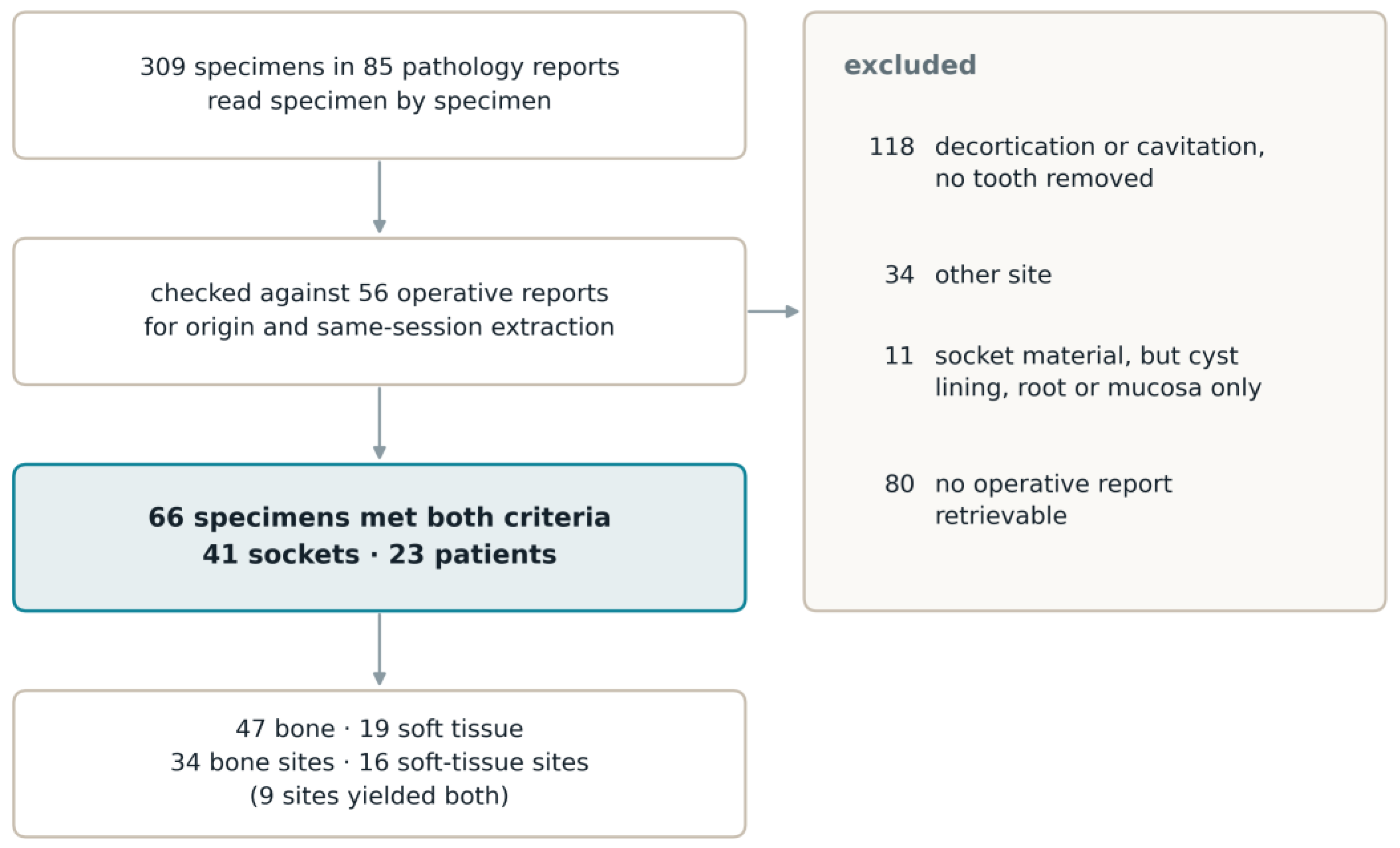
Figure 2.
Representative haematoxylin and eosin (HE)-stained sections illustrating the spectrum of bone alterations in the examined specimens. Each panel shows the whole section (left; the box marks the enlarged area) and a high-magnification detail (right). (a) Florid osteomyelitis, (b) osteonecrosis, (c) marrow fibrosis, (d) chronic inflammation. Scale bars 1 mm (overview) and 100 µm (detail).
Figure 2.
Representative haematoxylin and eosin (HE)-stained sections illustrating the spectrum of bone alterations in the examined specimens. Each panel shows the whole section (left; the box marks the enlarged area) and a high-magnification detail (right). (a) Florid osteomyelitis, (b) osteonecrosis, (c) marrow fibrosis, (d) chronic inflammation. Scale bars 1 mm (overview) and 100 µm (detail).
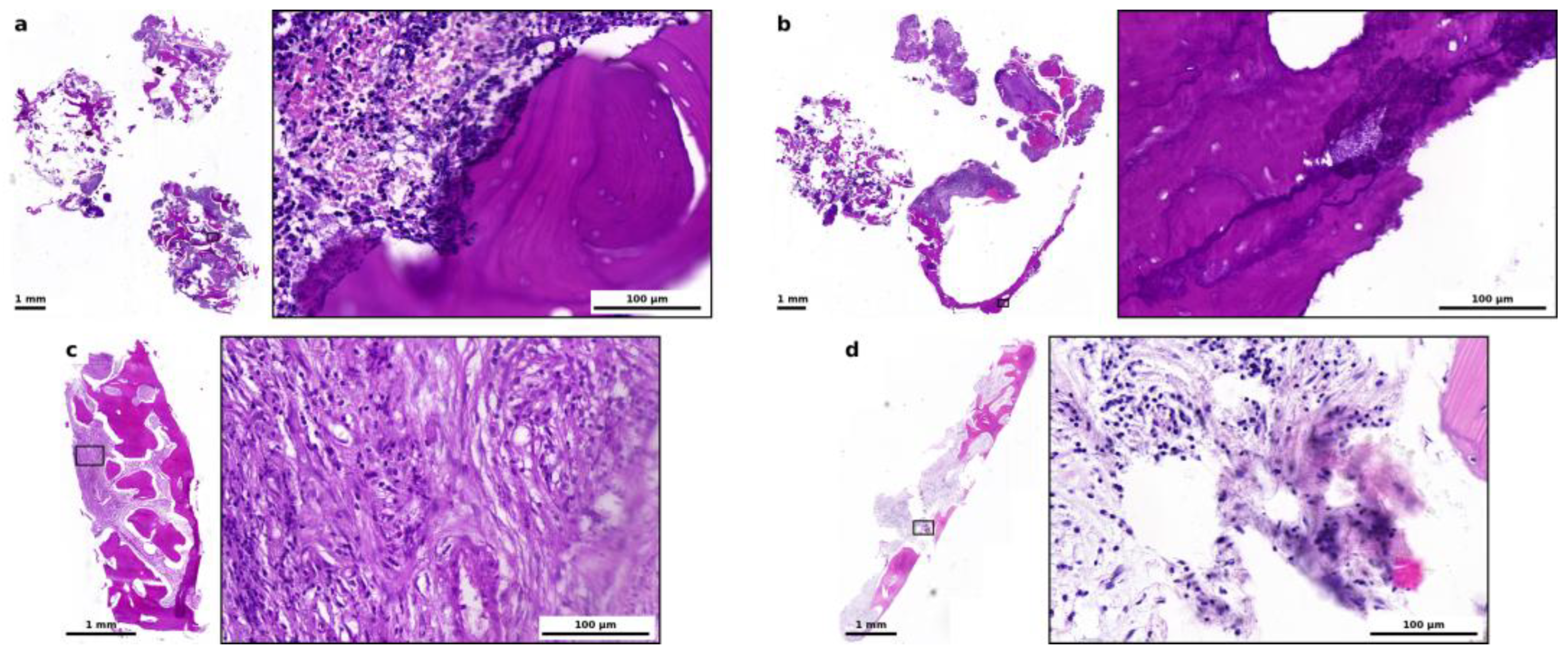
Figure 3.
Leading histopathological diagnosis at site level, bone against soft tissue. Bone carries the chronic picture (chronic inflammation and marrow fibrosis), whereas florid inflammation dominates the soft tissue of the socket.
Figure 3.
Leading histopathological diagnosis at site level, bone against soft tissue. Bone carries the chronic picture (chronic inflammation and marrow fibrosis), whereas florid inflammation dominates the soft tissue of the socket.
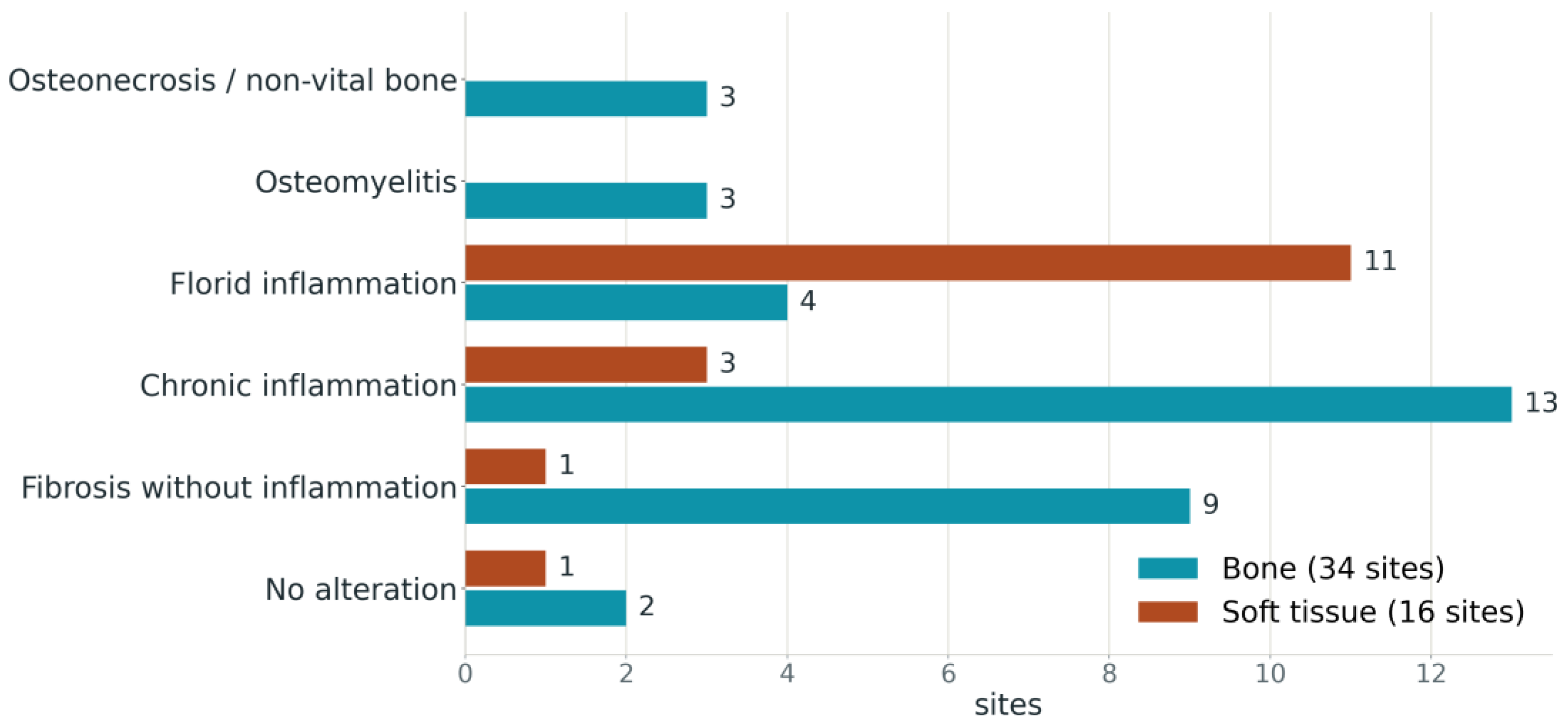
Figure 4.
Periapical pattern of the tooth-related bony lesion (TRBL) (a) and endodontic status (b) against histology of the socket wall, at site level. No socket with a purely hyperdense pattern occurred.
Figure 4.
Periapical pattern of the tooth-related bony lesion (TRBL) (a) and endodontic status (b) against histology of the socket wall, at site level. No socket with a purely hyperdense pattern occurred.
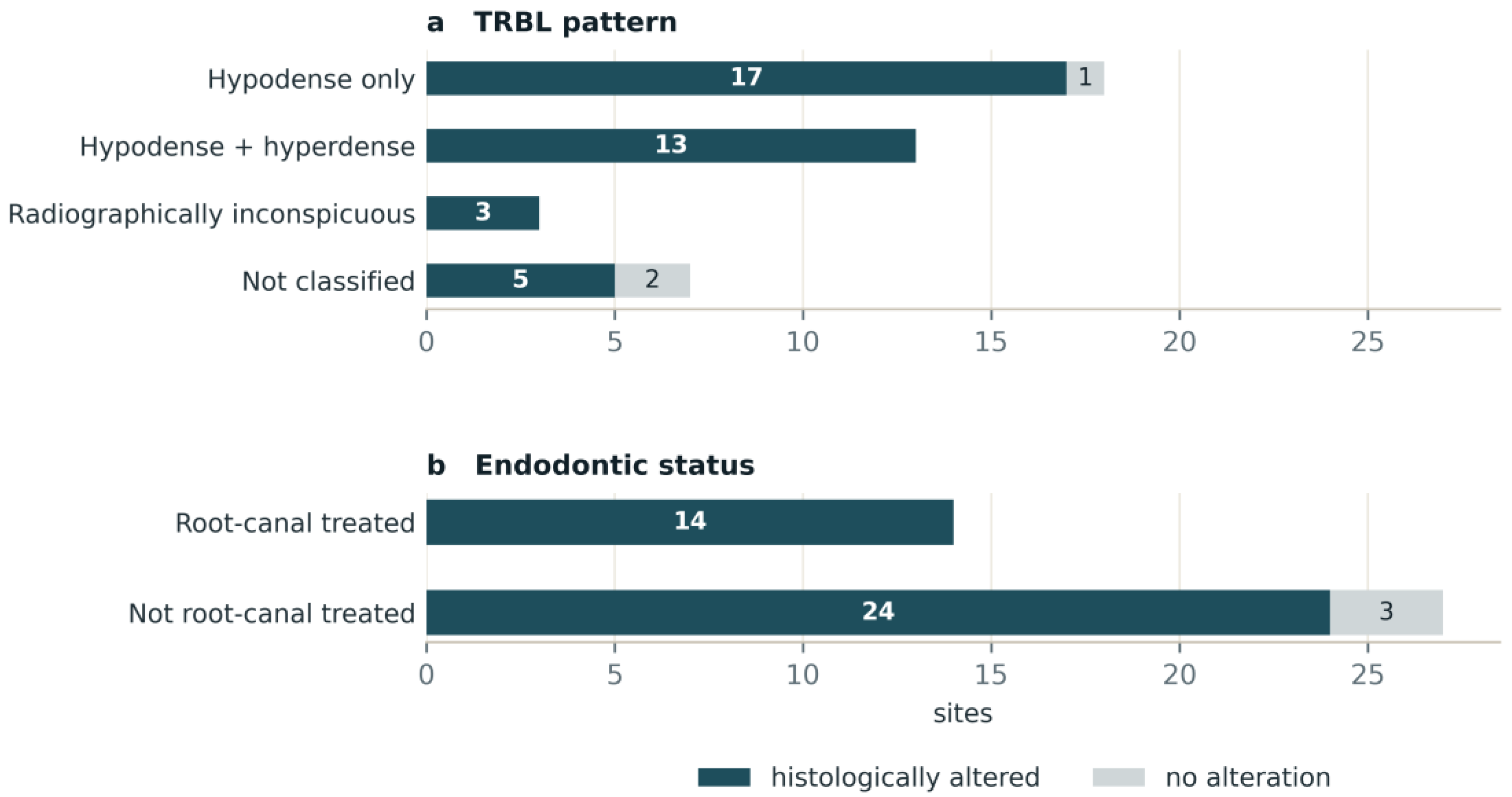

Table 1.
Leading histopathological diagnosis at site level, by tissue sampled. Nine sockets contributed both tissues and appear in both columns.
Table 1.
Leading histopathological diagnosis at site level, by tissue sampled. Nine sockets contributed both tissues and appear in both columns.
| Leading Diagnosis | Bone Sites (n = 34) | Soft-Tissue Sites (n = 16) |
|---|---|---|
| Osteonecrosis or non-vital bone | 3 | 0 |
| Osteomyelitis | 3 | 0 |
| Florid inflammation | 4 | 11 |
| Chronic inflammation | 13 | 3 |
| Fibrosis without inflammation | 9 | 1 |
| No detectable alteration | 2 | 1 |
| Altered, n (%) | 32 (94.1) | 15 (93.8) |
Table 2.
Preoperative periapical pattern of the tooth-related bony lesion (TRBL) against histology of the socket wall, at site level.
Table 2.
Preoperative periapical pattern of the tooth-related bony lesion (TRBL) against histology of the socket wall, at site level.
| TRBL Pattern | Sites | Histologically Altered | No Alteration |
|---|---|---|---|
| Hypodense only | 18 | 17 | 1 |
| Hypodense and hyperdense combined | 13 | 13 | 0 |
| Hyperdense only | 0 | – | – |
| Radiographically inconspicuous | 3 | 3 | 0 |
| Not classified | 7 | 5 | 2 |
| Total | 41 | 38 | 3 |
Table 3.
Endodontic status against histology, at site level.
| Endodontic Status | Sites | Histologically Altered | No Alteration |
|---|---|---|---|
| Root-canal treated | 14 | 14 | 0 |
| Not root-canal treated | 27 | 24 | 3 |
| Total | 41 | 38 | 3 |
Table 4.
Endodontic status and periapical radiographic pattern combined, at site level. A purely hyperdense pattern did not occur.
Table 4.
Endodontic status and periapical radiographic pattern combined, at site level. A purely hyperdense pattern did not occur.
| Endodontic Status and TRBL Pattern | Sites | Altered |
|---|---|---|
| Root-canal treated, hypodense | 7 | 7 |
| Root-canal treated, combined | 4 | 4 |
| Root-canal treated, radiographically inconspicuous | 3 | 3 |
| Not root-canal treated, hypodense | 11 | 10 |
| Not root-canal treated, combined | 9 | 9 |
| Not root-canal treated, pattern not classified | 7 | 5 |
| Total | 41 | 38 |
Disclaimer/Publisher’s Note: The statements, opinions and data contained in all publications are solely those of the individual author(s) and contributor(s) and not of MDPI and/or the editor(s). MDPI and/or the editor(s) disclaim responsibility for any injury to people or property resulting from any ideas, methods, instructions or products referred to in the content. |
© 2026 by the authors. Licensee MDPI, Basel, Switzerland. This article is an open access article distributed under the terms and conditions of the Creative Commons Attribution (CC BY) license (http://creativecommons.org/licenses/by/4.0/).
Copyright: This open access article is published under a Creative Commons CC BY 4.0 license, which permit the free download, distribution, and reuse, provided that the author and preprint are cited in any reuse.