

Submitted:
01 November 2026
Posted:
03 November 2026
You are already at the latest version
Abstract
Background: Advanced interventional pulmonology (IP) increasingly depends on cone-beam computed tomography (CBCT) and hybrid imaging workflows for the diagnosis and management of intrapulmonary lesions. However, globally, many IP teams still lack a dedicated pulmonology procedure suite in their institution and instead perform cases in rooms originally designed for cardiovascular, neurovascular, interventional radiology, or surgical use.
Methods: This review evaluates ceiling-mounted single-plane systems, floor-mounted systems, older and newer biplane angiography suites, robotic CBCT hybrid operating room systems and mobile CBCT systems and their suitability for IP procedures.
Results: Based on available literature and institutional experience, ceiling-mounted single-plane systems may offer ergonomic advantages for bronchoscopic IP because they preserve head-side access and procedural ergonomics. Floor-mounted systems remained usable, particularly in older institutions, but could restrict airway-side positioning. Biplane-suite capabilities varied by system generation and configuration; some newer configurations provided CBCT and overlay functions that may facilitate pulmonary workflows. Robotic hybrid systems may provide broad procedural flexibility for complex and combined procedures but require greater institutional support. Mobile CBCT systems represented a practical entry strategy for centers without dedicated infrastructure, while conventional multidetector CT remained valuable for selected percutaneous interventions. Successful adaptation across all platforms depended on room layout, workstation capability, team familiarity, anesthesia support, and multidisciplinary collaboration.
Conclusions: Advanced IP programs can be developed without a purpose-built pulmonology suite. Careful adaptation of existing imaging environments may allow centers to expand precision bronchoscopic and percutaneous capabilities according to local resources and workflow realities.
Keywords:
interventional pulmonology
; cone-beam computed tomography
; augmented fluoroscopy
; hybrid operating room
; peripheral pulmonary lesion
; pulmonary nodule
; microwave ablation
; transthoracic needle biopsy
; robotic bronchoscopy
; radiation safety
1. Introduction
The increasing detection of small, peripheral, and subsolid pulmonary lesions has created new diagnostic and therapeutic challenges for interventional pulmonology (IP). Lung cancer screening and contemporary pulmonary nodule management have increased the number of indeterminate lesions requiring accurate localization and tissue diagnosis [1,2], including subsolid and subcentimeter nodules that may be difficult to approach using conventional biopsy techniques [3]. Although advances in navigation bronchoscopy have improved access to peripheral targets, discrepancies between preprocedural CT anatomy and intraprocedural lesion position (or CT-to-body divergence) can contribute to geographic miss, increasing the importance of confirming the actual tool–lesion relationship during the procedure [4,5]. Cone-beam computed tomography (CBCT), augmented fluoroscopy, and hybrid image guidance provide intraprocedural target localization and tool-in-lesion confirmation [5,6], and growing clinical evidence supports their integration into bronchoscopic workflows across multiple navigation and localization strategies [7,8,9,10,11,12,13]. Mobile CBCT has further expanded the potential availability of advanced image guidance beyond fixed imaging suites [14]. Despite these advances, access to appropriately equipped procedural environments remains a major implementation challenge. Fixed C-arm CBCT and hybrid suites have traditionally been developed for established high-volume specialties such as cardiology, electrophysiology, neurovascular intervention, and interventional radiology, while dedicated pulmonary suites remain uncommon. Consequently, many IP programs must adapt existing shared, multi-specialty infrastructure to support increasingly complex pulmonary interventions.
Rather than advocating for a single standardized room design, this review provides a practical framework for implementing precision IP across available imaging environments while progressing toward optimal facilities. Drawing on published literature alongside clinical experience across multi-site academic and cancer center environments, we describe the implementation of IP procedures across ceiling-mounted and floor-mounted CBCT suites, biplane systems, robotic CBCT and hybrid operating rooms, mobile C-arm platforms, and conventional CT suites. The review focuses on parenchymal lesion interventions using both bronchoscopic and percutaneous approaches, with particular attention to procedure–room matching, workflow adaptation, equipment configuration, and multidisciplinary implementation. Pleural, airway, and other non-parenchymal procedures are discussed only where they directly influence procedural workflow or room design.
2. Materials and Methods
This narrative review synthesized published literature on cone-beam CT, augmented fluoroscopy, mobile CBCT, hybrid operating room workflows, percutaneous thoracic intervention, and advanced interventional pulmonology procedures. PubMed, Google Scholar, and reference lists of relevant articles were reviewed using combinations of the terms “cone-beam CT,” “CBCT,” “augmented fluoroscopy,” “interventional pulmonology,” “bronchoscopy,” “peripheral pulmonary lesion,” “hybrid operating room,” “percutaneous lung biopsy,” “microwave ablation,” “lung ablation,” and “mobile C-arm.” Priority was given to peer-reviewed clinical series, prospective studies, systematic reviews, technical reviews, and published experience from interventional pulmonology-led or multidisciplinary thoracic programs. This was not designed as a systematic review or meta-analysis; therefore, no formal risk-of-bias assessment or pooled quantitative analysis was performed.
3. Workflows Under CBCT
Advanced imaging broadens the scope of interventional pulmonology beyond bronchoscopic access alone. CBCT, augmented fluoroscopy, and hybrid imaging platforms support bronchoscopic biopsy, percutaneous transthoracic biopsy and ablation, image-guided localization, and combined single-stage procedures. Accordingly, procedural planning should prioritize the approach that provides the safest and most accurate access to the target rather than defaulting to a single route.
3.1. Bronchoscopic Applications
During bronchoscopy, CBCT can update lesion position after procedural changes, provide augmented fluoroscopic guidance, and confirm the tool–lesion relationship before tissue acquisition or treatment. These functions have been incorporated into workflows using radial endobronchial ultrasound (rEBUS), electromagnetic navigation, ultrathin bronchoscopy, and other navigation techniques [7,8,9,10,11,12,13,15,16,17]. Published experience has progressed from CBCT-derived augmented fluoroscopic localization and lung marking [18,19,20,21] to image-confirmed bronchoscopic biopsy [15,17]. Robotic and shape-sensing bronchoscopy further improve catheter stability and pathway control, while CBCT provides complementary confirmation of the instrument position relative to the intraprocedural target [22].
3.2. Percutaneous Applications
CBCT also supports percutaneous transthoracic biopsy and thermal ablation. Large series have demonstrated the feasibility and diagnostic performance of CBCT-guided transthoracic needle biopsy [23], while a recent 77-patient feasibility study reported CBCT laser-guided biopsy performed by an interventional pulmonologist in a hybrid operating room, with an overall diagnostic accuracy of 94.8% [24]. CBCT-guided microwave ablation, laser-guided lung ablation, combined ablation with thoracoscopic resection, and intrapulmonary trajectory adjustment further illustrate its therapeutic applications [25,26,27,28]. Transbronchial cryoablation is also undergoing early clinical evaluation [29]. These expanding applications reinforce the need to consider both bronchoscopic and percutaneous access when selecting and configuring procedural rooms.
3.3. Single-Stage and Combined Workflows
Hybrid imaging environments can integrate localization, diagnosis, and treatment within a single procedural episode. Reported workflows include augmented fluoroscopic bronchoscopic localization followed by thoracoscopic resection [30], image-guided pulmonary marking [31,32], and percutaneous ablation combined with thoracoscopic resection for multiple lesions [27]. Such approaches demonstrate the potential value of advanced imaging suites as shared procedural platforms capable of supporting complementary bronchoscopic, percutaneous, and surgical strategies according to lesion characteristics and multidisciplinary planning.
Taken together, the available evidence indicates that advanced interventional pulmonology and thoracic image-guided procedures can be performed across diverse imaging environments, including ceiling- and floor-mounted single-plane CBCT systems, biplane angiography platforms, robotic or hybrid operating-room CBCT systems, mobile three-dimensional C-arms, and conventional multidetector computed tomography (MDCT) suites. Table 1 provides a practical comparison of these platforms, highlighting their typical procedural environments, principal advantages for IP use, and workflow limitations, based on our experience. Because technical capability does not necessarily translate directly into procedural suitability, Table 2 further synthesizes the relative applicability of each platform across percutaneous, transbronchial, thoracoscopic, hybrid, and endovascular workflows.
4. Why a Dedicated Pulmonology CBCT Suite Is Uncommon
A dedicated pulmonary CBCT suite is an attractive reference model because it places airway access, anesthesia, bronchoscopy equipment, radiation protection, reconstruction workstations, and pulmonary staff within a single procedural environment. In practice, however, many institutions cannot begin with this model. The reasons are historical, spatial, financial, and organizational rather than purely technological [5,6].
First, the bronchoscopic use of CBCT is relatively recent. CBCT-guided bronchoscopy/endobronchial real-time navigation was first reported in 2014 by Hohenforst-Schmidt and colleagues, who described the use of CBCT for endobronchial real-time navigation [37]. This early report was later cited in technical reviews as the first published description of airway segmentation overlaid on fluoroscopic images during CBCT-guided bronchoscopy [5]. In contrast, C-arm CBCT-guided percutaneous transthoracic biopsy was already represented in larger thoracic intervention series from the early 2010s [23,33]. The later expansion of bronchoscopic CBCT literature—including CBCT-derived augmented fluoroscopy, tool-in-lesion confirmation, mobile CBCT-assisted bronchoscopy, robotic bronchoscopy with CBCT, and meta-analytic data—helps explain why most hospitals did not historically design fixed CBCT rooms around interventional pulmonology [5,6,7,8,9,10,11,12,13,14,15,16,17,22,30]. Conventional bronchoscopy suites were typically organized around airway access, endoscopy towers, anesthesia support, and fluoroscopy, rather than fixed angiographic CBCT, overlay workstations, and hybrid-room workflows [5,6].
Second, most fixed CBCT angiography systems were developed around cardiovascular, neurovascular, interventional radiology, or surgical workflows rather than the bronchoscope. As a result, room geometry, monitor placement, table controls, anesthesia access, C-arm parking, radiation shielding, and staff routines may not naturally match the needs of bronchoscopic or combined bronchoscopic-percutaneous IP procedures [5,6]. Third, hospital space is difficult to reclaim. Older hospitals may have limited expansion capacity, whereas newer hospitals may have already allocated advanced procedural rooms to established service lines. Finally, the capital cost of fixed CBCT or hybrid OR infrastructure, together with institutional hierarchy and room ownership, can make it unrealistic for an emerging IP program to request an immediately dedicated pulmonary CBCT suite.
This reality creates two practical pathways. The first is to upgrade an existing room so that it can support pulmonary procedures, including CBCT reconstruction, augmented fluoroscopy, laser guidance, compatible tables, bronchoscopy equipment, anesthesia access, storage, shielding, and staff training. The second is to share scheduled access to a room owned by another specialty. Both pathways require compromise. An IP team may need to work during protected procedural blocks, early time slots, or around cardiovascular, neurovascular, radiology, or surgical priorities. The advantage is that advanced IP can begin before a hospital commits to a purpose-built pulmonary CBCT suite [15,16,17,18,19,20,21,24,25,26,27,28,30,31,32,34,35], which is the purpose of this review article.
The first question for a shared room is therefore deceptively simple: can the C-arm perform the imaging required for the intended procedure? A fluoroscopy room without CBCT, a CBCT-capable room without practical reconstruction or overlay capability, or a system with inadequate image quality or field of view may appear advanced but fail at the decisive procedural step. Conversely, an older or non-dedicated room may still support safe and effective IP if its imaging capability, reconstruction workflow, table motion, anesthesia access, radiation behavior, and staff roles are understood [5,6,14,15,16,17,22,23,24,25,26,27,28,30,31,32,33,34,35,38]. The goal is not to force every room into a pulmonary template, but to match the room’s constraints to the lesion, the intended intervention, and the team’s procedural workflow.
5. The Standard Reference: Ceiling-Mounted CBCT Room for IP
For bronchoscopic IP, ceiling-mounted fixed C-arm CBCT systems may provide a practical starting configuration. The principal reason is physical access. A ceiling-mounted lateral approach can leave the patient’s head side relatively open for the bronchoscopist, anesthesiologist, bronchoscopy tower, ventilator circuit, and airway rescue (Figure 1A). This geometry may be relevant when procedures are performed under general anesthesia, when airway control and rapid team communication matter. It also reduces some of the crowding that occurs when a floor-mounted base competes for the same physical space as the bronchoscopist.
Ceiling-mounted systems can also support augmented fluoroscopic bronchoscopy when paired with a compatible workstation and software capable of CBCT reconstruction, target segmentation, image registration, and projection of three-dimensional information onto live fluoroscopy. This enables intraprocedural updating of lesion location and visualization of the tool–lesion relationship during bronchoscopy. Studies combining CBCT-derived augmented fluoroscopy with rEBUS, ultrathin bronchoscopy, electromagnetic navigation, and robotic platforms support the value of this approach for peripheral pulmonary lesion targeting [7,8,9,10,11,12,13]. Thus, for an IP program performing a high volume of peripheral pulmonary nodule procedures under anesthesia, a ceiling-mounted CBCT suite with appropriate workstation and overlay capabilities represents a practical reference configuration.
Still, ceiling-mounted systems also have workflow limitations and treating them as the only acceptable model can slow program development. Percutaneous procedures may be more constrained in some configurations, particularly when the needle path requires a skin entry site near the limits of the C-arm’s region of interest or when the gantry and table geometry make laser-guided alignment awkward. For both ceiling- and floor-mounted fixed systems, the scanned volume may not always capture both the lesion and skin entry point, especially in large patients, basal lesions, or oblique access paths. These limitations do not negate the value of ceiling systems; they simply mean that room choice should follow the procedure rather than a hierarchy of prestige.
However, most IP teams encounter a broader range of imaging environments than the ideal ceiling-mounted configuration. The practical differences among these rooms are not only related to CBCT capability, but also to airway access, C-arm parking, table movement, anesthesia positioning, workstation availability, and operator ergonomics (Figure 1 and Figure 2; Figure S1).
6. Adapting Non-Dedicated Rooms for IP
6.1. Floor-Mounted C-Arm CBCT Systems
Floor-mounted C-arm systems are often among the earliest fixed angiography platforms available to IP teams because they are common in older interventional radiology, vascular, and cardiovascular rooms. Compared with ceiling-mounted systems, where head-side access is generally more favorable for bronchoscopy and anesthesia (Figure 1A), the floor base and arc trajectory of floor-mounted systems may occupy the head-side working space and make bronchoscopy less ergonomic (Figure 1B). In some configurations, the bronchoscopist may stand near the foot of the C-arm or at an oblique angle, with implications for radiation exposure and hand position. Table movement, cable routing, and bronchoscopy tower placement should therefore be rehearsed before the first clinical case. These considerations are consistent with published reports describing workflow adaptation and room ergonomics as critical determinants of successful CBCT-guided bronchoscopy implementation [5,6,8].
Despite these limitations, floor-mounted systems can be clinically useful. They may provide excellent CBCT image quality, stable fluoroscopy, and laser guidance for percutaneous work [23,33]. In a center beginning a precision IP program, a floor-mounted room may be preferable to a conventional bronchoscopy suite if it offers reliable CBCT reconstruction, a usable workstation, and staff experienced with image-guided interventions. Accordingly, the suitability of a fixed CBCT suite is influenced by both the C-arm mounting configuration itself and its ability to provide reliable intraprocedural lesion localization, accurate tool–lesion assessment, and sufficient procedural access for safe instrument adjustment while maintaining airway or needle control. This principle underlies contemporary image-guided bronchoscopy workflows that emphasize confirmation imaging and real-time tool-lesion assessment rather than reliance on preprocedural navigation alone [4,7,9,13].
For bronchoscopic biopsy, floor-mounted systems require a disciplined setup: predefine the bronchoscopist position, park the C-arm before intubation when needed, confirm that the gantry can complete the spin without collision, and verify that the ventilator circuit, bronchoscope, and anesthesia tubing do not restrict C-arm rotation (Figure 1B). For percutaneous biopsy or ablation, floor-mounted systems may be particularly well suited to the workflow because their room configuration and imaging geometry are similar to those used for conventional image-guided percutaneous interventions. The table and gantry must still allow a laser or needle trajectory that includes both skin entry site and target lesion within the reconstructed field [23,33].
6.2. Robotic CBCT and Hybrid Operating-Room Systems
Robotic CBCT and hybrid operating-room platforms may offer multi-angle positioning and support multiple procedural workflows, while generally requiring substantial infrastructure, staffing, and scheduling resources. (Figure 1C). Compared with fixed ceiling or floor systems, robotic systems can often park away from the airway, approach the patient from multiple angles, and support laser-guided trajectories with automated patient- and table-referenced adjustment. These features may be relevant for complex percutaneous biopsy, microwave ablation, cryoablation, radiofrequency ablation, dye localization, and combined bronchoscopy-surgery workflows [24,25,26,27,28,30,31,32,35].
The hybrid operating room also solves a different problem: it permits single-stage care. A patient with multiple pulmonary lesions may undergo image-guided localization, ablation, and thoracoscopic resection without moving between rooms. Published experience from the NTUH/Biomedical Park group illustrates how hybrid operating-room CBCT can support single-stage augmented fluoroscopic bronchoscopy localization, thoracoscopic resection, microwave ablation, laser-guided ablation, and combined ablation-resection strategies [17,24,25,26,27,28,30,31,32,34,35]. Additional reports from thoracic surgery and interventional pulmonology literature have similarly demonstrated the feasibility of hybrid operating-room workflows for lesion localization, minimally invasive resection, and image-guided therapy [15,21]. Once image guidance, anesthesia, surgical backup, and pulmonary procedural expertise are co-located, the team can design lesion-centered pathways rather than room-centered pathways.
The limitations are predictable: cost, scheduling, staffing, anesthesia availability, room turnover, and competing surgical priorities. A hybrid operating room may provide technical capabilities suitable for complex combined workflows, although access may be limited by local scheduling, staffing, and competing surgical priorities. Programs should therefore avoid building a service that depends exclusively on the resource-intensive room unless procedural volume, institutional support, and scheduling authority are secure. These operational considerations have been highlighted in multiple reports evaluating implementation of advanced bronchoscopy and image-guided thoracic intervention programs [5,6].
6.3. Mobile C-Arm and Mobile CBCT Systems
Mobile CBCT and advanced three-dimensional C-arm systems are appealing because they lower the infrastructure barrier (Figure 1D). A mobile system can be brought to an operating room or procedural room that already supports anesthesia and bronchoscopy, avoiding the need for immediate construction. Early clinical experience suggests that mobile CBCT-assisted bronchoscopy can be performed safely and may improve peripheral lesion workflows, although image quality, field of view, reconstruction speed, software integration, and overlay capability vary by platform [14].
Mobile systems are particularly relevant for resource-variable hospitals. They may provide a feasible entry option for a center that cannot obtain a dedicated fixed suite, cannot control the angiography schedule, or needs to prove procedural demand before capital investment. The tradeoff is that mobile systems may not provide the same integrated workstation, laser-guided trajectory planning, or live augmented fluoroscopy as a mature fixed CBCT suite [6,14]. Their role should therefore be defined honestly: they are not always a substitute for a hybrid room with integrated CBCT and image-guidance capabilities, but they can be a bridge from conventional bronchoscopy toward precision IP. As with fixed CBCT systems, the key determinant of value is whether the platform can provide reliable intraprocedural confirmation of lesion and tool position [4,7,13].
6.4. Biplane Angiography Rooms
Biplane rooms are typically designed for neurovascular or cardiovascular intervention. For interventional pulmonology, the presence of two independently positioned imaging arms within the procedural field is both a principal advantage and a potential limitation. The second plane may restrict access to the patient, monitors, anesthesia machine, bronchoscopy workstation, or the operator’s preferred position. In many older biplane configurations, only one plane can perform CBCT while the other is essentially fluoroscopic. In such rooms, the pulmonary team must understand which plane provides CBCT, which plane can generate overlay, and how to park the unused arm without compromising sterility or safety.
Two practical configurations are illustrated in Figure 3. In Plan A (Figure 3A,B), the floor-mounted arm serves as the primary CBCT system. Figure 3A demonstrates its use during a bronchoscopic procedure, whereas Figure 3B demonstrates a percutaneous intervention using the same floor-mounted imaging geometry. This approach permits CBCT acquisition but may constrain head-side working space because the floor-mounted arm and its supporting structure remain close to the airway and operator position. Although Plan A can support both bronchoscopic and percutaneous procedures, its ergonomics may be less favorable when extensive head-side access is required.
A distinct Plan B configuration is shown in the lower panels of Figure 3. In Figure 3C, the floor-mounted arm is parked laterally, thereby removing it from the immediate head-side workspace and creating a geometry that more closely resembles that of a ceiling-mounted single-plane system. This provides substantially greater access around the patient’s head for the bronchoscopist, anesthesiologist, bronchoscopy tower, and airway equipment. Figure 3D demonstrates the operational configuration of Plan B, in which the ceiling-mounted arm is positioned around the patient and provides the additional rotational freedom required to complete CBCT acquisition while maintaining the wider head-side working space. Importantly, unlike conventional biplane systems in which the secondary ceiling-mounted arm may be limited to fluoroscopy, newer-generation systems may permit CBCT acquisition from the ceiling-mounted arm itself, allowing it to function independently while the floor-mounted arm remains parked.
The biplane suite may therefore be considered a shared-room opportunity rather than a compromise to be avoided. It may support IP workflows when its imaging capabilities, arm movement, room geometry, and workstation functions are understood by the procedural team. In appropriately equipped newer-generation systems, the ability to transition between a floor-mounted Plan A and a ceiling-arm Plan B configuration may permit the same room to support bronchoscopic, percutaneous, and potentially combined workflows while preserving procedural access. As with other advanced imaging environments, experience from bronchoscopy programs suggests that procedural success depends as much on workflow integration, imaging confirmation, and team familiarity as on the imaging platform itself [4,7,9].
6.5. Conventional MDCT-Guided Intervention as a Comparator and Partner
MDCT-guided thoracic intervention remains important, especially for percutaneous biopsy and ablation. Large C-arm CBCT-guided percutaneous transthoracic needle biopsy series show that needle-based lung procedures can achieve high accuracy when lesion visualization and trajectory control are strong [23,33]. The purpose of this review is not to imply that CBCT is the only acceptable pathway. For some lesions, especially those requiring a direct percutaneous route and not a bronchoscopic workflow, MDCT may remain efficient, familiar, and diagnostically robust.
The limitation of conventional CT is integration. It is less suited to live bronchoscopic navigation, fluoroscopic overlay, and one-room bronchoscopy-percutaneous-surgical workflows. CT rooms may also be difficult to coordinate with anesthesia, bronchoscopy equipment, sterile ablation setup, or thoracic surgery. A mature IP program should therefore view MDCT and CBCT as complementary rather than mutually exclusive. The question is which room best serves the lesion and planned intervention. Contemporary guidelines and expert reviews increasingly emphasize a multimodality approach in which bronchoscopy, CBCT, fluoroscopy, robotic navigation, and CT-guided intervention are selected according to lesion characteristics, procedural goals, and local expertise rather than adherence to a single technology platform [5,6].
The practical utility of an imaging platform for interventional pulmonology is determined not simply by the availability of CBCT, but by the interaction between imaging capability and procedural workflow, including room geometry, airway and anesthesia access, gantry mobility, patient positioning, workstation functionality, and compatibility with bronchoscopic, percutaneous, or combined interventions. As summarized in Table 3, successful applications have been reported across a broad range of configurations, including fixed ceiling- and floor-mounted systems, robotic C-arms, mobile three-dimensional C-arms, biplane angiography systems, and hybrid CT/CBCT environments. These platforms have supported transbronchial biopsy, localization, and ablation, as well as transthoracic biopsy, localization, and ablation, indicating that advanced thoracic image guidance is not confined to a single hardware architecture. Rather, the suitability of a platform should be assessed according to whether it can provide reproducible intraprocedural imaging, adequate procedural access, and reliable integration with the intended intervention.
8. Anesthesia and Procedural Control
Anesthesia planning in precision IP should account not only for procedural requirements but also for room geometry. In bronchoscopy, particularly robotic-assisted bronchoscopy (RAB), the anesthesia machine, robotic platform, bronchoscopy equipment, monitors, and imaging system may all compete for limited space around the patient. Equipment placement must preserve airway access while allowing unobstructed C-arm rotation and safe movement of the bronchoscopist and procedural team.
The use of an elongated breathing circuit or extension tubing can provide additional flexibility by allowing the anesthesia machine to be positioned farther from the procedural field while maintaining ventilation and airway control. This may be particularly useful in CBCT-guided bronchoscopy or RAB, where equipment crowding near the head of the patient can restrict imaging trajectories. Room setup should therefore be planned jointly by the anesthesia, bronchoscopy, and imaging teams before the procedure, with particular attention to C-arm clearance, robotic positioning, tubing length, and access to the airway [5,6]. Dry runs during early implementation can help identify spatial conflicts before clinical cases.
9. Radiation Safety and Occupational Exposure
Radiation safety deserves its own section because IP operators often stand differently from the specialists for whom the room was designed. A cardiovascular operator may work near the groin or radial access site behind established shielding, whereas a bronchoscopist may stand near the head, shoulder, or foot depending on the C-arm configuration. Percutaneous IP adds another exposure pattern because the operator may stand close to the beam during needle alignment, confirmation scans, or ablation checks [23,24,26,33].
The ALARA principle remains the foundation, but practical implementation must be room-specific. International guidance on CBCT radiation protection emphasizes dose optimization, staff training, dose monitoring, and special care when fluoroscopy and tomographic acquisition are combined [38]. Technical reviews of CBCT bronchoscopy also highlight the need to balance intraprocedural confirmation with radiation exposure [5,6]. Bronchoscopic CBCT learning-curve data show that both diagnostic accuracy and radiation performance can improve with experience, while the NTUH group’s radiation analysis of augmented fluoroscopic bronchoscopy reinforces that IP teams should measure and manage exposure rather than assume that dose patterns from cardiology or radiology translate directly to pulmonary workflows [9,34].
Practical dose reduction begins before the case. The team should select the lowest-dose CBCT protocol that still answers the procedural question, limit the scan range to the target region, avoid unnecessary repeat spins, and confirm that the lesion, tool, and intended pathway are within the field of view before acquisition. During fluoroscopy, operators should use pulsed fluoroscopy, collimation, last-image hold, low magnification when feasible, short fluoroscopy runs, and clear verbal commands before imaging. During CBCT acquisition, staff who are not required at the table should step back or move behind shielding, while the operator should maximize distance from the source whenever procedural control allows.
A shared suite should routinely track dose-area product, reference air kerma when available, fluoroscopy time, number of CBCT acquisitions, operator position, shielding use, and staff dosimetry. Protective measures should include lead aprons, thyroid shields, lead eyewear, ceiling-suspended or mobile barriers, table-side shielding when available, and consistent personal dosimeter use.
10. A Practical Decision Framework for Centers Without a Dedicated IP Suite
For centers without a dedicated pulmonary CBCT suite, precision IP can be developed through a stepwise, lesion-centered approach. The first step is to define the initial clinical service: peripheral bronchoscopic biopsy, bronchoscopic localization, percutaneous biopsy, ablation, or combined localization-resection. The second step is to inventory existing procedural environments, including the bronchoscopy suite, CT room, angiography room, biplane suite, hybrid operating room, mobile CBCT platform, and surgical operating room. The third step is to match each intended procedure to the room that can answer the key imaging and workflow question with the least compromise.
This approach reframes limited infrastructure as staged program development rather than a fixed barrier. A mobile CBCT platform may support early CBCT-assisted bronchoscopy; a shared angiography room may support percutaneous biopsy or ablation; a biplane suite may support selected bronchoscopic procedures once one plane can be parked; and a hybrid operating room may be reserved for complex combined cases. Early cases should be selected according to room capability, not only clinical ambition. From the beginning, programs should collect outcome, complication, radiation-dose, anesthesia, room-time, and scheduling data. These data can then justify protected room access, targeted equipment upgrades, or eventual construction of a dedicated pulmonary suite. Table S1 exhibits a pre-case checklist before adapting a shared imaging room to interventional pulmonology.
11. Future Directions
Future developments in precision IP will likely emphasize integration rather than any single platform. Robotic bronchoscopy with intraprocedural imaging, mobile CBCT, hybrid operating rooms, adaptive navigation, and expanding percutaneous techniques may increasingly support combined diagnosis, staging, localization, and treatment. Program development should evolve in parallel, with training encompassing imaging literacy, room selection, percutaneous anatomy, anesthesia coordination, radiation safety, and complication management.
However, evidence directly comparing imaging platforms and room configurations remains limited, with much of the available literature derived from technical reports, feasibility studies, and single-center experiences. Platform performance is also influenced by software capabilities, workflow design, room configuration, and operator experience; therefore, the assessments in this review should be interpreted as a practical synthesis rather than evidence of comparative superiority. Future multicenter studies should compare diagnostic and therapeutic outcomes, radiation exposure, workflow efficiency, and implementation across imaging environments to establish evidence-informed procedure–room matching. Ultimately, the optimal IP infrastructure may not be a single dedicated suite, but a coordinated network of rooms selected according to the needs of each procedure.
12. Conclusions
Advanced IP need not begin in a purpose-built pulmonary CBCT suite. Fixed single-plane, biplane, robotic or hybrid, mobile CBCT, and conventional CT environments can support selected pulmonary interventions when imaging capability, procedural access, anesthesia requirements, radiation safety, and local workflow are matched to the intended procedure. Structured room assessment and staged implementation can allow programs to expand toward increasingly complex bronchoscopic, percutaneous, and combined workflows.
Supplementary Materials
The following supporting information can be downloaded at the website of this paper posted on Preprints.org: Figure S1, top-view schematic spatial configuration of representative CBCT platforms in an interventional pulmonology suite; Table S1, pre-case checklist for adapting a shared imaging room to interventional pulmonology.
Author Contributions
Conceptualization, O.D.P., L.-K.C., and S.-M.Y.; methodology, O.D.P., S.M., L.-K.C., and S.-M.Y.; literature review and data curation, O.D.P. and S.M.; writing—original draft preparation, O.D.P.; writing—review and editing, O.D.P., L.-K.C., S.M., and S.-M.Y.; visualization, O.D.P. and S.M.; supervision, S.-M.Y. All authors have read and agreed to the published version of the manuscript.
Funding
This research received no external funding.
Institutional Review Board Statement
Not applicable for the review component of this manuscript. For procedural room photographs included in the figures, all images were de-identified before submission. For persons identifiable in the manuscript, written informed consent for publication has been obtained.
Data Availability Statement
No new datasets were generated or analyzed for this review. Data discussed in this article are derived from previously published studies cited in the reference list. Device-specific information presented in the supplementary materials was compiled from publicly available specifications, published reports, and author-compiled technical summaries; approximate values should be verified before publication.
Acknowledgments
The authors acknowledge the interventional pulmonology, thoracic surgery, anesthesiology, nursing, radiologic technology, and operating room teams whose multidisciplinary work has contributed to the development of shared-suite and hybrid imaging workflows for advanced interventional pulmonology. The authors thank Dr. Martin Kristoffer E. Ogbac for generously providing the images/photographs used in Figure 1.
Conflicts of Interest
S.M. is employed by Siemens Healthcare Limited and contributed technical and manuscript-review input. The manuscript was not funded by Siemens Healthcare Limited, and no manufacturer had control over the decision to submit, the interpretation of published literature, or the final conclusions. The review is intended as a workflow-focused synthesis and not as a product endorsement.
References
- National Lung Screening Trial Research Team. Reduced lung-cancer mortality with low-dose computed tomographic screening. N Engl J Med. 2011;365(5):395-409. [CrossRef]
- de Koning HJ, van der Aalst CM, de Jong PA, et al. Reduced lung-cancer mortality with volume CT screening in a randomized trial. N Engl J Med. 2020;382(6):503-513. [CrossRef]
- Gould MK, Donington J, Lynch WR, et al. Evaluation of individuals with pulmonary nodules: when is it lung cancer? Diagnosis and management of lung cancer, 3rd ed: American College of Chest Physicians evidence-based clinical practice guidelines. Chest. 2013;143(5 Suppl):e93S-e120S. [CrossRef]
- Kops SEP, Heus P, Korevaar DA, et al. Diagnostic yield and safety of navigation bronchoscopy: a systematic review and meta-analysis. Lung Cancer. 2023;180:107196. [CrossRef]
- Setser R, Chintalapani G, Bhadra K, Casal RF. Cone beam CT imaging for bronchoscopy: a technical review. J Thorac Dis. 2020;12(12):7416-7428. [CrossRef]
- Bruinen ARC, Verhoeven RLJ, van der Heijden EHFM. Cone beam computed tomography for navigational bronchoscopy. J Thorac Dis. 2025;17(8):6254-6264. [CrossRef]
- Pritchett MA, Schampaert S, de Groot JAH, et al. Cone-beam CT with augmented fluoroscopy combined with electromagnetic navigation bronchoscopy for biopsy of pulmonary nodules. J Bronchology Interv Pulmonol. 2018;25(4):274-282. [CrossRef]
- Verhoeven RLJ, Futterer JJ, Hoefsloot W, van der Heijden EHFM. Cone-beam CT image guidance with and without electromagnetic navigation bronchoscopy for biopsy of peripheral pulmonary lesions. J Bronchology Interv Pulmonol. 2021;28(1):60-69. [CrossRef]
- Verhoeven RLJ, van der Sterren W, Kong W, Langereis S, van der Tol P, van der Heijden EHFM. Cone-beam CT and augmented fluoroscopy-guided navigation bronchoscopy: radiation exposure and diagnostic accuracy learning curves. J Bronchology Interv Pulmonol. 2021;28(4):262-271. [CrossRef]
- Pritchett MA, Williams JC, Schirmer CC, Langereis S. Cone-beam CT-based navigation with augmented fluoroscopy of the airways for image-guided bronchoscopic biopsy of peripheral pulmonary nodules: a prospective clinical study. J Bronchology Interv Pulmonol. 2024;31(2):175-182. [CrossRef]
- DiBardino DM, Kim RY, Cao Y, et al. Diagnostic yield of cone-beam-derived augmented fluoroscopy and ultrathin bronchoscopy versus conventional navigational bronchoscopy techniques. J Bronchology Interv Pulmonol. 2023;30(4):335-345. [CrossRef]
- Brown MV, Badiei A, Arnold M, et al. The diagnostic yield of cone beam computed tomography combined with radial-endobronchial ultrasound for the diagnosis of peripheral pulmonary nodules: systematic review and meta-analysis. CHEST Pulm. 2024;2(2):100037. [CrossRef]
- Li Z, Yang L, Zhang J, et al. Efficacy and safety of cone-beam computed tomography-guided bronchoscopy for peripheral pulmonary lesions: a systematic review and meta-analysis. J Thorac Dis. 2025;17(2):551-563. [CrossRef]
- Salahuddin M, Chase S, Jaeger B, et al. Mobile cone-beam CT-assisted bronchoscopy for peripheral lung lesions. Diagnostics (Basel). 2023;13(5):827. [CrossRef]
- Yu KL, Yang SM, Ko HJ, et al. Efficacy and safety of cone-beam computed tomography-derived augmented fluoroscopy combined with endobronchial ultrasound in peripheral pulmonary lesions. Respiration. 2021;100(6):538-546. [CrossRef]
- Yang SM, Yu KL, Chen LC, et al. Augmented fluoroscopic bronchoscopy 2.0: image fusion for endobronchial roadmapping. J Bronchology Interv Pulmonol. 2021;28(4):303-306. [CrossRef]
- Chen LC, Yang SM, Malwade S, et al. Cone-beam computed-tomography-derived augmented fluoroscopy-guided biopsy for peripheral pulmonary nodules in a hybrid operating room: a case series. Diagnostics (Basel). 2023;13(6):1055. [CrossRef]
- Yang SM, Yu KL, Lin KH, et al. Real-time augmented fluoroscopy-guided lung marking for thoracoscopic resection of small pulmonary nodules: cumulative initial experience with 30 cases. J Formos Med Assoc. 2019;118(8):1232-1238. [CrossRef]
- Yang SM, Yu KL, Lin KH, et al. Real-time augmented fluoroscopy-guided lung marking for thoracoscopic resection of small pulmonary nodules. Surg Endosc. 2020;34(1):477-484. [CrossRef]
- Yang SM, Yu KL, Lin KH, et al. Lung tumor localization with augmented fluoroscopy: comparison with CT-guided dye localization. Surg Endosc. 2020;34(12):5393-5401. [CrossRef]
- Yang SM, Yu KL, Lin KH, et al. Lung nodule localization using augmented fluoroscopy: a single-center experience of 100 consecutive cases. World J Surg. 2020;44(7):2418-2425. [CrossRef]
- Revelo AE, Peng J, Ma J, et al. The additional accuracy gained by cone beam CT in shape-sensing robotic bronchoscopy. CHEST Pulm. 2025;3(4):100203. [CrossRef]
- Lee SM, Park CM, Lee KH, Bahn YE, Kim JI, Goo JM. C-arm cone-beam CT-guided percutaneous transthoracic needle biopsy of lung nodules: clinical experience in 1108 patients. Radiology. 2014;271(1):291-300. [CrossRef]
- Chen LC, Su PK, Hu GN, et al. Cone-beam computed tomography laser-guided transthoracic needle biopsy for pulmonary lesions in a hybrid operating room: feasibility study by an interventional pulmonologist. Diagnostics (Basel). 2026;16(2):226. [CrossRef]
- Chang LK, Yang SM, Chung WY, et al. Cone-beam computed tomography image-guided percutaneous microwave ablation for lung nodules in a hybrid operating room: an initial experience. Eur Radiol. 2024;34(5):3309-3319. [CrossRef]
- Chan PS, Chang LK, Malwade S, Chung WY, Yang SM. Cone beam CT derived laser-guided percutaneous lung ablation: minimizing needle-related complications under general anesthesia with lung separation. Acad Radiol. 2024;31(11):4676-4686. [CrossRef]
- Chang LK, Su PK, Chan PS, Malwade S, Chung WY, Yang SM. Single-stage image-guided percutaneous ablation with thoracoscopic resection for multiple pulmonary lesions in a hybrid operating room: a retrospective study. Cancers (Basel). 2024;16(20):3512. [CrossRef]
- Chang LK, Su PK, Malwade S, et al. Enhancing microwave ablation for lung lesions with cone-beam computed tomography guidance and intrapulmonary fine adjustment in a hybrid operating room. Acad Radiol. 2025;32(12):7591-7600. [CrossRef]
- Gu C, Tan L, Zhang P, et al. Transbronchial cryoablation in peripheral lung parenchyma with a novel thin cryoprobe and initial clinical testing. Thorax. 2024;79(7):633-643. [CrossRef]
- Yang SM, Chung WY, Ko HJ, et al. Single-stage augmented fluoroscopic bronchoscopy localisation and thoracoscopic resection of small pulmonary nodules in a hybrid operating room. Eur J Cardiothorac Surg. 2023;63(1):ezac541. [CrossRef]
- Yang SM, Malwade S, Chung WY, et al. Augmented fluoroscopy-guided dye localization for small pulmonary nodules in hybrid operating room: intrathoracic stamping versus transbronchial marking. Int J Comput Assist Radiol Surg. 2024;19(11):2203-2213. [CrossRef]
- Yang SM, Malwade S, Chung WY, et al. Nontraumatic intraoperative pulmonary nodule localization with laser guide stamping in a hybrid operating room. Updates Surg. 2024;76(7):2531-2540. [CrossRef]
- Choo JY, Park CM, Lee NK, Lee SM, Lee HJ, Goo JM. Percutaneous transthoracic needle biopsy of small (≤1 cm) lung nodules under C-arm cone-beam CT virtual navigation guidance. Eur Radiol. 2013;23(3):712-719. [CrossRef]
- Lian ME, Yee KH, Yu KL, Wu CH, Yang SM, Tsai HY. Statistical analysis of radiation exposure in augmented fluoroscopic bronchoscopy: insights from real-time image-guided bronchoscopy in thoracic surgical patients. J Radiol Prot. 2024;44(1):011502. [CrossRef]
- Chang LK, Yang SM, Chien N, et al. 2024 multidisciplinary consensus on image-guided lung tumor ablation from the Taiwan Academy of Tumor Ablation. Thorac Cancer. 2024;15(20):1607-1613. [CrossRef]
- Hiraki T, Matsui Y, Sakurai J, et al. Comparison of robotic versus manual needle insertion for CT-guided intervention: prospective randomized trial. Radiol Adv. 2025;2(2):umaf010. [CrossRef]
- Hohenforst-Schmidt W, Zarogoulidis P, Vogl T, et al. Cone beam computertomography (CBCT) in interventional chest medicine—high feasibility for endobronchial realtime navigation. J Cancer. 2014;5(3):231-241. [CrossRef]
- International Commission on Radiological Protection. Radiological protection in cone beam computed tomography (CBCT). ICRP Publication 129. Ann ICRP. 2015;44(1):9-127. [CrossRef]
- Lin CK, Ruan SY, Fan HJ, et al. Using cryoprobes of different sizes combined with cone-beam computed tomography-derived augmented fluoroscopy and endobronchial ultrasound to diagnose peripheral pulmonary lesions: a propensity-matched study. Respir Res. 2024;25(1):65. [CrossRef]
- Viscuso M, Verhoeven RLJ, Kops SEP, Hannink G, Trisolini R, van der Heijden EHFM. Diagnostic yield of cone beam CT based navigation bronchoscopy in patients with metastatic lesions: a propensity score matched case-control study. Eur J Surg Oncol. 2024;50(6):108341. [CrossRef]
- Casal RF, Sarkiss M, Jones AK, et al. Cone beam computed tomography-guided thin/ultrathin bronchoscopy for diagnosis of peripheral lung nodules: a prospective pilot study. J Thorac Dis. 2018;10(12):6950-6959. [CrossRef]
- Lin CK, Fan HJ, Yao ZH, et al. Cone-beam computed tomography-derived augmented fluoroscopy improves the diagnostic yield of endobronchial ultrasound-guided transbronchial biopsy for peripheral pulmonary lesions. Diagnostics (Basel). 2022;12(1):41. [CrossRef]
- Bruinen ARC, Verhoeven RLJ, Hannink G, van der Heijden EHFM. Learning shape-sensing robotic-assisted bronchoscopy after mastering advanced image-guided navigation bronchoscopy. Respiration. 2026;105(7):775-781. [CrossRef]
- Reihani AR, Zohouri M, Thomas J. Breaking new ground in interventional pulmonology: integrating cone beam CT and robotic-assisted bronchoscopy for high-risk pneumothorax in peripherally located solitary pulmonary nodules. Cureus. 2024;16(6):e62532. [CrossRef]
- Bashour SI, Khan A, Song J, et al. Improving shape-sensing robotic-assisted bronchoscopy outcomes with mobile cone-beam computed tomography guidance. Diagnostics (Basel). 2024;14(17):1955. [CrossRef]
- Kalchiem-Dekel O, Bergemann R, Ma X, et al. Determinants of radiation exposure during mobile cone-beam CT-guided robotic-assisted bronchoscopy. Respirology. 2024;29(9):803-814. [CrossRef]
- Husta BC, Cheng GZ, Batra H, et al. Shape-sensing robotic-assisted bronchoscopy with integrated mobile cone-beam CT for small nodules: results from the prospective multicentre CONFIRM study. Thorax. 2026;81(3):267-275. [CrossRef]
- Benn BS, Dugan J, Cuvillier Padilla CF, Wang Y, Wang X, Low SW. Cone beam CT with integration improves diagnostic yield of bronchoscopic peripheral pulmonary lesion biopsies. Respir Med. 2026;252:108666. [CrossRef]
- Cambiaghi T, Sarlin S, Santacruz JF, et al. Impact of intraoperative C-arm 3D imaging on the diagnostic yield of robotic shape-sensing navigation transbronchial biopsy. Ann Thorac Surg Short Rep. 2025;3(3):699-703. [CrossRef]
- Menghesha H, Arensmeyer J, Feodorovici P, et al. Double-lumen intubation facilitating a single-anesthesia workflow in robot-assisted navigational bronchoscopy and subsequent lung resection: a single-center, retrospective study. J Clin Med. 2026;15(3):1025. [CrossRef]
- Kawakita N, Toba H, Miyamoto N, et al. Intraoperative transbronchial metallic coil marking for small peripheral pulmonary lesions in a hybrid operation room. Cancers (Basel). 2024;16(23):4038. [CrossRef]
- Su PK, Hu GN, Malwade S, et al. Single-stage virtual-assisted lung mapping with mobile cone-beam CT confirmation followed by thoracoscopic sublobar lung resection. Surg Endosc. 2026;40(5):4468-4477. [CrossRef]
- Takizawa H, Miyamoto N, Kawakita N, et al. Intraoperative transbronchial metallic coil marking guided by mobile 3D C-arm for resection of a small peripheral lung lesion. J Thorac Dis. 2024;16(11):7933-7940. [CrossRef]
- Hong S, Ye L, Chen J, et al. Safety and efficacy of transbronchial radiofrequency ablation for stage IA peripheral lung cancer: a retrospective cohort study. Transl Lung Cancer Res. 2025;14(7):2736-2746. [CrossRef]
- Siu ICH, Chan JWY, Manuel TB 2nd, Ngai JCL, Lau RWH, Ng CSH. Bronchoscopic ablation of lung tumours: patient selection and technique. J Vis Surg. 2022;8:36. [CrossRef]
- Huang Y, Zhou L, Wang Y, Wang J, Hao Z, Fu X. Improving outcomes in electromagnetic navigation bronchoscopy-guided transbronchial microwave ablation for pulmonary nodules: the role of cone-beam computed tomography. Ther Adv Respir Dis. 2025;19:17534666251333287. [CrossRef]
- Beattie J, Parrish R, Ayasa L, et al. Safety and feasibility of bronchoscopic microwave ablation technology for peripheral lung cancer: a multi-center, prospective, single-arm study protocol. BMC Surg. 2025;25(1):497. [CrossRef]
- Cho SH, Yoon HJ, Lee Y, Kim I, Gil JR, Kim YJ. Fluoroscopy-guided percutaneous transthoracic needle lung biopsy with the aid of planning cone-beam CT: diagnostic accuracy and complications. Diagnostics (Basel). 2024;14(21):2441. [CrossRef]
- Jiao DC, Li TF, Han XW, et al. Clinical applications of the C-arm cone-beam CT-based 3D needle guidance system in performing percutaneous transthoracic needle biopsy of pulmonary lesions. Diagn Interv Radiol. 2014;20(6):470-474. [CrossRef]
- Cheng YC, Tsai SH, Cheng Y, Chen JH, Chai JW, Chen CCC. Percutaneous transthoracic lung biopsy: comparison between C-arm cone-beam CT and conventional CT guidance. Transl Oncol. 2015;8(4):258-264. [CrossRef]
- Yang L, Wang Y, Li L, et al. C-arm cone-beam CT virtual navigation versus conventional CT guidance in the transthoracic lung biopsy: a case-control study. Diagnostics (Basel). 2022;12(1):115. [CrossRef]
- Rotolo N, Floridi C, Imperatori A, et al. Comparison of cone-beam CT-guided and CT fluoroscopy-guided transthoracic needle biopsy of lung nodules. Eur Radiol. 2016;26(2):381-389. [CrossRef]
- Lempel JK, Raymond DP. Intraoperative percutaneous microcoil localization of small peripheral pulmonary nodules using cone-beam CT in a hybrid operating room. AJR Am J Roentgenol. 2019;213(4):778-781. [CrossRef]
- Hsieh MJ, Chou PL, Fang HY, Wen CT, Chao YK. Single-step localization and excision of small pulmonary nodules using a mobile 3D C-arm. Interact Cardiovasc Thorac Surg. 2021;33(6):885-891. [CrossRef]
- Kim MS, Hong HP, Ham SY, Koo DH, Kang DY, Oh TY. Complications after 100 sessions of cone-beam computed tomography-guided lung radiofrequency ablation: a single-center, retrospective experience. Int J Hyperthermia. 2020;37(1):763-771. [CrossRef]
Figure 1.
Representative imaging environments used for advanced interventional pulmonology. This composite figure juxtaposes representative images obtained from different procedural environments; borders demarcate separate images. (A) Ceiling-mounted single-plane cone-beam computed tomography (CBCT) systems generally preserve head-side access and are favorable for bronchoscopy–anesthesia integration. (B) Floor-mounted systems can support CBCT-guided procedures but may require adaptation because the floor base and gantry trajectory can compete with airway-side access. (C) Robotic or multi-axis C-arm CBCT hybrid operating-room (OR) systems may permit greater flexibility for complex percutaneous, bronchoscopic, and combined workflows. (D) Mobile 3D C-arm or mobile CBCT-capable systems lower the infrastructure barrier and may serve as an entry point for precision bronchoscopy. Written informed consent for publication was obtained for all potentially identifiable individuals shown in procedural room photographs.
Figure 1.
Representative imaging environments used for advanced interventional pulmonology. This composite figure juxtaposes representative images obtained from different procedural environments; borders demarcate separate images. (A) Ceiling-mounted single-plane cone-beam computed tomography (CBCT) systems generally preserve head-side access and are favorable for bronchoscopy–anesthesia integration. (B) Floor-mounted systems can support CBCT-guided procedures but may require adaptation because the floor base and gantry trajectory can compete with airway-side access. (C) Robotic or multi-axis C-arm CBCT hybrid operating-room (OR) systems may permit greater flexibility for complex percutaneous, bronchoscopic, and combined workflows. (D) Mobile 3D C-arm or mobile CBCT-capable systems lower the infrastructure barrier and may serve as an entry point for precision bronchoscopy. Written informed consent for publication was obtained for all potentially identifiable individuals shown in procedural room photographs.
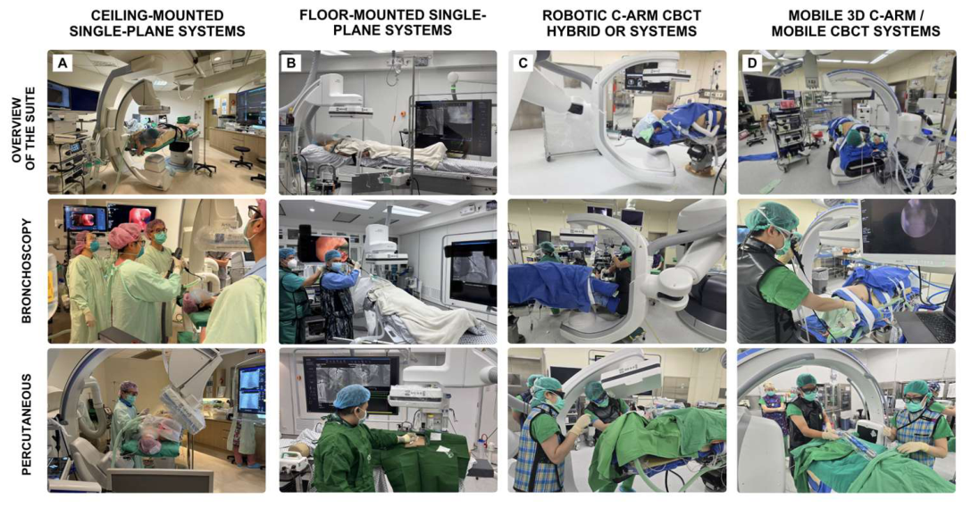
Figure 2.
Relative spatial footprint and free working space of representative CBCT platform configurations for interventional pulmonology.
Panels are labeled from left to right, top to bottom, as A–F. (A, B) Ceiling-mounted single-plane CBCT; (C, D) robotic CBCT / hybrid operating-room system; and (E, F) floor-mounted single-plane CBCT. Panels A, C, and E are semi-transparent composite overlays generated from photographs obtained from the same operator viewpoint behind the physician to illustrate the relative footprint of the C-arm system and the amount of residual free working space around the patient. Panels B, D, and F show the corresponding intra-procedural photographs. Across all configurations, the relative positions of the patient table, CBCT C-arm, bronchoscopy equipment, and anesthesia machine are shown for direct spatial comparison.
Figure 2.
Relative spatial footprint and free working space of representative CBCT platform configurations for interventional pulmonology.
Panels are labeled from left to right, top to bottom, as A–F. (A, B) Ceiling-mounted single-plane CBCT; (C, D) robotic CBCT / hybrid operating-room system; and (E, F) floor-mounted single-plane CBCT. Panels A, C, and E are semi-transparent composite overlays generated from photographs obtained from the same operator viewpoint behind the physician to illustrate the relative footprint of the C-arm system and the amount of residual free working space around the patient. Panels B, D, and F show the corresponding intra-procedural photographs. Across all configurations, the relative positions of the patient table, CBCT C-arm, bronchoscopy equipment, and anesthesia machine are shown for direct spatial comparison.
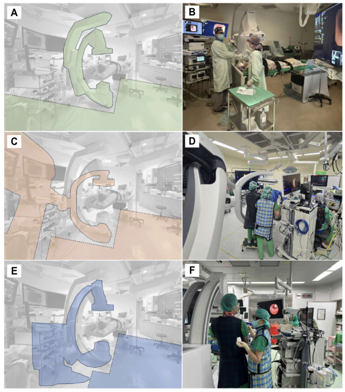
Figure 3.
Representative biplane CBCT room configurations for interventional pulmonology workflows.
Panels are labeled from left to right, top to bottom, as A–D. (A) Plan A, bronchoscopic configuration using the floor-mounted imaging arm. (B) Plan A, percutaneous configuration using the floor-mounted imaging arm. (C) Plan B, with the floor-mounted arm parked laterally to simulate a ceiling-mounted working geometry and provide increased head-side working space for bronchoscopy and anesthesia access. (D) Plan B, showing use of the ceiling-mounted arm to provide additional rotational freedom and enable complete CBCT acquisition while preserving a wide head-side working space. This configuration highlights an important distinction from conventional biplane systems, in which the secondary ceiling-mounted arm often provides fluoroscopy only and may not support CBCT acquisition. In contrast, this newer biplane platform allows the ceiling-mounted arm to perform CBCT, thereby enabling the primary arm to be parked aside and the room to function in a manner similar to a single-plane ceiling-mounted system.
Figure 3.
Representative biplane CBCT room configurations for interventional pulmonology workflows.
Panels are labeled from left to right, top to bottom, as A–D. (A) Plan A, bronchoscopic configuration using the floor-mounted imaging arm. (B) Plan A, percutaneous configuration using the floor-mounted imaging arm. (C) Plan B, with the floor-mounted arm parked laterally to simulate a ceiling-mounted working geometry and provide increased head-side working space for bronchoscopy and anesthesia access. (D) Plan B, showing use of the ceiling-mounted arm to provide additional rotational freedom and enable complete CBCT acquisition while preserving a wide head-side working space. This configuration highlights an important distinction from conventional biplane systems, in which the secondary ceiling-mounted arm often provides fluoroscopy only and may not support CBCT acquisition. In contrast, this newer biplane platform allows the ceiling-mounted arm to perform CBCT, thereby enabling the primary arm to be parked aside and the room to function in a manner similar to a single-plane ceiling-mounted system.
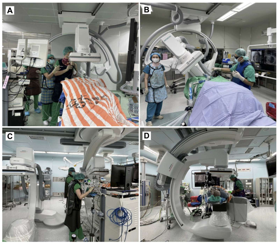

Table 1.
Practical comparison of imaging platforms for advanced interventional pulmonology.
| Imaging platform | Typical room / intended use | Benefits for IP Use | Limitations for IP Use |
|---|---|---|---|
| Ceiling-mounted single-plane CBCT | Angiography, multipurpose IR, or IP suite | Preserves head-side airway access; favorable for bronchoscopy, anesthesia integration, CBCT/augmented fluoroscopy, and selected percutaneous work | Limited availability; laser/table adjustment may be less automated; requires compatible workstation/software; often shared |
| Floor-mounted single-plane CBCT | Angiography, cardiovascular, or IR suite | Existing rooms may be repurposed; supports CBCT/augmented fluoroscopy, laser guidance, and percutaneous procedures | Floor base may obstruct airway-side access; bronchoscopy ergonomics and anesthesia positioning may be less favorable; often shared |
| Biplane angiography system | Cardiovascular or neurovascular angiography suite | May provide strong fluoroscopic capability; newer systems may support two-plane CBCT/overlay and complex targeting | Older systems may have limited CBCT/overlay; second arm may need to be parked; room capability must be verified; often shared |
| Robotic arm / hybrid OR CBCT | Hybrid OR or advanced surgical intervention suite | Multi-axis flexible C-arm; favorable for laser-guided ablation, localization, combined bronchoscopy–percutaneous–surgical workflows; large-volume scan capability; capable of one-stop shop workflow | High cost; limited availability; requires OR-level staffing, anesthesia support, and scheduling coordination |
| Mobile 3D C-arm / mobile CBCT | Standard OR, bronchoscopy room, or procedure room | Lower infrastructure requirement; feasible entry point for centers without fixed CBCT; may support mobile CBCT-assisted bronchoscopy | Image quality, field of view, overlay, workstation integration, and workflow efficiency vary by system |
| Conventional MDCT-guided thoracic intervention | Diagnostic CT or radiology suite | Strong platform for percutaneous biopsy, ablation, and localization; widely available comparator when bronchoscopy is not required | Limited real-time bronchoscopy integration; usually requires patient transfer if surgery follows; less suited for one-stop hybrid IP workflows |
CBCT = cone-beam computed tomography; IP = interventional pulmonology; IR = interventional radiology; MDCT = multidetector computed tomography, OR = operating room.
Table 2.
Literature-informed illustrative workflow applicability of imaging platforms for advanced interventional pulmonology procedures.
Table 2.
Literature-informed illustrative workflow applicability of imaging platforms for advanced interventional pulmonology procedures.
| Imaging platform | Percutaneous procedures (biopsy or ablation) | Transbronchial procedures (biopsy, ablation, navigational bronchoscopy) | VATS or thoracoscopic surgery | Hybrid procedure (localization, biopsy, or ablation + VATS) | Endovascular procedures (e.g., TAE) |
|---|---|---|---|---|---|
| Ceiling-mounted single-plane CBCT / AFB-capable systems | ++ | +++* | +§ | +§ | +§ |
| Floor-mounted single-plane CBCT / AFB-capable systems | ++ | ++*§ | +§ | +§ | +§ |
| Fixed biplane angiography / CBCT systems with overlay | ++ | ++*§ | +§ | ++§ | +++ |
| Multi-axis C-arm / CBCT hybrid OR systems | +++ | +++ | +++ | +++ | ++ |
| Mobile 3D C-arm / mobile CBCT systems | +§ | ++§ | +§ | +§ | ± |
| Conventional MDCT system | +++ | - | - | - | - |
Grades reflect workflow applicability rather than comparative clinical efficacy. Ratings were based on a narrative synthesis of published CBCT/augmented fluoroscopy bronchoscopy studies, NTUH Hsinchu/Biomedical Park/NTUH Cancer Center hybrid operating room experience, CT/C-arm CBCT-guided percutaneous thoracic intervention literature, mobile CBCT-assisted bronchoscopy reports, and the authors’ institutional experience. Actual workflow applicability depends on local room geometry, system generation, CBCT/overlay capability, installed hardware and software options, regulatory clearance, workstation availability, field of view, table design, anesthesia access, staff familiarity, local workflow and operator experience [5,6,7,8,9,10,11,12,13,14,15,16,17,18,19,20,21,22,23,24,25,26,27,28,29,30,31,32,33,34,35,36]. These ratings are an illustrative expert synthesis and do not constitute a validated comparative scoring system. +++, reported use with fewer identified workflow constraints; ++, reported use with workflow adaptation; +, reported use with substantial workflow adaptation or identified limitations; ±, applicability is primarily configuration- or institution-dependent ; −, use not identified in the literature reviewed or not typically reported; *, requires CBCT-derived overlay or augmented fluoroscopy capability; §, applicability is particularly dependent on room geometry, system generation, installed software, table design, anesthesia access, or local workflow. AFB = augmented fluoroscopic bronchoscopy; CBCT = cone-beam computed tomography; IP = interventional pulmonology; MDCT = multidetector computed tomography; VATS = video-assisted thoracoscopic surgery.
Table 3.
Representative CBCT platform configurations reported for advanced bronchoscopic and percutaneous thoracic procedures.
Table 3.
Representative CBCT platform configurations reported for advanced bronchoscopic and percutaneous thoracic procedures.
| Approach & Image-guided application |
CBCT platform/configuration | Representative system(s)* | Reference(s) |
|---|---|---|---|
| TRANSBRONCHIAL | |||
| CBCT-assisted biopsy | |||
| Fixed ceiling-/floor-mounted C-arm | Allura Clarity FD20; Azurion; Artis zeego; Artis zee | [9,39,40,42,43] | |
| Biplane angiography system | Artis zee | [15] | |
| Robotic C-arm | Artis zeego; ARTIS pheno, ARTIS pheno; Azurion, Allura Clarity FD20; Azurion; Artis zeego; ARTIS pheno; Artis dTA; Artis zee | [11,17,40,43,44] | |
| Mobile 3D C-arm | Cios Spin | [14,45,46,47,48,49,50] | |
| Localization ± VATS | |||
| Ceiling-mounted angiography C-arm | Alphenix Hybrid+ / INFX-8000H | [51] | |
| Biplane angiography system | Artis zee biplane | [21] | |
| Robotic C-arm | ARTIS pheno | [16,30,31] | |
| Mobile 3D C-arm | Cios Spin | [52,53] | |
| Ablation | |||
| Ceiling-mounted angiography C-arm | Artis Q | [54] | |
| Robotic C-arm | ARTIS pheno; Artis zeego | [55] | |
| Mobile 3D C-arm | Cios Spin | [54,56,57] | |
| TRANSTHORACIC | |||
| Biopsy | |||
| Fixed ceiling-/floor-mounted C-arm | Allura Xper FD20, Artis zee | [58] | |
| Robotic C-arm | ARTIS pheno; Artis zeego | [24,59] | |
| Hybrid CT/CBCT system | Brilliance 64 + Allura Xper FD20; Ingenuity Core128 + Artis zee; Aquilion 64 + Allura Xper FD20 | [60,61,62] | |
| Biplane angiography system | Artis zee; Allura Xper FD20 | [58] | |
| Localization ± VATS | |||
| Robotic C-arm | Artis zeego; ARTIS pheno | [31,32,63] | |
| Mobile 3D C-arm | Cios Spin | [64] | |
| Ablation | |||
| Fixed/ceiling-mounted C-arm | Allura Xper FD20 | [65] | |
| Robotic C-arm | ARTIS pheno | [25,26] | |
System capabilities vary by product generation, installed hardware and software options, configuration, and country-specific regulatory clearance. Device-specific capabilities should be verified using the applicable product documentation.
Disclaimer/Publisher’s Note: The statements, opinions and data contained in all publications are solely those of the individual author(s) and contributor(s) and not of MDPI and/or the editor(s). MDPI and/or the editor(s) disclaim responsibility for any injury to people or property resulting from any ideas, methods, instructions or products referred to in the content. |
© 2026 by the authors. Licensee MDPI, Basel, Switzerland. This article is an open access article distributed under the terms and conditions of the Creative Commons Attribution (CC BY) license (http://creativecommons.org/licenses/by/4.0/).
Copyright: This open access article is published under a Creative Commons CC BY 4.0 license, which permit the free download, distribution, and reuse, provided that the author and preprint are cited in any reuse.