

Submitted:
31 October 2026
Posted:
03 November 2026
You are already at the latest version
Abstract
Background: Idiopathic pulmonary fibrosis (IPF) is a chronic, progressive, and fatal interstitial lung disease characterized by excessive deposition of extracellular matrix and progressive destruction of the lung architecture. Repeated epithelial injury and aberrant tissue repair are strongly associated with its pathogenesis; however, the molecular mechanisms underlying disease progression remain incompletely understood. Altered microRNA (miRNA) expression has been extensively associated with IPF, but comparatively little attention has been given to the mechanisms regulating miRNA maturation and processing during pulmonary fibrosis. Objective: In this narrative review, we aimed to describe the relationship between IPF pathogenesis and alterations in miRNA maturation, with particular emphasis on post-transcriptional mechanisms that may disrupt the production and function of mature miRNAs. Methods: We conducted a narrative review of the available literature using PubMed, Scopus, and Google Scholar. No specific time frame was established because of the limited number of studies directly addressing miRNA maturation in IPF. Original research articles were prioritized, while relevant studies describing miRNA maturation mechanisms in other biological and disease contexts were also considered to provide mechanistic insight. Results: The available evidence suggests that alterations in miRNA maturation may contribute to miRNA dysregulation in IPF beyond changes in transcriptional control. Disruption of the Drosha/DGCR8 microprocessor complex, altered Dicer-dependent processing, and post-transcriptional modifications of miRNA precursors may affect the production of mature miRNAs. In addition, adenosine-to-inosine editing mediated by adenosine deaminases acting on RNA (ADARs) may modify miRNA sequences and influence their maturation, stability, and target selection. Mechanotransduction-associated pathways, including Hippo/YAP signaling, may further interfere with the miRNA-processing machinery. These alterations may contribute to the persistent dysregu-lation of antifibrotic miRNAs, including miR-29, thereby promoting extracellular matrix accumulation and sustaining profibrotic signaling. Conclusions: Although evidence directly linking defects in miRNA maturation to IPF remains limited, available findings support the hypothesis that alterations in post-transcriptional miRNA processing represent an additional regulatory layer contributing to pulmonary fibrosis. Further experimental studies are needed to determine how specific alterations in miRNA maturation and editing contribute to IPF pathogenesis and whether components of the miRNA-processing machinery may represent potential therapeutic targets.
Keywords:
miRNA biogenesis
; IPF
; SMAD
; ADAR
; Hippo/YAP
; p53
; drosha
; dicer
1. Introduction
Idiopathic pulmonary fibrosis (IPF) is a chronic and progressive interstitial lung disease characterized by excessive deposition of extracellular matrix (ECM) and irreversible deterioration of lung architecture and function. Although the etiology of IPF remains incompletely understood, repeated injury to the alveolar epithelium and aberrant tissue repair are strongly associated with disease pathogenesis [1,2,3,4,5]. The pathogenesis of IPF is complex, involving multiple cell types and a variety of cellular and molecular mechanisms [6]. Among these, lung fibroblast differentiation into myofibroblast has been widely recognized to be one of the most critical events during the pathological development of this disease [7,8].TGF-β1 has been shown to be an important mediator of lung fibrosis and can induce differentiation of pulmonary fibroblasts into myofibroblasts characterized by α-smooth muscle actin (α-SMA) expression and active synthesis of ECM proteins [9]. MicroRNAs (miRNAs) are small non-coding RNAs that regulate gene expression predominantly at the post-transcriptional level through mRNA degradation or translational repression [10,11,12]. Their biogenesis is a highly regulated multistep process involving several key proteins, including the Microprocessor components DROSHA and DGCR8 and the RNase III enzyme DICER [13,14,15,16,17]. Dysregulation of this machinery has been implicated in several fibrotic diseases, including IPF. Although alterations in miRNA expression have been extensively associated with IPF, growing evidence suggests that changes in miRNA abundance may result not only from altered transcription but also from defects in miRNA processing and maturation [10]. For example, reduced DICER1 expression has been observed in the myofibroblastic core of fibroblastic foci in IPF lungs and has been associated with impaired miRNA biogenesis. This alteration may reduce antifibrotic miRNAs, including miR-29, thereby promoting fibroblast activation and excessive extracellular matrix deposition [18]. Beyond the canonical miRNA biogenesis pathway, several signaling pathways and RNA-processing factors can regulate miRNA maturation post-transcriptionally. Transforming growth factor beta 1 (TGF-β1), a central profibrotic cytokine in IPF, can regulate miRNA biogenesis through SMAD-dependent modulation of pri-miRNA processing [13,15,19]. Similarly, the tumor suppressor p53 can interact with the DROSHA/DGCR8 microprocessor complex and the RNA helicases p68/p72, thereby facilitating the processing of specific pri-miRNAs in response to cellular stress. Mutations in p53 may disrupt this interaction and impair the processing of specific miRNAs [20,21]. Other mechanisms may also influence miRNA maturation. Adenosine deaminases acting on RNA (ADARs) can modify miRNA precursors through adenosine-to-inosine (A-to-I) RNA editing. They can also regulate miRNA processing through editing-independent interactions with components of the miRNA-processing machinery [20,22,23]
Furthermore, changes in the mechanical properties of the fibrotic microenvironment and activation of mechanotransduction pathways, including Hippo/YAP signaling, may interfere with miRNA-processing machinery activity [24] . Therefore, miRNA dysregulation in IPF may extend beyond alterations in transcriptional control and involve defects at different stages of miRNA maturation. Understanding these mechanisms may provide new insights into how persistent changes in miRNA activity contribute to fibroblast activation, extracellular matrix accumulation, and disease progression.
In this narrative review, we summarize the mechanisms involved in miRNA biogenesis and maturation, with particular emphasis on post-transcriptional regulation by TGF-β/SMAD signaling, p53, ADAR proteins, and mechanotransduction pathways. We then discuss the available evidence linking alterations in these mechanisms to miRNA dysregulation in IPF and highlight potential directions for future research.
2. Review Methodology
We conducted a narrative review of mechanisms regulating miRNA maturation and their potential relevance to idiopathic pulmonary fibrosis (IPF). We identified relevant literature through searches of PubMed, Scopus, and Google Scholar. We did not establish a specific publication time frame because few studies directly address miRNA maturation in IPF. When possible, we prioritized original research articles. In addition, we included relevant studies investigating miRNA maturation mechanisms in biological and disease contexts unrelated to IPF when they provided mechanistic evidence that could illustrate specific processes involved in miRNA biogenesis and maturation. These studies were considered particularly useful for mechanisms for which direct evidence in IPF remains limited.
3. Canonical miRNA Biogenesis and Maturation
3.1. Nuclear Processing of Pri-miRNAs by the Drosha/DGCR8 Microprocessor
RNA polymerase II transcribes most miRNA genes into primary miRNA transcripts (pri-miRNAs). miRNA genes are located in both intragenic and intergenic genomic regions, and pri-miRNAs contain characteristic hairpin structures that provide the structural information required for subsequent processing [18,19,25]. The nuclear RNase III enzyme DROSHA, together with its partner DGCR8, forms the Microprocessor complex that converts pri-miRNAs into precursor miRNAs (pre-miRNAs). DROSHA contains a double-stranded RNA-binding domain and two RNase III domains, while DGCR8 recognizes pri-miRNA structural features and positions it for cleavage. This processing step generates pre-miRNAs of approximately 55–70 nucleotides [26,27,28]. After nuclear processing, Exportin-5 (XPO5) exports the resulting pre-miRNAs to the cytoplasm in association with Ran-GTP [29]. Figure 1
3.2. Nuclear Export and Cytoplasmic Processing
Pre-miRNAs generally contain a 5′-monophosphate, a characteristic 3′ two-nucleotide overhang, a terminal loop, and a double-stranded stem. DICER recognizes these structural features during subsequent processing [30]. Exportin-5 recognizes the characteristic structure of pre-miRNAs and mediates their transport from the nucleus to the cytoplasm in a Ran-GTP-dependent manner [28,29].
3.3. Dicer-Mediated Processing and Mature miRNA Generation
In the cytoplasm, DICER recognizes pre-miRNAs and cleaves the double-stranded RNA stem to generate a small RNA duplex. Structural studies have shown that DICER uses its PAZ domain to recognize the 3′ dinucleotide overhang and measures an appropriate distance before cleavage, contributing to the characteristic length of mature miRNAs [20]. Figure 1M. The precise selection of cleavage sites can vary among different miRNA substrates. Structural features within the pre-miRNA, including the position of internal bulges and the terminal loop, can influence DICER cleavage and contribute to the generation of multiple miRNA isoforms (isomiRs) [30,31].
4. Post-Transcriptional Regulation of miRNA Maturation
4.1. Epitotranscriptomics
Post-translational modifications of Microprocessor-associated proteins provide another layer of regulation. PRMT1-mediated methylation of microprocessor-associated proteins has been shown to regulate miRNA biogenesis [32] . One of the most widespread and abundant RNA modifications is N6-methyladenosine (m6A) is the most conserved, reversible internal modification seen in cellular RNAs across all kingdoms of life, wherein a methyl group decorates the nitrogen at the 6th position of adenosine of the RNA molecule [33], Although m6A RNA methylation is a well-established post-transcriptional regulatory mechanism for mRNAs, its role in miRNAs remains poorly understood. Recent studies have suggested that m6A RNA methylation regulates the structure, maturation, and function of non-coding regulatory RNAs, including miRNAs, circRNAs, and lncRNAs [34]. For example, while m6A is known to regulate mRNA maturation, its role in miRNA biology remains less clearly defined. In this context, Garbo S. et al. used the HepG2 hepatoma cell line, which expresses methyltransferase-like 3 (METTL3), a catalytic subunit of the METTL3/METTL14 complex, to investigate miRNA methylation using RNA immunoprecipitation followed by sequencing (MeRIP-seq). Their results revealed distinct patterns of m6A methylation among miRNAs, with high methylation levels detected in miR-6767-5p, miR-548e-3p, miR-215-5p, miR-4786-5p, miR-3619-3p, and the liver-specific miR-122-5p; low methylation levels in miR-3074-5p, miR-374b-5p, let-7e-5p, miR-3692-5p, and miR-28-5p; and no detectable methylation in miR-301b-3p, miR-199a-5p, and miR-1180-3p. Furthermore, m6A modification impaired the interaction between the methylated miRNA fraction and Argonaute 2 (AGO2), thereby compromising miRNA function [35].
In 2015, Alarcón C. et al., using MDA-MB-231 cells in which METTL3 expression was inhibited by shRNA, reported that methyltransferase-like 3 (METTL3) methylates pri-miRNAs, marking them for recognition and processing by DGCR8. Depletion of this enzyme reduced DGCR8 binding to pri-miRNAs, resulting in a global decrease in mature miRNA levels. These findings indicate that m6A methylation plays an important role in miRNA maturation in these cells [36,37].
4.2. TGF-β/SMAD-Mediated Regulation of miRNA Processing
TGF-β signaling can regulate miRNA levels at both the transcriptional and post-transcriptional levels. Receptor-activated SMADs (R-SMADs) can influence miRNA biogenesis by modulating DROSHA-mediated processing of pri-miRNAs. R-SMADs interact with the RNA helicase p68 (DDX5), a Microprocessor component, and this interaction can facilitate DROSHA recruitment and promote pri-miRNA processing [8,12]. SMAD proteins can also associate with specific RNA sequences within pri-miRNAs. A conserved 5′-CAGAC-3′ sequence within the stem region of some pri-miRNAs, known as the RNA-SMAD-binding element (R-SBE), resembles the SMAD-binding element found in promoters of TGF-β/BMP-responsive genes. SMAD binding to this RNA element can facilitate Microprocessor recognition of the pri-miRNA and promote its processing [13]. The regulation of specific miRNAs can depend on the TGF-β/BMP stimulus. In COS cells, chromatin RNA immunoprecipitation experiments showed that SMAD1 associated with pri-miR-21 after BMP4 stimulation, whereas TGF-β1 stimulation increased the association of SMAD2 and SMAD3 with pri-miR-21. Similar observations were reported for miR-199a. In contrast, miR-214 associated with p68 and DROSHA but not with SMAD proteins, suggesting that BMP or TGF-β1 signaling does not directly regulate its processing [19]. R-SBEs have been identified in several miRNAs regulated by TGF-β/BMP signaling, including miR-105, miR-199a, miR-215, and miR-421 [13]. Together, these observations support a mechanism through which TGF-β/BMP signaling can directly influence miRNA maturation through SMAD-dependent regulation of the Microprocessor Figure 2.
4.3. p53-Mediated Regulation of miRNA Maturation
Several miRNAs are post-transcriptionally upregulated in a p53-dependent and p68/p72-dependent manner following DNA damage. Upon DNA damage, p53 accumulates and interacts with the DROSHA/DGCR8 Microprocessor complex in HCT116, WI-38, TIG-3, and MCF7 cells treated with doxorubicin. The association between DROSHA and p53 was significantly reduced following RNase A treatment and/or silencing of p68/p72 using RNA interference, suggesting that p53 interacts with DROSHA through specific RNA species and the RNA helicases p68/p72. p53 associates with DROSHA and p68 through its C-terminal region, and chromatin immunoprecipitation analyses showed that p53 associates with pri-miR-16-1 and pri-miR-143 regions following treatment of WI-38 human fetal lung fibroblasts and HCT116 colorectal carcinoma cells [21]. These findings support a role for p53 in regulating miRNA biogenesis through pri-miRNA processing. miR-143 and miR-145 have also been identified as tumor-suppressive miRNAs whose regulation is linked to the p53/p68/p72 axis. This mechanism is particularly relevant in colorectal cancer and illustrates how stress-responsive signaling can influence miRNA maturation in addition to transcriptional regulation [10]. The relevance of this mechanism is further highlighted by cancer-associated p53 mutations. Mutant p53 forms have been reported to reduce pre-miRNA and mature miRNA levels while promoting accumulation of corresponding pri-miRNAs, including pri-miR-16-1, pri-miR-143, and pri-miR-206 [25]. These findings suggest impaired pri-miRNA processing. Consistent with this interpretation, p53 mutants can reduce the interaction between DROSHA and p68, potentially compromising Microprocessor activity [10] Figure 3. However, direct evidence connecting p53-mediated Microprocessor regulation to specific miRNA deficits in IPF lung tissue remains to be established.
4.4. ADAR-Mediated Regulation and RNA Editing
Adenosine to inosine (A-to-I) RNA editing was originally discovered as a mysterious enzymatic activity causing unwinding of double-stranded RNA (dsRNA) in Xenopus laevis oocytes and embryos [38]. A limited number of editing sites were discovered serendipitously in protein-coding regions of mRNAs, when comparing human genomic DNA versus cDNA sequences. The most frequent and widespread targets of A-to-I RNA editing are dsRNAs made from inverted Alu repetitive elements (Alu dsRNAs), which are located within introns and untranslated regions [31,39,40]. The biogenesis and function of certain miRNAs is regulated by editing of the primary miRNAs (pri-miRNAs) [41]. Beyond transcriptional regulation, miRNA abundance also depends on the efficiency and fidelity of miRNA biogenesis and maturation. Adenosine deaminases acting on RNA (ADARs) catalyze adenosine-to-inosine (A-to-I) RNA editing, which can modify the sequence and structural properties of RNA molecules. When editing occurs within pri-miRNAs, it may alter structural features or recognition sites required for Microprocessor processing, thereby influencing pre-miRNA generation. Editing of pre-miRNAs may similarly affect their recognition and processing by DICER, ultimately altering the sequence and functional properties of mature miRNAs [21,22,42]. ADAR proteins can also regulate miRNA biogenesis through editing-independent mechanisms. ADAR1 isoforms p110 and p150 regulate let-7d processing, with a reported reduction in let-7d maturation [42], whereas ADAR1 has been reported to promote maturation of miR-155-5p through interaction with DICER1 [24]. ADAR1 has also been reported to interact with pri-miR-766 and negatively regulate maturation of miR-766-3p; increased ADAR1 expression was associated with reduced mature miR-766-3p, whereas ADAR1 depletion increased miR-766-3p abundance [43]. These findings indicate that ADAR proteins can influence miRNA maturation through both editing-dependent and editing-independent mechanisms, potentially contributing to disease-associated miRNA dysregulation Figure 4.
4.5. Mechanotransduction and Hippo/YAP-Dependent Regulation of the Microprocessor
Mechanotransduction refers to the process by which cells sense mechanical stimuli from their microenvironment, including matrix stiffness, topography, and tension, and convert them into biochemical signals that regulate gene expression. This process is mediated by integrin-based focal adhesions and downstream signaling pathways, particularly FAK and Rho/ROCK, which control cytoskeletal tension. Mechanical forces are transmitted to the nucleus through the LINC complex, formed by SUN proteins and nesprins, which physically connects the cytoskeleton to the nucleoskeleton. This connection allows mechanical inputs to directly influence nuclear organization and function [44]. One of the main pathways involved in this response is the Hippo pathway. When matrix stiffness is low or cell density is high, the MST1/2–LATS1/2 kinase cascade phosphorylates YAP and TAZ, causing their retention in the cytoplasm and subsequent proteasomal degradation. Under stiffer matrix conditions, however, Rho GTPase activity and actomyosin tension suppress this cascade, allowing YAP and TAZ to translocate to the nucleus. Once there they bind TEAD transcription factors and drive the expression of profibrotic genes, including CTGF and type I collagen [45,46,47,48]. Interestingly, it has been observed that, beyond their transcriptional functions, nuclear YAP and TAZ can directly interfere with miRNA biogenesis at the level of the Microprocessor complex. Mori et al. demonstrated that nuclear YAP physically binds to and sequesters p72 (DDX17). This RNA helicase acts as an essential cofactor for the DROSHA/DGCR8 Microprocessor complex. By sequestering p72, nuclear YAP reduces its availability to associate with DROSHA and primary miRNA (pri-miRNA) substrates, thereby decreasing the processing efficiency of pri-miRNAs and leading to a suppression of miRNA biogenesis.
This mechanism has been shown to be dependent on cell density: at low density, YAP is predominantly localized in the nucleus and miRNA levels are globally reduced. At high cell density the cytoplasmic retention of YAP mediated by the Hippo pathway releases p72, allowing it to associate with the Microprocessor complex and restore the efficiency of miRNA processing [17,24]. The relationship between YAP/TAZ signaling and miRNA regulation is bidirectional. YAP/TAZ can act as transcriptional activator of specific miRNAs like the profibrotic miRNA 130/301 family promoting ECM stiffening creating a positive mechanosensitive loop [49]. On the other hand, antifibrotic miRNAs such as miR-15a and miR-29a suppress YAP1 expression and stiffness-induced YAP activation, so when these miRNAs are lost through transcriptional or post-transcriptional mechanisms nuclear YAP/TAZ activity and profibrotic signaling may be further enhanced [50].
The efficiency of this sequestration mechanism is also shaped by post-translational modification of DDX17. Under hypoxia found in the fibrotic core of IPF lungs, DDX17 undergoes K63-linked polyubiquitination, which reduces its binding to pri-miRNA substrates while increasing its association with nuclear YAP and the coactivator p300. This shift redirects DDX17 away from the Microprocessor and toward transcriptional regulatory complexes, with a dual outcome: antifibrotic miRNA biogenesis is suppressed, and profibrotic gene programs are transcriptionally activated [Kao 2019]. Together this suggests that the hypoxic and mechanically altered microenvironment of fibrotic lung tissue may converge on DDX17, amplifying YAP-dependent Microprocessor suppression beyond what matrix stiffness alone would drive [51]. Other Post-translational modifications of Microprocessor-associated proteins such as PRMT1-mediated methylation of microprocessor-associated proteins has been shown to regulate miRNA biogenesis [32]. These mechanisms suggest that changes in the cellular and extracellular environment can influence miRNA maturation by modifying or redistributing the processing machinery (Figure 5). The fibrotic extracellular matrix, through mechanotransduction pathways, may disrupt the activity of the Drosha/DGCR8 microprocessor complex, potentially via post-translational modifications such as PRMT1-mediated arginine methylation and SUMOylation [52]
5. Dysregulation of miRNA Maturation in Idiopathic Pulmonary Fibrosis
5.1. DICER1 Deficiency and Impaired miRNA Biogenesis
Immunohistochemical analysis of the miRNA-processing machinery in IPF lung specimens revealed decreased DICER1 expression in the procollagen-rich myofibroblastic core of fibroblastic foci compared with the focus perimeter and adjacent alveolar walls. Mechanistically, IPF-derived extracellular matrix increased association of the DICER1 transcript with the RNA-binding protein AUF1, and DICER1 knockdown conferred cell-autonomous fibrogenic properties on primary human lung fibroblasts in zebrafish and mouse lung xenograft models [18] .
In conditional DICER knockout mice subjected to a model of renal fibrosis induced by unilateral ureteral obstruction (UUO), DICER depletion was found to dysregulate a protective antifibrotic axis comprising miR-9-5p, miR-344g-3p, and miR-7074-3p, thereby accelerating the progression of chronic kidney disease [53].
Hepatic stellate cells (HSC) activate and represent the main cell type responsible for the development of liver fibrosis, miR-21 was the microRNA that showed the strongest upregulation in HSC in multiple models of fibrogenesis, with an 8- to 24-fold induction compared to quiescent HSC, Cavigglia en 2018, Dicer deletion in hepatic stellate cells (HSCs) was shown to reduce the levels of several miRNAs but had limited effects on HSC activation and the development of liver fibrosis [54]. Therefore, it is important to emphasize that the effects of DICER1 deficiency depend on the cell type and pathological context and do not necessarily promote fibrosis in all organs.
Alterations in miRNA biogenesis may contribute to the progression of fibrotic processes. In idiopathic pulmonary fibrosis (IPF), reduced DICER1 expression in fibroblastic foci has been shown to impair the maturation of the miR-29 family, thereby promoting the expression of extracellular matrix-related genes and conferring profibrotic properties on fibroblasts. This mechanism is associated with the interaction between DICER1 mRNA and the RNA-binding protein AUF1, which promotes its degradation [18]. In models of renal fibrosis, Dicer deletion in mesenchymal cells also exacerbates interstitial fibrosis. However, its deletion in hepatic stellate cells had limited effects on fibrogenesis, suggesting that the consequences of disrupting miRNA biogenesis depend on the cellular context and the affected organ.
5.2. Impaired Maturation of Antifibrotic miRNAs and miR-29
The miR-29 family is downregulated in lungs with bleomycin-induced fibrosis and exhibits an inverse correlation with the expression of profibrotic genes and the severity of fibrosis. Its inhibition in lung fibroblasts promotes the expression of genes associated with the extracellular matrix and its remodeling, including collagens, laminins, and integrins. Furthermore, TGF-β1 suppresses miR-29 expression, contributing to the dysregulation of fibrosis-related genes. These findings suggest that miR-29 plays an antifibrotic role and represents a potential therapeutic target for pulmonary fibrosis [55]..
In IPF, suppression of antifibrotic miRNAs such as miR-29 may involve defects in miRNA processing in addition to transcriptional mechanisms. Because miR-29 regulates genes involved in extracellular matrix production, impaired maturation or reduced miR-29 abundance may contribute to sustained fibroblast activation and matrix accumulation [56]]. The available evidence therefore supports a model in which alterations in the extracellular matrix and intracellular miRNA-processing machinery establish a self-sustaining profibrotic feedback loop. However, the precise molecular sequence connecting matrix signals to specific defects in miRNA maturation remains incompletely defined Figure 5.
In a study conducted in ADAR2-deficient mice, miR-29b levels were found to be reduced, which correlated with increased expression of its target genes, COL1A2 and IGF1 [57]..
Dysregulation of ADAR enzymes may contribute to the development of cardiovascular diseases through alterations in RNA editing and miRNA expression. In patients with congenital heart disease, decreased ADAR2 expression and increased ADAR1 expression have been observed. Similarly, ADAR2 deficiency in mice reduced the expression of miR-29b, miR-405, and miR-19, which have been associated with cardiomyopathy and cardiac fibrosis, and increased the expression of miR-29b target genes, such as COL1A2 and IGF1. These findings suggest that ADAR2 may be involved in the regulation of fibrotic processes through miRNA modulation; however, this relationship requires direct validation in pulmonary fibrosis [55]..
5.3. Extracellular Matrix, Mechanotransduction, and the miRNA-Processing Machinery
Pathological remodeling in IPF involves an increase in extracellular matrix stiffness, which triggers mechanotransduction signaling that directly alters miRNA expression in lung tissue [58]. In stiff matrix environments YAP/TAZ signaling translates mechanical stress into intracellular signals that suppress antifibrotic miRNAs while upregulating profibrotic ones [59]. Among the antifibrotic miRNAs repressed by tissue stiffness, miR-29 functions as a master negative regulator of collagen and matrix production. Under high-stiffness conditions, YAP-dependent signaling sustains miR-29 repression, removing a key brake on collagen synthesis [58].
Other antifibrotic miRNAs, including miR-133a, miR-200, and miR-26a, which target critical nodes of TGF-β signaling, are similarly lost in the remodeled fibrotic lung . Mechanical stress and TGF-β1 stimulation also drive the overexpression of profibrotic miRNAs. miR-21, potentiated by both matrix stiffness and TGF-β1, silences Smad7, an endogenous inhibitor of TGF-β signaling, generating a positive feedback loop that amplifies profibrotic signaling and myofibroblast differentiation. Stiffness additionally promotes upregulation of miR-145, miR-155, and miR-199a-5p, which collectively accelerate fibroblast-to-myofibroblast transition and sustain the fibrotic cycle [58].
Emerging evidence indicates that miRNA dysregulation in IPF extends beyond transcriptional control and involves alterations in the miRNA biogenesis machinery. The fibrotic extracellular matrix may influence Microprocessor activity through mechanotransduction pathways and post-translational modifications of processing proteins. PRMT1-mediated methylation of microprocessor-associated proteins has been implicated in regulation of miRNA biogenesis [29]. In parallel, aberrant Hippo/YAP signaling may interfere with miRNA processing by affecting essential cofactors such as p72 [17,29]. Together, these mechanisms provide a potential molecular link between the altered mechanical environment of the fibrotic lung and persistent changes in miRNA maturation. However, direct evidence connecting individual mechanotransduction events to the processing of specific antifibrotic miRNAs in human IPF remains limited. (Figure 5)
5.4. ADAR-Mediated Regulation in IPF
ADAR-mediated regulation may represent an additional post-transcriptional mechanism relevant to IPF. ADAR1 isoforms have been shown to regulate let-7d processing in IPF, providing direct evidence that alterations in an RNA-editing protein can affect miRNA maturation in the fibrotic lung [27]. Because ADAR proteins can regulate miRNA maturation through both editing-dependent and editing-independent mechanisms, their dysregulation may affect miRNA abundance, sequence, target selection, and functional activity. Nevertheless, few studies have directly investigated ADAR-dependent miRNA maturation in IPF. Further work is required to determine whether specific editing events or ADAR–Microprocessor interactions contribute to the persistent miRNA abnormalities characteristic of pulmonary fibrosis. Table 1
ADAR2 is known to auto-regulate its own expression in various organs via a negative feedback loop that involves the editing of its own pre-mRNA. Soundararajan et al. (2022), using a transgenic mouse model overexpressing ADAR2 in the lungs, demonstrated that these mice developed spontaneous pulmonary fibrosis characterized by alveolar septal thickening, disruption of alveolar architecture, and neutrophil infiltration. Furthermore, these mice exhibited increased expression of connective tissue growth factor (CTGF), a profibrotic marker [60]..
Regarding ADAR1, its importance in hepatic homeostasis has been demonstrated. Mice with hepatocyte-specific ADAR1 knockout exhibited increased type I interferon signaling and elevated levels of proinflammatory cytokines. In vitro, ADAR1 silencing in HepG2 cells increased the expression of NF-κB target genes and the proinflammatory cytokines IL-6 and IL-8. Furthermore, paracrine signaling from these cells to hepatic stellate cells induced a profibrotic phenotype in the latter [61].
Another study showed that ADAR1 is downregulated in fibroblasts derived from lungs of patients with idiopathic pulmonary fibrosis (IPF), a finding associated with decreased levels of miR-21, PELI1, and SPRY2, which have been implicated in antifibrotic processes. Overexpression of ADAR1 and ADAR2 in these fibroblasts restored miR-21, PELI1, and SPRY2 levels [62].
In another disease associated with vascular remodeling, reduced ADAR1 expression, specifically of the p150 isoform, was observed in pulmonary artery smooth muscle cells (PASMCs) from patients with idiopathic pulmonary hypertension. To further investigate this association, researchers analyzed mice with PASMC-specific ADAR1 knockout and exposed them to hypoxia. Compared with control mice, ADAR1-deficient mice developed excessive vascular remodeling in distal arterioles, accompanied by increased endothelial permeability [63].
Continuing with vascular remodeling processes, another study showed that during phenotypic modulation of smooth muscle cells (SMCs), pre-mRNA accumulation and a decrease in mature mRNA levels of myosin heavy chain and smooth muscle α-actin were observed, suggesting impaired RNA processing. This phenomenon was associated with ADAR1-mediated A-to-I editing. ADAR1 silencing restored the expression of both markers, whereas mutation of its catalytic domain attenuated splicing alterations. Furthermore, ADAR1 knockdown inhibited neointima formation following vascular injury in mice, demonstrating its involvement in SMC phenotypic modulation and vascular remodeling [61,64].
Regarding the potential role of ADAR1 in hematopoietic cells, particularly in macrophages, ADAR1 deficiency reduced the formation and severity of abdominal aortic aneurysms (AAAs) by decreasing macrophage infiltration and vascular inflammation. Mechanistically, ADAR1 interacted with Drosha and promoted its degradation, thereby altering pri-miRNA-to-pre-miRNA processing and reducing the availability of microRNAs that inhibit NF-κB signaling. This increased IKKβ expression and promoted inflammatory and tissue-remodeling responses. These findings identify ADAR1 as a regulator of vascular inflammation and, potentially, fibrotic processes through the modulation of microRNA biogenesis [65].
6. Discussion and Future Perspectives
The evidence summarized in this review indicates that miRNA dysregulation in IPF should not be considered solely a consequence of altered transcription. Multiple levels of post-transcriptional regulation—including DROSHA/DGCR8 and DICER activity, TGF-β/SMAD and p53-dependent interactions, ADAR-mediated editing or protein–protein interactions, and mechanotransduction-associated changes—may influence the production and function of mature miRNAs. A major limitation of the current literature is that many mechanistic studies were performed in cellular or disease models other than IPF. Consequently, some mechanisms described in this review should be considered plausible regulatory pathways rather than established mechanisms of pulmonary fibrosis. Future studies should determine whether alterations in specific Microprocessor components, RNA modifications, or signaling-dependent interactions selectively affect antifibrotic miRNAs in human IPF.
A better understanding of these mechanisms could identify new molecular targets to restore miRNA maturation and potentially interrupt profibrotic signaling. In particular, defining how TGF-β/SMAD, p53, ADARs, and mechanical signaling converge on the miRNA-processing machinery may provide a more integrated view of miRNA dysregulation in IPF.
7. Conclusions
Alterations in miRNA maturation may represent an additional layer of regulation contributing to miRNA dysregulation in idiopathic pulmonary fibrosis. Current evidence supports roles for DICER1 deficiency, signaling-dependent regulation of the DROSHA/DGCR8 Microprocessor, ADAR-mediated mechanisms, and mechanotransduction pathways. Although direct evidence in IPF remains limited for several of these mechanisms, further characterization of miRNA processing may improve our understanding of pulmonary fibrosis pathogenesis and identify potential therapeutic opportunities.
Author Contributions
For research articles with several authors, a short paragraph specifying their individual contributions must be provided. The following statements should be used “Conceptualization, MCL, ELG, AAG, VR; FTM, EMM, writing—original draft preparation, MCL,VR, AAG, JC2, CB; writing—review and editing, RMO, X.X.; visualization, JC4, IH, AC; All authors have read and agreed to the published version of the manuscript.” Please turn to the CRediT taxonomy for the term explanation. Authorship must be limited to those who have contributed substantially to the work reported.
Funding
This research received no external funding.
Conflicts of Interest
The authors declare no conflicts of interest.
Abbreviations
The following abbreviations are used in this manuscript:
| ADAR | Adenosine deaminase acting on RNA |
| ADAR1 | Adenosine deaminase acting on RNA 1 |
| ADAR2 | Adenosine deaminase acting on RNA 2 |
| AGO2 | Argonaute 2 |
| A-to-I | Adenosine-to-inosine |
| AUF1 | AU-rich element RNA-binding protein 1 |
| BMP | Bone morphogenetic protein |
| CTGF | Connective tissue growth factor |
| DICER1 | Dicer 1 |
| DGCR8 | DiGeorge syndrome critical region gene 8 |
| DDX5 | DEAD-box helicase 5 |
| DDX17 | DEAD-box helicase 17 |
| ECM | Extracellular matrix |
| GABA | Gamma-aminobutyric acid |
| HSC | Hepatic stellate cell |
| IFN | Interferon |
| IKKβ | Inhibitor of nuclear factor kappa B kinase subunit beta |
| IL-6 | Interleukin 6 |
| IL-8 | Interleukin 8 |
| IPF | Idiopathic pulmonary fibrosis |
| m6A | N6-methyladenosine |
| MeRIP-seq | Methylated RNA immunoprecipitation sequencing |
| METTL3 | Methyltransferase-like 3 |
| METTL14 | Methyltransferase-like 14 |
| miRNA | MicroRNA |
| NF-κB | Nuclear factor kappa B |
| PAZ | Piwi–Argonaute–Zwille |
| PASMC | Pulmonary artery smooth muscle cell |
| PELI1 | Pellino E3 ubiquitin protein ligase 1 |
| pri-miRNA | Primary microRNA |
| pre-miRNA | Precursor microRNA |
| PRMT1 | Protein arginine methyltransferase 1 |
| R-SMAD | Receptor-regulated SMAD |
| R-SBE | RNA-SMAD-binding element |
| RNase | Ribonuclease |
| shRNA | Short hairpin RNA |
| SMAD | Small mothers against decapentaplegic |
| SMC | Smooth muscle cell |
| SPRY2 | Sprouty RTK signaling antagonist 2 |
| TGF-β | Transforming growth factor beta |
| TGF-β1 | Transforming growth factor beta 1 |
| UUO | Unilateral ureteral obstruction |
| YAP | Yes-associated protein |
| XPO5 | Exportin 5 |
References
- Pardo, A.; Selman, M. Lung Fibroblasts, Aging, and Idiopathic Pulmonary Fibrosis. Ann Am Thorac Soc 2016, 13 Suppl 5, S417–S421. [CrossRef]
- Sgalla, G.; Iovene, B.; Calvello, M.; Ori, M.; Varone, F.; Richeldi, L. Idiopathic Pulmonary Fibrosis: Pathogenesis and Management. Respir Res 2018, 19, 32. [CrossRef]
- Gill, S.K.; Gomer, R.H. Translational Regulators in Pulmonary Fibrosis: MicroRNAs, Long Non-Coding RNAs, and Transcript Modifications. Cells 2025, 14. [CrossRef]
- King, T.E., Jr; Pardo, A.; Selman, M. Idiopathic Pulmonary Fibrosis. Lancet 2011, 378, 1949–1961. [CrossRef]
- Selman, M.; Pardo, A. The Leading Role of Epithelial Cells in the Pathogenesis of Idiopathic Pulmonary Fibrosis. Cell Signal 2020, 66, 109482. [CrossRef]
- Wolters, P.J.; Collard, H.R.; Jones, K.D. Pathogenesis of Idiopathic Pulmonary Fibrosis. Annu Rev Pathol 2014, 9, 157–179. [CrossRef]
- Phan, S.H. The Myofibroblast in Pulmonary Fibrosis. Chest 2002, 122, 286S – 289S. [CrossRef]
- Scotton, C.J.; Chambers, R.C. Molecular Targets in Pulmonary Fibrosis: The Myofibroblast in Focus. Chest 2007, 132, 1311–1321. [CrossRef]
- Lee, C.G.; Cho, S.; Homer, R.J.; Elias, J.A. Genetic Control of Transforming Growth Factor-beta1-Induced Emphysema and Fibrosis in the Murine Lung. Proc Am Thorac Soc 2006, 3, 476–477. [CrossRef]
- Shang, R.; Lee, S.; Senavirathne, G.; Lai, E.C. microRNAs in Action: Biogenesis, Function and Regulation. Nat Rev Genet 2023, 24, 816–833. [CrossRef]
- Oliveto, S.; Mancino, M.; Manfrini, N.; Biffo, S. Role of microRNAs in Translation Regulation and Cancer. World J Biol Chem 2017, 8, 45–56. [CrossRef]
- Lee, Y.; Kim, M.; Han, J.; Yeom, K.-H.; Lee, S.; Baek, S.H.; Kim, V.N. MicroRNA Genes Are Transcribed by RNA Polymerase II. EMBO J 2004, 23, 4051–4060. [CrossRef]
- Davis, B.N.; Hilyard, A.C.; Nguyen, P.H.; Lagna, G.; Hata, A. Smad Proteins Bind a Conserved RNA Sequence to Promote microRNA Maturation by Drosha. Mol Cell 2010, 39, 373–384. [CrossRef]
- Lee, H.; Han, S.; Kwon, C.S.; Lee, D. Biogenesis and Regulation of the Let-7 miRNAs and Their Functional Implications. Protein Cell 2016, 7, 100–113. [CrossRef]
- Blahna, M.T.; Hata, A. Smad-Mediated Regulation of microRNA Biosynthesis. FEBS Lett 2012, 586, 1906–1912. [CrossRef]
- Cho, S.J.; Lee, M.; Stout-Delgado, H.W.; Moon, J.-S. DROSHA-Dependent miRNA and AIM2 Inflammasome Activation in Idiopathic Pulmonary Fibrosis. Int J Mol Sci 2020, 21. [CrossRef]
- Remenyi, J.; Bajan, S.; Fuller-Pace, F.V.; Arthur, J.S.C.; Hutvagner, G. The Loop Structure and the RNA Helicase p72/DDX17 Influence the Processing Efficiency of the Mice miR-132. Sci Rep 2016, 6, 22848. [CrossRef]
- Herrera, J.; Beisang, D.J.; Peterson, M.; Forster, C.; Gilbertsen, A.; Benyumov, A.; Smith, K.; Korenczuk, C.E.; Barocas, V.H.; Guenther, K.; et al. Dicer1 Deficiency in the Idiopathic Pulmonary Fibrosis Fibroblastic Focus Promotes Fibrosis by Suppressing MicroRNA Biogenesis. Am J Respir Crit Care Med 2018, 198, 486–496. [CrossRef]
- Suzuki, H.I. MicroRNA Control of TGF-β Signaling. Int J Mol Sci 2018, 19. [CrossRef]
- Zhang, X.; Gao, X.; Hu, J.; Xie, Y.; Zuo, Y.; Xu, H.; Zhu, S. ADAR1p150 Forms a Complex with Dicer to Promote miRNA-222 Activity and Regulate PTEN Expression in CVB3-Induced Viral Myocarditis. Int J Mol Sci 2019, 20. [CrossRef]
- Suzuki, H.I.; Yamagata, K.; Sugimoto, K.; Iwamoto, T.; Kato, S.; Miyazono, K. Modulation of microRNA Processing by p53. Nature 2009, 460, 529–533. [CrossRef]
- Widmark, A.; Sagredo, E.A.; Karlström, V.; Behm, M.; Biryukova, I.; Friedländer, M.R.; Daniel, C.; Öhman, M. ADAR1- and ADAR2-Mediated Regulation of Maturation and Targeting of miR-376b to Modulate GABA Neurotransmitter Catabolism. J Biol Chem 2022, 298, 101682. [CrossRef]
- Ilieva, M.; Uchida, S. Epitranscriptomics in Fibroblasts and Fibrosis. Am J Physiol Cell Physiol 2022, 322, C1110–C1116. [CrossRef]
- Mori, M.; Triboulet, R.; Mohseni, M.; Schlegelmilch, K.; Shrestha, K.; Camargo, F.D.; Gregory, R.I. Hippo Signaling Regulates Microprocessor and Links Cell-Density-Dependent miRNA Biogenesis to Cancer. Cell 2014, 156, 893–906. [CrossRef]
- Kim, K.; Baek, S.C.; Lee, Y.-Y.; Bastiaanssen, C.; Kim, J.; Kim, H.; Kim, V.N. A Quantitative Map of Human Primary microRNA Processing Sites. Mol Cell 2021, 81, 3422–3439.e11. [CrossRef]
- Maurin, T.; Cazalla, D.; Yang, S., Jr; Bortolamiol-Becet, D.; Lai, E.C. RNase III-Independent microRNA Biogenesis in Mammalian Cells. RNA 2012, 18, 2166–2173. [CrossRef]
- Seida, M.; Ogami, K.; Yoshino, S.; Suzuki, H.I. Fine Regulation of MicroRNAs in Gene Regulatory Networks and Pathophysiology. Int J Mol Sci 2025, 26. [CrossRef]
- Yi, R.; Qin, Y.; Macara, I.G.; Cullen, B.R. Exportin-5 Mediates the Nuclear Export of Pre-microRNAs and Short Hairpin RNAs. Genes Dev 2003, 17, 3011–3016. [CrossRef]
- Macrae, I.J.; Zhou, K.; Li, F.; Repic, A.; Brooks, A.N.; Cande, W.Z.; Adams, P.D.; Doudna, J.A. Structural Basis for Double-Stranded RNA Processing by Dicer. Science 2006, 311, 195–198. [CrossRef]
- Gu, S.; Jin, L.; Zhang, Y.; Huang, Y.; Zhang, F.; Valdmanis, P.N.; Kay, M.A. The Loop Position of shRNAs and Pre-miRNAs Is Critical for the Accuracy of Dicer Processing in Vivo. Cell 2012, 151, 900–911. [CrossRef]
- Bofill-De Ros, X.; Kasprzak, W.K.; Bhandari, Y.; Fan, L.; Cavanaugh, Q.; Jiang, M.; Dai, L.; Yang, A.; Shao, T.-J.; Shapiro, B.A.; et al. Structural Differences between Pri-miRNA Paralogs Promote Alternative Drosha Cleavage and Expand Target Repertoires. Cell Rep 2019, 26, 447–459.e4. [CrossRef]
- Leong, K.W.; Chong, M.M.W. Regulation of the Microprocessor by Post-Translational Modifications. Front Cell Dev Biol 2025, 13, 1721087. [CrossRef]
- Jiang, X.; Liu, B.; Nie, Z.; Duan, L.; Xiong, Q.; Jin, Z.; Yang, C.; Chen, Y. The Role of m6A Modification in the Biological Functions and Diseases. Signal Transduct Target Ther 2021, 6, 74. [CrossRef]
- Boulias, K.; Greer, E.L. Biological Roles of Adenine Methylation in RNA. Nat Rev Genet 2023, 24, 143–160. [CrossRef]
- Garbo, S.; D’Andrea, D.; Colantoni, A.; Fiorentino, F.; Mai, A.; Ramos, A.; Tartaglia, G.G.; Tancredi, A.; Tripodi, M.; Battistelli, C. m6A Modification Inhibits miRNAs’ Intracellular Function, Favoring Their Extracellular Export for Intercellular Communication. Cell Rep 2024, 43, 114369. [CrossRef]
- Jayasree, P.J.; Dutta, S.; Karemore, P.; Khandelia, P. Crosstalk Between m6A RNA Methylation and miRNA Biogenesis in Cancer: An Unholy Nexus. Mol Biotechnol 2024, 66, 3042–3058. [CrossRef]
- Alarcón, C.R.; Lee, H.; Goodarzi, H.; Halberg, N.; Tavazoie, S.F. N6-Methyladenosine Marks Primary microRNAs for Processing. Nature 2015, 519, 482–485. [CrossRef]
- Bass, B.L.; Weintraub, H. A Developmentally Regulated Activity That Unwinds RNA Duplexes. Cell 1987, 48, 607–613. [CrossRef]
- Li, J.B.; Levanon, E.Y.; Yoon, J.-K.; Aach, J.; Xie, B.; Leproust, E.; Zhang, K.; Gao, Y.; Church, G.M. Genome-Wide Identification of Human RNA Editing Sites by Parallel DNA Capturing and Sequencing. Science 2009, 324, 1210–1213. [CrossRef]
- Kim, D.D.Y.; Kim, T.T.Y.; Walsh, T.; Kobayashi, Y.; Matise, T.C.; Buyske, S.; Gabriel, A. Widespread RNA Editing of Embedded Alu Elements in the Human Transcriptome. Genome Res 2004, 14, 1719–1725. [CrossRef]
- Nishikura, K. A-to-I Editing of Coding and Non-Coding RNAs by ADARs. Nat Rev Mol Cell Biol 2016, 17, 83–96. [CrossRef]
- Díaz-Piña, G.; Rubio, K.; Ordoñez-Razo, R.M.; Barreto, G.; Montes, E.; Becerril, C.; Salgado, A.; Cabrera-Fuentes, H.; Aquino-Galvez, A.; Carlos-Reyes, A.; et al. ADAR1 Isoforms Regulate Processing in Idiopathic Pulmonary Fibrosis. Int J Mol Sci 2022, 23. [CrossRef]
- Shi, Z.; Li, J.; Ding, J.; Zhang, Y.; Min, W.; Zhu, Y.; Hou, Y.; Yuan, K.; Sun, C.; Wang, X.; et al. ADAR1 Is Required for Acute Myeloid Leukemia Cell Survival by Modulating Post-Transcriptional Wnt Signaling through Impairing miRNA Biogenesis. Leukemia 2025, 39, 599–613. [CrossRef]
- Graham, D.M.; Burridge, K. Mechanotransduction and Nuclear Function. Curr Opin Cell Biol 2016, 40, 98–105. [CrossRef]
- Noguchi, S.; Saito, A.; Mikami, Y.; Urushiyama, H.; Horie, M.; Matsuzaki, H.; Takeshima, H.; Makita, K.; Miyashita, N.; Mitani, A.; et al. TAZ Contributes to Pulmonary Fibrosis by Activating Profibrotic Functions of Lung Fibroblasts. Sci Rep 2017, 7, 42595. [CrossRef]
- Liu, F.; Lagares, D.; Choi, K.M.; Stopfer, L.; Marinković, A.; Vrbanac, V.; Probst, C.K.; Hiemer, S.E.; Sisson, T.H.; Horowitz, J.C.; et al. Mechanosignaling through YAP and TAZ Drives Fibroblast Activation and Fibrosis. Am J Physiol Lung Cell Mol Physiol 2015, 308, L344–L357. [CrossRef]
- Ma, S.; Meng, Z.; Chen, R.; Guan, K.-L. The Hippo Pathway: Biology and Pathophysiology. Annu Rev Biochem 2019, 88, 577–604. [CrossRef]
- Dupont, S.; Morsut, L.; Aragona, M.; Enzo, E.; Giulitti, S.; Cordenonsi, M.; Zanconato, F.; Le Digabel, J.; Forcato, M.; Bicciato, S.; et al. Role of YAP/TAZ in Mechanotransduction. Nature 2011, 474, 179–183. [CrossRef]
- Kim, J.; Kwon, H.; Shin, Y.K.; Song, G.; Lee, T.; Kim, Y.; Jeong, W.; Lee, U.; Zhang, X.; Nam, G.; et al. MAML1/2 Promote YAP/TAZ Nuclear Localization and Tumorigenesis. Proc Natl Acad Sci U S A 2020, 117, 13529–13540. [CrossRef]
- Chen, Y.; Zhao, X.; Sun, J.; Su, W.; Zhang, L.; Li, Y.; Liu, Y.; Zhang, L.; Lu, Y.; Shan, H.; et al. YAP1/Twist Promotes Fibroblast Activation and Lung Fibrosis That Conferred by miR-15a Loss in IPF. Cell Death Differ 2019, 26, 1832–1844. [CrossRef]
- Kao, S.-H.; Cheng, W.-C.; Wang, Y.-T.; Wu, H.-T.; Yeh, H.-Y.; Chen, Y.-J.; Tsai, M.-H.; Wu, K.-J. Regulation of miRNA Biogenesis and Histone Modification by K63-Polyubiquitinated DDX17 Controls Cancer Stem-like Features. Cancer Res 2019, 79, 2549–2563. [CrossRef]
- Gross, T.J.; Powers, L.S.; Boudreau, R.L.; Brink, B.; Reisetter, A.; Goel, K.; Gerke, A.K.; Hassan, I.H.; Monick, M.M. A microRNA Processing Defect in Smokers’ Macrophages Is Linked to SUMOylation of the Endonuclease DICER. J Biol Chem 2014, 289, 12823–12834. [CrossRef]
- Sakuma, H.; Maruyama, K.; Aonuma, T.; Kobayashi, Y.; Hayasaka, T.; Kano, K.; Kawaguchi, S.; Nakajima, K.-I.; Kawabe, J.-I.; Hasebe, N.; et al. Inducible Deletion of microRNA Activity in Kidney Mesenchymal Cells Exacerbates Renal Fibrosis. Sci Rep 2024, 14, 10963. [CrossRef]
- Caviglia, J.M.; Yan, J.; Jang, M.-K.; Gwak, G.-Y.; Affo, S.; Yu, L.; Olinga, P.; Friedman, R.A.; Chen, X.; Schwabe, R.F. MicroRNA-21 and Dicer Are Dispensable for Hepatic Stellate Cell Activation and the Development of Liver Fibrosis. Hepatology 2018, 67, 2414–2429. [CrossRef]
- Cushing, L.; Kuang, P.P.; Qian, J.; Shao, F.; Wu, J.; Little, F.; Thannickal, V.J.; Cardoso, W.V.; Lü, J. miR-29 Is a Major Regulator of Genes Associated with Pulmonary Fibrosis. Am J Respir Cell Mol Biol 2011, 45, 287–294. [CrossRef]
- van Rooij, E.; Sutherland, L.B.; Thatcher, J.E.; DiMaio, J.M.; Naseem, R.H.; Marshall, W.S.; Hill, J.A.; Olson, E.N. Dysregulation of microRNAs after Myocardial Infarction Reveals a Role of miR-29 in Cardiac Fibrosis. Proc Natl Acad Sci U S A 2008, 105, 13027–13032. [CrossRef]
- Altaf, F.; Vesely, C.; Sheikh, A.M.; Munir, R.; Shah, S.T.A.; Tariq, A. Modulation of ADAR mRNA Expression in Patients with Congenital Heart Defects. PLoS One 2019, 14, e0200968. [CrossRef]
- Wang, S.; Yu, H.; Liu, S.; Liu, Y.; Gu, X. Regulation of Idiopathic Pulmonary Fibrosis: A Cross-Talk between TGF- Signaling and MicroRNAs. Front Med (Lausanne) 2024, 11, 1415278. [CrossRef]
- Herrera, J.A.; Schwartz, M.A. MicroRNAs in Mechanical Homeostasis. Cold Spring Harb Perspect Med 2022, 12. [CrossRef]
- Soundararajan, R.; Varanasi, S.M.; Patil, S.S.; Srinivas, S.; Hernández-Cuervo, H.; Czachor, A.; Bulkhi, A.; Fukumoto, J.; Galam, L.; Lockey, R.F.; et al. Lung Fibrosis Is Induced in ADAR2 Overexpressing Mice via HuR-Induced CTGF Signaling. FASEB J 2022, 36, e22143. [CrossRef]
- Ben-Shoshan, S.O.; Kagan, P.; Sultan, M.; Barabash, Z.; Dor, C.; Jacob-Hirsch, J.; Harmelin, A.; Pappo, O.; Marcu-Malina, V.; Ben-Ari, Z.; et al. ADAR1 Deletion Induces NFκB and Interferon Signaling Dependent Liver Inflammation and Fibrosis. RNA Biol 2017, 14, 587–602. [CrossRef]
- Díaz-Piña, G.; Ordoñez-Razo, R.M.; Montes, E.; Páramo, I.; Becerril, C.; Salgado, A.; Santibañez-Salgado, J.A.; Maldonado, M.; Ruiz, V. The Role of ADAR1 and ADAR2 in the Regulation of miRNA-21 in Idiopathic Pulmonary Fibrosis. Lung 2018, 196, 393–400. [CrossRef]
- Kim, Y.; Maroli, G.; Woodcock, C.-S.C.; Kim, H.; Liu, Y.; Klouda, T.; Li, Y.; Li, Q.; Hao, Y.; Schumacher, V.; et al. Deficiency of Smooth Muscle Adar1 Exacerbates Vascular Remodeling and Pulmonary Hypertension. Circulation Research 2026. [CrossRef]
- Fei, J.; Cui, X.-B.; Wang, J.-N.; Dong, K.; Chen, S.-Y. ADAR1-Mediated RNA Editing, A Novel Mechanism Controlling Phenotypic Modulation of Vascular Smooth Muscle Cells. Circ Res 2016, 119, 463–469. [CrossRef]
- Cai, D.; Sun, C.; Murashita, T.; Que, X.; Chen, S.-Y. ADAR1 Non-Editing Function in Macrophage Activation and Abdominal Aortic Aneurysm. Circ Res 2023, 132, e78–e93. [CrossRef]
Figure 1.
General mechanism of miRNA biogenesis. RNA polymerase II transcribes pri-miRNA, which the Drosha–DGCR8 Microprocessor complex then processes to generate pre-miRNA. Exportin-5 exports the resulting pre-miRNA from the nucleus to the cytoplasm in association with Ran-GTP. In the cytoplasm, Dicer further processes the pre-miRNA to generate the mature miRNA. "Created in BioRender. Ruiz, V. M. (2026) https://BioRender.com/xwj37in ".
Figure 1.
General mechanism of miRNA biogenesis. RNA polymerase II transcribes pri-miRNA, which the Drosha–DGCR8 Microprocessor complex then processes to generate pre-miRNA. Exportin-5 exports the resulting pre-miRNA from the nucleus to the cytoplasm in association with Ran-GTP. In the cytoplasm, Dicer further processes the pre-miRNA to generate the mature miRNA. "Created in BioRender. Ruiz, V. M. (2026) https://BioRender.com/xwj37in ".
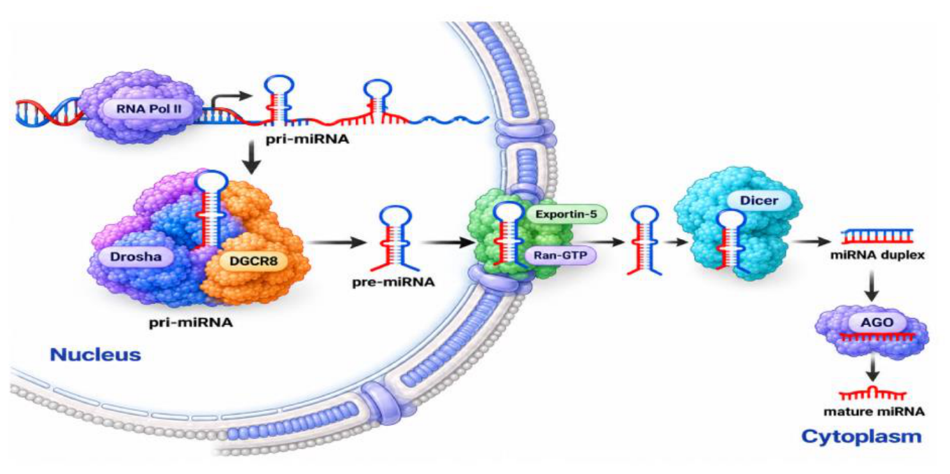
Figure 2.
Regulation of miRNA biogenesis through the TGF-β1–SMAD axis. Regulation of miRNA biogenesis through the TGF-β1–SMAD axis. miRNA biogenesis of miR-21 is increased by TGF-β1–SMAD axis. Created in BioRender. Ruiz, V. M. (2026) https://BioRender.com/p86rsaf.
Figure 2.
Regulation of miRNA biogenesis through the TGF-β1–SMAD axis. Regulation of miRNA biogenesis through the TGF-β1–SMAD axis. miRNA biogenesis of miR-21 is increased by TGF-β1–SMAD axis. Created in BioRender. Ruiz, V. M. (2026) https://BioRender.com/p86rsaf.
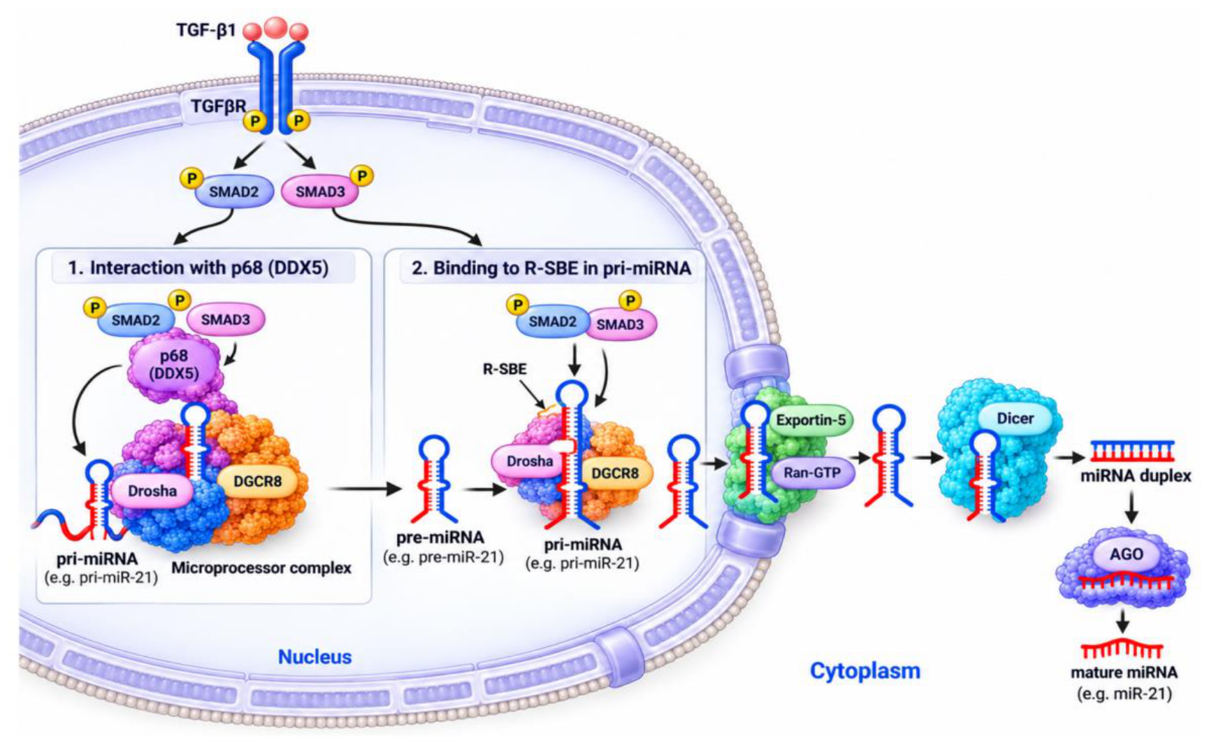
Figure 3.
p53-mediated regulation of miRNA biogenesis through the DROSHA/DGCR8 Microprocessor com-plex. Wild-type p53 can regulate miRNA biogenesis in response to cellular stress, such as DNA damage. Fol-lowing p53 accumulation and activation, p53 interacts with the DROSHA/DGCR8 Microprocessor complex through the C-terminal region of p53 and the p68/p72 helicases, facilitating the processing of pri-miRNAs into pre-miRNAs. In contrast, cancer-associated p53 mutations can impair the interaction between p53 and p68/p72 and reduce the association of the Microprocessor complex with pri-miRNAs, resulting in decreased Microprocessor activity and reduced production of pre-miRNAs. Thus, loss or alteration of p53 function may contribute to dysregulation of miRNA biogenesis.. "Created in BioRender. Ruiz, V. M. (2026) https://BioRender.com/niwc36d.
Figure 3.
p53-mediated regulation of miRNA biogenesis through the DROSHA/DGCR8 Microprocessor com-plex. Wild-type p53 can regulate miRNA biogenesis in response to cellular stress, such as DNA damage. Fol-lowing p53 accumulation and activation, p53 interacts with the DROSHA/DGCR8 Microprocessor complex through the C-terminal region of p53 and the p68/p72 helicases, facilitating the processing of pri-miRNAs into pre-miRNAs. In contrast, cancer-associated p53 mutations can impair the interaction between p53 and p68/p72 and reduce the association of the Microprocessor complex with pri-miRNAs, resulting in decreased Microprocessor activity and reduced production of pre-miRNAs. Thus, loss or alteration of p53 function may contribute to dysregulation of miRNA biogenesis.. "Created in BioRender. Ruiz, V. M. (2026) https://BioRender.com/niwc36d.
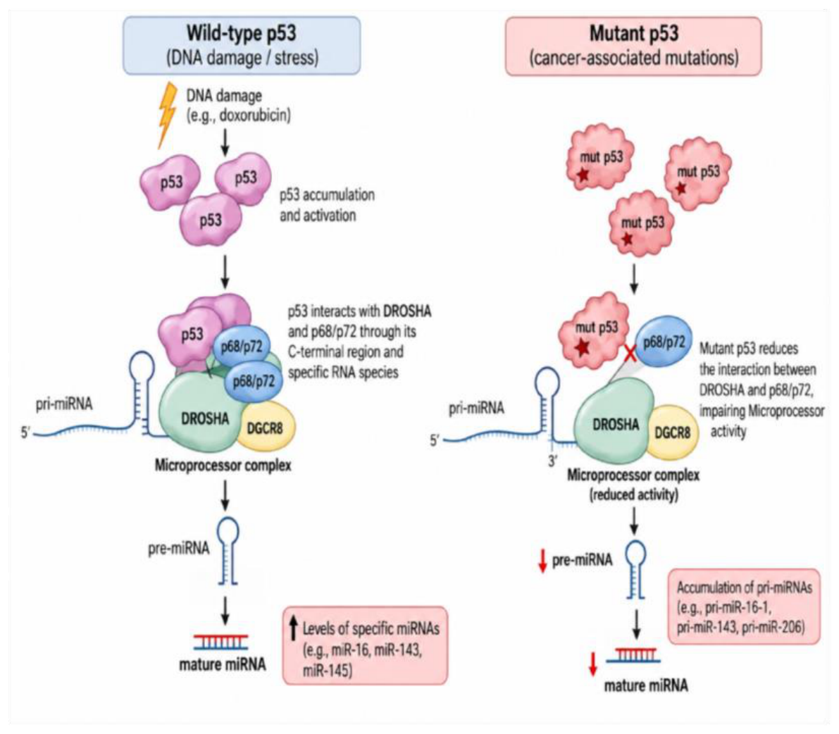
Figure 4.
ADAR-mediated regulation of miRNA biogenesis and maturation. (A) Editing-dependent mech-anisms. ADAR proteins catalyze adenosine-to-inosine (A-to-I) editing within pri-miRNAs or pre-miRNAs. Ed-iting of pri-miRNAs may alter their secondary structure or recognition elements, thereby affecting processing by the DROSHA/DGCR8 Microprocessor and the generation of pre-miRNAs. Similarly, editing of pre-miRNAs may modify their structure and recognition by DICER, potentially altering cleavage efficiency, mature miRNA sequence, and functional properties. (B) Editing-independent mechanisms. ADAR1 can modulate miRNA maturation through direct interactions with components of the miRNA-processing machinery or with miRNA precursors. ADAR1 isoforms p110 and p150 have been associated with reduced let-7d maturation, whereas interaction of ADAR1 with DICER1 promotes miR-155-5p maturation. Conversely, ADAR1 interaction with pri-miR-766 negatively regulates miR-766-3p maturation, resulting in reduced levels of the mature miRNA. Collectively, these mechanisms illustrate the dual role of ADAR proteins in regulating miRNA biogenesis through both editing-dependent and editing-independent pathways. "Created in BioRender. Ruiz, V. M. (2026) https://BioRender.com/7cdmmrj".
Figure 4.
ADAR-mediated regulation of miRNA biogenesis and maturation. (A) Editing-dependent mech-anisms. ADAR proteins catalyze adenosine-to-inosine (A-to-I) editing within pri-miRNAs or pre-miRNAs. Ed-iting of pri-miRNAs may alter their secondary structure or recognition elements, thereby affecting processing by the DROSHA/DGCR8 Microprocessor and the generation of pre-miRNAs. Similarly, editing of pre-miRNAs may modify their structure and recognition by DICER, potentially altering cleavage efficiency, mature miRNA sequence, and functional properties. (B) Editing-independent mechanisms. ADAR1 can modulate miRNA maturation through direct interactions with components of the miRNA-processing machinery or with miRNA precursors. ADAR1 isoforms p110 and p150 have been associated with reduced let-7d maturation, whereas interaction of ADAR1 with DICER1 promotes miR-155-5p maturation. Conversely, ADAR1 interaction with pri-miR-766 negatively regulates miR-766-3p maturation, resulting in reduced levels of the mature miRNA. Collectively, these mechanisms illustrate the dual role of ADAR proteins in regulating miRNA biogenesis through both editing-dependent and editing-independent pathways. "Created in BioRender. Ruiz, V. M. (2026) https://BioRender.com/7cdmmrj".
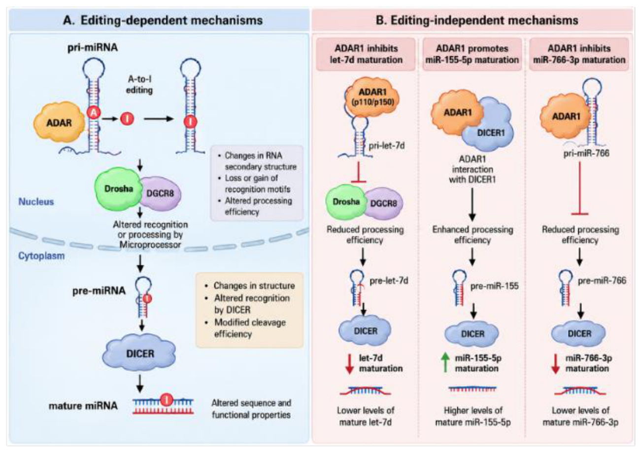
Figure 5.
Mechanotransduction pathways disrupt Drosha/DGCR8 microprocessor activity through post-translational modifications, such as PRMT1-mediated arginine methylation. Aberrant activation of the Hippo pathway and nuclear localization of YAP may further impair microRNA processing by sequestering essential cofactors, such as p72. These mechanisms contribute to a sustained reduction in miR-29 levels, promoting extracellular matrix accumulation and establishing a profibrotic positive feedback loop that drives disease progression. "Created in BioRender. Ruiz, V. M. (2026) https://BioRender.com/09unhfh ".
Figure 5.
Mechanotransduction pathways disrupt Drosha/DGCR8 microprocessor activity through post-translational modifications, such as PRMT1-mediated arginine methylation. Aberrant activation of the Hippo pathway and nuclear localization of YAP may further impair microRNA processing by sequestering essential cofactors, such as p72. These mechanisms contribute to a sustained reduction in miR-29 levels, promoting extracellular matrix accumulation and establishing a profibrotic positive feedback loop that drives disease progression. "Created in BioRender. Ruiz, V. M. (2026) https://BioRender.com/09unhfh ".
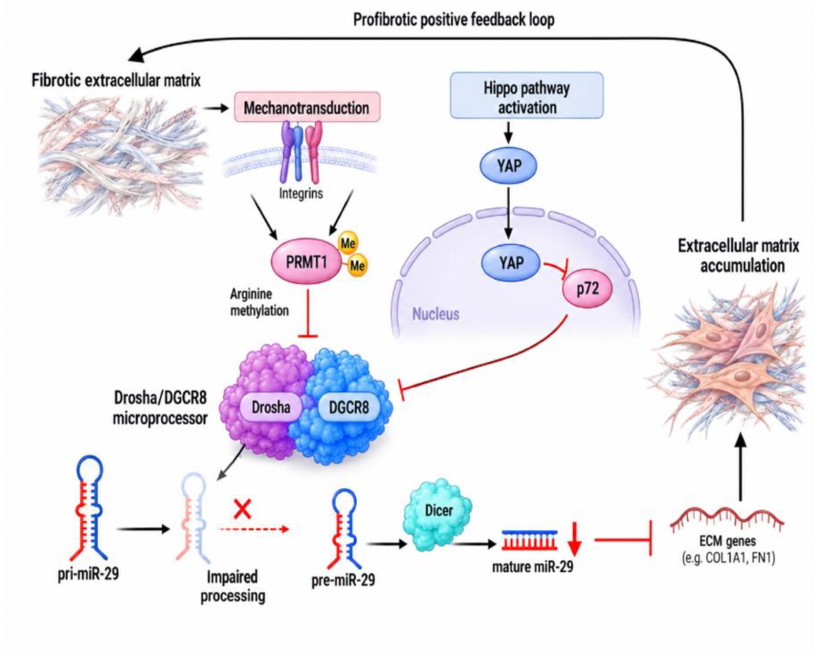

Table 1.
ADAR. Mechanism affecting miRNA maturation.
| Study | ADAR protein | miRNA | Cellular/biological context | Mechanism affecting miRNA maturation | Effect on mature miRNA | Main biological consequence |
|---|---|---|---|---|---|---|
| Yu et al., 2022 | ADAR1 | miR-155-5p | Adipogenesis / obesity | Editing-independent interaction with DICER1. ADAR1 interacts with DICER1 and promotes processing of the miRNA precursor. | ↑ miR-155-5p | ↓ C/EBPβ, ↓ adipogenic transcription factors, and inhibition of adipocyte differentiation. |
| Widmark et al., 2022 | ADAR1 / ADAR2 | miR-376b and miRNAs of the miR-379–410 cluster | Neuronal/GABAergic system | Editing-dependent and editing-independent mechanisms. A-to-I editing can alter precursor processing; ADAR2 can also interfere with miRNA processing independently of catalytic activity. | Context-dependent | Altered miRNA maturation and targeting, including regulation of GABA catabolism. |
| Díaz-Piña et al., 2022 | ADAR1-p110 / ADAR1-p150 | let-7d | Idiopathic pulmonary fibrosis (IPF) | Isoform-dependent regulation of miRNA processing. Differential expression and subcellular distribution of ADAR1 isoforms are associated with altered let-7d precursor and mature miRNA levels. | ↓ mature let-7d in IPF; restored following ADAR1 overexpression | ↑ COL3A1 and SMAD2 expression, potentially contributing to the fibrotic phenotype. |
| Shi et al., 2025 | ADAR1 | miR-766-3p | Acute myeloid leukemia (AML) | Editing-independent binding to pri-miR-766. ADAR1 interacts with the primary miRNA transcript and impairs its maturation. | ↓ miR-766-3p |
Disclaimer/Publisher’s Note: The statements, opinions and data contained in all publications are solely those of the individual author(s) and contributor(s) and not of MDPI and/or the editor(s). MDPI and/or the editor(s) disclaim responsibility for any injury to people or property resulting from any ideas, methods, instructions or products referred to in the content. |
© 2026 by the authors. Licensee MDPI, Basel, Switzerland. This article is an open access article distributed under the terms and conditions of the Creative Commons Attribution (CC BY) license (http://creativecommons.org/licenses/by/4.0/).
Copyright: This open access article is published under a Creative Commons CC BY 4.0 license, which permit the free download, distribution, and reuse, provided that the author and preprint are cited in any reuse.