
Submitted:
30 October 2026
Posted:
02 November 2026
You are already at the latest version
Abstract
Background/ Objectives: Hyperlactatemia is a nonspecific biochemical phenotype with a broad acquired and inherited differential, complicating genomic interpretation. We aimed to identify and curate genes associated with inherited metabolic hyperlactatemia and develop an evidence-tiered virtual diagnostic panel. Methods: NCBI Gene was searched on 8 August 2026 using four lactate-related terms. Human records were deduplicated by GeneID and curated for gene–disease validity, inheritance, mechanism, and lactate-specific clinical relevance. Targeted review of ClinGen, OMIM, GenCC, PanelApp, GeneReviews, and primary literature identified established causes missed by keyword retrieval. Results: The searches yielded 802 raw records. After removal of duplicate export blocks, 401 human query records resolved to 240 unique genes. Submission-version reconciliation identified 34 search-derived genes in the 85-gene active panel, while 206 search-derived genes were not retained and remain available for audit and future reanalysis. Targeted review contributed 51 additional active genes. The final panel comprised 40 Tier 1, 26 Tier 2, and 19 Tier 3 genes. Conclusions: Phenotype-keyword retrieval showed low specificity and incomplete sensitivity. Integrating gene–disease validity with lactate-specific relevance provides a transparent, updateable framework for phenotype-informed genomic prioritization.
Keywords:
hyperlactatemia
; lactic acidosis
; inherited metabolic disorders
; mitochondrial disease
; gene–disease validity
; virtual gene panel
; genomic diagnosis
; variant prioritisation
1. Introduction
Hyperlactatemia is common in tissue hypoxia, impaired perfusion, and critical systemic illness [1]. Lactate is a central intermediate of energy metabolism, generated from pyruvate through the reversible reaction catalysed by lactate dehydrogenase [2,3]. In clinical practice, blood lactate concentrations >2 mmol/L are generally considered elevated, whereas values >4 mmol/L are often used to indicate more severe hyperlactatemia; lactic acidosis denotes lactate elevation accompanied by metabolic acidemia [4,5,6,7]. Hyperlactatemia is classified mainly into Type A and Type B, based on whether tissue oxygen delivery is compromised. Type A occurs due to inadequate tissue oxygenation or global or local tissue hypoperfusion. Type B occurs when oxygen delivery is normal, but lactate rises due to altered cellular metabolism, underlying systemic illness, toxin exposure, or clearance failure [8]. Although acquired causes predominate, inherited metabolic and mitochondrial disorders are important causes of type B hyperlactatemia and may be overlooked, particularly in attenuated adolescent or adult presentations [9,10].
Inherited metabolic disorders (IMDs) can cause hyperlactatemia through defects of pyruvate transport or oxidation, pyruvate dehydrogenase function, oxidative phosphorylation, mitochondrial translation or mtDNA maintenance, gluconeogenesis, and secondary disruption of mitochondrial function [11,12,13,14,15,16,17]. The resulting phenotypes span severe neonatal lactic acidosis and encephalopathy to intermittent, stress-induced abnormalities associated with hypoglycaemia, cardiomyopathy, liver disease, myopathy, exercise intolerance, seizures, or multisystem involvement [18,19].
Lactate elevation is neither sensitive nor specific for IMDs. Pre-analytical factors and acquired conditions—including seizures, hypoxia, shock, hepatic dysfunction, medications, toxins, malignancy, inflammation, and recent exertion—can increase lactate concentrations [20,21], whereas patients with established mitochondrial or metabolic disease may have normal values between episodes. Interpretation therefore depends on the magnitude, persistence, and reproducibility of the elevation; the physiological context; associated biochemical abnormalities; and the broader phenotype [22].
Although next-generation sequencing has expanded molecular diagnosis of IMDs and mitochondrial disease, gene selection and variant prioritisation for hyperlactatemia remain difficult. Phenotype-keyword searches can retrieve genes involved in lactate transport, signalling, hypoxic responses, inflammation, tumour metabolism, or experimental pathways without an established role in inherited metabolic hyperlactatemia. Conversely, clinically relevant disease genes may be missed when lactate elevation is intermittent, phenotype-restricted, or incompletely annotated [21].
Virtual gene panels can prioritise diagnostically relevant genes within exome- or genome-scale data while remaining updatable as evidence evolves. Their clinical value, however, depends on separating two related dimensions: the strength of the underlying gene–disease relationship and the relevance of hyperlactatemia to that disorder. A definitive disease gene may be associated with lactate elevation only in particular phenotypes or during metabolic stress, whereas mechanistic plausibility alone does not establish a clinically valid gene–disease association [23].
We therefore aimed to systematically identify and clinically curate genes associated with inherited metabolic hyperlactatemia and to develop an evidence-tiered virtual panel for molecular diagnosis. Phenotype-derived candidates were assessed for gene–disease validity, inheritance, molecular mechanism, and the frequency, context, and diagnostic relevance of lactate elevation, then supplemented by targeted review of established gene–disease resources and primary literature. The resulting framework distinguishes core, extended, and context-dependent diagnostic associations from limited or indirect evidence retained for audit and future reanalysis.
1.1. Biochemical Basis and Mechanisms of Inherited Metabolic Hyperlactatemia
Systemic lactate homeostasis reflects the balance among production, utilisation, and clearance. Lactate is generated continuously under aerobic conditions in skeletal muscle, brain, erythrocytes, the gastrointestinal tract, and other tissues; therefore, accumulation cannot be interpreted as a marker of oxygen deficiency in isolation [24]. Lactate also functions as an oxidative fuel, a major gluconeogenic substrate, and a metabolic signalling molecule [2]. Systemic disposal occurs predominantly through hepatic gluconeogenesis and glucose recycling via the Cori cycle, with the kidneys contributing through gluconeogenesis and direct oxidation, particularly during fasting [25].
The lactate–pyruvate equilibrium is tightly coupled to cellular redox state. Reduction of pyruvate to lactate by lactate dehydrogenase oxidises NADH to NAD+, supporting glycolytic flux when mitochondrial oxidative capacity is limited or pyruvate generation exceeds mitochondrial utilisation [26]. A more reduced cytosolic NADH/NAD+ state therefore favours lactate formation even when oxygen delivery is adequate [27] (Figure 1).
1.2. Mechanisms of Lactate Elevation
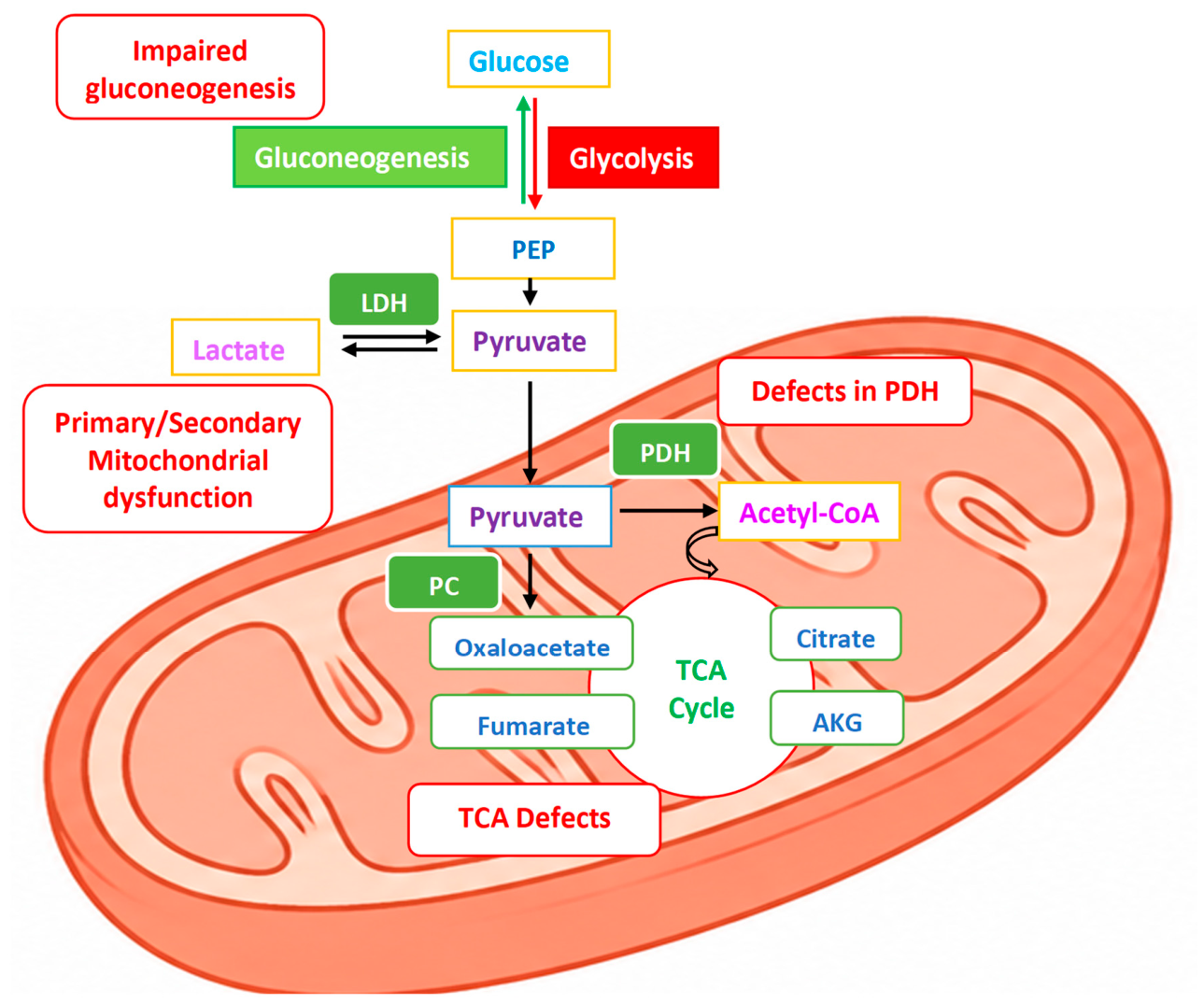
Inherited metabolic hyperlactatemia can be organised into three overlapping mechanisms: increased pyruvate flux toward lactate, impaired lactate utilisation or gluconeogenic clearance, and disturbed intracellular redox homeostasis. These mechanisms frequently coexist and may become clinically apparent only during fasting, infection, exercise, surgery, pregnancy, or other metabolic stress, producing persistent or episodic lactate elevation in blood or cerebrospinal fluid [28] (Figure 2).
1.2.1. Increased Lactate Production
Lactate production increases when glycolytic generation of pyruvate exceeds the capacity for mitochondrial transport or oxidation. Cellular energy deficiency drives compensatory glycolysis and diversion of pyruvate toward lactate [12,29]. This pattern is characteristic of mitochondrial pyruvate-carrier defects, pyruvate dehydrogenase (PDH) complex deficiency, and respiratory-chain or oxidative-phosphorylation disorders [30]. In PDH deficiency, impaired conversion of pyruvate to acetyl-CoA limits entry of glycolytic carbon into the tricarboxylic acid cycle, leading to accumulation of pyruvate, lactate, and often alanine; the high dependence of the central nervous system on oxidative metabolism contributes to the prominent neurological phenotype [31,32]. Similar abnormalities may arise when toxic metabolites secondarily inhibit pyruvate oxidation or mitochondrial respiration, as in selected organic acidemias [33].
Figure 2.
Major metabolic and mitochondrial causes of hyperlactatemia. Abbreviations: AKG, Alpha-ketoglutarate; LDH, lactate dehydrogenase; PC, Pyruvate Carboxylase; PDH, pyruvate dehydrogenase complex; PEP, phosphoenolpyruvate; TCA, tricarboxylic acid.
Figure 2.
Major metabolic and mitochondrial causes of hyperlactatemia. Abbreviations: AKG, Alpha-ketoglutarate; LDH, lactate dehydrogenase; PC, Pyruvate Carboxylase; PDH, pyruvate dehydrogenase complex; PEP, phosphoenolpyruvate; TCA, tricarboxylic acid.
1.2.2. Impaired Lactate Utilisation and Clearance
Hyperlactatemia may also reflect reduced hepatic or renal disposal rather than primary overproduction. Disorders that impair hepatic glucose production or gluconeogenic flux limit incorporation of lactate into glucose and can produce marked lactate accumulation during fasting or catabolic stress. Fructose-1,6-bisphosphatase deficiency typically presents with fasting- or illness-induced hypoglycaemia, ketosis, and lactic acidosis [34]. In glycogen storage disease type I (GSD I), disruption of the glucose-6-phosphatase system blocks the terminal step shared by gluconeogenesis and glycogenolysis, producing persistent hyperlactatemia with hypoglycaemia, hyperuricaemia, and hypertriglyceridaemia [17].
1.2.3. Altered Intracellular Redox State
Respiratory-chain dysfunction impairs mitochondrial NADH oxidation, increases the intracellular NADH/NAD+ ratio, and shifts the lactate dehydrogenase equilibrium toward lactate formation. Hyperlactatemia in this setting is therefore a redox consequence of impaired oxidative metabolism rather than a marker of tissue hypoxia alone [35,36].
2. Materials and Methods
2.1. Phenotype-Based Gene Identification and Dataset Construction
A phenotype-based search of NCBI Gene was performed on 8 August 2026 using the terms “increased lactate”, “high lactate”, “lactic acidosis”, and “hyperlactatemia”. The four intended searches returned 352, 348, 98, and 4 records, respectively (802 raw records). Row-level reconciliation of the retained raw-data file showed that the 352-record and 348-record export blocks had each been duplicated in the assembled DOCX. These duplicate file blocks were removed before analysis. Across the four intended search blocks, 401 Homo sapiens records remained. Records were deduplicated using the NCBI Gene identifier (GeneID) as the primary key, with approved gene symbols used for secondary verification, yielding 240 unique human genes.
2.2. Clinical Curation and Dataset Augmentation
Each phenotype-derived gene was manually reviewed for an established inherited human gene–disease association, mode of inheritance, molecular function, biochemical mechanism, and the frequency, reproducibility, clinical context, and diagnostic relevance of lactate elevation in blood or cerebrospinal fluid. Hyperlactatemia relevance was classified as characteristic/recurrent, variable/phenotype-dependent, context-dependent/secondary, or limited/indirect. Classification reflected the overall clinical and biochemical phenotype rather than an isolated report of increased lactate.
Because phenotype-keyword retrieval can miss established disorders when biochemical features are incompletely annotated, the dataset was supplemented by targeted review of ClinGen Gene–Disease Validity classifications where available, Online Mendelian Inheritance in Man (OMIM), the Gene Curation Coalition (GenCC), Genomics England PanelApp, GeneReviews, and relevant primary literature. Genes added through targeted review were evaluated using the same criteria as phenotype-derived genes. For submission-version provenance, the recovered 240-gene phenotype-search dataset was maintained as a distinct layer and cross-referenced against the active panel.
2.3. Evidence Dimensions and Assessment
Two evidence dimensions were assessed separately. Gene–disease validity represented the strength of evidence that variation in a gene causes an inherited disorder. Hyperlactatemia relevance represented the frequency, reproducibility, mechanistic relationship, clinical context, and diagnostic usefulness of lactate elevation within that disorder. External classifications informed gene–disease assessment but were not mapped directly onto the study-specific diagnostic tiers.
Mechanistic plausibility for lactate elevation was not sufficient for active-panel inclusion without adequate human gene–disease evidence. Conversely, a well-established gene–disease relationship did not justify high hyperlactatemia priority when lactate elevation was inconsistent, phenotype-restricted, or confined to specific physiological circumstances.
2.4. Diagnostic Tier Assignment and Virtual-Panel Development
Clinically relevant genes were assigned to three diagnostic tiers. Tier 1 comprised established disorders in which hyperlactatemia or lactic acidosis is characteristic or recurrent and mechanistically and diagnostically informative. Tier 2 comprised established disorders in which lactate elevation is clinically relevant but variably expressed, age- or genotype-dependent, or phenotype-restricted. Tier 3 comprised established disorders in which hyperlactatemia occurs predominantly during metabolic decompensation, fasting, hypoglycaemia, organ dysfunction, seizures, or other physiological stress.
Genes not meeting the criteria for active-panel inclusion were not assigned an active diagnostic tier. Where such genes were recovered by the phenotype searches, they were retained for audit and future reanalysis. For the recovered search-derived genes, the submission-version disposition does not imply recovery of the historical manual exclusion categories because those labels are absent from the retained raw data. The framework included nuclear and mitochondrial causes, and tier assignment reflected the combined assessment of gene–disease validity and hyperlactatemia relevance rather than retrieval by a lactate-related term alone. A smaller representative gene subset was used only for mechanistic presentation and did not constitute an additional tier.
2.5. Submission-Version Raw-Data Reconciliation and Provenance
At manuscript finalisation, the retained NCBI Gene raw-data file was recovered and re-parsed at row level. Supplementary Table S2B provides all 240 unique human search-derived genes with GeneID, search-term membership, and submission-version disposition. Cross-referencing these genes against the independently curated 85-gene active panel in Supplementary Table S2A identified 34 search-derived active genes and 206 search-derived genes not retained in the active panel. The 206 genes are retained for audit and future reanalysis, but no historical exclusion category is inferred from the recovered raw data. Fifty-one active genes were added through targeted review of authoritative gene–disease resources.
The supplementary workbook records active-panel tier assignment, the study assessment of gene–disease validity, lactate-specific relevance, evidence sources, curation status, and discovery provenance, together with the complete recovered phenotype-search dataset. Submission-version curation and reconciliation were performed in August 2026, and the supplementary workbook was finalised on 16 August 2026. AI was used to support organization and language refinement of the submission-version reconciliation.
3. Results
3.1. Phenotype-Search Yield and Dataset Construction
The four intended phenotype searches generated 802 raw records. The retained raw-data contained duplicate copies of the 352-record and 348-record exports; after removal of these duplicate file blocks, the intended search data comprised 401 human records. GeneID-based deduplication yielded 240 unique human genes. Cross-referencing the recovered search-derived genes against the submission-version active panel identified 34 active-panel genes and 206 genes not retained in the active panel; the latter are retained for audit and future reanalysis. Targeted review of authoritative gene–disease resources contributed 51 additional active genes that were absent from the recovered phenotype searches.
The retrieval profile demonstrated two complementary limitations of keyword-based discovery: low specificity, because 206 of 240 recovered search-derived genes were not retained in the submission-version active panel, and incomplete sensitivity, because 51 of 85 active genes were identified only through targeted resource review.
3.2. Evidence-Tiered Virtual Panel
Submission-version reconciliation yielded an 85-gene active panel comprising 40 Tier 1, 26 Tier 2 and 19 Tier 3 genes. Of these, 34 genes were present in the recovered 240-gene phenotype-search dataset and 51 were added through targeted review. The remaining 206 search-derived genes were not retained in the active panel and are retained for audit and future reanalysis. Accordingly, 291 unique genes are represented in the reconciled submission-version dataset: 240 recovered search-derived genes plus 51 targeted augmentations (Table 1; Supplementary Tables S2A and S2B).
3.3. Mechanistic Representation of the Curated Framework
Despite substantial genetic heterogeneity, curated genes converged on a limited number of proximal biochemical mechanisms. Representative groups included mitochondrial pyruvate transport and oxidation, PDH-complex function and lipoylation, gluconeogenesis, oxidative phosphorylation, mitochondrial translation and mtDNA maintenance, tricarboxylic-acid-cycle metabolism, iron–sulphur biology, and mtDNA-encoded oxidative-phosphorylation function (Table 2). Table 2 is illustrative and does not define tier membership.
3.4. Diagnostic Application of the Curated Framework
To support clinical application, recurrent biochemical and phenotypic patterns were linked to representative inherited disorders and complementary investigations (Table 3). This pattern-based framework is intended to guide phenotype-informed genomic prioritisation while complementing, rather than replacing, standard biochemical assessment and disorder-specific diagnostic pathways.
4. Discussion
4.1. Mechanistic Interpretation of the Curated Framework
The principal finding is that diagnostic relevance cannot be inferred from a lactate-related database hit or pathway association alone. Clinically relevant genes converged on a limited set of proximal mechanisms—pyruvate handling, oxidative phosphorylation and redox balance, mitochondrial translation and mtDNA maintenance, gluconeogenesis, and secondary disruption of mitochondrial energy metabolism. This mechanistic organisation improves interpretability, but tier assignment depends on how consistently hyperlactatemia contributes to the human disease phenotype rather than on pathway membership alone.
4.1.1. Mitochondrial Pyruvate Transport and Oxidation
Defects of the mitochondrial pyruvate carrier, PDH complex, and mitochondrial lipoylation occupy a proximal position in lactate metabolism. Their direct effect on mitochondrial pyruvate disposal explains why recurrent lactate elevation—often accompanied by pyruvate or alanine accumulation and, in PDH deficiency, a normal or only mildly increased lactate-to-pyruvate ratio—can be diagnostically informative. These disorders illustrate the distinction between direct perturbation of pyruvate utilisation and more distal defects of cellular energy metabolism.
4.1.2. Respiratory-Chain and Oxidative-Phosphorylation Disorders
Respiratory-chain and oxidative-phosphorylation disorders form a genetically broad group that includes mtDNA variants and nuclear defects affecting respiratory-chain subunits, assembly factors, cofactors, and related mitochondrial processes. Their shared biochemical consequence is impaired oxidative metabolism, but the diagnostic behaviour of lactate is heterogeneous.
Reduced ATP production and impaired NADH reoxidation increase reliance on glycolysis and favour conversion of pyruvate to lactate. In severe phenotypes, including Leigh and MELAS-spectrum disease, hyperlactatemia may be recurrent and highly informative; in attenuated or tissue-restricted disease it may be intermittent or absent between episodes. This variability explains why genes within a common mechanistic pathway can appropriately occupy different diagnostic tiers.
4.1.3. Mitochondrial Translation and mtDNA Maintenance
Defects of mitochondrial translation can impair synthesis of multiple mtDNA-encoded respiratory-chain subunits, whereas defects of mtDNA replication, nucleotide supply, or genome stability can cause depletion, multiple deletions, or secondary respiratory-chain failure. Both groups therefore converge on combined oxidative-phosphorylation dysfunction rather than a single enzyme defect.
Their phenotypic breadth—including encephalopathy, epilepsy, myopathy, cardiomyopathy, neuropathy, ophthalmoplegia, and liver disease—also means that lactate elevation is strongly context dependent. Heteroplasmy, tissue involvement, disease stage, and metabolic stress can modify biochemical expression, supporting phenotype-specific prioritisation rather than uniform tier assignment across all mitochondrial genes.
4.1.4. Gluconeogenesis and Hepatic Glucose Production
Gluconeogenic disorders are mechanistically distinct because hyperlactatemia primarily reflects impaired lactate utilisation for hepatic glucose production. Fructose-1,6-bisphosphatase deficiency typically causes fasting- or illness-induced ketotic hypoglycaemia with lactic acidosis, whereas GSD I produces more persistent lactate elevation through disruption of the glucose-6-phosphatase system.
This biochemical context is diagnostically valuable: fasting dependence, hypoglycaemia, hepatomegaly, hyperuricaemia, and hypertriglyceridaemia can distinguish impaired lactate clearance from primary respiratory-chain disease. By contrast, other hepatic glycogen-storage disorders warrant lower lactate-specific priority when hyperlactatemia is variable, secondary, or absent.
4.1.5. Secondary Hyperlactatemia in other Metabolic Disorders
Organic acidemias and related intermediary metabolic disorders can cause secondary hyperlactatemia when accumulated metabolites inhibit PDH activity, respiratory-chain enzymes, or other mitochondrial processes. In these disorders, lactate is usually interpreted within a broader crisis phenotype that may include ketosis, hyperammonaemia, high-anion-gap metabolic acidosis, and characteristic organic-acid or acylcarnitine abnormalities.
Similarly, fatty acid oxidation disorders (FAODs) can reduce ATP availability during fasting and catabolic stress, increasing dependence on glucose and glycolysis. Because hypoketotic hypoglycaemia, rhabdomyolysis, cardiomyopathy, hepatic dysfunction, and acylcarnitine abnormalities are generally more specific than lactate elevation, these disorders are best prioritised according to the strength and consistency of the lactate association rather than broad membership of an energy-metabolism pathway.
4.1.6. Metabolic Stress and Episodic Hyperlactatemia
Reduced metabolic reserve is a unifying feature of many IMDs. Fasting, intercurrent infection, surgery, strenuous exercise, pregnancy, vomiting, and selected medications can increase energy demand, accelerate catabolism, impair mitochondrial function, or reduce substrate availability, thereby unmasking previously compensated defects—particularly in adolescents and adults with attenuated phenotypes [100].
Consequently, hyperlactatemia may be episodic and absent during metabolic stability. Acute decompensation can resemble sepsis, tissue hypoperfusion, or drug toxicity, so a recurrent or disproportionate rise in lactate during physiological stress is more informative than a single isolated measurement.
4.2. Clinical Phenotype and Diagnostic Red Flags
Inherited metabolic hyperlactatemic disorders are phenotypically heterogeneous because clinical expression reflects the affected pathway, residual function, age at presentation, tissue-specific energy requirements, heteroplasmy, and exposure to metabolic stress. No single clinical feature is sufficiently specific; diagnostic value arises from recurrent patterns across organ systems and biochemical states.
In neonates and children, red flags include recurrent vomiting, feeding difficulty, failure to thrive, hypotonia, developmental delay, seizures, encephalopathy, cardiomyopathy, hepatomegaly, hypoglycaemia, and metabolic acidosis. Severe disorders may present with persistent neonatal lactic acidosis, whereas milder conditions may manifest only during fasting or intercurrent illness.
Adolescent and adult presentations may be subtler, with exercise intolerance, myopathy, cardiomyopathy, neuropathy, progressive neurological or psychiatric manifestations, recurrent encephalopathic or “sepsis-like” episodes, unexplained liver disease, or deterioration during infection, fasting, surgery, pregnancy, strenuous exercise, or medication exposure [101]. A late presentation does not exclude inherited disease; consanguinity, similarly, affected relatives, maternal inheritance, or unexplained early deaths can further support a genetic aetiology.
4.3. Diagnostic Application of the Curated Framework
Evaluation of unexplained hyperlactatemia requires a structured, mechanism-based approach integrating clinical assessment, biochemical phenotyping, imaging, genomic analysis, and, when necessary, functional studies [22]. Because lactate elevation can arise from multiple primary and secondary mechanisms, interpretation should emphasise the magnitude, persistence, and physiological context of the elevation, associated biochemical abnormalities, the lactate-to-pyruvate ratio, and the overall phenotype [21].
4.3.1. Confirmation and Exclusion of Acquired Causes
Before pursuing an IMD diagnosis, common acquired causes and pre-analytical artefacts should be addressed. Tissue hypoxia, shock, sepsis, seizures, hepatic dysfunction, toxins, medications, recent exercise, prolonged tourniquet use, struggling during venepuncture, and delayed sample processing can all increase lactate. Importantly, inherited and acquired mechanisms may coexist.
An unexpected lactate result that is discordant with the clinical presentation should be repeated under controlled conditions when clinically appropriate. Lactate and pyruvate should be measured simultaneously whenever the lactate-to-pyruvate ratio is being used to support mechanistic localisation.
4.3.2. Biochemical Phenotyping
Paired lactate and pyruvate measurements provide an indirect assessment of cellular redox state and can support biochemical localisation. A markedly increased lactate-to-pyruvate (L/P) ratio may support impaired mitochondrial NADH reoxidation, as in respiratory-chain dysfunction, whereas a normal or mildly increased ratio may be more consistent with defects of mitochondrial pyruvate transport or utilisation, particularly PDH-complex deficiency [32,102]. Interpretation is meaningful only when lactate is genuinely elevated and both analytes are collected and processed appropriately. Neither lactate nor the L/P ratio is sufficiently sensitive or specific to establish a diagnosis in isolation; results should be integrated with absolute analyte concentrations, sample quality, glucose, ketones, ammonia, alanine, urine organic acids, acylcarnitines, and the broader clinical context [103].
Secondary hyperalaninaemia may indicate chronic pyruvate accumulation because excess pyruvate is transaminated to alanine [104], but alanine elevation is not disorder-specific and may also occur during catabolism or prolonged lactic acidemia. Plasma amino-acid analysis can provide additional pathway-specific clues, including hyperglycinaemia in mitochondrial lipoylation defects and amino-acid abnormalities associated with organic acidemias or mitochondrial hepatopathies [105,106].
Urine organic-acid analysis is particularly important when organic acidemias, intermediary metabolic defects, or secondary mitochondrial dysfunction are suspected [64]. Urinary lactate is nonspecific; disease-associated metabolites such as methylmalonic acid, methylcitrate, 3-hydroxypropionic acid, isovalerylglycine, and other characteristic organic acids are more informative [107]. Diagnostic yield is often highest during acute metabolic decompensation.
Plasma or dried-blood-spot acylcarnitine profiling is essential for identifying FAODs and several organic acidemias [108]. Results should be interpreted with glucose, ketones, free fatty acids, free and total carnitine, creatine kinase (CK), liver biochemistry, and the clinical circumstances of sampling because diagnostic abnormalities may diminish after treatment or during metabolic stability.
Additional first-line investigations may include blood gases, glucose, β-hydroxybutyrate, ammonia, electrolytes, liver and renal biochemistry, CK, uric acid, triglycerides, free fatty acids, carnitine, and selected endocrine studies. The pattern of accompanying abnormalities can substantially narrow the differential diagnosis [109,110,111]: Hypoketotic hypoglycaemia suggests impaired fatty acid oxidation or ketogenesis. Fasting-induced lactic acidosis with ketosis suggests a gluconeogenic defect. Hypoglycaemia, hepatomegaly, hyperuricaemia, and hypertriglyceridaemia suggest GSD I. High-anion-gap acidosis, ketosis, and hyperammonaemia should prompt urgent investigation for an organic acidemia.
The most informative samples are often obtained during acute decompensation. When feasible without delaying treatment, crisis sampling should include lactate, pyruvate, blood gas, glucose, ketones, ammonia, plasma amino acids, acylcarnitines, and urine organic acids, with additional plasma, serum, urine, and dried blood spots stored for subsequent analysis.
4.3.3. Neuroimaging and Functional Studies
Brain magnetic resonance imaging provides important supportive evidence when neurological manifestations suggest mitochondrial or pyruvate-metabolism disease. Relevant findings include bilateral symmetrical basal-ganglia, thalamic, or brainstem lesions; Leigh syndrome-like abnormalities; stroke-like cortical lesions that do not conform to vascular territories; cerebral or cerebellar atrophy; and characteristic white-matter patterns [68]. Magnetic resonance spectroscopy may demonstrate a cerebral lactate peak, although this finding is neither universal nor specific [112]. Normal neuroimaging does not exclude mitochondrial disease, particularly in early, mild, intermittent, or adult-onset presentations [113,114].
Muscle biopsy now has a selective rather than routine diagnostic role because of advances in genomic testing. Ragged-red fibres, cytochrome c oxidase-negative fibres, and subsarcolemmal mitochondrial accumulation can support mitochondrial dysfunction but are neither universal nor disease-specific [115]. Respiratory-chain enzyme analysis, PDH activity, mitochondrial oxygen-consumption studies, coenzyme Q10 quantification, mtDNA copy-number analysis, metabolomics, or proteomics may provide functional evidence in unresolved cases [116].
Invasive or tissue-based investigations are generally reserved for cases in which biochemical and genomic testing is inconclusive, functional confirmation is required to interpret a variant of uncertain significance, or the suspected defect may not be reliably detectable in blood [117]. Tissue selection should reflect the suspected disorder because heteroplasmy, disease stage, and tissue-specific expression can materially affect diagnostic sensitivity.
4.3.4. Genomic Investigation and Application of the Curated Framework
Genomic testing should be selected according to phenotype, biochemical pattern, diagnostic urgency, and suspected inheritance. Mechanism-directed panels may be appropriate when findings strongly implicate a defined pathway, such as mitochondrial pyruvate transport or oxidation, gluconeogenesis, respiratory-chain function, fatty acid oxidation, or organic acid metabolism.
The curated framework is intended to focus interpretation on genes in which pathogenic variation is associated with a clinically credible inherited hyperlactatemic phenotype. This is important because broad phenotype- or pathway-based lists may include genes involved in lactate transport, sensing, inflammation, hypoxic responses, tumour metabolism, or secondary metabolic regulation without an established causal relationship to systemic inherited hyperlactatemia, increasing variant burden without improving diagnostic prioritisation.
Whole-exome and whole-genome sequencing are particularly valuable for multisystem disease, atypical presentations, negative targeted testing, suspected blended phenotypes, or possible novel gene–disease associations [118]. Genome sequencing can improve detection of structural, noncoding, and some mitochondrial variants, although analytical performance remains platform dependent.
Mitochondrial DNA requires dedicated analytical consideration. Heteroplasmy, tissue-specific variant distribution, low variant fractions, large-scale deletions, and mtDNA depletion can produce false-negative results when testing is restricted to blood or conventional exome sequencing. Analysis of urine epithelial cells, skeletal muscle, liver, or another clinically affected tissue may therefore be necessary, with copy-number and structural-variant analysis included where technically appropriate.
A molecular diagnosis can inform treatment, surveillance, prognosis, cascade testing, recurrence risk, and reproductive counselling. However, a variant should not be assumed to explain hyperlactatemia solely because the affected gene has a plausible role in energy metabolism; interpretation requires concordant clinical, biochemical, segregation, and, when necessary, functional evidence [119].
4.4. Implications for Gene-Panel Design and Variant Interpretation
The observed retrieval profile quantifies the limitations of phenotype-keyword search as a stand-alone panel-design strategy. Of 240 unique search-derived genes, 34 were retained in the submission-version active panel and 206 were not retained in the active panel; the latter are retained for audit and future reanalysis. Targeted review then contributed 51 active genes absent from the phenotype searches. These findings demonstrate both the low specificity and incomplete sensitivity of purely keyword-driven discovery.
False-positive retrieval is biologically unsurprising because lactate participates in hypoxic signalling, inflammation, tumour metabolism, cellular stress responses, transport, and general metabolic regulation. Genes influencing these processes may alter lactate production or handling without causing an established inherited systemic hyperlactatemia phenotype. Indiscriminate inclusion therefore risks increasing incidental findings and variants of uncertain significance without improving clinical prioritisation.
The two-dimensional assessment of gene–disease validity and hyperlactatemia relevance is central to the framework. These dimensions overlap but are not interchangeable: an established disease gene may show lactate elevation only in severe, age-specific, tissue-specific, or stress-related phenotypes, whereas a biologically plausible effect on lactate metabolism does not establish human disease causality. The study-specific tiers are therefore phenotype-specific prioritisation categories, not substitutes for external gene–disease validity classifications.
These findings support virtual panels as interpretation layers applied to broader genomic data rather than as fixed laboratory assays. Exome or genome sequencing remains important for atypical, multisystem, or unresolved disease, while the curated tiers can focus variant review according to the biochemical phenotype. Dedicated mtDNA analysis remains necessary because heteroplasmy, tissue-specific distribution, depletion, and large-scale rearrangements may not be reliably captured by conventional blood-based exome analysis.
Targeted augmentation also underscores the need for periodic reanalysis. Established disorders may be absent from phenotype searches when lactate elevation is intermittent, tissue-dependent, stress-induced, or incompletely represented in database annotations, while search-derived genes not retained in the current active panel may acquire stronger evidence over time. The panel should therefore be versioned and updated rather than treated as definitive.
4.5. Limitations and Future Directions
This study has several limitations. Discovery depended on four lactate-related NCBI Gene search terms and on the completeness and consistency of linked phenotype annotations. For genes outside the active panel, that disposition indicates non-retention in the submission-version panel and does not infer a historical exclusion category. Manual classification of the active panel required expert judgement about the frequency, severity, and diagnostic significance of lactate elevation, and boundaries between characteristic, variable, and context-dependent hyperlactatemia are not absolute. The published evidence base is also uneven, particularly for recently described disorders represented by small patient series.
This was an evidence-curation study, not a diagnostic-yield or clinical-validation study. The virtual panel has not been prospectively evaluated in an independent patient cohort and should not be interpreted as a validated laboratory assay. External validation, multidisciplinary review, and prospective assessment of diagnostic yield, variant burden, and reclassification frequency are therefore important next steps.
Periodic reassessment will be required as gene–disease relationships are established or revised and phenotypic spectra expand. Future versions should use versioned evidence review with documented changes in tier assignment and, where clinically appropriate, integrate nuclear and mitochondrial genomic data with biochemical, metabolomic, transcriptomic, and tissue-specific functional evidence.
5. Conclusions
Phenotype-keyword retrieval alone produces a heterogeneous mixture of clinically relevant disease genes, secondary associations, and nonspecific candidates while missing some established inherited causes of hyperlactatemia. Separating gene–disease validity from lactate-specific clinical relevance enables transparent classification of active genes into core, extended, and context-dependent tiers while retaining non-active search-derived genes for audit and future reanalysis. The resulting evidence-tiered virtual panel provides an updatable framework for phenotype-informed prioritisation of molecular findings in patients with unexplained or recurrent hyperlactatemia.
Supplementary Materials
The following supporting information can be downloaded at the website of this paper posted on Preprints.org, Table S1, detailed clinical and mechanistic annotation of Tier 1 core genes associated with inherited causes of hyperlactatemia; Table S2A, Submission-version evidence-tiered active panel for inherited causes of hyperlactatemia; Table S2B, Recovered NCBI phenotype-search dataset and submission-version disposition; Table S3, Phenotype- and biochemical-pattern recognition framework for inherited hyperlactatemia. Table S2B provides row-level provenance for all 240 unique human phenotype-search genes and records whether each gene is retained in the active panel; genes outside the active panel are retained for audit and future reanalysis without inferring a historical exclusion category.
Author Contributions
Conceptualization, methodology, investigation, formal analysis, data curation, validation, visualization, writing—original draft preparation, and writing—review and editing were performed by the author. The author has read and agreed to the published version of the manuscript.
Funding
This research received no external funding.
Institutional Review Board Statement
Not applicable.
Informed Consent Statement
Not applicable.
Data Availability Statement
The submission-version active panel detailed clinical and mechanistic annotations, and the recovered phenotype-search dataset supporting this study are provided in Supplementary Tables S1–S3. No identifiable patient-level data were generated or analyzed in this study.
Acknowledgments
OpenAI ChatGPT was used for language editing, structural refinement, and assistance in organizing the submission-version reconciliation. The author remains responsible for the final content of the manuscript.
Conflicts of Interest
The author declares no conflicts of interest.
References
- Janotka, M.; Ostadal, P. Biochemical markers for clinical monitoring of tissue perfusion. Mol. Cell Biochem 2021, 476, 1313–1326. [Google Scholar] [CrossRef] [PubMed]
- Brooks, G.A. Lactate as a fulcrum of metabolism. Redox Biol. 2020, 35, 101454. [Google Scholar] [CrossRef] [PubMed]
- Li, J.; Ma, P.; Liu, Z.; Xie, J. L- and D-Lactate: unveiling their hidden functions in disease and health. Cell Commun. Signal 2025, 23, 134. [Google Scholar] [CrossRef] [PubMed]
- Haas, S.A.; Lange, T.; Saugel, B.; Petzoldt, M.; Fuhrmann, V.; Metschke, M.; Kluge, S. Severe hyperlactatemia, lactate clearance and mortality in unselected critically ill patients. Intensive Care Med. 2016, 42, 202–210. [Google Scholar] [CrossRef] [PubMed]
- Suetrong, B.; Walley, K.R. Lactic Acidosis in Sepsis: It's Not All Anaerobic: Implications for Diagnosis and Management. Chest 2016, 149, 252–261. [Google Scholar] [CrossRef] [PubMed]
- Smith, Z.R.; Horng, M.; Rech, M.A. Medication-Induced Hyperlactatemia and Lactic Acidosis: A Systematic Review of the Literature. Pharmacother. J. Hum. Pharmacol. Drug Ther. 2019, 39, 946–963. [Google Scholar] [CrossRef] [PubMed]
- Vieira, I.H.; Petrova, M.; Moura, J.P. Does the Same Hyperlactatemia Cut-Off in the Context of Acute Diseases Hold the Same Meaning in Diabetes Mellitus? Cureus 2022, 14, e25163. [Google Scholar] [CrossRef] [PubMed]
- Handy, J. The origin and interpretation of hyperlactataemia during low oxygen delivery states. Crit. Care 2007, 11, 104. [Google Scholar] [CrossRef] [PubMed]
- Stacpoole, P.W. Lactic Acidosis. Endocrinol. Metab. Clin. N. Am. 1993, 22, 221–245. [Google Scholar] [CrossRef]
- Belu, A.; Filip, N.; Trandafir, L.M.; Spoială, E.L.; Țarcă, E.; Zamosteanu, D.; Ghiga, G.; Bernic, J.; Jehac, A.; Cojocaru, E. Lactate, an Essential Metabolic Marker in the Diagnosis and Management of Pediatric Conditions. Diagnostics 2025, 15, 816. [Google Scholar] [CrossRef] [PubMed]
- Ferreira, C.R.; Rahman, S.; Keller, M.; Zschocke, J. An international classification of inherited metabolic disorders (ICIMD). J. Inherit. Metab. Dis. 2021, 44, 164–177. [Google Scholar] [CrossRef] [PubMed]
- Ma, F.; Yu, W. The Roles of Lactate and Lactylation in Diseases Related to Mitochondrial Dysfunction. Int. J. Mol. Sci. 2025, 26. [Google Scholar] [CrossRef] [PubMed]
- Zhang, L.; Li, Q.; Deng, Y.; Zou, Y.; Wang, L.; Li, J. Glycolysis to lactylation: Unraveling the metabolic and epigenetic landscape in tissue fibrosis (Review). Mol. Med. Rep. 2025, 32. [Google Scholar] [CrossRef] [PubMed]
- Schwab, M.A.; Sauer, S.W.; Okun, J.G.; Nijtmans, L.G.; Rodenburg, R.J.; van den Heuvel, L.P.; Dröse, S.; Brandt, U.; Hoffmann, G.F.; Ter Laak, H.; et al. Secondary mitochondrial dysfunction in propionic aciduria: a pathogenic role for endogenous mitochondrial toxins. Biochem J. 2006, 398, 107–112. [Google Scholar] [CrossRef] [PubMed]
- Haas, R.H.; Parikh, S.; Falk, M.J.; Saneto, R.P.; Wolf, N.I.; Darin, N.; Cohen, B.H. Mitochondrial disease: a practical approach for primary care physicians. Pediatrics 2007, 120, 1326–1333. [Google Scholar] [CrossRef] [PubMed]
- Patel, K.P.; O'Brien, T.W.; Subramony, S.H.; Shuster, J.; Stacpoole, P.W. The spectrum of pyruvate dehydrogenase complex deficiency: clinical, biochemical and genetic features in 371 patients. Mol. Genet Metab. 2012, 105, 34–43. [Google Scholar] [CrossRef] [PubMed]
- Chou, J.Y.; Jun, H.S.; Mansfield, B.C. Glycogen storage disease type I and G6Pase-β deficiency: etiology and therapy. Nat. Rev. Endocrinol. 2010, 6, 676–688. [Google Scholar] [CrossRef] [PubMed]
- Elboraee, M. The Association between Hyperlactatemia and Adverse Long-Term Outcome in Infants With Moderate-To-Severe Hypoxic Ischemic Encephalopathy. Paediatr. Child Health 2016, 21, e85a. [Google Scholar] [CrossRef]
- Montanari, C.; Tagi, V.M.; Tosi, M.; Stucchi, E.; Pisano, E.M.; Raso, I.; Fini, G.; Fiori, L.; Mannarino, S.; Zuccotti, G.; et al. An overview on cardiac involvement in Inborn Errors of Metabolism: from clinical clues to nutritional management strategies. Front. Cardiovasc. Med. 2025, 12–2025. [Google Scholar] [CrossRef] [PubMed]
- Zhang, S.; Wang, J.; Xu, Z.; Cheng, Z.; Shao, B.; Yu, J. Lactate: elucidating its indispensable role in human health. Mol. Cancer 2026, 25, 2. [Google Scholar] [CrossRef] [PubMed]
- Andersen, L.W.; Mackenhauer, J.; Roberts, J.C.; Berg, K.M.; Cocchi, M.N.; Donnino, M.W. Etiology and therapeutic approach to elevated lactate levels. Mayo Clin. Proc. 2013, 88, 1127–1140. [Google Scholar] [CrossRef] [PubMed]
- Ijaz, A.; Abbas, S.; Shabbir, M.; Badshah, Y.; Abid, F.; Afsar, T.; Razak, S. Inherited metabolic disorders: presentation, clinical types, laboratory diagnosis and genetic markers. Orphanet J. Rare Dis. 2025, 20, 422. [Google Scholar] [CrossRef] [PubMed]
- Sheikh Hassani, M.; Jain, R.; Ramaswamy, S.; Sinha, S.; El Naofal, M.; Halabi, N.; Alyafei, S.; Alfalasi, R.; Shenbagam, S.; Taylor, A.; et al. Virtual Gene Panels Have a Superior Diagnostic Yield for Inherited Rare Diseases Relative to Static Panels. Clin. Chem. 2025, 71, 169–184. [Google Scholar] [CrossRef] [PubMed]
- Li, X.; Yang, Y.; Zhang, B.; Lin, X.; Fu, X.; An, Y.; Zou, Y.; Wang, J.X.; Wang, Z.; Yu, T. Lactate metabolism in human health and disease. Signal Transduct. Target Ther. 2022, 7, 305. [Google Scholar] [CrossRef] [PubMed]
- Bobok, N. Regulation and clinical manifestations of gluconeogenesis dysfunction. npj Metab. Health Dis. 2026, 4. [Google Scholar] [CrossRef] [PubMed]
- Lin, Y.; Wang, Y.; Li, P.F. Mutual regulation of lactate dehydrogenase and redox robustness. Front Physiol. 2022, 13, 1038421. [Google Scholar] [CrossRef] [PubMed]
- Kane, D.A. Lactate oxidation at the mitochondria: a lactate-malate-aspartate shuttle at work. Front Neurosci. 2014, 8, 366. [Google Scholar] [CrossRef] [PubMed]
- Baddam, S.; Tubben, R.E. Lactic Acidosis. In StatPearls; StatPearls Publishing Copyright © 2026; StatPearls Publishing LLC: Treasure Island (FL), 2026. [Google Scholar]
- Cai, X.; Ng, C.P.; Jones, O.; Fung, T.S.; Ryu, K.W.; Li, D.; Thompson, C.B. Lactate activates the mitochondrial electron transport chain independently of its metabolism. Mol. Cell 2023, 83, 3904–3920.e3907. [Google Scholar] [CrossRef] [PubMed]
- Yiew, N.K.H.; Finck, B.N. The mitochondrial pyruvate carrier at the crossroads of intermediary metabolism. Am. J. Physiol. Endocrinol. Metab. 2022, 323, E33–e52. [Google Scholar] [CrossRef] [PubMed]
- Fluge, Ø.; Mella, O.; Bruland, O.; Risa, K.; Dyrstad, S.E.; Alme, K.; Rekeland, I.G.; Sapkota, D.; Røsland, G.V.; Fosså, A.; et al. Metabolic profiling indicates impaired pyruvate dehydrogenase function in myalgic encephalopathy/chronic fatigue syndrome. JCI Insight 2016, 1, e89376. [Google Scholar] [CrossRef] [PubMed]
- Ganetzky, R.; McCormick, E.M.; Falk, M.J. Primary Pyruvate Dehydrogenase Complex Deficiency Overview. In GeneReviews(®); University of Washington, Seattle Copyright © 1993-2026, University of Washington, Seattle. GeneReviews is a registered trademark of the University of Washington, Seattle. All rights reserved; Adam, M.P., Bick, S., Mirzaa, G.M., Pagon, R.A., Wallace, S.E., Amemiya, A., Eds.; Test.: Seattle (WA), 1993. [Google Scholar]
- Du, M.; Li, M.; Wu, S.; Wu, X.; Chen, Y.; Zhu, C. Metabolic toxicity and neurological dysfunction in methylmalonic acidemia: from mechanisms to therapeutics. Mol. Med. 2025, 31, 333. [Google Scholar] [CrossRef] [PubMed]
- Bijarnia-Mahay, S.; Bhatia, S.; Arora, V. Fructose-1,6-Bisphosphatase 1 Deficiency. In GeneReviews(®); University of Washington, Seattle Copyright © 1993-2026, University of Washington, Seattle. GeneReviews is a registered trademark of the University of Washington, Seattle. All rights reserved; Adam, M.P., Bick, S., Mirzaa, G.M., Pagon, R.A., Wallace, S.E., Amemiya, A., Eds.; Test.: Seattle (WA), 1993. [Google Scholar]
- Chinopoulos, C. Impact of mitochondrial reductive stress due to respiratory chain limitation on cancer via posttranslational modifications. Redox Biol. 2026, 94, 104224. [Google Scholar] [CrossRef] [PubMed]
- Glancy, B.; Kane, D.A.; Kavazis, A.N.; Goodwin, M.L.; Willis, W.T.; Gladden, L.B. Mitochondrial lactate metabolism: history and implications for exercise and disease. J. Physiol. 2021, 599, 863–888. [Google Scholar] [CrossRef] [PubMed]
- Jiang, H.; Alahmad, A.; Fu, S.; Fu, X.; Liu, Z.; Han, X.; Li, L.; Song, T.; Xu, M.; Liu, S.; et al. Identification and characterization of novel MPC1 gene variants causing mitochondrial pyruvate carrier deficiency. J. Inherit. Metab. Dis. 2022, 45, 264–277. [Google Scholar] [CrossRef] [PubMed]
- McCommis, K.S.; Finck, B.N. The Hepatic Mitochondrial Pyruvate Carrier as a Regulator of Systemic Metabolism and a Therapeutic Target for Treating Metabolic Disease. Biomolecules 2023, 13, 261. [Google Scholar] [CrossRef] [PubMed]
- McWilliam, C.A.; Ridout, C.K.; Brown, R.M.; McWilliam, R.C.; Tolmie, J.; Brown, G.K. Pyruvate dehydrogenase E2 deficiency: A potentially treatable cause of episodic dystonia. Eur. J. Paediatr. Neurol. 2010, 14, 349–353. [Google Scholar] [CrossRef] [PubMed]
- Head, R.A.; Brown, R.M.; Zolkipli, Z.; Shahdadpuri, R.; King, M.D.; Clayton, P.T.; Brown, G.K. Clinical and genetic spectrum of pyruvate dehydrogenase deficiency: Dihydrolipoamide acetyltransferase (E2) deficiency. Ann. Neurol. 2005, 58, 234–241. [Google Scholar] [CrossRef] [PubMed]
- Brown, R.M.; Head, R.A.; Morris, A.A.; Raiman, J.A.; Walter, J.H.; Whitehouse, W.P.; Brown, G.K. Pyruvate dehydrogenase E3 binding protein (protein X) deficiency. Dev. Med. Child Neurol. 2006, 48, 756–760. [Google Scholar] [CrossRef] [PubMed]
- Brown, R.M.; Head, R.A.; Brown, G.K. Pyruvate dehydrogenase E3 binding protein deficiency. Hum. Genet. 2002, 110, 187–191. [Google Scholar] [CrossRef] [PubMed]
- Wongkittichote, P.; Chhay, C.; Zerafati-Jahromi, G.; Weisenberg, J.L.; Mian, A.; Jensen, L.T.; Grange, D.K. Novel LIAS variants in a patient with epilepsy and profound developmental disabilities. Mol. Genet. Metab. 2023, 138, 107373. [Google Scholar] [CrossRef] [PubMed]
- Mayr, J.A.; Zimmermann, F.A.; Fauth, C.; Bergheim, C.; Meierhofer, D.; Radmayr, D.; Zschocke, J.; Koch, J.; Sperl, W. Lipoic acid synthetase deficiency causes neonatal-onset epilepsy, defective mitochondrial energy metabolism, and glycine elevation. Am. J. Hum. Genet 2011, 89, 792–797. [Google Scholar] [CrossRef] [PubMed]
- Habarou, F.; Hamel, Y.; Haack, T.B.; Feichtinger, R.G.; Lebigot, E.; Marquardt, I.; Busiah, K.; Laroche, C.; Madrange, M.; Grisel, C.; et al. Biallelic Mutations in LIPT2 Cause a Mitochondrial Lipoylation Defect Associated with Severe Neonatal Encephalopathy. Am. J. Hum. Genet 2017, 101, 283–290. [Google Scholar] [CrossRef] [PubMed]
- Kishita, Y.; Tsuda, M.; Sato-Miyata, Y.; Takeo, S.; Aigaki, T. Loss of LipT2 disrupts mitochondrial protein lipoylation and metabolic homeostasis in Drosophila melanogaster. FEBS Lett. [CrossRef] [PubMed]
- Marin-Valencia, I.; Roe, C.R.; Pascual, J.M. Pyruvate carboxylase deficiency: Mechanisms, mimics and anaplerosis. Mol. Genet. Metab. 2010, 101, 9–17. [Google Scholar] [CrossRef] [PubMed]
- Cappel, D.A.; Deja, S.; Duarte, J.A.G.; Kucejova, B.; Iñigo, M.; Fletcher, J.A.; Fu, X.; Berglund, E.D.; Liu, T.; Elmquist, J.K.; et al. Pyruvate-Carboxylase-Mediated Anaplerosis Promotes Antioxidant Capacity by Sustaining TCA Cycle and Redox Metabolism in Liver. Cell Metab. 2019, 29, 1291–1305.e1298. [Google Scholar] [CrossRef] [PubMed]
- Habarou, F.; Brassier, A.; Rio, M.; Chrétien, D.; Monnot, S.; Barbier, V.; Barouki, R.; Bonnefont, J.P.; Boddaert, N.; Chadefaux-Vekemans, B.; et al. Pyruvate carboxylase deficiency: An underestimated cause of lactic acidosis. Mol. Genet Metab. Rep. 2015, 2, 25–31. [Google Scholar] [CrossRef] [PubMed]
- Sharma, A.G.; Kanwal, S.K.; Chhapola, V.; Kumar, V. Novel fructose bisphosphatase 1 gene mutation presenting as recurrent episodes of vomiting in an Indian child. J. Postgrad. Med. 2018, 64, 180–182. [Google Scholar] [CrossRef] [PubMed]
- Leslie, N.; Wang, X.; Peng, Y.; Valencia, C.A.; Khuchua, Z.; Hata, J.; Witte, D.; Huang, T.; Bove, K.E. Neonatal multiorgan failure due to ACAD9 mutation and complex I deficiency with mitochondrial hyperplasia in liver, cardiac myocytes, skeletal muscle, and renal tubules. Hum. Pathol. 2016, 49, 27–32. [Google Scholar] [CrossRef] [PubMed]
- Garone, C.; Donati, M.A.; Sacchini, M.; Garcia-Diaz, B.; Bruno, C.; Calvo, S.; Mootha, V.K.; Dimauro, S. Mitochondrial encephalomyopathy due to a novel mutation in ACAD9. JAMA Neurol. 2013, 70, 1177–1179. [Google Scholar] [CrossRef] [PubMed]
- Braczynski, A.K.; Vlaho, S.; Müller, K.; Wittig, I.; Blank, A.E.; Tews, D.S.; Drott, U.; Kleinle, S.; Abicht, A.; Horvath, R.; et al. ATP synthase deficiency due to TMEM70 mutation leads to ultrastructural mitochondrial degeneration and is amenable to treatment. BioMed Res. Int. 2015, 2015, 462592. [Google Scholar] [CrossRef] [PubMed]
- Tauchmannová, K.; Pecinová, A.; Houštěk, J.; Mráček, T. Variability of Clinical Phenotypes Caused by Isolated Defects of Mitochondrial ATP Synthase. Physiol. Res. 2024, 73, S243–s278. [Google Scholar] [CrossRef] [PubMed]
- Oláhová, M.; Hardy, S.A.; Hall, J.; Yarham, J.W.; Haack, T.B.; Wilson, W.C.; Alston, C.L.; He, L.; Aznauryan, E.; Brown, R.M.; et al. LRPPRC mutations cause early-onset multisystem mitochondrial disease outside of the French-Canadian population. Brain 2015, 138, 3503–3519. [Google Scholar] [CrossRef] [PubMed]
- Debray, F.G.; Morin, C.; Janvier, A.; Villeneuve, J.; Maranda, B.; Laframboise, R.; Lacroix, J.; Decarie, J.C.; Robitaille, Y.; Lambert, M.; et al. LRPPRC mutations cause a phenotypically distinct form of Leigh syndrome with cytochrome c oxidase deficiency. J. Med. Genet 2011, 48, 183–189. [Google Scholar] [CrossRef] [PubMed]
- Mayr, J.A.; Merkel, O.; Kohlwein, S.D.; Gebhardt, B.R.; Böhles, H.; Fötschl, U.; Koch, J.; Jaksch, M.; Lochmüller, H.; Horváth, R.; et al. Mitochondrial phosphate-carrier deficiency: a novel disorder of oxidative phosphorylation. Am. J. Hum. Genet 2007, 80, 478–484. [Google Scholar] [CrossRef] [PubMed]
- Kwong, J.Q.; Davis, J.; Baines, C.P.; Sargent, M.A.; Karch, J.; Wang, X.; Huang, T.; Molkentin, J.D. Genetic deletion of the mitochondrial phosphate carrier desensitizes the mitochondrial permeability transition pore and causes cardiomyopathy. Cell Death Differ. 2014, 21, 1209–1217. [Google Scholar] [CrossRef] [PubMed]
- Mishra, G.; Coyne, L.P.; Chen, X.J. Adenine nucleotide carrier protein dysfunction in human disease. IUBMB Life 2023, 75, 911–925. [Google Scholar] [CrossRef] [PubMed]
- Bao, S.; Ye, J.; Zhou, J.; Zheng, C.; Xu, Y.; Chen, S. Mitochondrial DNA depletion syndrome and its cardiac complication. Front Cardiovasc Med. 2025, 12, 1582219. [Google Scholar] [CrossRef] [PubMed]
- Brea-Calvo, G.; Haack, T.B.; Karall, D.; Ohtake, A.; Invernizzi, F.; Carrozzo, R.; Kremer, L.; Dusi, S.; Fauth, C.; Scholl-Bürgi, S.; et al. COQ4 mutations cause a broad spectrum of mitochondrial disorders associated with CoQ10 deficiency. Am. J. Hum. Genet 2015, 96, 309–317. [Google Scholar] [CrossRef] [PubMed]
- Xie, J.; Jiang, J.; Guo, Q. Primary Coenzyme Q10 Deficiency-7 and Pathogenic COQ4 Variants: Clinical Presentation, Biochemical Analyses, and Treatment. Front Genet 2021, 12, 776807. [Google Scholar] [CrossRef] [PubMed]
- Ballout, R.A.; Al Alam, C.; Bonnen, P.E.; Huemer, M.; El-Hattab, A.W.; Shbarou, R. FBXL4-Related Mitochondrial DNA Depletion Syndrome 13 (MTDPS13): A Case Report With a Comprehensive Mutation Review. Front. Genet. 2019, Volume 10 - 2019. [Google Scholar] [CrossRef] [PubMed]
- Ferreira Gonçalves, C.; Lipari Pinto, P.; Claro, A.R.; Coelho, J.; Quintas, S.; Rodrigues, M.; Costa, P.; Margalha Miranda, A.; Rebelo, M.; Nogueira, C.; et al. New variant in the FBXL4 gene – leading to mitochondrial DNA depletion syndrome. J. Pediatr. Neonatal Individ. Med. (JPNIM) 2024, 13, e130111. [Google Scholar] [CrossRef] [PubMed]
- Rahman, S.; Copeland, W.C. POLG-related disorders and their neurological manifestations. Nat. Rev. Neurol. 2019, 15, 40–52. [Google Scholar] [CrossRef] [PubMed]
- Wen, H.; Deng, H.; Li, B.; Chen, J.; Zhu, J.; Zhang, X.; Yoshida, S.; Zhou, Y. Mitochondrial diseases: from molecular mechanisms to therapeutic advances. Signal Transduct. Target. Ther. 2025, 10, 9. [Google Scholar] [CrossRef] [PubMed]
- El-Hattab, A.W.; Scaglia, F. SUCLG1-Related Mitochondrial DNA Depletion Syndrome, Encephalomyopathic Form with Methylmalonic Aciduria. In GeneReviews(®); Adam, M.P., Bick, S., Mirzaa, G.M., Pagon, R.A., Wallace, S.E., Amemiya, A., Eds.; University of Washington: Seattle.
- Copyright © 1993-2026, University of Washington, Seattle. GeneReviews is a registered trademark of the University of Washington, Seattle. All rights reserved; Test.: Seattle (WA), 1993.
- Lancaster, M.S.; Graham, B.H. Succinyl-CoA Synthetase Dysfunction as a Mechanism of Mitochondrial Encephalomyopathy: More than Just an Oxidative Energy Deficit. Int. J. Mol. Sci. 2023, 24, 10725. [Google Scholar] [CrossRef] [PubMed]
- Steenweg, M.E.; Ghezzi, D.; Haack, T.; Abbink, T.E.; Martinelli, D.; van Berkel, C.G.; Bley, A.; Diogo, L.; Grillo, E.; Te Water Naudé, J.; et al. Leukoencephalopathy with thalamus and brainstem involvement and high lactate 'LTBL' caused by EARS2 mutations. Brain 2012, 135, 1387–1394. [Google Scholar] [CrossRef] [PubMed]
- Biancheri, R.; Lamantea, E.; Severino, M.; Diodato, D.; Pedemonte, M.; Cassandrini, D.; Ploederl, A.; Trucco, F.; Fiorillo, C.; Minetti, C.; et al. Expanding the Clinical and Magnetic Resonance Spectrum of Leukoencephalopathy with Thalamus and Brainstem Involvement and High Lactate (LTBL) in a Patient Harboring a Novel EARS2 Mutation. JIMD Rep. 2015, 23, 85–89. [Google Scholar] [CrossRef] [PubMed]
- Shahni, R.; Wedatilake, Y.; Cleary, M.A.; Lindley, K.J.; Sibson, K.R.; Rahman, S. A distinct mitochondrial myopathy, lactic acidosis and sideroblastic anemia (MLASA) phenotype associates with YARS2 mutations. Am. J. Med. Genet A 2013, 161a, 2334–2338. [Google Scholar] [CrossRef] [PubMed]
- Sommerville, E.W.; Ng, Y.S.; Alston, C.L.; Dallabona, C.; Gilberti, M.; He, L.; Knowles, C.; Chin, S.L.; Schaefer, A.M.; Falkous, G.; et al. Clinical Features, Molecular Heterogeneity, and Prognostic Implications in YARS2-Related Mitochondrial Myopathy. JAMA Neurol. 2017, 74, 686–694. [Google Scholar] [CrossRef] [PubMed]
- Neyroud, A.S.; Rudinger-Thirion, J.; Frugier, M.; Riley, L.G.; Bidet, M.; Akloul, L.; Simpson, A.; Gilot, D.; Christodoulou, J.; Ravel, C.; et al. LARS2 variants can present as premature ovarian insufficiency in the absence of overt hearing loss. Eur. J. Hum. Genet 2023, 31, 453–460. [Google Scholar] [CrossRef] [PubMed]
- Riley, L.G.; Rudinger-Thirion, J.; Schmitz-Abe, K.; Thorburn, D.R.; Davis, R.L.; Teo, J.; Arbuckle, S.; Cooper, S.T.; Campagna, D.R.; Frugier, M.; et al. LARS2 Variants Associated with Hydrops, Lactic Acidosis, Sideroblastic Anemia, and Multisystem Failure. JIMD Rep. 2016, 28, 49–57. [Google Scholar] [CrossRef] [PubMed]
- França, M.M.; Mendonca, B.B. Genetics of ovarian insufficiency and defects of folliculogenesis. Best Pract. Res. Clin. Endocrinol. Metab. 2022, 36, 101594. [Google Scholar] [CrossRef] [PubMed]
- Baruffini, E.; Dallabona, C.; Invernizzi, F.; Yarham, J.W.; Melchionda, L.; Blakely, E.L.; Lamantea, E.; Donnini, C.; Santra, S.; Vijayaraghavan, S.; et al. MTO1 mutations are associated with hypertrophic cardiomyopathy and lactic acidosis and cause respiratory chain deficiency in humans and yeast. Hum. Mutat. 2013, 34, 1501–1509. [Google Scholar] [CrossRef] [PubMed]
- Ghezzi, D.; Baruffini, E.; Haack, Tobias B.; Invernizzi, F.; Melchionda, L.; Dallabona, C.; Strom, Tim M.; Parini, R.; Burlina, Alberto B.; Meitinger, T.; et al. Mutations of the Mitochondrial-tRNA Modifier MTO1 Cause Hypertrophic Cardiomyopathy and Lactic Acidosis. Am. J. Hum. Genet. 2012, 90, 1079–1087. [Google Scholar] [CrossRef] [PubMed]
- Tekmenuray-Unal, A.; Tas, M.; Kangin, M.; Bozaci, A.E. Arg339Gln Is a Recurrent Variant in Rare Combined Oxidative Phosphorylation Deficiency 4: A New Patient with Biallelic TUFM Gene Variant. Mol. Syndromol. 2025. [Google Scholar] [CrossRef] [PubMed]
- Villeneuve-Cloutier, N.; Warman-Chardon, J.; Bourque, D.K. Expanding the Phenotype of TUFM -Related Combined Oxidative Phosphorylation Deficiency 4. Am. J. Med. Genet A 2026, 200, 1884–1889. [Google Scholar] [CrossRef] [PubMed]
- Yang, J.O.; Shaybekyan, H.; Zhao, Y.; Kang, X.; Fishbein, G.A.; Khanlou, N.; Alejos, J.C.; Halnon, N.; Satou, G.; Biniwale, R.; et al. Case Report: Whole Exome Sequencing Identifies Compound Heterozygous Variants in TSFM Gene Causing Juvenile Hypertrophic Cardiomyopathy. Front. Cardiovasc. Med. 2022, Volume 8 - 2021. [Google Scholar] [CrossRef] [PubMed]
- Perli, E.; Pisano, A.; Glasgow, R.I.C.; Carbo, M.; Hardy, S.A.; Falkous, G.; He, L.; Cerbelli, B.; Pignataro, M.G.; Zacara, E.; et al. Novel compound mutations in the mitochondrial translation elongation factor (TSFM) gene cause severe cardiomyopathy with myocardial fibro-adipose replacement. Sci. Rep. 2019, 9, 5108. [Google Scholar] [CrossRef] [PubMed]
- Parisi, L.; Escher, R. Myopathy, lactic acidosis and sideroblastic anemia syndrome 1 (MLASA1): clinical hallmarks in a large pedigree with a novel PUS1 R144Q mutation, remarkable response to somatropin, and review of the literature. Haematologica 2025, 110, 2819–2822. [Google Scholar] [CrossRef] [PubMed]
- Woods, J.; Cederbaum, S. Myopathy, lactic acidosis and sideroblastic anemia 1 (MLASA1): A 25-year follow-up. Mol. Genet Metab. Rep. 2019, 21, 100517. [Google Scholar] [CrossRef] [PubMed]
- Yap, Z.Y.; Strucinska, K.; Matsuzaki, S.; Lee, S.; Si, Y.; Humphries, K.; Tarnopolsky, M.A.; Yoon, W.H. A biallelic pathogenic variant in the OGDH gene results in a neurological disorder with features of a mitochondrial disease. J. Inherit. Metab. Dis. 2021, 44, 388–400. [Google Scholar] [CrossRef] [PubMed]
- Whittle, E.F.; Chilian, M.; Karimiani, E.G.; Progri, H.; Buhas, D.; Kose, M.; Ganetzky, R.D.; Toosi, M.B.; Torbati, P.N.; Badv, R.S.; et al. Biallelic variants in OGDH encoding oxoglutarate dehydrogenase lead to a neurodevelopmental disorder characterized by global developmental delay, movement disorder, and metabolic abnormalities. Genet. Med. 2023, 25, 100332. [Google Scholar] [CrossRef] [PubMed]
- Yang, H.; Yu, D. Clinical, biochemical and metabolic characterization of patients with short-chain enoyl-CoA hydratase(ECHS1) deficiency: two case reports and the review of the literature. BMC Pediatr. 2020, 20, 50. [Google Scholar] [CrossRef] [PubMed]
- Haack, T.B.; Jackson, C.B.; Murayama, K.; Kremer, L.S.; Schaller, A.; Kotzaeridou, U.; de Vries, M.C.; Schottmann, G.; Santra, S.; Büchner, B.; et al. Deficiency of ECHS1 causes mitochondrial encephalopathy with cardiac involvement. Ann. Clin. Transl. Neurol. 2015, 2, 492–509. [Google Scholar] [CrossRef] [PubMed]
- Rusecka, J.M.; Ceccatelli Berti, C.; Szczęśniak, D.; Bednarska-Makaruk, M.; Mroczek, M.; Kacprzak, M.M.; Sobczyńska-Tomaszewska, A.; Goffrini, P. Autosomal dominant myopathy caused by a novel ISCU variant. Front Genet 2025, 16, 1605440. [Google Scholar] [CrossRef] [PubMed]
- Mochel, F.; Knight, M.A.; Tong, W.-H.; Hernandez, D.; Ayyad, K.; Taivassalo, T.; Andersen, P.M.; Singleton, A.; Rouault, T.A.; Fischbeck, K.H.; et al. Splice Mutation in the Iron-Sulfur Cluster Scaffold Protein ISCU Causes Myopathy with Exercise Intolerance. Am. J. Hum. Genet. 2008, 82, 652–660. [Google Scholar] [CrossRef] [PubMed]
- Wei, X.; Du, M.; Xie, J.; Luo, T.; Zhou, Y.; Zhang, K.; Li, J.; Chen, D.; Xu, P.; Jia, M.; et al. Mutations in TOMM70 lead to multi-OXPHOS deficiencies and cause severe anemia, lactic acidosis, and developmental delay. J. Hum. Genet 2020, 65, 231–240. [Google Scholar] [CrossRef] [PubMed]
- Dutta, D.; Briere, L.C.; Kanca, O.; Marcogliese, P.C.; Walker, M.A.; High, F.A.; Vanderver, A.; Krier, J.; Carmichael, N.; Callahan, C.; et al. De novo mutations in TOMM70, a receptor of the mitochondrial import translocase, cause neurological impairment. Hum. Mol. Genet 2020, 29, 1568–1579. [Google Scholar] [CrossRef] [PubMed]
- Mei, J.; Ding, P.; Gao, C.; Zhou, J.; Li, Z.; Zhang, C.; Gao, J. Mitochondrial Diseases: Molecular Pathogenesis and Therapeutic Advances. MedComm (2020) 2025, 6, e70385. [Google Scholar] [CrossRef] [PubMed]
- Chin, H.-L.; Lai, P.S.; Tay, S.K.H. A clinical approach to diagnosis and management of mitochondrial myopathies. Neurotherapeutics 2024, 21, e00304. [Google Scholar] [CrossRef] [PubMed]
- Ng, Y.S.; Lax, N.Z.; Maddison, P.; Alston, C.L.; Blakely, E.L.; Hepplewhite, P.D.; Riordan, G.; Meldau, S.; Chinnery, P.F.; Pierre, G.; et al. MT-ND5 Mutation Exhibits Highly Variable Neurological Manifestations at Low Mutant Load. EBioMedicine 2018, 30, 86–93. [Google Scholar] [CrossRef] [PubMed]
- Magro, G.; Laterza, V.; Tosto, F. Leigh Syndrome: A Comprehensive Review of the Disease and Present and Future Treatments. Biomedicines 2025, 13. [Google Scholar] [CrossRef] [PubMed]
- Ganetzky, R.D.; Stendel, C.; McCormick, E.M.; Zolkipli-Cunningham, Z.; Goldstein, A.C.; Klopstock, T.; Falk, M.J. MT-ATP6 mitochondrial disease variants: Phenotypic and biochemical features analysis in 218 published cases and cohort of 14 new cases. Hum. Mutat. 2019, 40, 499–515. [Google Scholar] [CrossRef] [PubMed]
- Carli, S.; Levarlet, A.; Diodato, D.; Bertini, E.S.; Martinelli, D.; Malandrini, A.; Lopergolo, D.; Gallus, G.N.; Ganetzky, R.D.; La Morgia, C.; et al. Natural History of Patients With Mitochondrial ATPase Deficiency Due to Pathogenic Variants of MT-ATP6 and MT-ATP8. Neurology 2025, 104, e213462. [Google Scholar] [CrossRef] [PubMed]
- Mancuso, M.; Bellusci, M.; Carelli, V.; de Coo, I.; Diodato, D.; Distelmaier, F.; Hikmat, O.; Hirano, M.; Horvath, R.; Karaa, A.; et al. Diagnostic Criteria and Management of MELAS and Stroke-Like Episodes: Consensus-Based Statements. Eur. J. Neurol. 2026, 33, e70588. [Google Scholar] [CrossRef] [PubMed]
- El-Hattab, A.W.; Adesina, A.M.; Jones, J.; Scaglia, F. MELAS syndrome: Clinical manifestations, pathogenesis, and treatment options. Mol. Genet. Metab. 2015, 116, 4–12. [Google Scholar] [CrossRef] [PubMed]
- Garcia-Alvarez, M.; Marik, P.; Bellomo, R. Stress hyperlactataemia: present understanding and controversy. Lancet Diabetes Endocrinol. 2014, 2, 339–347. [Google Scholar] [CrossRef] [PubMed]
- Lipiński, P.; Doroba, A. The Usefulness of Basic Laboratory Analyses in Diagnostics of Inherited Metabolic Diseases in Children. Diagnostics 2025, 15. [Google Scholar] [CrossRef] [PubMed]
- Redant, S.; Hussein, H.; Mugisha, A.; Attou, R.; De Bels, D.; Honore, P.M.; De Laet, C.C. Differentiating Hyperlactatemia Type A From Type B: How Does the Lactate/pyruvate Ratio Help? J. Transl. Int. Med. 2019, 7, 43–45. [Google Scholar] [CrossRef] [PubMed]
- Feldman, A.G.; Sokol, R.J.; Hardison, R.M.; Alonso, E.M.; Squires, R.H.; Narkewicz, M.R. Lactate and Lactate: Pyruvate Ratio in the Diagnosis and Outcomes of Pediatric Acute Liver Failure. J. Pediatr. 2017, 182, 217–222.e213. [Google Scholar] [CrossRef] [PubMed]
- Yiew, N.K.H.; Vazquez, J.H.; Martino, M.R.; Kennon-McGill, S.; Price, J.R.; Allard, F.D.; Yee, E.U.; Layman, A.J.; James, L.P.; McCommis, K.S.; et al. Hepatic pyruvate and alanine metabolism are critical and complementary for maintenance of antioxidant capacity and resistance to oxidative insult. Mol. Metab. 2023, 77, 101808. [Google Scholar] [CrossRef] [PubMed]
- Ferreira, C.R.; van Karnebeek, C.D.M. Inborn errors of metabolism. Handb. Clin. Neurol. 2019, 162, 449–481. [Google Scholar] [CrossRef] [PubMed]
- Shatla, H.M.; Tomoum, H.Y.; Elsayed, S.M.; Elagouza, I.A.; Shatla, R.H.; Mohsen, M.M.; Hamed, A.N. Role of plasma amino acids and urinary organic acids in diagnosis of mitochondrial diseases in children. Pediatr. Neurol. 2014, 51, 820–825. [Google Scholar] [CrossRef] [PubMed]
- Forny, P.; Hörster, F.; Ballhausen, D.; Chakrapani, A.; Chapman, K.A.; Dionisi-Vici, C.; Dixon, M.; Grünert, S.C.; Grunewald, S.; Haliloglu, G.; et al. Guidelines for the diagnosis and management of methylmalonic acidaemia and propionic acidaemia: First revision. J. Inherit. Metab. Dis. 2021, 44, 566–592. [Google Scholar] [CrossRef] [PubMed]
- Schmidt-Sommerfeld, E.; Penn, D.; Duran, M.; Rinaldo, P.; Bennett, M.J.; Santer, R.; Stanley, C.A. Detection and quantitation of acylcarnitines in plasma and blood spots from patients with inborn errors of fatty acid oxidation. Prog. Clin. Biol. Res. 1992, 375, 355–362. [Google Scholar] [PubMed]
- Al-Thihli, K.; Sinclair, G.; Sirrs, S.; Mezei, M.; Nelson, J.; Vallance, H. Performance of serum and dried blood spot acylcarnitine profiles for detection of fatty acid β-oxidation disorders in adult patients with rhabdomyolysis. J. Inherit. Metab. Dis. 2014, 37, 207–213. [Google Scholar] [CrossRef] [PubMed]
- Saneto, R.P.; Friedman, S.D.; Shaw, D.W. Neuroimaging of mitochondrial disease. Mitochondrion 2008, 8, 396–413. [Google Scholar] [CrossRef] [PubMed]
- Baldacci, J.; Calderisi, M.; Fiorillo, C.; Santorelli, F.M.; Rubegni, A. Automatic Recognition of Ragged Red Fibers in Muscle Biopsy from Patients with Mitochondrial Disorders. Healthcare 2022, 10. [Google Scholar] [CrossRef] [PubMed]
- Lin, D.D.; Crawford, T.O.; Barker, P.B. Proton MR spectroscopy in the diagnostic evaluation of suspected mitochondrial disease. AJNR Am. J. Neuroradiol. 2003, 24, 33–41. [Google Scholar] [PubMed]
- Gropman, A.L. Neuroimaging in mitochondrial disorders. Neurotherapeutics 2013, 10, 273–285. [Google Scholar] [CrossRef] [PubMed]
- Friedman, S.D.; Shaw, D.W.W.; Ishak, G.; Gropman, A.L.; Saneto, R.P. The use of neuroimaging in the diagnosis of mitochondrial disease. Dev. Disabil. Res. Rev. 2010, 16, 129–135. [Google Scholar] [CrossRef] [PubMed]
- Hautakangas, M.-R.; Niskanen, T.; Vieira, P.; Helander, H.; Portaankorva, A.M.; Rantala, H.; Hinttala, R.; Uusimaa, J. Muscle biopsy and mitochondrial disease criteria as diagnostic tools for paediatric patients presenting with neuromuscular phenotypes: highlighting the role of secondary mitochondrial dysfunction. Neuromuscul. Disord. 2026, 61, 106370. [Google Scholar] [CrossRef] [PubMed]
- Turton, N.; Cufflin, N.; Dewsbury, M.; Fitzpatrick, O.; Islam, R.; Watler, L.L.; McPartland, C.; Whitelaw, S.; Connor, C.; Morris, C.; et al. The Biochemical Assessment of Mitochondrial Respiratory Chain Disorders. Int. J. Mol. Sci. 2022, 23. [Google Scholar] [CrossRef] [PubMed]
- Koga, Y.; Nonaka, I.; Sunohara, N.; Yamanaka, R.; Kumagai, K. Variability in the activity of respiratory chain enzymes in mitochondrial myopathies. Acta Neuropathol. 1988, 76, 135–141. [Google Scholar] [CrossRef] [PubMed]
- Davis, R.L.; Kumar, K.R.; Puttick, C.; Liang, C.; Ahmad, K.E.; Edema-Hildebrand, F.; Park, J.S.; Minoche, A.E.; Gayevskiy, V.; Mallawaarachchi, A.C.; et al. Use of Whole-Genome Sequencing for Mitochondrial Disease Diagnosis. Neurology 2022, 99, e730–e742. [Google Scholar] [CrossRef] [PubMed]
- Record, C.J.; Reilly, M.M. Lessons and pitfalls of whole genome sequencing. Pract. Neurol. 2024, 24, 263–274. [Google Scholar] [CrossRef] [PubMed]
Figure 1.
Biochemistry of lactate and pyruvate metabolism. Abbreviations: ATP, Adenosine triphosphate; LDH, lactate dehydrogenase; NAD+/NADH, Nicotinamide Adenine Dinucleotide; PDH, pyruvate dehydrogenase complex.
Figure 1.
Biochemistry of lactate and pyruvate metabolism. Abbreviations: ATP, Adenosine triphosphate; LDH, lactate dehydrogenase; NAD+/NADH, Nicotinamide Adenine Dinucleotide; PDH, pyruvate dehydrogenase complex.
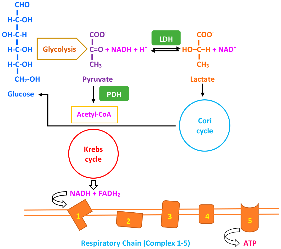

Table 1.
Submission-version active-panel classification and search-derived gene disposition.
| Category | Definition | Panel role |
|---|---|---|
| Tier 1: Core hyperlactatemia genes (n = 40) | Established disorders in which hyperlactatemia or lactic acidosis is characteristic or recurrent and mechanistically and diagnostically informative. | Core diagnostic panel |
| Tier 2: Extended diagnostic genes (n = 26) |
Established disorders in which lactate elevation is clinically relevant but variably expressed, age- or genotype-dependent, or phenotype-restricted. | Extended diagnostic panel |
| Tier 3: Context-dependent or secondary associations (n = 19) | Established disorders in which lactate elevation occurs predominantly during metabolic decompensation or physiological stress rather than as a consistent primary biochemical feature. | Extended differential-diagnosis panel |
| Search-derived genes not retained in active panel (n = 206) | Recovered by one or more phenotype searches but not retained in the submission-version active panel. The recovered raw data do not support assignment to the historical manual exclusion categories. | Audit and future reanalysis; not included in active panel |
Note: Submission-version reconciliation comprises 85 active genes (40 Tier 1, 26 Tier 2 and 19 Tier 3) plus 206 search-derived genes not retained in the active panel, for 291 unique genes represented in the submission-version dataset. Supplementary Table S2A provides the 85-gene active panel; Supplementary Table S2B provides all 240 recovered phenotype-search genes, including the 34 active overlaps and 206 non-active search-derived genes. The 206 genes are retained for audit and future reanalysis; no historical exclusion category is inferred from the recovered raw data.
Table 2.
Mechanistically representative genes associated with inherited metabolic causes of hyperlactatemia.
Table 2.
Mechanistically representative genes associated with inherited metabolic causes of hyperlactatemia.
| Mechanistic category | Representative genes | Representative disorders/phenotypes | Principal mechanism of lactate elevation | Ref. |
|---|---|---|---|---|
| Mitochondrial pyruvate transport | MPC1 | Mitochondrial pyruvate-carrier deficiency | Impaired mitochondrial pyruvate entry reduces oxidative utilisation and redirects pyruvate toward lactate. | [37,38] |
| Pyruvate dehydrogenase complex | PDHA1, PDHB, DLAT, PDHX | Primary PDH deficiency | Impaired conversion of pyruvate to acetyl-CoA causes accumulation of pyruvate, alanine, and lactate. | [16,32,39,40,41,42] |
| Mitochondrial lipoylation | LIAS, LIPT2 | Lipoylation defects / multiple mitochondrial dehydrogenase deficiency | Defective lipoylation impairs pyruvate and α-ketoglutarate dehydrogenase activity. | [43,44,45,46] |
| Pyruvate carboxylation | PC | Pyruvate carboxylase deficiency | Impaired conversion of pyruvate to oxaloacetate compromises anaplerosis and gluconeogenesis, promoting pyruvate-to-lactate conversion. | [47,48,49] |
| Gluconeogenesis and hepatic glucose production | FBP1, G6PC1, SLC37A4 | Fructose-1,6-bisphosphatase deficiency; GSD I | Impaired glucose production reduces lactate utilisation and promotes lactate accumulation, particularly during fasting. | [34,50] |
| Respiratory-chain and oxidative-phosphorylation dysfunction | ACAD9, NDUFV1, SURF1, TMEM70, LRPPRC | Complex I, complex IV, and ATP-synthase disorders | Impaired oxidative phosphorylation alters mitochondrial redox balance and ATP production, increasing reliance on glycolysis and conversion of pyruvate to lactate. | [51,52,53,54,55,56] |
| Mitochondrial metabolite and nucleotide transport | SLC25A3, SLC25A4 | Phosphate-carrier and adenine-nucleotide translocator disorders | Impaired substrate or ADP/ATP transport compromises oxidative phosphorylation and mitochondrial energy production. | [57,58,59,60] |
| Coenzyme Q biosynthesis | COQ4 | Primary coenzyme Q deficiency | Reduced electron transfer through the respiratory chain impairs oxidative phosphorylation and promotes secondary lactate accumulation. | [61,62] |
| Mitochondrial DNA maintenance and depletion | FBXL4, POLG, SUCLG1, TWNK | mtDNA depletion and maintenance disorders | mtDNA depletion, instability, or defective replication causes secondary respiratory-chain failure. | [63,64,65,66,67,68] |
| Mitochondrial translation | DARS2, EARS2, YARS2, MTO1, PUS1, TUFM, TSFM | Combined oxidative-phosphorylation deficiencies, mitochondrial aminoacyl-tRNA synthetase disorders, and translation-factor defects | Impaired synthesis or maturation of mtDNA-encoded respiratory-chain proteins causes combined oxidative-phosphorylation dysfunction. | [69,70,71,72,73,74,75,76,77,78,79,80,81,82,83] |
| TCA-cycle and related mitochondrial metabolism | OGDH, ECHS1 | OGDH-related disease; ECHS1 deficiency | Impaired TCA-cycle metabolism or toxic metabolite accumulation reduces oxidative metabolism and may secondarily inhibit pyruvate utilisation. | [84,85,86,87] |
| Iron–sulphur cluster and mitochondrial protein function | ISCU, TOMM70* | ISCU myopathy; TOMM70-related mitochondrial disease (emerging association) | Defective Fe–S-dependent enzymes or mitochondrial protein import compromises respiratory-chain and TCA-cycle function. | [88,89,90,91] |
| Mitochondrial DNA-encoded OXPHOS disorders | MT-ND1, MT-ND5, MT-ATP6 | Leigh syndrome, MELAS-spectrum disease, NARP and related mitochondrial disorders | Pathogenic mtDNA variants impair respiratory-chain or ATP-synthase function, promoting redox-driven lactate accumulation. | [92,93,94,95,96,97] |
| Mitochondrial tRNA disorders | MT-TL1 | MELAS and related mitochondrial disease | Defective mitochondrial translation causes multisystem oxidative-phosphorylation dysfunction and recurrent lactic acidosis. | [98,99] |
Table 2 presents representative examples across major biochemical mechanisms while limiting redundancy within genetically heterogeneous pathways. It is not exhaustive and should not be used as a substitute for evidence-tier assignment; version-specific assignments and evidence sources are provided in Supplementary Table S2. TOMM70 is not retained in the submission-version active panel and is shown here only as an emerging mechanistic literature example. Abbreviations: ADP, adenosine diphosphate; ATP, adenosine triphosphate; CoQ, coenzyme Q; CSF, cerebrospinal fluid; Fe–S, iron–sulphur; GSD I, glycogen storage disease type I; MELAS, mitochondrial encephalomyopathy, lactic acidosis, and stroke-like episodes; mtDNA, mitochondrial DNA; NARP, neuropathy, ataxia, and retinitis pigmentosa; OXPHOS, oxidative phosphorylation; PDH, pyruvate dehydrogenase; TCA, tricarboxylic acid.
Table 3.
Phenotype- and biochemical-pattern framework for diagnostic evaluation of suspected inherited metabolic hyperlactatemia.
Table 3.
Phenotype- and biochemical-pattern framework for diagnostic evaluation of suspected inherited metabolic hyperlactatemia.
| Clinical and biochemical pattern | Representative inherited disorders and diagnostic considerations | Suggested additional investigations |
|---|---|---|
| Persistent or recurrent hyperlactatemia with an increased L/P ratio | Respiratory-chain and oxidative-phosphorylation disorders; Leigh syndrome spectrum; MELAS; mitochondrial DNA maintenance and translation disorders; primary coenzyme Q₁₀ deficiency | Paired lactate and pyruvate measurements; blood gas; glucose and ketones; plasma amino acids; urine organic acids; acylcarnitine profile; CK; liver and renal biochemical assessment; genomic testing |
| Hyperlactatemia with a normal or only mildly increased L/P ratio | Mitochondrial pyruvate-carrier deficiency; primary PDH deficiency; mitochondrial lipoylation defects | Paired lactate and pyruvate measurements; blood gas; glucose and ketones; plasma amino acids; urine organic acids; brain MRI; genomic testing; enzyme/functional studies |
| Severe neonatal or infantile lactic acidosis with hypoglycaemia, hyperammonaemia and neurological dysfunction | PC deficiency; other anaplerotic or multiple-carboxylase disorders should be considered according to the accompanying biochemical findings | Blood gas, glucose, lactate, pyruvate, ammonia and ketones; plasma amino acids; urine organic acids; acylcarnitine profile; biotinidase activity; CSF lactate and pyruvate; genomic testing; enzyme studies. |
| Hyperlactatemia with developmental delay, epilepsy, encephalopathy, stroke-like episodes or movement disorder | Leigh syndrome spectrum; MELAS; PDH deficiency; POLG-related disease; mitochondrial respiratory-chain, translation, and mtDNA-maintenance disorders | Brain MRI, including diffusion-weighted imaging and MR spectroscopy; EEG; plasma amino acids; urine organic acids; acylcarnitine profile; CK; CSF lactate and pyruvate; nuclear genomic and mtDNA analysis. |
| Hyperlactatemia with myopathy, exercise intolerance, rhabdomyolysis or cardiomyopathy | Mitochondrial myopathies and respiratory-chain disorders; FAODs, including ACADM- and ACADVL-related disease; MADD; mitochondrial membrane and substrate-transport disorders | CK; glucose and ketones; plasma acylcarnitines; free and total carnitine; urine organic acids; liver biochemical assessment; ECG and echocardiography; cardiac biomarkers; phenotype-directed genomic testing. |
| Hyperlactatemia with hypoketotic hypoglycaemia | MCADD (ACADM); VLCADD (ACADVL); multiple acyl-CoA dehydrogenase deficiency (ETFA, ETFB, ETFDH); other FAODs or ketogenesis disorders | Glucose, β-hydroxybutyrate, free fatty acids, lactate, blood gas and ammonia; plasma acylcarnitines; free and total carnitine; urine organic acids and acylglycines; CK; functional/genomic testing |
| Fasting-induced ketotic hypoglycaemia and lactic acidosis | Fructose-1,6-bisphosphatase deficiency (FBP1) and other disorders of gluconeogenesis | Glucose, lactate, pyruvate, ketones and blood gas; plasma amino acids; urine organic acids; genomic testing |
| Persistent or recurrent hyperlactatemia with fasting intolerance, hepatomegaly, hyperuricaemia or hypertriglyceridaemia | GSD I caused by G6PC1 or SLC37A4 pathogenic variants | Serial glucose and lactate; triglycerides, cholesterol and uric acid; blood gas; liver and renal biochemical assessment; urine albumin-to-creatinine ratio; abdominal imaging; nutritional assessment; targeted testing of G6PC1 and SLC37A4. |
| Hyperlactatemia with high-anion-gap metabolic acidosis, ketosis, hyperammonaemia or acute neonatal metabolic decompensation | Propionic acidemia (PCCA, PCCB); methylmalonic acidemia (MMUT and intracellular cobalamin disorders); isovaleric acidemia (IVD); other organic acidemias | Ammonia, blood gas, glucose, lactate and ketones; plasma acylcarnitines and amino acids; urine organic acids; methylmalonic acid; total homocysteine; confirmatory genomic, enzyme studies. |
| Episodic hyperlactatemia precipitated by fasting, infection, surgery, pregnancy or strenuous exercise | Attenuated mitochondrial disease; partial pyruvate-metabolism defects; FAODs; gluconeogenic disorders; other later-onset IMDs | Lactate, pyruvate, blood gas, glucose, ketones, ammonia, plasma amino acids, acylcarnitines and urine organic acids; exome/genome sequencing according to the phenotype. |
| Hyperlactatemia with multisystem neurological, cardiac, hepatic, renal, endocrine, ophthalmological or auditory involvement | mtDNA disorders; nuclear-encoded respiratory-chain, mitochondrial translation and mtDNA-maintenance disorders; other multisystem IMDs | Brain and organ-directed imaging; ECG and echocardiography; ophthalmological and audiological assessment; endocrine, hepatic and renal evaluation; nuclear genomic sequencing combined with mtDNA analysis. |
| Persistent unexplained hyperlactatemia after an appropriate first-line biochemical evaluation | Rare or newly recognised mitochondrial and metabolic disorders; atypical presentations of established IMDs; mtDNA sequence variants, rearrangements or low-level mosaic variants; non-genetic causes not identified during the initial assessment | Exome or genome sequencing with dedicated mtDNA analysis; copy-number and structural variants; RNA analysis, enzyme assays, metabolomics, and tissue studies. |
Abbreviations: CK, creatine kinase; CoQ₁₀, coenzyme Q₁₀; CSF, cerebrospinal fluid; ECG, electrocardiogram; EEG, electroencephalogram; FAODs, fatty acid oxidation disorders; IMD, inherited metabolic disorder; L/P ratio, lactate-to-pyruvate; MADD, multiple acyl-CoA dehydrogenase deficiency; MCADD, medium-chain acyl-CoA dehydrogenase deficiency; MELAS, mitochondrial encephalomyopathy, lactic acidosis and stroke-like episodes; MR, magnetic resonance; MRI, magnetic resonance imaging; mtDNA, mitochondrial DNA; NADH, reduced nicotinamide adenine dinucleotide; OXPHOS, oxidative phosphorylation; PDH, pyruvate dehydrogenase; PC, pyruvate carboxylase.
Disclaimer/Publisher’s Note: The statements, opinions and data contained in all publications are solely those of the individual author(s) and contributor(s) and not of MDPI and/or the editor(s). MDPI and/or the editor(s) disclaim responsibility for any injury to people or property resulting from any ideas, methods, instructions or products referred to in the content. |
© 2026 by the authors. Licensee MDPI, Basel, Switzerland. This article is an open access article distributed under the terms and conditions of the Creative Commons Attribution (CC BY) license (http://creativecommons.org/licenses/by/4.0/).
Copyright: This open access article is published under a Creative Commons CC BY 4.0 license, which permit the free download, distribution, and reuse, provided that the author and preprint are cited in any reuse.