
Submitted:
28 October 2026
Posted:
29 October 2026
You are already at the latest version
Abstract
Polycystic ovary syndrome (PCOS) is a heterogeneous multisystem disorder characterized by reproductive, metabolic, and neuroendocrine abnormalities. Current management primarily targets individual manifestations through lifestyle modifications and pharmacological therapy, while growing evidence implicates altered autonomic and neuroendocrine regulation in PCOS pathophysiology. Electroceuticals (ECs) and neuromodulatory interventions may represent potential non-pharmacological approaches for modulating neural pathways involved in reproductive and metabolic regulation. This review examines the biological rationale, experimental evidence, and available clinical findings supporting the investigation of such interventions in PCOS. From a biomedical perspective, ECs may offer potential advantages in PCOS management; however, the limited evidence currently available precludes definitive conclusions regarding their short- and long-term therapeutic effects and potential adverse effects in humans. Therefore, further specialized preclinical and clinical studies are warranted to evaluate the safety and efficacy of this promising approach.
Keywords:
polycystic ovary syndrome
; electroceuticals
; autonomic nervous system
; neuroendocrine regulation
1. Introduction
Polycystic ovary syndrome (PCOS) affects an estimated 6–13% of women of reproductive age globally, depending on diagnostic criteria, making it one of the most common women’s health disorders (Teede et al. 2023; World Health Organization 2026). PCOS is a heterogeneous disorder involving reproductive abnormalities, metabolic dysfunction, and neuroendocrine imbalance. Hallmark features include irregular menstruation, anovulation, hyperandrogenism, insulin resistance (IR), obesity, and an increased risk of type 2 diabetes and cardiovascular disease (Teede et al. 2023; Yu et al. 2024).
Conventional management strategies for PCOS focus on symptom control using hormonal contraceptives, insulin sensitizers, and lifestyle modifications. Although these interventions can effectively address specific clinical manifestations, treatment response and adherence vary, and individual therapies generally target selected reproductive or metabolic features rather than the full multisystem pathophenotype of PCOS (Teede et al. 2023). Increasing recognition of PCOS as a disorder of integrated control systems rather than isolated ovarian pathology has prompted interest in alternative therapeutic paradigms, particularly those addressing autonomic nervous system dysregulation, including sympathetic hyperactivity and reduced parasympathetic tone (Lansdown and Rees 2012; Yu et al. 2024).
Electroceuticals (ECs), recognized as tools for bioelectronic medicines, represent a novel class of interventions that target neural and sometimes muscular or vascular signaling to possibly modulate organ function (Famm et al. 2013). This technology can be classified into various types based on relevant characteristics, including the energy source, the mode of intervention, and the ultimate target, with the crucial requirement being that it produces a significant and reproducible biological/therapeutic effect. Because neural pathways contribute to endocrine, metabolic, inflammatory, and ovarian regulation, neuromodulatory interventions provide a biologically plausible framework for investigating whether targeted modification of neural activity can influence selected PCOS phenotypes (Pikov et al. 2018; Zhang et al. 2023). Accordingly, this review examines the biological rationale for electroceutical and neuromodulatory approaches in PCOS, summarizes available clinical and preclinical evidence, and discusses key limitations and priorities for future investigation.
Search Strategy and Scope of the Review
This article was not designed or reported as a systematic review or meta-analysis. A structured literature search was performed in PubMed, Embase, Scopus, Web of Science, Google Scholar, and ClinicalTrials.gov to identify relevant studies. The search strategy combined Medical Subject Headings (MeSH), where applicable, with free-text keywords using Boolean operators. The keywords included PCOS and related terms (definition, symptoms, epidemiology, diagnostic features, treatments, and history); ECs and related terms (definition, types, therapeutic applications, history, and U.S. Food and Drug Administration [FDA]-approved applications); neural control of endocrine function; bioelectricity and related biological pathways; ECs and PCOS; obesity; diabetes; brain-derived neurotrophic factor (BDNF); and the advantages and disadvantages of ECs. Original research articles, clinical studies, randomized and non-randomized trials, observational studies, systematic reviews, and high-quality narrative reviews eligible for inclusion. Literature that was irrelevant to the review topic, letters, and duplicate reports were excluded.
2. Neuroendocrine and Metabolic Pathophysiology of PCOS
PCOS, or Stein–Leventhal syndrome (SLS), is one of the most common endocrine disorders affecting women of reproductive age worldwide (Sati et al. 2023). Reported PCOS burden has increased over recent decades, although estimates vary substantially according to population characteristics, study methodology, and diagnostic criteria (AlHussain et al. 2020; Che et al. 2023; Saei Ghare et al. 2023; Hameed et al. 2024; Salari et al. 2024; Vatier and Christin-Maitre 2024). However, variation in reported PCOS prevalence estimates is attributable to differences in study methods, demographic characteristics, including genetics and living and nutritional conditions, and the diagnostic criteria (Sutaji et al. 2022; Luo et al. 2024; Pinto et al. 2024; Sikarwar and Kazim 2024; Talebi et al. 2023). PCOS is a major cause of anovulatory infertility (Hossain et al. 2013; Khan et al. 2019; Nautiyal et al. 2022; Guan et al. 2023; Elnashar 2024; Huang et al. 2024a). However, estimates of its contribution to infertility depend on the population studied and the definition of infertility used. Therefore, any reported percentage should be linked to the specific population and type of infertility evaluated in the cited source.
Chronic anovulation, hyperandrogenism, and the exclusion of other causes of infertility through differential diagnosis, together with polycystic ovarian morphology, have been proposed as the main criteria for diagnosing PCOS (Gleicher et al. 2022; Nautiyal et al. 2022; Siddiqui et al. 2022). Confirmation of two of the three Rotterdam diagnostic criteria is used as the diagnostic basis for PCOS in current clinical practice. According to the 2023 International Evidence-based Guideline for the Assessment and Management of PCOS, polycystic ovarian morphology in adults is generally defined by the presence of at least 20 follicles in one ovary when high-quality ultrasound imaging and adequate follicle counting are available. When accurate follicle counting is not feasible, an ovarian volume of at least 10 mL or other guideline-specified criteria may be used. These ultrasound findings should be interpreted within the overall diagnostic framework for PCOS and should not be considered diagnostic in isolation (Coutinho and Kauffman 2019; Barlampa et al. 2021; Bai et al. 2022; Dybciak et al. 2023; Nikrad et al. 2023; Teede et al. 2023; Bai et al. 2024; Huang et al. 2024b).
The multisystemic nature of PCOS includes the reproductive, nervous, endocrine, immune, and metabolic systems (Sun et al. 2020; Gunn 2022) (Figure 1). Therefore, the numerous but interrelated clinical manifestations and disorders associated with this syndrome include hyperinsulinemia and glucose intolerance (Legro 2012; Behboudi-Gandevani et al. 2018), dyslipidemia (Wild et al. 2011), hypertension (Andrade et al. 2016), chronic inflammation (Zhou et al. 2024), immune dysfunction (Wang et al. 2023), endometrial hyperplasia (AlHussain et al. 2020), gut dysbiosis (Cardinale et al. 2022), neuroendocrine impairment (Zhang et al. 2022), hyperandrogenism (Luo et al. 2024), hyperestrogenism (Yang and Chen, 2024; Stener-Victorin et al. 2009), defective ovulation and ovulatory failure (Blasco et al. 2019), chronic anovulation (Esparza et al. 2020), luteinizing hormone (LH) hyperpulsatility (Esparza et al. 2020), disruption of hypothalamic–pituitary–ovarian (HPO) function (Witchel et al. 2019), abnormalities in energy expenditure (Laven 2019), and mitochondrial dysfunction (Zhang et al. 2022). Eating, mood, and sleep disorders, and psychological problems have been reported in these patients (Khadilkar 2016; Barlampa et al. 2021; Dybciak et al. 2023; Vatier and Christin-Maitre 2024). Thus, PCOS can affect not only the ovaries and reproductive system but also the liver, pancreas, intestines, adipose tissue, brain, and even the skin and hair.
This multifactorial syndrome involves numerous genetic, epigenetic, and environmental factors, and many of the causative factors and pathological pathways underlying this condition remain unknown (Saei Ghare Naz et al. 2019; Nautiyal et al. 2022). Neuroendocrine dysregulation, including involvement of the HPO axis, has been reported in PCOS, where increased gonadotropin-releasing hormone (GnRH) pulse frequency leads to increased LH secretion, resulting in ovarian androgen overproduction and impaired follicular development. Another consequence of of this axis dysfunction is altered follicle-stimulating hormone (FSH) levels. Together with increased LH levels, these changes may contribute to ovarian theca-cell hyperplasia, alterations in ovarian follicular development, cyst formation, and anovulation, as well as excessive production of estrogen and androgens, subsequent hirsutism, alterations in insulin, leptin, and adiponectin levels, increased anti-Müllerian hormone (AMH), IR, and decreased sex hormone-binding globulin (Coutinho and Kauffman 2019; Liao et al. 2021; Sarahian et al. 2021; Barbotin et al. 2023; Ruddenklau and Campbell 2019; Sati et al. 2023; Bai et al. 2024). Autonomic nervous system imbalance, characterized by increased sympathetic activity and decreased parasympathetic tone, may contribute to ovarian dysfunction, IR, and chronic inflammation. Hyperinsulinemia may further contribute to a self-reinforcing pathological loop by increasing androgen synthesis.
Another important pathological mechanism in PCOS is primary or secondary inflammation, which is associated with increased levels of inflammatory mediators such as tumor necrosis factor alpha (TNF-α), interleukin (IL)-1, and IL-6, as well as hyperinsulinemia, hyperandrogenism, increased oxidative stress, and nuclear factor kappa B (NF-κB) activation. These alterations may contribute to the development of IR, apoptosis, and infertility and may increase the susceptibility of patients with PCOS to infections and autoimmune diseases (Liu et al. 2020; Huffman et al. 2021; Sarahian et al. 2021; Bai et al. 2022; Gao et al. 2022; Gunn 2022; Radenović et al. 2022; Siddiqui et al. 2022; Geng et al. 2023; Mukherjee et al. 2023; Nikrad et al. 2023; Wang et al. 2023; Bai et al. 2024; Chen et al. 2024; Huang et al. 2024a; Juber et al. 2024; Luo et al. 2024).
It should be noted that no definitive treatment completely halts the progression of PCOS, and current management strategies are primarily symptomatic rather than etiologic. These include lifestyle modifications, traditional medicine, pharmacological treatments, particularly hormonal therapy and insulin-related drugs such as metformin, surgical interventions, psychological counseling, assisted reproductive technologies, and novel emerging approaches, including gene and immunotherapy (Hosseinkhani et al. 2017; Takasaki et al. 2018; Hassan et al. 2021; Franik et al. 2022; Lu et al. 2022; Han et al. 2023; Liu et al. 2023; Mohammad 2023; Saei Ghare Naz et al. 2023; Sati et al. 2023; Szczesnowicz et al. 2023; Calcaterra et al. 2024; Chico-Sordo and García-Velasco 2024; Melin et al. 2024; Yang et al. 2024; Muhammed Saeed et al. 2025). There is an urgent need to develop and investigate innovative treatments for PCOS. Given its multisystem nature and interconnected neural and endocrine pathways, PCOS may be amenable to neuromodulatory interventions.
2.1. Electroceuticals
Bioelectrical signaling contributes to the function of excitable tissues and to numerous cellular processes, including membrane-potential regulation, ion transport, neurotransmission, intercellular communication, cellular detoxification, production and secretion of strategic cellular molecules such as proteins, hormones and neurotransmitters (NTs). Therapeutic electrical or electromagnetic stimulation can therefore modify neural activity and, depending on the target and stimulation parameters, influence downstream physiological processes (Tuszynski et al. 2017; Huang et al. 2004; Kulbacka et al. 2017; Levin et al. 2019; Speer et al. 2019; Martin et al. 2020; Sen et al. 2020; Levin 2021; Zhang et al. 2021; Lu et al. 2023). Therefore, sometimes by strengthening or modifying this electrical current from the outside, effective therapeutic interventions may be performed for possible improve of human health.
ECs is the knowledge of stimulations and interventions on the bioelectricity, intercellular signaling and neurohormonal circuits of a living being using magnetic, electric, and ultrasound currents that can be used in the form of implantation or application of non-permanent currents (Bok et al. 2023; Balasubramanian et al. 2024; Jo et al. 2024; Perez et al. 2025) that include vagus nerve stimulation (VNS), cranial electrotherapy stimulation (CES), transcranial direct current stimulation (tDCS), transcranial alternating current stimulation (tACS), transcutaneous vagus nerve stimulation (tVNS), electroconvulsive treatment (ECT), and chronic deep brain stimulation (DBS) (Bok et al. 2023; Perez et al. 2025); single transcranial magnetic stimulation (TMS), paired-pulse TMS, repetitive transcranial magnetic stimulation (rTMS), infrared, electromagnetic field (EMF), repeated electromagnetic field stimulation (REMFS) (Tsai and Hamblin 2017; Brunyé et al. 2021; Perez et al. 2021; Bok et al. 2023; Jung et al. 2023; Jo et al. 2024; Perez et al. 2025). Studies have shown the possible effectiveness of this technology in the clinical field of neurological, metabolic, and inflammatory disorders (Jung et al. 2023; Zouali et al. 2023; Mishra et al. 2024); therefore, when the factors involved in PCOS are affected by this approach, it may also be used as a therapeutic strategy in this syndrome itself.
Given the increased sympathetic nerve activity in PCOS, which has consequences such as increased androgen production by theca cells, impaired follicular maturation, and ultimately ovarian stromal hypertrophy; a rationale may be provided for investigating whether EC interventions that reduce sympathetic activity or enhance parasympathetic signaling can influence ovarian function in PCOS (Stener-Victorin et al. 2009; Lansdown and Rees, 2012; Zhang et al. 2023). This has been shown in studies conducted using neuromodulation methods as well as electroacupuncture (EA), which have led to a decrease in sympathetic activity and expression of nerve growth factor (Stener-Victorin et al., 2000; Bai et al. 2004; Stener-Victorin et al. 2009; Zhang et al. 2023).
Another pathway involved in this syndrome was changes in the HPO axis, which led to increased LH production, increased ovarian androgen production, and anovulation (McCartney et al. 2022). Neuromodulation, EA and ECs may influence gonadotropin secretion and ovulatory function closer to their physiological state by affecting hypothalamic neuronal networks affecting GnRH, including kisspeptin, neurokinin B, and dynorphin (KNDy) neurons, which are disturbed in PCOS; however, direct evidence that these interventions normalize gonadotropin secretion in women with PCOS remains limited (McCartney et al. 2022; Xu et al. 2023).
Possible control of IR, HbA1c, and diabetes, factors involved in PCOS, with ECs and EA is one of the areas where many clinical and preclinical studies have been conducted on this subject, and this control can reduce metabolic stress on ovarian tissue (Stener-Victorin et al. 2016; Benrick et al. 2017; Peng et al. 2020a; Li et al. 2026).
Given the involvement of the HPA pathway in PCOS, activation of the cholinergic anti-inflammatory pathway, especially through vagal stimulation, may reduce the production and release of pro-inflammatory cytokines and modulate immune cell activity, and may also be effective in restoring metabolic-reproductive balance by restoring cortisol secretion to baseline and finally possibly reversing HPA overactivity (Pavlov and Tracey 2005; Pasquali et al. 2012; Bonaz et al. 2016).
2.2. Neurovascular, Anatomical, and Immune Connectivity of the Ovary in PCOS
2.2.1. Ovarian Vascular and Anatomical Connectivity
The ovary is mainly supplied by the ovarian arteries along with the uterine arteries and utero-ovarian anastomoses (Figure 2). The suspensory ligament, containing ovarian vessels, autonomic nervous networks, and lymphatic vessels, connects the ovary to the pelvic wall, while the ovarian ligament provides a medial connection with the uterus (Ouyang et al. 2012; Gibson and Mahdy 2023). Vascular abnormalities such as increased stromal blood vessels and decreased ovarian vascular resistance (Aleem and Predanic 1996; Ozkan et al.2007), irregular ovarian angiogenesis including abnormalities related to VEGF and other angiogenic mediators (Tal et al. 2015; Di Pietro et al. 2018) have been reported in PCOS, all of which can affect ovarian structure and function.
2.2.2. Ovarian Innervation and Neuroendocrine Connectivity
The autonomic nervous system, including sympathetic and parasympathetic, is primarily responsible for ovarian innervation (Figure 2). Studies have reported that sympathetic hyperactivity in PCOS is manifested by increased norepinephrine content and release and altered beta-adrenergic responsiveness, which may affect ovarian steroidogenesis and follicular function (Barria et al. 1993; Lara et al. 1993). On the other hand, increased systemic sympathetic nervous activity, altered cardiac autoregulation, and impaired post-exercise recovery in patients with this syndrome are associated with increased systemic sympathetic activity (Lansdown and Aled Rees 2012; Duval et al. 2023).
2.2.3. Immune and Humoral Connectivity
The immune–endocrine microenvironment appears to be disrupted in PCOS, as studies have shown increased circulating inflammatory mediators, including IL-1β, IL-6, IL-17A/F, IL-18, TNF-α, MCP-1, and C-reactive protein, along with alterations in immune cell populations such as Th17 and regulatory T cells in this syndrome, consistent with similar inflammatory changes in the ovarian microenvironment, and these two groups of findings suggest a link between ovarian inflammation and systemic inflammatory conditions (Zhai and Pang 2022; Banerjee et al. 2023). The result of these inflammatory changes could be diagnostic features of PCOS, such as hyperandrogenism, IR, and adipose tissue dysfunction, abnormal folliculogenesis, and anovulation. Because autonomic pathways can modulate systemic inflammatory signaling, neuroimmune mechanisms represent an additional hypothesis through which neuromodulation might influence PCOS-related inflammation. Direct demonstration of this mechanism in women with PCOS is currently lacking.
Given the material discussed in section 5, it may be possible to arrive at an acceptable biological framework for investigating targeted neuromodulation and ECs as therapeutic strategies in PCOS. However, given the limited direct studies on the clinical effects of electrical stimulation of peripheral nerve pathways and EC methods on the ovary, and especially clinical cases of PCOS, it is currently not possible to make a definitive statement about the effectiveness of this approach in PCOS, and more precise research is required.
3. Discussion: Experimental and Clinical Evidence Synthesis
The autonomic nervous system contributes to the regulation of ovarian function, including follicular development, steroidogenesis, and ovulation (Astudillo-Guerrero et al. 2025). Experimental studies involving autonomic or uterine nerve denervation have reported alterations in follicular development, estrous cyclicity, and ovarian steroid production (Astudillo-Guerrero et al. 2025). These effects may involve adrenergic signaling, which can influence ovarian blood flow and steroid secretion through autonomic neural pathways (Uchida and Kagitani 2015; Astudillo-Guerrero et al. 2025). However, the effects of autonomic nerve stimulation or denervation depend on the specific neural pathway, experimental conditions, and stimulation parameters. Such findings provide a biological rationale for investigating targeted neuromodulation in PCOS but do not, by themselves, establish its clinical efficacy.
VNS has been investigated in several conditions involving autonomic, metabolic, and neuroendocrine regulation. However, its potential therapeutic role in PCOS remains insufficiently established by direct clinical evidence. Proposed effects on hormonal regulation, reproductive function, insulin resistance, body weight, and mood-related symptoms should therefore be interpreted cautiously, particularly when they are extrapolated from studies in other populations or disease settings (Radhika and Ramesh Babu 2023; Zhang et al. 2023; Stańczak et al. 2024; Li et al. 2025). Further PCOS-specific studies are needed to determine the efficacy, safety, and clinical relevance of VNS in this population.
Transcutaneous auricular vagus nerve stimulation (taVNS) offers a non-invasive alternative to conventional VNS, avoiding surgical implantation and associated postoperative complications. The anti-inflammatory effects of VNS and taVNS are related to the effects of these systems on the hypothalamic-pituitary-adrenal (HPA) axis, the cholinergic anti-inflammatory pathway, and the splenic sympathetic anti-inflammatory pathway (Zhang et al. 2023). Non-invasive CES may modulate HPA axis activity and neurotransmitter systems, potentially modulating cortisol, GABAergic, and acetylcholinergic signaling to regulate central and peripheral nervous system activity and alleviate stress and anxiety (Okano et al. 2025). These technologies should currently be regarded as mechanistically plausible investigational approaches rather than established therapies for PCOS.
EMF-based interventions have also been investigated in relation to PCOS, including studies of pulsed EMF in women with the syndrome (Abd Al Samea et al. 2018; Esmael Hasanin et al. 2019). These reports indicate that clinical investigation of EMF-based approaches in PCOS has been undertaken, although the available evidence remains limited and requires critical evaluation of study design, intervention parameters, comparator groups, and reported outcomes. The findings should be distinguished from those of electroacupuncture and other forms of electrical neuromodulation because the underlying physical modalities and proposed mechanisms differ. Experimental studies have also examined electromagnetic exposures in relation to ovarian function and reproductive outcomes (Mohammed et al. 2021a; Mohammed et al. 2021b; Mohammed et al. 2024). However, the biological effects of electromagnetic exposure depend on factors such as frequency, intensity, waveform, exposure duration, and tissue characteristics. Findings from one exposure protocol cannot therefore be generalized to other electromagnetic interventions. Further studies are needed to establish the efficacy and safety of each modality specifically for reproductive and metabolic outcomes in PCOS. It is worth noting that in a study on PCOS model rats, even transcutaneous electrical muscle stimulation results in a decrease in plasma testosterone, LH, glucose, insulin, and body weight, and an increase in estradiol-progesterone and even catalase (Morsy and Abd-El Hamid 2015). A summary of the potential logic for ECs advantages in PCOS is shown in Figure 3.
3.1. Category 1: Direct Clinical Evidence on PCOS
Direct clinical studies on the effects of neuromodulation and ECs on PCOS patients are very limited and mostly focus on EA. Therefore, it is still too early to draw definitive conclusions about the effectiveness of this technology in this area. However, a study by Jedel et al. in 2011 on 84 women with PCOS showed that 16 weeks of low-frequency EA significantly reduced total circulating testosterone (approximately 25%), as well as decreased androsterone glucuronide and androstane-3α,17β-diol-3-glucuronide, increased the number of menstrual cycles from 0.28 to 0.69 per month, and improved acne (Jedel et al. 2011). An important point in this study is the comparison of the results of the EA group with the groups without intervention and the group with physical exercise. Because the comparator groups consisted of physical exercise and no intervention rather than an electrically matched sham procedure, the study cannot clearly distinguish specific electrical effects from contextual, procedural, or nonspecific treatment effects.
The next randomized controlled study by Johansson et al. 2013 is related to the investigation of the effect of acupuncture incorporating low-frequency electrical stimulation on ovulation in 32 women with PCOS divided into two control and intervention groups (Johansson et al. 2013). During the 10–13-week intervention, a significant increase in ovulation frequency, a decrease in several circulating sex steroids and androgen-related metabolites, including testosterone, free testosterone, dehydroepiandrosterone sulfate (DHEA/DHEAS), and several androgen glucuronides, was reported in the group undergoing EA intervention. So, it is possible that ECs can also affect ovulation rates (Johansson et al. 2013).
The next section is related to the possible effects of EAs on the metabolic parameters of PCOS. In a prospective clinical study on 80 women with PCOS and IR, significant reductions in homeostatic model assessment for IR (HOMA-IR), fasting plasma glucose, and fasting insulin were reported following 6 months of acupuncture combined with manual and low-frequency electrical stimulation (Li et al. 2020). Interpretation is limited due to the absence of a concurrent control group, which prevents separation of treatment effects from temporal, behavioral, or regression-to-the-mean effects.
According to a double-blind randomized clinical trial conducted by Muharam et al. 2022, combining EA with pharmacological therapy can improve insulin sensitivity in women with PCOS, with a significant reduction in HOMA-IR. (Muharam et al. 2022) However, given the limited studies in this section, a definitive conclusion still does not seem very logical.
Obesity is a contributing factor in PCOS and may be a potential therapeutic target for the ECs. A sham-controlled randomized trial in 106 obese women with PCOS showed that 12 weeks of acupuncture compared with sham acupuncture reduced waist-to-hip ratio, body fat mass, and body mass index (BMI). In addition, other factors such as fasting glucose, fasting insulin, and HOMA-IR, total cholesterol, triglycerides, and low-density lipoprotein cholesterol (LDL-C) also improved (Wang et al. 2026).
There has also been a randomized clinical study report on the possible impact of EA on the psyche and quality of life of people with this syndrome, which led to improvements in depression and anxiety in 72 women (Jin et al. 2016). This section also needs more detailed and extensive studies.
Another technique used in the field of neuromodulation and ECs is transcutaneous electrical acupoint stimulation (TEAS). The results of a prospective cohort study on 345 obese women with PCOS examined the difference in the effectiveness of letrozole with or without transcutaneous electrical acupoint stimulation, and the results indicated a positive effect on ovulation-induced pregnancy rates following the combination of the two methods (Su et al. 2023).
3.1. Category 2: PreClinical Evidence on PCOS
Preclinical models are valuable for identifying potential neural and metabolic mechanisms, but experimental PCOS models reproduce selected features of the human syndrome rather than its complete heterogeneous phenotype. Consequently, therapeutic effects in rodents should not be interpreted as evidence of clinical efficacy.
One study that demonstrates the potential of using ECs and neuromodulation to control PCOS beyond the ovarian level, i.e., in the hypothalamic reproductive neuroendocrine circuitry, is Xu et al. 2023 on a letrozole-induced PCOS rat model (Xu et al. 2023). Following low-frequency EA in this animal model, estrous cycles were restored, ovarian morphology improved, circulating testosterone and LH levels were reduced, and even kisspeptin, GnRH, and dynorphin levels in the hypothalamic arcuate nucleus were reduced (Xu et al. 2023). It should be noted that to draw definitive conclusions in this case, it is necessary to repeat several preclinical studies with the specific aim of investigating these points. These findings provide mechanistic support for an effect of EA on hypothalamic reproductive circuitry in this PCOS model, but whether comparable neuroendocrine modulation occurs in women remains unknown.
Improving and increasing ovarian blood supply and innervation, especially through autonomic nerves, could be one of the possible mechanisms of the effectiveness of neuromodulation and ECs techniques in PCOS (Raja-Khan et al. 2011), and this could be an aim for animal models of PCOS research.
Pikov et al. (2018) investigated high-frequency electrical modulation of the superior ovarian nerve (SON) in a rat model of PCOS. Kilohertz-frequency alternating current (KHFAC) was applied to modulate SON activity over a period of 2–3 weeks. The intervention reversed several PCOS-associated abnormalities, including irregular or absent estrous cycles, ovarian cyst formation, reduced numbers of corpora lutea, and increased ovarian norepinephrine concentration. However, fertility capacity was similar between the untreated PCOS and KHFAC-treated PCOS groups. These findings provide preclinical evidence supporting the investigation of targeted ovarian nerve modulation, but they do not establish clinical efficacy in women with PCOS.
Peng et al. 2020 study showed that EA suppressed SREBP1 expression in DHEA-induced PCOS-like rats, which may be effective in improved insulin signaling, reduced IR and metabolic/ovarian dysfunction. (Peng et al. 2020b) On the other hand, in DHEA-induced PCOS-like rats, EA was able to improve IR, mitochondrial dysfunction and endoplasmic reticulum (ER) stress, which is likely EA was associated with inhibition of the mTOR/4E-BP1 pathway (Peng et al. 2020a).
One of the possible mechanisms by which EA improves ovarian morphology and function is through the control of oxidative stress. In the study of Cong et al. 2023, the increased expression of ovarian aminolevulinate synthase 2 (Alas2) in mice with PCOS induced by DHEA was reduced following EA treatment, which may inhibit apoptosis, oxidative stress, and mitochondrial dysfunction in ovarian granulosa cells, suggesting that EA may exert protective effects on ovarian granulosa cells. (Cong et al. 2023).
The metabolic effects of electroacupuncture (EA) have also been investigated in preclinical PCOS models. Huang et al. (2024c) evaluated low-frequency EA in a rat model of PCOS induced by letrozole combined with a high-fat diet, which exhibited insulin resistance and hepatic steatosis. EA reduced fasting blood glucose, fasting insulin, HOMA-IR, triglyceride levels, and free fatty acid levels, and alleviated hepatic steatosis. At the molecular level, EA downregulated hepatic mRNA expression of tumor necrosis factor-alpha (TNF-α) and interleukin-6 (IL-6), while upregulating peroxisome proliferator-activated receptor-alpha (PPAR-α). EA also increased hepatic protein expression of phosphorylated Akt, phosphorylated glycogen synthase kinase-3 beta (GSK3β), and glucose transporter 4 (GLUT4), increased the ratio of phosphorylated GSK3β to total GSK3β, and decreased total GSK3β protein expression. These findings suggest that EA may improve glucose and lipid metabolism and alleviate hepatic steatosis in this experimental PCOS model, potentially involving modulation of hepatic PI3K/Akt insulin-signaling pathways. However, these preclinical findings require further validation and cannot be directly extrapolated to clinical efficacy in women with PCOS.
Although few studies have been conducted, which are mostly focused on EA, future studies are needed to take a more specialized look at the short and long-term effects of this class of technologies in animal models. Despite the limited direct clinical and preclinical studies conducted, this area of ECs in this syndrome has not yet been comprehensively and mechanistically investigated to reveal its true short-term and long-term positive and negative aspects. Therefore, at present, the evidence is insufficient to support EC and neuromodulation interventions as established PCOS treatments. The existing literature instead provides proof-of-concept and mechanistic justification for further investigation, particularly as adjunctive approaches to standard management. The studies reviewed in categories 1 and 2 are summarized in Tables 1 and 2 of Supplementary File S1, respectively.
4. Limitations and Future Perspectives
Safety considerations vary substantially according to the EC modality, anatomical target, stimulation parameters, exposure duration, and whether the intervention is invasive or non-invasive. Among these points is the possibility of EMRs being harmful to the reproductive system and gamete health, which has divided this field into three parts: hazardous, neutral and beneficial [Mohammed et al. 2021a]. According to reports, EMR can lead to apoptosis of ovarian granulosa cells, and there are even cases of the possibility of neurotoxicity of some of the applied EMR frequencies (Mohammed et al. 2021a; Mohammed et al. 2021b). This means the need to know the type of exposure and the current applied, appropriate for minimal damage, duration and frequency of effective and safe exposure, which is the definition of standard treatment protocols (Mohammed et al. 2021b). This point becomes even more prominent when considering the side effects of each type of EC field, which range from surgical complications and infections to respiratory, vocal, skin, and neurological complications (Cabrera et al. 2021; Magisetty and Park 2022; Jung et al. 2023; Yang et al. 2024; Perez et al. 2025). Future studies should systematically evaluate the reproductive, developmental, and long-term safety of electroceutical interventions. Where scientifically justified by the intervention’s mechanism, exposure characteristics, and preclinical findings, potential effects on gamete function and reproductive outcomes should also be investigated. These considerations represent areas for safety assessment rather than evidence of established mutagenic effects. Another point is the possibility of damage to other tissues and organs affected by EC treatments, which should be investigated and recognized in order to seek preventive and protective approaches for other parts.
In today’s advanced world, the competition is for efficient devices with minimum dimensions, weight, energy consumption, manufacturing and maintenance costs, and in everyday applications, perhaps personalization of devices, which devices used in the field of ECs will also be subject to the same law. Another important point in the optimal design of these devices, especially implantable ones, is the use of biodegradable, biocompatible materials, with long battery life, and perhaps in the near future, self-charging batteries, which can overcome one of the main challenges, namely hardware and software problems and constant communication (Magisetty and Park 2022; Bok et al. 2023; Jung et al. 2023; Perez et al. 2025). Easy learning, ease of use, and training of trained medical professionals are other gaps in this medical field. Perhaps the lack of defined protocols for examining real effectiveness and defining how to use this knowledge can be considered other limitations in the present era, but despite all the above, the bright future potential of this new therapeutic approach cannot be ignored. By identifying gaps, limitations, and potential disadvantages in each area more quickly and accurately, we can work to address them and take fundamental steps to improve that sector as quickly as possible.
Future research may explore closed-loop bioelectronic systems in which stimulation parameters are adapted according to physiological biomarkers, potentially including autonomic, hormonal, or metabolic signals. Computational and machine-learning approaches may eventually assist in identifying patient-specific stimulation parameters. Such systems would require rigorous validation, clinically meaningful biomarkers, and careful evaluation of safety and reproducibility. In order to advance this field, interdisciplinary collaboration of neuroscientists, endocrinologists, and bioengineers will be required.
5. Conclusions
PCOS involves interacting reproductive, metabolic, neuroendocrine, autonomic, and inflammatory abnormalities, providing a biologically plausible framework for investigating neuromodulatory interventions. Current direct clinical evidence is limited and predominantly concerns EA and related electrical acupoint stimulation techniques. These studies suggest potential effects on selected androgen-related, metabolic, and reproductive outcomes, while preclinical findings provide additional mechanistic support involving hypothalamic reproductive circuits, insulin signaling, mitochondrial function, oxidative stress, and inflammatory pathways. However, the available evidence does not yet establish ECs as an effective treatment class for PCOS, and findings from EA should not be generalized to other neuromodulatory technologies. Future research should prioritize well-controlled clinical trials, standardized stimulation protocols, mechanistic biomarkers, appropriate sham interventions, and systematic assessment of long-term efficacy and safety. The potential advantages of ECs and neuromodulation are summarized in Figure 3, while Figure 4 provides a summary of the discussion covered in this review article.
Supplementary Materials
The following supporting information can be downloaded at the website of this paper posted on Preprints.org.
Author Contributions
S.M. and I.K. conceived and designed the study. S.M.: Conceptualization, Writing – original draft, Data curation, Methodology, Formal Analysis, Investigation, Visualization, Writing – review & editing. I.K.: Conceptualization, Methodology, Writing – review & editing, Investigation, Formal Analysis, Validation, Resources, Project administration, Supervision. L.J.M.: Visualization, Data curation, Writing – review & editing, and HBS: Conceptualization, Writing – original draft, Writing – review & editing, Supervision, Validation, Funding acquisition, Methodology.
Funding
The authors declare that they did not receive any funds. Razi University and Babylon University provided the facilities and institutional support necessary to conduct this work. Uppsala University provided scientific supervision and financial support for the publication fee. The funders had no role in the study conception, the literature selection, data analysis, interpretation of the findings, or the decision to submit the manuscript for publication.
Institutional Review Board Statement
Not applicable.
Informed Consent Statement
Not applicable.
Data Availability Statement
Supplementary data accompanying this article are available online and include all additional tables and figures.
Acknowledgments
The authors sincerely thank Razi University and Uppsala University for their support.
Conflicts of Interest
The authors declare that the research was conducted without any commercial or financial relationships that could be construed as a potential conflict of interest.
References
- World Health Organization. Polycystic ovary syndrome. (2026) https://www.who.int/news-room/fact-sheets/detail/polycystic-ovary-syndrome.
- Teede HJ, Tay CT, Laven J, Dokras A, Moran LJ, Piltonen TT, et al. Recommendations from the 2023 international evidence-based guideline for the assessment and management of polycystic ovary syndrome. J. Clin. Endocrinol. Metab. 2023; 108:2447–2469. [CrossRef]
- Yu Y, Chen T, Zheng Z, Jia F, Liao Y, Ren Y, et al. The role of the autonomic nervous system in polycystic ovary syndrome. Front Endocrinol (Lausanne). 2024;14:1295061. [CrossRef]
- Lansdown A, Rees DA. The sympathetic nervous system in polycystic ovary syndrome: a novel therapeutic target. Clin Endocrinol (Oxf). 2012;77:791-801. [CrossRef]
- Famm K, Litt B, Tracey KJ, Boyden ES, Slaoui M. Drug discovery: a jump-start for electroceuticals. Nature. 2013;496:159–161. [CrossRef]
- Pikov V, Sridhar A , Lara HE. High-frequency electrical modulation of the superior ovarian nerve as a treatment of polycystic ovary syndrome in the rat. Front Physiol. 2018;9:459. [CrossRef]
- Zhang S, He H, Wang Y, Wang X, Liu X. Transcutaneous auricular vagus nerve stimulation as a potential novel treatment for polycystic ovary syndrome. Sci Rep. 2023;13:7721. [CrossRef]
- Sati A, Desroziers E, Campbell RE. Microglia: Sculptors of the polycystic ovary syndrome (PCOS)- like brain? Reproduction. 2023;166:R51–R61. [CrossRef]
- AlHussain F, AlRuthia Y, Al-Mandeel H, Bellahwal A, Alharbi F, Almogbel Y, et al. Metformin improves the depression symptoms of women with polycystic ovary syndrome in a lifestyle modification program. Patient. Prefer. Adherence. 2020;14:737–746. [CrossRef]
- Che Y, Yu J, Li Y.Sh, Zhu Y.C, Tao T. Polycystic ovary syndrome: challenges and possible solutions. J. Clin. Med. 2023;12:1500. [CrossRef]
- Saei Ghare Naz M, Rahnemaei F, Ramezani F, Sayehmiri F, Ghasemi V, Banaei M, et al. Possible cognition changes in women with polycystic ovary syndrome: a narrative review. Obstet. Gynecolo. Sci. 2023;66:347-363. [CrossRef]
- Salari N, Nankali A, Ghanbari AH, Jafarpour S, Ghasemi H, Dokaneheifard S, et al. Global prevalence of polycystic ovary syndrome in women worldwide: a comprehensive systematic review and meta-analysis. Arch. Gynecolo. Obstet. 2024;310:1303-13014. [CrossRef]
- Hameed L, Khan MN, Dar Farooq A, Muhammad Siddiqui SM. Knowledge and prevalence of polycystic ovarian syndrome among undergraduate female students in Karachi. J. Popul. Ther. Clin. Pharmacol. 2024;31:588-594. [CrossRef]
- Vatier C, Christin-Maitre S. Epigenetic/circadian clocks and PCOS. Hum. Reprod. 2024;39:1167–1175. [CrossRef]
- Sutaji Z, Elias MH, Ahmad MF, Karim AKA, Abu MA. A systematic review and integrated bioinformatic analysis of candidate genes and pathways in the endometrium of patients with polycystic ovary syndrome during the implantation window. Front. Endocrinol. (Lausanne). 2022;13:900767. [CrossRef]
- Talebi S, Zeraattalab-Motlagh S, Jalilpiran Y, et al. The effects of pro-, pre-, and synbiotics supplementation on polycystic ovary syndrome: an umbrella review of metaanalyses of randomized controlled trials. Front. Nutr. 2023;10:1178842. [CrossRef]
- Luo X, Dong Y, Zheng H, Zhou X, Rong L, Liu X, et al. CAPN2 correlates with insulin resistance states in PCOS as evidenced by multi-dataset analysis. J. Ovarian. Res. 2024;17:79. [CrossRef]
- Pinto J, Cera N, Pignatelli D. Psychological symptoms and brain activity alterations in women with PCOS and their relation to the reduced quality of life: a narrative review. J. Endocrinol. Invest. 2024;47:1599–1620. [CrossRef]
- Sikarwar NS, Kazim F. Assessing the prevalence and implications of PCOS in women: a comprehensive study. Eur. J. Cardiovasc. Med. 2024;14:487-494.
- Hossain M, Cao M, Wang Q, Kim J.Y, Schellander K, Tesfaye D, Tsang B.K. Altered expression of miRNAs in a dihydrotestosterone-induced rat PCOS model. J. Ovarian. Res. 2013;6:36. [CrossRef]
- Khan MJ, Ullah A, Basit S. Genetic basis of polycystic ovary syndrome (PCOS): current perspectives. Appl. Clin. Genet. 2019;12:249–260. [CrossRef]
- Nautiyal H, Imam SS, Alshehri S, Ghoneim M, Afzal M, Alzarea S, et al. Polycystic ovarian syndrome: a complex disease with a genetics approach. Biomedicines. 2022; 10:540. [CrossRef]
- Guan HR, Li B, Zhang ZH, Wu HS, He XL, Dong YJ, et al. Integrated bioinformatics and network pharmacology to explore the therapeutic target and molecular mechanisms of Bailing capsule on polycystic ovary syndrome. BMC. Complement. Med. Ther. 2023;23:458. [CrossRef]
- Elnashar A. Lean polycystic ovary syndrome: a narrative review. Clin. Exp. Obstet. Gynecol. 2024;51:142. doi:10.31083/j.ceog5106142.
- Huang J, Fan H, Li C, Yang K, Xiong C, Xiong S, et al. Dysregulation of ferroptosis-related genes in granulosa cells associates with impaired oocyte quality in polycystic ovary syndrome. Front. Endocrinol. 2024a;15:1346842. [CrossRef]
- Siddiqui S, Mateen S, Ahmad R, Moin S. A brief insight into the etiology, genetics, and immunology of polycystic ovarian syndrome (PCOS). J. Assist. Reprod. Genet. 2022;39:2439–2473. [CrossRef]
- Gleicher N, Darmon S, Patrizio P, Barad DH. Reconsidering the polycystic ovary syndrome (PCOS). Biomedicines. 2022;10:1505. [CrossRef]
- Huang MC, Chen PL, Hsu CL. Transcriptome analysis of mesenchymal progenitor cells revealed molecular insights into metabolic dysfunction and inflammation in polycystic ovary syndrome. Int. J. Mol. Sci. 2024b; 25:7948. [CrossRef]
- Barlampa D, Bompoula MS, Bargiota A, Kalantaridou S, Mastorakos G, Valsamakis G. Hypothalamic inflammation as a potential pathophysiologic basis for the heterogeneity of clinical, hormonal, and metabolic presentation in PCOS. Nutrients. 2021;13:520. [CrossRef]
- Dybciak P, Raczkiewicz D, Humeniuk E, Powrózek T, Gujski M, Małecka-Massalska T, et al. Depression in polycystic ovary syndrome: a systematic review and meta-analysis. J. Clin. Med. 2023;12:6446. [CrossRef]
- Coutinho EA, Kauffman AS. The role of the brain in the pathogenesis and physiology of polycystic ovary syndrome (PCOS). Med Sci (Basel). 2019;7:84. [CrossRef]
- Bai H, Ding H, Wang M. Polycystic ovary syndrome (PCOS): symptoms, causes, and treatment. Clin. Exp. Obstet. Gynecol. 2024;51:126. [CrossRef]
- Bai L, Gong J, Guo Y, Li Y, Huang H, Liu X. Construction of a ceRNA network in polycystic ovary syndrome (PCOS) driven by exosomal lncRNA. Front. Genet. 2022;13:979924. [CrossRef]
- Nikrad N, Abbasalizad Farhangi M, Pourteymour Fard Tabrizi F, Vaezi M, Mahmoudpour A, Mesgari-Abbasi M. The effect of calorie-restriction along with thylakoid membranes of spinach on the gut-brain axis pathway and oxidative stress biomarkers in obese women with polycystic ovary syndrome: a randomized, double-blind, placebo-controlled clinical trial. J. Ovarian. Res. 2023;16:216. [CrossRef]
- Gunn, HM. Proteomic and clinical insights into polycystic ovary syndrome in adolescents [PhD thesis]. London: University College London; 2022.
- Sun X, Yan X, Liu K, Wu M, Li Z, Wang Y, et al. lncRNA H19 acts as a ceRNA to regulate the expression of CTGF by targeting miR-19b in polycystic ovary syndrome. Braz. J. Med. Biol. Res. 2020;53:e9266. [CrossRef]
- Behboudi-Gandevani S, Amiri M, Bidhendi Yarandi R, Noroozzadeh M, Farahmand M, Rostami Dovom M, Ramezani Tehrani F. The risk of metabolic syndrome in polycystic ovary syndrome: a systematic review and meta-analysis. Clin. Endocrinol. 2018;88:169–184. [CrossRef]
- Legro RS. Obesity and PCOS: implications for diagnosis and treatment. Semin. Reprod. Med. 2012; 30:496–506. [CrossRef]
- Wild RA, Rizzo M, Clifton S, Carmina E. Lipid levels in polycystic ovary syndrome: systematic review and meta-analysis. Fertil. Steril. 2011;95:1073–1079. [CrossRef]
- Andrade VHLD, Mata AM, Borges RS, Costa-Silva DR, Martins LM, Ferreira PM et al. Current aspects of polycystic ovary syndrome: A literature review. Rev. Assoc. Med. Bras. 2016;62:867-871. [CrossRef]
- Zhou M, Yu J, Li X, Ruan Z, Yu S. Role of the gut microbiota and innate immunity in polycystic ovary syndrome: Current updates and future prospects. J. Cell. Mol. Med. 2024;28:e18258. [CrossRef]
- Wang J, Yin T, Liu S. Dysregulation of immune response in PCOS organ system. Front. Immunol. 2023;14:1169232. [CrossRef]
- Cardinale V, Lepore E, Basciani S, Artale S, Nordio M, Bizzarri M, et al. Positive effects of α-lactalbumin in the management of symptoms of polycystic ovary syndrome. Nutrients. 2022;14:3220. [CrossRef]
- Zhang Y, Chen D, Wang D, Wang L, Weng Y, Wang H, et al. Moderate aerobic exercise regulates follicular dysfunction by initiating brain-derived neurotrophic factor (BDNF)-mediated anti-apoptotic signaling pathways in polycystic ovary syndrome. J. Clin. Med. 2022;11:5584. [CrossRef]
- Yang J, Chen C. Hormonal changes in PCOS. J. Endocrinol. 2024;261:e230342. [CrossRef]
- Stener-Victorin E, Teede H, Norman RJ, Legro R, Goodarzi MO, Dokras A, et al. Polycystic ovary syndrome.Nat. Rev. Dis. Primers. 2024;10:27. [CrossRef]
- Blasco V, Pinto FM, Fernández-Atucha A, Prados N, Tena-Sempere M, Fernández-Sánchez M, Candenas L. Altered expression of the kisspeptin/KISS1R and neurokinin B/NK3R systems in mural granulosa and cumulus cells of patients with polycystic ovarian syndrome. J. Assist. Reprod. Genet. 2019;36:113–120. [CrossRef]
- Esparza LA, Schafer D, Ho BS, Thackray VG, Kauffman AS. Hyperactive LH pulses and elevated kisspeptin and NKB gene expression in the arcuate nucleus of a PCOS mouse model. Endocrinology. 2020;161:1–15. [CrossRef]
- Witchel SF, Oberfield SO, Peña AS. Polycystic ovary syndrome: pathophysiology, presentation, and treatment with emphasis on adolescent girls. J. Endocr. Soc. 2019;3:1545-1573. [CrossRef]
- Laven JSE. Follicle stimulating hormone receptor (FSHR) polymorphisms and polycystic ovary syndrome (PCOS). Front. Endocrinol. 2019;10:23. [CrossRef]
- Khadilkar SS. Polycystic ovarian Syndrome: is it time to rename PCOS to HA-PODS? J. Obstet. Gynecolo. India. 2016;66:81–87. [CrossRef]
- Saei Ghare Naz M, Ramezani Tehrani F, Alavi Majd H, Ahmadi F, Ozgoli G, Rashidi Fakari F, et al. The prevalence of polycystic ovary syndrome in adolescents: a systematic review and meta-analysis. Int. J. Reprod. Biomed. 2019;17:533-542. [CrossRef]
- Ruddenklau A, Campbell RE. Neuroendocrine impairments of polycystic ovary syndrome. Endocrinology. 2019;160:2230–2242. [CrossRef]
- Liao B, Qiao J, Pang Y. Central regulation of PCOS: abnormal neuronal-reproductive-metabolic circuits in PCOS pathophysiology. Front. Endocrinol. 2021;12:667422. [CrossRef]
- Sarahian N, Sarvazad H, Sajadi E, Rahnejat N, Eskandari Roozbahani N. Investigation of common risk factors between polycystic ovary syndrome and Alzheimer’s disease: a narrative review. Reprod. Health. 2021;18:156. [CrossRef]
- Barbotin AL, Mimouni NAH, Kuchcinski G, Lopes R, Viard R, Rasika S, et al. Hypothalamic neuroglial plasticity is regulated by antiMüllerian hormone and disrupted in polycystic ovary syndrome. EBioMedicine. 2023;90:104535. [CrossRef]
- Liu M, Murthi S, Poretsky L. Polycystic ovary syndrome and gender identity. Yale. J. Biol. Med. 2020; 93:529-537.
- Geng X, He Z, Bao Z, Di W, Gu Z. Aberrant HPO axis alterations and autoimmune abnormalities in PCOS patients with DOR: a retrospective analysis. J. Clin. Med. 2023;12:5212. [CrossRef]
- Huffman AM, Rezq S, Cardozo BJC, Romero DG. SARS-CoV-2 viral entry proteins in hyperandrogenemic female mice: implications for women with PCOS and COVID-19. Int. J. Mol. Sci. 2021;22:4472. [CrossRef]
- Gao Q, Ma C, Meng S, Wang G, Xing Q, Xu Y, et al. Exploration of molecular features of PCOS with different androgen levels and immune-related prognostic biomarkers associated with implantation failure. Front. Endocrinol. 2022;13:946504. [CrossRef]
- Radenovi’c S, Pupovac M, Andji’c M, Bila J, Srećković S, Gudović A, et al. Prevalence, risk factors, and pathophysiology of nonalcoholic fatty liver disease (NAFLD) in women with polycystic ovary syndrome (PCOS). Biomedicines. 2022;10:131. [CrossRef]
- Mukherjee A.G, Wanjari U.R, Kannampuzha S, Murali R, Namachivayam A, Ganesan R, et al. The implication of mechanistic approaches and the role of the microbiome in polycystic ovary syndrome (PCOS): a review. Metabolites. 2023;13:129. [CrossRef]
- Chen H, Li T, Gao R, Cheng M, Zhang Q, Liu X, Chen M, Liao X, Qin L. RNA editing landscape of adipose tissue in polycystic ovary syndrome provides insight into the obesity-related immune responses. Front. Endocrinol. 2024;15:1379293. [CrossRef]
- Juber N.F, Abdulle A, Ahmad A, AlAnouti F, Loney T, Idaghdour Y, et al. Associations between polycystic ovary syndrome (PCOS) and antibiotic use: results from the UAEHFS. Antibiotics. 2024;13:397. [CrossRef]
- Hosseinkhani A, Asadi N, Pasalar M, Zarshenas MM. Traditional Persian medicine and management of metabolic dysfunction in polycystic ovary syndrome. J. Tradit. Complement. Med. 2017;8:17-23. [CrossRef]
- Takasaki A, Tamura I, Okada-Hayashi M, Orita T, Tanabe M, Maruyama S, et al. Usefulness of intermittent clomiphene citrate treatment for women with polycystic ovarian syndrome that is resistant to standard clomiphene citrate treatment. Reprod. Med. Biol. 2018;17:454-458. [CrossRef]
- Hassan MF, Sengupta P, Dutta S. Assisted reproductive technologies for women with polycystic ovarian syndrome. Biomed. Pharmacol. J. 2021;14:1305-1308. [CrossRef]
- Franik S, Le QK, Kremer JAM, Kiesel L, Farquhar C. Aromatase inhibitors (letrozole) for ovulation induction in infertile women with polycystic ovary syndrome. Cochrane. Database. Syst. Rev. 2022; 9:CD010287. [CrossRef]
- Lu Z, Chen C, Gao Y, Li Y, Zhao X, Zhang H, et al. Screening target genes for the treatment of PCOS via analysis of single-cell sequencing data. Ann. Med. 2022;54:2975–2989. [CrossRef]
- Han Y, Wu H, Sun S, Zhao R, Deng Y, Zeng S, Chen J. Effect of high fat diet on disease development of polycystic ovary syndrome and lifestyle intervention strategies. Nutrients. 2023;15:2230. [CrossRef]
- Liu D, Gao X, Pan X, Zhou T, Zhu C, Li F, et al. The hepato-ovarian axis: genetic evidence for a causal association between non-alcoholic fatty liver disease and polycystic ovary syndrome. BMC Medicine. 2023; 21:62. [CrossRef]
- Mohammad S. The effect of laparoscopic ovarian drilling in patients with anovulatory polycystic ovary syndrome and high serum levels of anti-Müllerian hormone. J. Med. Life. 2023;16:1047–1049. [CrossRef]
- Szczesnowicz A, Szeliga A, Niwczyk O, Bala G, Meczekalski B. Do GLP-1 analogs have a place in the treatment of PCOS? new insights and promising therapies. J. Clin. Med. 2023;12:5915. [CrossRef]
- Calcaterra V, Magenes VC, Massini G, De Sanctis L, Fabiano V, Zuccotti G. High fat diet and polycystic ovary syndrome (PCOS) in adolescence: an overview of nutritional strategies. Nutrients. 2024;16:938. [CrossRef]
- Chico-Sordo L, García-Velasco JA. MicroRNAs as biomarkers and therapeutic targets in female infertility. Int. J. Mol. Sci. 2024;25:12979. [CrossRef]
- Melin JM, Forslund M, Alesi SJ, Piltonen T, Romualdi D, Spritzer P, et al. Effects of different insulin sensitisers in the management of polycystic ovary syndrome: a systematic review and meta-analysis. Clin. Endocrinol (Oxf). 2024;100:149-163. [CrossRef]
- Yang Y, Zhang H, Huang BY, Lu YH, Fukuzawa I, Yang S, et al. Relationship between smoking, excessive androgen and negative emotions in women with polycystic ovary syndrome (PCOS). J. Ovarian. Res. 2024;17:211. [CrossRef]
- Muhammed Saeed AA, Noreen S, Awlqadr FH, Farooq MI, Qadeer M, Rai N, et al. Nutritional and herbal interventions for polycystic ovary syndrome (PCOS): a comprehensive review of dietary approaches, macronutrient impact, and herbal medicine in management. J. Health. Popul. Nutr. 2025;44:143. [CrossRef]
- Tuszynski J, Tilli TM, Levin M. Ion channel and neurotransmitter modulators as electroceutical approaches to the control of cancer. Curr. Pharm. Des. 2017;23:4827-4841. [CrossRef]
- Huang Y, Anderle P, Bussey KJ, Barbacioru C, Shankavaram U, Dai Z, et al. Membrane transporters and channels: role of the transportome in cancer chemosensitivity and chemoresistance. Cancer. Res. 2004;64:4294-4301. [CrossRef]
- Kulbacka J, Choromańska A, Rossowska J, Weżgowiec J, Saczko J, Rols MP. Cell membrane transport mechanisms: ion channels and electrical properties of cell membranes. Adv. Anat. Embryol. Cell. Biol. 2017; 227:39-58. [CrossRef]
- Levin M, Selberg J, Rolandi M. Endogenous bioelectrics in development, cancer, and regeneration: drugs and bioelectronic devices as electroceuticals for regenerative medicine. iScience. 2019;22:519-533. [CrossRef]
- Speer JE, Gunasekara DB, Wang Y, Fallon JK, Attayek PJ, Smith PC, et al. Molecular transport through primary human small intestinal monolayers by culture on a collagen scaffold with a gradient of chemical cross-linking. J. Biol. Eng. 2019;13:36. [CrossRef]
- Martin EA, Lasseigne AM, Miller AC. Understanding the molecular and cell biological mechanisms of electrical synapse formation. Front. Neuroanat. 2020;14:12. [CrossRef]
- Sen CK, Mathew-Steiner SS, Das A, Sundaresan VB, Roy S. Electroceutical management of bacterial biofilms and surgical infection. Antioxid. Redox. Signal. 2020;33:713-724. [CrossRef]
- Levin M. Bioelectric signaling: reprogrammable circuits underlying embryogenesis, regeneration, and cancer. Cell. 2021;184:1971-1989. [CrossRef]
- Zhang T, Hu W, Chen W. Plasma membrane integrates biophysical and biochemical regulation to trigger immune receptor functions. Front. Immunol. 2021;12:613185. [CrossRef]
- Lu X, Luo C, Wu J, Deng Y, Mu X, Zhang T, et al. Ion channels and transporters regulate nutrient absorption in health and disease. J. Cell. Mol. Med. 2023;27:2631–2642. [CrossRef]
- Bok J, Ha J, Ahn BJ, Jang Y. Disease-modifying effects of non-invasive electroceuticals on-amyloid plaques and tau tangles for Alzheimer’s disease. Int. J. Mol. Sci. 2023;24:679. [CrossRef]
- Balasubramanian S, Weston DA, Levin M, Davidian D. Charging ahead: examining the future therapeutic potential of electroceuticals. Adv. Therap. 2024;7:2300344. [CrossRef]
- Jo D, Lee H, Jang Y, Oh P, Kwon Y. The development of a new vagus nerve simulation electroceutical to improve the signal attenuation in a living implant environment. Sensors. 2024;24:3172. [CrossRef]
- Perez FP, Walker B, Morisaki J, Kanakri H, Rizkalla M. Neurostimulation devices to treat Alzheimer’s disease. Explor. Neurosci. 2025;4:100674. [CrossRef]
- Jung B, Yang C, Lee SH. Electroceutical and bioelectric therapy: its advantages and limitations. Clin. Psychopharmacol. Neurosci. 2023;21:19-31. [CrossRef]
- Tsai ST, Hamblin MR. Biological effects and medical applications of infrared radiation. J. Photochem. Photobiol B. 2017;170:197–207. [CrossRef]
- Brunyé TT, Patterson JE, Wooten T, Hussey EK. A critical review of cranial electrotherapy stimulation for neuromodulation in clinical and non-clinical samples. Front. Hum. Neurosci. 2021;15:625321. [CrossRef]
- Perez FP, Maloney B, Chopra N, Morisaki JJ, Lahiri DK. Repeated electromagnetic field stimulation lowers amyloid-β peptide levels in primary human mixed brain tissue cultures. Sci. Rep. 2021;11:621. [CrossRef]
- Zouali M. Pharmacological and electroceutical targeting of the cholinergic anti-inflammatory pathway in autoimmune diseases. Pharmaceuticals (Basel). 2023;16:1089. [CrossRef]
- Mishra I, Chaudhary K, Sharma V, Krishna G, Mishra R. Electroceuticals: unlocking the promise of therapies. Daru. 2024;33:7. [CrossRef]
- Stener-Victorin E, Jedel E, Janson PO, Sverrisdottir YB. Low-frequency electroacupuncture and physical exercise decrease high muscle sympathetic nerve activity in polycystic ovary syndrome. Am. J. Physiol. Regul. Integr. Comp. Physiol. 2009;297:R387-395. [CrossRef]
- Stener-Victorin E, Lundeberg T, Waldenström U, Manni L, Aloe L, Gunnarsson S, Janson PO. Effects of electro-acupuncture on nerve growth factor and ovarian morphology in rats with experimentally induced polycystic ovaries. Biol. Reprod. 2000;63:1497-1503. [CrossRef]
- Bai YH, Lim SC, Song CH, Bae CS, Jin CS, Choi BC, et al. Electro-acupuncture reverses nerve growth factor abundance in experimental polycystic ovaries in the rat. Gynecol. Obstet. Invest. 2004;57:80-85. [CrossRef]
- McCartney CR, Campbell RE, Marshall JC, Moenter SM. The role of gonadotropin-releasing hormone neurons in polycystic ovary syndrome. J. Neuroendocrinol. 2022;34:e13093. [CrossRef]
- Xu G, Zhao X, Li Z, Hu J, Li X, Li J, et al. Effects of electroacupuncture on the kisspeptin-gonadotropin-releasing hormone (GnRH) /luteinizing hormone (LH) neural circuit abnormalities and androgen receptor expression of kisspeptin/neurokinin B/dynorphin neurons in PCOS rats. J. Ovarian. Res. 2023; 16:15. [CrossRef]
- Stener-Victorin E, Maliqueo M, Soligo M, Protto V, Manni L, Jerlhag E, et al. Changes in HbA1c and circulating and adipose tissue androgen levels in overweight-obese women with polycystic ovary syndrome in response to electroacupuncture. Obes. Sci. Pract. 2016;2:426-435. [CrossRef]
- Benrick A, Kokosar M, Hu M, Larsson M, Maliqueo M, Rodrigues Marcondes RR, et al. Autonomic nervous system activation mediates the increase in whole-body glucose uptake in response to electroacupuncture. FASEB J. 2017;31:3288-3297. [CrossRef]
- Li X, Xu S, Xie L, Kuang H, Liu J, Li Y. Effectiveness and safety of auricular therapy for polycystic ovary syndrome: a systematic review and meta-analysis. Front. Endocrinol. 2026;17:1726938. [CrossRef]
- Peng Y, Guo L, Gu A, Shi B, Ren Y, Cong J, et al. Electroacupuncture alleviates polycystic ovary syndrome-like symptoms through improving insulin resistance, mitochondrial dysfunction, and endoplasmic reticulum stress via enhancing autophagy in rats. Mol. Med. 2020a;26:73. [CrossRef]
- Pavlov VA, Tracey KJ. The cholinergic anti-inflammatory pathway. Brain. Behav. Immun. 2005;19:493-499. [CrossRef]
- Pasquali R, Gambineri A. Cortisol and the polycystic ovary syndrome. Expert. Rev. Endocrinol. Metab. 2012;7:555-566. [CrossRef]
- Bonaz B, Sinniger V, Pellissier S. Anti-inflammatory properties of the vagus nerve: potential therapeutic implications of vagus nerve stimulation. J. Physiol. 2016;594:5781-5790. [CrossRef]
- Ouyang Z, Liu P, Yu Y, Chen C, Song X, Liang B, et al. Role of ovarian artery-to-uterine artery anastomoses in uterine artery embolization: initial anatomic and radiologic studies. Surg. Radiol. Anat. 2012;34:737-741. [CrossRef]
- Gibson E, Mahdy H. Anatomy, abdomen and pelvis, ovary. In: StatPearls [Internet]. Treasure Island (FL): StatPearls Publishing; 2023. Available from: https://www.ncbi.nlm.nih.gov/books/NBK545187/.
- Aleem FA, Predanic M. Transvaginal color Doppler determination of the ovarian and uterine blood flow characteristics in polycystic ovary disease. Fertil. Steril. 1996;65:510-516. [CrossRef]
- Ozkan S, Vural B, Calişkan E, Bodur H, Türköz E, Vural F. Color Doppler sonographic analysis of uterine and ovarian artery blood flow in women with polycystic ovary syndrome. J. Clin. Ultrasound. 2007;35:305-313. [CrossRef]
- Tal R, Seifer DB, Arici A. The emerging role of angiogenic factor dysregulation in the pathogenesis of polycystic ovarian syndrome. Semin. Reprod. Med. 2015;33:195-207. [CrossRef]
- Di Pietro M, Pascuali N, Parborell F, Abramovich D. Ovarian angiogenesis in polycystic ovary syndrome. Reproduction. 2018;155:R199-R209. [CrossRef]
- Barria A, Leyton V, Ojeda SR, Lara HE. Ovarian steroidal response to gonadotropins and beta-adrenergic stimulation is enhanced in polycystic ovary syndrome: role of sympathetic innervation. Endocrinology. 1993;133:2696-2703. [CrossRef]
- Lara HE, Ferruz JL, Luza S, Bustamante DA, Borges Y, Ojeda SR. Activation of ovarian sympathetic nerves in polycystic ovary syndrome. Endocrinology. 1993;133:2690-2695. [CrossRef]
- Duval A, Huckins W, Berbrier DE, Usselman CW. Is polycystic ovary syndrome associated with elevated muscle sympathetic nerve activity? Exp. Physiol. 2023;108:1250-1256. [CrossRef]
- Zhai Y, Pang Y. Systemic and ovarian inflammation in women with polycystic ovary syndrome. J. Reprod. Immunol. 2022;151:103628. [CrossRef]
- Banerjee S, Cooney LG, Stanic AK. Immune dysfunction in polycystic ovary syndrome. Immunohorizons. 2023;7:323-332. [CrossRef]
- Astudillo-Guerrero C, Paredes AH, Escobar J, Fernandois D, Barra R, Cruz G. Metabolic control of ovarian function through the sympathetic nervous system: role of leptin. Front. Endocrinol. 2025;15:1484939. [CrossRef]
- Uchida S, Kagitani F. Autonomic nervous regulation of ovarian function by noxious somatic afferent stimulation. J. Physiol. Sci. 2015;65:1-9. [CrossRef]
- Radhika N, Ramesh Babu K. Exploring the merits of vagus nerve stimulation in the treatment of PCOS condition in women using yoga. IJHSR. 2023;13:417-427. [CrossRef]
- Stańczak NA, Grywalska E, Dudzińska E. The latest reports and treatment methods on polycystic ovary syndrome. Ann. Med. 2024;56:2357737. [CrossRef]
- Li C, Cheng D, Ren H, Zhang T. Unraveling the gut microbiota’s role in PCOS: a new frontier in metabolic health. Front. Endocrinol. 2025;16:1529703. [CrossRef]
- Okano K, Lee MM, Hart-Pomerantz H, Smith M, Sandone MK, Harvey T, et al. Effects of repeated cranial electrotherapy stimulation on physiological and behavioral responses to acute stress: a double-blind randomized clinical trial. Front. Hum. Neurosci. 2025;19:1641801. [CrossRef]
- Mohammed S, Sundaram V, Zyuzikov N. Effect of 150 kHz electromagnetic radiation on the development of polycystic ovaries induced by estradiol Valerate in Sprague Dawley rats. J. Ovarian. Res. 2021a;14:26. [CrossRef]
- Mohammed S, Sundaram V, Adidam Venkata CR, Zyuzikov N. Polycystic ovary rat model exposure to 150 kHz intermediate frequency: hypothalamic-pituitary-ovarian axis at the receptor, cellular, tissue, and hormone levels. J. Ovarian. Res. 2021b;14:173. [CrossRef]
- Mohammed S, Cockburn BN, Sundaram V. Exploring the novel alternative and modern treatment options for polycystic ovary syndrome-a review. Integrative. Medicine. Reports. 2024;3:33-42. [CrossRef]
- Abd Al Samea GA, Mahmoud NF, Hamada HA, Gabr AA. Influence of pulsed electromagnetic field on dermatological symptoms of hyperandrogen in obese women with polycystic ovarian syndrome. J. Clin. Anal. Med. 2018; 9:493-497. [CrossRef]
- Esmael Hasanin M, Abdelsamea GA, Barmoud Nashed A. Effect of pulsed electromagnetic field versus aerobic exercises on women with polycystic ovary syndrome: A single-blind randomized controlled trial. Ann. Clin. Anal. Med. 2019;10:676-681. [CrossRef]
- Morsy WE, Abd-El Hamid MS. Effect of transcutaneous electrical muscle stimulation on reproductive dysfunction in female rats with letrozole induced polycystic ovarian syndrome. J Am Sci. 2015;11:10-21.
- Jedel E, Labrie F, Odén A, Holm G, Nilsson L, Janson PO, et al. Impact of electro-acupuncture and physical exercise on hyperandrogenism and oligo/amenorrhea in women with polycystic ovary syndrome: a randomized controlled trial. Am. J. Physiol. Endocrinol. Metab. 2011;300:E37-45. [CrossRef]
- Johansson J, Redman L, Veldhuis PP, Sazonova A, Labrie F, Holm G, et al. Acupuncture for ovulation induction in polycystic ovary syndrome: a randomized controlled trial. Am. J. Physiol. Endocrinol. Metab. 2013;304:E934-43. [CrossRef]
- Li J, Wu W, Stener-Victorin E, Ng EHY, Li RHW, Li M, et al. A prospective pilot study of the effect of acupuncture on insulin sensitivity in women with polycystic ovary syndrome and insulin resistance. Acupunct. Med. 2020;38:310-318. [CrossRef]
- Muharam R, Srilestari A, Mihardja H, Juvanni Callestya L, Kemal Harzif A. Combination of electroacupuncture and pharmacological treatment improves insulin resistance in women with polycystic ovary syndrome: double-blind randomized clinical trial. Int. J. Reprod. Biomed. 2022;20:289–298. [CrossRef]
- Wang Y, Fu L, Wang J, Fang X, Xu S, Li S, et al. Effect of electroacupuncture on metabolic level and quality of life in patients with obese polycystic ovary syndrome: a randomized controlled trial. Front. Endocrinol. 2026;16:1723419. [CrossRef]
- Jin C, Pang R, Huang J, Jing X, Wu Z, Zhao J. Impacts on physical and mental health of patients with polycystic ovary syndrome treated with electroacupuncture: a randomized controlled trial. Zhongguo. Zhen. Jiu. 2016;36:591-595. [CrossRef]
- Su NJ, Huang CY, Li JS, Huang ZP, Zhang B, Wang SL, et al. Letrozole + Ziwu Liuzhu transcutaneous electrical acupoint stimulation improves ovulation-induced pregnancy rate in obese women with polycystic ovary syndrome: a prospective cohort study. Med. Acupunct. 2023;35:334-341. [CrossRef]
- Raja-Khan N, Stener-Victorin E, Wu X, Legro RS. The physiological basis of complementary and alternative medicines for polycystic ovary syndrome. Am. J. Physiol. Endocrinol. Metab. 2011;301:E1–E10. [CrossRef]
- Peng Y, Yang X, Luo X , Liu C, Cao X, Wang H, et al. Novel mechanisms underlying anti-polycystic ovary like syndrome effects of electroacupuncture in rats: suppressing SREBP1 to mitigate insulin resistance, mitochondrial dysfunction and oxidative stress. Biol. Res. 2020b;53:50. [CrossRef]
- Cong J, Li M, Wang Y, Ma H, Yang X, Gao J, et al. Protective effects of electroacupuncture on polycystic ovary syndrome in rats: Down-regulating Alas2 to inhibit apoptosis, oxidative stress, and mitochondrial dysfunction in ovarian granulosa cells. Tissue. Cell. 2023;82:102090. [CrossRef]
- Huang S, Yu C, Hu M, Wen Q, Wen X, Li S, et al. Electroacupuncture ameliorates hepatic defects in a rat model of polycystic ovary syndrome induced by letrozole and a high-fat diet. Acupunct. Med. 2024c; 42:87-99. [CrossRef]
- Cabrera LY, Nowak GR, McCright AM, Achtyes E, Bluhm R. A qualitative study of key stakeholders’ perceived risks and benefits of psychiatric electroceutical interventions. Health. Risk. Soc. 2021;23:217-235. [CrossRef]
- Magisetty R, Park SM. New era of electroceuticals: clinically driven smart implantable electronic devices moving towards precision therapy. Micromachines. 2022;13:161. [CrossRef]
- Yang M, Wang L, Liu W, Li W, Huang Y, Jin Q, et al. Highly-stable, injectable, conductive hydrogel for chronic neuromodulation. Nat Commun. 2024;15:7993. [CrossRef]
Figure 1.
Pathological mechanisms involved in polycystic ovary syndrome (PCOS). This figure illustrates factors contributing to the development of PCOS criteria. Disruption of the HPA and HPO pathways leads to disruption of the secretion of LH and FSH hormones, which affect the ovary and hormonal secretions of this organ, and may lead to the complications of PCOS, namely hirsutism, ovarian cysts, infertility, vascular disorders, inflammation, and secondary disorders related to these changes, such as obesity, insulin resistance, diabetes, and behavioral-cognitive disorders. E2, estradiol; FSH, follicle-stimulating hormone; HPA, hypothalamic-pituitary-adrenal; HPO, hypothalamic-pituitary-ovarian; IL, interleukin; LH, luteinizing hormone; P4, progesterone; OS, oxidative stress; TNFα, tumor necrosis factor α. Note: Solid arrows indicate well-established mechanistic relationships supported by substantial biological evidence, whereas dashed arrows represent indirect, multifactorial, context-dependent, or potentially bidirectional associations for which a direct causal relationship has not been firmly established. The arrows illustrate the proposed pathophysiological interactions involved in PCOS and do not imply that all pathways operate uniformly in every patient. ↑ indicates an increase; ↓ indicates a decrease.
Figure 1.
Pathological mechanisms involved in polycystic ovary syndrome (PCOS). This figure illustrates factors contributing to the development of PCOS criteria. Disruption of the HPA and HPO pathways leads to disruption of the secretion of LH and FSH hormones, which affect the ovary and hormonal secretions of this organ, and may lead to the complications of PCOS, namely hirsutism, ovarian cysts, infertility, vascular disorders, inflammation, and secondary disorders related to these changes, such as obesity, insulin resistance, diabetes, and behavioral-cognitive disorders. E2, estradiol; FSH, follicle-stimulating hormone; HPA, hypothalamic-pituitary-adrenal; HPO, hypothalamic-pituitary-ovarian; IL, interleukin; LH, luteinizing hormone; P4, progesterone; OS, oxidative stress; TNFα, tumor necrosis factor α. Note: Solid arrows indicate well-established mechanistic relationships supported by substantial biological evidence, whereas dashed arrows represent indirect, multifactorial, context-dependent, or potentially bidirectional associations for which a direct causal relationship has not been firmly established. The arrows illustrate the proposed pathophysiological interactions involved in PCOS and do not imply that all pathways operate uniformly in every patient. ↑ indicates an increase; ↓ indicates a decrease.
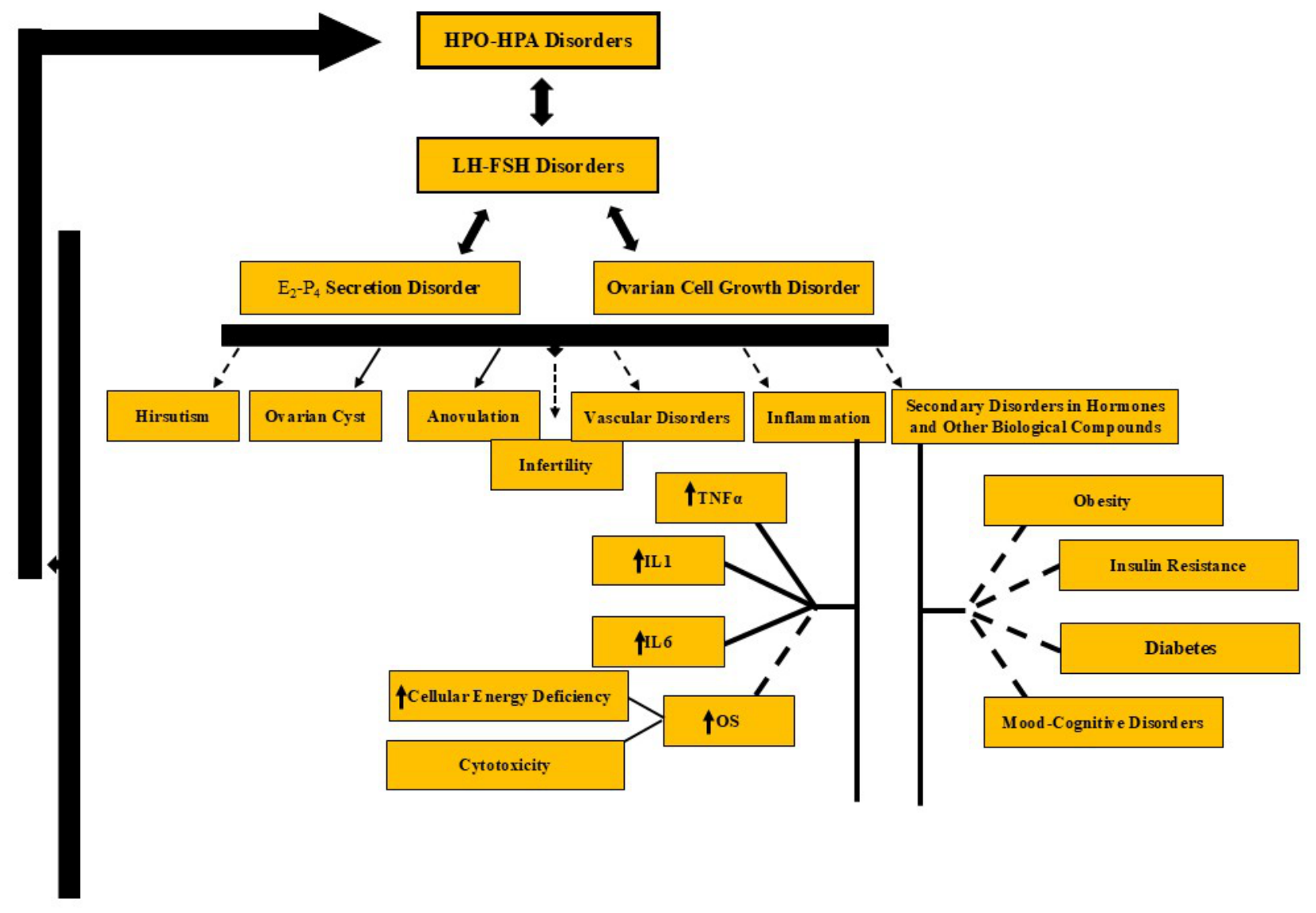
Figure 2.
Vascular, neural, lymphatic, and peripheral connections of the ovary. (A) Schematic representation of the anatomical and functional connections of the ovary with the central nervous system, peripheral nervous system, circulation, lymphatic system, and peripheral organs and tissues. The ovarian artery and vein provide oxygen, nutrients, and hormonal transport, while the ovarian nerve plexus conveys autonomic neural signals that may influence ovarian function. Lymphatic vessels facilitate immune surveillance and the transport of immune cells and signaling molecules. The ovarian stroma and extracellular matrix provide structural support and contribute to local cellular communication. Bidirectional interactions between the ovary and peripheral organs, including the uterus, liver, pancreas, intestines, adipose tissue, and skin, may contribute to the integration of reproductive, metabolic, endocrine, and immune functions. Red and blue arrows represent vascular connections, yellow arrows indicate neuronal pathways, green arrows denote lymphatic connections, and purple arrows represent supportive interactions involving the ovarian stroma and extracellular matrix. (B) Schematic illustration of the autonomic neural pathways associated with ovarian innervation, emphasizing sympathetic input through the splanchnic nerves and celiac plexus, as well as the potential contribution of parasympathetic pathways. Sympathetic nerve fibers reach the ovary through the ovarian plexus and accompany the ovarian vasculature, potentially modulating ovarian blood flow, steroidogenesis, follicular development, and other aspects of reproductive function. Parasympathetic influences, including potential vagal contributions, are also depicted; however, the extent and functional significance of direct parasympathetic innervation of the ovary remain incompletely characterized. The magnified inset shows the close anatomical association between nerve fibers and ovarian blood vessels and follicular structures. Orange and green lines indicate sympathetic and parasympathetic pathways, respectively, whereas red and blue lines represent arteries and veins. Yellow structures represent Ganglia. These pathways illustrate potential neurovascular interactions through which autonomic activity may influence ovarian physiology and its integration with systemic homeostatic mechanisms.
Figure 2.
Vascular, neural, lymphatic, and peripheral connections of the ovary. (A) Schematic representation of the anatomical and functional connections of the ovary with the central nervous system, peripheral nervous system, circulation, lymphatic system, and peripheral organs and tissues. The ovarian artery and vein provide oxygen, nutrients, and hormonal transport, while the ovarian nerve plexus conveys autonomic neural signals that may influence ovarian function. Lymphatic vessels facilitate immune surveillance and the transport of immune cells and signaling molecules. The ovarian stroma and extracellular matrix provide structural support and contribute to local cellular communication. Bidirectional interactions between the ovary and peripheral organs, including the uterus, liver, pancreas, intestines, adipose tissue, and skin, may contribute to the integration of reproductive, metabolic, endocrine, and immune functions. Red and blue arrows represent vascular connections, yellow arrows indicate neuronal pathways, green arrows denote lymphatic connections, and purple arrows represent supportive interactions involving the ovarian stroma and extracellular matrix. (B) Schematic illustration of the autonomic neural pathways associated with ovarian innervation, emphasizing sympathetic input through the splanchnic nerves and celiac plexus, as well as the potential contribution of parasympathetic pathways. Sympathetic nerve fibers reach the ovary through the ovarian plexus and accompany the ovarian vasculature, potentially modulating ovarian blood flow, steroidogenesis, follicular development, and other aspects of reproductive function. Parasympathetic influences, including potential vagal contributions, are also depicted; however, the extent and functional significance of direct parasympathetic innervation of the ovary remain incompletely characterized. The magnified inset shows the close anatomical association between nerve fibers and ovarian blood vessels and follicular structures. Orange and green lines indicate sympathetic and parasympathetic pathways, respectively, whereas red and blue lines represent arteries and veins. Yellow structures represent Ganglia. These pathways illustrate potential neurovascular interactions through which autonomic activity may influence ovarian physiology and its integration with systemic homeostatic mechanisms.
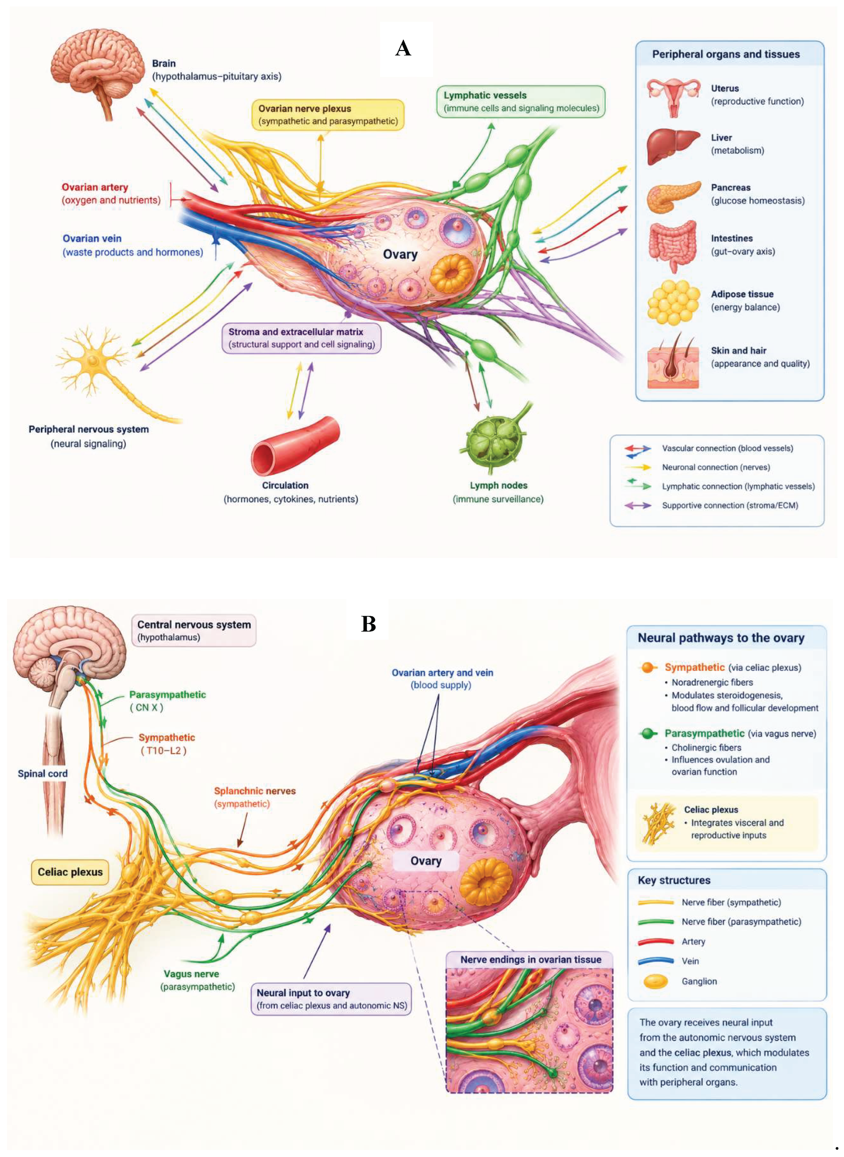
Figure 3.
A summary of the hypothesized mechanisms and potential outcomes of electroceutical interventions in polycystic ovary syndrome (PCOS). This image shows the effects of the ECs cascade mechanisms, starting with the neural effects involved in PCOS, which may affect hormonal and neurotransmitter status, metabolism, and appetite. These modifications may therefore affect the involved factors and diagnostic signs in this syndrome. PΣ, parasympathetic nervous system; PCOS, polycystic ovary syndrome; PNS, peripheral nervous system; Σ, sympathetic nervous system; VNS, vagus nerve stimulation. Note: Solid arrows indicate mechanistic relationships supported by direct and relatively strong evidence for the specific intervention, whereas dashed arrows represent hypothesized, indirect, or incompletely established pathways and potential clinical outcomes. The proposed pathways illustrate the possible effects of electroceutical interventions in PCOS and should not be interpreted as evidence of established clinical efficacy.↑ indicates an increase; ↓ indicates a decrease.
Figure 3.
A summary of the hypothesized mechanisms and potential outcomes of electroceutical interventions in polycystic ovary syndrome (PCOS). This image shows the effects of the ECs cascade mechanisms, starting with the neural effects involved in PCOS, which may affect hormonal and neurotransmitter status, metabolism, and appetite. These modifications may therefore affect the involved factors and diagnostic signs in this syndrome. PΣ, parasympathetic nervous system; PCOS, polycystic ovary syndrome; PNS, peripheral nervous system; Σ, sympathetic nervous system; VNS, vagus nerve stimulation. Note: Solid arrows indicate mechanistic relationships supported by direct and relatively strong evidence for the specific intervention, whereas dashed arrows represent hypothesized, indirect, or incompletely established pathways and potential clinical outcomes. The proposed pathways illustrate the possible effects of electroceutical interventions in PCOS and should not be interpreted as evidence of established clinical efficacy.↑ indicates an increase; ↓ indicates a decrease.
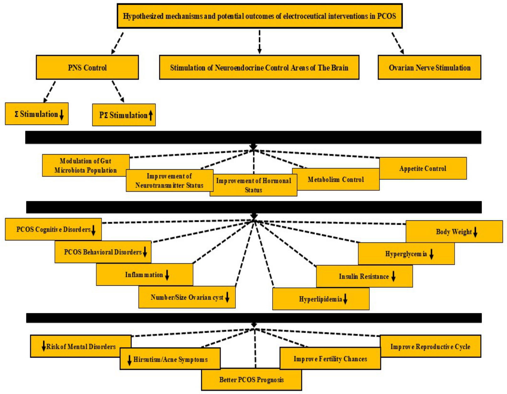
Figure 4.
Conceptual framework linking the pathophysiology and management of polycystic ovary syndrome (PCOS) with bioelectricity-based perspectives and the potential role of electroceuticals (ECs). The figure comprises two interconnected conceptual domains separated by a vertical line. The left panel illustrates the pathophysiology of PCOS and its associated management and control strategies. The right panel presents a multilevel overview of bioelectricity at the cellular, tissue, organ-system, and organ levels. Downward arrows indicate the conceptual progression from bioelectric alterations to bioelectric dysfunction and their potential association with pathological conditions, including PCOS. Subsequent arrows illustrate the proposed role of ECs in modulating bioelectric dysfunction and the potential development of novel therapeutic approaches. At the bottom of the right panel, a balance scale represents the potential advantages and limitations of electroceutical approaches, while the question mark emphasizes the need for further investigation to determine their overall therapeutic value. This framework is conceptual and does not imply that the efficacy or clinical applicability of electroceuticals in PCOS has been established. Solid arrows indicate well-established associations or recognized pathophysiological relationships, whereas dashed arrows represent proposed, indirect, or incompletely established associations. The arrows indicate conceptual relationships and do not necessarily imply causality.
Figure 4.
Conceptual framework linking the pathophysiology and management of polycystic ovary syndrome (PCOS) with bioelectricity-based perspectives and the potential role of electroceuticals (ECs). The figure comprises two interconnected conceptual domains separated by a vertical line. The left panel illustrates the pathophysiology of PCOS and its associated management and control strategies. The right panel presents a multilevel overview of bioelectricity at the cellular, tissue, organ-system, and organ levels. Downward arrows indicate the conceptual progression from bioelectric alterations to bioelectric dysfunction and their potential association with pathological conditions, including PCOS. Subsequent arrows illustrate the proposed role of ECs in modulating bioelectric dysfunction and the potential development of novel therapeutic approaches. At the bottom of the right panel, a balance scale represents the potential advantages and limitations of electroceutical approaches, while the question mark emphasizes the need for further investigation to determine their overall therapeutic value. This framework is conceptual and does not imply that the efficacy or clinical applicability of electroceuticals in PCOS has been established. Solid arrows indicate well-established associations or recognized pathophysiological relationships, whereas dashed arrows represent proposed, indirect, or incompletely established associations. The arrows indicate conceptual relationships and do not necessarily imply causality.
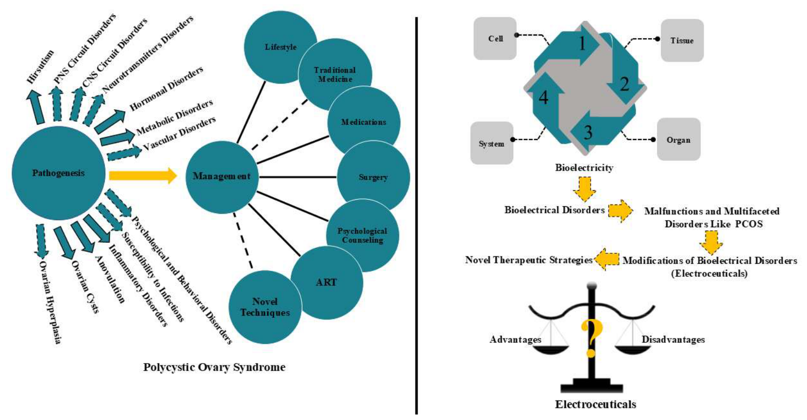
Disclaimer/Publisher’s Note: The statements, opinions and data contained in all publications are solely those of the individual author(s) and contributor(s) and not of MDPI and/or the editor(s). MDPI and/or the editor(s) disclaim responsibility for any injury to people or property resulting from any ideas, methods, instructions or products referred to in the content. |
© 2026 by the authors. Licensee MDPI, Basel, Switzerland. This article is an open access article distributed under the terms and conditions of the Creative Commons Attribution (CC BY) license (http://creativecommons.org/licenses/by/4.0/).
Copyright: This open access article is published under a Creative Commons CC BY 4.0 license, which permit the free download, distribution, and reuse, provided that the author and preprint are cited in any reuse.