

Submitted:
27 October 2026
Posted:
29 October 2026
You are already at the latest version
Abstract
The perinatal period has emerged as a critical window for the development of overweight and obesity and has therefore become a focus of scientific interest. According to the concept of developmental origins of health and disease (DOHaD), the first 1000 days after conception are of critical importance for the proper programming of stem cells. Multipotent mesenchymal stem cells (MSCs) have the capacity to differentiate into adipocytes both prenatally and postnatally. The aim of this review is to provide translational evidence for increased adipogenic programming of MSCs due to maternal obesity, cesarean delivery, and infant formula feeding. All three perinatal exposures plausibly converge on impaired canonical Wnt/β-catenin signaling in MSCs; this is directly supported for maternal obesity and indirectly for cesarean delivery and formula feeding through gut–endocrine mechanisms. Deficient Wnt/β-catenin signaling plays a pivotal role for the early committment of MSCs to the adipocyte lineage. These insights open up a comprehensive new approach to the understanding of the perinatal sequence of molecular events promoting early obesity development. Common medical practices during the perinatal period require reconsideration for the primary prevention of obesity.
Keywords:
adipogenesis
; cesarean delivery
; enteroendocrine cell
; glucagon-like peptide 1
; infant formula feeding
; intestinal stem cell
; maternal obesity
; mesenchymal stem cell
; obesity
; Wnt/β-catenin signaling
1. Introduction
The global epidemics of obesity and diabetes continue to advance unabatedly. In the United States, 70% of adults are overweight or obese. Childhood obesity has nearly doubled over the past two decades [1]. Children and adolescents with obesity are approximately five times more likely to have obesity in adulthood compared with their normal-weight peers [2].
Obesity, in turn, increases the risk of type 2 diabetes mellitus [3]. The perinatal period has emerged as a critical window for the development of obesity and diabetes and has therefore become a focus of scientific interest [4]. In particular, it is becoming increasingly evident that external and internal influences during the first 1000 days of life after conception have a substantial programming effect on the development of obesity [5,6,7,8,9].
Accumulating evidence indicates that dysregulated programming of mesenchymal stem cells (MSCs) toward adipocytes and, consequently, early adipogenesis, may already begin during fetal life [10,11,12,13,14,15,16,17,18]. Multipotent MSCs can self-renew or, in response to specific signals, further differentiate towards adipocytes, osteocytes, chondrocytes or myocytes [19,20].
A signaling pathway that is critical for MSC differentiation is canonical Wnt/β-catenin signaling. When activated, MSCs preferentially differentiate into osteocytes (osteogenesis) [21,22,23,24], whereas attenuation of Wnt/β-catenin signaling promotes their differentiation into adipocytes (adipogenesis) [25,26,27,28,29,30,31].
Boyle et al. [10] compared Wnt signaling of umbilical cord (UC)-MSCs from infants born to obese mothers (Ob-MSCs) versus mothers with normal weight (NW-MSCs) and found reduced Wnt/β-catenin signaling and a greater potential for adipogenesis in Ob-MSCs. This important observation in concert with literature evidence raised our research questions: (1) Is reduced Wnt/β-catenin signaling the fundamental mechanism driving adipogenic commitment of MSCs during the perinatal period of life? (2) Are maternal obesity, cesarean delivery and artifical infant formula feeding, which are all epidemilogically associated with an enhanced obesity risk, related to impaired Wnt signaling of MSCs? To follow these questions, the reader should be familiar with the mode of action of canonical Wnt/β-catenin signaling. The aim of this review is to synthesize mechanistic literature evidence and propose a unifying hypothesis that maternal obesity, cesarean delivery, and infant formula feeding converge on impaired Wnt/β-catenin signaling to drive adipogenic programming of MSCs.
2. Basic Principles of Canonical Wnt/β-Catenin Signaling
Among the molecular pathways reported to be altered in Ob-MSCs, including redox/forkhead box protein O1 (FOXO1) signaling, DNA methylation, and mitochondrial function, canonical Wnt/β-catenin is uniquely positioned at the intersection of intestinal stem cell homeostasis, enteroendocrine glucagon-like peptide 1 (GLP-1) secretion, and mesenchymal lineage choice, which motivates its selection as the organizing framework of this review. For a deeper understanding of canonical and non-canonical Wnt signaling we refer to recent excellent expert reviews [32,33,34,35,36]. Figure 1 illustrates basic principles of canonical Wnt/β-catenin signaling modified according to Rim et al. [32].
Among the best-characterized Wnt target genes are LGR5 (leucine-rich repeat-containing G protein-coupled receptor 5), a widely used marker of intestinal stem cells (ISCs), and TCF7L2 (transcription factor 7-like 2, also known as TCF4). LGR5 binds R-spondins and thereby potentiates Wnt signaling, supporting the maintenance and expansion of the ISC compartment [32,37]. The Wnt/β-catenin signaling pathway plays a critical role in increasing the activity of ISCs and promotes intestinal development [38,39,40,41,42]. Appropriate ISC activity plays also a key role for further differentiation of intestinal cells into nonsecretory and secretory intestinal cells. Knockdown of the nuclear Wnt effector TCF7L2 in mice negatively affected the ISC population and abrogated the development of enteroendocrine cells (EECs) [42]. Thus, Wnt signaling maintains intestinal homeostasis and the development of enteroendocrine cells.
2.1. Canonical Wnt/β-Catenin/TCF7L2 Signaling Enhances GLP-1 Signaling
Importantly, β-catenin/TCF7L2 binds the GCG promoter/enhancer and increases proglucagon expression. Experiments using the glycogen synthase kinase 3β (GSK3β) inhibitor lithium activates β-catenin signaling and constitutively active β-catenin increases both GCG mRNA and GLP-1 production in intestinal endocrine L-cells [43]. Shao et al. [44] confirmed that the Wnt signaling pathway effector TCF7L2 controls gut and brain proglucagon gene expression and glucose homeostasis. The TCF7L2 polymorphism rs7903146 is considered one of the strongest and most consistently replicated genetic risk factors for type 2 diabetes mellitus among common genetic variants. The T allele has been associated with reduced insulin content and secretion, and impaired incretin effects. GLP-1 is a proteolytic cleavage product of proglucagon [45]. Further evidence supports that GLP-1/GLP-1 receptor (GLP1R) signaling is upregulated by appropriate Wnt signaling [46,47,48,49,50,51]. As shown in pancreatic β-cells, Wnt/TCF7L2 signaling upregulates the LIM homeodomain protein ISL1, which enhances the expression of GLP1R [51].
2.2. Wnt/β-Catenin Signaling and C/EBPβ- and PPARγ-Mediated MSC Adipogenic Fate Determination
Wnt/β-catenin signaling inhibits MSC adipogenic differentiation but stimulates osteogenic differentiation [19,20]. The adipogenic transcription factor peroxisome proliferator-activated receptor-γ (PPARγ) plays a key role for adipogenic differentiation and promotes the expression of crucial genes driving adipogenesis [52,53], whereas Runt-related transcription factor 2 (RUNX2) is a pivotal transcription factor promoting osteogenesis [54,55] (Figure 2).
Two decades ago, an important crosstalk between Wnt/β-catenin signaling and PPARγ activity was established [56,57,58,59]. The differentiation of preadipocytes into adipocytes requires suppression of canonical Wnt signaling, which involves PPARγ-associated targeting of β-catenin to the proteasome [57]. Indeed, sustained activation of β-catenin through the expression of Wnt1 or Wnt10b in preadipocytes blocks adipogenesis by inhibiting PPARγ-associated gene expression. Liu et al. [57] showed that activation of PPARγ, by exposing Swiss mouse fibroblasts to troglitazone, stimulates GSK3β-dependent degradation of β-catenin, whereas a β-catenin mutant defective in GSK3β-mediated phosphorylation inhibits the expression of a subset of adipogenic genes. These pioneering data provide evidence that GSK3β is a key regulator of the balance between β-catenin and PPARγ activity. Activation of canonical Wnt signaling downstream of PPARγ blocks the expression of a select subset of adipogenic genes [57].
Furthermore, Liu et al. [58] identified a direct functional interaction between β-catenin and PPARγ involving the TCF/LEF-binding domain of β-catenin and a catenin-binding domain (CBD) within PPARγ, which enhances the proteasomal degradation of β-catenin. In addition, Wnt signaling induces the expression of WNT1-inducible signaling pathway protein 2 (WISP2), which inactivates the transcription factor zinc finger protein 423 (ZFP423) [60]. WISP2, a secreted protein encoded by a Wnt target gene, acts as a mesenchymal cell activator that enhances Wnt signaling while inhibiting PPARγ activation and adipogenic differentiation [60]. WISP2 is both secreted by mesenchymal precursor cells and highly expressed in their cytosol, where it binds the PPARγ transcriptional activator ZFP423 [61], thereby preventing its nuclear translocation [62].
Another Wnt target gene encodes WNT1-inducible signaling pathway protein 1 (WISP1), which inactivates PPARγ. Inactivation of PPARγ transcriptional activity was mediated, at least in part, by a direct physical association between WISP1 and PPARγ, followed by proteasome-dependent degradation of PPARγ [63]. Furthermore, Xu et al. [64] showed that Wnt5a represses PPARγ transactivation through H3K9 histone methyltransferase protein complexes.
CCAAT/enhancer-binding protein β (C/EBPβ) is rapidly induced during the early stages of adipogenesis and is responsible for the transcriptional induction of two major adipogenic transcription factors: PPARγ and CCAAT/enhancer-binding protein α (C/EBPα). Remarkably, overexpression of C/EBPβ substantially inhibited Wnt signaling. C/EBPβ is involved in the suppression of Wnt10b through direct binding to specific regions of the WNT10B promoter [65]. PPARγ also inhibits RUNX2 expression [66,67,68]. In particular, PPARγ physically interacts with RUNX2 and can interfere with RUNX2 binding and transactivation at osteoblast-specific element 2 (OSE2), thereby reducing RUNX2-driven gene transcription [68].
2.3. Molecular Crosstalk between GLP-1- and Wnt-Signaling Restricts Adipogenic Differentiation but Promotes Osteogenic Differentiation of MSCs
2.4. GLP-1 Augments Canonical Wnt Signaling
Wnt and GLP-1 signals exert synergistic effects on stem cell regulation. Wnt signaling not only enhances GLP-1 signaling [46,47,48,49,50,51], but GLP-1 also promotes Wnt/β-catenin signaling [73,76,79].
To understand the synergistic effects of GLP-1 and Wnt signaling, it is important to note that the activated GLP1R preferentially couples to Gαs, increasing cAMP levels and subsequently activating protein kinase A (PKA) [80,81,82]. Hino et al. [83] showed that phosphorylation of β-catenin by PKA inhibits β-catenin ubiquitination. Ser675 was identified as a PKA phosphorylation site. Substitution of this serine residue with alanine attenuated PKA-mediated inhibition of β-catenin ubiquitination, PKA-induced stabilization of β-catenin, and PKA-dependent activation of TCF. GLP-1/PKA activation enhances the nuclear translocation of β-catenin and the initiation of osteogenic gene expression. Activated PKA also phosphorylates phosphoinositide 3-kinase (PI3K), leading to AKT activation. AKT subsequently phosphorylates GSK3β, inhibiting its activity [79]. GSK3β-mediated degradation of β-catenin is thus inhibited, and nuclear accumulation of β-catenin is enhanced.
Notably, Zhao et al. [84] reported that AMP-activated protein kinase (AMPK) phosphorylates β-catenin at Ser552, thereby enhancing β-catenin/TCF-mediated transcription, in C3H10T1/2 mesenchymal cells. This murine embryonic fibroblast/mesenchymal progenitor cell line can differentiate into osteogenic and adipogenic lineages. Figure 3 illustrates the molecular interaction between Wnt and GLP-1 signals in MSC differentiation.
3. Maternal Obesity
The influence of maternal overweight and obesity on the perinatal differentiation of adipocyte precursor cells as a predisposing factor for subsequent overweight in offspring is currently a focus of scientific interest [85]. An important question is: How does maternal overweight or obesity during pregnancy influence the adipogenic potential of fetal MSCs? An elegant, noninvasive retrospective approach that imposes no burden on either mother or child during pregnancy is the isolation of MSCs from the umbilical cord (UC-MSCs) [10,16] or umbilical cord blood [86]. However, differences in MSC adipogenic potential have been observed depending on the tissue source, in the following order: adipose tissue-derived MSCs (AD-MSCs) > bone marrow-derived MSCs (BM-MSCs) > umbilical cord-derived MSCs (UC-MSCs) [87]. In particular, PPARγ mRNA expression was highest in AD-MSCs, lowest in UC-MSCs, and intermediate in BM-MSCs. These results suggest that differences in adipogenic potential among the three MSC types are associated with their basal PPARγ mRNA expression levels [87].
Bae et al. [86] investigated the adipogenic differentiation of MSCs isolated from umbilical cord blood and confirmed that increased Wnt5a/β-catenin signaling inhibits their adipogenic differentiation. The addition of Wnt5a inhibited adipogenic differentiation and reduced PPARγ expression and lipid storage. In contrast, depletion of β-catenin increased the adipogenic potential of umbilical cord blood MSCs. Interestingly, in a study involving 44 neonates, lower GLP-1 concentrations were measured in the umbilical cord blood of macrosomic neonates than in that of normal-weight neonates (6.9±2.96 ±2.9 vs. 10.3±3.7 pg/mL) [88]. Thus, MSCs in the umbilical cord and/or umbilical cord blood of neonates delivered by obese mothers may have been exposed to reduced GLP-1 signaling prenatally.
Undifferentiated UC-MSCs from infants of mothers with obesity (Ob-MSCs) exhibited higher protein levels of the transcription factor ZNF423 (zinc finger protein 423) than UC-MSCs from infants of normal-weight mothers (NW-MSCs), a difference associated with adipocyte hypertrophy following MSC differentiation [13]. ZNF423 is of major importance for adipogenesis because it induces the expression of the adipogenic transcription factor PPARγ [61]. Notably, ZNF423 activity is negatively regulated by Wnt signaling. Wnt signaling induces the expression of WISP1 and WISP2, both of which inhibit PPARγ activity [62,63].
In this context, the study by Boyle et al. [10] is highly informative. The authors investigated Wnt/β-catenin signaling and adipogenic differentiation in UC-MSCs from infants of normal-weight mothers (NW-MSCs) and infants of mothers with obesity (Ob-MSCs). Following differentiation, Ob-MSCs exhibited increased adipogenesis, with a 50% increase in PPARγ protein levels compared with NW-MSCs. Ob-MSCs showed 25% lower inhibitory phosphorylation of GSK3β, suggesting increased β-catenin degradation. However, this study did not investigate the effects of GLP-1 on PKA-mediated phosphorylation of β-catenin or AKT-mediated inhibitory phosphorylation of GSK3β.
Bellalta et al. [18] recently characterized the proteins in the MSC secretome following the isolation of UC-MSCs from Wharton’s jelly of infants born to normal-weight mothers (NW-MSCs) and infants born to mothers with obesity (Ob-MSCs). They found increased expression of the Wnt inhibitor SFRP1 (secreted frizzled-related protein 1) in Ob-MSCs compared with NW-MSCs. SFRP1 is a proadipogenic protein that inhibits Wnt signaling [89]. Similarly, the Wnt antagonist SFRP4 (secreted frizzled-related protein 4) increases adipogenic differentiation in human adipose tissue-derived MSCs [90].
In a further study, Bellalta et al. [17] showed that maternal overweight increases oxidative stress in MSCs, representing another potential mechanism associated with PPARγ activation. Compared with NW-MSCs, Ob-MSCs exhibited reduced stemness characteristics, including lower OCT4 (octamer-binding transcription factor 4, POU5F1) expression and decreased clonogenic capacity.
Remarkably, Wnt signaling and the transcription factor OCT4 are interconnected in determining early cell fate during differentiation and development [91]. OCT4 is a major regulator of the pluripotency network, controlling whether cells remain stem cells or begin differentiating into specialized cell types. Thus, OCT4 is associated with MSC stemness. Its overexpression increases MSC proliferation and colony-forming capacity [92,93]. Importantly, Li et al. [94] identified OCT4 as a novel target of Wnt signaling and demonstrated that the POU5F1 promoter is regulated by β-catenin/TCF. Indeed, OCT4 is a direct target of β-catenin/TCF-mediated transcription, and the binding site at positions −875/−881 of the POU5F1 promoter is critical for β-catenin/TCF-dependent gene expression.
In addition, Kelly et al. [95] provided evidence that β-catenin enhances OCT4 activity and reinforces pluripotency through a TCF-independent mechanism. They showed that stabilized β-catenin forms a complex with OCT4 and enhances its activity. OCT4 is a core component of the transcriptional network regulating pluripotency. Hade et al. [96] showed that, during the differentiation of preadipocytes into adipocytes, OCT4 levels declined significantly, whereas levels of PPARγ, the master transcription factor of mature adipocytes, increased. Intriguingly, Hannan et al. [97] observed that human embryonic stem cells (ESCs) transduced with an artificial OCT4-targeting short hairpin RNA displayed remarkably high lineage-specific spontaneous differentiation into adipocytes. Of note, the GLP1R agonist semaglutide stimulated the proliferation and maintenance of stemness in jaw bone marrow MSCs, accompanied by increased OCT4 and RUNX2 expression and enhanced osteogenesis [98].
Boyle et al. [99] observed reduced fatty acid oxidation (FAO) in UC-MSCs from infants of mothers with obesity (Ob-MSCs). In the Ob-MSC group with the most pronounced metabolic disturbances, correlations were found with neonatal adiposity and elevated umbilical cord blood insulin levels. Ob-MSCs with reduced FAO also showed lower AMPK activity. This finding again suggests a relationship with reduced Wnt signaling: AMPK, like GLP-1-activated PKA, phosphorylates β-catenin, thereby increasing its transcriptional activity, whereas AMPK inhibition resulted in increased PPARγ levels [84].
Erickson et al. [12] compared three metabolic profiles in UC-MSCs: (1) MSCs with normal fatty acid oxidation (FAO) in the setting of normal maternal weight, (2) MSCs with normal FAO in the setting of maternal obesity, and (3) MSCs with reduced FAO in the setting of maternal obesity. MSC FAO was negatively correlated with maternal fasting blood glucose and insulin levels, positively correlated with HDL cholesterol levels, and negatively associated with infant fat mass.
Notably, FAO is also regulated by Wnt/β-catenin signaling. In osteoblasts, activation of the Wnt signaling pathway increased oleic acid oxidation, whereas depletion of β-catenin reduced FAO and the expression of enzymes involved in β-oxidation [100]. In liver cells, loss of β-catenin likewise reduced FAO [101].
Iaffaldano et al. [102] observed that mitochondrial basal respiration (control: 46.9 ± 0.7; Ob: 32.6 ± 0.8 pmol/min), ATP-linked respiration (control: 29.3 ± 1.9; Ob: 20.1 ± 0.3 pmol/min), and maximal respiration (control: 75.2 ± 5.3; Ob: 50.5 ± 4.1 pmol/min) were significantly (P < 0.0001) lower in Ob-UC-MSCs versus control UC-MSCs. Paz et al. [103] reported also a trend toward lower maximal respiration and spare respiratory capacity of Ob-MSCs compared to NW-MSCs. Unexpectedly and in contrast to all other UC-MSC studies, Ob-MSCs exhibited an impaired adipogenic phenotype, characterized by reduced protein levels of C/EBPα and PPARγ during differentiation.
In summary, most UC-MSC studies reported enhanced adipogenic differentiation and decreased FAO in association with maternal obesity, findings that can be linked to reduced Wnt/β-catenin signaling.
4. Gestational Diabetes Mellitus
Overweight and obesity increase the risk of gestational diabetes mellitus (GDM) [104]. However, findings regarding the adipogenic potential of MSCs under hyperglycemic conditions are conflicting [105]. Cramer et al. [106] observed increased adipogenic potential and reduced osteogenic and chondrogenic differentiation in adipose tissue-derived MSCs exposed to high glucose concentrations. Incubation of adult bone marrow MSCs with sera from patients with type 2 diabetes mellitus increased adipogenic gene expression and lipid storage [107]. In contrast, Kim et al. [108] found reduced adipogenic and osteogenic differentiation in UC-MSCs from mothers with GDM compared with those from normoglycemic mothers. Bone marrow MSCs also showed reduced adipogenic differentiation following exposure to high glucose concentrations [109]. Cheng et al. [110] compared human adipose-derived stem cells obtained from the subcutaneous adipose tissue of donors with diabetes (dASCs) and donors without diabetes (nASCs). Although dASCs and nASCs treated with high glucose concentrations (4.5 g/L) exhibited decreased proliferation, increased expression of the pluripotency markers SOX2 (sex determining region Y)-box 2), OCT4, and homeobox protein NANOG was observed. Moreover, nASCs treated with high glucose concentrations exhibited decreased cell migration, enhanced senescence, and significantly higher intracellular reactive oxygen species levels. Their adipogenic and osteogenic differentiation capacities, however, remained comparable to those of cells treated with low glucose concentrations (1.0 g/L). Hyperglycemic conditions appear, in general, to reduce MSC differentiation potential and accelerate MSC senescence [105].
5. Cesarean Delivery
Mothers with overweight or obesity give statistically more births by cesarean delivery than normal-weight mothers [111,112]. The Federal Statistical Office of Germany had reported that cesarean delivery accounted for 33% of all births in Germany in 2024 [113], and a further global increase is expected. According to Betran et al. [114], the latest available data from 2010–2018, covering 154 countries and 94.5% of live births worldwide, indicate that 21.1% of women gave birth by cesarean delivery, with regional averages ranging from 5% in sub-Saharan Africa to 42.8% in Latin America and the Caribbean. Epidemiological data indicate that cesarean delivery is associated with a significantly increased risk of obesity compared with vaginal delivery. In a meta-analysis of 28 studies, Kuhle et al. [115] demonstrated that cesarean delivery was associated with an increased risk of childhood obesity compared with vaginal delivery (RR: 1.34; 95% CI: 1.18–1.51). A recently published meta-analysis of 20 studies likewise confirmed an increased risk of obesity in early childhood following cesarean delivery compared with vaginal delivery (RR: 1.23; 95% CI: 1.12–1.34) [116].
Since the influence of intrauterine factors on MSCs ends with the birth, the question arises whether, and if yes, how cesarean delivery might further promote adipogenic differentiation of MSCs. One possible explanation is the absence of the effects of vaginal delivery on the infant gut microbiota. In particular, cesarean delivery results in less efficient transmission of Bifidobacteria than vaginal delivery. Indeed, during the first postnatal months, the intestinal microbiome of infants delivered by cesarean delivery contains significantly reduced numbers of Bifidobacteria and Bacteroides species [117,118,119,120,121,122,123,124,125].
Acetate and lactate are the main products of human milk oligosaccharide (HMO) fermentation by Bifidobacterium species [126,127]. Beyond carbohydrate fermentation, Bifidobacterium species convert aromatic amino acids, such as tryptophan, phenylalanine, and tyrosine, into aromatic lactic acids—indole-3-lactic acid (ILA), phenyllactic acid, and 4-hydroxyphenyllactic acid—through an aromatic lactate dehydrogenase present in B. longum, B. breve, and B. bifidum [126,127]. Gastrointestinal Bacteroidota species produce acetate as one of their major short-chain fatty acid (SCFA) end products [128,129,130]. Notably, Bacteroides thetaiotaomicron can utilize human milk carbohydrates and release acetate [131].
Martin et al. [117] demonstrated that the delivery mode affected the organic acid profile produced by the intestinal microbiome. During the first and second months of life, the probability of detecting acetic and lactic acids was significantly lower in infants born by cesarean delivery than in those born vaginally. This is of critical importance because microbiota-derived lactate and acetate bind to their respective receptors, G protein-coupled receptor 81 (GPR81) and free fatty acid receptor 2 (FFAR2), and stimulate Wnt3 production by Paneth cells. Wnt3 then activates the Wnt3/β-catenin signaling pathway in neighboring ISCs [132,133]. Consistent with these findings, Wu et al. [134] recently reported that Lactobacillus amylovorus–lactate–GPR81 signaling stimulated β-catenin production and the differentiation of jejunal organoids.
Thus, impaired microbiota-derived lactate/acetate signaling due to reduced abundance of Bifidobacteria and Bacteroides species in the early intestinal microbiome after cesarean delivery may impair Wnt3-dependent ISC homeostasis (Figure 4).
Accumulating evidence indicates that gut microbiota can influence obesity. The gut microbiota contributes to host metabolism through several mechanisms, including increased dietary energy harvest, modulation of lipid metabolism, altered endocrine function, and increased inflammatory activity [135,136,137,138,139,140,141].
Yahagi et al. [142] investigated longitudinal changes in the gut microbiota and organic acid concentrations during the first two years of life in 12 Japanese infants. They confirmed that Bifidobacteria contribute significantly to organic acid production in healthy infants. In particular, acetate, lactate, and formiate, reported as rarely observed in adults in this context, are characteristic of breastfed infancy. Bifidobacterium longum subsp. infantis and the symbiotic activity of Bifidobacterium bifidum and Bifidobacterium breve efficiently produce these organic acids by metabolizing HMOs. Thus, HMO-utilizing Bifidobacteria play an important role in regulating infant gut organic acid profiles.
FFAR2, also known as G protein-coupled receptor 43 (GPR43), binds SCFAs produced by microbial carbohydrate fermentation [143]. FFAR2/GPR43 and FFAR3/GPR41 are expressed on enteroendocrine L cells and participate in GLP-1 secretion [144]. It is also important to note that lactate, through binding to its receptor GPR81, and acetate and propionate, through binding to FFAR2, increase GLP-1 secretion [145,146].
Pedersen et al. [147] observed that oral administration of 25 g of sodium lactate plus 6 g of sodium chloride to young adult men significantly increased plasma GLP-1 levels. Treatment with Bifidobacterium animalis subsp. lactis GCL2505 (BlaG) increased acetate and GLP-1 levels in both the gut and plasma [148].
Activation of FFAR2 by acetate and propionate has been identified as an important trigger for GLP-1 production and release by enteroendocrine L cells in the lower intestine. Notably, fructooligosaccharide supplementation in rats increased the density of FFAR2-positive EECs in the proximal colon by more than twofold compared with controls, alongside an increase in GLP-1-containing L cells [149]. In contrast, FFAR2-knockout (GPR43−/−) mice exhibit reduced SCFA-induced GLP-1 secretion [150].
Importantly, Bifidobacterium species associated with breastfeeding produce aromatic lactic acids in the infant gut [151]. Tryptophan metabolites, such as indole, stimulate GLP-1 release from enteroendocrine L cells through mechanisms involving the aryl hydrocarbon receptor (AhR) [152]. Vrzalová et al. [153] demonstrated that mixtures of intestinal microbial tryptophan metabolites influence AhR activity. Notably, Bacteroides thetaiotaomicron, B. eggerthii, B. ovatus, and B. fragilis produce indole-3-acetic acid (IAA) from tryptophan [154]. Another study involving 596 infants found that Bacteroides species were significantly less frequently detected, or were present at very low abundance, in infants born by cesarean delivery. Higher Bacteroides abundance was detected in 49.0% of vaginally delivered infants, compared with only 0.4% of infants born by cesarean delivery [119].
Indole-3-lactate has been shown to promote ISC differentiation into EECs [155]. In addition, IAA activates AhR and increases GLP-1 secretion by upregulating GCG expression and promoting stem cell differentiation into L cells [156]. Furthermore, supplementation with an AhR agonist or a Lactobacillus strain with a high capacity to produce AhR ligands improved intestinal barrier function and GLP-1 secretion [157].
Importantly, acetic and lactic acid levels are lower in infants born by cesarean delivery than in those born vaginally [117]. Compared with formula-fed infants, breastfed infants have higher concentrations of acetate, lactate, and indole-3-lactate in their stool [158,159,160].
Taken together, cesarean delivery may impair SCFA and indole/indole derivative-mediated enteroendocrine GLP-1 signaling. This may subsequently reduce GLP-1-stimulated Wnt/β-catenin signaling in MSCs, providing a further stimulus for their adipogenic differentiation [69,70,71,72,73,74,75,76,77,78,79].
6. Infant Formula Feeding
The metabolic relay race toward obesity continues with infant formula feeding. Cesarean delivery before the onset of labor is associated with delayed or less successful initiation of breastfeeding [161,162,163]. One possible explanation is that cesarean delivery, particularly emergency cesarean section performed before the onset of labor, reduces maternal endogenous release of oxytocin and prolactin [164,165].
A Canadian study by Rossiter et al. [166] concluded that exclusive breastfeeding without formula supplementation has a protective effect against the development of childhood obesity. The U.S. NHANES study showed that even short-term breastfeeding was associated with a lower risk of childhood obesity than formula feeding (OR: 0.31; 95% CI: 0.17–0.59). Children who were breastfed for three to six months had a lower risk of obesity at three to four years of age (OR: 0.56; 95% CI: 0.35–0.89) [167]. A recent meta-analysis including 26 publications (n=332,297) confirmed an inverse relationship between breastfeeding and obesity risk in children aged two to six years [168]. A dose–response relationship with breastfeeding duration was observed: one day to less than three months, OR: 1.07 (95% CI: 0.94–1.21); three to less than six months, OR: 0.96 (95% CI: 0.60–1.54); and six months or longer, OR: 0.67 (95% CI: 0.58–0.77). Each additional month of breastfeeding was associated with 4.0% lower odds of obesity (OR: 0.96 per month; 95% CI: 0.95–0.97).
Excessive postnatal protein intake, known as the “early protein hypothesis of obesity,” has been proposed as a possible explanation for the adipogenic effects of infant formula feeding [169]. Melnik et al. [170] recently provided a mechanistic explanation for this hypothesis based on impaired Wnt/β-catenin signaling in white adipose tissue MSCs. Excessive protein intake resulting from uncontrolled formula feeding or the use of high-protein formula increases serum levels of insulin, insulin-like growth factor 1 (IGF-1), and essential branched-chain amino acids (BCAAs), particularly leucine, in human infants [171,172] and newborn rhesus monkeys (Macaca mulatta) [173]. Notably, the abundance of fecal microbes known to vary in their ability to utilize complex carbohydrates, including Bifidobacterium and Ruminococcus from the Ruminococcaceae family, also increased with reduced protein intake [173].
High protein intake increases serum insulin, IGF-1, and BCAA levels, all of which stimulate the mammalian target of rapamycin complex 1-ribosomal protein S6 kinase beta-1 (mTORC1–S6K1) signaling pathway, a pathway linked to the pathogenesis of early childhood obesity associated with formula feeding [174]. After entering the nucleus, S6K1 can suppress the expression of key Wnt genes [175]. In particular, S6K1-mediated phosphorylation of histone H2B at Ser36 recruit enhancer of zeste homolog 2 (EZH2) to histone H3, where it mediates H3K27 trimethylation. This suppresses the expression of Wnt genes, including Wnt6, Wnt10a, and Wnt10b, while upregulating PPARγ and C/EBPα and increasing adipogenesis.
Scavo et al. [176] demonstrated that IGF-1 promotes cell growth and lipogenesis during the differentiation of human MSCs into adipocytes. Hu et al. [177] observed a critical attenuation of Wnt/β-catenin signaling in adipose tissue MSCs in response to elevated IGF-1 levels. They showed that IGF-1 increases PPARγ expression. PPARγ, in turn, increases the expression of Axin2, an inhibitor of Wnt/β-catenin signaling. Because circulating IGF-1 can reach the vascular stem cell niche of adipose tissue, excessive postnatal protein intake, accompanied by increased IGF-1/mTORC1/S6K1 signaling, may inhibit Wnt/β-catenin signaling in MSCs and thereby promote adipogenic differentiation [170,174,175,176,177,178].
Luk et al. [179] explored the role of the endothelial IGF-1 receptor (IGF1R) as a paracrine modulator of the white adipose tissue phenotype. They showed that reduced endothelial IGF1R expression in male mice receiving a high-fat diet leads to depot-specific, beneficial remodeling of white adipose tissue. Rajashekhar et al. [180] investigated the hypothesis that MSC fate within the perivascular niche could be modulated through interactions with endothelial cells (ECs), which regulate MSC adipogenic potential. An EC-specific effect was accompanied by increased expression of factors involved in Wnt signaling, most prominently Wnt1, Wnt4, and Wnt10a, which are well-known inhibitors of adipogenesis.
Consistent with the reported suppression of Wnt expression by excessive mTORC1–S6K1 signaling in MSCs [175], enhanced nuclear S6K1 signaling in ECs due to high protein intake and elevated plasma insulin, IGF-1, and BCAA levels may reduce EC-derived Wnt expression. This potential mechanism requires further experimental investigation.
Dysregulation of Wnt/β-catenin signaling in MSCs may also be promoted by additional factors associated with formula feeding. Bifidobacteria are not an inherent or mandatory component of infant formula. Some formulas are supplemented with specific Bifidobacterium strains as probiotics, while others contain no added Bifidobacteria. Infant formula does not reproduce the broad diversity and composition of HMOs naturally present in human milk. Human milk contains more than 100 structurally distinct HMOs, whereas HMO-supplemented infant formulas generally contain only a limited number of individual HMOs. Their HMO composition and diversity therefore differ substantially from those of human milk [181,182,183]. As shown in rhesus monkeys, high-protein infant formula reduced the abundance of intestinal Bifidobacteria [173].
Furthermore, infant formula contains no bioactive milk exosomes and exosomal microRNAs [184,185], which support intestinal development [186] and the growth and proliferation of Bifidobacteria [187]. Weil et al. [188] studied the gastrointestinal passage, intestinal epithelial cell uptake, and Argonaute 2 (AGO2) loading of milk microRNAs in neonates using xenomiRs as tracers. Of note, human milk extracellular vesicles (hmEVs) survive neonatal digestion and are taken up by neonatal intestinal enteroids [189].
Milk exosomes activate Wnt/β-catenin signaling and promote ISC activity and cell proliferation [190,191]. Treatment of ISCs with human milk-derived exosomes significantly upregulated the mRNA expression of Axin2, c-Myc, Cyclin D1, and LGR5 compared with exosome-free controls. In the presence of carnosic acid, a specific Wnt/β-catenin signaling inhibitor, cell viability was significantly reduced [190]. Consistent with these findings, Maghraby et al. [191] demonstrated in mice that bovine milk exosomes induced the proliferation of Lgr5+ ISCs in association with Wnt pathway activation.
Gross et al. [192] identified exosomes as carriers of Wnt proteins. The potential transfer of Wnts by milk-derived exosomes is currently under investigation.
Exosomal transfer of microRNA-148a, which suppresses the Wnt inhibitor DKK1 [194], may represent an indirect exosome-mediated mechanism that enhances intestinal Wnt/β-catenin signaling [195].
Unlike infant formula, human milk contains GLP-1 as a bioactive component. GLP-1 concentrations in milk collected at the end of a breastfeeding session, termed hindmilk, were negatively correlated with infant weight gain at six months of age [196]. Human milk also contains oxytocin, in contrast to infant formula [197,198]. Notably, oxytocin upregulates microRNA-148a-3p expression in milk exosomes [199]. Recent findings suggest that oxytocin may also contribute to increased ISC activity [200] and inhibit adipogenesis in adipose tissue and bone marrow stem cells while promoting osteogenesis [201].
Recently, Seitz et al. [202] investigated changes in the milk proteome from colostrum to mature milk. Wnt5a was identified as one of 50 proteins upregulated in colostrum compared with mature milk. Intriguingly, Wnt5a has been shown to promote osteogenic differentiation and inhibit adipogenic differentiation of human MSCs [203]. Bae et al. [86] used qPCR to examine the expression of Wnt family members (Wnt1, Wnt3a, Wnt4, Wnt5a, Wnt5b, and Wnt10b) in two lots of human umbilical cord blood MSCs with high (MSCs-H) and low (MSCs-L) adipogenic differentiation potential. Only Wnt5a mRNA levels were significantly decreased in MSCs-H following adipogenic induction. Under control conditions, β-catenin in MSCs-H was distributed throughout the cytoplasm and strongly localized to the plasma membrane. Adipogenic cells, by contrast, exhibited a reduced area of cytoplasmic β-catenin fluorescence, suggesting an active role for the canonical Wnt pathway during adipogenic differentiation. Western blotting showed higher Wnt5a and β-catenin protein levels in MSCs-L than in MSCs-H. Furthermore, Xu et al. [64] showed that Wnt5a represses PPARγ transactivation through H3K9 histone methyltransferase protein complexes. Intriguingly, Smith et al. [204] demonstrated in a human intestinal organoid model that the addition of human milk, compared with infant formula, increased β-catenin signaling, promoted organoid growth, and enhanced the proliferation of both ISCs and EECs.
Unlike infant formula, human milk contains stem cells with multilineage potential that are transferred to offsprings during breastfeeding [205,206,207]. Remarkably, Widjaja et al. [208] observed mesenchymal stem-like cells with high expression of the surface markers CD44, CD73, CD90, and CD105. They identified Wnt11 in the human milk stem cell secretome under hypoxic conditions and Wnt10b under nonhypoxic conditions. At present, it is uncertain whether human milk-derived MSCs (1) differentiate into adipocytes themselves, (2) promote or inhibit adipogenesis in other cells, or (3) regulate infant fat development during breastfeeding. These are three distinct possibilities that do not apply to MSC-free formula feeding.
Taken together, accumulating evidence suggests that infant formula feeding may also be associated with insufficient activation of Wnt signaling in MSCs, thereby contributing to adipogenic differentiation.

| (A) Maternal obesity | |||
| Finding | References | Finding | References |
| In UC blood MSCs Wnt5a mRNA and β-catenin decreased during adipogenic differentiation | [86] | Inhibition of adipogenic differentiation after addition of Wnt5a | [86] |
| In Ob-MSCs higher protein levels of ZNF423 | [13] | Inhibition of ZNF423 by Wnt1-induced WISP2 | [62] |
| In Ob-MSCs increased increased levels of PPARγ protein | [10,17] | The Wnt1-induced proteins WISP1 and WISP2 inhibit PPARγ expression | [63] |
| Lower inhibitory phosphorylation of GSK3β, suggesting increased degradation of β-catenin. | [10] | Lithium chloride inhibition of GSK3β increased nuclear β-catenin content and normalized nuclear PPARγ in Ob-MSCs. | [10] |
| Increased expression of the Wnt inhibitor SFRP1 in Ob-MSC secretome | [18] | Proadipogenic SFRP1 inhibits Wnt signaling | [89] |
| Lower OCT4 (POU5F1) expression in Ob-MSCs | [17] | POU5F1 expression is upregulated by β-catenin/TCF | [94,95] |
| OCT4 shRNA-transduced human ESCs displayed high lineage-specific differentiation toward adipocytes | [97] | OCT4 levels declined during preadipocyte to adipocyte differentiation, whereas PPARγ increased. | [96] |
| Reduced fatty acid oxidation (FAO) in Ob-MSCs | [99] | Depletion of β-catenin reduced FAO in osteoblasts and hepatocytes | [100,101] |
| (B) Cesarean delivery | |||
| Finding | References | Finding | References |
| Lactate deficiency in the infant gut due to cesarean delivery with reduced transmission of lactate-producing Bifidobacteria | [117] | Lactate via GPR81 stimulates Wnt3 expression of Paneth cells that activate Wnt signaling of intestinal stem cells | [132] |
| Acetate deficiency in the infant gut due to cesarean delivery with reduced transmission of acetate-producing Bifidobacteria and Bacteroides | [117] | Acetate via FFAR2 stimulates Wnt3 expression of Paneth cells that activate Wnt signaling of intestinal stem cells | [133] |
| Acetate, lactate and indoles and indol-derivatives via FFAR2, GPR81 and AhR, respectively, stimulate GLP1-1 secretion of EECs | [145,146,147,152] | GLP-1 activates MSCs; GLP1R activates PKA, which phosphorylates β-catenin thereby enhancing β-catenin signaling | [69,79] |
| (C) Formula feeding | |||
| Finding | References | Finding | References |
| Increased IGF-1-TORC1-S6K1 signaling due to excessive formula protein intake | [171,174] | Nuclear S6K1-mediated Wnt gene silencing | [175] |
| IGF-1 promotes differentiation of human MSCs into adipocytes | [176] | Attenuation of Wnt/β-catenin signaling in adipose tissue MSCs in response to elevated IGF-1 levels due to PPARγ-mediated expression of Axin2, an inhibitor of Wnt/β-catenin signaling | [177] |
| Absence of milk exosomes in infant formula | [184,185,186] | Human and bovine milk-derived exosomes enhance Wnt/β-catenin signaling of rat and murine intestinal stem cells increasing LGR5 expression | [190,191] |
| Absent transfer of milk exosomal microRNAs including exosomal microRNA-148a | [184,185,186] | Absent milk microRNA-148a-mediated inhibition of the Wnt inhibitor DKK1 | [194] |
| Fecal deficiencies of acetate, lactate and indole-lactate in formula-fed compared to breastfed infants | [158,159,160] | Yet uncharacterized effects on ISC Wnt signaling and enteroendocrine L cell GLP-1 secretion | |
| Absence of MSCs in infant formula | [205,206,207] | human breastmilk stem cell secretome: Wnt11 secretion under hypoxic, Wnt10b under non-hypoxic conditions | [208] |
| Wnt5 has been detected in the human breast milk proteome, whereas standard formulas are not known to contain Wnt5a, | [202] | Wnt5a mRNA decreased in MSCs with high adipogenic potential by adipogenic induction. Wnt5a promotes osteogenic but inhibits adipogenic differentiation of human MSCs | [203] |
| Exposure of fetal small intestine-derived organoids to human breast milk or infant formula | [204] | Organoids supplemented with formula exhibited reduced proliferation and showed decreased expression of β-catenin, TCF3 and TCF7 | [204] |
7. Combined Effects of Adipogenic Perinatal Factors
Maternal overweight and metabolic disturbances, together with impaired microbiome signaling associated with cesarean delivery and formula feeding rather than vaginal delivery and breastfeeding, may act synergistically to dysregulate Wnt signaling in MSCs during the perinatal period.
Which of these perinatal factors predominates in adipogenic dysregulation remains unclear and warrants further research. Two combinations represent contrasting scenarios: normal maternal weight, vaginal delivery, and breastfeeding versus maternal overweight or obesity, cesarean delivery, and formula feeding. Overall, there are eight possible combinations, whose effects on the endpoint of interest—childhood obesity—remain to be clarified (Table 3).
Studies examining only a single obesogenic factor, without accounting for all of the aforementioned perinatal influences, may therefore incompletely characterize the pathogenic molecular mechanisms underlying childhood obesity. Of particular importance, breastfeeding can partially compensate for the microbiome deficiencies associated with cesarean delivery [209,210,211,212]. Breastfeeding should therefore be promoted, particularly after cesarean delivery, as a potentially important measure for obesity prevention.
This presentation focuses on canonical Wnt/β-catenin signaling as a unifying node, but we acknowledge that redox-dependent FOXO1 signaling [15], epigenetic DNA methylation changes [99], and mitochondrial/AMPK alterations are reported in the same primary studies and may act in parallel to, rather than solely upstream or downstream of Wnt. A comparative pathway analysis across these frameworks remains to be performed.
8. Conclusions
Maternal obesity, cesarean delivery, and infant formula feeding are perinatal factors associated with an increased risk of childhood obesity and may influence the developmental trajectory during the first 1,000 days after conception. All three epidemiologically confirmed obesity risk constellations are associated with molecular alterations in MSCs that include impaired canonical Wnt/β-catenin signaling. We propose Wnt as a candidate unifying node, while recognizing that parallel pathways (redox, epigenetic, inflammatory) are also implicated and may co-operate in the perinatal programming of adipogenesis. Fetal MSCs derived from the umbilical cord tissue or umbilical cord blood of infants born to mothers with obesity exhibit increased adipogenic potential, which has been experimentally linked to impaired canonical Wnt/β-catenin signaling [10,86]. However, umbilical cord-derived MSCs may not fully reflect the adipogenic potential of adipose tissue-derived MSCs [87,213].
Cesarean delivery-associated disturbances in microbiota-derived signals in the postnatal gut, including lactate, acetate, and indole derivatives, can influence Wnt signaling in ISCs [132,133] and GLP-1 secretion by EECs [145,146]. Reduced EEC-derived GLP-1 signaling may, in turn, impair Wnt/β-catenin signaling in MSCs [69,79]. Similarly, compared with breastfeeding, formula feeding may provide a less favorable environment for microbiota-regulated signaling during early postnatal development. Supporting this concept, Smith et al. [204] demonstrated that exposure to infant formula, compared with human milk, was associated with reduced intestinal organoid growth, impaired β-catenin expression, and compromised EEC development.
At present, the strongest direct human evidence for altered MSC adipogenic differentiation during the perinatal period concerns maternal obesity. Cesarean delivery and formula feeding represent plausible additional contributing factors. However, their independent contributions and relative effect sizes remain uncertain. Maternal overweight and obesity are associated with an increased likelihood of cesarean delivery, which may be followed by a lower likelihood of breastfeeding. The co-occurrence of these three interconnected factors may therefore constitute a particularly relevant combination of exposures that could contribute to continued perinatal adipogenic programming, i.e., the perinatal march toward obesity. These interconnected risk factors should be addressed early through appropriate preventive strategies at the individual, clinical, and public health levels.
Author Contributions
Conceptualization and manuscript writing, B.C.M.; methodology, software, validation, formal analysis, B.C.M., R.W., G.S., J.C.G., S.M.J., W.S., and E.O.; investigation, B.C.M., R.W., J.C.G. and G.S.; visualization, S.W.; project administration, R.W.; supervision, G.S. and R.W.; B.C.M. holds the primary responsibility for the final content. All authors have read and agreed to the published version of the manuscript.
Funding
This research received no external funding.
Institutional Review Board Statement
Not applicable.
Informed Consent Statement
Not applicable.
Data Availability Statement
No new data were created or analyzed in this study. Data sharing is not applicable to this article.
Conflicts of Interest
The authors declare no conflicts of interest.
Abbreviations
The following abbreviations are used in this manuscript:
| AhR | Arylhydrocarbon receptor |
| AKT | AKT kinase (protein kinase B) |
| AMPK | AMP-activated protein kinase |
| APC | Adenomatous polyposis coli |
| ASC | Adipose-derived stem cell |
| BCAA | Branched-chain amino acid |
| CBD | Catenin-binding domain |
| C/EBPα | CCAAT/enhancer-binding protein α |
| C/EBPβ | CCAAT/enhancer-binding protein β |
| CK1α | Casein kinase 1α |
| DKK1 | Dickkopf 1 |
| DOHaD | Developmental origins of health and disease |
| DVL1 | (mammalian) Dishevelled |
| EC | Endothelial cell |
| EEC | Enteroendocrine cell |
| ESC | Embryonic stem cell |
| EV | Extracellular vesicle |
| FAO | Fatty acid oxidation |
| FFAR2 | Free fatty acid receptor 2 |
| FOXO1 | Forkhead box protein O1 |
| FZD | Frizzled receptor |
| GCG | Glucagon |
| GDM | Gestational diabetes mellitus |
| GLP-1 | Glucagon-like peptide 1 |
| GLP1R | GLP-1 receptor |
| GPR43 | G protein-coupled receptor 43 (FFAR2) |
| GPR81 | G protein-coupled receptor 81 (lactate receptor) |
| GSK3β | Glycogen synthase kinase 3β |
| H2B | Histone 2 B |
| H3K27 | Histone H3 lysine 27 |
| HDL | High-density lipoprotein |
| HMO | Human milk oligosaccharide |
| IAA | Indole-3-acetic acid |
| IGF-1 | Insulin-like growth factor 1 |
| IGF1R | IGF-1 receptor |
| ISC | Intestinal stem cell |
| LGR5 | Leucine-rich repeat-containing G protein-coupled receptor 5 |
| LPL | Lipoprotein lipase |
| LRP | Low-density lipoprotein receptor-related protein |
| microRNA | Micro-ribonucleic acid |
| mTORC1 | Mechanistic target of rapamycin complex 1 |
| MSC | Mesenchymal stem cell |
| NHANES | National Health and Nutrition Examination Survey |
| OCT4 | POU domain, class 5, transcription factor 1/Octamer-binding transcription factor 4 |
| OSE2 | Osteoblast-specific element 2 |
| OXPHOS | Oxidative phosphorylation |
| PI3K | Phosphoinositide 3-kinase |
| PKA | Protein kinase A |
| POU5F1 | POU domain, class 5, transcription factor 1 (OCT4) |
| PPARγ | Peroxisome proliferator-activated receptor γ |
| RUNX2 | Runt-related transcription factor 2 |
| SCFA | Short-chain fatty acid |
| SFRP1 | Secreted frizzled-related protein 1 |
| SOX2 | (Sex determining region Y)-box 2 |
| S6K1 | Ribosomal protein S6 kinase beta-1 (also called p70S6K) |
| TCF7L2 | Transcription factor 7-like 2 |
| UC | Umbilical cord |
| UCB | Umbilical cord blood |
| Wnt | Wnt protein |
| WISP1 | WNT1 inducible signaling pathway protein 1 |
| WISP2 | WNT1 inducible signaling pathway protein 2 |
| ZFP423 | Zinc finger protein 423 |
References
- World Health Organization. Obesity and Overweight. Fact Sheet. December 8, 2025. https://www.who.int/news-room/fact-sheets/detail/obesity-and-overweight (accessed on October 10, 2026.
- Simmonds, M.; Llewellyn, A.; Owen, C.G.; Woolacott, N. Predicting adult obesity from childhood obesity: a systematic review and meta-analysis. Obes Rev. 2016, 17, 95-107. [CrossRef]
- Yu, H.J.; Ho, M.; Liu, X.; Yang, J.; Chau, P.H.; Fong, D.Y.T. Association of weight status and the risks of diabetes in adults: a systematic review and meta-analysis of prospective cohort studies. Int. J. Obes. (Lond). 2022, 46, 1101-1113. [CrossRef]
- Umano, G.R.; Bellone, S.; Buganza, R.; Calcaterra, V.; Corica, D.; De Sanctis, L.; Di Sessa, A.; Faienza, M.F.; Improda, N.; Licenziati, M.R.; et al. Early roots of childhood obesity: risk factors, mechanisms, and prevention strategies. Int. J. Mol. Sci. 2025, 26, 7388. [CrossRef]
- Mameli, C.; Mazzantini, S.; Zuccotti, G.V. Nutrition in the first 1000 days: the origin of childhood obesity. Int. J. Environ. Res. Public Health. 2016, 13, 838. [CrossRef]
- Rossiter, C.; Cheng, H.; Appleton, J.; Campbell, K.J.; Denney-Wilson, E. Addressing obesity in the first 1000 days in high risk infants: Systematic review. Matern. Child Nutr. 2021, 17, e13178. [CrossRef]
- Wu, A.J.; Oken, E. Developmental contributions to obesity: nutritional exposures in the first thousand days. Gastroenterol. Clin. North Am. 2023, 52, 333-345. [CrossRef]
- Kracht, C.L.; Neshteruk, C.D.; Moding, K.J.; Rolke, L.J.; Wagner, B.E.; Kielb, E.; Ferrante, M.J.; Robinson, C.; Keinsley, J.; Colella, J.; et al. Community-based diet and obesity-related policy, system, and environmental interventions for obesity prevention during the first 1000 days: A scoping review. Obes. Rev. 2024, 25, e13815. [CrossRef]
- Schipper, M.C.; Manshanden, A.; Philippe, K.; Ferré, N.; Luque, V.; Lecorguillé, M.; Aubert, A.M.; Guerlich, K.; Le Gal, C.; Feher, S.; et al. First 1000 days strategies to prevent childhood obesity: a narrative review and recommendations from the EndObesity Consortium. Pediatr. Obes. 2026, 21, e70060. [CrossRef]
- Boyle, K.E.: Patinkin, Z.W.; Shapiro, A.L.; Baker, P.R. 2nd; Dabelea, D.; Friedman, J.E. Mesenchymal stem cells from infants born to obese mothers exhibit greater potential for adipogenesis: the Healthy Start BabyBUMP Project. Diabetes. 2016, 65, 647-659. [CrossRef]
- Baker, P.R. 2nd; Patinkin, Z.; Shapiro, A.L.; De La Houssaye, B.A.; Woontner, M.; Boyle, K.E.; Vanderlinden, L.; Dabelea, D.; Friedman, J.E. Maternal obesity and increased neonatal adiposity correspond with altered infant mesenchymal stem cell metabolism. JCI Insight. 2017, 2, e94200. [CrossRef]
- Erickson, M.L.; Patinkin, Z.W.; Duensing, A.M.; Dabelea, D.; Redman, L.M.; Boyle, K.E. Maternal metabolic health drives mesenchymal stem cell metabolism and infant fat mass at birth. JCI Insight. 2021, 6, e146606. [CrossRef]
- Keleher, M.R.; Shubhangi, S.; Brown, A.; Duensing, A.M.; Lixandrão, M.E.; Gavin, K.M.; Smith, H.A.; Kechris, K.J.; Yang, I.V.; Dabelea, D.; et al. Adipocyte hypertrophy in mesenchymal stem cells from infants of mothers with obesity. Obesity (Silver Spring). 2023, 31, 2090-2102. [CrossRef]
- Bellalta, S.; Plösch, T.; Faas, M.; Casanello, P. The role of mesenchymal stem cells in early programming of adipose tissue in the offspring of women with obesity. Pediatr. Obes. 2024, 19, e13120. [CrossRef]
- Gyllenhammer, L.E.; Zaegel, V.; Duensing, A.M.; Lixandrao, M.E.; Dabelea, D.; Bergman, B.C.; Boyle, K.E. Lipidomics of infant mesenchymal stem cells associate with the maternal milieu and child adiposity. JCI Insight. 2024, 9, e180016. [CrossRef]
- Gyllenhammer, L.E.; Boyle, K.E. New frontiers: umbilical cord mesenchymal stem cells uncover developmental roots and biological underpinnings of obesity susceptibility. Curr. Obes. Rep. 2025, 14, 10. [CrossRef]
- Bellalta, S.; Pinheiro-Machado, E.; Borghuis, T.; Prins, J.; Plösch, T.; Casanello, P.; Faas, M. Maternal obesity programs adipogenic commitment in neonatal mesenchymal stem cells: a link to redox-dependent FOXO1 signaling. J. Cell Physiol. 2026, 241, e70178. [CrossRef]
- Bellalta, S.; Pinheiro-Machado, E.; Casanello, P.; Faas, M.; Plösch, T. Profiling the secretome: maternal obesity impacts redox and adipogenic signaling during neonatal mesenchymal stem cell adipogenesis. Stem Cells. 2026, 44, sxag001. [CrossRef]
- Chen, Q.; Shou, P.; Zheng, C.; Jiang, M.; Cao, G.; Yang, Q.; Cao, J.; Xie, N.; Velletri, T.; Zhang, X.; et al. Fate decision of mesenchymal stem cells: adipocytes or osteoblasts? Cell Death Differ. 2016, 23, 1128-1139. [CrossRef]
- Hu, L.; Yin, C.; Zhao, F.; Ali, A.; Ma, J.; Qian, A. Mesenchymal stem cells: cell fate decision to osteoblast or adipocyte and application in osteoporosis treatment. Int. J. Mol. Sci. 2018, 19, 360. [CrossRef]
- Glass, D.A. 2nd; Bialek, P.; Ahn, J.D.; Starbuck, M.; Patel, M.S.; Clevers, H.; Taketo, M.M.; Long, F.; McMahon, A.P.; Lang, R.A.; et al. Canonical Wnt signaling in differentiated osteoblasts controls osteoclast differentiation. Dev Cell. 2005, 8, 751-764. [CrossRef]
- Liu, G.; Vijayakumar, S.; Grumolato, L.; Arroyave, R.; Qiao, H.; Akiri, G.; Aaronson, S.A. Canonical Wnts function as potent regulators of osteogenesis by human mesenchymal stem cells. J. Cell Biol. 2009, 185, 67-75. [CrossRef]
- Kim, J.H.; Liu, X.; Wang, J.; Chen, X.; Zhang, H.; Kim, S.H.; Cui, J.; Li, R.; Zhang, W.; Kong, Y.; et al. Wnt signaling in bone formation and its therapeutic potential for bone diseases. Ther. Adv. Musculoskelet. Dis. 2013, 5, 13-31. [CrossRef]
- Hu, L.; Chen, W.; Qian, A.; Li, Y.P. Wnt/β-catenin signaling components and mechanisms in bone formation, homeostasis, and disease. Bone Res. 2024, 12, 39. [CrossRef]
- Ross, S.E.; Hemati, N.; Longo, K.A.; Bennett, C.N.; Lucas, P.C.; Erickson, R.L.; MacDougald, O.A. Inhibition of adipogenesis by Wnt signaling. Science. 2000, 289, 950-953. [CrossRef]
- Laudes, M. Role of WNT signalling in the determination of human mesenchymal stem cells into preadipocytes. J. Mol. Endocrinol. 2011, 46, R65-R72. [CrossRef]
- Yuan, Z.; Li, Q.; Luo, S.; Liu, Z.; Luo, D.; Zhang, B.; Zhang, D.; Rao, P.; Xiao, J. PPARγ and Wnt signaling in adipogenic and osteogenic differentiation of mesenchymal stem cells. Curr. Stem Cell Res. Ther. 2016, 11, 216-225. [CrossRef]
- Hu, L.; Yin, C.; Zhao, F.; Ali, A.; Ma, J.; Qian, A. Mesenchymal stem cells: cell fate decision to osteoblast or adipocyte and application in osteoporosis treatment. Int. J. Mol. Sci. 2018, 19, 360. [CrossRef]
- Yu, M.; Qin, K.; Fan, J.; Zhao, G.; Zhao, P.; Zeng, W.; Chen, C.; Wang, A.; Wang, Y.; Zhong, J.; et al. The evolving roles of Wnt signaling in stem cell proliferation and differentiation, the development of human diseases, and therapeutic opportunities. Genes Dis. 2023, 11, 101026. [CrossRef]
- de Winter, T.J.J.; Nusse, R. Running against the Wnt: how Wnt/β-catenin suppresses adipogenesis. Front. Cell Dev. Biol. 2021, 9, 627429. [CrossRef]
- Bagchi, D.P.; MacDougald, O.A. Wnt signaling: from mesenchymal cell fate to lipogenesis and other mature adipocyte functions. Diabetes. 2021, 70, 1419-1430. [CrossRef]
- Rim, E.Y.; Clevers, H.; Nusse, R. The Wnt pathway: from signaling mechanisms to synthetic modulators. Annu. Rev. Biochem. 2022, 91, 571-598. [CrossRef]
- Qin, K.; Yu, M.; Fan, J.; Wang, H.; Zhao, P.; Zhao, G.; Zeng, W.; Chen, C.; Wang, Y.; Wang, A.; et al. Canonical and noncanonical Wnt signaling: Multilayered mediators, signaling mechanisms and major signaling crosstalk. Genes Dis. 2023, 11, 103-134. [CrossRef]
- Yu, M.; Qin, K.; Fan, J.; Zhao, G.; Zhao, P.; Zeng, W.; Chen, C.; Wang, A.; Wang, Y.; Zhong, J.; et al. The evolving roles of Wnt signaling in stem cell proliferation and differentiation, the development of human diseases, and therapeutic opportunities. Genes Dis. 2023, 11, 101026. [CrossRef]
- Holzem, M.; Boutros, M.; Holstein, T.W. The origin and evolution of Wnt signalling. Nat. Rev. Genet. 2024, 25, 500-512. [CrossRef]
- Xue, C.; Chu, Q.; Shi, Q.; Zeng, Y.; Lu, J.; Li, L. Wnt signaling pathways in biology and disease: mechanisms and therapeutic advances. Signal Transduct. Target Ther. 2025, 10, 106. [CrossRef]
- Barker, N.; van Es, J.H.; Kuipers, J.; Kujala, P.; van den Born, M.; Cozijnsen, M.; Haegebarth, A.; Korving, J.; Begthel, H.; Peters, P.J.; et al. Identification of stem cells in small intestine and colon by marker gene Lgr5. Nature. 2007, 449, 1003-1007. . [CrossRef]
- Barker, N. Adult intestinal stem cells: critical drivers of epithelial homeostasis and regeneration. Nat. Rev. Mol. Cell Biol. 2014, 15, 19-33. [CrossRef]
- Clevers, H.; Loh, K.M.; Nusse, R. Stem cell signaling. An integral program for tissue renewal and regeneration: Wnt signaling and stem cell control. Science. 2014, 346, 1248012. [CrossRef]
- Mah, A.T.; Yan, K.S.; Kuo, C.J. Wnt pathway regulation of intestinal stem cells. J. Physiol. 2016, 594, 4837-4847. [CrossRef]
- Perochon, J.; Carroll, L.R.; Cordero, J.B. Wnt signalling in intestinal stem cells: lessons from mice and flies. Genes (Basel). 2018, 9, 138. [CrossRef]
- Korinek, V.; Barker, N.; Moerer, P.; van Donselaar, E.; Huls, G.; Peters, P.J.; Clevers, H. Depletion of epithelial stem-cell compartments in the small intestine of mice lacking Tcf-4. Nat. Genet. 1998, 19, 379-383. [CrossRef]
- Ni, Z.; Anini, Y.; Fang, X.; Mills, G.; Brubaker, P.L.; Jin, T. Transcriptional activation of the proglucagon gene by lithium and beta-catenin in intestinal endocrine L cells. J. Biol. Chem. 2003, 278, 1380-2387. [CrossRef]
- Shao, W.; Wang, D.; Chiang, Y.T.; Ip, W.; Zhu, L.; Xu, F.; Columbus, J.; Belsham, D.D.; Irwin, D.M.; Zhang, H.; et al. The Wnt signaling pathway effector TCF7L2 controls gut and brain proglucagon gene expression and glucose homeostasis. Diabetes. 2013, 62, 789-800. [CrossRef]
- Gasbjerg, L.S.; Nielsen, C.K.; Suppli, M.P.; Grøndahl, M.F.G.; Holst, J.J.; Knop, F.K.; Jeppesen, P.B.; Lund, A.B. Proglucagon-derived peptides: human physiology and therapeutic potential. Physiol. Rev. 2026, 106, 529-586. [CrossRef]
- Chiang, Y.T.; Ip, W.; Jin, T. The role of the Wnt signaling pathway in incretin hormone production and function. Front. Physiol. 2012, 3, 273. [CrossRef]
- Yi, F.; Brubaker, P.L.; Jin, T. TCF-4 mediates cell type-specific regulation of proglucagon gene expression by beta-catenin and glycogen synthase kinase-3beta. J. Biol. Chem. 2005, 280, 1457-1464. [CrossRef]
- Yi, F.; Sun. J.; Lim. G.E.; Fantus, I.G.; Brubaker, P.L.; Jin, T. Cross talk between the insulin and Wnt signaling pathways: evidence from intestinal endocrine L cells. Endocrinology. 2008, 149, 2341-2351. [CrossRef]
- García-Jiménez, C. Wnt and incretin connections. Vitam. Horm. 2010, 84, 355-387. [CrossRef]
- Wang, J.; Wie, L.R.; Liu, Y.L.; Ding, C.Z.; Guo, F.; Wang, J.; Qin, Q.; Huang, F.J.; Xin, Y.; Ma, S.N.; et al. Berberine activates the β-catenin/TCF4 signaling pathway by down-regulating miR-106b to promote GLP-1 production by intestinal L cells. Eur. J. Pharmacol. 2021, 911, 174482. [CrossRef]
- Shao, W.; Szeto, V.; Song, Z.; Tian, L.; Feng, Z.P.; Nostro, M.C.; Jin, T. The LIM homeodomain protein ISL1 mediates the function of TCF7L2 in pancreatic beta cells. J. Mol. Endocrinol. 2018, 61, 1-12. [CrossRef]
- Chawla, A.; Schwarz, E.J.; Dimaculangan, D.D.; Lazar, M.A. Peroxisome proliferator-activated receptor (PPAR) gamma: adipose-predominant expression and induction early in adipocyte differentiation. Endocrinology. 1994, 135, 798-800. [CrossRef]
- Rosen, E.D.; Sarraf, P.; Troy, A.E.; Bradwin, G.; Moore, K.; Milstone, D.S.; Spiegelman, B.M.; Mortensen, R.M. PPAR gamma is required for the differentiation of adipose tissue in vivo and in vitro. Mol. Cell. 1999, 4, 611-617. [CrossRef]
- Bruderer, M.; Richards, R.G.; Alini, M.; Stoddart, M.J. Role and regulation of RUNX2 in osteogenesis. Eur. Cell Mater. 2014, 28, 269-286. [CrossRef]
- Vimalraj, S.; Arumugam, B.; Miranda, P.J.; Selvamurugan, N. Runx2: Structure, function, and phosphorylation in osteoblast differentiation. Int. J. Biol. Macromol. 2015, 78, 202-208. [CrossRef]
- Farmer, S.R. Regulation of PPARgamma activity during adipogenesis. Int. J. Obes (Lond). 2005, 29, Suppl 1, S13-S16. [CrossRef]
- Liu, J.; Farmer, S.R. Regulating the balance between peroxisome proliferator-activated receptor gamma and beta-catenin signaling during adipogenesis. A glycogen synthase kinase 3beta phosphorylation-defective mutant of beta-catenin inhibits expression of a subset of adipogenic genes. J. Biol. Chem. 2004, 279, 45020-45027. [CrossRef]
- Liu, J.; Wang, H.; Zuo, Y.; Farmer, S.R. Functional interaction between peroxisome proliferator-activated receptor gamma and beta-catenin. Mol. Cell Biol. 2006, 26, 5827-5837. [CrossRef]
- Moldes, M.; Zuo, Y.; Morrison, R.F.; Silva, D.; Park, B.H.; Liu, J.; Farmer, S.R. Peroxisome-proliferator-activated receptor gamma suppresses Wnt/beta-catenin signalling during adipogenesis. Biochem. J. 2003, 376, 607-613. [CrossRef]
- Grünberg, J.R.; Hammarstedt, A.; Hedjazifar, S.; Smith, U. The novel secreted adipokine WNT1-inducible Signaling Pathway Protein 2 (WISP2) is a mesenchymal cell activator of canonical WNT. J. Biol. Chem. 2014, 289, 6899-6907. [CrossRef]
- Gupta, R.K.; Arany, Z.; Seale, P.; Mepani, R.J.; Ye, L.; Conroe, H.M.; Roby, Y.A.; Kulaga, H.; Reed, R.R.; Spiegelman, B.M. Transcriptional control of preadipocyte determination by Zfp423. Nature. 2010, 464, 619-623. [CrossRef]
- Hammarstedt, A.; Hedjazifar, S.; Jenndahl, L.; Gogg, S.; Grünberg, J.; Gustafson, B.; Klimcakova, E.; Stich, V.; Langin, D.; Laakso, M.; et al. WISP2 regulates preadipocyte commitment and PPARγ activation by BMP4. Proc. Natl. Acad. Sci. U S A. 2013, 110, 2563-2568. [CrossRef]
- Ferrand, N.; Béreziat, V.; Moldes, M.; Zaoui, M.; Larsen, A.K.; Sabbah, M. WISP1/CCN4 inhibits adipocyte differentiation through repression of PPARγ activity. Sci. Rep. 2017, 7, 1749. [CrossRef]
- Xu, C.; Wang, J.; Zhu, T.; Shen, Y.; Tang, X.; Fang, L.; Xu, Y. Cross-talking between PPAR and WNT signaling and its regulation in mesenchymal stem cell differentiation. Curr. Stem Cell Res. Ther. 2016, 11, 247-254. [CrossRef]
- Chung, S.S.; Lee, J.S.; Kim, M.; Ahn, B.Y.; Jung, H.S.; Lee, M.; Kim, J.W.; Park, K.S. Regulation of Wnt/β-catenin signaling by CCAAT/enhancer binding protein β during adipogenesis. Obesity (Silver Spring). 2012, 20, 482-487. [CrossRef]
- Liu, L.F.; Shen, W.J.; Zhang, Z.H.; Wang, L.J.; Kraemer, F.B. Adipocytes decrease Runx2 expression in osteoblastic cells: roles of PPARγ and adiponectin. J. Cell Physiol. 2010, 225, 837-845. [CrossRef]
- Ge, C.; Cawthorn, W.P.; Li, Y., Zhao, G.; Macdougald, O.A.; Franceschi, R.T. Reciprocal control of osteogenic and adipogenic differentiation by ERK/MAP kinase phosphorylation of Runx2 and PPARγ transcription factors. J. Cell Physiol. 2016, 231, 587-596. [CrossRef]
- Jeon, M.J.; Kim, J.A.; Kwon, S.H.; Kim, S.W.; Park, K.S.; Park, S.W.; Kim, S.Y.; Shin, C.S. Activation of peroxisome proliferator-activated receptor-gamma inhibits the Runx2-mediated transcription of osteocalcin in osteoblasts. J. Biol. Chem. 2003, 278, 23270-23277. [CrossRef]
- Weber, L.; Hashemnia Sharbabaki, M.; Fuchs, B.; Alberton, P.; Giunta, R.; Mert, S.; Thierfelder, N. The influence of GLP-1 agonists on human mesenchymal stem cells: a systematic review. Stem Cell Rev. Rep. 2026, 22, 26-46. [CrossRef]
- Sanz, C.; Vázquez, P.; Blázquez, C.; Barrio, P.A.; Alvarez Mdel, M.; Blázquez, E. Signaling and biological effects of glucagon-like peptide 1 on the differentiation of mesenchymal stem cells from human bone marrow. Am. J. Physiol. Endocrinol. Metab. 2010, 298, E634-E643. [CrossRef]
- El Bekay, R.; Coín-Aragüez, L.; Fernández-García, D.; Oliva-Olivera, W.; Bernal-López, R.; Clemente-Postigo, M.; Delgado-Lista, J.; Diaz-Ruiz, A.; Guzman-Ruiz, R.; Vázquez-Martínez, R.; et al. Effects of glucagon-like peptide-1 on the differentiation and metabolism of human adipocytes. Br. J. Pharmacol. 2016, 173, 1820-1834. [CrossRef]
- Cantini, G.; Di Franco, A.; Samavat, J.; Forti, G.; Mannucci, E.; Luconi, M. Effect of liraglutide on proliferation and differentiation of human adipose stem cells. Mol. Cell Endocrinol. 2015, 402, 43-50. [CrossRef]
- Liu, H.; Zhan, Y.L.; Luo, G.J.; Zou, L.L.; Li, Y.; Lu, H.Y. Liraglutide and insulin have contrary effects on adipogenesis of human adipose-derived stem cells via Wnt pathway. Diabetes Metab. Syndr. Obes. 2020, 13, 3075-3087. [CrossRef]
- Lee, H.M.; Joo, B.S.; Lee, C.H.; Kim, H.Y.; Ock, J.H.; Lee, Y.S. Effect of glucagon-like peptide-1 on the differentiation of adipose-derived stem cells into osteoblasts and adipocytes. J. Menopausal Med. 2015, 21, 93-103. [CrossRef]
- Deng, B.; Zhu, W.; Duan, Y.; Hu, Y.; Chen, X.; Song, S.; Yi, Z.; Song, Y. Exendin-4 promotes osteogenic differentiation of adipose-derived stem cells and facilitates bone repair. Mol Med Rep. 2019, 20, 4933-4942. [CrossRef]
- Li, Y.; Fu, H.; Wang, H.; Luo, S.; Wang, L.; Chen, J.; Lu, H. GLP-1 promotes osteogenic differentiation of human ADSCs via the Wnt/GSK-3β/β-catenin pathway. Mol. Cell Endocrinol. 2020, 515, 110921. [CrossRef]
- Yang, Z.; Li, X.; Wu, N.; Wang, L.; Lu, Z.; Wang, L.; Han, S.; Lu, H. GLP-1 regulates osteo-adipogenic fate of BMSCs via HIF-2-AKT signaling and supports trabecular bone in glucocorticoid-induced osteoporosis. Stem Cell Reports. 2026, 21, 103008. [CrossRef]
- Tian, Y.; Liu, H.; Bao, X.; Li, Y. Semaglutide promotes the proliferation and osteogenic differentiation of bone-derived mesenchymal stem cells through activation of the Wnt/LRP5/β-catenin signaling pathway. Front. Pharmacol. 2025, 16, 1539411. [CrossRef]
- Meng, J.; Ma, X.; Wang, N.; Jia, M.; Bi, L.; Wang, Y.; Li, M.; Zhang, H.; Xue, X.; Hou, Z.; et al. Activation of GLP-1 receptor promotes bone marrow stromal cell osteogenic differentiation through β-catenin. Stem Cell Reports. 2016, 6, 579-591. Erratum in: Stem Cell Reports. 2016, 6, 633. doi: 10.1016/j.stemcr.2016.03.010. [CrossRef]
- Light, P.E.; Manning Fox, J.E.; Riedel, M.J.; Wheeler, M.B. Glucagon-like peptide-1 inhibits pancreatic ATP-sensitive potassium channels via a protein kinase A- and ADP-dependent mechanism. Mol. Endocrinol. 2002, 16, 2135-2144. [CrossRef]
- Meloni, A.R.; DeYoung, M.B.; Lowe, C.; Parkes, D.G. GLP-1 receptor activated insulin secretion from pancreatic β-cells: mechanism and glucose dependence. Diabetes Obes. Metab. 2013, 15, 15-27. [CrossRef]
- Pabreja, K.; Mohd, M.A.; Koole, C.; Wootten, D.; Furness, S.G. Molecular mechanisms underlying physiological and receptor pleiotropic effects mediated by GLP-1R activation. Br. J. Pharmacol. 2014, 171, 1114-1128. [CrossRef]
- Hino, S.; Tanji, C.; Nakayama, K.I.; Kikuchi, A. Phosphorylation of beta-catenin by cyclic AMP-dependent protein kinase stabilizes beta-catenin through inhibition of its ubiquitination. Mol. Cell Biol. 2005, 25, 9063-9072. [CrossRef]
- Zhao, J.; Yue, W.; Zhu, M.J.; Sreejayan, N.; Du, M. AMP-activated protein kinase (AMPK) cross-talks with canonical Wnt signaling via phosphorylation of beta-catenin at Ser 552. Biochem. Biophys. Res. Commun. 2010, 395, 146-151. [CrossRef]
- Lecoutre, S.; Maqdasy, S.; Lambert, M.; Breton, C. The impact of maternal obesity on adipose progenitor cells. Biomedicines. 2023, 11, 3252. [CrossRef]
- Bae, Y.K.; Kwon, J.H.; Kim, M.; Kim, G.H.; Choi, S.J.; Oh, W.; Yang, Y.S.; Jin, H.J.; Jeon, H.B. Intracellular calcium determines the adipogenic differentiation potential of human umbilical cord blood-derived mesenchymal stem cells via the Wnt5a/β-catenin signaling pathway. Stem Cells Int. 2018, 2018, 6545071. [CrossRef]
- Chi, Y.; Han, Z.B.; Xu, F.Y.; Wang, Y.W.; Feng, X.M.; Chen, F.; Ma, F.X.; Du, W.J.; Han, Z.C. Adipogenic potentials of mesenchymal stem cells from human bone marrow, umbilical cord and adipose tissue are different. Zhongguo Shi Yan Xue Ye Xue Za Zhi. 2014, 22, 588-594.
- Aktulay, A.; Engin-Ustun, Y.; Yasar, O.G.; Yilmaz, C.; Erkaya, S.; Ozgu-Erdinc, A.S. Levels of glucagon-like peptide 1 are decreased in macrosomic neonates from non-diabetic mothers. Z. Geburtshilfe Neonatol. 2019, 223, 48-53. [CrossRef]
- Lagathu, C.; Christodoulides, C.; Tan, C.Y.; Virtue, S.; Laudes, M.; Campbell, M.; Ishikawa, K.; Ortega, F.; Tinahones, F.J.; Fernández-Real, J.M.; et al. Secreted frizzled-related protein 1 regulates adipose tissue expansion and is dysregulated in severe obesity. Int. J. Obes. (Lond). 2010, 34, 1695-1705. [CrossRef]
- Visweswaran, M.; Schiefer, L.; Arfuso, F.; Dilley, R.J.; Newsholme, P.; Dharmarajan, A. Wnt antagonist secreted frizzled-related protein 4 upregulates adipogenic differentiation in human adipose tissue-derived mesenchymal stem cells. PLoS One. 2015, 10, e0118005. [CrossRef]
- Sineva, G.S.; Pospelov, V.A. β-Catenin in pluripotency: adhering to self-renewal or Wnting to differentiate? Int. Rev. Cell Mol Biol. 2014, 312, 53-78. [CrossRef]
- Liu, T.M.; Tew, W.; Yang, Z.; Lim, B.; Hui, J.H.P.; Lee, E.H.; Loh, Y.H.; Cool, S. Understanding the molecular basis of mesenchymal stem cell stemness: implications for clinical applications. Cell Death Dis. 2025, 16, 778. [CrossRef]
- Zeineddine, D.; Hammoud, A.A.; Mortada, M.; Boeuf, H. The Oct4 protein: more than a magic stemness marker. Am. J. Stem Cells. 2014, 3, 74-82.
- Li, J.; Li, J.; Chen, B. Oct4 was a novel target of Wnt signaling pathway. Mol. Cell Biochem. 2012, 362, 233-240. [CrossRef]
- Kelly, K.F.; Ng, D.Y.; Jayakumaran, G.; Wood, G.A.; Koide, H.; Doble, B.W. β-catenin enhances Oct-4 activity and reinforces pluripotency through a TCF-independent mechanism. Cell Stem Cell. 2011, 8, 214-227. [CrossRef]
- Hade, M.D.; Butsch, B.L.; Loreto Palacio, P.; Nguyen, K.T.; Shantaram, D.; Noria, S.F.; Brethauer, S.A.; Needleman, B.J.; Hsueh, W.; Reátegui, E.; et al. Human differentiated adipocytes as surrogate mature adipocytes for adipocyte-derived extracellular vesicle analysis. Cells. 2025, 14, 757. [CrossRef]
- Hannan, N.R.; Wolvetang, E.J. Adipocyte differentiation in human embryonic stem cells transduced with Oct4 shRNA lentivirus. Stem Cells Dev. 2009, 18, 653-660. [CrossRef]
- Liu, H.; Tian, Y.; Bao, X.; Li, Y. GLP-1RA in JBMMSCs promoted osteogenic differentiation via modulating CREB and BRD4 signaling mediated proliferation and stemness. Sci Rep. 2025, 15, 40773. [CrossRef]
- Boyle, K.E.; Patinkin, Z.W.; Shapiro, A.L.B.; Bader, C.; Vanderlinden, L.; Kechris, K.; Janssen, R.C.; Ford, R.J.; Smith, B.K.; Steinberg, G.R.; et al. Maternal obesity alters fatty acid oxidation, AMPK activity, and associated DNA methylation in mesenchymal stem cells from human infants. Mol. Metab. 2017, 6, 1503-1516. [CrossRef]
- Frey, J.L.; Kim, S.P.; Li, Z.; Wolfgang, M.J.; Riddle, R.C. β-Catenin directs long-chain fatty acid catabolism in the osteoblasts of male mice. Endocrinology. 2018, 159, 272-284. [CrossRef]
- Lehwald, N.; Tao, G.Z.; Jang, K.Y.; Papandreou, I.; Liu, B.; Liu, B.; Pysz, M.A.; Willmann, J.K.; Knoefel, W.T.; Denko, N.C.; et al. β-Catenin regulates hepatic mitochondrial function and energy balance in mice. Gastroenterology. 2012, 143, 754-764. [CrossRef]
- Iaffaldano, L.; Nardelli, C.; D'Alessio, F.; D'Argenio, V.; Nunziato, M.; Mauriello, L.; Procaccini, C.; Maruotti, G.M.; Martinelli, P.; Matarese, G.; et al. Altered bioenergetic profile in umbilical cord and amniotic mesenchymal stem cells from newborns of obese women. Stem Cells Dev. 2018, 27, 199-206. [CrossRef]
- Paz, H.A.; Zhong, Y.; Williams, D.K.; Shankar, K.; Andres, A.; Wankhade, U.D. Maternal obesity alters adipogenic potential and mitochondrial maximal respiration in infant mesenchymal stem cells. Front. Endocrinol (Lausanne). 2026, 17, 1786389. [CrossRef]
- Najafi, F.; Hasani, J.; Izadi, N.; Hashemi-Nazari, S.S.; Namvar, Z.; Shamsi, H.; Erfanpoor, S. Risk of gestational diabetes mellitus by pre-pregnancy body mass index: a systematic review and meta-analysis. Diabetes Metab. Syndr. 2021, 15, 102181. [CrossRef]
- Yin, M.; Zhang, Y.; Yu, H.; Li, X. Role of hyperglycemia in the senescence of mesenchymal stem cells. Front. Cell Dev. Biol. 2021, 9, 665412. [CrossRef]
- Cramer, C.; Freisinger, E.; Jones, R.K.; Slakey, D.P.; Dupin, C.L.; Newsome, E.R.; Alt, E.U.; Izadpanah, R. Persistent high glucose concentrations alter the regenerative potential of mesenchymal stem cells. Stem Cells Dev. 2010, 19, 1875-1884. [CrossRef]
- Moseley, K.F.; Doyle, M.E.; Jan De Beur, S.M. Diabetic serum from older women increases adipogenic differentiation in mesenchymal stem cells. Endocr. Res. 2018, 43, 155-165. [CrossRef]
- Kim, J.; Piao, Y.; Pak, Y.K.; Chung, D.; Han, Y.M.; Hong, J.S.; Jun, E.J.; Shim, J.Y.; Choi, J.; Kim, C.J. Umbilical cord mesenchymal stromal cells affected by gestational diabetes mellitus display premature aging and mitochondrial dysfunction. Stem Cells Dev. 2015, 24, 575-586. Erratum in: Stem Cells Dev. 2015, 24, 1263. doi: 10.1089/scd.2014.0349.cxn. [CrossRef]
- Rharass, T.; Lucas, S. High glucose level impairs human mature bone marrow adipocyte function through increased ROS production. Front. Endocrinol (Lausanne). 2019, 10, 607. [CrossRef]
- Cheng, N.C.; Hsieh, T.Y.; Lai, H.S.; Young, T.H. High glucose-induced reactive oxygen species generation promotes stemness in human adipose-derived stem cells. Cytotherapy. 2016, 18, 371-383. [CrossRef]
- Chu, S.Y., Kim, S.Y., Schmid, C.H., Dietz, P.M., Callaghan, W.M., Lau, J., Curtis, K.M. Maternal obesity and risk of cesarean delivery: a meta-analysis. Obes. Rev. 2007, 8, 385-394. [CrossRef]
- Al-Omari, B.; Alam, Z.; Farhat, J.; Mohamed, S.A.; Mehrin, S.; Östlundh, L.; Abdalla, M.A.; Al-Rifai, RH. Associations between maternal overweight and obesity with cesarean section delivery in Middle East and North Africa region: a systematic review, meta-analysis, and attributable risk. Syst. Rev. 2025, 15, 15. [CrossRef]
- DESTATIS. Kaiserschnittrate im Jahr 2024 mit 33,0 % auf Höchststand. https://www.destatis.de/DE/Presse/Pressemitteilungen/2026/05/PD26_N028_23.html (accessed September 25, 2026).
- Betran, P.; Ye, J.; Moller, A.B.; Souza, J.P.; Zhang, J. Trends and projections of caesarean section rates: global and regional estimates. BMJ Glob. Health. 2021, 6, e005671. [CrossRef]
- Kuhle, S.; Tong, O.S.; Woolcott, C.G. Association between caesarean section and childhood obesity: a systematic review and meta-analysis. Obes. Rev. 2015, 16, 295-303. [CrossRef]
- Zhou, C.; Tang, L.; Zhou, L.; Retnakaran, R.; Krewski, D.; Wen, S.W.; Xie, R.H. Risk of early childhood overweight/obesity following cesarean section: A systematic review and meta-analysis. Int. J Gynaecol. Obstet. 2025, 171, 1092-1103. [CrossRef]
- Martin, R.; Makino, H.; Cetinyurek Yavuz, A.; Ben-Amor, K.; Roelofs, M.; Ishikawa, E.; Kubota, H.; Swinkels, S.; Sakai, T.; Oishi, K.; Kushiro, A.; et al. Early-life events, including mode of delivery and type of feeding, siblings and gender, shape the developing gut microbiota. PLoS One. 2016, 11, e0158498. [CrossRef]
- Rutayisire, E.; Huang, K.; Liu, Y.; Tao, F. The mode of delivery affects the diversity and colonization pattern of the gut microbiota during the first year of infants' life: a systematic review. BMC Gastroenterol. 2016, 16, 86. [CrossRef]
- Shao, Y.; Forster, S.C.; Tsaliki, E.; Vervier, K.; Strang, A.; Simpson, N.; Kumar, N.; Stares, M.D.; Rodger, A.; Brocklehurheimbachst, P.; et al. Stunted microbiota and opportunistic pathogen colonization in caesarean-section birth. Nature. 2019, 574, 117-121. [CrossRef]
- Akagawa, S.; Tsuji, S.; Onuma, C.; Akagawa, Y.; Yamaguchi, T.; Yamagishi, M.; Yamanouchi, S.; Kimata, T.; Sekiya, S.I.; Ohashi, A.; et al. Effect of delivery mode and nutrition on gut microbiota in neonates. Ann. Nutr. Metab. 2019, 74, 132-139. [CrossRef]
- Mueller, N.T.; Differding, M.K.; Østbye, T.; Hoyo, C.; Benjamin-Neelon, S.E. Association of birth mode of delivery with infant faecal microbiota, potential pathobionts, and short chain fatty acids: a longitudinal study over the first year of life. BJOG. 2021, 128, 1293-1303. [CrossRef]
- Shaterian, N.; Abdi, F.; Ghavidel, N.; Alidost, F. Role of cesarean section in the development of neonatal gut microbiota: A systematic review. Open Med. (Wars). 2021, 16, 624-639. [CrossRef]
- Lai, C.; Huang, L.; Wang, Y.; Huang, C.; Luo, Y.; Qin, X.; Zeng, J. Effect of different delivery modes on intestinal microbiota and immune function of neonates. Sci. Rep. 2024, 14, 17452. [CrossRef]
- Singleton, H.; Mantzouratou, A.; Maqsood, R.; Brown, H.; Corbin-Clarke, S.; Long, C.; Khashu, M. Impact of mode of birth and perinatal antibiotics on infant gut microbiota and health: a systematic review and meta-analysis. BMJ Paediatr. Open. 2026, 10, e004170. [CrossRef]
- Jurgiel, J.; Gromowski, T.; Król, J.; Bomba-Opoń, D.; Kościółek, T.; Wielgoś, M. The impact of maternal microbial transfer on the infant gut microbiome after cesarean delivery: a systematic review. Am. J. Obstet. Gynecol. 2026, 233, S541.e1-S541.e16. [CrossRef]
- O'Callaghan, A.; van Sinderen, D. Bifidobacteria and their role as members of the human gut microbiota. Front. Microbiol. 2016, 7, 925. [CrossRef]
- Vásquez-Dean, J.; Canales, A.; Orellana, C.A.; Garrido, D. Bifidobacterium as keystone species driving microbial interactions in the infant digestive tract. Annu. Rev. Food Sci. Technol. 2026, 17, 255-285. [CrossRef]
- Porter, N.T.; Larsbrink, J. Investigation and alteration of organic acid synthesis pathways in the mammalian gut symbiont Bacteroides thetaiotaomicron. Microbiol. Spectr. 2022, 10, e0231221. [CrossRef]
- Shin, J.H.; Tillotson, G.; MacKenzie, T.N.; Warren, C.A.; Wexler, H.M.; Goldstein, E.J.C. Bacteroides and related species: The keystone taxa of the human gut microbiota. Anaerobe. 2024, 85, 102819. [CrossRef]
- Alsharairi, N.A. Therapeutic potential of gut microbiota and its metabolite short-chain fatty acids in neonatal necrotizing enterocolitis. Life (Basel). 2023, 13, 561. [CrossRef]
- Chia, L.W.; Mank, M.; Blijenberg, B.; Aalvink, S.; Bongers, R.S.; Stahl, B.; Knol, J.; Belzer, C. Bacteroides thetaiotaomicron fosters the growth of butyrate-producing anaerostipes caccae in the presence of lactose and total human milk carbohydrates. Microorganisms. 2020, 8, 1513. [CrossRef]
- Lee, Y.S.; Kim, T.Y.; Kim, Y.; Lee, S.H.; Kim, S.; Kang, S.W.; Yang, J.Y.; Baek, I.J.; Sung, Y.H.; Park, Y.Y.; et al. Microbiota-derived lactate accelerates intestinal stem-cell-mediated epithelial development. Cell Host Microbe. 2018, 24, 833-846.e6. [CrossRef]
- Song, B.; Deng, F.; Jiang, H.; Zhang, M.; Zhao, J.; Chai, J.; Li, Y. Microbial regulation of intestinal stem cell function: implications for alleviating intestinal injury of weaned piglets. NPJ Biofilms Microbiomes. 2026, 12, 102. [CrossRef]
- Wu, H.; Mu, C.; Li, X.; Fan, W.; Shen, L.; Zhu, W. Breed-driven microbiome heterogeneity regulates intestinal stem cell proliferation via Lactobacillus-lactate-GPR81 signaling. Adv. Sci. (Weinh). 2024, 11, e2400058. [CrossRef]
- Greiner, T.; Bäckhed, F. Effects of the gut microbiota on obesity and glucose homeostasis. Trends Endocrinol. Metab. 2011, 22, 117-123. [CrossRef]
- Sonnenburg, J.L.; Bäckhed, F. Diet-microbiota interactions as moderators of human metabolism. Nature. 2016, 535, 56-64. [CrossRef]
- Gérard, P. Gut microbiome and obesity. How to prove causality? Ann. Am. Thorac. Soc. 2017, 14, S354-S356. [CrossRef]
- Cuevas-Sierra, A.; Ramos-Lopez, O.; Riezu-Boj, J.I.; Milagro, F.I., Martinez, J.A. Diet, gut microbiota, and obesity: links with host genetics and epigenetics and potential applications. Adv. Nutr. 2019, 10, S17-S30. [CrossRef]
- Muscogiuri, G.; Cantone, E.; Cassarano, S.; Tuccinardi, D.; Barrea, L.; Savastano, S.; Colao, A.; on behalf of the Obesity Programs of Nutrition, Education, Research and Assessment (OPERA) group. Gut microbiota: a new path to treat obesity. Int. J. Obes. Suppl. 2019, 9, 10-19. [CrossRef]
- Thingholm, L.B.; Rühlemann, M.C.; Koch, M.; Fuqua, B.; Laucke, G.; Boehm, R.; Bang, C.; Franzosa, E.A.; Hübenthal, M.; Rahnavard, A.; et al. Obese individuals with and without type 2 diabetes show different gut microbial functional capacity and composition. Cell Host Microbe. 2019, 26, 252-264.e10. [CrossRef]
- Zeng, Q.; Yang, Z.; Wang, F.; Li, D.; Liu, Y.; Wang, D.; Zhao, X.; Li, Y.; Wang, Y.; Feng, X.; et al. Association between metabolic status and gut microbiome in obese populations. Microb. Genom. 2021, 7, 000639. [CrossRef]
- Yahagi, K. Fucosylated human milk oligosaccharide-utilizing bifidobacteria regulate the gut organic acid profile of infants. Biosci. Microbiota Food Health. 2024, 43, 92-99. [CrossRef]
- Bindels, L.B.; Dewulf, E.M.; Delzenne, N.M. GPR43/FFA2: physiopathological relevance and therapeutic prospects. Trends Pharmacol. Sci. 2013, 34, 226-232. [CrossRef]
- Psichas, A.; Sleeth, M.L.; Murphy, K.G.; Brooks, L.; Bewick, G.A.; Hanyaloglu, A.C.; Ghatei, M.A.; Bloom, S.R.; Frost, G. The short chain fatty acid propionate stimulates GLP-1 and PYY secretion via free fatty acid receptor 2 in rodents. Int. J. Obes. (Lond). 2015, 39, 424-429. [CrossRef]
- Lu, V.B.; Gribble, F.M.; Reiman, F. Free fatty acid receptors in enteroendocrine cells. Endocrinology. 2018, 159, 2826-2835. [CrossRef]
- Brubaker, P.L. Linking the gut microbiome to metabolism through endocrine hormones. Endocrinology. 2018, 159, 2978-2979. [CrossRef]
- Pedersen, M.G.B.; Søndergaard, E.; Nielsen, C.B.; Johannsen, M.; Gormsen, L.C.; Møller, N.; Jessen, N.; Rittig, N. Oral lactate slows gastric emptying and suppresses appetite in young males. Clin. Nutr. 2022, 41, 517-525. Erratum in: Clin. Nutr. 2024, 43, 1180. doi: 10.1016/j.clnu.2024.03.019. [CrossRef]
- Aoki, R.; Kamikado, K.; Suda, W.; Takii, H.; Mikami, Y.; Suganuma, N.; Hattori, M.; Koga, Y. A proliferative probiotic Bifidobacterium strain in the gut ameliorates progression of metabolic disorders via microbiota modulation and acetate elevation. Sci. Rep. 2017, 7, 43522. [CrossRef]
- Kaji, I.; Karaki, S.; Tanaka, R.; Kuwahara, A. Density distribution of free fatty acid receptor 2 (FFA2)-expressing and GLP-1-producing enteroendocrine L cells in human and rat lower intestine, and increased cell numbers after ingestion of fructo-oligosaccharide. J. Mol. Histol. 2011, 42, 27-38. [CrossRef]
- Tolhurst, G.; Heffron, H.; Lam, Y.S.; Parker, H.E.; Habib, A.M.; Diakogiannaki, E.; Cameron, J.; Grosse, J.; Reimann, F.; Gribble, F.M. Short-chain fatty acids stimulate glucagon-like peptide-1 secretion via the G-protein-coupled receptor FFAR2. Diabetes. 2012, 61, 364-371. [CrossRef]
- Laursen, M.F.; Sakanaka, M.; von Burg, N.; Mörbe, U.; Andersen, D.; Moll, J.M.; Pekmez, C.T.; Rivollier, A.; Michaelsen, K.F.; Mølgaard, C.; et al. Bifidobacterium species associated with breastfeeding produce aromatic lactic acids in the infant gut. Nat. Microbiol. 2021, 6, 1367-1382. [CrossRef]
- Chimerel, C.; Emery, E.; Summers, D.K.; Keyser, U.; Gribble, F.M.; Reimann, F. Bacterial metabolite indole modulates incretin secretion from intestinal enteroendocrine L cells. Cell Rep. 2014, 9, 1202-1208. [CrossRef]
- Vrzalová, A.; Pečinková, P.; Illés, P.; Gurská, S.; Džubák, P.; Szotkowski, M.; Hajdúch, M.; Mani, S.; Dvořák, Z. Mixture effects of tryptophan intestinal microbial metabolites on aryl hydrocarbon receptor activity. Int. J. Mol. Sci. 2022, 23, 10825. [CrossRef]
- Russell, W.R.; Duncan, S.H.; Scobbie, L.; Duncan, G.; Cantlay, L.; Calder, A.G.; Anderson, S.E.; Flint, H.J. Major phenylpropanoid-derived metabolites in the human gut can arise from microbial fermentation of protein. Mol. Nutr. Food Res. 2013, 57, 523-535. [CrossRef]
- Li, Q.; Tan, D.; Xiong, S.; Yu, K.; Su, Y.;GLP1 Zhu, W. Time-restricted feeding promotes glucagon-like peptide-1 secretion and regulates appetite via tryptophan metabolism of gut Lactobacillus in pigs. Gut Microbes. 2025, 17, 2467185. [CrossRef]
- Wang, P.; Sun, H.; Maitiabula, G.; Zhang, L.; Yang, J.; Zhang, Y.; Gao, X.; Li, J.; Xue, B.; Li, C.J.; et al. Total parenteral nutrition impairs glucose metabolism by modifying the gut microbiome. Nat. Metab. 2023, 5, 331-348. [CrossRef]
- Natividad, J.M.; Agus, A.; Planchais, J.; Lamas, B.; Jarry, A.C.; Martin, R.; Michel, M.L.; Chong-Nguyen, C.; Roussel, R.; Straube, M.; et al. Impaired aryl hydrocarbon receptor ligand production by the gut microbiota is a key factor in metabolic syndrome. Cell Metab. 2018, 28, 737-749.e4. [CrossRef]
- Siigur, U.; Ormisson, A.; Tamm, A. Faecal short-chain fatty acids in breast-fed and bottle-fed infants. Acta Paediatr. 1993, 82, 536-538. [CrossRef]
- Edwards, C.A.; Parrett, A.M.; Balmer, S.E.; Wharton, B.A. Faecal short chain fatty acids in breast-fed and formula-fed babies. Acta Paediatr. 1994, 83, 459-462. [CrossRef]
- Brink, L.R.; Mercer, K.E.; Piccolo, B.D.; Chintapalli, S.V.; Elolimy, A.; Bowlin, A.K.; Matazel, K.S.; Pack, L.; Adams, S.H.; Shankar, K.; et al. Neonatal diet alters fecal microbiota and metabolome profiles at different ages in infants fed breast milk or formula. Am. J. Clin. Nutr. 2020, 111, 1190-1202. [CrossRef]
- Prior, E.; Santhakumaran, S.; Gale, C.; Philipps, L.H.; Modi, N.; Hyde, M.J. Breastfeeding after cesarean delivery: a systematic review and meta-analysis of world literature. Am. J. Clin. Nutr. 2012, 95, 1113-1135. [CrossRef]
- Zhao, J.; Zhao, Y.; Du, M.; Binns, C.W.; Lee, A.H. Does caesarean section affect breastfeeding practices in China? A systematic review and meta-Analysis. Matern. Child Health J. 2017, 21, 2008-2024. [CrossRef]
- Yisma, E.; Mol, B.W.; Lynch, J.W.; Smithers, L.G. Impact of caesarean section on breastfeeding indicators: within-country and meta-analyses of nationally representative data from 33 countries in sub-Saharan Africa. BMJ Open. 2019, 9, e027497. [CrossRef]
- Nissen, E.; Uvnäs-Moberg, K.; Svensson, K.; Stock, S.; Widström, A.M.; Winberg, J. Different patterns of oxytocin, prolactin but not cortisol release during breastfeeding in women delivered by caesarean section or by the vaginal route. Early Hum. Dev. 1996, 45, 103-118. [CrossRef]
- Uvnäs Moberg, K.; Ekström-Bergström, A.; Buckley, S.; Massarotti, C.; Pajalic, Z.; Luegmair, K.; Kotlowska, A.; Lengler, L.; Olza, I.; Grylka-Baeschlin, S.; et al. Maternal plasma levels of oxytocin during breastfeeding-A systematic review. PLoS One. 2020, 15, e0235806. [CrossRef]
- Rossiter, M.D.; Colapinto, C.K.; Khan, M.K.; McIsaac, J.L.; Williams, P.L.; Kirk, S.F.; Veugelers, P.J. Breast, formula and combination feeding in relation to childhood obesity in Nova Scotia, Canada. Matern. Child Health J. 2015, 19, 2048-2056. [CrossRef] [PubMed]
- Zhou, M.; Hu, L.; Li, F.; Wen, J.; Liang, Z.; Chen, D. Beneficial effects of short-term breastfeeding versus non-breastfeeding in early life against childhood obesity: findings from the US-based population study NHANES. Int. Breastfeed J. 2024, 19, 56. [CrossRef]
- Qiao, J.; Dai, L.J.; Zhang, Q.; Ouyang, Y.Q. A Meta-analysis of the association between breastfeeding and early childhood obesity. J. Pediatr. Nurs. 2020, 53, 57-66. [CrossRef]
- Koletzko, B.; Broekaert, I.; Demmelmair, H.; Franke, J.; Hannibal, I.; Oberle, D.; Schiess, S.; Baumann, B.T.; Verwied-Jorky, S.; EU Childhood Obesity Project. Protein intake in the first year of life: a risk factor for later obesity? The E.U. childhood obesity project. Adv. Exp. Med. Biol. 2005, 569, 69-79. [CrossRef]
- Melnik, B.C.; Weiskirchen, R.; John, S.M.; Stremmel, W.; Leitzmann, C.; Weiskirchen, S.; Schmitz, G. White adipocyte stem cell expansion through infant formula feeding: new insights into epigenetic programming explaining the early protein hypothesis of obesity. Int. J. Mol. Sci. 2025, 26, 4493. [CrossRef]
- Socha, P.; Grote, V.; Gruszfeld, D.; Janas, R.; Demmelmair, H.; Closa-Monasterolo, R.; Subías, J.E.; Scaglioni, S.; Verduci, E.; Dain, E.; et al; European Childhood Obesity Trial Study Group. Milk protein intake, the metabolic-endocrine response, and growth in infancy: data from a randomized clinical trial. Am. J. Clin. Nutr. 2011, 94, 1776S-1784S. [CrossRef]
- Socha, P.; Hellmuth, C.; Gruszfeld, D.; Demmelmair, H.; Rzehak, P.; Grote, V.; Weber, M.; Escribano, J.; Closa-Monasterolo, R.; Dain, E., et al.; European Childhood Obesity Trial Study Group. Endocrine and metabolic biomarkers predicting early childhood obesity risk. Nestle Nutr. Inst. Workshop Ser. 2016, 85, 81-88. [CrossRef]
- He, X.; Sotelo-Orozco, J.; Rudolph, C.; Lönnerdal, B.; Slupsky, C.M. The role of protein and free amino acids on intake, metabolism, and gut microbiome: a comparison between breast-fed and formula-fed Rhesus monkey infants. Front. Pediatr. 2020, 7, 563. [CrossRef]
- Melnik, B.C. Excessive leucine-mTORC1-signalling of cow milk-based infant formula: the missing link to understand early childhood obesity. J. Obes. 2012, 2012, 197653. [CrossRef]
- Yi, S.A.; Um, S.H.; Lee, J.; Yoo, J.H.; Bang, S.Y.; Park, E.K.; Lee, M.G.; Nam, K.H.; Jeon, Y.J.; Park, J.W.; et al. S6K1 phosphorylation of H2B mediates EZH2 trimethylation of H3: a dDeterminant of early adipogenesis. Mol. Cell. 2016, 62, 443-452. [CrossRef]
- Scavo, L.M.; Karas, M.; Murray, M.; Leroith, D. Insulin-like growth factor-I stimulates both cell growth and lipogenesis during differentiation of human mesenchymal stem cells into adipocytes. J. Clin. Endocrinol. Metab. 2004, 89, 3543-3553. [CrossRef]
- Hu, L.; Yang, G.; Hägg, D.; Sun, G.; Ahn, J.M.; Jiang, N.; Ricupero, C.L.; Wu, J.; Rodhe, C.H.; Ascherman, J.A.; et al. IGF1 promotes adipogenesis by a lineage bias of endogenous adipose stem/progenitor cells. Stem Cells. 2015, 33, 2483-2495. [CrossRef]
- Melnik, B.C.; Weiskirchen, R.; Weiskirchen, S.; Stremmel, W.; John, S.M.; Leitzmann, C.; Schmitz, G. Diabetes-preventive molecular mechanisms of breast versus formula feeding: new insights into the impact of milk on stem cell Wnt signaling. Front. Nutr. 2025, 12, 1652297. [CrossRef]
- Luk, C.; Bridge, K.I.; Warmke, N.; Simmons, K.J.; Drozd, M.; Moran, A.; MacCannell, A.D.V.; Cheng, C.W.; Straw, S.; Scragg, J.L.: et al. Paracrine role of endothelial IGF-1 receptor in depot-specific adipose tissue adaptation in male mice. Nat. Commun. 2025, 16, 170. [CrossRef]
- Rajashekhar, G.; Traktuev, D.O.; Roell, W.C.; Johnstone, B.H.; Merfeld-Clauss, S.; Van Natta, B.; Rosen, E.D.; March, K.L.; Clauss, M. IFATS collection: Adipose stromal cell differentiation is reduced by endothelial cell contact and paracrine communication: role of canonical Wnt signaling. Stem Cells. 2008, 26, 2674-2681. [CrossRef]
- Bode, L.; Jantscher-Krenn, E. Structure-function relationships of human milk oligosaccharides. Adv. Nutr. 2012, 3, 383S-391S. [CrossRef]
- Ames, S.R.; Lotoski, L.C.; Azad, M.B. Comparing early life nutritional sources and human milk feeding practices: personalized and dynamic nutrition supports infant gut microbiome development and immune system maturation. Gut Microbes. 2023, 15, 2190305. [CrossRef]
- Wichmann, A. Biological effects of combinations of structurally diverse human milk oligosaccharides. Front. Pediatr. 2024, 12, 1439612. [CrossRef]
- Leiferman, A.; Shu, J.; Upadhyaya, B.; Cui, J.; Zempleni, J. Storage of extracellular vesicles in human milk, and microRNA profiles in human milk exosomes and infant formulas. J. Pediatr. Gastroenterol Nutr. 2019, 69, 235-238. [CrossRef]
- Carr, L.E.; Virmani, M.D.; Rosa, F.; Munblit, D.; Matazel, K.S.; Elolimy, A.A.; Yeruva, L. Role of human milk bioactives on infants' gut and immune health. Front. Immunol. 2021, 12, 604080. [CrossRef]
- Melnik, B.C.; Stremmel, W.; Weiskirchen, R.; John, S.M.; Schmitz, G. Exosome-derived microRNAs of human milk and their effects on infant health and development. Biomolecules. 2021, 11, 851. [CrossRef]
- Luo, Y.; Bi, J.; Lin, Y.; He, J.; Wu, S.; Zhang Y.; Wang, Y.; Song, S.; Guo, H. Milk-derived small extracellular vesicles promote bifidobacteria growth by accelerating carbohydrate metabolism. LWT Food Sci. Technol. 2023; 182, 114866. [CrossRef]
- Weil, P.P.; Reincke, S.; Hirsch, C.A.; Giachero, F.; Aydin, M.; Scholz, J.; Jönsson, F.; Hagedorn, C.; Nguyen, D.N.; Thymann, T.; et al. Uncovering the gastrointestinal passage, intestinal epithelial cellular uptake, and AGO2 loading of milk miRNAs in neonates using xenomiRs as tracers. Am. J. Clin. Nutr. 2023, 117, 1195-1210. [CrossRef]
- Yung, C.; Zhang, Y.; Kuhn, M.; Armstrong, R.J.; Olyaei, A.; Aloia, M.; Scottoline, B.; Andres, S.F. Neonatal enteroids absorb extracellular vesicles from human milk-fed infant digestive fluid. J. Extracell. Vesicles. 2024, 13, e12422. [CrossRef]
- Dong, P.; Zhang, Y.; Yan, D.Y.; Wang, Y.; Xu, X.; Zhao, Y.C.; Xiao, T.T. Protective effects of human milkd exosomes on intestinal stem cells damaged by oxidative stress. Cell Transplant. 2020, 29, 963689720912690. [CrossRef]
- Maghraby, M.K.; Li, B.; Chi, L.; Ling, C.; Benmoussa, A.; Provost, P.; Postmus, A.C.; Abdi, A.; Pierro, A.; Bourdon, C.; et al. Extracellular vesicles isolated from milk can improve gut barrier dysfunction induced by malnutrition. Sci. Rep. 2021, 11, 7635. [CrossRef]
- Gross, J.C.; Chaudhary, V.; Bartscherer, K.; Boutros, M. Active Wnt proteins are secreted on exosomes. Nat. Cell Biol. 2012, 14, 1036-1045. [CrossRef]
- Weiskirchen, R.; Schröder, S.K.; Weiskirchen, S.; Buhl, E.M.; Melnik, B. Isolation of bovine and human milk extracellular vesicles. Biomedicines. 2023, 11, 2715. [CrossRef]
- Sheng, W.; Jiang, H.; Yuan, H.; Li, S. miR-148a-3p facilitates osteogenic differentiation of fibroblasts in ankylosing spondylitis by activating the Wnt pathway and targeting DKK1. Exp. Ther. Med. 2022, 23, 365. [CrossRef]
- Kuhnert, F.; Davis, C.R.; Wang, H.T.; Chu, P.; Lee, M.; Yuan, J.; Nusse, R.; Kuo, C.J. Essential requirement for Wnt signaling in proliferation of adult small intestine and colon revealed by adenoviral expression of Dickkopf-1. Proc. Natl. Acad. Sci. U S A. 2004, 101, 266-271. [CrossRef]
- Schueler, J.; Alexander, B.; Hart, A.M.; Austin, K.; Larson-Meyer, D.E. Presence and dynamics of leptin, GLP-1, and PYY in human breast milk at early postpartum. Obesity (Silver Spring). 2013, 21, 1451-1458. [CrossRef]
- Takeda, S.; Kuwabara, Y.; Mizuno, M. Concentrations and origin of oxytocin in breast milk. Endocrinol. Jpn. 1986, 33, 821-826. [CrossRef]
- Lindley Baron-Cohen, K.; Fearon, P.; Feldman, R.; Hardiman, P.; Zagoory-Sharon, O.; Meins, E.; Fonagy, P. Intranasal oxytocin increases breast milk oxytocin, but has a reduced effect in depressed mothers: A randomized controlled trial. Psychoneuroendocrinology. 2025, 174, 107374. [CrossRef]
- Gutman-Ido, E.; Reif, S.; Musseri, M.; Schabes, T.; Golan-Gerstl, R. Oxytocin regulates the expression of selected colostrum-derived microRNAs. J. Pediatr. Gastroenterol. Nutr. 2022, 74, e8-e15. [CrossRef]
- Liu, J.; Guo, M.; Geng, M.; Lv, W.; Liang, Y.; Zhang, Y.; Meng, X.; Li, J.; Wang, X.; Zuo, X. Oxytocin regulation of intestinal stem cell self-renewal and differentiation. Cell Signal. 2026, 139, 112337. [CrossRef]
- Elabd, C.; Basillais, A.; Beaupied, H.; Breuil, V.; Wagner, N.; Scheideler, M.; Zaragosi, L.E.; Massiéra, F.; Lemichez, E.; Trajanoski, Z.; et al. Oxytocin controls differentiation of human mesenchymal stem cells and reverses osteoporosis. Stem Cells. 2008, 26, 2399-2407. [CrossRef]
- Seitz, T.; Viitaharju, J.; Benchraka, C.; Merilahti, J.; Kalliomäki, M.; Polari, L.; Toivola, D.; Lahti, L.; Kauko, O.; Laitinen, K. Breast milk proteome: changes in the different stages of lactation and impacts of gestational diabetes mellitus and body mass index. Mol. Nutr. Food Res. 2025, 69, e70225. [CrossRef]
- Bilkovski, R.; Schulte, D.M.; Oberhauser, F.; Gomolka, M.; Udelhoven, M.; Hettich, M.M.; Roth, B.; Heidenreich, A.; Gutschow, C.; Krone, W.; et al. Role of WNT-5a in the determination of human mesenchymal stem cells into preadipocytes. J. Biol. Chem. 2010, 285, 6170-6178. [CrossRef]
- Smith, L.; Santiago, E.G.; Eke, C.; Gu, W.; Wang, W.; Llivichuzhca-Loja, D.; Kehoe, T.; St Denis, K.; Strine, M.; Taylor, S.;L et al. Human milk supports robust intestinal organoid growth, differentiation, and homeostatic cytokine production. Gastro. Hep. Adv. 2024, 3, 1030-1042. [CrossRef]
- Hassiotou, F.; Hartmann, P.E. At the dawn of a new discovery: the potential of breast milk stem cells. Adv. Nutr. 2014, 5, 770-778. [CrossRef]
- Ninkina, N.; Kukharsky, M.S.; Hewitt, M.V.; Lysikova, E.A.; Skuratovska, L.N.; Deykin, A.V.; Buchman, V.L. Stem cells in human breast milk. Hum. Cell. 2019, 32, 223-230. [CrossRef]
- Mane, S.; Taneja, S.; Madala, J.S.; Agarkhedkar, S.; Khetan M. Study of stem cells in human milk. Cureus. 2022, 14, e23701. [CrossRef]
- Widjaja, S.L.; Salimo, H.; Yulianto, I.; Soetrisno. Proteomic analysis of hypoxia and non-hypoxia secretome mesenchymal stem-like cells from human breastmilk. Saudi J. Biol. Sci. 2021, 28, 4399-4407. [CrossRef]
- Ho, N.T.; Li, F.; Lee-Sarwar, K.A.; Tun, H.M.; Brown, B.P.; Pannaraj, P.S.; Bender, J.M.; Azad, M.B.; Thompson, A.L.; Weiss, S.T.; et al. Meta-analysis of effects of exclusive breastfeeding on infant gut microbiota across populations. Nat. Commun. 2018, 9, 4169. [CrossRef]
- Liu, Y.; Qin, S.; Song, Y.; Feng, Y.; Lv, N.; Xue, Y.; Liu, F.; Wang, S.; Zhu, B.; Ma, J.; et al. The perturbation of infant gut microbiota caused by cesarean delivery is partially restored by exclusive breastfeeding. Front. Microbiol. 2019, 10, 598. [CrossRef]
- Guo, C.; Zhou, Q.; Li, M.; Zhou, L.; Xu, L.; Zhang, Y.; Li, D.; Wang, Y.; Dai, W.; Li, S.; et al. Breastfeeding restored the gut microbiota in caesarean section infants and lowered the infection risk in early life. BMC Pediatr. 2020, 20, 532. [CrossRef]
- Pivrncova, E.; Kotaskova, I.; Thon, V. Neonatal diet and gut microbiome development after C-section during the first three months after birth: a systematic review. Front. Nutr. 2022, 9, 941549. [CrossRef]
- Liu, L.; Liu, H.; Chen, M.; Ren, S.; Cheng, P.; Zhang, H. miR-301b~miR-130b-PPARγ axis underlies the adipogenic capacity of mesenchymal stem cells with different tissue origins. Sci. Rep. 2017, 7, 1160. [CrossRef]
Figure 1.
Schematic representation of canonical Wnt/β-catenin signaling. Canonical Wnt proteins secreted by neighboring cells bind to Frizzled (FZD) receptors and the co-receptor low-density lipoprotein receptor-related protein (LRP) on the plasma membrane of target cells. Ligand binding activates the LRP/FZD receptor complex and inhibits the β-catenin destruction complex. In the absence of Wnt signaling, β-catenin is phosphorylated by the destruction complex, a process involving glycogen synthase kinase 3β (GSK3β), and subsequently targets β-catenin to proteasomal degradation. Wnt-dependent inhibition of the destruction complex results in intracellular accumulation and nuclear translocation of β-catenin. In the nucleus, β-catenin interacts with transcription factor 7-like 2 (TCF7L2) and other transcriptional regulators to induce the expression of Wnt target genes. Among the extracellular antagonists of Wnt signaling is Dickkopf-1 (DKK1), which interferes with LRP/FZD-mediated signal transduction. Further abbreviations: APC: adenomatosis polyposis coli, CK1α: casein kinase 1α, DVL: (mammalian) disheveled, LGR5: leucine-rich repeat-containing G-protein receptor 5.
Figure 1.
Schematic representation of canonical Wnt/β-catenin signaling. Canonical Wnt proteins secreted by neighboring cells bind to Frizzled (FZD) receptors and the co-receptor low-density lipoprotein receptor-related protein (LRP) on the plasma membrane of target cells. Ligand binding activates the LRP/FZD receptor complex and inhibits the β-catenin destruction complex. In the absence of Wnt signaling, β-catenin is phosphorylated by the destruction complex, a process involving glycogen synthase kinase 3β (GSK3β), and subsequently targets β-catenin to proteasomal degradation. Wnt-dependent inhibition of the destruction complex results in intracellular accumulation and nuclear translocation of β-catenin. In the nucleus, β-catenin interacts with transcription factor 7-like 2 (TCF7L2) and other transcriptional regulators to induce the expression of Wnt target genes. Among the extracellular antagonists of Wnt signaling is Dickkopf-1 (DKK1), which interferes with LRP/FZD-mediated signal transduction. Further abbreviations: APC: adenomatosis polyposis coli, CK1α: casein kinase 1α, DVL: (mammalian) disheveled, LGR5: leucine-rich repeat-containing G-protein receptor 5.
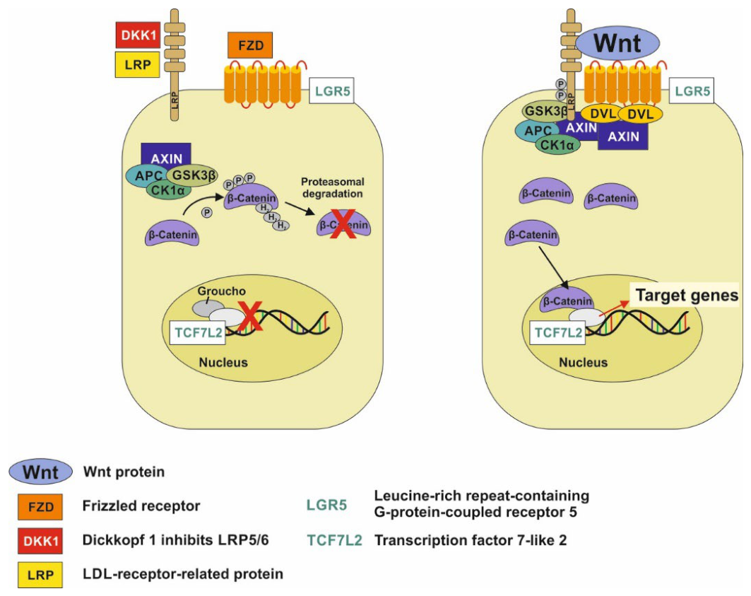
Figure 2.
Influence of Wnt signaling on the differentiation of mesenchymal stem cells (MSCs). A) Reduced Wnt signaling activity decreases the inhibition of the adipogenic transcription factor PPARγ (peroxisome proliferator-activated receptor-γ), thereby promoting the differentiation of MSCs into adipocytes. B) In contrast, increased Wnt signaling activity promotes osteogenic differentiation by upregulaltion of RUNX2 (runt-related transcription factor 2) as well as inhibition of PPARγ. Wnt signaling during MSC commitment remains to be delineated across MSC sources. In most MSC models, Wnt/β-catenin inhibits adipogenesis and supports osteogenesis, with dose–time dependence across contexts. A clinical example is osteoporosis, characterized by loss of bone tissue accompanied by an increase in adipose tissue in the bone marrow.
Figure 2.
Influence of Wnt signaling on the differentiation of mesenchymal stem cells (MSCs). A) Reduced Wnt signaling activity decreases the inhibition of the adipogenic transcription factor PPARγ (peroxisome proliferator-activated receptor-γ), thereby promoting the differentiation of MSCs into adipocytes. B) In contrast, increased Wnt signaling activity promotes osteogenic differentiation by upregulaltion of RUNX2 (runt-related transcription factor 2) as well as inhibition of PPARγ. Wnt signaling during MSC commitment remains to be delineated across MSC sources. In most MSC models, Wnt/β-catenin inhibits adipogenesis and supports osteogenesis, with dose–time dependence across contexts. A clinical example is osteoporosis, characterized by loss of bone tissue accompanied by an increase in adipose tissue in the bone marrow.
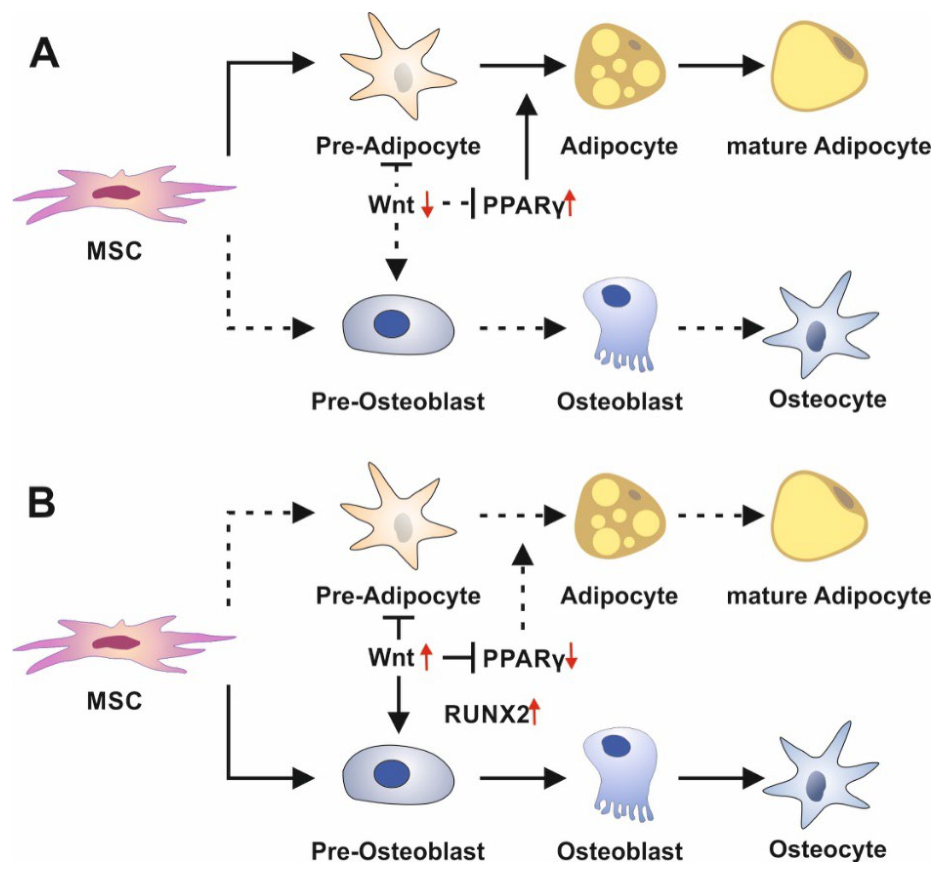
Figure 3.
Enhancement of Wnt/β-catenin signaling by GLP-1 promotes osteogenesis and inhibits adipogenesis. Binding of Wnt proteins to the Frizzled receptor/LRP complex inhibits the β-catenin destruction complex, which includes GSK3β (glycogen synthase kinase 3β), Axin, and APC. This allows increased amounts of the signal transducer β-catenin to reach the nucleus, where it binds to TCF7L2 (transcription factor 7-like 2), thereby activating the transcription of Wnt-dependent target genes. In the absence of Wnt proteins, GSK3β promotes β-catenin degradation. Under the influence of Wnt/TCF7L2 signaling, enteroendocrine cells (EECs) in the intestine produce glucagon-like peptide 1 (GLP-1). Through cAMP formation, GLP-1 signaling activates protein kinase A (PKA). PKA-mediated phosphorylation of β-catenin increases its transcriptional activity. PKA also activates phosphoinositide 3-kinase (PI3K) and AKT. AKT-mediated phosphorylation inhibits GSK3β, thereby increasing the cytoplasmic amount of β-catenin available for intracellular signal transduction. Activated GLP-1/Wnt signaling promotes osteogenesis but inhibits adipogenesis. Adapted from Meng et al. [79].
Figure 3.
Enhancement of Wnt/β-catenin signaling by GLP-1 promotes osteogenesis and inhibits adipogenesis. Binding of Wnt proteins to the Frizzled receptor/LRP complex inhibits the β-catenin destruction complex, which includes GSK3β (glycogen synthase kinase 3β), Axin, and APC. This allows increased amounts of the signal transducer β-catenin to reach the nucleus, where it binds to TCF7L2 (transcription factor 7-like 2), thereby activating the transcription of Wnt-dependent target genes. In the absence of Wnt proteins, GSK3β promotes β-catenin degradation. Under the influence of Wnt/TCF7L2 signaling, enteroendocrine cells (EECs) in the intestine produce glucagon-like peptide 1 (GLP-1). Through cAMP formation, GLP-1 signaling activates protein kinase A (PKA). PKA-mediated phosphorylation of β-catenin increases its transcriptional activity. PKA also activates phosphoinositide 3-kinase (PI3K) and AKT. AKT-mediated phosphorylation inhibits GSK3β, thereby increasing the cytoplasmic amount of β-catenin available for intracellular signal transduction. Activated GLP-1/Wnt signaling promotes osteogenesis but inhibits adipogenesis. Adapted from Meng et al. [79].
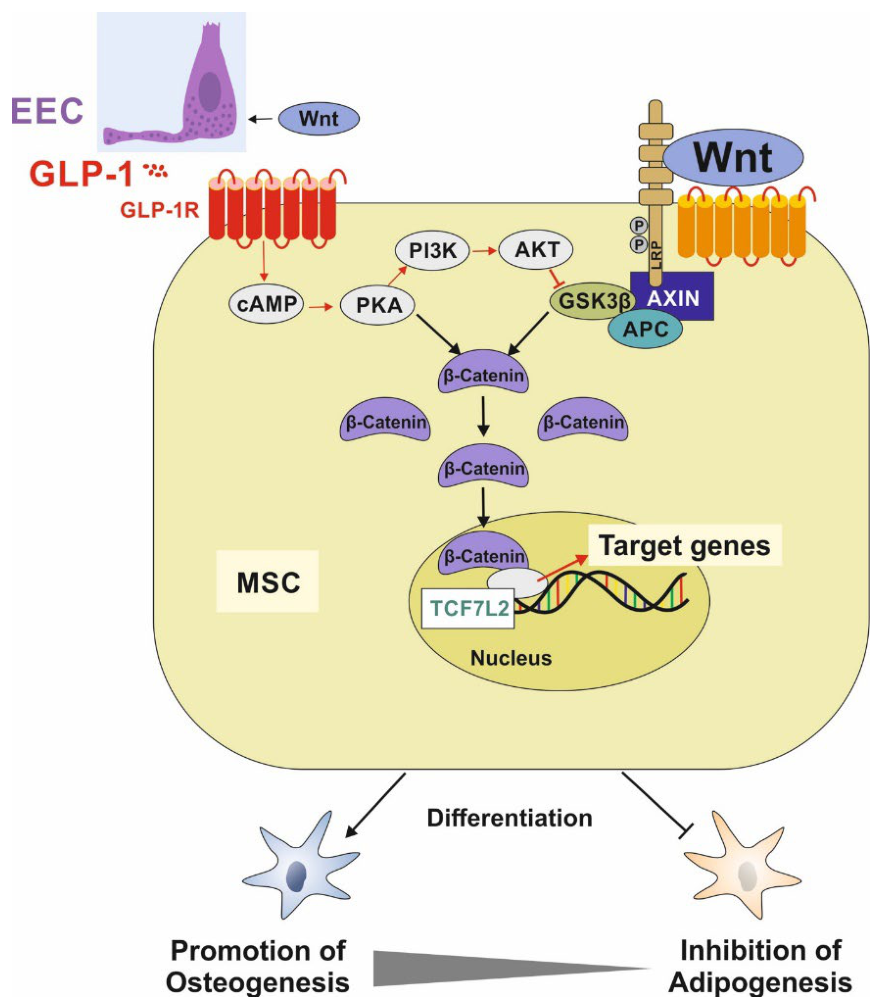
Figure 4.
Microbial metabolites, intestinal Wnt signaling, and GLP-1 secretion. Bifidobacteria and Bacteroides species produce metabolites that include acetate and lactate. Within the intestinal stem cell niche, acetate and lactate activate FFAR2 and GPR81, respectively, on Paneth cells, stimulating Wnt3 production. Wnt3 subsequently binds to Frizzled receptors on intestinal stem cells (ISCs), thereby enhancing Wnt signaling in these cells. In addition, Paneth cells produce lactate, which is made available to ISCs as a substrate for energy production via oxidative phosphorylation (OXPHOS). The Wnt gradient decreases as intestinal cells become increasingly differentiated, contributing to the spatial organization and specialization of intestinal cell types. Enteroendocrine cells also respond to microbial metabolites, such as acetate, lactate, and indole derivatives, produced by bifidobacteria and other microorganisms. These metabolites can promote the release of glucagon-like peptide 1 (GLP-1). GLP-1 reaches various target tissues through the bloodstream and can also transmit signals to central satiety and regulatory centers in the brain through afferent vagal nerve pathways.
Figure 4.
Microbial metabolites, intestinal Wnt signaling, and GLP-1 secretion. Bifidobacteria and Bacteroides species produce metabolites that include acetate and lactate. Within the intestinal stem cell niche, acetate and lactate activate FFAR2 and GPR81, respectively, on Paneth cells, stimulating Wnt3 production. Wnt3 subsequently binds to Frizzled receptors on intestinal stem cells (ISCs), thereby enhancing Wnt signaling in these cells. In addition, Paneth cells produce lactate, which is made available to ISCs as a substrate for energy production via oxidative phosphorylation (OXPHOS). The Wnt gradient decreases as intestinal cells become increasingly differentiated, contributing to the spatial organization and specialization of intestinal cell types. Enteroendocrine cells also respond to microbial metabolites, such as acetate, lactate, and indole derivatives, produced by bifidobacteria and other microorganisms. These metabolites can promote the release of glucagon-like peptide 1 (GLP-1). GLP-1 reaches various target tissues through the bloodstream and can also transmit signals to central satiety and regulatory centers in the brain through afferent vagal nerve pathways.
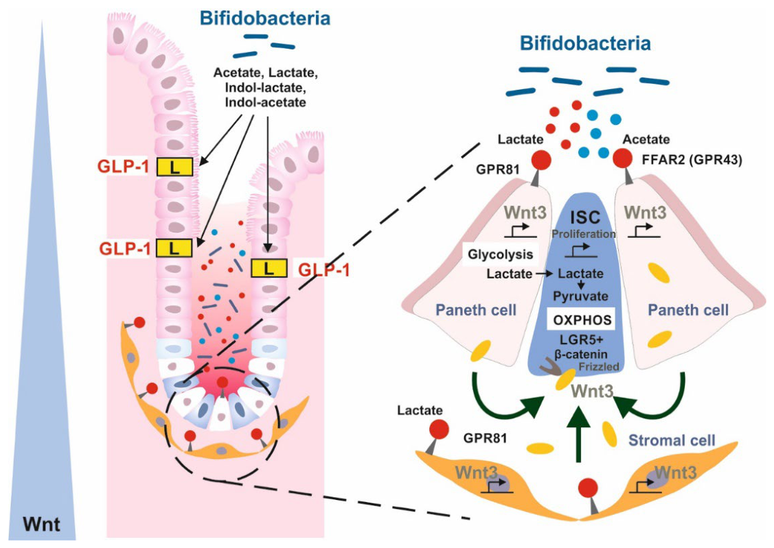
Table 1.
Observed effects of GLP-1 and GLP1R agonists on stem cells.
| Stem cell source | GLP-1/GLP-1 receptor agonist-mediated effects | Reference |
|---|---|---|
| Bone marrow MSC | GLP-1-mediated inhibiton of MSC differentiaon into adipocytes, reduced expression of PPARγ, C/EBPβ, and lipoprotein lipase (LPL). | [70] |
| Human visceral adipose-tissue | GLP-1 decreased the expression of PPARγ mRNA | [71] |
| Human adipose-derived stem cells | Liraglutide inhibits adipogenesis | [72] |
| Human adipose-derived stem cells | Liraglutide activates Wnt signaling and inhibits adipogenic differentiation | [73] |
| Human adipose- derived stem cells | GLP-1 inhibits adipogenic differentiation, reduces the expression of PPARγ, LPL and adipocyte protein 2 (AP2) but enhances osteogenic differentiation | [74] |
| Adipose-derived stem cells | Exendin-4 inhibits adipogenic differentiation but promotes osteogenic differentiation | [75] |
| Human adipose- derived stem cell | GLP-1 promotes osteogenic differentiation via the Wnt/GSK-3β/β-catenin pathway | [76] |
| Bone marrow-derived MSC | GLP-1 inhibits adipogenesis and enhances osteogenesis | [77] |
| Bone marrow-derived MSC | Semaglutide promotes proliferation and osteogenic differentiation of bone-derived MSCs through activation of the Wnt/LRP5/β-catenin signaling pathway | [78] |
| Bone marrow stromal cell | Exendin-4 inhibits adipogenic differentiation but promotes osteogenic differentiation through β-catenin | [79] |
Table 3.
Overview of the possible combinations of factors affecting adipogenic differentiation of MSCs during the perinatal period.
Table 3.
Overview of the possible combinations of factors affecting adipogenic differentiation of MSCs during the perinatal period.
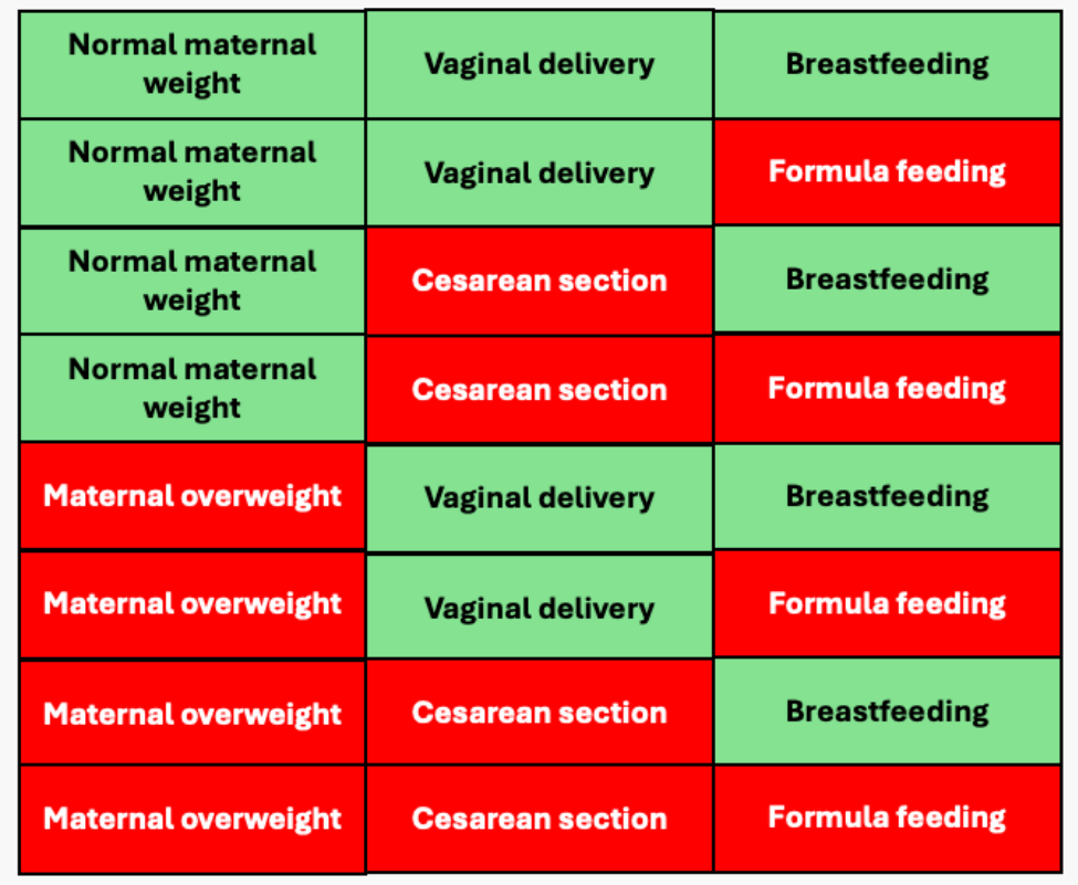 |
Disclaimer/Publisher’s Note: The statements, opinions and data contained in all publications are solely those of the individual author(s) and contributor(s) and not of MDPI and/or the editor(s). MDPI and/or the editor(s) disclaim responsibility for any injury to people or property resulting from any ideas, methods, instructions or products referred to in the content. |
© 2026 by the authors. Licensee MDPI, Basel, Switzerland. This article is an open access article distributed under the terms and conditions of the Creative Commons Attribution (CC BY) license (http://creativecommons.org/licenses/by/4.0/).
Copyright: This open access article is published under a Creative Commons CC BY 4.0 license, which permit the free download, distribution, and reuse, provided that the author and preprint are cited in any reuse.