

Submitted:
23 October 2026
Posted:
27 October 2026
You are already at the latest version
Abstract
Hereditary sensory and autonomic neuropathy type IV [HSAN IV] is a rare ge-netic disorder of children characterized by the inability to feel pain and to sweat [anhidrosis]. There is insensitivity to temperature. Dysfunction of the sensory nerves which are responsible for pain and temperature causes the sensory loss. NTRK1 gene variants cause this disorder, inherited in an autosomal recessive pattern. This review aims to synthesize current research on the clinical, molecular, and diagnostic aspects of Hereditary Sensory and Autonomic Neuropathy -IV [HSAN IV] and identify gaps for future research. The objectives include reviewing studies on HSAN's clinical features and genetic variations, assessing the effectiveness of diagnostic methods, genetic testing and highlighting inconsist-encies in genotype-phenotype correlations. We searched PubMed, Scopus, Web of Science, Embase and Google Scholar using terms 'HSAN' and 'genetic vari-ants', with no time frame restrictions. The literature on the clinical and molec-ular characteristics, diagnostics, therapeutics including recent advances of HSAN IV has been reviewed. This review highlights advancements in understanding HSAN's clinical and genetic aspects, while emphasizing the need for further re-search to address gaps in genetic knowledge and improve diagnosis and treatment outcomes.
Keywords:
HSAN
; genetic diseases
; paediatric neuropathy
; rare diseases
1. Introduction
Hereditary Sensory and Autonomic Neuropathy [HSAN] type IV or Congenital Insensitivity to Pain with Anhidrosis [CIPA] eponymously the Nishida syndrome, is an autosomal recessive condition with congenital insensitivity to pain, temperature, anhidrosis and cognitive impairment. Swanson et al.,first described this in 1963 [1,2]. HSAN IV is one of the rarer types of HSANs [2]. The NTRK1[Neurotrophic Receptor Tyrosine Kinase 1] gene, coding for the Tyrosine Related Kinase A [TRK-A] protein, is mutated in this condition [3]. TRK-A a Nerve Growth Factor [NGF] receptor by binding to NGFs maintains the functionality of the neurons - NGF-dependent primary efferent nerves responsible for the nociceptive and homeostatic afferent pathways. The interaction between TRK-A and NGF is disrupted in HSAN IV [4]. This results in neurological, orthopaedic, psychiatric oral, dermatological and ophthalmological symptoms in children [5,6,7,8,9,10,11,12]. The clinical presentation of HSAN-IV is reduced perception of pain and temperature stimuli with intact tactile, vibratory and position perception [13]. Deep Tendon reflexes are preserved. Nerve conduction studies demonstrate normal conduction velocities. nerve biopsies reveal loss of unmyelinated and thin myelinated Aδ-fibres [12]. Sweat glands are intact, but without nervous innervation on histopathological examination. Genetic testing is the definitive diagnostic test for this condition -direct DNA sequencing to identify point variations, small insertions or deletions [14]. Next Generation Sequencing [NGS] enables parallel processing of multiple sequencing. Whole Exome Sequencing [WES] helps identify novel anomalies in addition to existing ones.
The literature on the prevalence of HSAN IV and diagnostic guidelines are vague and sparse [15,16]. Multi-organ system involvement and recurrent exacerbations limit the lifespan of these patients. Consanguineous marriages, prevalent in India, increase the risk of this condition [2,16]. This review aims to understand this condition better- by exploring the pathophysiology, the clinical and molecular characteristics, occupational support for patients enhancing their quality of life and recent advances.
2. Materials and Methods
Google Scholar, Scopus, PubMed and Web of Science were used for the literature search. The MeSH terms [for PubMed] and the free-text keywords [for Google Scholar, Scopus and Web of Science] - “Genetic diseases”, “Hereditary Sensory Autonomic Neuropathy IV”, “Congenital Insensitivity to Pain with Anhidrosis” and “Paediatric Neurology” were combined using the Boolean operators “OR” and “AND”. The search was done on 26/05/2026. The inclusion criteria were all papers in English after 1990. Expert opinion articles, conference abstracts and non-peer reviewed articles were excluded. The papers were selected and screened by two authors [NSU and SG] and any discrepancy was resolved by a third author [BS] resolved through discussion. A total of 75 articles were screened and 49 were included for this review. AI assistance was used for summarizing literature, drafting a table and to generate infographic figures.
3. Results and Discussion
3.1. General Overview of HSAN
Hereditary neuropathy comprises a diverse group of disorders affecting the peripheral nervous system, with widely varying clinical presentations and genetic etiologies. These disorders are broadly divided into two categories: those in which neurons are primarily affected either alone or as the prime element of the disorder , and those in which the neuropathy is merely one component of a more generalized or systemic illness. Hereditary sensory and autonomic neuropathies [HSAN] represents a heterogeneous class of hereditary neuropathies in which sensory, and occasionally autonomic neuronal elements are affected. HSANs are subclassified into types I through V, with type I being the most common and type IV the second most common. Types IV and V are closely related and are reportedly associated with mutations in the NTRK1 gene. The triad of pain, insensitivity, anhidrosis and cognitive impairment in Type V help differentiate it from Type IV [17]. Hereditary Sensory Autonomic Neuropathy IV [HSAN IV] is the second most common HSAN and, like other recessively transmitted HSANs, has its onset in infancy. What distinguishes HSAN IV from the other subtypes is the extensive involvement of ectodermal structures, [skin, bone, and the nervous system] with absent or markedly decreased sweating representing the cardinal feature of the disorder [18,19,20,21].Mutations of the NGF - Beta gene [NGF-B] with milder anhidrosis are seen in Type V. Fungiform tongue papillae and intact lacrimal gland function in Type IV is further distinguished by preservation of lacrimal gland function and of the fungiform tongue papillae, features that differentiate it from Type III HSAN [17].
HSAN IV must also be differentiated from the other Hereditary Sensory Autonomic Neuropathies [HSAN]. Type I [Hereditary Sensory Radicular Neuropathy] is a relatively mild condition manifesting between the second and fourth decades of life and affecting the lower limbs predominantly. Type II [Congenital Sensory Neuropathy] is also associated with loss of sweating, but temperature regulation and blood pressure control remain normal. Type III [Familial Dysautonomia, or Riley–Day syndrome] presents with a multisystem picture that involves multiple systems and includes postural hypotension, Ataxia, Kyphoscoliosis, Oropharyngeal dyscoordination, and abnormal Gastro-Esophageal motility resulting in feeding difficulties and recurrent aspiration Pneumonia. The Type V variant disease resembles type IV but is milder , without intellectual disability or significant anhidrosis, and selectively affects nociception. Lesch–Nyhan syndrome which is a differential diagnosis, resulting from Hypoxanthine–Gguanine Phospho-Ribosyl Transferase [HGPRT] deficiency, is characterized by hyperuricemia, self-induced trauma, intellectual disability, Chorea, and Athetosis [21].
Historically, Type IV HSAN, also known as [Nishida syndrome], was first described as early as the 1900s by Dearborn as a case of ‘pure congenital analgesia,’ and was more thoroughly investigated and reported some sixty years later by Swanson [17].
3.2. Etiopathogenesis
HSAN IV was first described in 1963 by Swanson in two male siblings in two brothers affected by defective temperature sensation and insensitivity to pain. Consanguineous marriages are seen in approximately half of the reported cases.Approximately half of reported cases have occurred in consanguineous marriages. HSAN IV arises from mutations in NTRK1, located on chromosome 1, which encodes the tyrosine kinase receptor for Nerve growth factor [NGF]. Mutations in this gene lead to failure of differentiation and migration of neural crest cells, resulting in the near-complete absence of small myelinated and unmyelinated nerve fibers, with consequent loss of pain and temperature sensation; sweat glands are also left uninnervated, producing anhidrosis [24].
Loss-of-function mutations of the NTRK1 [Neurotrophic Receptor Tyrosine Kinase 1] on chromosome 1q21-22 [OMIM 256800] causes this condition.This gene plays a crucial role in the development of nociceptive sensory and sympathetic autonomic neurons within the dorsal root sensory ganglia, -which is reflected in this disorder - histopathologically by the absence of small unmyelinated nerve fibers and by the denervation and atrophic changes of the eccrine sweat glands. Clinically, this is evident as anhidrosis with unresponsiveness to Pilocarpine in affected patients [17].
It is the anhidrosis that gives rise to episodic fevers and extreme hyperpyrexia, usually the earliest sign of the disorder, and this can precipitate recurrent febrile convulsions secondary to elevated environmental temperature. The anhidrosis is thought to be secondary to impaired thoracolumbar sympathetic outflow. It is evident on the trunk and upper extremities in all cases, while other body regions are variably affected, and it results in thickened, callused skin with lichenification of the palms, dystrophic nails, and areas of hypotrichosis on the scalp [23,24,25].
HSAN IV is associated with both decreased sensation and autonomic dysfunction. While anhidrosis [complete loss of sweating] produces cutaneous changes such as severe xerosis, lichenification and scaling, eczematous dermatitis, blisters, ulcers and scars, the accompanying sensory insensitivity is far more profound, resulting in self-mutilation, auto-amputation, and corneal scarring. Although no immunological abnormality is present, individuals with HSAN IV have marked difficulty healing ectodermal structures such as skin and bone; fractures are slow to heal, and large weight-bearing joints are particularly susceptible to repeated trauma, frequently progressing to Charcot’s joints and osteomyelitis. Speech is usually clear, and although hypotonia and delayed developmental milestones are common in early childhood, strength and tone tend to normalize with age; nevertheless, severe learning problems, hyperactivity, and emotional lability are frequently observed. Intellectual disability in HSAN IV is variable, ranging from severe to mild, and some patients initially considered to have normal cognition have later exhibited mild impairment. [22].
Although no other disorder exactly mimics HSAN IV, impairment of pain sensation together with oral mutilation has also been reported in conditions such as Lesch–Nyhan syndrome, Tourette syndrome, and De Lange syndrome. Absence of nail and hair abnormalities and the presence of normal sweat glands on skin biopsy help to exclude anhidrotic or hypohydrotic ectodermal dysplasia caused by abnormality in EDA [Ectodysplasin A] gene which also causes developmental issues in skin, hair and teeth. Because nerve conduction studies assess only one to two percent of large myelinated fibers, they may lack the sensitivity to detect abnormalities confined to small fibers. Nerve conduction studies are limited in their sensitivity to detect abnormalities of large myelinated fibres [1-2% sensitivity] rendering them useful for only small fibres. An appropriate number of sweat glands on skin biopsy despite anhidrosis has been reported in this condition. A normal number of sweat glands on skin biopsy, despite clinical anhidrosis, is consistent with previous reports in the literature [23].
3.3. NTRK1 Mutations in HSAN IV
Several variation of NTRK1 gene mutations have been reported, exceeding fifty in number, including frameshift, altered splice-site, missense, intragenic polymorphic, and deletion mutations - with frameshift mutations representing the most common category and the 586fs variant being the most frequently observed site [13,14].
At the molecular level, a novel NTRK1 mutation associated with HSAN IV has been described as consisting of a G→C transversion at nucleotide 2405, causing an Arg→Pro substitution at codon 774. A loss-of-function effect is caused by this mutation. Biological and biochemical studies were consistent with a loss-of-function effect, confirming the pathogenic role of this mutation. Residue Arg774 is located at the C-terminus of the NTRK1 tyrosine kinase domain and is surrounded by residues conserved across members of the TRK receptor family. In spite of this mutation being unique to NTRK1, the functional importance highlighted by data, its precise role remains unclear. All the reported mutations associated with HSAN IV occur within the NTRK1 tyrosine kinase domain, whereas point mutations in the extracellular portion of NTRK1 and mutations in other genes encoding neurotrophin receptors, might give rise to other variants of HSAN [26].
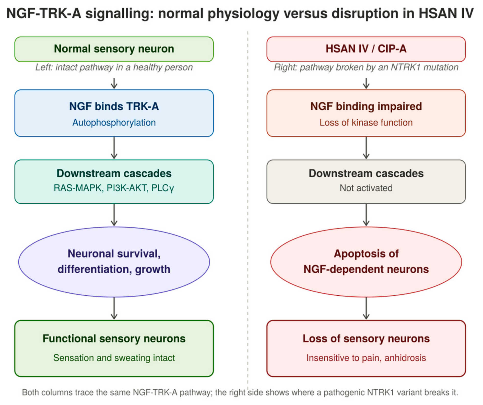
TRKA [Tropomysin receptor Kinase A], encoded by NTRK1, is the receptor tyrosine kinase receptor for nerve growth factor [NGF] - mutation of this gene is responsible and is the gene responsible for HSAN IV. [Figure 1] Defects in NGF signal transduction at the TRKA receptor lead to the death or failure of survival of sympathetic ganglion neurons and nociceptive sensory neurons derived from the neural crest. Thirty-seven different NTRK1 mutations have been identified in patients across various countries, - comprising nine frameshift, seven nonsense, seven splice, and fourteen missense mutations,. are They are distributed across an extracellular domain involved in NGF binding as well as the intracellular signal-transduction domain. Naturally occurring TRKA mis-sense mutations with loss of function provide considerable insight into the structure–function relationship within the receptor tyrosine kinase receptor family, and the continued characterization of HSAN IV-associated mutations aids diagnosis and genetic counseling for this severe disorder [27].
A comprehensive functional analysis of seven novel NTRK1 missense mutations — c.1550G>A, c.1565G>A, c.1970T>C, c.2096T>C, c.2254T>A, c.2288G>C, and c.2311C>T, corresponding to p.G517E, p.G522E, p.L657P, p.I699T, p.C752S, p.C763S, and p.R771C — found all seven to contribute to the pathophysiology of the disease by in-silico analysis. Each mutation was systematically evaluated for the TRKA glycosylation status, intracellular and cell-membrane expression, NGF-stimulated TRKA auto-phosphorylation, TRKA-Y496 phosphorylation, PLCγ activity, and neurite outgrowth. A diverse range of functional effects was noted, with one mutation appearing fully functional, another retaining partial activity across assays, one affecting only the PLCγ pathway, and four proving functionally null across all assays. This demonstrated that complete abolition of TRKA kinase activity is not the only pathogenic mechanism underlying HSAN IV [28].
A 9-bp deletion [c.354_359+3delTCGCCTGAA], resulting in skipping of exons 2 and 3, was identified in a HSAN IV patient of Turkish origin [CMT-860.01]; the main features in this case were the absence of obvious mental retardation and less pronounced anhidrosis [29]
Additional NTRK1 mutations have been reported, including c.1549G>C [p.Gly517Arg], a novel mutation not previously described, and c.851-33C>T, which was linked with ‘mild’ manifestations and identified as one of the founder mutations among Chinese HSAN IV patients. A total of four different NTRK1 mutations — c.851-33T>A, c.44G>A [p.Trp15*], c.287+2dupT, and c.1549G>C [p.Gly517Arg] — were identified across these families, expanding the mutational spectrum of NTRK1 and providing further insight into genotype–phenotype correlations in HSAN IV [30].
Sequencing of NTRK1 in a further proband revealed two novel compound heterozygous missense mutations: c.1635G>C [p.Ala527Pro] in exon 13, inherited from a heterozygous father, and c.2197G>A [p.Gly714Asp] in exon 16, which arose de novo since neither parent carried the variant. Bioinformatic analyses, including PolyPhen-2, predicted both substitutions as probably damaging to protein function, and alignment across species showed that both affected residues conserved from zebrafish to humans during evolution, supporting their pathogenicity in HSAN IV [31].
Functional splicing studies validated the impact of a novel intronic variant, c.850+5G>A, on NTRK1 pre-mRNA splicing, showing that it generated two truncated protein products along with decreased mRNA and protein expression. This variant was identified in a HSAN IV pedigree alongside the known East Asian hotspot splicing variant c.851-33T>A, together expanding the variant spectrum of NTRK1 relevant to genetic diagnosis of HSAN IV [32].
A broader mutational survey identified 27 NTRK1 mutations, of which 15 were novel. The twelve previously known, HGMD-recorded pathogenic variants included five missense mutations [c.1784T>G, c.1927C>T, c.2056C>T, c.2152G>A, and c.2293C>T, producing p.L595R, p.R643W, p.R686C, p.G718S, and p.R765C respectively], one nonsense mutation [c.1786C>T, p.R596*], two frameshift mutations [c.963delG, c.1736delT], and four intronic splicing mutations [c.851-33T>A, c.287+2dupT, c.850+1G>A, and c.2188-11G>A]. The two most common mutations, c.851-33T>A and c.287+2dupT, together accounted for 24 of 68 alleles. A large intragenic deletion, c.429-374_717+485del [~1403 bp], mediated by recombination between Alu elements spanning exons 5–6 and introns 4 and 6, was also identified in several probands as either homozygotes or compound heterozygotes, suggesting a possible founder effect which is a loss of genetic variation that happens when a new population is set up by a very small group of individuals from a larger parent group [33].
Whole-genome sequencing further identified two recurrent deep intronic variants in intron 7, c.851-794C>G and c.851-798C>T, absent from public population databases [1000 Genomes, dbSNP141, gnomAD]. RNA analysis demonstrated five forms of abnormal alternative splicing between exons 7 and 8, with c.851-794C>G predicted to create a cryptic donor splice site that activates upstream cryptic acceptor sites, leading to inclusion of variably sized pseudo-exons and consequent frameshift. Separately, a heterozygous intronic variant, c.575-19G>A — found in three unrelated families and homozygous together with c.326A>G in another — was shown by mini-gene assay to create a novel splice acceptor site, resulting in inclusion of a 17-bp intronic sequence, premature translational termination, and a truncated protein product [p.P194Lfs*9] [33].
Structurally, the TrkA receptor encoded by NTRK1 comprises three functional domains: an extracellular domain encoded by exons 1–8, including two immunoglobulin-like domains important for NGF binding; a transmembrane domain encoded by exon 11; and an intracellular tyrosine kinase domain encoded by exons 13–17 that is essential for signal transduction. The observation that most reported mutations cluster within the extracellular and intracellular tyrosine kinase domains underscores the functional importance of these regions [33].

Table 1.
NTRK1 Mutations Reported Across Different Gene Regions.
| Region / Exon-Intron | Nucleotide Change [cDNA] | Protein Change | Mutation Type | Notes |
| Extracellular domain [NGF-binding] & intracellular signal-transduction domain — general distribution | — | — | Aggregate: 9 frameshift, 7 nonsense, 7 splice, 14 missense | 37 different TRKA mutations identified across patients in various countries |
| - | c.1550G>A | p.G517E | Missense | Novel; predicted pathogenic by in-silico analysis |
| - | c.1565G>A | p.G522E | Missense | Novel; predicted pathogenic by in-silico analysis |
| - | c.1970T>C | p.L657P | Missense | Novel; predicted pathogenic by in-silico analysis |
| - | c.2096T>C | p.I699T | Missense | Novel; predicted pathogenic by in-silico analysis |
| - | c.2254T>A | p.C752S | Missense | Novel; predicted pathogenic by in-silico analysis |
| - | c.2288G>C | p.C763S | Missense | Novel; predicted pathogenic by in-silico analysis |
| - | c.2311C>T | p.R771C | Missense | Novel; predicted pathogenic by in-silico analysis |
| Extracellular domain — Exon 3 | c.354_359+3delTCGCCTGAA | — | 9-bp deletion [splicing] | Causes skipping of exon 3 [and exons 2–3]; identified in a CIPA patient of Turkish origin [CMT-860.01] |
| - | c.1549G>C | p.Gly517Arg | Missense | Novel mutation, not previously reported |
| Intronic [splice region] | c.851-33C>T | — | Splice-region | Associated with a “mild” phenotype; founder mutation in Chinese CIPA patients |
| Intronic [splice region] | c.851-33T>A | — | Splice-region | “Mild” phenotype; East Asian hotspot variant; one of the two most common NTRK1 mutations [24/68 alleles] |
| - | c.44G>A | p.Trp15* | Nonsense | — |
| Intronic [splice region] | c.287+2dupT | — | Splice-site duplication | One of the two most common NTRK1 mutations [24/68 alleles] |
| Extracellular domain — Exon 13 | c.1635G>C | p.Ala527Pro | Missense | Compound heterozygous; paternally inherited |
| Intracellular tyrosine kinase domain — Exon 16 | c.2197G>A | p.Gly714Asp | Missense | Compound heterozygous; de novo mutation [absent in both parents] |
| Intronic [splice region, near exon 7/8 boundary] | c.850+5G>A | — | Splice-region [novel] | Generates two truncated proteins; decreased mRNA and protein expression; leads to CIPA |
| Extracellular domain — Exons 5–6 / Introns 4 & 6 | c.429-374_717+485del | — | Large intragenic deletion [~1403 bp] | Alu-element mediated recombination; homozygous/compound heterozygous in probands; possible “founder effect” |
| Intron 7 | c.851-798C>T | — | Deep intronic substitution | Recurrent variant; part of compound change c.[851-798C>T;851-794C>G]; not present in 1000 Genomes, dbSNP141, gnomAD |
| Intron 7 | c.851-794C>G | — | Deep intronic substitution | Creates a cryptic donor splice site; activates upstream cryptic acceptor sites; causes pseudo-exon inclusion and frameshift |
| - | c.1784T>G | p.L595R | Missense | Known pathogenic variant [HGMD] |
| - | c.1786C>T | p.R596* | Nonsense | Known pathogenic variant [HGMD] |
| - | c.1927C>T | p.R643W | Missense | Known pathogenic variant [HGMD] |
| - | c.2056C>T | p.R686C | Missense | Known pathogenic variant [HGMD] |
| - | c.2152G>A | p.G718S | Missense | Known pathogenic variant [HGMD] |
| - | c.2293C>T | p.R765C | Missense | Known pathogenic variant [HGMD] |
| - | c.963delG | — | Frameshift | Known pathogenic variant [HGMD] |
| - | c.1736delT | — | Frameshift | Known pathogenic variant [HGMD] |
| Intronic [splice region] | c.850+1G>A | — | Splice-site | Known pathogenic variant [HGMD] |
| Intronic [splice region] | c.2188-11G>A | — | Splice-site | Known pathogenic variant [HGMD] |
| Compound genotype | c.[326A>G;575-19G>A] | — | Compound [missense + intronic splice] | Homozygous in one family |
| Intronic [splice region] | c.575-19G>A | p.P194Lfs*9 [predicted] | Splice-site [creates novel acceptor site] | Heterozygous in three unrelated families [Families 19, 28, 32]; 17-bp intronic sequence inclusion; premature termination/truncated protein |
NTRK1 mutations - inheritance patterns - genotype and phenotype variations as reported.
Figure 1.
NTRK signalling pathway- normal versus disruption in HSAN IV.
3.4. Clinical Presentation
The cardinal constant features of HSAN IV, present in most patients, are anhidrosis, episodes of pyrexia-of-unknown origin, episodes of unexplained fever [usually the earliest sign of the disorder], intellectual disability, insensitivity to pain, and self-mutilating behavior. Additional features frequently present include multiple fractures, joint dislocations and deformities, skin infections, bruising, corneal ulceration, and aggressive behavior. Less commonly, the condition can present with impaired immune response, proteinuria, renal failure, anemia, hyperkeratosis, dry skin, and early primary tooth loss. Death from hyperpyrexia within the first three years of life occurs in approximately 20% of cases. Pain insensitivity leads to deep ulceration over the knees and elbows, overuse of bones and joints with recurrent fractures, osteomyelitis, joint dislocations, and Charcot joints -affected children commonly sustain self-inflicted injuries in the form of skin ulcers, burns, bone fractures, and auto-amputation of the fingertips and tongue. Cutaneous features result from a combination of anhidrosis and repeated trauma. Anhidrosis contributes to xerosis and a thick, callused appearance of the skin with lichenification of the palms, while skin infections, bruises, and scars arise from repeated trauma or self-mutilation. Typical oral manifestations include ulceration of the ventral tongue surface from repeated biting during infancy, which usually leads to loss of the tongue tip by the second year of life; lip wounds are most often confined to the lower lip, and multiple missing teeth from auto-extraction may be seen in up to 50% of patients. Histologically, the skin and appendageal structures are normal, with absence of non-myelinated and small myelinated nerve fibers. Sweat glands appear structurally normal but lack innervation by small-diameter neurons. Motor and sensory peripheral nerve conduction velocities are usually normal on electromyographic examination [24].
A case report describes a 22-month-old girl born to consanguineous parents who presented with high-grade fever, insensitivity to painful stimuli, and absence of perspiration, along with self-mutilating behavior and insensitivity to temperature - hot or cold stimuli applied to the body. Examination revealed loss of the tongue-tip , missing teeth, generalized xerosis, ulcers at sites of minor trauma, dysplastic nails, and digital ulcers. Sensory examination demonstrated complete absence of pain and temperature perception, while vibration and fine touch remained intact and lacrimation was normal. Differential diagnoses included HSAN, Lesch–Nyhan syndrome, hypohidrotic ectodermal dysplasia, and leprosy -; routine investigations, including serum uric acid, were normal. Clinical exome sequencing confirmed a diagnosis of autosomal recessive HSAN IV, identifying a novel, predicted pathogenic variant at exon- 16 of the NTRK1 gene [25].
Hyperpyrexia, though common, remains a major cause of mortality in these patients; death occurs in approximately 20% of cases by three years of age, and in up to 43% over the lifespan when combined with sepsis, reflecting the poor tolerance of these patients to physiological stress. Staphylococcal infections of the skin and soft tissue are among the most commonly reported infections in HSAN IV and carry a high rate of complications, underscoring the importance of aggressive management and early use of local antiseptics.Congenital Insensitivity to Pain with Anhidrosis is an exceedingly rare disease, with only 31 cases reported at the time of one review. It is a severe autosomal recessive condition that produces self-mutilation within the first few months of life.. Bone fractures, multiple scars, Osteomyelitis, joint deformities, limb auto-amputations and intellectual disability is seen as these children grow older. and death from hyperpyrexia occurs within the first three years of life in almost 20% of patients. Ultrastructural and morphometric studies of peripheral nerves demonstrate loss of unmyelinated and small myelinated fibers, although the precise physiopathologic mechanism of this developmental disorder remains unknown [30].
3.4.1. Orthopaedic Manifestations of HSAN IV
The orthopaedic manifestations of HSAN IV include atypical fractures, infections, growth disturbance, joint subluxation, and Charcot joints of the limbs and spine. Preventive strategies include padded footwear, gait and posture modification, parental education regarding environmental thermoregulation, and behavioural support [34].
Healing of ectodermal structures such as skin and bone is challenging in patients with HSAN IV. Fractures are slow to heal, and large weight-bearing joints appear particularly susceptible to repeated trauma, frequently progressing to Charcot joints and osteomyelitis [1,3,12].
A nine-year-old female patient presenting with painless fractures, delayed healing, and abnormal callus formation was diagnosed with HSAN IV; a negative sweat test, indicating anhidrosis, supported the clinical diagnosis of this condition [9].
Three clinical groups of HSAN-IV patients have been described based on orthopaedic presentation: type A, characterised by multiple infections; type B, characterised by fractures, growth disturbances, and avascular necrosis; and type C, characterised by Charcot arthropathy and joint dislocations in addition to fractures and infections. Aspirates and cultures should be used to diagnose infection, thereby limiting the need for unnecessary surgery, while wide surgical debridement is the advocated treatment for established infections. Footwear modification, corrective osteotomies, and epiphysiodesis have been suggested as surgical measures for limb-length discrepancies arising from this condition, while patient education, footwear modification, and periods of non-weight-bearing can serve as both preventative and therapeutic measures for decubitus ulcers [35].
Kohler et al. described the orthopaedic course of a 10-year-old patient with HSAN IV who presented with a range of distinctive orthopaedic challenges. While several of her orthopaedic injuries healed adequately following treatment, other injuries such charcot joint neuropathy, delayed healing, infections, osteomyelitis, etc resulted in severe complications and progressive joint destruction [36].
Beyond isolated fractures, affected children commonly display multiple fractures together with joint dislocations and deformities. Insensitivity to pain leads to deep ulceration over the knees and elbows and to chronic overuse of bones and joints, resulting in recurrent fractures, osteomyelitis, joint dislocations, and Charcot joint deformities; self-inflicted injuries in these children frequently extend to auto-amputation of the fingertips in addition to skin ulcers, burns, and bone fractures [24].
Because these fractures and joint injuries are not perceived as painful, they often present late and require frequent surgical intervention; operating surgeons should also bear in mind that intraoperative or postoperative tachycardia and hypertension may reflect an unconscious physiological pain response, despite the patient’s lack of conscious pain awareness due to a reduced number of peripheral pain fibres [24].
Early recognition of the orthopaedic complications of HSAN IV, together with prevention of accidental injury, regular follow-up, and coordinated multidisciplinary management, has been proposed as a means of reducing the frequency and severity of these complications over the course of the disease [37].
Congenital insensitivity to pain with anhidrosis [HSAN IV] is a rare hereditary sensory neuropathy comprising congenital insensitivity to pain, anhidrosis, and mental retardation. A 4-year-old child with HSAN IV and a calcaneal ulcer was treated with double opposing rotation flaps, which eventually healed. [38]
A 10-year-old Caucasian boy was admitted with swelling of the right thigh and high fever, having received an initial diagnosis of HSAN IV at 3 years of age on the basis of recurrent painless fever episodes, inability to sweat, corneal opacities, and mental retardation, later confirmed by genetic and dermal studies. His family had first noticed painless swelling around the knee joint at age 4, which went unreported until a febrile illness prompted hospital admission. He underwent knee debridement and intravenous antibiotics for septic arthritis, followed by a second operation three weeks later for recurrence and spread of infection to the right femoral metaphysis, and subsequently underwent several further operations on the right knee and thigh prior to the index admission. He also had dystrophic nails, healthy permanent teeth, and a history of premature loss of deciduous teeth around age 3, along with bilateral central corneal opacities suggestive of prior neurotrophic keratitis. On examination, the right thigh was edematous but non-tender, permitting pain-free walking, and was 10 cm shorter than the left, producing a limping gait; multiple wound scars were present on all extremities. The right thigh circumference measured 47 cms compared with 26 cms on the left, pyrexia with warmth and fluctuation on palpation but no discharge. The haemoglobin was 8.4 g/dL, white cell count 20,000, ESR 90, and CRP 50. Radiographs of the lower extremities demonstrated shortening, deformity, and sequestrum in the distal right femur with proximal tibial thickening, consistent with an acute flare of chronic osteomyelitis. Open debridement and irrigation were performed, followed by six weeks of intravenous Teicoplanin guided by intraoperative culture growth of Methicillin-Resistant Staphylococcus Aureus; at six months postoperativelyThere was no wound discharge and no major radiographic change.[39]
3.4.2. Oral Manifestations of HSAN IV
Oral anomalies as a pointing sign for HSAN IV were highlighted by O’Hagan-Wong et al. in their report of a five-month-old infant presenting with a bifid tongue. The scanty literature available on this presentation was noted to make diagnosis particularly challenging [10].
In a retrospective review of eight patients, Ashwin DP et al. reported oral and digital injuries, including auto-extraction of teeth and severe bite injuries of the fingertips and oral soft tissues. The authors concluded that prompt diagnosis coupled with a supportive management plan could help prevent or reduce the severity of such injuries [40].
Oral and craniofacial manifestations reported in the literature include a large number of missing teeth, nasal malformation, submucous cleft palate, severe soft tissue injuries, dental caries, and malocclusion [41].
A case reported in the literature described a patient born after a normal pregnancy and delivery who, from the third month of life onward, experienced recurrent unexplained fevers. An absence of sweating with dry, warm skin was consistently noted during these febrile episodes. The patient was also diagnosed with congenital corneitis and submucous cleft palate, and began biting his tongue and fingers after his teeth erupted at eight months of age [41].
Scratch marks, erosions, and cicatricial scarring resulting from self-inflicted soft tissue trauma were documented, along with congenital nasal defects such as a depressed nasal bridge, malformed nasal apex, narrow nostrils, a dysmorphic columella, and a deviated nasal septum. Skin erosions on the left nasal alae and corneal ulcerations in the right eye, attributable to scratching, were also observed. Cranial CT scans demonstrated a submucous cleft palate along with alveolar bone loss of the maxilla and mandible [41].
Oral examination in this case revealed erosions, scarring, and hyperkeratosis of the soft tissues, including the tongue, lips, gingiva, and palatal and buccal mucosa. A decreased ability to open the mouth was attributed to a fibrous band of scar tissue in the cheeks and lips. Early tooth loss was noted, involving almost all primary teeth with the exception of a carious and mobile left mandibular primary second molar, along with several permanent teeth. Panoramic radiographs revealed that fifteen permanent teeth were missing, including the lower central incisors, the right premolars, and all maxillary teeth except the second molars and the left canine; the remaining teeth exhibited short-root anomalies. The patient’s guardians confirmed that the missing teeth had not been congenitally absent but had erupted before being lost to auto-extraction or self-mutilation [41].
With respect to dental history, another reported patient had no established pattern of dental care at home and sought emergency dental treatment only in response to pain and swelling. Oral hygiene practices were suboptimal, with irregular toothbrushing [four times a week] and no use of dental floss. The dental history revealed a recurrent pattern of dental infections leading to extra-oral facial cellulitis over a four-year period, caused by carious teeth. Owing to the patient’s lack of cooperation, no definitive dental treatment could be performed in the clinic beyond antibiotic prescription, and the patient subsequently underwent two dental rehabilitation procedures under general anesthesia, the most recent occurring a year prior to presentation; these interventions included multiple restorations and extractions of both primary and permanent teeth [42].
Clinically, this patient presented as cheerful and friendly during the initial dental examination, with developmental delay and dry skin evident on general appearance. Extra-oral examination revealed self-inflicted nail-biting behavior resulting in nearly absent fingernails, as well as biting marks on the fingers and multiple injuries on the toes and soles of the feet. Intra-oral examination showed ulceration and crusting on the left side of the lower lip, multiple traumatic ulcers on the ventral and dorsal aspects of the tongue, and extensive ulcers on the right buccal mucosa and retromolar pad region, along with old scars and bite marks on the buccal mucosa. Limited mouth opening was again attributed to thick fibrous scar tissue on the cheek secondary to repeated biting [42].
At the permanent dentition stage, this patient exhibited multiple missing teeth resulting from a combination of congenitally missing teeth and extraction of unrestorable teeth necessitated by caries and Grade III mobility, procedures carried out during two dental rehabilitation sessions under general anesthesia. The maxillary teeth were otherwise sound, and the permanent mandibular incisors showed no caries, although Grade II mobility was detected, suggesting a degree of tooth movement. Cold testing to assess pulp vitality could not be performed owing to the patient’s medical condition, and although the mother denied any history of trauma, the tooth mobility was considered likely related to the underlying neuropathic features of HSAN IV [42].
A key challenge in the dental management of these patients is controlling self-mutilating behavior while avoiding serious damage to the oral structures, hands, and fingers. A Brazilian case of HSAN IV was documented and discussed with clinical follow-up of oral-related problems over a four-year period, in which a conservative treatment approach using a mouthguard-like appliance was proposed with the objective of avoiding full-mouth extraction [43].
Histopathological evaluation of skin samples in this case revealed severe peripheral nerve fiber loss along with mild loss and absent innervation of the sweat glands. Ultrastructural and morphometric studies of a shed tooth further demonstrated dental abnormalities, including hypomineralization, dentin hypoplasia, cementogenesis defects, and a dysplastic periodontal ligament [43].
3.4.3. Ophthalmic Manifestations of HSAN IV
Ophthalmic abnormalities such as corneal scarring, together with multiple fractures, joint deformities, osteomyelitis, and disabling self-mutilation, have been described in a 12-year-old patient with HSAN IV, alongside other systemic anomalies [7].
A systematic review identified 20 distinct ocular manifestations of HSAN IV/HSAN IV. Absent corneal sensitivity was the most frequent finding [47.5%, n = 56], followed by corneal ulceration [39.8%, n = 47] and corneal opacity [27.96%, n = 33]. Bilateral involvement was present in 47.45% of patients overall and in 100% of those with absent corneal reflex. Across 27 studies, the five most common manifestations were absent corneal reflex, corneal ulceration, corneal opacity, decreased lacrimation [22%, n = 26], and superficial punctate keratopathy [16.1%, n = 19]. [49] Shatzky et al. reported corneal ulceration with preserved corneal sensitivity in 10.2% of patients. Elsana et al., reviewing 32 HSAN IV patients, reported corneal ulceration, corneal opacities, decreased lacrimation, and absent corneal reflex, identifying self-mutilation as a probable risk factor for ulceration, particularly in children under 10 years. Comparable findings of corneal ulcers and opacities were reported by Yagev et al., Jarade et al., López-Cortés et al., Masri et al., and Shorer et al. Single cases of corneal ulceration were reported by Levy et al., Gao et al., Rapp et al., Ofluoglo et al., Schalka et al., and Suresh et al.. Amano et al. described superficial punctate keratitis, corneal opacities, decreased lacrimation, and absent corneal sensation among 18 HSAN IV patients. Bilateral corneal opacities were reported by Altassan et al., Guven et al., Brahim et al., and Kucukdurmaz et al.. Vardy et al. described bilateral opacities with decreased lacrimation, absent corneal sensitivity, and self-mutilating behavior, while Sethi et al. reported neurotrophic keratitis with superficial punctate keratitis, decreased lacrimation, and self-mutilation. Geographically, absent corneal reflex, decreased lacrimation, corneal ulceration, and corneal opacity were most frequently reported in Israel, followed by Japan and China, whereas superficial punctate keratopathy was reported more often in Japan than in Israel. Fourteen studies reported rarer findings, including blepharoptosis [the most common, n = 8, 6.7%], neurotrophic keratitis, exotropia, esotropia, and corneal scarring [each n = 4, 3.3%], as well as eye redness, ciliary entropion, keratoconus, epiphora, astigmatism, congenital keratitis, hypopyon, sluggishly reactive pupil, blurred vision, and tortuous retinal arteries. [34]
A 30-year-old man with lifelong pain insensitivity, self-injurious behavior, anhidrosis, and recurrent hyperthermia presented with worsening right corneal ulceration. Examination revealed absent pain and thermal sensation, reduced tear secretion, absent corneal reflex, and markedly decreased bilateral corneal nerve fibers. The findings suggested hereditary sensory and autonomic neuropathy with trigeminal involvement causing neurotrophic keratopathy. Treatment resulted in ulcer healing with residual stromal opacity. [45]
Superficial punctate keratopathy was observed in 64% of eyes and corneal opacity in 8.3%. All eyes showed reduced tear break-up time, while Schirmer’s I values [measure of total aqueous tear production [both basal and reflex tearing] over a period of 5 minutes] were largely normal. These findings suggest that patients with HSAN IV commonly develop ocular surface abnormalities, increasing their susceptibility to corneal infections. [46]
Another study with 15 children with HSAN IV, all showed bilateral loss of corneal sensation, with frequent corneal opacities and poorly healing or recurrent ulcers requiring surgical intervention in some cases. The findings highlight absent corneal sensation as a major risk factor for neurotrophic keratitis and impaired corneal healing in HSAN IV. [47]
A 6-year-old boy with HSAN IV presented with bilateral neurotrophic corneal ulcers and reduced corneal sensitivity despite normal tearing. The case highlights neurotrophic corneal ulceration as an ocular manifestation of HSAN IV and emphasizes early diagnosis and treatment to prevent scarring and perforation.[48]
3.4.4. Cognitive Phenotype/Intellectual Disability In HSAN IV
Children with HSAN IV may show delays in attaining developmental milestones [developmental delays] and learning disabilities are common. Intellectual disability has been reported in some people, but the severity varies widely, and some children are only mildly affected. Behavioral problems including irritability, hyperactivity, inability to control one’s emotions [emotional lability] and episodes of anger or rage have also been reported. [49]
Pain insensitivity in HSAN IV results from an absence of small-diameter, thinly myelinated or unmyelinated primary afferent neurons, while anhidrosis is caused by a deficiency of sympathetic postganglionic neurons. [50] Phenotypic heterogeneity in the occurrence of intellectual disability has been reported among patients with HSAN IV. A reported case involving a homozygous c.851-33T>A NTRK1 mutation demonstrated severe intellectual disability and aberrant splicing. Literature suggests that mutations disrupting critical NTRK1 functional domains are generally associated with more severe neurological impairment, whereas mutations outside these regions may produce milder phenotypes. However, variability among patients with similar mutations indicates that additional factors influence the genotype–phenotype relationship.[51]
Most individuals with NTRK1-HSAN IV show varying degrees of intellectual disability and characteristic behavioral features, including defects in conceptual thinking, abstract reasoning, and social behavior, along with moderate-to-severe emotional disturbance and occasional rage. Cognitive and adaptive assessments suggest many affected children have intellectual or learning disability together with severe attention-deficit/hyperactivity disorder, though irritability, hyperactivity, impulsivity, and acting-out behaviors tend to improve with age; the prognosis for independent functioning varies. Batouk et al., reporting a child with HSAN IV and multiple Charcot joints, emphasized that no standardized curative intervention exists for HSAN IV, making education and prevention the most important strategies for improving quality of life [52].
3.5. Diagnostic Approach
HSAN IV being a rare and clinically complex disorder, and because the absence of pain and typical signs and symptoms, complicates the early recognition of injury. The management is best approached through a specialized, multidisciplinary team. Orthopedic surgeons, pediatricians, pain specialists, psychologists, and rehabilitation experts must work together to tailor diagnostic and treatment strategies to the wide range of clinical presentations reported in the literature. [53,54]
Figure 2.
Approaches to diagnose HSAN IV.
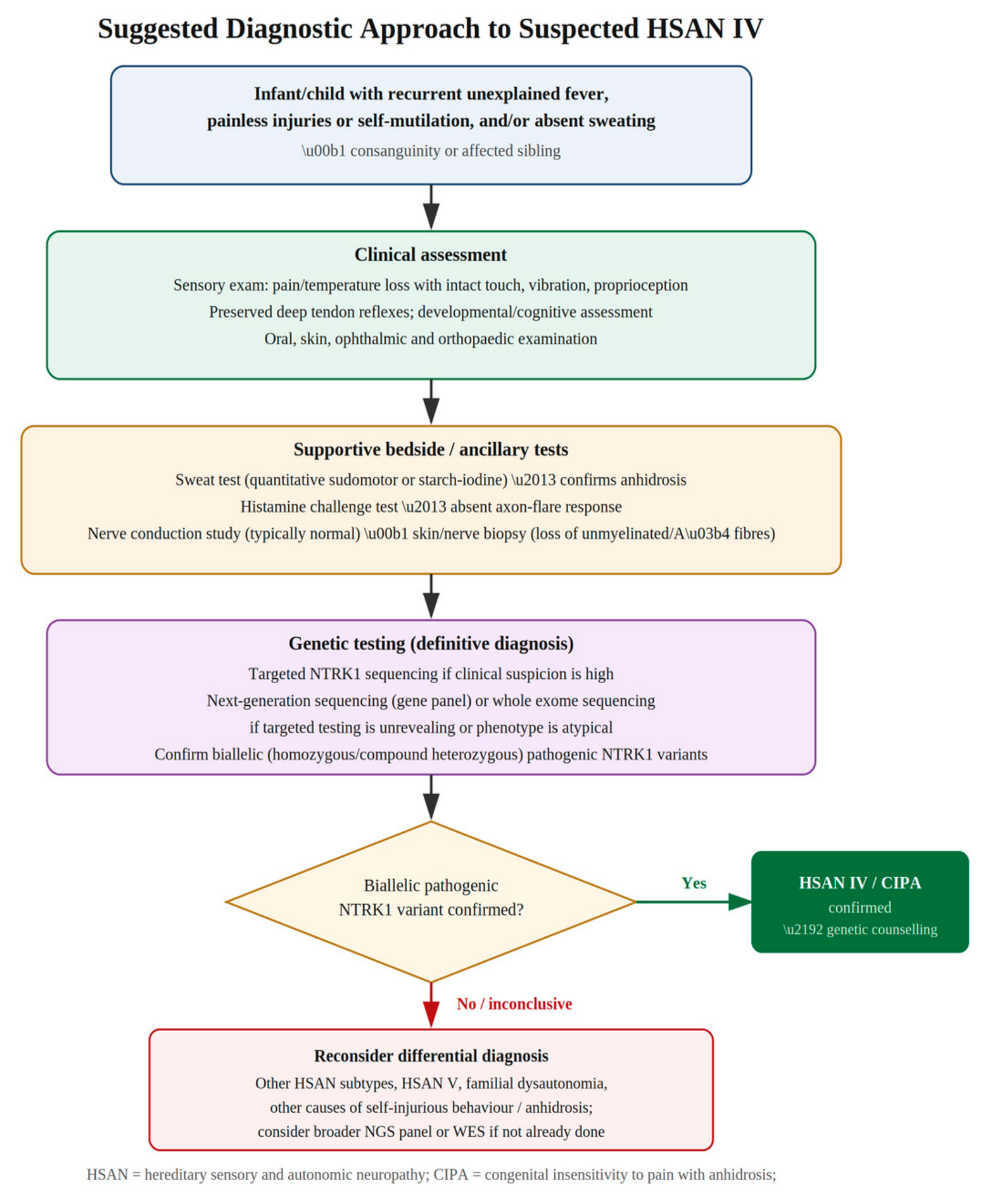
Genetic testing is the single most definitive diagnostic test for this condition which includes direct DNA sequencing to identify point variations, small insertions or deletions. [14] Identification of NTRK1 mutations remains the only tool available for prenatal diagnosis. Genetic counselling is advised to discourage consanguineous marriages, especially when there is family history as this condition is an autosomal recessive [24].
Pain insensitivity drastically delays injury recognition without a sign of prevalent injury or infection. Various behavioural cues such as reluctance to walk or agitation may be the only warning signs, so vigilant monitoring for occult unnoticed fractures, pressure ulcers, and infections risking sepsis is essential. Radiological imaging is crucial for identifying osteolytic change, whereas inflammatory markers such as CRP and ESR are often unreliable. [16]
Oral findings have been highlighted by multiple researchers. They include bifid tongue, oral injuries like auto-extraction of teeth, severe bite injuries or both oral soft tissues and finger digits. [10,45]. Oral and craniofacial anomalies include a large number of missing teeth, nasal malformation, submucous cleft palate, severe soft tissue injuries, dental caries, submucous cleft palate and malocclusion [41].
Ophthalmic abnormalities to look for include such as neurotrophic corneal ulceration, corneal scarring and corneal perforation. [7]
3.6. Management Strategies for HSAN IV
3.6.1. General Supportive Care
Treatment is supportive and best provided by a multidisciplinary team comprising - spanning pediatrics, occupational therapy, physiotherapy, psychology orthopedics, dentistry, ophthalmology, and dermatology. Body-temperature monitoring helps pre-empt hyperthermia and hypothermia caused by anhidrosis., The while a child’s activities should be modified as far as reasonable to reduce injury risk — though intolerance to immobilization impaired immobilization tolerance often delays orthopedic healing, and bracing or invasive procedures raise increase the risk of infection risk. Tooth extraction, filing of sharp incisal edges and mouth guards minimize oral injury from self-mutilation. Oral injury from self-mutilation is minimized through tooth extraction, filing of sharp incisal edges, and mouth guards. Regular moisturization limits palmar and plantar hyperkeratosis and cracking, while neurotrophic keratitis requires routine ocular care, infection prevention measures, and daily surface observation. Behavioral, developmental, and motor interventions, along with educational and social support, are recommended for school-age children and adolescents. Ongoing surveillance includes daily caregiver evaluation for unrecognized injury, regular specialist review, and annual follow-up at a comprehensive care center, with hot or cold environments, extreme-temperature foods and baths, and high-impact activities should be avoided. Where familial NTRK1 variants are known, molecular testing of at-risk infants allows early monitoring for hyperpyrexia and oral injury as primary teeth erupt. Molecular testing of at-risk infants [known familial NTRK1 variants] allows for early monitoring of hyperpyrexia and oral injury as primary teeth erupt. [47]
The molecular mechanisms underlying the HSANs remain incompletely understood, though emerging evidence implicates axonal transport, control of neuronal membrane excitability, and neuronal development. In the absence of a curative, mechanism-targeted therapy is not yet available, supportive care of the kind described above remains the only established therapy for HSAN; future studies should aim to identify common disease mechanisms that could serve as therapeutic targets.[55]
3.6.2. Orthopedic and Skeletal Management
Musculo-skeletal complications form the most demanding aspect of HSAN IV management. Pain insensitivity substantially delays injury recognition, as illustrated by cases in which tibial fractures have progressed to osteomyelitis without redness or pain . behavioral cues such as reluctance to walk or agitation may be the only warning signs, so vigilant monitoring for occult unnoticed fractures, pressure ulcers, and infections risking sepsis is essential. Radiological imaging is crucial for identifying osteolytic change, whereas inflammatory markers such as CRP and ESR are often unreliable. Nabiyev et al. documented multiple cases of osteomyelitis in HSAN IV, underscoring the difficulty of early diagnosis and the need for prolonged treatment following delayed detection Zhang and Haga reported musculo-skeletal complications in 65% of HSAN IV patients, with 91% of fractures affecting the lower limbs, most commonly in children aged one to seven years.
Once a fracture is identified, orthopedic decision-making requires balancing conservative and surgical approaches. Hartono et al. found that surgically treated articular and metaphyseal fractures in HSAN IV often develop infection and non-union, whereas diaphyseal fractures show better outcomes with conservative management such as cast immobilization. However, conservative casting is effective mainly in mild cases, given poor compliance, recurrent localized infection, and delayed healing, while external fixation is frequently undermined by mechanical instability requiring multiple revisions. Prolonged, culture-guided antibiotic therapy is essential for managing recurrent infection and preventing sepsis; Staphylococcus aureus is consistently reported as the predominant pathogen, with methicillin resistance documented in nearly half of cases in one retrospective cohort.[16]
Building on this, the optimal therapeutic approach centers on prophylactic measures, such as braces for early weight-bearing in non-surgical fractures, together with rigorous follow-up to avoid missed complications. An alternative strategy proposes early surgical repair of long-bone fractures combined with early bisphosphonate use — surgical fracture repair to achieve early functional recovery and avoid a destructive end-stage, paired with bisphosphonates to manage osteoporosis. Ultimately, addressing the underlying cause of HSAN IV, rather than treating symptoms alone, is likely the optimal approach. When HSAN IV arises from NTRK1 loss-of-function mutations affecting the TRKA receptor, molecular therapy targeting the NGF receptor tyrosine kinase would represent the most effective intervention. [56]
In practice, fractures in HSAN IV often heal poorly, forming hypertrophic pseudarthrosis, with healing that typically takes three weeks to six months or failing altogether with an enhanced risk of infected non-union. Hyperbaric oxygen therapy [as an adjunct] bisphosphonate therapy [IV Pamidronate], including intravenous pamidronate has shown promise in reducing fracture recurrence, with no adverse effects reported at five years of follow-up. Looking further ahead, experimental approaches such as gene therapy, nerve growth factor administration, and stem cell-based strategies may offer future avenues to enhance neuronal function and reduce complications.[56]
Summarising, the complementary strategies as below improve outcomes and reduce long term morbidity in HSAN IVTaken together, the literature supports a set of complementary strategies to improve outcomes and reduce long-term morbidity in HSAN IV:
- Enhanced infection control through early, culture-guided antibiotic therapy, long-term suppressive regimens, and aggressive wound management such as vacuum-assisted closure.
- Bone grafting, including vascularized fibular grafts or synthetic substitutes, to promote structural stability and reduce non-union.
- Custom orthotic solutions tailored to biomechanical needs, improving mobility and reducing pressure-ulcer and deformity risk.
- Early amputation, though controversial, may become necessary when limb salvage leads to recurrent hospitalization and poor quality of life. These decisions require careful risk-benefit discussion with patients and families, weighing mobility gains against surgical and psychological risk.A high risk of auto-amputation from self-harm has also been reported. [57]
- Multidisciplinary rehabilitation, including physiotherapy, occupational therapy, and psychological support, is crucial for functional outcomes and adaptation, particularly given that repeated surgeries contribute to post-traumatic stress disorder in up to 16% of children and 23% of parents, linked to prolonged hospitalization, multiple procedures, and insufficient psychosocial support. [58]
3.6.3. Treatment of Ocular Abnormalities:
Jarade et al. achieved complete corneal ulcer healing using preservative-free artificial tears, bandage contact lenses, and topical antibiotics. Levy et al. resolved corneal erosion with protective tarsorrhaphy, while Guven et al. and Yagev et al. used lateral tarsorrhaphy, corneal patch grafts, and penetrating keratoplasty for corneal opacity and ulceration. John et al. managed dry eye and recurrent corneal erosion with lubricants, topical antibiotics, and bilateral punctal cautery followed by tarsorrhaphy, although punctate erosions persisted despite treatment. Elsana et al. used fortified topical antibiotics, photoactivated chromophores, lubricants, and surgical interventions, including tarsorrhaphy and amniotic membrane transplantation, for nonhealing ulcers. Verity et al. found that cyclopentolate–phenylephrine drops, rather than cocaine–hydroxyamphetamine, successfully relieved ptosis and induced mydriasis.[59] Suresh et al. treated neurotrophic keratitis with tobramycin, atropine, and carboxymethyl cellulose drops, and corneal ulceration with bilateral lateral tarsorrhaphy. Overall, medical therapy appears effective as first-line management for corneal ulceration and opacity, with surgical procedures such as tarsorrhaphy and keratoplasty reserved for recurrent, nonhealing lesions. [39]
3.6.4. Genetic Counselling
NTRK1-HSAN IV follows autosomal recessive inheritance, though uniparental isodisomy accounts for some cases. For AR inheritance, each sibling of an affected individual has a 25% chance of being affected, a 50% chance of being an asymptomatic carrier, and a 25% chance of being unaffected and non-carrier; sibling risk is not increased above the general population for uniparental isodisomy. Once familial pathogenic variants are identified, carrier testing, prenatal testing, and preimplantation genetic testing become possible — complementing the early monitoring strategy for at-risk infants described above. [32]
3.6.5. Prognosis and Follow-Up Care
Given its progressive, chronic nature, HSAN IV requires lifelong follow-up to monitor skeletal deformity, infection, and neurological complications. Survival into adulthood is possible, with reported ages ranging from 6 months to 33 years, though mortality remains a concern, most often from septic shock or post-surgical cardiac arrest linked to dysautonomia. Regular radiographic assessment, infection monitoring, and orthopedic review should therefore be integral to long-term care; one seven-year follow-up documented progressive acro-osteolysis, reinforcing the need for lifelong specialist supervision given the disease’s progressive course. Custom orthotics and adaptive physical therapy improve long-term function and mobility, while psychological support remains essential for addressing self-harm and sensory-seeking behaviors.
These long-term risks are illustrated by comparison with a previously reported Turkish child with HSAN IV, born to non-consanguineous heterozygous carriers, who shared recurrent fevers, delayed speech, eczematous lesions, hyperkeratosis, oral self-mutilation, and multiple tibial and ankle fractures at a young age; fracture management and complications were not elaborated in that report, however, underscoring persistent gaps in the literature on HSAN IV management. Unlike that patient, other reported cases have lacked ataxic gait and convulsions, raising the possibility that such features may emerge only with age. More broadly, complications such as osteomyelitis, fractures, and joint instability increase the risk of limb deformity, mobility loss, and chronic infection over time: progressive valgus knee deformity and hip flexion contracture may lead to wheelchair dependence, recurrent S. aureus infection heightens sepsis risk, and anhidrosis increases susceptibility to life-threatening hyperthermia. Amputation, though often initially refused by families, may ultimately remain a viable option to prevent further deterioration. [39]
More broadly, there is no specific or targeted therapeutic agent for HSAN IV. Severe hyperpyrexia and traumatic complications require frequent medical attention, and close supervision of affected children is necessary to prevent injuries, burns, fractures, corneal ulceration, and self-mutilation. Parents should be counseled on simple preventive measures such as avoiding excessive wrapping, preventing dehydration, and controlling hyperpyrexia, all of which can reduce otherwise preventable deaths. Treating physicians should also be made aware of the diagnosis, as this can help avoid unnecessary antibiotic use and investigation during febrile episodes. Frequent surgical intervention may be required for fractures, and operating surgeons should consider the possibility of inadequate analgesia if tachycardia or hypertension occurs postoperatively, reflecting an unconscious physiological pain response despite the patient’s lack of conscious awareness due to the reduced number of peripheral pain fibers. Identification of NTRK1 mutations remains the only tool available for prenatal diagnosis. Genetic counselling is advised to discourage consanguineous marriages, especially when there is family history as this condition is an autosomal recessive disorder., and, as HSAN IV is an autosomal recessive disorder, genetic counseling is advised to discourage consanguineous marriage, particularly where there is a positive family history [17].
In conclusion, congenital insensitivity to pain with anhidrosis is an uncommon cause of fever of unknown origin and should be considered in the context of self-mutilation, particularly in patients from regions with high rates of consanguinity. To date, no targeted therapeutic agent exists for this disorder, and affected families should be counseled and trained to keep the child well hydrated and to avoid injury and physiological stress [17].
3.7. Multicentre Study Data
Congenital insensitivity to pain with anhidrosis [HSAN IV] is a rare autosomal recessive disorder caused by mutations in NTRK1, encoding the neurotrophic tyrosine kinase-1 receptor. A multicenter French retrospective study identified seven HSAN IV patients from laboratories sequencing NTRK1, originating from France [2], Suriname [2], Mali [1], Kazakhstan [1], and Algeria [1]. Mean age was 9.8 years [range 4–20], 57% were female, 57% had delayed developmental milestones, and 57% had a family history of consanguinity. Diagnosis occurred at a mean age of 4.8 months, with all patients presenting pain insensitivity, anhidrosis, intellectual disability, self-mutilation, febrile episodes, impaired temperature perception, and autonomic dysfunction. Associated findings included hyperactivity [86%], emotional lability [86%], joint deformities [71%], bone fractures [57%], abnormal touch/vibration/position sense [50%], skin/hair/nail abnormalities [28%], and hypothermia episodes [28%]. Two patients died [ages 9 and 12] from infection. Nerve conduction studies showed absent lower-limb sensory nerve action potentials in three cases, and one sensory nerve biopsy showed complete absence of unmyelinated fibers. Nine NTRK1 pathogenic variants were identified, including three novel mutations, confirming HSAN IV as an extremely rare disorder and expanding its genotypic spectrum. [60]
A ten-year retrospective cohort study at Soroka University Medical Center, Israel [2014–2023], reviewed electronic records to identify 63 HSAN IV patients, all from consanguineous Bedouin families — the largest HSAN IV cohort reported to date. Data spanned demographics, clinical presentation, genetic analysis, infections, wound sites, hospitalization duration, and surgical intervention. Staphylococcus aureus, notably methicillin-resistant strains, predominated, with Gram-negative organisms common in lower-limb infections; extended-spectrum beta-lactamase-producing bacteria were comparatively less frequent, and demographic factors correlated with infection patterns, antibiotic resistance, and surgical needs. The findings highlight dynamic shifts in microbial composition and resistance over time, drawing parallels with infection dynamics in diabetic foot ulcers due to shared neuropathy- and ischemia-related susceptibility, and support development of tailored, targeted management strategies for HSAN IV and related neuropathic conditions.[61]
A study of three Korean HSAN IV patients described their clinical and genetic features, identifying one novel gross deletion variant alongside varying disease severity; patients with previously reported attenuated variants showed milder clinical manifestations. This expanded the known spectrum of NTRK1 variants and reinforced that clarifying genotype–phenotype relationships is key to understanding HSAN IV’s pathogenesis and clinical prognosis. [62]
Collectively, these multicenter and cohort-based reports remain the principle source of HSAN IV source of population-level data on HSAN IV, yet each is limited by small sample size, geographic or ethnic clustering [e.g., consanguineous Bedouin families], and reliance on retrospective record review. Pooling multicenter and multinational cohort data, with standardized reporting of genotype, phenotype, infection burden, and outcomes, is needed to close the epidemiological gap and generate the population-based prevalence and natural history data currently lacking for HSAN IV/HSAN. [62]
3.8. Future Directions
Research on HSAN IV remains limited, comprising mostly case reports of new cases and phenotypes, which highlights the need for further studies on treatment strategies and long-term outcomes [33]. A systematic synthesis of findings and management approaches from reported cases is needed to fully characterize HSAN IV’s clinical and genetic spectrum, alongside long-term follow-up studies to clarify outcomes and risk factors for disease progression; future work should prioritize early diagnostic markers, optimized therapeutic strategies, and standardized treatment protocols. Building on the experimental avenues noted above, investigations into bone regeneration, nerve growth factor therapies, and advanced orthopedic interventions such as implants may open new possibilities for managing HSAN IV-related complications, while non-pharmacological pain-management approaches warrant exploration to improve quality of life. Enhanced genetic counseling and early screening can aid monitoring, and collaboration among researchers, clinicians, and patient advocacy groups — including multicenter and international efforts — could accelerate research and support consensus guidelines. Finally, the ethical and sociolegal dilemmas surrounding invasive interventions such as amputation should continue to be examined, with psychosocial support integrated into care plans to address the emotional and social burden of living with this rare genetic disorder. [44]
4. Conclusion
HSAN-IV with its orthopedic and neurological manifestation poses a unique challenge during the formative years of a child, leading to difficulty in achieving unrestricted mobility and optimal social interaction - thereby hindering the holistic growth of the child, with its negative consequence on the society at large. Lack of clinician awareness, heterogeneity in its presentation, clusters of consanguinity, debilitating multisystem involvement requiring long term multidisciplinary care increases burden on the family and the health care system. With the recent advances in molecular/genetic diagnostics, coupled with clinical heterogeneity of HSAN; clinical phenotype to genotype correlation is often complex leading to diagnostic dilemma. A comprehensive review to gather robust data on prevalence, documentation of the varied manifestations of the disease including a long term follow up, genotype to phenotype correlation and lastly, to build awareness for establishing support systems is the need of the hour.
Table 2.
Summary of the Literature Review.
| S NO | AUTHOR & YEAR | PURPOSE OF STUDY | KEY FINDINGS OF THE STUDY | STRENGTHS AND LIMITATIONS OF THE STUDY |
| 1 | National Organization for Rare Disorders [NORD] [2024/2026] | Provide academic and medical information about Hereditary Sensory and Autonomic Neuropathy Type IV [HSAN IV]. | Outlines the clinical features, NTRK1 genetic etiology, and multidisciplinary supportive care strategies. | Strength: Accessible and updated. Limitation: it is an overview of Secondary reference. |
| 2 | Indo et al. [1996] | Identify the genetic etiology of HSAN IV. | Loss-of-function mutations in the TRKA/NGF receptor gene [NTRK1] discovered- Molecular basis of HSAN IV. | Strength: Landmark gene discovered. Limitation: Initial discovery, therefore cohort size was small. |
| 3 | Kim et al. [2013] | Evaluate orthopedic complications, interventions, and outcomes in patients with HSAN IV. | Documented recurrent bone fractures, frequent Charcot joints, osteomyelitis, and high rate of surgical complications. | Strength: Focused and detailed analysis of orthopedic and surgical outcomes. Limitation: Retrospective design with a small patient sample size. |
| 4 | Marik et al. [2009] | Report specific orthopedic manifestations and complications in children with HSAN IV. | Detailed severe pediatric skeletal pathology, joint dislocations, painless fractures and chronic osteomyelitis. | Strength: Detailed descriptions of pediatric orthopedic manifestations. Limitation: Very small sample size/case series. |
| 5 | Indo [2002] | Examine biological, molecular and clinical aspects of NTRK1 gene mutations causing HSAN IV. | Mapped diverse mutation types across domain structures of the TRKA receptor kinase causing neurodevelopmental failure. | Strength: Detailed molecular [structural and functional] mapping of NTRK variants. Limitation: Unilateral Focus on molecular genetics. Long-term clinical trial outcomes not analysed. |
| 6 | Yozu et al. [2016] | Review rehabilitation associated challenges in HSAN IV/V and propose novel strategies for physical rehabilitation. | Proposed sensory-substitution, joint protection, and behavioral modifications to mitigate self-mutilation and joint destruction. | Strength: Pragmatic emphasis on rehabilitation and non-surgical/interventional management. Limitation: Rehabilitation protocols proposals require larger validation studies. |
| 7 | Indo [2018] | Explore the role of NGF-dependent neurons in human feelings, emotion, and pain keeping HSAN spectrum as a model. | Linked survival of NGF associated sensory/autonomic networks to central processing of thermal regulation, affect and interoception. | Strength: Broad conceptual framework bridging emotional expression and neurobiology. Limitation: Theoretical synthesis, therefore requires deeper research. |
| 8 | Hutchison [2007] | Review the evolution and clinical transition of DNA sequencing technologies. | Documented developments in DNA sequencing efficacy and cost-effective strategies enabling discovery of genes involved in human disease. | Strength: Technical and complete review of sequencing methodology. Limitation: Non-specific methodological review |
| 9 | Almutairi & Tabassum [2022] | Case report of HSAN and review of literature. | Highlighted diagnostic difficulties, thermal injuries management, and crucial caregiver instructions. | Strength: Pragmatic demonstration of the disease integrated with literature review. Limitation: Single-patient report. |
| 10 | Schwarzkopf et al. [2005] | Review clinical and genetic aspects of HSAN | Overviewed clinical diagnosis, orthopedic complications, and genetic implications of HSAN. | Strength: Detailed clinical narrative overview. Limitation: Narrative review pertaining to one geographic location. |
| 11 | Tanaka et al. [1990] | Examine iron-dependent cytotoxic effects of L-dopa on cultured dorsal root ganglion [DRG] neurons. | Showed that L-dopa causes iron-dependent neurotoxicity in cultured sensory DRG neurons. | Strength: Specific experimental sensory neurotoxicity data along with detailed understanding of nerve conduction. Limitation: Study explains neurotransmission mechanisms in detail, but not specific to HSAN |
| 12 | Udhayashankar et al. [2012] | Present a case report of HSAN highlighting cutaneous findings in a South Indian child. | Showed significant dermatological markers linked to regional consanguinity. | Strength: Emphasizes diagnostic capacity for skin lesions. Limitation: Single-case report. |
| 13 | Sreenivasan et al. [2024] |
Report a case of HSAN in a 22-month-old child of consanguineous parents. | Overviewed early toddler presentations including tongue biting, unexplained fevers, and painless fractures. | Strength: Focuses on very early pediatric presentation. Limitation: No long-term data & single case presentation. |
| 14 | Greco et al. [2000] | Identify a novel NTRK1 mutation associated with HSAN | Expansion of pathogenic NTRK1 variants catalog that leads to HSAN spectrum. | Strength: Precise molecular mapping of a novel mutation variant. Limitation: Dynamic expression assays absent & Small cohort size. |
| 15 | Indo [2001] | Assess mutations and polymorphisms in the [NTRK] gene in HSAN. | Detailed various missense, nonsense, and splice-site mutations across functional domains of the TRKA receptor. | Strength: Robust reporting of HSAN variants globally. Limitation: Study was limited to descriptive genetic mapping. |
| 16 | Shaikh et al. [2017] | To conduct functional analysis of NTRK1 missense mutations. | Demonstrated that missense variants cause loss of function via autophosphorylation, impaired receptor trafficking or cell-surface localization. | Strength: Comprehensive in vitro functional cellular assays validating variant pathogenicity. Limitation: In vitro studies may not fully reflect native human sensory neurons. |
| 17 | Rotthier et al. [2009] | Conduct genotype-phenotype correlation assessment across a large cohort of HSAN patients. | Showed that NTRK1 mutations specifically lead to the severe HSAN IV phenotype | Strength: Large, international multi-center cohort analysis. Limitation: Variable retrospective data aggregation. |
| 18 | Wang WB et al. [2018] | Identify a novel mutation in the NTRK1 gene in Chinese HSAN patients | Confirmed a novel pathogenic NTRK1 mutation contributing to the East Asian genetic mutation database. | Strength: Reporting a molecular spectrum in an under-represented population. Limitation: Small sample cohort. |
| 19 | Wang Q et al. [2015] | Report novel and de novo mutations in NTRK1 associated with HSAN. | Showed that HSAN can result from de novo mutations in addition to conventional genetic inheritance. | Strength: An insight into non-inherited de novo variants. Limitation: Single case reported. |
| 20 | Sun et al. [2024] | Demonstrate a novel NTRK1 splice-site variant in a Chinese family with HSAN. | Confirmed aberrant RNA splicing leading to functional loss of the TRKA protein. | Strength: Combined genomic sequencing with transcript analysis. Limitation: Study restricted to one family. |
| 21 | Geng et al. [2018] | Investigate NTRK1 mutations and clinical features in Chinese patients with HSAN. | Demonstrated novel mutations and evaluated phenotypic variations. Identified impairment of cognitive development. | Strength: Multi-patient cohort combining molecular and clinical data. Limitation: Retrospective design depending on past medical records. |
| 22 | Kayani et al. [2017] | Describe orthopedic manifestations and surgical outcomes in patients with HSAN IV. | Reported frequent orthopedic operations, deep infections, high rates of nonunion and Charcot joint progression. | Strength: Long-term specialized tertiary center outcome data. Limitation: Descriptive study, retrospective cohort, tertiary referral selection bias. |
| 23 | Bar-On et al. [2002] | Assess orthopedic management and outcomes in HSAN. | Demonstrated that conservative and non-operative management of orthopedic manifestations often yields better and long-term results than surgery. | Strength: Proven evidence-based surgical and management strategies. Limitation: Small sample size. |
| 24 | Kohler et al. [2023] | Report orthopedic manifestations of HSAN IV in a 10-year-old child. | Highlighted management of Charcot arthropathy and severe lower extremity fractures in late childhood. | Strength: Detailed pediatric-orthopedic case presentation. Limitation: Single-patient case study. |
| 25 | Nishida [1951] | Report on clinical congenital anhidrosis. | Early historical description of congenital anhidrosis and sensory impairment in Japan. | Strength: Early foundation historical report. Limitation: Published prior to genetic and modern diagnostic tools. |
| 26 | Tunçbilek et al. [2005] | Describe management of a calcaneal ulcer in a child with HSAN. | Outlined reconstructive plastic surgery procedures and protective protocols for non-healing weight-bearing ulcers. | Strength: Insight on Practical surgical reconstruction for HSAN patients. Limitation: Single case presentation. |
| 27 | Küçükdurmaz et al. [2015] | Report a case of HSAN presenting initially with chronic osteomyelitis. | Painless bone infections masking the underlying diagnosis of HSAN. | Strength: Highlighting diagnostic pitfalls in orthopedic manifestations in HSAN. Limitation: Single case report. |
| 28 | Ashwin et al. [2015] | Demonstrate oral and maxillofacial manifestations of HSAN IV in a case series and review. | Reported dental trauma, severe oral self-mutilation [tongue/lip biting] and premature tooth loss. | Strength: Focused analysis of specialized maxillofacial complications. Limitation: Small patient series. |
| 29 | Gao et al. [2013] | Investigate craniofacial findings and correlate with 2 novel missense NTRK1 mutations in HSAN. | Linked novel mutations to specific craniofacial anomalies, severe self-inflicted oral trauma, and dental defects. | Strength: Integrated oral pathology workup with molecular genetic mutation. Limitation: Small primary sample group. |
| 30 | Sulaiman & Alyahya [2023] | Reported a case of HSAN detailing clinical presentation and protective care. | Highlighted preventive care measures to reduce thermal injury risks and severe self-mutilation. | Strength: Focus on protective care on a regular basis. Limitation: Single case report. |
| 31 | Baker et al. [2025] | Assess ocular manifestations in HSAN patients. | Reported high prevalence of alacrima, neurotrophic keratitis, corneal scarring with increased risk of vision loss. | Strength: Specific and detailed ophthalmological characterization. Limitation: Small sample size due to rarity of disease presentation. |
| 32 | Schalka et al. [2006] | Present a 4-year dental follow-up of a patient with HSAN. | Demonstrated that customized oral appliances and early dental intervention can prevent oral soft tissue self-mutilation. | Strength: Longitudinal 4-year management follow-up data. Limitation: Single patient evaluation. |
| 33 | Zhu et al. [2022] | Document ophthalmic findings and a novel NTRK1 mutation in a HSAN patient. | Linked a novel NTRK1 variant with early-onset neurotrophic corneal perforation risk. | Strength: Comprehensive correlation between NTRK mutation and ophthalmic pathology. Limitation: Single-patient report. |
| 34 | Amano et al. [2006] | Describe ocular manifestations among HSAN patients. | Noted reduced tear secretion, severe corneal hypaesthesia, and high rates of ulceration. | Strength: Evaluation of eye complications in a moderate cohort. Limitation: Retrospective design |
| 35 | Yagev et al. [1999] | Understand ophthalmic and systemic manifestations of HSAN. | Documented that alacrima and neurotrophic keratitis are major ophthalmic features that accompany systemic anhidrosis. | Strength: Early integration of systemic and eye features. Limitation: Small sample size. |
| 36 | Jarade et al. [2002] | Report corneal ulcers in HSAN and review of literature. | Emphasised that indolent corneal ulceration can be an early sign of HSAN and outlined the significance of protective lens treatments. | Strength: Reliable clinical guidance on managing corneal ulcers. Limitation: Evidence is limited to one patient. |
| 37 | Gabreels-Festen [1999] | Review morphological hallmarks and mechanisms of hereditary neuropathies in childhood. | Demonstrated selective depletion of unmyelinated fibers on nerve biopsy across pediatric sensory neuropathies. | Strength: Clear histopathological overview of peripheral neuropathies in childhood. Limitation: Archival records prior to molecular testing. |
| 38 | Liu et al. [2018] | Evaluate phenotypic heterogeneity of intellectual disability in HSAN patients. | Revealed variable cognitive capacity ranging from normal IQ to severe intellectual impairment in HSAN patients. | Strength: Robust assessment of cognitive spectrum. Limitation: Different cognitive assessment tools across historical literature. |
| 39 | Batouk et al. [2021] | Reported a case of HSAN presenting with multiple Charcot joints in a child. | Demonstrated severe multi-joint destruction in early childhood thereby stressing early diagnostic protocols. | Strength: High-impact clinical illustration of the disease. Limitation: Single-patient case report. |
| 40 | Higashimoto et al. [2024] | Demonstrate an atypical presentation of HSAN causing a diagnostic challenge. | Highlighted how unconventional symptoms can delay genetic testing and definitive diagnosis. | Strength: Explains real-world diagnostic challenges and importance of genomic sequencing. Limitation: Single case report. |
| 41 | Yang et al. [2023] | Review cellular mechanisms of cognitive impairment induced by NTRK1 knockdown in mice. | Demonstrated that NTRK1 knockdown suppresses AMPK/ULK1/FUNDC1-mediated hippocampal cell damage and cognitive impairment. | Strength: Robust experimental study defining intracellular pathways. Limitation: Experimental study |
| 42 | Rotthier et al. [2012] | Overview of cellular mechanisms and genetics of HSAN spectrum. | Grouped various pathways across the HSAN spectrum, emphasising on axonal degradation and neurotrophin signaling failure. | Strength: Detailed, highly structured pathophysiological explanation. Limitation: Review article without experimental data. |
| 43 | Li et al. [2019] | Determine clinical variability and NTRK1 mutations in Han Chinese HSAN patients. | Documented novel NTRK1 variants and phenotypic variety in Chinese HSAN cohorts. | Strength: Detailed regional patient cohort genetic analysis. Limitation: Limited geographical representation obscures global perspective. |
| 44 | Miura et al. [2000] | Document complete paternal uniparental isodisomy for chromosome-1 revealing an NTRK1 mutation in HSAN. | Demonstrated that paternal uniparental isodisomy can lead to homozygous NTRK1 mutation expression from a single carrier parent. | Strength: Significant novel genetic transmission mechanism discovery. Limitation: Single-patient presentation. |
| 45 | Miura et al. [2000] | Conduct polymorphism/mutation analysis of TRKA [NTRK1] in HSAN families. | Noted pathogenic mutations and non-pathogenic polymorphisms in the high-affinity NGF receptor gene. | Strength: Early detailed genetic mapping of TRKA variants. Limitation: Long term follow-up required. |
| 46 | Verity et al. [1982] | Evaluate children with reduced pain sensitivity to differentiate HSAN types II and IV. | Successfully differentiated HSAN spectrum with clinical criteria. | Strength: Robust early clinical and neurophysiological differentiation. Limitation: Conducted prior to molecular genetic sequence discovery. |
| 47 | Echaniz-Laguna et al. [2021] | Conduct a nationwide multicentric retrospective study on NTRK1-related HSAN in France. | Produced national cohort data on life expectancy, mutation spectrum, joint destruction, and long-term outcomes. | Strength: Large, unbiased nationwide cohort with rigorous follow-up data. Limitation: Retrospective design leading to discrepancy in historical data collection. |
| 48 | Klaitman et al. [2024] | Perform a 10-year study of patients living with HSAN. | Documented long-term survival, joint preservation and overall quality of life along with awareness status of patients for over a decade. | Strength: 10-year longitudinal design providing realistic outcome and reliable data. Limitation: Regionally focused patient group. |
| 49 | Cho et al. [2024] |
Demonstrate clinical and genetic characteristics of 3 HSAN patients and review of literature. | Broadened the NTRK1 mutation database and overview of pediatric clinical management strategies. | Strength: Rigorous advanced clinical and genomic integration. Limitation: Small sample size. |
Author Contributions
Conceptualization, NSU and BS.; methodology, NSU and SG.; data curation, NSU and SG.; writing—original draft preparation, NSU and SG.; writing—review and editing, NSU, UVK, KNVR.; visualization, NSU, SG and BS.; supervision, NSU and KNVR.; project administration, SG. All authors have read and agreed to the published version of the manuscript.
Funding
This research received no external funding.
Institutional Review Board Statement
Not applicable for studies not involving humans or animals.
Data Availability Statement
No new data were created as this is a review paper.
Acknowledgments
AI assistance was used for summarizing literature, drafting a table and to generate infographic figures. Tool: Claude AI, Version: Claude-Sonnet 5.
Conflicts of Interest
The authors declare no conflicts of interest.
Abbreviations
The following abbreviations are used in this manuscript:
| MDPI | Multidisciplinary Digital Publishing Institute |
| DOAJ | Directory of open access journals |
| TLA | Three letter acronym |
| LD | Linear dichroism |
| HSAN IV/HSAN Type IV- | Hereditary and Sensory Autonomic Neuropathy Type IV |
| CIPA | Congenital Insensitivity to Pain with Anhidrosis |
| NTRK1 | Neurotrophic Receptor Tyrosine Kinase 1 |
| TRK-A | Tyrosine Related Kinase A |
| NGF | Nerve Growth Factor |
| DNA | Deoxyribonucleic Acid |
| NGS | Next Generation Sequencing |
| WES | Whole Exome Sequencing |
References
- National Organization for Rare Disorders (NORD). Hereditary sensory and autonomic neuropathy type IV (Internet). Available online: https://rarediseases.org/rare-diseases/hereditary-sensory-and-autonomic-neuropathy-type-iv/ (accessed on 19 Jun 2026).
- Dyck, P.J.; Mellinger, J.F.; Reagan, T.J.; Horowitz, S.J.; McDonald, J.W.; Litchy, W.J.; et al. Not “indifference to pain” but varieties of hereditary sensory and autonomic neuropathy. Brain J. Neurol. 1983, 106 Pt 2, 373–90. [Google Scholar] [CrossRef] [PubMed]
- Indo, Y.; Tsuruta, M.; Hayashida, Y.; Karim, M.A.; Ohta, K.; Kawano, T.; et al. Mutations in the TRKA/NGF receptor gene in patients with congenital insensitivity to pain with anhidrosis. Nat. Genet. 1996, 13(4), 485–8. [Google Scholar] [CrossRef] [PubMed]
- Indo, Y. Nerve growth factor and the physiology of pain: lessons from congenital insensitivity to pain with anhidrosis. Clin. Genet. 2012, 82(4), 341–50. [Google Scholar] [CrossRef] [PubMed]
- Schon, K.R.; Parker, A.P.; Woods, C.G. Congenital insensitivity to pain overview. In Europe PMC; University of washington, 2019; p. 29419974. [Google Scholar]
- Haga, N.; Kubota, M.; Miwa, Z. Pain JRG on CI to. Hereditary sensory and autonomic neuropathy types IV and V in Japan. Pediatr. Int. 2015, 57(1), 30–6. [Google Scholar] [PubMed]
- Kim, W.; Guinot, A.; Marleix, S.; Chapuis, M.; Fraisse, B.; Violas, P. Hereditary sensory and autonomic neuropathy type IV and orthopaedic complications. Orthop. Traumatol. Surg. Res. 2013, 99(7), 881–5. [Google Scholar] [CrossRef] [PubMed]
- Nagasako, E.M.; Oaklander, A.L.; Dworkin, R.H. Congenital insensitivity to pain: an update. Pain 2003, 101(3), 213–9. [Google Scholar] [CrossRef] [PubMed]
- Marik, I.; Kuklik, M.; Kuklikova, D.; Kozlowsk, K. Hereditary sensory and autonomic neuropathy type IV orthopaedic complications. J. Pediatr. Orthop. B 2009, 18(3), 138–40. [Google Scholar] [CrossRef] [PubMed]
- Indo, Y. Genetics of congenital insensitivity to pain with anhidrosis (CIPA) or hereditary sensory and autonomic neuropathy type IV. Clinical, biological and molecular aspects of mutations in TRKA(NTRK1) gene encoding the receptor tyrosine kinase for nerve growth factor. Clin. Auton. Res. Off. J. Clin. Auton. Res. Soc. 2002, 12 Suppl 1, I20–32. [Google Scholar] [CrossRef] [PubMed]
- Swanson, A.G. Congenital insensitivity to pain with anhydrosis. A unique syndrome in two male siblings. Arch. Neurol. 1963, 8, 299–306. [Google Scholar] [PubMed]
- Yozu, A.; Haga, N.; Funato, T.; Owaki, D.; Chiba, R.; Ota, J. Hereditary sensory and autonomic neuropathy types 4 and 5: Review and proposal of a new rehabilitation method. Neurosci. Res. 2016, 104, 105–11. [Google Scholar] [CrossRef] [PubMed]
- Indo, Y. NGF-dependent neurons and neurobiology of emotions and feelings. Lessons from congenital insensitivity to pain with anhidrosis. Neurosci. Biobehav Rev. 2018, 87, 1–16. [Google Scholar] [CrossRef] [PubMed]
- Hutchison, C. A. DNA sequencing: bench to bedside and beyond. Nucleic Acids Res. 2007, 35(18), 6227–6237. [Google Scholar] [CrossRef] [PubMed]
- Lischka, A.; Lassuthova, P.; Çakar, A.; et al. Genetic pain loss disorders. Nat. Rev. Dis. Primers 2022, 8, 41. [Google Scholar] [CrossRef] [PubMed]
- Rajbansh, P.; Yadav, M.; Kumar, P.; Das, A. Congenital insensitivity to pain with anhidrosis: a rare entity. Indian Dermatol. Online J. 2020, 11(2), 274–7. [Google Scholar] [CrossRef] [PubMed]
- Almutairi, M.M.; Tabassum, S. Congenital insensitivity to pain with anhidrosis: a case report and review of the pertinent literature. Cureus 2022, 14(11), e31019. [Google Scholar] [CrossRef] [PubMed]
- Dearborn G: A case of congenital general pure analgesia. J. Nerv. Ment. Dis. 1932, 75, 612–5. [CrossRef]
- Wiesmann, U.N.; DiDonato, S.; Herschkowitz, N.N. Effect of chloroquine on cultured fibroblasts: release of lysosomal hydrolases and inhibition of their uptake; University of Washington: Seattle, WA, 1975. [Google Scholar] [CrossRef] [PubMed]
- Suriu, C.; Khayat, M.; Weiler, M.; et al. Skoura - a genetic island for congenital insensitivity to pain and anhidrosis among Moroccan Jews, as determined by a novel mutation in the NTRK1 gene. Clin. Genet. 2009, 75, 230–6. [Google Scholar] [CrossRef] [PubMed]
- Algahtani, H.; Naseer, M.I.; Al-Qahtani, M.; Abdulrahman, S.A.; Boker, F.; Shirah, B. Congenital insensitivity to pain with anhidrosis: a report of two siblings with a novel mutation in (TrkA) NTRK1 gene in a Saudi family. J. Neurol. Sci. 2016, 370, 35–8. [Google Scholar] [CrossRef] [PubMed]
- Schwarzkopf, R.; Pinsk, V.; Weisel, Y.; Atar, D.; Gorzak, Y. Clinical and genetic aspects of congenital insensitivity to pain with anhidrosis. Harefuah 2005, 144, 433–7. [Google Scholar] [PubMed]
- Tanaka, M.; Sotomatsu, A.; Kanai, H.; Hirai, S. Iron-dependent cytotoxic effects of dopa on cultured neurons of the dorsal root ganglia. Clin. Neurol. 1990, 30, 379–83. [Google Scholar]
- Udayashankar, C.; Oudeacoumar, P.; Nath, A.K. Congenital insensitivity to pain and anhidrosis: A case report from South India. Indian J. Dermatol. 2012, 57(6), 503. [Google Scholar] [CrossRef] [PubMed]
- Sreenivasan, V.; Karunakar, P.; Madhileti, S.; Govindaswamy Ramamoorthy, J.; Gulati, R. A rare case of congenital insensitivity to pain with anhidrosis. Paediatr. Int. Child Health 2024, 44(2), 59–62. [Google Scholar] [CrossRef] [PubMed]
- Greco, A.; Villa, R.; Tubino, B.; Romano, L.; Penso, D.; Pierotti, M.A. A novel NTRK1 mutation associated with congenital insensitivity to pain with anhidrosis. Am. J. Hum. Genet. 2000, 66(5), 1671–1676. [Google Scholar] [CrossRef] [PubMed]
- Indo, Y. Molecular basis of congenital insensitivity to pain with anhidrosis (CIPA): mutations and polymorphisms in TRKA (NTRK1) gene encoding the receptor tyrosine kinase for nerve growth factor. Hum. Mutat. 2001, 18(6), 462–471. [Google Scholar] [CrossRef] [PubMed]
- Shaikh, S.S.; Chen, Y.C.; Halsall, S.A.; Nahorski, M.S.; Omoto, K.; Young, G.T.; Phelan, A.; Woods, C.G. A comprehensive functional analysis of NTRK1 missense mutations causing hereditary sensory and autonomic neuropathy type IV (HSAN IV). Hum. Mutat. 2017, 38(1), 55–63. [Google Scholar] [CrossRef] [PubMed]
- Rotthier, A.; Baets, J.; De Vriendt, E.; Jacobs, A.; Auer-Grumbach, M.; Lévy, N.; Bonello-Palot, N.; Kilic, S.S.; Weis, J.; Nascimento, A.; Swinkels, M.; Kruyt, M.C.; Jordanova, A.; De Jonghe, P.; Timmerman, V. Genes for hereditary sensory and autonomic neuropathies: a genotype-phenotype correlation. Brain 2009, 132 Pt 10, 2699–2711. [Google Scholar] [CrossRef] [PubMed]
- Wang, W.B.; Cao, Y.J.; Lyu, S.S.; Zuo, R.T.; Zhang, Z.L.; Kang, Q.L. Identification of a novel mutation of the NTRK1 gene in patients with congenital insensitivity to pain with anhidrosis (CIPA). Gene 2018, 679, 253–259. [Google Scholar] [CrossRef] [PubMed]
- Wang, Q.; Guo, S.; Duan, G.; Xiang, G.; Ying, Y.; Zhang, Y.; Zhang, X. Novel and novel de novo mutations in NTRK1 associated with congenital insensitivity to pain with anhidrosis: a case report. Medicine 2015, 94(20), e871. [Google Scholar] [CrossRef] [PubMed]
- Sun, L.; Dai, J.; Zhang, Y.; Zhou, L.; Ren, Y.; Wang, H. A novel NTRK1 splice site variant causing congenital insensitivity to pain with anhidrosis in a Chinese family. Front Genet. 2024, 15, 1345081. [Google Scholar] [CrossRef] [PubMed]
- Geng, X.; Liu, Y.; Ren, X.; Guan, Y.; Wang, Y.; Mao, B.; Zhao, X.; Zhang, X. Novel NTRK1 mutations in Chinese patients with congenital insensitivity to pain with anhidrosis. Mol. Pain 2018, 14, 1744806918781140. [Google Scholar] [CrossRef] [PubMed]
- Kayani, B.; Sewell, M.D.; Platinum, J.; Olivier, A.; Briggs, T.W.; Eastwood, D.M. Orthopaedic manifestations of congenital indifference to pain with anhidrosis (hereditary sensory and autonomic neuropathy type IV). Eur. J. Paediatr. Neurol. 2017, 21(2), 318–326. [Google Scholar] [CrossRef] [PubMed]
- Bar-On, E.; Weigl, D.; Parvari, R.; Katz, K.; Weitz, R.; Steinberg, T. Congenital insensitivity to pain: orthopaedic manifestations. J. Bone Jt. Surg. Br. Vol. 2002, 84(2), 252–257. [Google Scholar] [CrossRef]
- Kohler, J.; Metcalf, R.; Kowalski, H. Orthopedic manifestations of hereditary sensory and autonomic neuropathy IV in a 10-year-old patient. Iowa Orthop. J. 2023, 43(1), 95. [Google Scholar] [PubMed]
- Nishida, G. Congenital anhidrosis. Saishin Igaku 1951, 6, 1100–4. [Google Scholar] [CrossRef] [PubMed]
- Tunçbilek, G.; Öztekin, C.; Kayıkçıoğlu, A. Calcaneal ulcer in a child with congenital insensitivity to pain syndrome. Scand. J. Plast. Reconstr. Surg. Hand Surg. 2005, 39(3), 180–183. [Google Scholar] [CrossRef] [PubMed]
- Küçükdurmaz, F.; İmren, Y.; Uruç, V.; Şen, C. Congenital insensitivity to pain with anhidrosis (CIPA) manifested with chronic osteomyelitis: a case report. J. Clin. Anal. Med. 2015, 6, 230–232. [Google Scholar] [CrossRef]
- Ashwin, D.P.; Chandan, G.D.; Jasleen, H.K.; Rajkumar, G.C.; Rudresh, K.B.; Prashanth, R. Hereditary sensory and autosomal peripheral neuropathy-type IV: case series and review of literature. Oral Maxillofac. Surg. 2015, 19(2), 117–23. [Google Scholar] [CrossRef] [PubMed]
- Gao, L.; Guo, H.; Ye, N.; Bai, Y.; Liu, X.; Yu, P.; Xue, Y.; Ma, S.; Wei, K.; Jin, Y.; Wen, L. Oral and craniofacial manifestations and two novel missense mutations of the NTRK1 gene identified in the patient with congenital insensitivity to pain with anhidrosis. PLoS ONE 2013, 8(6), e66863. [Google Scholar] [CrossRef] [PubMed]
- Sulaiman, N.M.; Alyahya, E. Congenital insensitivity to pain with anhidrosis: a case report. Cureus 2023, 15(11), e48294. [Google Scholar] [CrossRef] [PubMed]
- Baker, M.; Abedal-Kareem, K.; Eid, S.; Alkhawaldeh, M.; Albashaireh, Y.; Joulani, J.; Bani Amer, S.; Hazaimeh, E.; Jbarah, O.F.; Aleshawi, A.; Al-Dwairi, R. Ocular manifestations in congenital insensitivity to pain with anhidrosis: a window into a rare syndrome. Vision 2025, 9(3), 62. [Google Scholar] [CrossRef] [PubMed]
- Schalka, M.M.S.; Corrêa, M.S.N.P.; Ciamponi, A.L. Congenital insensitivity to pain with anhidrosis (CIPA): a case report with 4-year follow-up. Oral Surg. Oral Med. Oral Pathol. Oral Radiol. Endod. 2006, 101(6), 769–773. [Google Scholar] [CrossRef] [PubMed]
- Zhu, R.; Zhu, Y.; Xu, M.; Gu, Z. Ophthalmic findings of congenital insensitivity to pain with anhidrosis with a novel neurotrophic tyrosine kinase receptor type 1 gene mutation: a case report. Front Med. 2022, 9, 955929. [Google Scholar] [CrossRef] [PubMed]
- Amano, S.; Fukuoka, S.; Usui, T.; Honda, N.; Ideta, R.; Ochiai, M.; Yamagami, S.; Araie, M.; Awaya, Y. Ocular manifestations of congenital insensitivity to pain with anhidrosis. Am. J. Ophthalmol. 2006, 141(3), 472–477. [Google Scholar] [CrossRef] [PubMed]
- Yagev, R.; Levy, J.; Shorer, Z.; Lifshitz, T. Congenital insensitivity to pain with anhidrosis: ocular and systemic manifestations. Am. J. Ophthalmol. 1999, 127(3), 322–326. [Google Scholar] [CrossRef] [PubMed]
- Jarade, E.F.; El-Sheikh, H.F.; Tabbara, K.F. Indolent corneal ulcers in a patient with congenital insensitivity to pain with anhidrosis: a case report and literature review. Eur. J. Ophthalmol. 2002, 12(1), 60–65. [Google Scholar] [CrossRef] [PubMed]
- Gabreels-Festin, A. Hereditary neuropathies in childhood: morphologic hallmarks and pathophysiologic mechanisms. J. Child Neurol. 1999, 14, 52–53. [Google Scholar] [PubMed]
- Liu, Z.; Liu, J.; Liu, G.; Cao, W.; Liu, S.; Chen, Y.; Zuo, Y.; Chen, W.; Chen, J.; Zhang, Y.; Huang, S.; Qiu, G.; Giampietro, P.F.; Zhang, F.; Wu, Z.; Wu, N. Phenotypic heterogeneity of intellectual disability in patients with congenital insensitivity to pain with anhidrosis: a case report and literature review. J. Int. Med. Res. 2018, 46(6), 2445–2457. [Google Scholar] [CrossRef] [PubMed]
- Batouk, O.A.; Almutairi, M.M.; Saemaldahar, M.A.; Ambon, B.Z. Congenital insensitivity to pain with anhidrosis and multiple Charcot joints in a child: a case report. J. Musculoskelet. Surg. Res. 2021, 5(4), 298–302. [Google Scholar] [CrossRef]
- Higashimoto, T.; Garber, M.E.; Hipp, L.; Damon, J.; Li, Q. Atypical presentation of congenital insensitivity to pain with anhidrosis leading to diagnostic odyssey. Mol. Genet. Genom. Med. 2024, 12(10), e70027. [Google Scholar] [CrossRef] [PubMed]
- Yang, K.; Wu, J.; Li, S.; Wang, S.; Zhang, J.; Wang, Y.P.; Yan, Y.S.; Hu, H.Y.; Xiong, M.F.; Bai, C.B.; Sun, Y.Q. NTRK1 knockdown induces mouse cognitive impairment and hippocampal neuronal damage through mitophagy suppression via inactivating the AMPK/ULK1/FUNDC1 pathway. Cell Death Discov. 2023, 9(1), 404. [Google Scholar] [CrossRef] [PubMed]
- Rotthier, A.; Baets, J.; Timmerman, V.; Janssens, K. Mechanisms of disease in hereditary sensory and autonomic neuropathies. Nat. Rev. Neurol. 2012, 8(2), 73–85. [Google Scholar] [CrossRef] [PubMed]
- Li, N.; Guo, S.; Wang, Q.; Duan, G.; Sun, J.; Liu, Y.; Zhang, J.; Wang, C.; Zhu, C.; Liu, J.; Zhang, X. Heterogeneity of clinical features and mutation analysis of NTRK1 in Han Chinese patients with congenital insensitivity to pain with anhidrosis. J. Pain Res. 2019, 12, 453–65. [Google Scholar] [CrossRef] [PubMed]
- Miura, Y.; Hiura, M.; Torigoe, K.; Numata, O.; Kuwahara, A.; Matsunaga, M.; Hasegawa, S.; Boku, N.; Ino, H.; Mardy, S.; Endo, F.; Matsuda, I.; Indo, Y. Complete paternal uniparental isodisomy for chromosome 1 revealed by mutation analyses of the TRKA (NTRK1) gene encoding a receptor tyrosine kinase for nerve growth factor in a patient with congenital insensitivity to pain with anhidrosis. Hum. Genet. 2000a, 107, 205–9. [Google Scholar] [CrossRef] [PubMed]
- Miura, Y.; Mardy, S.; Awaya, Y.; Nihei, K.; Endo, F.; Matsuda, I.; Indo, Y. Mutation and polymorphism analysis of the TRKA (NTRK1) gene encoding a high-affinity receptor for nerve growth factor in congenital insensitivity to pain with anhidrosis (CIPA) families. Hum. Genet. 2000b, 106, 116–24. [Google Scholar] [CrossRef] [PubMed]
- Verity, C.M.; Dunn, H.G.; Berry, K. Children with reduced sensitivity to pain: Assessment of hereditary sensory neuropathy types II and IV. Dev. Med. Child Neurol. 1982, 24, 785–797. [Google Scholar] [CrossRef] [PubMed]
- Echaniz-Laguna, A.; Altuzarra, C.; Verloes, A.; et al. NTRK1 gene-related congenital insensitivity to pain with anhidrosis: a nationwide multicenter retrospective study. Neurogenetics 2021, 22, 333–341. [Google Scholar] [CrossRef] [PubMed]
- Klaitman, S.S.; Ling, G.; Kristal, E.; David, O.; Elamour, S.; Hershkovitz, E.; Ling, E. Living without pain: a 10-year study of congenital insensitivity to pain with anhidrosis. Pediatr. Res. 2024. [Google Scholar] [CrossRef] [PubMed]
- Cho, J.H.; Hwang, S.; Kwak, Y.H.; Yum, M.S.; Seo, G.H.; Koh, J.Y.; Ju, Y.S.; Yoon, J.H.; Kang, M.; Do, H.S.; Kim, S.; Kim, G.H.; Bae, H.; Lee, B.H. Clinical and genetic characteristics of three patients with congenital insensitivity to pain with anhidrosis: case reports and a review of the literature. Mol. Genet Genom. Med. 2024, 12(4), e2430. [Google Scholar] [CrossRef] [PubMed]
Disclaimer/Publisher’s Note: The statements, opinions and data contained in all publications are solely those of the individual author(s) and contributor(s) and not of MDPI and/or the editor(s). MDPI and/or the editor(s) disclaim responsibility for any injury to people or property resulting from any ideas, methods, instructions or products referred to in the content. |
© 2026 by the authors. Licensee MDPI, Basel, Switzerland. This article is an open access article distributed under the terms and conditions of the Creative Commons Attribution (CC BY) license (http://creativecommons.org/licenses/by/4.0/).
Copyright: This open access article is published under a Creative Commons CC BY 4.0 license, which permit the free download, distribution, and reuse, provided that the author and preprint are cited in any reuse.