

Submitted:
26 October 2026
Posted:
27 October 2026
You are already at the latest version
Abstract
The formation of the cardiac and skeletal striated muscles is a highly coordinated process during embryogenesis, emanating in both cases as mesodermal derivatives. Over the last decades the transcriptional regulatory networks that govern cardiac and skeletal muscle morphogenesis have been extensively studied. However, non-coding RNAs (ncRNAs) have emerged as new players modulating different aspects of gene expression, adding thus a novel layer of complexity to those developmental programs, by acting at epigenetic, transcriptional and post-transcriptional levels. In this review, we provide a state-of-the-art view of the functional contribution of long non-coding RNAs during cardiac and skeletal muscle development, highlighting the distinct molecular mechanisms currently described in these morphogenic processes.
Keywords:
lncRNAs
; cardiac development
; skeletal muscle development
; molecular mechanisms
1. Introduction
Cardiac and skeletal muscle development are highly coordinated processes during embryogenesis. Both lineages originate from the mesoderm through signaling pathways that modulate the specification, proliferation and differentiation of progenitor cells. Initially, research has focused on transcriptional networks controlled by key transcription factors, such as Mesp1, Nkx2.5, Gata4 or Tbx5 during heart development [1,2,3] and Pax3, MyoD, Myf5 or MyoG during skeletal myogenesis [4,5]. However, non-coding RNAs (ncRNAs) have emerged as a new layer of gene regulatory that has added complexity in these developmental programs at the epigenetic, transcriptional and post-transcriptional levels [6,7].
ncRNAs can be classified according to several criteria, with transcript length being one of the most widely used. Based on size, ncRNAs are divided into small non-coding RNAs (less than 200 nucleotides) and long non-coding RNAs (longer than 200 nucleotides) [8,9]. Among small ncRNAs, microRNAs (miRNAs) are small molecules of ~22 nucleotides which primarily act post-transcriptionally by targeting specific mRNAs for degradation or translational repression, regulating specific cardiac and skeletal muscle gene networks [10,11]. More recently, long non-coding RNAs (lncRNAs) have also emerged as key regulators of gene expression. Unlike miRNAs, lncRNAs exhibit diverse mechanisms of action depending on their subcellular localization and molecular interactions [9]. Specifically, in the nucleus they can act as chromatin modifiers by recruiting epigenetic complexes, function as transcriptional co-activators or repressors or maintain 3D genomic architecture [12,13,14]. In contrast, in the cytoplasm lncRNAs can act as molecular scaffolds to mediate protein interactions, regulate mRNA stability and translation, act as competitive endogenous RNAs (ceRNAs) by sponging miRNAs or even encode functional micropeptides that directly modulate cellular processes [15,16,17]. Through these varied mechanisms, lncRNAs orchestrate early mesodermal commitment, cell fate specification, lineage differentiation and functional maturation in both cardiac and skeletal muscle lineages. In the review, we provide a comprehensive analysis of the distinct lncRNAs that modulate different phases of striated muscle development.
2. LncRNAs in Cardiac Development
Heart development is a complex developmental process that begins shortly after gastrulation with the specification of cardiac progenitors within the first heart field (FHF) and the second heart field (SHF) [18,19,20]. Progenitors from the FHF converge at the embryonic midline to form the primary linear heart tube, composed of an inner endocardial layer and an outer myocardial wall, while SHF cells enter at the cardiac poles to drive its elongation [21,22,23]. This linear structure then undergoes rightward cardiac looping in which the asymmetric tube expands to form the primitive atrial and ventricular chambers [24,25,26]. Additionally, cells from the proepicardium migrate over the myocardial surface to form the epicardium, establishing the three structural layers of the heart [27] and cardiac neural crest also contribute to shape the developing heart [28,29]. Subsequently, specialized endocardial cushions develop to form the cardiac valves, while a coordinated septation process divides the internal spaces into a four chambered heart [26,30]. Finally, the embryonic heart completes its development through the formation of the coronary blood vessels and the cardiac conduction system [31,32].
These stages of cardiac development are highly regulated at the transcriptional level by key transcription factors that modulate early cell lineage decisions, structural patterning and tissue maturation [33,34]. In addition to this transcriptional control, lncRNAs have emerged as essential novel regulators within these developmental processes [35]. In this context, lncRNAs can act at multiple levels to regulate gene expression across early cardiac differentiation, cardiomyocyte differentiation and functional maturation, as well as the fate of non-cardiomyocyte lineages and overall cardiac morphogenesis, as described in the following sections.
2.1. LncRNAs in Early Cardiac Differentiation
Early cardiac differentiation involves the progression of pluripotent cells toward the cardiogenic mesoderm and the subsequent specification of cardiovascular progenitors [36,37,38], a process coordinated by a network of genes and transcription factors that act in a temporally regulated and hierarchical manner [39]. The involvement of lncRNAs in cardiac differentiation begins at early developmental stages, already at the transition from pluripotency toward the precardiac mesoderm (Figure 1A). In this context, transcriptomic analysis of human pluripotent stem cells during cardiovascular differentiation identified three lncRNAs with stage-specific expression patterns throughout development [40]. Among these, Terminator is a predominantly nuclear lncRNA that is expressed in pluripotent stem cells and contributes to the maintenance of their identity [40]. Likewise, Alien is a lncRNA with nuclear, perinuclear and cytoplasmic localization that is expressed in cardiovascular progenitors and its loss affects mesodermal specification and cardiovascular lineage development [40]. In the study by Kurian et al. (2015), the functions of both lncRNAs were confirmed in vivo in zebrafish and both contain conserved regions among zebrafish, mice and humans [40].
As cells progress toward the cardiogenic mesoderm, other lncRNAs participate in the regulation of this process, including Fendrr and Moshe. Fendrr is a nuclear lncRNA described in mice that is expressed in the lateral mesoderm and participates in the regulation of genes involved in mesodermal development through its interaction with the epigenetic complexes PRC2 and TrxG/MLL [41]. In addition, Fendrr directly binds to promoter regions of genes such as Pitx2 and Foxf1, contributing to mediate their epigenetic regulation. Consequently, loss of Fendrr results in ventricular thinning, myocardial hypoplasia and reduced cardiomyocyte proliferation [42]. Similarly, Moshe is a Gata6-associated antisense transcript that is expressed during cardiac mesoderm formation and contributes to cardiac progenitor specification by directly binding to the Nkx2.5 promoter to regulate its expression. Notably, Moshe lncRNA is conserved among mice, pigs and humans [42].
At early stages of differentiation, the transcription factor Eomes contributes to cardiogenic mesoderm specification and activates the Mesp1 program, a central regulator of cardiovascular fate [43,44]. Subsequently, Mesp1 contributes to the activation of cardiac identity factors, including Nkx2.5, Gata4 and Tbx5, which promote the consolidation of the cardiogenic program and subsequent differentiation toward cardiomyocytes [45,46,47]. In this context, different lncRNAs can modulate this regulatory network by acting at different levels of the cardiogenic program. Associated with this early specification process, the lncRNA Meteor has been related to the regulation of Eomes during the transition of pluripotent stem cells toward the cardiogenic mesoderm. Alexanian et al. (2017) demonstrated that the lncRNA Meteor is located proximal to Eomes and that its transcription is required for Eomes activation in embryonic stem cells (ESCs) [48]. The deletion of the Meteor lncRNA or its promoter reduced Eomes expression, whereas specific deletion of Meteor RNA did not affect this transcription factor [49]. These results support the notion that Meteor function depends on transcriptional activity at the locus rather than the presence of the mature RNA. Specifically, Gil et al. (2023) described that the transcription through the Meteor locus promotes a chromatin state compatible with Eomes expression. Conversely, interruption of this transcription was associated with accumulation of the repressive H3K27me3 marks, alterations in the three-dimensional organization of the locus and reduced communication with Eomes, thereby decreasing its expression. Therefore, the Meteor locus acts as a cis-regulatory element of Eomes, highlighting that lncRNA function may depend on transcriptional activity and chromatin regulation rather than on the RNA product itself [49].
In addition to regulating its transcription, Eomes mRNA translation is also regulated by lncRNAs such as Cpmer (Cytoplasmic mesoderm regulator), which is specifically expressed in the cytoplasm of cardiomyocytes and is conserved between mice and humans [50]. In vitro loss-of-function assays in mESCs demonstrated that Cpmer inhibition reduced the expression of cardiac progenitor markers, whereas lncRNA overexpression produced the opposite effect, supporting its role in the regulation of cardiac differentiation. Particularly, Lyu et al. (2022) identified the translation elongation factor eEF1A2, as well as Eomes mRNA, as molecules that directly interact with Cpmer. Moreover, loss- and gain-of-function assays targeting eEF1A2 and Eomes showed effects similar to those observed following Cpmer modification, supporting its involvement in this process [50]. At the molecular level, Cpmer exerts a post-transcriptional function by acting as a scaffold between eEF1A2 and Eomes mRNA, as recognition of Eomes mRNA through RNA-RNA base pairing enables Cpmer to facilitate the recruitment of eEF1A2, thereby promoting Eomes translation and consequently, cardiac differentiation [50].
Similarly, other lncRNAs have been described acting as scaffolds to promote interaction between different complexes. One example is the lncRNA Linc1405, which mediates the interaction between Eomes, the Trithorax group subunit WDR5 and the histone acetyltransferase GCN5 at the Mesp1 enhancer region, thereby activating the expression of this gene and regulating cardiogenic mesoderm specification in ESCs [51]. The function of this lncRNA, whose expression has been detected in the primitive streak, was studied in Linc1405 mutant mice, in which alterations in the thickness of the interventricular septum (IVS) and the left ventricular wall were observed, together with reduced cardiac function [51]. Therefore, these studies demonstrate that lncRNAs can act at different levels within the same regulatory network during the specification and early differentiation of cardiac cells. In this context, different lncRNAs can regulate Eomes through transcriptional or post-transcriptional mechanisms, whereas others, such as Linc1405, facilitate the transcriptional activity of Mesp1 by assembling a regulatory complex.
The transcription factor Mesp1 is also regulated by lncRNAs, including Braveheart (Bvht), one of the first lncRNAs described as regulators of cardiovascular lineage specification. This lncRNA, which is mouse-specific and distributed in both the nucleus and cytoplasm, acts upstream of Mesp1 during ESCs differentiation [52]. Klattenhoff et al. (2013) demonstrated that Bvht down-regulation reduces the activation of genes involved in cardiac specification, including Mesp1, Hand1/2, Tbx5 and Nkx2.5. In addition, they identified an interaction between Bvht and Suz12, a subunit of the repressive complex. In the absence of Bvht, Suz12 and the repressive H3K27me3 mark remain associated with cardiac gene promoters, thereby impairing their activation [52]. Therefore, these results suggest that Braveheart lncRNA participates in the epigenetic regulation of the cardiogenic program through its interaction with PRC2.
More recently, a study carried out by Xue et al. (2016) demonstrated that Braveheart has a second mechanisms of action that is independent of its interaction with PRC2 and is related to its secondary structure [53]. Specifically, they identified a motif at its 5′ end termed AGIL (Asymmetric G-rich Internal Loop), whose integrity is required for proper cardiovascular lineage specification and cardiomyocyte differentiation. This motif is not required for Mesp1 activation, but is needed for the subsequent activation of the cardiac transcription factor network, indicating that the two mechanisms operate at different stages of differentiation. Through molecular interaction experiments, Xue et al. (2016) also identified CNBP as a protein that binds to the AGIL motif. This interaction functionally antagonizes the activity of this protein, a negative regulator of cardiac differentiation, thereby promoting activation of the cardiogenic program [53]. Thus, Braveheart may contribute to cardiac differentiation through both epigenetic and structural mechanisms. Complementarily, Hou et al. (2017) extended these observations to mouse mesenchymal stem cells (MSCs), showing that Bvht overexpression promotes the acquisition of a cardiogenic phenotype in vitro and increases the expression of Mesp1 and several cardiac transcription factors, whereas Mesp1 inhibition attenuates these effects [54].
In addition to lncRNAs whose expression is associated with the early stages of differentiation, other lncRNAs have been identified to participate in mesodermal commitment toward the cardiogenic lineage. An example is CARMEN, a mainly nuclear lncRNA associated with a cardiac super-enhancer and conserved between mice and humans [55]. Specifically, CARMEN participates in the regulation of genes involved in cardiac mesoderm specification through its interaction with the PRC2 components Suz12 and Ezh2, and its silencing inhibits cardiac differentiation [55]. At this same stage, the lncRNA Platr4, which is also nuclear, exhibits high expression in the blastocyst (E4.0), which progressively decreases until disappearing at E10.5-E12.5 [56]. In vitro experiments conducted by Hazra et al. (2022) demonstrated that Platr4 promotes the expression of cardiac mesoderm markers (Gata4, Tbx5) and endothelium/endocardium lineage markers (Sox17/Foxa2), supporting its role in cardiac mesoderm specification and early cardiomyocyte differentiation [56]. Platr4 interacts with Yap and Tead4 and promotes Tead4 binding to regulatory regions of Ctgf, thereby promoting its expression and, consequently, cardiac mesoderm and cardiomyocyte differentiation. The relevance of this function was also demonstrated in vivo, as Platr4 mutant mice exhibited severe valvular defects and alterations in cardiac functions along with reduced expression of cardiac genes such as Ctgf, Gata4, Nkx2.5, Mef2c and Tbx5 [56].
The lncRNA LncCMRR represents another example of nuclear lncRNA that regulates cardiac differentiation through its interaction with a regulatory protein [57]. In this context, in vitro and in vivo experiments demonstrated that loss of LncCMRR function alters cardiac mesoderm formation and cardiomyocyte differentiation by affecting Flk1 expression [57]. LncCMRR interacts with Purb, a transcriptional repressor of Flk1 and prevents its binding to the Flk1 promoter, thereby increasing its expression [57,58]. Overall, these studies show that lncRNAs participate at multiple levels in the regulation of early cardiac differentiation through transcriptional, epigenetic and post-transcriptional mechanisms.
2.2. LncRNAs in Cardiomyocyte Differentiation and Maturation
Once cardiovascular progenitors are specified, they progressively give rise to distinct cardiac cell lineages, including cardiomyocytes (CMs), endothelial cells (ECs) and smooth muscle cells (SMCs) [59,60], a process in which lncRNAs participate through diverse regulatory mechanisms. Among these cell populations, the roles of lncRNAs in cardiomyocyte differentiation, maturation and function have been particularly well characterized (Figure 1B), as described in the following paragraphs.
Several transcription factors participate in the regulation of CM differentiation, including the transcription factor Gata6 [61]. Similarly, the lncRNA GATA6-AS1 has been identified as a participant in this process. GATA6-AS1 is a human lncRNA located adjacent to the gene encoding GATA6 and its transcription occurs coordinately with that of this transcription factor. In vitro assays performed in human pluripotent stem cells (hPSCs) show that loss-of-function of this nuclear lncRNA reduces the expression of cardiac progenitors markers such as Mesp1/2, suggesting that GATA6-AS1 participates in the regulation of the early stages of cardiac differentiation [62].
Numerous studies have demonstrated that other lncRNAs participate in CM differentiation and maturation by modulating the expression of cardiac lineage-specific transcription factors. Many of these studies have been performed in vitro using P19 cells (mouse embryonic carcinoma cell line), a model used to analyze CM differentiation through the formation of embryoid bodies (EBs) in the presence of dimethyl sulfoxide [63]. However, it is important to consider that this model may have some limitations when extrapolating these findings to physiological cardiac development. One example is LncRNA-uc.40, whose overexpression in P19 cells inhibits CM induction and maturation [64], whereas its silencing promotes the differentiation of these cells into CMs through the regulation of Pbx1 [65]. Similarly, lncRNA-uc.245 increases the expression of the transcription factor Fog2, promoting CM differentiation [66], whereas lncRNA-uc.167 can epigenetically modulate the expression of cardiac-specific genes through changes in DNA methylation [67,68]. In contrast, gain-of-function of lncRNA-uc.457 inhibits the differentiation of P19 cells into CMs and reduces the expression of genes such as Mef2c, Nppa or cTnt. This latter lncRNA is conserved among mice, primates and humans, which further supports its potential relevance in the regulation of cardiac differentiation [69].
Other examples of lncRNAs conserved between primates and humans include STX18-AS1 and VENTHEART. STX18-AS1 is predominantly expressed in the outflow tract (OFT), atrial septum (AS), atrioventricular cushion (AVC) and parts of the ventricles, whereas its expression becomes progressively restricted to the septum and valves during development [70]. In vitro CRISPR loss-of-function experiments in hESCs demonstrated a reduced capacity for differentiation into CMs, together with a lower beating frequency and reduced expression of cardiac transcription factors such as NKX2.5, GATA4 and TBX5 [70], supporting a role for STX18-AS1 in CM differentiation. Similarly, VENTHEART (VHRT) is another primate and human conserved lncRNA that is specifically expressed in ventricular CMs. Its loss-of-function is associated with alterations in sarcomere structure and organization, as well as changes in the expression of genes involved in ventricular specification and maturation, including MYL2, MYH7, HEY2 and PLN [71]. In vitro experiments showed that only the expression of the lncRNA was insufficient to restore the correct ventricular transcriptional program. Further analyses also demonstrated that the VENTHEART and MYL2 loci share a topologically associated domain and enhancer elements, suggesting that the VENTHEART locus is required to maintain chromatin loops that allow these enhancers to properly activate the expression of MYL2 and other ventricular genes [71]. Thus, the VENTHEART locus contributes to the specification and maturation of ventricular CMs through the regulation of chromatin architecture. Although the authors describe VHRT as predominantly cytoplasmic, their analyses mainly focus on its nuclear function, raising the possibility that additional cytoplasmic functions remain to be characterized [71].
The subcellular localization of lncRNAs is an important determinant of their potential mechanisms of action [72,73]. A representative example is the lncRNA Charme, for which two isoforms, pCharme and mCharme, have been identified during mouse embryonic development [74]. Both isoforms reach their highest expression at E15.5 and subsequently decrease after birth, although pCharme is more abundant than mCharme. Importantly, pCharme is predominantly nuclear whereas mCharme is located in the cytoplasm of CMs. Taliani et al. (2023) demonstrated that pCharme directly binds to the Tbx5 promoter and increases CM maturation, with low pCharme levels being associated with an altered Myh6/Myh7 ratio. Thus, the nuclear isoform appears to be required for the activation of morphogenetic programs involved in CM maturation and myocardial organization [74]. In addition, pCharme directly interacts with Matr3, a protein with a structural role in the nucleus, promoting the formation of specific nuclear condensates required for the expression of genes such as Cacna1c, Notch3 and Myo18b, which participate in cardiac morphogenesis, function and embryonic development [74,75]. Therefore, pCharme acts as a nuclear scaffold that recruits and concentrates Matr3 in nuclear ribonucleoprotein condensates, facilitating the processing and regulation of transcripts involved in cardiac development and maturation [74,75].
More recently, Buonaiuto et al. (2025) demonstrated that this nuclear lncRNA is conserved in humans, where it is referred to as HSCHARME (Human Syntenic CHARME) and is expressed in both fetal and adult CMs [76]. Two isoforms were also identified, the nuclear pCHARME isoform, which retains the first intron and the cytoplasmic mCHARME isoform, corresponding to the mature transcript. The study focused on the nuclear isoform and demonstrated that pCHARME regulates the alternative splicing of key genes involved in CM differentiation by acting as a decoy for PTBP1, a protein that functions as a splicing repressor, thereby promoting the expression of these genes.
Although numerous studies have described lncRNAs that promote the expression of genes contributing to CM maturation, other lncRNAs have been identified that negatively regulate CM maturation, such as the lncRNA Carma (CARdiomyocyte Maturation-Associated lncRNA), present in both mice and humans [77]. Although Carma levels increase during the late stages of CM maturation, loss-of-function studies demonstrated that it acts as a negative regulator of the maturation rate, as its reduction promotes the activation of cardiac transcriptional networks and accelerates the acquisition of mature CM features. Although primarily localized to the nucleus, a direct interaction between Carma and miR-132 has been reported, which targets Rbpj and mediates Notch pathway to regulate cardiac mesoderm lineage commitment [77,78,79]. Specifically, Kay et al. (2022) demonstrated that loss of Carma decreases Rbpj and Notch expression, which results in an increase in two lncRNAs (LINC1230 and LINC1335 in humans and Gm33322 and Gm37245 in mice). These lncRNAs block neuronal differentiation and promote cardiogenesis, thereby contributing to commitment to the cardiac lineage [77].
This process of late differentiation and maturation of CMs has also been associated with several lncRNAs with cytoplasmic localization, for example, CARDEL in humans or Cmarr in mice [80,81]. For CARDEL (CArdiac DEvelopment LncRNA), in vitro analyses in hPSCs showed that it is expressed mainly in the cytoplasm of fetal CMs, but not at more advanced stages. Although its specific mechanism of action has not been described to date, Pereira et al. (2024) demonstrated that the loss-of-function of this lncRNA leads to a decrease in cTNT and other genes involved in CM differentiation and maturation, as well as reduced contractile capacity. Therefore, CARDEL promotes human cardiogenic differentiation by remodeling the cardiac transcriptional program and enhancing electrophysiological properties associated with CM contractile function [80].
Another cytoplasmic lncRNA that participates in CM maturation and electrophysiology is Cmarr (Cardiomyocyte Maturation-Related RNA), although it has only been described in mice [81]. Cmarr promotes the physiological maturation of mESC-derived CMs by improving cellular electrophysiology, calcium handling and mitochondrial maturation, promoting the acquisition of functional characteristics typical of adult CMs [81]. Regarding its mechanism of action, a function as a ceRNA for miRNAs has been attributed to this cytoplasmic lncRNA. Specifically, Cmarr binds to miR-540-3p acting as a molecular sponge and reducing the availability of this miRNA in the cytoplasm, increasing the expression of genes associated with cardiac maturation [81]. Specifically, miR-540-3p targets the 3′UTR region of Dtna, an intracellular component of the dystrophin-associated glycoprotein complex (DGC) that binds to Yap in CMs to control their maturation status [81,82]. Recently, the XR_951230.1 lncRNA was also identified acting as a ceRNA for miRNAs [83]. This human lncRNA is controlled by SIRT1, which exerts an important function during cardiac development and CM maturation [84,85]. Although the authors describe these mechanisms as preliminary results since they were conducted only in vitro with loss-of-function of SIRT1, they demonstrated that SIRT1 modulates the expression of XR_951230.1. This lncRNA acts as a ceRNA by binding miR-3663-3p to regulate the expression of SMYD family proteins involved in cardiac development [83].
Once CM differentiation and maturation are completed and these cells fully acquire their identity, they must adopt behaviors typical of these cells, such as contractile capacity. Another functional feature of CMs regulated by lncRNAs is their migratory capacity, where the murine and human conserved lncRNA Airn exert an important role [86]. In vitro loss-of-function assays in HL1 cardiomyocytes revealed that, in addition to controlling apoptosis, the inhibition of Airn reduced CM migration. Regarding its mechanism of action, the authors validated that this nuclear lncRNA directly binds to Igf2bp2, an RNA-binding protein that controls the post-transcriptional regulation of several genes involved in this function [86]. Thus, these results suggest that Airn acts by facilitating the interaction between Igf2bp2 and some messenger RNAs, promoting their expression [86]. In addition, another lncRNA with a similar function, BANCR, has also been described in primates and humans. BANCR originated from an endogenous retrovirus (ERV) called MER41, which integrated into the primate genome [87]. The inhibition of BANCR in hESCs showed no alteration in proliferation, electrophysiology or sarcomere structure, although genes related to the extracellular matrix and cytoskeleton were altered, suggesting a potential role in regulating CM migration [87]. Wilson et al. (2020) conducted HiChIP, ChIP-seq, ATAC-seq and RNA-seq assays that revealed a highly active enhancer in hESC-derived CMs located less than 1 kb upstream of BANCR. Furthermore, they indicated that this enhancer forms chromatin loops with BANCR and two genes, TJP2 and FAM189A2, both involved in modulating cell migration [87]. Curiously, they also showed that the transcription factors TBX5 and TEAD4 bind to this active enhancer and regulate BANCR expression, activating the expression of genes involved in migratory programs, thereby promoting proper CM migration [87].
In addition to functional properties, lncRNAs also govern the specification of specialized CM subtypes, such as pacemaker cells. This lineage restriction toward specialized subtypes is controlled by epigenetic mechanisms mediated by lncRNAs. An example is the lncRNA RCPCD (Regulator of Cardiac Pacemaker Cell Differentiation), a murine nuclear lncRNA whose expression progressively decreases during differentiation toward this lineage [88]. Overexpression of this lncRNA in EBs derived from mESCs led to a decrease in the expression of characteristic gene markers such as Hcn4, Shox2 or Cx45, as well as lower beating frequency and an inhibition of pacemaker cell differentiation. Conversely, loss-of-function assays showed the opposite effects, suggesting an epigenetic repressive role over these genes [88]. Specifically, Zhu et al. (2021) demonstrated that RCPCD directly binds to DNA methyltransferases (DNMTs) and to the Hcn4 promoter, enhancing its methylation and inhibiting its expression, acting as a negative regulator of pacemaker cell differentiation during cardiac development [88]. Taken together, these findings demonstrate that lncRNAs act as a spatial and functional regulators of CM development, modulating lineage commitment, physiological maturation and functional competence through diverse nuclear and cytoplasmic mechanisms.
2.3. LncRNAs in the Differentiation and Maturation of Non-Cardiomyocyte Lineages
Along with their well-established role in CM development and function, lncRNAs act as key regulators in the specification, differentiation and maturation of other cardiac cell lineages. Particularly, lncRNAs also play an important role in vascular development, coordinating EC specification, angiogenesis and homeostasis (Figure 1C). In the early stages of this process, the lncRNA SENCR participates in early endothelial differentiation, whose expression correlates with the transcription factor FLI1, a regulator of endothelial development [89]. Specifically, SENCR facilitates the transition toward mesoderm and subsequently the endothelial lineage, while also promoting endothelial cell migration and proliferation in vitro in HUVECs. Thus, SENCR not only directs differentiation at early stages but also maintains an active endothelial phenotype in mature stages. However, the molecular mechanism for this lncRNA, being present in both the nucleus and cytoplasm, has not yet been fully elucidated [89]. Meanwhile, the lncRNA LINC00961, conserved between mice and humans, regulates endothelial proliferation and migration through a bifunctional mechanism characterized by the production of the micropeptide SPAAR, with antagonistic functions to those of the lncRNA [90]. Curiously, Spencer et al. (2020) demonstrated that LINC00961 interacts with thymosin β4, inhibiting actin dynamics and vascular network formation. In contrast, the micropeptide SPAAR interacts with the cytoskeletal protein SYNE1 to exert the opposite effect, promoting angiogenesis [90]. Therefore, this locus exhibits a complex dual regulation in ECs through functional antagonism between the lncRNA and its encoded micropeptide.
Beyond cytoskeletal dynamics, the recruitment of chromatin remodeling complexes and cell cycle regulation are also mechanisms through which lncRNAs enhance angiogenesis and subsequent vascular homeostasis. On one hand, the nuclear lncRNA SCDAL (Stem-Cell Derived Angiogenic LncRNA), conserved in primates and humans, acts as key promoter of angiogenesis [91]. Using in vitro neovascularization models with human embryonic stem cell-derived mesenchymal stem cells (hESC-MSCs), Wu et al. (2021) indicated that SCDAL recruits SNF5, a subunit of the chromatin-remodeling complex, to the growth differentiation factor 6 (GDF6) promoter, inducing its transcription and enhancing angiogenic capacity. These results were confirmed in vivo, where engraftment of cells with lncRNA inhibition reduced capillary density and increased fibrosis, thereby impairing tissue recovery [91]. On the other hand, once the vascular network is formed, restricting proliferation is necessary to preserve tissue integrity. In this context, the lncRNA PCAT19 acts as a negative regulator of cell cycle progression [92]. This lncRNA exhibits dynamic cellular localization in hiPSCs and HUVECs, accumulating in the nucleus upon reaching confluence. In the nucleus, PCAT19 binds to the RPA2 protein, preventing its uncontrolled phosphorylation and regulating DNA replication and repair processes. In this way, the lncRNA PCAT19 prevents DNA damage and promotes a state of quiescence and homeostasis in the mature endothelium [92].
The maintenance of vascular smooth muscle cell (VSMC) identity also depends on specific lncRNA networks. An example is Carmn (CARdiac Mesoderm-progenitor-associated Non-coding RNA), a lncRNA whose role in differentiation processes has been extensively studied [55]. More recently, Dong et al. (2021) demonstrated that this lncRNA is transiently expressed in cardiomyocytes during early development, but its expression becomes progressively restricted to the nucleus of VSMCs [93]. Through in vitro and in vivo assays, the authors showed that loss of Carmn induces a phenotype transition from a contractile state to a proliferative and inflammatory phenotype. Specifically, they identified that Carmn binds directly to the transcriptional coactivator Myocardin (Myocd), enhancing its transcriptional activity and promoting the expression of the contractile gene program in VSMC [93]. Linked to this regulatory network, MYOSLID has also been identified as a human-specific vascular smooth muscle lncRNA that promotes contractile differentiation [94]. Mechanistically, this cytoplasmic lncRNA regulates actin filament dynamics, controlling the nucleus-cytoplasmic translocation of MKL1, a transcriptional coactivator of SRF [94,95]. Therefore, MYOSLID and Carmn act as modulators of the VSMC phenotype, coordinating cytoskeletal remodeling and the activation of essential transcription factors to preserve the vascular contractile state.
In addition to intrinsic cell-autonomous functions, lncRNAs also modulate paracrine communication across cardiac cell types, highlighting the murine lncRNA Charme. Alongside its function in cardiomyocyte differentiation [74], Charme is required for cardiac fibroblast (CF) maturation, as its deficiency retains CFs in an immature and impaired differentiation state [96]. Floris et al. (2026) observed that CMs and ECs cultured with conditioned medium from Charme-deficient CFs displayed reduced maturation and a decreased ability to form capillary structures, respectively. Thus, in addition to modulating the extracellular matrix and angiogenesis, Charme is essential for the proper differentiation of CFs and acts as a key node in paracrine communication, coordinating multicellular maturation and overall cardiac tissue homeostasis through intercellular interactions [96]. Taken together, these findings demonstrate that lncRNAs are also relevant in non-cardiomyocyte lineages, driving endothelial, vascular smooth muscle and fibroblast differentiation to maintain vascular homeostasis and overall cardiac tissue maturation.
2.4. LncRNAs in Cardiac Morphogenesis and Structural Development
Heart formation requires precise spatiotemporal regulation to coordinate complex developmental programs, including chamber formation, outflow tract development and structural maturation. In this context, several lncRNAs act as key morphogenetic regulators, controlling the chromatin organization of transcription factors as well as the specification of vascular and valvular structures (Figure 1D). In this process, the cis-regulation of cardiac transcription factors represents an important mechanism for proper cardiac development. For instance, the lncRNA Upperhand (Uph) modulates the expression of Hand2 [97], an essential transcription factor for cardiac morphogenesis and ventricular specification [98,99]. Present in both the nucleus and cytoplasm, this lncRNA is transcribed from a bidirectional promoter shared with Hand2. Mechanistically, the transcription of Uph itself maintains the Hand2 enhancer active, facilitating GATA4 binding and increasing the H3K27ac marks. The importance of this mechanism was demonstrated in vivo, where Uph mutant mice exhibited embryonic lethality around E10.5 due to defective right ventricular development [97].
This regulation over Hand2 is complemented by the action of Handsdown (Hdn), a nuclear lncRNA needed for cardiac development [100]. Ritter et al. (2019) observed that both in vitro and in vivo alterations in Hdn transcript levels did not modify Hand2 expression. Instead, they demonstrated that transcriptional activity itself at the Hdn locus regulates Hand2 and other cardiac genes in cis. This process occurs through a mechanism dependent on 3D genome architecture, where active Hdn transcription coordinates chromatin contacts between specific regulatory regions and topologically associating domains during cardiac morphogenesis [100]. Conversely, regulation by lncRNAs within this same family of transcription factors also exhibits some exceptions, highlighting the lncRNA associated with the Hand1 enhancer in the proepicardium and epicardium [101]. In this study, specific deletion of the lncRNA reduced its tissue levels but did not alter Hand1 gene expression or cause any cardiac morphogenesis defects in vivo [101]. This finding demonstrated that, at the Hand1 locus, the enhancer DNA sequence is the primary regulatory element, whereas the lncRNA transcript is not required during development, unlike the lncRNAs associated with the Hand2 locus.
At the structural level, heart formation requires coordinated remodeling of specific anatomical regions, including the valves and OFT. In this context, the human lncRNA GATA6-AS1, named Moshe in mice, functions as a regulator for OFT and aortic valve formation [102], in addition to its previously role in early cardiac maturation [62]. Using in vitro models of cardiac neural crest cell differentiation (hESCs-CNCs) and in vivo mice models, Kim et al. (2025) showed that loss of this transcript compromises valvular and OFT development by reducing the expression of key Notch signaling components, such as Notch1 and Jag1 [102]. In parallel, OFT development is controlled by post-transcriptional mechanisms mediated by Lnc-Tssk2-8, which regulates Wnt signaling [103]. This lncRNA, localized in both the nucleus and cytoplasm of mammalian cell models, acts as a ceRNA for miR-6721-5p and miR-491-5p. By binding these miRNAs, Lnc-Tssk2-8 prevents the repression of their target mRNAs, Hspa6 and Cryab, whose proteins stabilize β-catenin and drive LEF1-dependent transcriptional activation, a central cascade for adequate OFT development [103,104].
Finally, late stages of cardiac morphogenesis require structural maturation of the ventricular walls through coordinated processes of myocardial compaction and epicardial development. The murine lncRNA MIR503HG (also known as Gm28730) has been identified as an essential modulator of ventricular compaction [105]. Although no phenotype was observed in early embryonic stages in MIR503HG KO mice, a reduction in left ventricular compaction alongside an expanded trabecular zone was observed at E17.5, leading to its association with a left ventricular non-compaction (LVNC) phenotype [105]. In parallel, myocardial maturation is closely coordinated with epicardial cell proliferation, a process which involves lncRNAs localized within the Dlk1-Dio3 domain [106], such as Gtl2 (Meg3 in humans) and Rian (Meg8 in humans), which are highly conserved between mice and humans [107]. These transcripts are broadly expressed across various structures of the developing heart, including the epicardium, ventricular wall, trabecular myocardium and OFT [107]. Using a mouse model designed to block transcription of this domain, Teng et al. (2024) observed marked thinning of both ventricular walls starting at stage E14.5, evidencing a defect in myocardial compaction and maturation. Particularly, they demonstrated that the inhibition of Gtl2 and Rian reduces the proliferative capacity of epicardial cells, which impairs normal epicardial development and triggers failures in ventricular compaction, contributing to structural cardiac abnormalities [107]. More recently, other lncRNAs have been shown to play essential roles in these processes. For instance, the nuclear lncRNA Alien interacts with Wt1 to regulate epicardial cell migration and EMT, which are needed for proper epicardial development [108]. Similarly, Gm14014 lncRNA, which is also conserved in chicken, contributes to epicardial cell migration by interacting with Myl9 to promote cytoskeletal remodeling [109]. In addition to structural regulation, the murine lncRNA Elektra, present in the nuclear and cytoplasmic compartments, acts in cardiomyocytes by interacting with the Qki protein to control the expression of ion channel genes in the myocardium [108]. In summary, these findings demonstrate that lncRNAs provide an additional layer of control that orchestrates complex cardiac architecture.
2.5. lncRNAs in Skeletal Muscle Development
Skeletal muscle is a heterogeneous tissue representing approximately 40% of the human body mass. Each skeletal muscle is composed of individual muscle fibers that are diverse in size, shape and contractile protein content. Such distinct characteristics allows them to fulfill the different functional needs of the vertebrate body such as maintaining body posture, locomotor impulse or pulmonary ventilation [110]. From a developmental perspective, skeletal muscles can be subdivided into somite-derived, composing all the muscles of the trunk and the limbs and non-somite derived, constituting the cranial muscles [111].
The formation of skeletal muscle during embryonic and postnatal development is a process exquisitely controlled by regulatory networks that combines the interplay of extrinsic (e.g., morphogens, neurohormonal input) and intrinsic elements (gene regulatory elements). The intrinsic elements form hierarchical interactions between transcriptional regulators, regulatory RNAs and chromatin-remodeling factors. Over the last decade a large body of evidence has unraveled the regulatory mechanisms provides by morphogens and transcriptional regulators, such as Pax3 and Pax7 [112,113] and the myogenic regulators (MRFs), including MyoD, Myf5 and myogenin (MyoG), as well as their co-regulator myocyte enhancer 2 (Mef2) family [114,115], particularly on somite-derived skeletal muscles. However, the functional role of non-coding RNAs, particularly long non-coding RNAs is just emerging [116,117]. In this context, growing evidence has shown that lncRNAs participate in gene expression regulation in a variety of vital processes, such as cancer, lipid metabolism, diabetes and also muscle development [118,119,120].
Understanding of the plausible role of lncRNAs have been explored in different skeletal muscle developmental settings by analyzing the differential expression of lncRNAs by microarrays and RNAseq approaches [121,122,123,124,125,126,127,128,129,130]. These experiments highlighted the large number of differentially expressed lncRNAs in proliferating vs differentiating myoblasts, embryonic and postnatal muscle development and fast vs slow twitch muscles. Importantly, myogenic factors regulated lncRNAs, such as those regulated by MyoD have also been identified [126]. Overall these data support the notion that lncRNAs play essential roles during skeletal muscle differentiation.
Seminar papers reported the functional role in myoblast proliferation vs differentiation (Figure 2), as detailed below. Overexpression of different lncRNAs have been reported to increase myoblast differentiation [131,132,133,134] and to decrease both proliferation and differentiation [135,136]. Kang et al. (2026) reported that the nuclearly enriched Ppp1r1b lncRNA interacts with PRC2 and hnRNAP1 regulating the promoter occupancy of muscle-specific transcription factors such as MyoD and MyoG in murine C2C12 skeletal muscle myoblasts [131]. Importantly, these authors also demonstrated that Ppp1r1b lncRNA was similarly capable of modulating human skeletal muscle differentiation in vitro [131]. Another lncRNA that modulates myoblast differentiation is Mir22hg, that is equally expressed in both nuclear and cytoplasmic subcellular compartments. Mir22hg expression leads to enhanced expression of the microRNA miR-22 that in turn targets Hdac4 mRNA. Decreased expression of Hdac4 as well as experimental silencing in vitro promotes Mef2c expression and thus skeletal muscle differentiation as described by Li et al. (2021) [132]. Furthermore, these authors also demonstrated that such molecular mechanisms are also operative during in vivo skeletal muscle regeneration. Additionally, Sui et al. (2019) reported that nuclearly restricted Irm lncRNA can enhance myogenic differentiation by interacting with Mef2d [133]. In vitro overexpression of Irm lncRNA in C2C12 myoblasts leads to increased myosin heavy chain (MyHC) expression and increased fusion index while silencing had the opposite effect. Similar effects were also reported in vivo during skeletal muscle regeneration. Mechanistically, Mef2d was identified to interact with Irm lncRNA by pull down-mass spectrometry assays and impairment of such interactions hampered MyoD/Mef2d recruitment to the MyoG and miR-206 promoters, halting therefore myogenic differentiation. Thus, these data support that Irm may act as a scaffold lncRNA to recruit MyoD/Mef2d to the promoters of MyoG and miR-206 and thereby transactivate the transcription of MyoG and miR-206, leading to the initiation of myogenesis [133]. Finally, Song et al. (2018) demonstrated that overexpression of linc-Smad7 promoted arrest of myoblasts in G1 phase, inhibited DNA replication and induced myoblast differentiation [134]. On the other hand, Wang et al. (2022) reported that silencing of Gm10561, a cytoplasmic enriched lncRNA, inhibited both proliferation and differentiation of both C2C12 myoblasts and satellite cells in pigs. Mechanistically, Gm10561 acts as sponge of miR-431, which directly targets Mef2c and E2f3 and thus leading to MyHC and MyoG protein downregulation [135]. Similarly, Wang et al. (2020) reported that lncRNA Dleu2 inhibits both proliferation, as evidenced by decreased EdU positive cells, and differentiation of skeletal muscle myoblasts, as indicated by reduced MyoD and MyoG expression, both in vitro and in vivo, by acting as a miR-181 sponge and thus regulating Sepp1 expression [136]. In sum these data demonstrate that lncRNAs can modulate proliferation and differentiation of skeletal muscle cells by distinct molecular mechanisms, acting both as chromatin regulators in the nucleus or as microRNA competing endogenous RNA in the cytoplasm.
On the other hand, silencing of multiple lncRNAs has been reported to modulate skeletal muscle cellular differentiation in vitro (Figure 2) [124,126,128,137,138,139,140,141,142,143]. In this context, Mueller et al. (2014) identified MUNC lncRNA as a MyoD upstream noncoding RNA that is encoded 5kb upstream of the transcription start site of MyoD. Silencing MUNC reduced myoblast differentiation and particularly reduced the interaction of the MyoD distal regulatory region with the basal promoter. Contrary, MUNC overexpression increased MyoD, MyoG and Myh3 transcript expression but not at protein level, opening the possibility that MUNC can as in trans [142]. However additional experiments are required to fully elucidate its mechanism of action during myoblast differentiation.
Zhou et al. (2015) reported that linc-YY1, a MyoD regulated lncRNA, is required for myoblast differentiation. Linc-YY1 knockdown delays myoblast differentiation as myogenic markers such as MyoG, MyHC, Tnni2 and α-actin are downregulated, all of which are known to be direct transcriptional targets of YY1/PRC2 [143]. Complementary results were obtained after linc-YY1 overexpression, both in C2C12 myoblasts and freshly isolated satellite cells. Mechanistically, this nuclear lncRNA modulates YY1 transcription by associating with PRC2 and thus regulating epigenetic marks in myogenic regulatory elements [143]. More recently, Chen et al. (2022) demonstrated that the lncRNA Has2os is also required for skeletal muscle differentiation, since knockdown of Has2os leads to impaired expression of MyoD, MyoG, MyHC and Mef2c without altering Has2 expression, a process that seems to modulate JNK/MAPK signaling [137]. Similarly, Chen et al. (2024) described that 4930581F22Rik lncRNA inhibition resulted in decreased expression of MyoD, MyoG, MyHC and Mef2c expression, a process that is modulated by ERK/MAPK signaling [124]. However, in both cases, it remains to be established the subcellular distribution of these lncRNAs and the precise molecular mechanisms remain to be fully elucidated.
In contrast, Zhang et al. (2021) reported that lncMyoD inhibits myoblast differentiation while promotes proliferation by modulating miR-370-3p expression. Silencing lncMyoD resulted in increased EdU labelling and upregulation of Cdk4, Cdk6 and CyclinD1, while overexpression had the opposite trends in C2C12 myoblasts. Additionally, lncMyoD inhibition resulted in decreased MyoD, MyoG, Myf5 and Mrf4 expression as well as MyHC labelling, indicating this inhibitory effect on myogenic differentiation. At the molecular level, lncMyoD acts as miR-370-3p sponge that in turn modulated Cdk4, Cdk6 and CyclinD1, promoting cell proliferation and acyl-coenzime A dehydrogenease (Acabsb) promoting fast-twitch fiber transition [138].
Both nuclear and cytoplasmic lncRNAs contribute to the regulation of myogenesis through interactions with key myogenic genes, including MyoD, regulating their expression while also being regulated by them. For example, Dum is a lncRNA similarly located in both subcellular compartments in skeletal myoblasts that can silence the neighboring gene, Dppa2, with the Dum-Dppa2 interaction be required to promote skeletal myogenesis, as reported by Wang et al. (2015) [126]. Importantly, Dum expression is regulated by MyoD. These authors demonstrated that Dum silencing leads to decreased expression, at both mRNA and protein levels, of myogenic differentiation markers such as MyoG, MyHC and troponin in C2C12 myoblasts, isolated satellite cells as well as in vivo. Furthermore, they demonstrated that Dum can modulate Dppa promoter methylation by interacting with Dnmts, particularly, Dnmt1, Dnmt3a and Dnmt3b [126]. Altogether, these data suggested that Dum mainly functions in cis to silence Dppa2 expression upon myogenic differentiation.
Militello et al. (2018) described that Myolinc, a preferentially nuclearly located lncRNA, is indispensable for the expression of myogenic regulatory factors (MyoD and MyoG) and the formation of multi-nucleated myotubes in myoblasts [140]. By binding to TAR DN-binding protein 43 (Tdp-43), Myolinc regulates the expression of MyoD and Acta1, while its silencing impairs myogenic differentiation. Such mechanism was also demonstrated both in vitro as well as in vivo in regenerating skeletal muscles. Another lncRNA that modulates the expression of MyoD is Myoparr, a lncRNA transcribed from the upstream regulatory region of the MyoG promoter and primarily located in the nucleus in skeletal myoblasts [139]. Myoparr silencing results in depleted MyoG, MyoD1 and MyHC expression, a process mediated by decreased RNA Pol II occupancy of both MyoG and MyoD1 promoters. Mechanistically, these authors demonstrate that Myoparr interacts with Ddx17 and histone acetyltransferase PCAF modulating therefore MyoG and MyoD1 transcription [139]. Interestingly, cell proliferation is halted by modulating the expression of distinct microRNAs, such as miR-133b and miR-206. Similarly, Malat-1 has been shown to inhibit myocyte differentiation by acting as ceRNA of miR-133, which subsequently regulates Srf expression, a process that is mediated in the cytoplasm [141]. Additionally, modulation of Malat1 lncRNA was reported to display no significant changes in proliferation, despite there was a significant reduction on MyoG expression, with no significant differences in differentiation or fusion index in murine C2C12 cells [144]. All these data nicely support a pivotal role of distinct lncRNAs during proliferation and differentiation of skeletal myoblasts.
More recently, Dill et al. (2020) reported that silencing of Meg3 lncRNA induces epithelial-to-mesenchymal transition (EMT) of C2C12 myoblasts and suppressed cell state transition required for differentiation [128]. Short hairpin Meg3 stably transfected myoblasts exhibited downregulation of MyoD1, Mef2c, Myh4, muscle creatine kinase (Mck) and skeletal alpha-actin (Acta1) as well as reduced myotube fusion index, while restoring endogenous Meg3 reverted this phenotype. However, gene expression analyses of Meg3 silenced myoblasts revealed an upregulation of EMT markers, a process that seemed to be mediated by Meg3/Ezh2 interactions that lead to epigenetic methylation of the promoters of EMT associated regulatory genes such as Tgfβ and Bmp4 [128].
Moreover, inhibition of other lncRNAs has been reported to act in opposite manner, i.e., enhancing myoblast cellular differentiation while halting cell proliferation (Figure 2) [135,145,146,147,148]. For example, Guo et al. (2017) reported that AK143003, a lncRNA preferentially located in the cytoplasm, if inhibited leads to accumulation of myogenic markers (MyoG and MyHC) while its overexpression resulted in the opposite effect [147]. More recently, Cai et al. (2021) reported that lnc-ORA knockdown repressed myoblast proliferation while facilitated myoblast differentiation. Mechanistically, lnc-ORA inhibited skeletal muscle myogenesis by acting as a sponge of miR-532-3p, which targets phosphate and tension homologue gene, resulting in suppression of the PI3K/protein kinase B signaling [145].
Knockdown of Neat1 lncRNA accelerates myoblast proliferation while suppressing myoblast differentiation as reported by Wang et al. (2019). Mechanistically, Neat1 physically interacts with Ezh2 recruiting it to target gene promoters such as P21, Myog, Mhy4 and Tnni2 [146]. Likewise, Ma et al. (2023) demonstrated that lncRNA-TBP inhibits myoblast proliferation but promotes myogenic differentiation. Specifically, lncRNA-TBP acts as a regulatory RNA that directly interacts with TBP protein to regulating the transcriptional activity of TBP-targets genes such as Klf4, Gpi, Tnni2 and Cdkn1a [148].
Similarly, distinct lncRNAs have also been reported to perform similar modulatory actions in human myoblasts, such as lncFAM, as described by Chang et al. (2022) [149] as well as in human induced pluripotent stem cells skeletal muscle myoblasts for H19, as reported by Li et al. (2021) [150]. Thus, these data demonstrate that multiple lncRNAs can distinctly modulate skeletal myoblasts proliferation and differentiation and provide evidence of distinct molecular mechanism driving such regulatory knots including both nuclear and cytoplasmic mechanisms as detailed below.
Given the importance of skeletal muscle growth and differentiation as a source for the meat industry, the role of distinct lncRNAs in myoblast proliferation and differentiation has also been studied in primary myoblast cultures of different experimental species, such as lnc237 in ducks [151], lncMD in caws [127], lncRNA gene11007 in buffalo [125], LOC106505926 in pigs [121], lnc-Meg8 in yaks [152] and lncMPD2 in chicken [122], supporting in all cases a vital role of these lncRNAs regulation proliferation vs differentiation as summarized on Figure 2.
Importantly, the role of lncRNAs has also been investigated in murine satellite cells, in which several of these lncRNAs are capable of modulating cell proliferation and/or cell differentiation [143,153,154]. Among these, lncMyoD, lnc-YY1 and lnc-Rewind have been functionally characterized, as detailed in Figure 2. Similarly, the functional role of lncRNAs modulating proliferation and differentiation of skeletal muscle satellite cells have also been analyzed in experimental models such as pigs [121,123,155,156,157,158,159], cows [127,160,161,162,163], chicken [122], sheep [164,165], ducks [151] and yaks [152]. Interestingly, many of these lncRNAs act in the cytoplasm as ceRNAs modulating miRNAs availability and regulating downstream target genes (Figure 2).
In pigs, several lncRNAs have been reported to regulate satellite cell proliferation and differentiation through lncRNA-miRNA interactions. For example, LOC106505926 acts as ceRNA of miR-22-5p modulating CXXC5 expression [121], while the lncRNA H19 has been reported to regulate miR-935 and miR-296-5p availability, modulating p38 MAPK signaling [155]. Similarly, Meg3 can act on miR-423-5p targeting Srf in porcine skeletal muscle satellite cells [159]. In cattle, lncMD regulates miR-125b and thus Ifg2 expression [127], whereas lncA2B1 has been demonstrated to interact with miR-206, modulating HNRNPA2B1 expression in bovine skeletal muscle satellite cells [161]. In sheep, Wu et al. (2020) also reported that lncRNA CTTN1-IT1 can modulate miR-29 and thus regulate YAP1 expression in ovine skeletal muscle satellite cells [165]. Furthermore, in chickens, lncMPD2 modulates miR-34a-5p availability and thus targets Thbs1 [122], while in ducks, lnc237 has been reported to regulate miR-22-5p leading to Myl9 modulation [151]. Finally, in yaks the lncRNA Meg8 modulates miR-22-3p regulating Rtl1, which in turn controls the expression of Myf5 and MyoG [152]. In sum, these data demonstrate that lncRNAs can act as ceRNAs to distinct microRNAs that in turn modulate skeletal myoblast proliferation and differentiation as well as in satellite cells in different species.
Furthermore, additional evidence reported by different authors demonstrated that such modulation can reach even the fast/slow twitch skeletal muscle differentiation in murine myoblasts. In this context, Zhang et al. (2021) reported that lncMyoD regulates miR-370-3p function as a ceRNA, leading to the modulation of Acadsb and thus determinates the muscle fiber-type composition [138]. More recently, Wang et al. (2022) described that Gm10561 lncRNA, a cytoplasmic enriched lncRNA interacts with miR-432, modulating Mef2c and E2f3 expression and therefore determining the slow/fast type fiber composition, since both slow fiber marker MyHC I and the fast fiber markers MyHC IIA and MyHC IIB were modulated (decreased) [135], while He et al. (2022) reported that COPS3 AS lncRNA can modulate miR-762 that targets MyoD1 promoted myogenic differentiation and fast-MyHC expression [166].
Several studies demonstrate that lncRNAs can directly influence the transcriptional regulation of key cell cycle and/or myogenic skeletal muscle determinant transcription factors modulating therefore the proliferation and differentiation of skeletal muscle satellite cells (Figure 2). Specifically, Meg3 lncRNA has been reported to interact with CDC23-HMGA1, leading to ERK signalling activation and thus promoting porcine skeletal muscle satellite cells differentiation [156]. Li et al. (2023) also reported in porcine skeletal muscle satellite cells that TCONS_00323213 lncRNA directly interacts with MyoD promoter, modulating thus cell differentiation [157] while Cipriano et al. (2021) demonstrated that Rewind lncRNA increase proliferation of murine skeletal muscle satellite cells by interacting with G9a histone lysine methyltransferase at the Wnt7b promoter, modulating H3K9me2 methylation [154].
In addition to those previously described lncRNAs that primarily act within the cytoplasmic compartment as ceRNA and those lncRNAs that primarily act within the nucleus modulating chromatin epigenetic marks and thus transcriptional regulation, there are a set of lncRNAs that similarly modulate skeletal murine muscle myoblast proliferation and differentiation for which their molecular mechanisms remain largely unsolved. Has2os and 4930581F22Rik lncRNAs decrease differentiation and modulate MAPK/JNK and MAPK/ERK signaling, respectively but it remains to be elucidated their direct role in differentiation [124,137]. Similar examples have also been described in different experimental models such as primary myoblasts cultures of pigs [123], sheep [164,167] and cattle [163] as well as in satellite cells [123,158,160,162,168].
While there is ample evidence on the functional role of lncRNAs in vitro in skeletal muscle myogenesis, as described below, there is more limited evidence in vivo, particularly during embryonic and postnatal myogenesis. Seminal studies by Zhu et al. (2017) demonstrated that overexpressing and silencing Lnc-mg in mice led to muscle atrophy and hypertrophy respectively [169]. More recently, Ballarino et al. (2018) reported that Charme lncRNA deficient mice displayed cardiac and skeletal muscle defects, as previously mentioned. Mechanistically, Charme depletion resulted on the disassembly of a specific chromosomal domain and the downregulation of those myogenic genes contained therein [170].
Additionally, given the pivotal role of lncRNAs modulating satellite cell proliferation and differentiation, as previously detailed, and given the role of these muscle-specific stem cells in regeneration and muscle growth, there is multiple evidence of the functional role of lncRNAs in regeneration [124,128,134,142,146,171,172,173,174], growth [175,176], atrophy and hypertrophy [145,148,169] as well as in skeletal muscle-fiber type composition [138,148,177] in vivo in different experimental species including mouse [124,128,134,138,142,146,148,169,172,173,174,175], chicken [148,171,177] and sheep [176]. However, detail analyses of the functional role of these lncRNAs in skeletal muscle pathology is beyond the scope of the present study. Collectively, these findings highlight the functional diversity of lncRNAs and their involvement in multiples aspects of skeletal muscle differentiation, proliferation and regeneration.
3. Conclusions and Perspectives
Cardiac and skeletal muscle formation are complex developmental processes in which a large cascade of morphogens, transcriptional factors and signaling pathways are intricately interconnected [111,112,113]. Over the last decades we have witnessed great advances on the understanding of the transcriptional regulatory networks governing both cardiac and skeletal muscle development [114,115]. The discovery of non-coding RNAs as a novel layer of gene regulation has added complexity to understanding of the fine tune of these regulatory networks. In the last decade our understanding of the functional role of long non-coding RNAs in cardiac and skeletal muscle development has greatly emerged [116,117,118,119]. As we have illustrated in this review, multiple lncRNAs have been described influencing key aspects of cardiac morphogenesis already since the precardiac mesoderm is starting to be configured until the complex morphogenetic processes of cardiac looping, chamber acquisition and septation is taken place. LncRNAs have been demonstrated to play essential role on the early steps of the formation of the heart. Similarly, multiple lncRNAs are described to play pivotal role in the proliferation and differentiation of skeletal muscle cells, both in vitro and in vivo, yet there are some gaps that remain to further explored, such as the role of lncRNA in the early myoblast commitment and myotube formation in vivo as well as their role in non-somite derived skeletal muscle development.
Additionally, emerging evidence are reported regarding the molecular mechanisms driven by lncRNAs. Depending on their subcellular localization, nuclear lncRNAs primarily act as chromatin modulators and/or chromatin conformation modulators, directly influencing target gene transcription while cytoplasmic lncRNAs usually act as competing endogenous RNAs, modulating microRNA function and thus microRNA-targeted signaling pathways. However, in multiple cases our understanding of the molecular mechanisms governed by lncRNAs is still scarce and therefore additional efforts should be made in this front.
Curiously, it is important to highlight that although an increasing number of lncRNAs have been described to modulate cardiac and skeletal muscle development only a handful of them have been reported to modulate both striated muscle formation [169,170]. Thus, it remains to be elucidated if such lack commonality is based on our incipient and fragmented understanding of the functional role of the lncRNAs during cardiac and skeletal muscle development or if they are indeed demonstrating that divergent lncRNA-mediated regulatory networks are operative within each striated muscle type.
Finally, it is important to emphasize that our increasing understanding of the functional role of lncRNAs during cardiac and skeletal muscle development, as well as in cardiac and skeletal muscle diseases and regeneration is pivotal to design novel therapeutic tools to heal the damaged heart and skeletal muscle [178,179].
Author Contributions
Conceptualization, D.F., S. C-C. Resources, D.F., S. C-C. Data Curation, D.F., S. C-C. Writing—Original Draft Preparation, D.F., S. C-C. Writing—Review & Editing, D.F., S. C-C. Funding Acquisition, D.F.
Funding
This work was supported by grants of the Ministerio de Innovación y Ciencia of the Spanish Government to DF (PID2022-138163OB-C32), Consejería de Universidad, Investigación e Innovación of the Junta de Andalucía Regional Council to DF (ProyExcel_00409) and UJA Impulsa to DF (R1B_2025_016).
Conflicts of Interest
The authors declare that they have no competing interest.
References
- Kuwahara, K.; Nishikimi, T.; Nakao, K. Transcriptional Regulation of the Fetal Cardiac Gene Program. J. Pharmacol. Sci. 2012, 119(3), 198–203. [Google Scholar] [CrossRef] [PubMed]
- Masino, A.M.; Gallardo, T.D.; Wilcox, C.A.; Olson, E.N.; Williams, R.S.; Garry, D.J. Transcriptional Regulation of Cardiac Progenitor Cell Populations. Circ. Res. 2004, 95(4), 389–97. [Google Scholar] [CrossRef] [PubMed]
- Porrello, E.R.; Lee, C.J.M.; Foo, R.S.Y.; Elliott, D.A. Transcriptional regulation in heart development, disease and regeneration: reassessing the fetal gene hypothesis. Nat. Rev. Cardiol. 2026, 23(3), 183–96. [Google Scholar] [CrossRef] [PubMed]
- Yablonka-Reuveni, Z.; Day, K.; Vine, A.; Shefer, G. Defining the transcriptional signature of skeletal muscle stem cells1,2. J. Anim. Sci. 2008, 86 (suppl_14), E207–16. [Google Scholar] [CrossRef] [PubMed]
- Braun, T.; Gautel, M. Transcriptional mechanisms regulating skeletal muscle differentiation, growth and homeostasis. Nat. Rev. Mol. Cell Biol. 2011, 12(6), 349–61. [Google Scholar] [CrossRef] [PubMed]
- Peschansky, V.J.; Wahlestedt, C. Non-coding RNAs as direct and indirect modulators of epigenetic regulation. Epigenetics 2014, 9(1), 3–12. [Google Scholar] [CrossRef] [PubMed]
- Diniz, G.P.; Chan, J.; Mably, J.D.; Wang, D.Z. Impact of microRNAs and long noncoding RNAs in skeletal and cardiac muscles. Curr. Opin. Physiol. 2025, 44, 100829. [Google Scholar] [CrossRef] [PubMed]
- Ma, L.; Bajic, V.B.; Zhang, Z. On the classification of long non-coding RNAs. RNA Biol. 2013, 10(6), 924–33. [Google Scholar] [CrossRef] [PubMed]
- Mattick, J.S.; Amaral, P.P.; Carninci, P.; Carpenter, S.; Chang, H.Y.; Chen, L.L.; Chen, R.; Dean, C.; Dinger, M.E.; Fitzgerald, K.A.; et al. Long non-coding RNAs: definitions, functions, challenges and recommendations. Nat. Rev. Mol. Cell Biol. 2023, 24(6), 430–47. [Google Scholar] [CrossRef] [PubMed]
- O’Brien, J.; Hayder, H.; Zayed, Y.; Peng, C. Overview of MicroRNA Biogenesis, Mechanisms of Actions, and Circulation. Front Endocrinol. 2018, 9, 402. [Google Scholar] [CrossRef] [PubMed]
- Callis, T.E.; Chen, J.F.; Wang, D.Z. MicroRNAs in Skeletal and Cardiac Muscle Development. DNA Cell Biol. 2007, 26(4), 219–25. [Google Scholar] [CrossRef] [PubMed]
- Yu, B.; Shan, G. Functions of long noncoding RNAs in the nucleus. Nucleus 2016, 7(2), 155–66. [Google Scholar] [CrossRef] [PubMed]
- Devaux, Y.; Zangrando, J.; Schroen, B.; Creemers, E.E.; Pedrazzini, T.; Chang, C.P.; Dorn, G.W., 2nd; Thum, T.; Heymans, S. Cardiolinc network. Long noncoding RNAs in cardiac development and ageing. Nat. Rev. Cardiol. 2015, 12(7), 415–25. [Google Scholar] [CrossRef] [PubMed]
- Guh, C.Y.; Hsieh, Y.H.; Chu, H.P. Functions and properties of nuclear lncRNAs—from systematically mapping the interactomes of lncRNAs. J. BioMed Sci. 2020, 27(1), 44. [Google Scholar] [CrossRef] [PubMed]
- Noh, J.H.; Kim, K.M.; McClusky, W.G.; Abdelmohsen, K.; Gorospe, M. Cytoplasmic functions of long noncoding RNAs. WIREs RNA [Internet] 2018, 9(3). Available online: https://wires.onlinelibrary.wiley.com/doi/10.1002/wrna.1471. [CrossRef] [PubMed]
- Zhang, K.; Shi, Z.M.; Chang, Y.N.; Hu, Z.M.; Qi, H.X.; Hong, W. The ways of action of long non-coding RNAs in cytoplasm and nucleus. Gene 2014, 547(1), 1–9. [Google Scholar] [CrossRef] [PubMed]
- Zhong, D.; Wang, J.; Li, Q.; Wang, C.; Huang, Y.; Cao, Y.; Li, H. Revisiting noncoding RNAs: emerging coding functions and their impact on skeletal muscle development. Exp. Mol. Med. 2026, 58(1), 32–42. [Google Scholar] [CrossRef] [PubMed]
- Buckingham, M.; Meilhac, S.; Zaffran, S. Building the mammalian heart from two sources of myocardial cells. Nat. Rev. Genet. 2005, 6(11), 826–35. [Google Scholar] [CrossRef] [PubMed]
- Srivastava, D. Making or Breaking the Heart: From Lineage Determination to Morphogenesis. Cell 2006, 126(6), 1037–48. [Google Scholar] [CrossRef] [PubMed]
- Li, Y.; Du, J.; Deng, S.; Liu, B.; Jing, X.; Yan, Y.; Liu, Y.; Wang, J.; Zhou, X.; She, Q. The molecular mechanisms of cardiac development and related diseases. Sig Transduct. Target Ther. 2024, 9(1), 368. [Google Scholar] [CrossRef] [PubMed]
- Meilhac, S.M.; Buckingham, M.E. The deployment of cell lineages that form the mammalian heart. Nat. Rev. Cardiol. 2018, 15(11), 705–24. [Google Scholar] [CrossRef] [PubMed]
- Kelly, R.G.; Brown, N.A.; Buckingham, M.E. The Arterial Pole of the Mouse Heart Forms from Fgf10-Expressing Cells in Pharyngeal Mesoderm. Dev. Cell 2001, 1(3), 435–40. [Google Scholar] [CrossRef] [PubMed]
- Zaffran, S.; Kelly, R.G.; Meilhac, S.M.; Buckingham, M.E.; Brown, N.A. Right Ventricular Myocardium Derives From the Anterior Heart Field. Circ. Res. 2004, 95(3), 261–8. [Google Scholar] [CrossRef] [PubMed]
- Le Garrec, J.F.; Domínguez, J.N.; Desgrange, A.; Ivanovitch, K.D.; Raphaël, E.; Bangham, J.A.; Torres, M.; Coen, E.; Mohun, T.J.; Meilhac, S.M. A predictive model of asymmetric morphogenesis from 3D reconstructions of mouse heart looping dynamics. eLife 2017, 6, e28951. [Google Scholar] [CrossRef]
- Männer, J. The anatomy of cardiac looping: A step towards the understanding of the morphogenesis of several forms of congenital cardiac malformations. Clin. Anat. 2009, 22(1), 21–35. [Google Scholar] [CrossRef] [PubMed]
- Christoffels, V.M.; Habets, P.E.M.H.; Franco, D.; Campione, M.; De Jong, F.; Lamers, W.H.; Bao, Z.Z.; Palmer, S.; Biben, C.; Harvey, R.P.; et al. Chamber Formation and Morphogenesis in the Developing Mammalian Heart. Dev. Biol. 2000, 223(2), 266–78. [Google Scholar] [CrossRef] [PubMed]
- Männer, J.; Pérez-Pomares, J.M.; Macías, D.; Muñoz-Chápuli, R. The Origin, Formation and Developmental Significance of the Epicardium: A Review. Cells Tissues Organs 2001, 169(2), 89–103. [Google Scholar] [CrossRef] [PubMed]
- Waldo, K.; Zdanowicz, M.; Burch, J.; Kumiski, D.H.; Stadt, H.A.; Godt, R.E.; Creazzo, T.L.; Kirby, M.L. A novel role for cardiac neural crest in heart development. J. Clin. Invest. 1999, 103(11), 1499–507. [Google Scholar] [CrossRef] [PubMed]
- Erhardt, S.; Zheng, M.; Zhao, X.; Le, T.P.; Findley, T.O.; Wang, J. The Cardiac Neural Crest Cells in Heart Development and Congenital Heart Defects. JCDD 2021, 8(8), 89. [Google Scholar] [CrossRef] [PubMed]
- Armstrong, E.J.; Bischoff, J. Heart Valve Development: Endothelial Cell Signaling and Differentiation. Circ. Res. 2004, 95(5), 459–70. [Google Scholar] [PubMed]
- Red-Horse, K.; Ueno, H.; Weissman, I.L.; Krasnow, M.A. Coronary arteries form by developmental reprogramming of venous cells. Nature 2010, 464(7288), 549–53. [Google Scholar] [CrossRef] [PubMed]
- Mohan, R.A.; Boukens, B.J.; Christoffels, V.M. Developmental Origin of the Cardiac Conduction System: Insight from Lineage Tracing. Pediatr. Cardiol. 2018, 39(6), 1107–14. [Google Scholar] [CrossRef] [PubMed]
- Olson, E.N. Gene Regulatory Networks in the Evolution and Development of the Heart. Science 2006, 313(5795), 1922–7. [Google Scholar] [CrossRef] [PubMed]
- Bruneau, B.G. Signaling and Transcriptional Networks in Heart Development and Regeneration. Cold Spring Harb. Perspect. Biol. 2013, 5(3), a008292–a008292. [Google Scholar] [CrossRef] [PubMed]
- Alexanian, M.; Ounzain, S. Long Noncoding RNAs in Cardiac Development. Cold Spring Harb. Perspect. Biol. 2020, 12(11), a037374. [Google Scholar] [CrossRef] [PubMed]
- Camp, E. Ingression, migration and early differentiation of cardiac progenitors. Front Biosci. 2011, 16(1), 2416. [Google Scholar] [CrossRef] [PubMed]
- Farrell, M.J.; Kirby, M.L. Cell biology of cardiac development. In International Review of Cytology [Internet]; Elsevier, 2001; pp. 99–158. Available online: https://linkinghub.elsevier.com/retrieve/pii/S0074769601020046.
- Xiong, H.; He, A. Single-Cell Transcriptomic Analysis of Cardiac Progenitor Differentiation. Curr. Cardiol. Rep. 2020, 22(6), 38. [Google Scholar] [CrossRef] [PubMed]
- Latif, S.; Masino, A.; Garry, D. Transcriptional Pathways Direct Cardiac Development and Regeneration. Trends Cardiovasc. Med. 2006, 16(7), 234–40. [Google Scholar] [CrossRef] [PubMed]
- Kurian, L.; Aguirre, A.; Sancho-Martinez, I.; Benner, C.; Hishida, T.; Nguyen, T.B.; Reddy, P.; Nivet, E.; Krause, M.N.; Nelles, D.A.; et al. Identification of Novel Long Noncoding RNAs Underlying Vertebrate Cardiovascular Development. Circulation 2015, 131(14), 1278–90. [Google Scholar] [CrossRef] [PubMed]
- Grote, P.; Wittler, L.; Hendrix, D.; Koch, F.; Währisch, S.; Beisaw, A.; Macura, K.; Bläss, G.; Kellis, M.; Werber, M.; et al. The Tissue-Specific lncRNA Fendrr Is an Essential Regulator of Heart and Body Wall Development in the Mouse. Dev. Cell 2013, 24(2), 206–14. [Google Scholar] [CrossRef] [PubMed]
- Kim, N.J.; Lee, K.H.; Son, Y.; Nam, A.R.; Moon, E.H.; Pyun, J.H.; Park, J.; Kang, J.S.; Lee, Y.J.; Cho, J.Y. Spatiotemporal expression of long noncoding RNA Moshe modulates heart cell lineage commitment. RNA Biol. 2021, 18(sup2), 640–54. [Google Scholar] [CrossRef] [PubMed]
- Costello, I.; Pimeisl, I.M.; Dräger, S.; Bikoff, E.K.; Robertson, E.J.; Arnold, S.J. The T-box transcription factor Eomesodermin acts upstream of Mesp1 to specify cardiac mesoderm during mouse gastrulation. Nat. Cell Biol. 2011, 13(9), 1084–91. [Google Scholar] [CrossRef] [PubMed]
- Bondue, A.; Lapouge, G.; Paulissen, C.; Semeraro, C.; Iacovino, M.; Kyba, M.; Blanpain, C. Mesp1 Acts as a Master Regulator of Multipotent Cardiovascular Progenitor Specification. Cell Stem Cell 2008, 3(1), 69–84. [Google Scholar] [CrossRef] [PubMed]
- Lindsley, R.C.; Gill, J.G.; Murphy, T.L.; Langer, E.M.; Cai, M.; Mashayekhi, M.; Wang, W.; Niwa, N.; Nerbonne, J.M.; Kyba, M.; et al. Mesp1 Coordinately Regulates Cardiovascular Fate Restriction and Epithelial-Mesenchymal Transition in Differentiating ESCs. Cell Stem Cell 2008, 3(1), 55–68. [Google Scholar] [CrossRef] [PubMed]
- Den Hartogh, S.C.; Schreurs, C.; Monshouwer-Kloots, J.J.; Davis, R.P.; Elliott, D.A.; Mummery, C.L.; Passier, R. Dual Reporter MESP1mCherry/w-NKX2-5eGFP/w hESCs Enable Studying Early Human Cardiac Differentiation. Stem Cells 2015, 33(1), 56–67. [Google Scholar] [CrossRef] [PubMed]
- Yilbas, A.E.; Hamilton, A.; Wang, Y.; Mach, H.; Lacroix, N.; Davis, D.R.; Chen, J.; Li, Q. Activation of GATA4 gene expression at the early stage of cardiac specification. Front Chem. [Internet] 2014, 2. Available online: http://journal.frontiersin.org/article/10.3389/fchem.2014.00012/abstract. [CrossRef] [PubMed]
- Alexanian, M.; Maric, D.; Jenkinson, S.P.; Mina, M.; Friedman, C.E.; Ting, C.C.; Micheletti, R.; Plaisance, I.; Nemir, M.; Maison, D.; et al. A transcribed enhancer dictates mesendoderm specification in pluripotency. Nat. Commun. 2017, 8(1), 1806. [Google Scholar] [CrossRef] [PubMed]
- Gil, N.; Perry, R.B.T.; Mukamel, Z.; Tuck, A.; Bühler, M.; Ulitsky, I. Complex regulation of Eomes levels mediated through distinct functional features of the Meteor long non-coding RNA locus. Cell Rep. 2023, 42(6), 112569. [Google Scholar] [CrossRef] [PubMed]
- Lyu, Y.; Jia, W.; Wu, Y.; Zhao, X.; Xia, Y.; Guo, X.; Kang, J. Cpmer: A new conserved eEF1A2-binding partner that regulates Eomes translation and cardiomyocyte differentiation. Stem Cell Rep. 2022, 17(5), 1154–69. [Google Scholar] [CrossRef] [PubMed]
- Guo, X.; Xu, Y.; Wang, Z.; Wu, Y.; Chen, J.; Wang, G.; Lu, C.; Jia, W.; Xi, J.; Zhu, S.; et al. A Linc1405/Eomes Complex Promotes Cardiac Mesoderm Specification and Cardiogenesis. Cell Stem Cell 2018, 22(6), 893–908.e6. [Google Scholar] [CrossRef] [PubMed]
- Klattenhoff, C.A.; Scheuermann, J.C.; Surface, L.E.; Bradley, R.K.; Fields, P.A.; Steinhauser, M.L.; Ding, H.; Butty, V.L.; Torrey, L.; Haas, S.; et al. Braveheart, a Long Noncoding RNA Required for Cardiovascular Lineage Commitment. Cell 2013, 152(3), 570–83. [Google Scholar] [CrossRef] [PubMed]
- Xue, Z.; Hennelly, S.; Doyle, B.; Gulati, A.A.; Novikova, I.V.; Sanbonmatsu, K.Y.; Boyer, L.A. A G-Rich Motif in the lncRNA Braveheart Interacts with a Zinc-Finger Transcription Factor to Specify the Cardiovascular Lineage. Mol. Cell 2016, 64(1), 37–50. [Google Scholar] [CrossRef] [PubMed]
- Hou, J.; Long, H.; Zhou, C.; Zheng, S.; Wu, H.; Guo, T.; Wu, Q.; Zhong, T.; Wang, T. Long noncoding RNA Braveheart promotes cardiogenic differentiation of mesenchymal stem cells in vitro. Stem Cell Res. Ther. 2017, 8(1), 4. [Google Scholar] [CrossRef] [PubMed]
- Ounzain, S.; Micheletti, R.; Arnan, C.; Plaisance, I.; Cecchi, D.; Schroen, B.; Reverter, F.; Alexanian, M.; Gonzales, C.; Ng, S.Y.; et al. CARMEN, a human super enhancer-associated long noncoding RNA controlling cardiac specification, differentiation and homeostasis. J. Mol. Cell. Cardiol. 2015, 89, 98–112. [Google Scholar] [CrossRef] [PubMed]
- Hazra, R.; Brine, L.; Garcia, L.; Benz, B.; Chirathivat, N.; Shen, M.M.; Wilkinson, J.E.; Lyons, S.K.; Spector, D.L. Platr4 is an early embryonic lncRNA that exerts its function downstream on cardiogenic mesodermal lineage commitment. Dev. Cell 2022, 57(21), 2450–2468.e7. [Google Scholar] [CrossRef] [PubMed]
- Yang, Y.; Wang, X.; Tan, Y.; Xu, Y.; Guo, X.; Wu, Y.; Wang, W.; Jing, R.; Zhu, F.; Ye, D.; et al. LncCMRR Plays an Important Role in Cardiac Differentiation by Regulating the Purb/Flk1 Axis. Stem Cells 2023, 41(1), 11–25. [Google Scholar] [CrossRef] [PubMed]
- Wang, J.; Xiang, D.; Mei, S.; Jin, Y.; Sun, D.; Chen, C.; Hu, D.; Li, S.; Li, H.; Wang, Y.; et al. The novel long noncoding RNA Lnc19959.2 modulates triglyceride metabolism-associated genes through the interaction with Purb and hnRNPA2B1. Mol. Metab. 2020, 37, 100996. [Google Scholar] [CrossRef] [PubMed]
- Wu, S.M.; Chien, K.R.; Mummery, C. Origins and Fates of Cardiovascular Progenitor Cells. Cell 2008, 132(4), 537–43. [Google Scholar] [CrossRef] [PubMed]
- Kinder, S.J.; Loebel, D.A.F.; Tam, P.P.L. Allocation and Early Differentiation of Cardiovascular Progenitors in the Mouse Embryo. Trends Cardiovasc. Med. 2001, 11(5), 177–84. [Google Scholar] [CrossRef] [PubMed]
- Yoon, C.H.; Kim, T.W.; Koh, S.J.; Choi, Y.E.; Hur, J.; Kwon, Y.W.; Cho, H.J.; Kim, H.S. Gata6 in pluripotent stem cells enhance the potential to differentiate into cardiomyocytes. BMB Rep. 2018, 51(2), 85–91. [Google Scholar] [CrossRef] [PubMed]
- Jha, R.; Li, D.; Wu, Q.; Ferguson, K.E.; Forghani, P.; Gibson, G.C.; Xu, C. A long non-coding RNA GATA6-AS1 adjacent to GATA6 is required for cardiomyocyte differentiation from human pluripotent stem cells. FASEB J. 2020, 34(11), 14336–52. [Google Scholar] [CrossRef] [PubMed]
- Skerjanc, I. Cardiac and Skeletal Muscle Development in P19 Embryonal Carcinoma Cells. Trends Cardiovasc. Med. 1999, 9(5), 139–43. [Google Scholar] [CrossRef] [PubMed]
- Li, H.; Jiang, L.; Yu, Z.; Han, S.; Liu, X.; Li, M.; Zhu, C.; Qiao, L.; Huang, L. The Role of a Novel Long Noncoding RNA TUC40- in Cardiomyocyte Induction and Maturation in P19 Cells. Am. J. Med. Sci. 2017, 354(6), 608–16. [Google Scholar] [CrossRef] [PubMed]
- Wu, R.; Xue, P.; Wan, Y.; Wang, S.; Gu, M. LncRNA-uc.40 silence promotes P19 embryonic cells differentiation to cardiomyocyte via the PBX1 gene. In Vitr. CellDevBiol-Anim. 2018, 54(8), 600–9. [Google Scholar] [CrossRef] [PubMed]
- Liu, H.; Hu, Y.; Yin, J.; Yan, X.; Chen, W.; Wang, X.; Han, S.; Yu, Z.; Li, M. Effects of long non-coding RNA uc.245 on cardiomyocyte-like differentiation in P19 cells via FOG2. Gene 2019, 694, 83–92. [Google Scholar] [CrossRef] [PubMed]
- Song, G.; Shen, Y.; Ruan, Z.; Li, X.; Chen, Y.; Yuan, W.; Ding, X.; Zhu, L.; Qian, L. LncRNA-uc.167 influences cell proliferation, apoptosis and differentiation of P19 cells by regulating Mef2c. Gene 2016, 590(1), 97–108. [Google Scholar] [CrossRef] [PubMed]
- Yin, A.; Feng, M.; Cheng, Z.; Zhang, Q.; Li, H.; Xu, J.; Zhang, H.; Li, Y.; Qian, L. Altered DNA Methylation of Long Noncoding RNA uc.167 Inhibits Cell Differentiation in Heart Development. BioMed Res. Int. 2018, 2018, 4658024. [Google Scholar] [CrossRef] [PubMed]
- Zhang, Q.; Cheng, Z.; Yu, Z.; Zhu, C.; Qian, L. Role of lncRNA uc.457 in the differentiation and maturation of cardiomyocytes. Mol Med Report [Internet]. 4 [ Apr 2019. Available online: http://www.spandidos-publications.com/10.3892/mmr.2019.10132.
- Liu, Y.; M kit, Choy; Abraham, S.; Tenin, G.; Black, G.C.; Keavney, B.D. Data on cardiac lncRNA STX18-AS1 expression in developing human hearts and function during in vitro hESC-cardiomyocyte differentiation. Data Brief. 2022, 45, 108770. [Google Scholar] [CrossRef] [PubMed]
- Yang, Y.; Dashi, A.; Soong, P.L.; Lin, K.H.; Tan, W.L.W.; Pan, B.; Autio, M.I.; Tiang, Z.; Hartman, R.J.G.; Wei, H.; et al. Long noncoding RNA VENTHEART is required for ventricular cardiomyocyte specification and function. J. Mol. Cell. Cardiol. 2024, 197, 90–102. [Google Scholar] [CrossRef] [PubMed]
- Bridges, M.C.; Daulagala, A.C.; Kourtidis, A. LNCcation: lncRNA localization and function. J. Cell Biol. 2021, 220(2), e202009045. [Google Scholar] [CrossRef] [PubMed]
- Sang, L.; Yang, L.; Ge, Q.; Xie, S.; Zhou, T.; Lin, A. Subcellular distribution, localization, and function of noncoding RNAs. WIREs RNA 2022, 13(6), e1729. [Google Scholar] [CrossRef] [PubMed]
- Taliani, V.; Buonaiuto, G.; Desideri, F.; Setti, A.; Santini, T.; Galfrè, S.; Schirone, L.; Mariani, D.; Frati, G.; Valenti, V.; et al. The long noncoding RNA Charme supervises cardiomyocyte maturation by controlling cell differentiation programs in the developing heart. eLife 2023, 12, e81360. [Google Scholar] [CrossRef] [PubMed]
- Desideri, F.; Cipriano, A.; Petrezselyova, S.; Buonaiuto, G.; Santini, T.; Kasparek, P.; Prochazka, J.; Janson, G.; Paiardini, A.; Calicchio, A.; et al. Intronic Determinants Coordinate Charme lncRNA Nuclear Activity through the Interaction with MATR3 and PTBP1. Cell Rep. 2020, 33(12), 108548. [Google Scholar] [CrossRef] [PubMed]
- Buonaiuto, G.; Desideri, F.; Setti, A.; Palma, A.; D’Angelo, A.; Storari, G.; Santini, T.; Laneve, P.; Trisciuoglio, D.; Ballarino, M. LncRNA HSCHARME is altered in human cardiomyopathies and promotes stem cell-derived cardiomyogenesis via splicing regulation. Nat. Commun. 2025, 16(1), 7880. [Google Scholar] [CrossRef] [PubMed]
- Kay, M.; Soltani, B.M.; Nemir, M.; Aghagolzadeh, P.; Pezzuto, I.; Chouvardas, P.; Ruberto, F.; Movahedi, F.; Ansari, H.; Baharvand, H.; et al. The conserved long non-coding RNA CARMA regulates cardiomyocyte differentiation. Cardiovasc. Res. 2022, 118(10), 2339–53. [Google Scholar] [CrossRef] [PubMed]
- Nemir, M.; Croquelois, A.; Pedrazzini, T.; Radtke, F. Induction of Cardiogenesis in Embryonic Stem Cells via Downregulation of Notch1 Signaling. Circ. Res. 2006, 98(12), 1471–8. [Google Scholar] [CrossRef] [PubMed]
- Lowell, S.; Benchoua, A.; Heavey, B.; Smith, A.G. Notch Promotes Neural Lineage Entry by Pluripotent Embryonic Stem Cells. In PLoS Biol.; Lovell-Badge, R., Ed.; 11 Apr 2006; Volume 4, 5. [Google Scholar]
- Pereira, I.T.; Gomes-Júnior, R.; Hansel-Frose, A.; França, R.S.V.; Liu, M.; Soliman, H.A.N.; Chan, S.S.K.; Dudley, S.C.; Kyba, M.; Dallagiovanna, B. Cardiac Development Long Non-Coding RNA (CARDEL) Is Activated during Human Heart Development and Contributes to Cardiac Specification and Homeostasis. Cells 2024, 13(12), 1050. [Google Scholar] [CrossRef] [PubMed]
- Wu, Y.; Guo, X.; Han, T.; Feng, K.; Zhang, P.; Xu, Y.; Yang, Y.; Xia, Y.; Chen, Y.; Xi, J.; et al. Cmarr/miR-540-3p axis promotes cardiomyocyte maturation transition by orchestrating Dtna expression. Mol. Ther. Acids 2022, 29, 481–97. [Google Scholar] [CrossRef] [PubMed]
- Morikawa, Y.; Heallen, T.; Leach, J.; Xiao, Y.; Martin, J.F. Dystrophin–glycoprotein complex sequesters Yap to inhibit cardiomyocyte proliferation. Nature 2017, 547(7662), 227–31. [Google Scholar] [CrossRef] [PubMed]
- Li, C.; Mahemuti, M.; Maimaiti, Y.; Wang, T.; Zhang, X.; Jiapaer, Z. Sirt1 Promotes Cardiomyocyte Differentiation Through the XR_951230.1/miR-3663-3p/SMYD1 Axis. Genes. 2026, 17(3), 282. [Google Scholar] [CrossRef] [PubMed]
- Cheng, H.L.; Mostoslavsky, R.; Saito, S.; Manis, J.P.; Gu, Y.; Patel, P.; Bronson, R.; Appella, E.; Alt, F.W.; Chua, K.F. Developmental defects and p53 hyperacetylation in Sir2 homolog (SIRT1)-deficient mice. Proc. Natl. Acad. Sci. USA 2003, 100(19), 10794–9. [Google Scholar] [CrossRef] [PubMed]
- Fang, Y.; Fan, W.; Xu, X.; Janoshazi, A.K.; Fargo, D.C.; Li, X. SIRT1 regulates cardiomyocyte alignment during maturation. J. Cell Sci. 2022, 135(7), jcs259076. [Google Scholar] [CrossRef] [PubMed]
- Hosen, M.R.; Militello, G.; Weirick, T.; Ponomareva, Y.; Dassanayaka, S.; Moore, J.B.; Döring, C.; Wysoczynski, M.; Jones, S.P.; Dimmeler, S.; et al. Airn Regulates Igf2bp2 Translation in Cardiomyocytes. Circ. Res. 2018, 122(10), 1347–53. [Google Scholar] [CrossRef] [PubMed]
- Wilson, K.D.; Ameen, M.; Guo, H.; Abilez, O.J.; Tian, L.; Mumbach, M.R.; Diecke, S.; Qin, X.; Liu, Y.; Yang, H.; et al. Endogenous Retrovirus-Derived lncRNA BANCR Promotes Cardiomyocyte Migration in Humans and Non-human Primates. Dev. Cell 2020, 54(6), 694–709.e9. [Google Scholar] [CrossRef] [PubMed]
- Zhu, Y.; You, J.; Wei, W.; Gu, J.; Xu, C.; Gu, X. Downregulated lncRNA RCPCD promotes differentiation of embryonic stem cells into cardiac pacemaker-like cells by suppressing HCN4 promoter methylation. Cell Death Dis. 2021, 12(7), 667. [Google Scholar] [CrossRef] [PubMed]
- Boulberdaa, M.; Scott, E.; Ballantyne, M.; Garcia, R.; Descamps, B.; Angelini, G.D.; Brittan, M.; Hunter, A.; McBride, M.; McClure, J.; et al. A Role for the Long Noncoding RNA SENCR in Commitment and Function of Endothelial Cells. Mol. Ther. 2016, 24(5), 978–90. [Google Scholar] [CrossRef] [PubMed]
- Spencer, H.L.; Sanders, R.; Boulberdaa, M.; Meloni, M.; Cochrane, A.; Spiroski, A.M.; Mountford, J.; Emanueli, C.; Caporali, A.; Brittan, M.; et al. The LINC00961 transcript and its encoded micropeptide, small regulatory polypeptide of amino acid response, regulate endothelial cell function. Cardiovasc. Res. 2020, 116(12), 1981–94. [Google Scholar] [CrossRef] [PubMed]
- Wu, R.; Hu, W.; Chen, H.; Wang, Y.; Li, Q.; Xiao, C.; Fan, L.; Zhong, Z.; Chen, X.; Lv, K.; et al. A Novel Human Long Noncoding RNA SCDAL Promotes Angiogenesis through SNF5-Mediated GDF6 Expression. Adv. Sci. 2021, 8(18), 2004629. [Google Scholar] [CrossRef] [PubMed]
- Oo, J.A.; Pálfi, K.; Warwick, T.; Wittig, I.; Prieto-Garcia, C.; Matkovic, V.; Tomašković, I.; Boos, F.; Izquierdo Ponce, J.; Teichmann, T.; et al. Long non-coding RNA PCAT19 safeguards DNA in quiescent endothelial cells by preventing uncontrolled phosphorylation of RPA2. Cell Rep. 2022, 41(7), 111670. [Google Scholar] [CrossRef] [PubMed]
- Dong, K.; Shen, J.; He, X.; Hu, G.; Wang, L.; Osman, I.; Bunting, K.M.; Dixon-Melvin, R.; Zheng, Z.; Xin, H.; et al. CARMN Is an Evolutionarily Conserved Smooth Muscle Cell–Specific LncRNA That Maintains Contractile Phenotype by Binding Myocardin. Circulation 2021, 144(23), 1856–75. [Google Scholar] [CrossRef] [PubMed]
- Zhao, J.; Zhang, W.; Lin, M.; Wu, W.; Jiang, P.; Tou, E.; Xue, M.; Richards, A.; Jourd’heuil, D.; Asif, A.; et al. MYOSLID Is a Novel Serum Response Factor–Dependent Long Noncoding RNA That Amplifies the Vascular Smooth Muscle Differentiation Program. ATVB 2016, 36(10), 2088–99. [Google Scholar] [CrossRef] [PubMed]
- Miralles, F.; Posern, G.; Zaromytidou, A.I.; Treisman, R. Actin Dynamics Control SRF Activity by Regulation of Its Coactivator MAL. Cell 2003, 113(3), 329–42. [Google Scholar] [CrossRef] [PubMed]
- Floris, E.; Cozzolino, C.; Buonaiuto, G.; Taliani, V.; Bordin, A.; Palma, A.; Picchio, V.; Dhori, X.; Nutile, F.; Nicoletti, C.; et al. Effect of the cardiac long non-coding RNA Charme depletion on the maturation and paracrine signaling of resident cardiac fibroblasts. Cell Death Dis. 2026, 17(1), 507. [Google Scholar] [CrossRef] [PubMed]
- Anderson, K.M.; Anderson, D.M.; McAnally, J.R.; Shelton, J.M.; Bassel-Duby, R.; Olson, E.N. Transcription of the non-coding RNA upperhand controls Hand2 expression and heart development. Nature 2016, 539(7629), 433–6. [Google Scholar] [CrossRef] [PubMed]
- Thomas, T.; Yamagishi, H.; Overbeek, P.A.; Olson, E.N.; Srivastava, D. The bHLH Factors, dHAND and eHAND, Specify Pulmonary and Systemic Cardiac Ventricles Independent of Left–Right Sidedness. Dev. Biol. 1998, 196(2), 228–36. [Google Scholar] [CrossRef] [PubMed]
- Tsuchihashi, T.; Maeda, J.; Shin, C.H.; Ivey, K.N.; Black, B.L.; Olson, E.N.; Yamagishi, H.; Srivastava, D. Hand2 function in second heart field progenitors is essential for cardiogenesis. Dev. Biol. 2011, 351(1), 62–9. [Google Scholar] [CrossRef] [PubMed]
- Ritter, N.; Ali, T.; Kopitchinski, N.; Schuster, P.; Beisaw, A.; Hendrix, D.A.; Schulz, M.H.; Müller-McNicoll, M.; Dimmeler, S.; Grote, P. The lncRNA Locus Handsdown Regulates Cardiac Gene Programs and Is Essential for Early Mouse Development. Dev. Cell 2019, 50(5), 644–657.e8. [Google Scholar] [CrossRef] [PubMed]
- George, R.M.; Firulli, A.B. Deletion of a Hand1 lncRNA-Containing Septum Transversum Enhancer Alters lncRNA Expression but Is Not Required for Hand1 Expression. JCDD 2021, 8(5), 50. [Google Scholar] [CrossRef] [PubMed]
- Kim, N.J.; Moon, E.H.; Oh, J.H.; Kim, H.M.; Sung, S.H.; Kim, H.S.; Kim, C.Y.; Im, Y.J.; Turner, J.E.; Lee, Y.J.; et al. Tissue-specific lncRNA GATA6-AS1 and its ortholog Moshe as essential regulators of aortic valve development. BMB Rep. 2025, 58(4), 175–82. [Google Scholar] [CrossRef] [PubMed]
- Fa, J.; Zhang, X.; Zhang, X.; Qi, M.; Zhang, X.; Fu, Q.; Xu, Z.; Gao, Y.; Wang, B. Long Noncoding RNA lnc-TSSK2-8 Activates Canonical Wnt/β-Catenin Signaling Through Small Heat Shock Proteins HSPA6 and CRYAB. Front Cell Dev. Biol. 2021, 9, 660576. [Google Scholar] [CrossRef] [PubMed]
- Mitra, A.; Basak, T.; Datta, K.; Naskar, S.; Sengupta, S.; Sarkar, S. Role of α-crystallin B as a regulatory switch in modulating cardiomyocyte apoptosis by mitochondria or endoplasmic reticulum during cardiac hypertrophy and myocardial infarction. Cell Death Dis. 2013, 4(4), e582–e582. [Google Scholar] [CrossRef] [PubMed]
- Monteiro, J.P.; Saha, D.; Spiroski, A.M.; Mahesh, S.; Kaltzis, P.; Nadavallil, A.; Janbandhu, V.; Murray, N.J.; Severi, F.; De Pace, A.L.; et al. Loss of conserved long non-coding RNA MIR503HG leads to altered NOTCH pathway signalling and left ventricular non-compaction cardiomyopathy. Cardiovasc. Res. 2025, 121(7), 988–91. [Google Scholar] [CrossRef] [PubMed]
- Rocha, S.T.D.; Edwards, C.A.; Ito, M.; Ogata, T.; Ferguson-Smith, A.C. Genomic imprinting at the mammalian Dlk1-Dio3 domain. Trends Genet. 2008, 24(6), 306–16. [Google Scholar] [CrossRef] [PubMed]
- Teng, X.; He, H.; Yu, H.; Zhang, X.; Xing, J.; Shen, J.; Li, C.; Wang, M.; Shao, L.; Wang, Z.; et al. LncRNAs in the Dlk1-Dio3 Domain Are Essential for Mid-Embryonic Heart Development. IJMS 2024, 25(15), 8184. [Google Scholar] [CrossRef] [PubMed]
- Caño-Carrillo, S.; Lozano-Velasco, E.; Franco, D. Elektra-Qki and Alien-Wt1 lncRNA-protein interaction controls myocardial ion channel expression and epicardial EMT during heart development in mice. Cell Mol Life Sci [Internet]. 7 [ Jul 2026. Available online: https://link.springer.com/10.1007/s00018-026-06322-x.
- Caño-Carrillo, S.; Garcia-Padilla, C.; Aranega, A.E.; Lozano-Velasco, E.; Franco, D. Mef2c- and Nkx2-5-Divergent Transcriptional Regulation of Chick WT1_76127 and Mouse Gm14014 lncRNAs and Their Implication in Epicardial Cell Migration. IJMS 2024, 25(23), 12904. [Google Scholar] [CrossRef] [PubMed]
- Janssen, I.; Heymsfield, S.B.; Wang, Z.; Ross, R. Skeletal muscle mass and distribution in 468 men and women aged 18–88 yr. J. Appl. Physiol. 2000, 89(1), 81–8. [Google Scholar] [CrossRef] [PubMed]
- Tajbakhsh, S.; Relaix, F. Advances in skeletal myogenesis: from molecular regulation to cell biology and pathology. Exp. Cell Res. 2023, 424(1), 113457. [Google Scholar] [CrossRef] [PubMed]
- Lagha, M.; Sato, T.; Bajard, L.; Daubas, P.; Esner, M.; Montarras, D.; Relaix, F.; Buckingham, M. Regulation of Skeletal Muscle Stem Cell Behavior by Pax3 and Pax7. Cold Spring Harb. Symp. Quant. Biol. 2008, 73(0), 307–15. [Google Scholar] [CrossRef] [PubMed]
- Buckingham, M.; Relaix, F. PAX3 and PAX7 as upstream regulators of myogenesis. Semin. Cell Dev. Biol. 2015, 44, 115–25. [Google Scholar] [CrossRef] [PubMed]
- Esteves De Lima, J.; Relaix, F. Master regulators of skeletal muscle lineage development and pluripotent stem cells differentiation. Cell Regen. 2021, 10(1), 31. [Google Scholar] [CrossRef] [PubMed]
- Vicente-García, C.; Hernández-Camacho, J.D.; Carvajal, J.J. Regulation of myogenic gene expression. Exp. Cell Res. 2022, 419(1), 113299. [Google Scholar] [CrossRef] [PubMed]
- Yang, Y.; Wu, J.; Liu, W.; Zhao, Y.; Chen, H. The Function and Regulation Mechanism of Non-Coding RNAs in Muscle Development. IJMS 2023, 24(19), 14534. [Google Scholar] [CrossRef] [PubMed]
- Maegdefessel, L.; Fasolo, F. Long Non-Coding RNA Function in Smooth Muscle Cell Plasticity and Atherosclerosis. ATVB 2025, 45(2), 172–85. [Google Scholar] [CrossRef] [PubMed]
- Yao, X.; Huang, X.; Chen, J.; Lin, W.; Tian, J. Roles of non-coding RNA in diabetic cardiomyopathy. Cardiovasc Diabetol. 2024, 23(1), 227. [Google Scholar] [CrossRef] [PubMed]
- Biferali, B.; Mocciaro, E.; Runfola, V.; Gabellini, D. Long non-coding RNAs and their role in muscle regeneration. Curr. Top. Dev. Biol. 2024, 158, 433–65. [Google Scholar] [CrossRef] [PubMed]
- Yan, H.; Bu, P. Non-coding RNA in cancer. In Essays in Biochemistry; Hon, C.C., Ed.; 27 Oct 2021; Volume 65, 4, pp. 625–39. [Google Scholar]
- Shi, M.; Yang, S.; Zhao, X.; Sun, D.; Li, Y.; Yang, J.; Li, M.; Cai, C.; Guo, X.; Li, B.; et al. Effect of LncRNA LOC106505926 on myogenesis and Lipogenesis of porcine primary cells. BMC Genom. 2024, 25(1), 530. [Google Scholar] [CrossRef] [PubMed]
- Niu, Y.; Zhang, Y.; Tian, W.; Wang, Y.; Liu, Y.; Ji, H.; Cai, H.; Han, R.; Tian, Y.; Liu, X.; et al. The long noncoding RNA lncMPD2 inhibits myogenesis by targeting the miR-34a-5p/THBS1 axis. Int. J. Biol. Macromol. 2024, 275, 133688. [Google Scholar] [CrossRef] [PubMed]
- Li, L.; Cheng, X.; Chen, L.; Li, J.; Luo, W.; Li, C. Long Noncoding Ribonucleic Acid MSTRG.59589 Promotes Porcine Skeletal Muscle Satellite Cells Differentiation by Enhancing the Function of PALLD. Front Genet. 2019, 10, 1220. [Google Scholar] [CrossRef] [PubMed]
- Chen, W.C.; Chen, W.X.; Tan, Y.Y.; Xu, Y.J.; Luo, Y.; Qian, S.Y.; Xu, W.Y.; Huang, M.C.; Guo, Y.H.; Zhou, Z.G.; et al. LncRNA 4930581F22Rik promotes myogenic differentiation by regulating the ERK/MAPK signaling pathway. Heliyon 2024, 10(9), e30640. [Google Scholar] [CrossRef] [PubMed]
- Zhang, N.; Xu, G.; Sun, P.; Wang, S.; Zhu, Y.; Duan, S.; Jiang, M.; Li, H.; Wei, X.; Ma, Y. Buffalo long non-coding RNA gene11007 promotes myoblasts proliferation. Front Vet. Sci. 2022, 9, 857044. [Google Scholar] [CrossRef] [PubMed]
- Wang, L.; Zhao, Y.; Bao, X.; Zhu, X.; Kwok, YK yin; Sun, K.; Chen, X.; Huang, Y.; Jauch, R.; Esteban, M.A.; et al. LncRNA Dum interacts with Dnmts to regulate Dppa2 expression during myogenic differentiation and muscle regeneration. Cell Res. 2015, 25(3), 335–50. [Google Scholar] [CrossRef] [PubMed]
- Sun, X.; Li, M.; Sun, Y.; Cai, H.; Lan, X.; Huang, Y.; Bai, Y.; Qi, X.; Chen, H. The developmental transcriptome sequencing of bovine skeletal muscle reveals a long noncoding RNA, lncMD, promotes muscle differentiation by sponging miR-125b. Biochim. Et. Biophys. Acta (BBA) —Molecular Cell Res. 2016, 1863(11), 2835–45. [Google Scholar] [CrossRef] [PubMed]
- Dill, T.L.; Carroll, A.; Pinheiro, A.; Gao, J.; Naya, F.J. The long noncoding RNA Meg3 regulates myoblast plasticity and muscle regeneration through epithelial-mesenchymal transition. Development 2020, dev, 194027. [Google Scholar] [CrossRef] [PubMed]
- Chen, R.; Jiang, T.; She, Y.; Xie, S.; Zhou, S.; Li, C.; Ou, J.; Liu, Y. Comprehensive analysis of lncRNAs and mRNAs with associated co-expression and ceRNA networks in C2C12 myoblasts and myotubes. Gene 2018, 647, 164–73. [Google Scholar] [CrossRef] [PubMed]
- Zhang, Z.; Li, J.; Guan, D.; Liang, C.; Zhuo, Z.; Liu, J.; Lu, A.; Zhang, G.; Zhang, B. A newly identified lncRNA MAR1 acts as a miR-487b sponge to promote skeletal muscle differentiation and regeneration. J. Cachexia Sarcopenia Muscle 2018, 9(3), 613–26. [Google Scholar] [CrossRef] [PubMed]
- Kang, X.; Zhao, Y.; Nelson, S.F.; Pyle, A.; Lusis, A.J.; Touma, M. Heterogeneous nuclear ribonucleoprotein A1 (hnRNPA1) maintains muscle progenitor identity by stabilizing the Ppp1r1b-lncRNA –PRC2 complex. Nucleic Acids Res. 2026, 54(9), gkag497. [Google Scholar] [CrossRef] [PubMed]
- Li, R.; Li, B.; Cao, Y.; Li, W.; Dai, W.; Zhang, L.; Zhang, X.; Ning, C.; Li, H.; Yao, Y.; et al. Long non-coding RNA Mir22hg-derived miR-22-3p promotes skeletal muscle differentiation and regeneration by inhibiting HDAC4. Mol. Ther. Nucleic Acids 2021, 24, 200–11. [Google Scholar] [CrossRef] [PubMed]
- Sui, Y.; Han, Y.; Zhao, X.; Li, D.; Li, G. Long non-coding RNA Irm enhances myogenic differentiation by interacting with MEF2D. Cell Death Dis. 2019, 10(3), 181. [Google Scholar] [CrossRef] [PubMed]
- Song, C.; Wang, J.; Ma, Y.; Yang, Z.; Dong, D.; Li, H.; Yang, J.; Huang, Y.; Plath, M.; Ma, Y.; et al. Linc-smad7 promotes myoblast differentiation and muscle regeneration via sponging miR-125b. Epigenetics 2018, 13(6), 591–604. [Google Scholar] [CrossRef] [PubMed]
- Wang, S.; Tan, B.; Xiao, L.; Zeng, J.; Zhao, X.; Hong, L.; Li, Z.; Cai, G.; Zheng, E.; Gu, T.; et al. Long non-coding RNA Gm10561 promotes myogenesis by sponging miR-432. Epigenetics 2022, 17(13), 2039–55. [Google Scholar] [CrossRef] [PubMed]
- Wang, Y.; Zhao, Z.J.; Kang, X.R.; Bian, T.; Shen, Z.M.; Jiang, Y.; Sun, B.; Hu, H.B.; Chen, Y.S. lncRNA DLEU2 acts as a miR-181a sponge to regulate SEPP1 and inhibit skeletal muscle differentiation and regeneration. Aging 2020, 12(23), 24033–56. [Google Scholar] [CrossRef] [PubMed]
- Chen, W.; Chen, W.; Liu, P.; Qian, S.; Tao, S.; Huang, M.; Xu, W.; Li, C.; Chen, X.; Lin, H.; et al. Role of lncRNA Has2os in Skeletal Muscle Differentiation and Regeneration. Cells 2022, 11(21), 3497. [Google Scholar] [CrossRef] [PubMed]
- Zhang, P.; Du, J.; Guo, X.; Wu, S.; He, J.; Li, X.; Shen, L.; Chen, L.; Li, B.; Zhang, J.; et al. LncMyoD Promotes Skeletal Myogenesis and Regulates Skeletal Muscle Fiber-Type Composition by Sponging miR-370-3p. Genes 2021, 12(4), 589. [Google Scholar] [CrossRef] [PubMed]
- Hitachi, K.; Nakatani, M.; Takasaki, A.; Ouchi, Y.; Uezumi, A.; Ageta, H.; Inagaki, H.; Kurahashi, H.; Tsuchida, K. Myogenin promoter-associated lncRNA Myoparr is essential for myogenic differentiation. EMBO Rep. 2019, 20(3), EMBR201847468. [Google Scholar] [CrossRef] [PubMed]
- Militello, G.; Hosen, M.R.; Ponomareva, Y.; Gellert, P.; Weirick, T.; John, D.; Hindi, S.M.; Mamchaoui, K.; Mouly, V.; Döring, C.; et al. A novel long non-coding RNA Myolinc regulates myogenesis through TDP-43 and Filip1. In Journal of Molecular Cell Biology; Wang, Z., Ed.; 1 Apr 2018; Volume 10, 2, pp. 102–17. [Google Scholar]
- Han, X.; Yang, F.; Cao, H.; Liang, Z. Malat1 regulates serum response factor through miR-133 as a competing endogenous RNA in myogenesis. FASEB J. 2015, 29(7), 3054–64. [Google Scholar] [CrossRef] [PubMed]
- Mueller, A.C.; Cichewicz, M.A.; Dey, B.K.; Layer, R.; Reon, B.J.; Gagan, J.R.; Dutta, A. MUNC, a Long Noncoding RNA That Facilitates the Function of MyoD in Skeletal Myogenesis. Mol. Cell Biol. 2015, 35(3), 498–513. [Google Scholar] [CrossRef] [PubMed]
- Zhou, L.; Sun, K.; Zhao, Y.; Zhang, S.; Wang, X.; Li, Y.; Lu, L.; Chen, X.; Chen, F.; Bao, X.; et al. Linc-YY1 promotes myogenic differentiation and muscle regeneration through an interaction with the transcription factor YY1. Nat. Commun. 2015, 6(1), 10026. [Google Scholar] [CrossRef] [PubMed]
- Watts, R.; Johnsen, V.L.; Shearer, J.; Hittel, D.S. Myostatin-induced inhibition of the long noncoding RNA Malat1 is associated with decreased myogenesis. Am. J. Physiol.-Cell Physiol. 2013, 304(10), C995–1001. [Google Scholar] [CrossRef] [PubMed]
- Cai, R.; Zhang, Q.; Wang, Y.; Yong, W.; Zhao, R.; Pang, W. Lnc-ORA interacts with microRNA-532-3p and IGF2BP2 to inhibit skeletal muscle myogenesis. J. Biol. Chem. 2021, 296, 100376. [Google Scholar] [CrossRef] [PubMed]
- Wang, S.; Zuo, H.; Jin, J.; Lv, W.; Xu, Z.; Fan, Y.; Zhang, J.; Zuo, B. Long noncoding RNA Neat1 modulates myogenesis by recruiting Ezh2. Cell Death Dis. 2019, 10(7), 505. [Google Scholar] [CrossRef] [PubMed]
- Guo, Y.; Wang, J.; Zhu, M.; Zeng, R.; Xu, Z.; Li, G.; Zuo, B. Identification of MyoD-Responsive Transcripts Reveals a Novel Long Non-coding RNA (lncRNA-AK143003) that Negatively Regulates Myoblast Differentiation. Sci. Rep. 2017, 7(1), 2828. [Google Scholar] [CrossRef] [PubMed]
- Ma, M.; Cai, B.; Zhou, Z.; Kong, S.; Zhang, J.; Xu, H.; Zhang, X.; Nie, Q. LncRNA-TBP mediates TATA-binding protein recruitment to regulate myogenesis and induce slow-twitch myofibers. Cell Commun. Signal. 2023, 21(1), 7. [Google Scholar] [CrossRef] [PubMed]
- Chang, M.W.; Yang, J.H.; Tsitsipatis, D.; Yang, X.; Martindale, J.L.; Munk, R.; Pandey, P.R.; Banskota, N.; Romero, B.; Batish, M.; et al. Enhanced myogenesis through lncFAM-mediated recruitment of HNRNPL to the MYBPC2 promoter. Nucleic Acids Res. 2022, 50(22), 13026–44. [Google Scholar] [CrossRef] [PubMed]
- Li, Y.; Zhang, Y.; Hu, Q.; Egranov, S.D.; Xing, Z.; Zhang, Z.; Liang, K.; Ye, Y.; Pan, Y.; Chatterjee, S.S.; et al. Functional significance of gain-of-function H19 lncRNA in skeletal muscle differentiation and anti-obesity effects. Genome Med. 2021, 13(1), 137. [Google Scholar] [CrossRef] [PubMed]
- Cao, C.; Zhou, J.; Guo, T.; Zhang, H.; Zhi, Z.; Wang, P.; Li, T.; Cui, M.; Hu, Z.; Zhang, J. Lnc237 regulates duck myoblasts proliferation and differentiation via the miR-22-5p/MYL9 axis. Poult. Sci. 2025, 104(12), 105885. [Google Scholar] [CrossRef] [PubMed]
- Ma, X.; La, Y.; Wang, T.; Huang, C.; Feng, F.; Guo, X.; Bao, P.; Wu, X.; Chu, M.; Liang, C.; et al. Lnc-MEG8 regulates yak myoblast differentiation via the miR-22-3p/RTL1 axis. BMC Genom. 2024, 25(1), 1146. [Google Scholar] [CrossRef] [PubMed]
- Dong, A.; Preusch, C.B.; So, W.K.; Lin, K.; Luan, S.; Yi, R.; Wong, J.W.; Wu, Z.; Cheung, T.H. A long noncoding RNA, LncMyoD, modulates chromatin accessibility to regulate muscle stem cell myogenic lineage progression. Proc. Natl. Acad. Sci. USA 2020, 117(51), 32464–75. [Google Scholar] [CrossRef] [PubMed]
- Cipriano, A.; Macino, M.; Buonaiuto, G.; Santini, T.; Biferali, B.; Peruzzi, G.; Colantoni, A.; Mozzetta, C.; Ballarino, M. Epigenetic regulation of Wnt7b expression by the cis-acting long noncoding RNA Lnc-Rewind in muscle stem cells. eLife 2021, 10, e54782. [Google Scholar] [CrossRef] [PubMed]
- Liu, Q.; Zhou, H.; Fu, C.; Han, M.; Xie, S.; Li, M.; Li, C. MAZ-induced lncRNA H19 regulates proliferation and differentiation of porcine skeletal muscle satellite cells via sponge miR-935/miR-296-5p and the p38 MAPK pathway. Int. J. Biol. Macromol. 2025, 308, 142675. [Google Scholar] [CrossRef] [PubMed]
- Liu, Q.; Zhou, H.; Fu, C.; Xie, S.; Li, M.; Li, C. LncRNA MEG3 regulates the development of porcine skeletal muscle satellite cells by enhancing HMGA1 stability. Cell Mol. Biol. Lett. 2026, 31(1), 59. [Google Scholar] [CrossRef] [PubMed]
- Li, M.; Liu, Q.; Xie, S.; Fu, C.; Li, J.; Tian, C.; Li, X.; Li, C. LncRNA TCONS_00323213 Promotes Myogenic Differentiation by Interacting with PKNOX2 to Upregulate MyoG in Porcine Satellite Cells. IJMS 2023, 24(7), 6773. [Google Scholar] [CrossRef] [PubMed]
- Li, C.; Cheng, X.; Li, T.; Xie, X.; Li, Q.; Chen, F.; Xu, H.; Zhang, P.; Xu, Y. Long non-coding RNA and mRNA expression profiling of porcine satellite cells using strand-specific RNA-seq. Sci. Data 2025, 12(1), 700. [Google Scholar] [CrossRef] [PubMed]
- Cheng, X.; Li, L.; Shi, G.; Chen, L.; Fang, C.; Li, M.; Li, C. MEG3 Promotes Differentiation of Porcine Satellite Cells by Sponging miR-423-5p to Relieve Inhibiting Effect on SRF. Cells 2020, 9(2), 449. [Google Scholar] [CrossRef] [PubMed]
- Zhang, X.; Chen, M.; Liu, X.; Zhang, L.; Ding, X.; Guo, Y.; Li, X.; Guo, H. A novel lncRNA, lnc403, involved in bovine skeletal muscle myogenesis by mediating KRAS/Myf6. Gene 2020, 751, 144706. [Google Scholar] [CrossRef] [PubMed]
- Zhang, J.; Sheng, H.; Zhang, L.; Li, X.; Guo, Y.; Wang, Y.; Guo, H.; Ding, X. Bta-miR-206 and a Novel lncRNA-lncA2B1 Promote Myogenesis of Skeletal Muscle Satellite Cells via Common Binding Protein HNRNPA2B1. Cells 2023, 12(7), 1028. [Google Scholar] [CrossRef] [PubMed]
- Chen, M.; Li, X.; Zhang, X.; Li, Y.; Zhang, J.; Liu, M.; Zhang, L.; Ding, X.; Liu, X.; Guo, H. A novel long non-coding RNA, lncKBTBD10, involved in bovine skeletal muscle myogenesis. In Vitr. CellDevBiol-Anim. 2019, 55(1), 25–35. [Google Scholar] [CrossRef] [PubMed]
- Li, X.; Xing, S.S.; Meng, S.B.; Hou, Z.Y.; Yu, L.; Chen, M.J.; Yuan, D.D.; Xu, H.F.; Cai, H.F.; Li, M. SOX6 AU controls myogenesis by cis-modulation of SOX6 in cattle. Epigenetics 2024, 19(1), 2341578. [Google Scholar] [CrossRef] [PubMed]
- Chen, Q.; Bao, J.J.; Zhang, H.C.; Huang, C.; Zhao, Q.; Pu, Y.B.; Jiang, L.; Hosseiny, A.; Ibrahim, M.; Hussain, T.; et al. LncRNA GTL2 regulates myoblast proliferation and differentiation via the PKA-CREB pathway in Duolang sheep. Zool. Res. 2024, 45(6), 1261–75. [Google Scholar] [CrossRef] [PubMed]
- Wu, T.; Wang, S.; Wang, L.; Zhang, W.; Chen, W.; Lv, X.; Li, Y.; Hussain, Z.; Sun, W. Long Noncoding RNA (lncRNA) CTTN-IT1 Elevates Skeletal Muscle Satellite Cell Proliferation and Differentiation by Acting as ceRNA for YAP1 Through Absorbing miR-29a in Hu Sheep. Front Genet. 2020, 11, 843. [Google Scholar] [CrossRef] [PubMed]
- He, Z.Z.; Zhao, T.; Qimuge, N.; Tian, T.; Yan, W.; Yi, X.; Jin, J.; Cai, R.; Yu, T.; Yang, G.; et al. COPS3 AS lncRNA enhances myogenic differentiation and maintains fast-type myotube phenotype. Cell. Signal. 2022, 95, 110341. [Google Scholar] [CrossRef] [PubMed]
- Tan, D.; Chen, Y.; Han, X.; Fu, Y.; Chen, J.; Lai, C.; Yang, X.; Shan, X.; Silva, L.H.P.; Jiang, H. A novel long non-coding RNA, Lnc-GAS7, modulates myosin heavy chain isoform switching via AMPK signaling in ovine muscle. Int. J. Biol. Macromol. 2025, 330, 148291. [Google Scholar] [CrossRef] [PubMed]
- Chen, M.; Zhang, L.; Guo, Y.; Liu, X.; Song, Y.; Li, X.; Ding, X.; Guo, H. A novel lncRNA promotes myogenesis of bovine skeletal muscle satellite cells via PFN1-RhoA/Rac1. J. Cell. Mol. Medi 2021, 25(13), 5988–6005. [Google Scholar] [CrossRef] [PubMed]
- Zhu, M.; Liu, J.; Xiao, J.; Yang, L.; Cai, M.; Shen, H.; Chen, X.; Ma, Y.; Hu, S.; Wang, Z.; et al. Lnc-mg is a long non-coding RNA that promotes myogenesis. Nat. Commun. 2017, 8(1), 14718. [Google Scholar] [CrossRef] [PubMed]
- Ballarino, M.; Cipriano, A.; Tita, R.; Santini, T.; Desideri, F.; Morlando, M.; Colantoni, A.; Carrieri, C.; Nicoletti, C.; Musarò, A.; et al. Deficiency in the nuclear long noncoding RNA Charme causes myogenic defects and heart remodeling in mice. EMBO J. 2018, 37(18), EMBJ201899697. [Google Scholar] [CrossRef] [PubMed]
- Guo, Y.; Geng, W.; Chen, Z.; Zhi, Y.; Zhang, K.; Li, Z.; Li, G.; Kang, X.; Tian, W.; Li, H.; et al. LncRNA lncMGR regulates skeletal muscle development and regeneration by recruiting CDK9 and sponging miRNAs. Int. J. Biol. Macromol. 2024, 266, 131049. [Google Scholar] [CrossRef] [PubMed]
- Lv, W.; Jiang, W.; Luo, H.; Tong, Q.; Niu, X.; Liu, X.; Miao, Y.; Wang, J.; Guo, Y.; Li, J.; et al. Long noncoding RNA lncMREF promotes myogenic differentiation and muscle regeneration by interacting with the Smarca5/p300 complex. Nucleic Acids Res. 2022, 50(18), 10733–55. [Google Scholar] [PubMed]
- Lei, S.; Chen, R.; Shi, H.; Zhou, S.; She, Y. lncRNA AK159072 Promotes Myoblast Proliferation and Muscle Regeneration Through Activation of Akt/Foxo1 Pathway. J. Biochem Amp Mol. Tox 2025, 39(5), e70292. [Google Scholar] [CrossRef] [PubMed]
- Yu, X.; Zhang, Y.; Li, T.; Ma, Z.; Jia, H.; Chen, Q.; Zhao, Y.; Zhai, L.; Zhong, R.; Li, C.; et al. Long non-coding RNA Linc-RAM enhances myogenic differentiation by interacting with MyoD. Nat. Commun. 2017, 8(1), 14016. [Google Scholar] [CrossRef] [PubMed]
- Liang, T.; Zhou, B.; Shi, L.; Wang, H.; Chu, Q.; Xu, F.; Li, Y.; Chen, R.; Shen, C.; Schinckel, A.P. IncRNA AK017368 promotes proliferation and suppresses differentiation of myoblasts in skeletal muscle development by attenuating the function of miR-30c. FASEB J. 2018, 32(1), 377–89. [Google Scholar] [CrossRef] [PubMed]
- Wei, C.; Wu, M.; Wang, C.; Liu, R.; Zhao, H.; Yang, L.; Liu, J.; Wang, Y.; Zhang, S.; Yuan, Z.; et al. Long Noncoding RNA Lnc-SEMT Modulates IGF2 Expression by Sponging miR-125b to Promote Sheep Muscle Development and Growth. Cell Physiol. Biochem. 2018, 49(2), 447–62. [Google Scholar] [CrossRef] [PubMed]
- Cai, B.; Ma, M.; Zhang, J.; Kong, S.; Zhou, Z.; Li, Z.; Abdalla, B.A.; Xu, H.; Zhang, X.; Lawal, R.A.; et al. Long noncoding RNA ZFP36L2-AS functions as a metabolic modulator to regulate muscle development. Cell Death Dis. 2022, 13(4), 389. [Google Scholar] [CrossRef] [PubMed]
- Beermann, J.; Piccoli, M.T.; Viereck, J.; Thum, T. Non-coding RNAs in Development and Disease: Background, Mechanisms, and Therapeutic Approaches. Physiol. Rev. 2016, 96(4), 1297–325. [Google Scholar] [CrossRef] [PubMed]
- Das, S.; Shah, R.; Dimmeler, S.; Freedman, J.E.; Holley, C.; Lee, J.M.; Moore, K.; Musunuru, K.; Wang, D.Z.; Xiao, J.; et al. Noncoding RNAs in Cardiovascular Disease: Current Knowledge, Tools and Technologies for Investigation, and Future Directions: A Scientific Statement From the American Heart Association; Genomic and Precision Medicine: Circ, Aug 2020; Volume 13, 4, p. e000062. [Google Scholar]
Figure 1.
Molecular mechanisms of lncRNAs in cardiovascular lineage commitment and morphogenesis. A) Schematic representation of the nuclear and cytoplasmic molecular mechanisms of lncRNAs regulating cardiovascular progenitor specification. B) Regulatory networks mediated by nuclear and cytoplasmic lncRNAs during cardiomyocyte differentiation and maturation. C) Functional roles of lncRNAs in differentiation towards non-myocardial lineages, including fibroblasts, epicardial, endothelial and smooth muscle cells. D) Schematic model of the involvement of lncRNAs in key structural processes during cardiac morphogenesis. Schemes were created using Biorender (https:/www.biorender.com/) and Adobe Illustrator 2022.
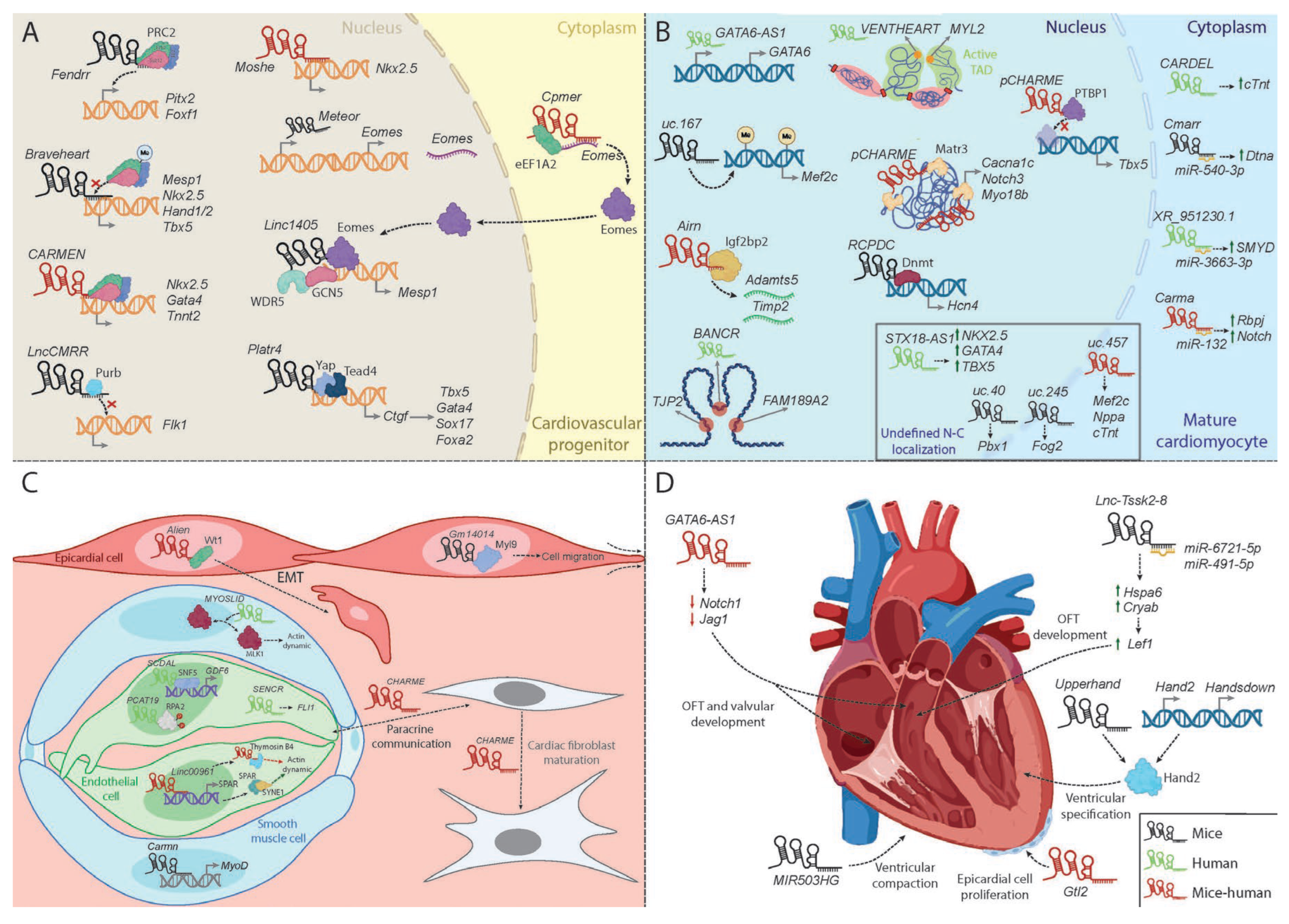
Figure 2.
Molecular mechanisms of lncRNAs during skeletal muscle development. Representative summary table and schematic overview of key lncRNAs regulating proliferation and differentiation in myoblasts (including C2C12 cells) and satellite cells. In the table, lncRNAs highlighted in green indicate processes promoted by elevated lncRNA expression, whereas those in red indicate those promoted upon downregulation. The schematic model describes lncRNA-mediated regulatory mechanisms, including competing endogenous RNA (ceRNA) networks (top) and chromatin epigenetic modulation regulating proliferation and differentiation in mice (red) and other species. Schemes were created using Biorender (https:/www.biorender.com/) and Adobe Illustrator 2022.
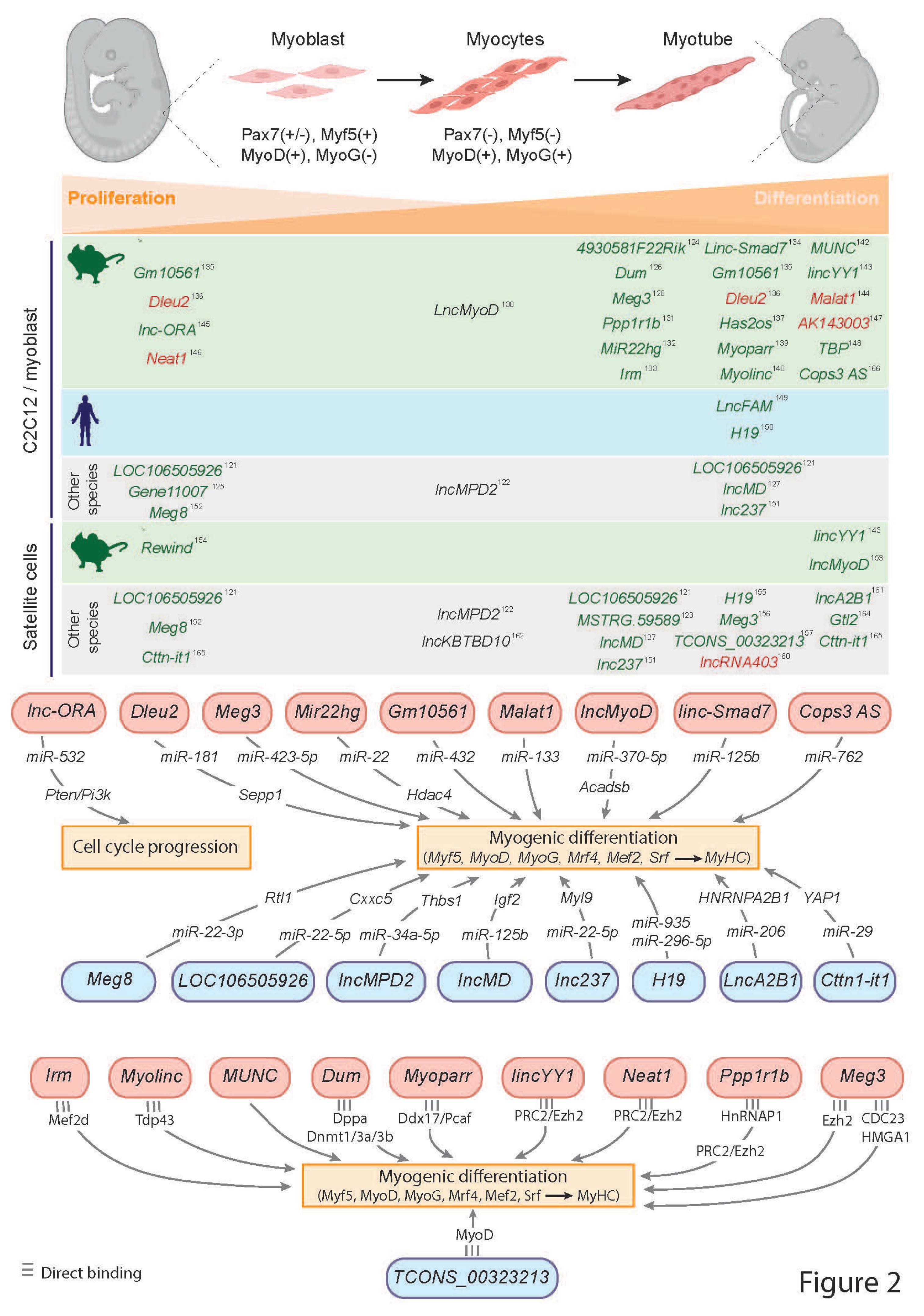
Disclaimer/Publisher’s Note: The statements, opinions and data contained in all publications are solely those of the individual author(s) and contributor(s) and not of MDPI and/or the editor(s). MDPI and/or the editor(s) disclaim responsibility for any injury to people or property resulting from any ideas, methods, instructions or products referred to in the content. |
© 2026 by the authors. Licensee MDPI, Basel, Switzerland. This article is an open access article distributed under the terms and conditions of the Creative Commons Attribution (CC BY) license (http://creativecommons.org/licenses/by/4.0/).
Copyright: This open access article is published under a Creative Commons CC BY 4.0 license, which permit the free download, distribution, and reuse, provided that the author and preprint are cited in any reuse.