

Submitted:
26 October 2026
Posted:
27 October 2026
You are already at the latest version
Abstract
Background and Objectives: Passive release — a release technique performed while the target tissue is held in a passively stretched position — applies to the joint capsule, ligaments, tendons, muscles, peripheral nerves, vessels and subcutaneous tissue; when the release is delivered by injection it is termed “passive release with hydrorelease”. In our experience, no local anesthetic is required, the stretched position makes tender stacking fascia easier to localize, and range of motion tends to improve more than with hydrorelease in a relaxed position. The aim of this article is to describe, in a reproducible form, the treatment points and ultrasound technique of passive release with hydrorelease across multiple body regions. Materials and Methods: This protocol is based on our clinical experience. Eight treatment points in six regions (forearm; shoulder; hip, anterior and posterior; gluteal region; posterior thigh; posterior knee) were selected from our practice. For each point, the patient position, the assistant-maintained passive stretch, the ultrasound visualization, the release procedure, and the safety considerations are described with demonstration videos. Results: The protocol is presented as eight point-by-point descriptions (P1–P8), each pairing a passively stretched position with hydrorelease of the target fascia, ligament or capsule, with the anatomical rationale and corresponding video (Videos S1–S8). Conclusions: This article describes passive release with hydrorelease as a reproducible multi-region protocol. It is a technique description based on our clinical experience; no claim of effectiveness is made, the technique is proposed as one element of multimodal care rather than as a stand-alone treatment, and its prospective evaluation remains a task for future studies.
Keywords:
passive release with hydrorelease
; tight fascia sign
; ultrasound-guided
; fascia
; joint capsule
; iliofemoral ligament
; deep gluteal syndrome
; sciatic nerve
; oblique popliteal ligament
1. Introduction
Reduced fascial extensibility and glide can cause pain and restrict range of motion (ROM). Such changes may involve the joint capsule, ligaments, tendons, epimysium, and epineurium — that is, the fascial system in its broad sense [1,2,3]. Within the loose connective tissue of the deep fascia, a hyaluronan-rich layer permits gliding between fascial sublayers, and altered density of this layer has been proposed as a basis of myofascial pain [4].
Passive release with hydrorelease [5,6] is a fascia-directed technique in which hydrorelease is performed while the target tissue is held in a passively stretched position; hereafter it is referred to as passive release.‡ The Japanese Non-surgical Orthopedics Society (JNOS) classifies it as one form of combination release [6], and it has been presented at an international meeting [7]; the present article describes the protocol in a reproducible form.
Passive release therefore combines hydrorelease with manual control of a passive-stretch position; this manual passive-stretch component distinguishes it from standard ultrasound-guided fascia hydrorelease (US-FHR) [8]. Depending on the target tissue, the two approaches may be used selectively or together.
Candidate sites are identified by stacking fascia under passive stretch — a JNOS term for the ultrasound appearance of several fascial layers that are stacked and move as a unit instead of gliding [5,6] — and by reduced extensibility and gliding, with the tight fascia sign — a dynamic ultrasound finding of a taut-like fascial layer movement [5,6] — as a supplementary finding where visible. A related assessment, the fascia stretch test (FST), has been conceptualized by JNOS [6]; however, the procedure and positivity criteria are not yet fully standardized; thus, their standardization and validation remain topics for future work. In the present protocol, the release target is stacking fascia identified under passive stretch; the tight fascia sign is the appearance of such fascia when it is visibly tensioned, which is most evident in capsular and ligamentous structures like the coracohumeral ligament, and may be less conspicuous in other anatomical regions.
The technique developed from the following observations in our practice, none of which has been tested in a controlled study. No local anesthetic is used; therefore, no related adverse events arise. Applying tension to the tissue accentuates tender points, which in our experience aids localization of the stacking fascia requiring treatment. The improvement in ROM can be confirmed on the spot during the injection, and in our experience it tends to be greater than with hydrorelease in a relaxed position [5]. Because the procedure is frequently performed in conjunction with a physical therapist, subsequent rehabilitation is easily integrated.
The joint capsule is a principal target of passive release. In knee flexion contracture associated with osteoarthritis, a trend toward a more collagenous (fibrotic) composition of the posterior capsule has been reported histologically, although the difference did not reach statistical significance [9], and genome-wide expression analysis has shown significant changes in genes associated with tissue fibrosis [10]; together, these findings provide a pathological rationale for targeting the capsule. Moreover, a flexion contracture is not merely a consequence of osteoarthritis: in the Osteoarthritis Initiative cohort, it was itself a risk factor for the incidence and progression of knee osteoarthritis and for earlier arthroplasty [11]. Restoring end-range motion — extension in the knee — is therefore clinically meaningful in its own right, and passive release addresses the capsule precisely at that end of range, with the joint held by an assistant at the limit of its available motion.
The aim of this article is to describe, in a reproducible form, the treatment points and the ultrasound technique for passive release across multiple body regions. This article is a technique description and protocol; it is not a study of clinical effectiveness, and no claim of efficacy is made. The 19-point protocol for groin and hip-related pain is presented in a separate paper (in preparation); the two are complementary — the present article describes the technique (how), whereas that paper maps the pain sources (where and why).
2. Materials and Methods
2.1. Nature of the Study, Protocol Development and Ethical Considerations
This clinical protocol is based on our clinical experience. The treatment points come from our practice; the supporting literature was then assembled in three steps. (i) PubMed was searched point by point for the anatomy of the target (cadaveric and imaging work), its ultrasound appearance and needle approach, the gliding or strain behaviour of the tissue, and complications of comparable injection procedures in the region, pairing the name of the structure with terms such as “anatomy”, “cadaver”, “ultrasound-guided”, “injection”, “hydrodissection”, “strain” and “complication” (e.g., “coracohumeral ligament” AND “strain”; “sciatic nerve” AND “hydrodissection”). Searches were run from August to October 2026 with no date restriction, giving preference to English-language papers. (ii) The technique and the positions were cross-checked against Japanese-language textbooks edited by us [5]. (iii) Every statement attributed to a source was verified against its full text, or its abstract where the full text was unavailable, before it was included. The references were chosen for their direct bearing on each point, not by a systematic search, and selection bias is possible (Section 4.1).
This article is a clinical protocol and technique description; no patients were enrolled. Positioning is shown by demonstration, and ultrasound images and a cadaveric specimen are used for anatomical illustration. The positioning photographs show a demonstration by the operator (H.K.) and the assistant (R.A.) with two models (H.H. and one volunteer); all persons shown have given written consent for their publication. No injection was performed during the demonstration; the needle remained capped. The ultrasound images and videos are anonymized, and written informed consent for their publication was obtained from the patients in accordance with the consent policy of Kimura Pain Clinic (effective 1 October 2016). The cadaveric image was obtained during an anatomy course at Nihon University School of Medicine and approved by the Ethics Committee of Nihon University School of Medicine (approval no. 28-8-0, 20 December 2016). Personal identifiers are not shown, and figures are presented without names burned into the images.
2.2. Identification of Stacking Fascia Under Passive Stretch; the Tight Fascia Sign as a Supplementary Finding
Treatment points are identified primarily by stacking fascia seen under passive stretch: the region is scanned in short and long axis with the target tissue held in a passively stretched position, and stacking fascia — several fascial layers that are stacked and move as a unit instead of gliding [5,6] — is identified as the release target. Where it is visibly tensioned, the tight fascia sign (a dynamic ultrasound finding of a taut-like, “tethered” movement of the fascial layer [5,6]) serves as a supplementary finding, and reduced extensibility/gliding and pain reproduction are checked. The FST is used only as an introductory concept: it is an evaluation method that makes tender points clearer by elongating the fascia, positioned in the JNOS scheme as “evaluation and treatment of the fascial pain syndrome: fascia stretch test (with tight fascia sign) → passive release” [6]. As the procedure and positivity criteria lack full specification, they are not prescribed in this study. Consequently, their standardization, validation, and inter-rater reproducibility remain tasks for future research.
2.3. Selection Criteria for Treatment Points
Candidate points were selected from our practice, by consensus among us, when they satisfied the following: (a) stacking fascia under passive stretch (with the tight fascia sign where visible); (b) safe ultrasound visualization and an accessible approach; (c) high clinical frequency; (d) an anatomical rationale; and (e) reproducible demonstration on video. The eight points are representative examples rather than an exhaustive list.
2.4. General Technique
For each point, an assistant maintains the joint at the end of its available range; hydrorelease is then used to dissect and separate the target fascia, while the joint range is continuously controlled to keep the fascia under tension as the range increases. A tendency toward greater improvement when injecting into fascia held under tension, rather than in a relaxed position, has been described [5]. The injectate is normal saline or bicarbonate Ringer’s solution. A 27-gauge 38–40 mm needle is generally used; at the shoulder, hip and gluteal points (P2–P6), a 25-gauge 60 mm Cathelin (spinal) needle can be used depending on body habitus, although the 27-gauge needle may suffice. The volume is titrated to the visible ultrasonographic separation of the fascial layers, typically 1–5 mL per point and no more than 10 mL for broad targets. The effect of the release is difficult to show on imaging; during the injection, improvement in ROM is confirmed by both the operator and assistant via joint mobility assessment and manual palpation of tissue resistance.
2.5. Ultrasound and Video Acquisition
All procedures are performed with a Konica Minolta SONIMAGE HS ultrasound system (linear L18-4, 4–18 MHz; convex C5-2, 2–5 MHz). Ultrasound visualization uses a probe appropriate to the depth of the target. In keeping with the protocol format of this series, the video recordings constitute the primary demonstration of results and are provided as Supplementary Videos (S1–S8); the videos are deposited in a public repository (Zenodo, Creative Commons Attribution 4.0 International [CC BY 4.0] license), with deposition performed by H.K.
2.6. Treatment Points and Positioning (Overview)
Eight treatment points across six regions are covered. The body positions and passive maneuvers in Table 1 follow our demonstration (H.K., R.A. and H.H.); the per-point protocol (visualization, release maneuver, video, and anatomical rationale) is given in the Results.
For the coracohumeral ligament point (P3), the chosen position (extension, adduction, external rotation) is consistent with a cadaveric strain study showing that the coracohumeral ligament is strained by passive external rotation at lower elevation and by extension with adduction [12]. For the superior joint capsule (P2), the position instead combines scapular elevation and upward rotation with longitudinal traction and adduction of the humerus, guiding the humerus into relative glenohumeral adduction to tension the superior capsule.
2.7. Terminology
The technique name is used as passive release throughout, with the full term passive release with hydrorelease given at first mention (Section 1). The registered-trademark symbol is not used in the main text; the trademark is noted once, at first mention.
2.8. Safety Considerations
Operator competence. Every point in this protocol presupposes that the operator can keep the needle tip in view for the whole procedure; this is a skill of the operator rather than a property of the technique. Clinicians who cannot reliably track the tip on ultrasound should not perform passive release, and the needle must not be advanced while the tip is out of sight. Apart from direct B-mode imaging, the needle tip position can be inferred from the displacement of adjacent tissue during advancement, and further confirmed by a small test injection (P6–P8). Prior to the procedure, patients are instructed to report any pain or paresthesia immediately. The onset of paresthesia, a sudden exacerbation of pain, or unexpected resistance during injection necessitates immediate withdrawal and repositioning of the needle.
Contraindications and training. Local or systemic infection, inability to lie still, and inability to tolerate the stretched position are contraindications. At the deep points (P4–P8), the balance of risk and benefit is judged case by case in patients on anticoagulants or antiplatelet drugs and in those with a bleeding tendency. Training in ultrasound-guided injection is sufficient for the superficial points (P1–P3); the points that lie next to major nerves and vessels — the femoral neurovascular bundle at P4, the sciatic nerve at P5–P7, and the popliteal artery and tibial nerve at P8 — require supervised training first.
Adverse events in our experience. Ultrasound-guided fascia hydrorelease, which includes passive release, is routinely performed at Kimura Pain Clinic. During the previous video-archive study period (October 2015–March 2026), the first author performed an estimated 360,000 US-FHR procedures (approximately 150 procedures/day × 20 days/month × 12 months/year × 10 years) [13]. We have not seen a serious complication — nerve or vascular injury, haematoma needing treatment, or infection — with the procedures described here. Occasional minor events are soreness at the puncture site for a short time and a vasovagal reaction with brief hypotension, both of which settle quickly. Local-anaesthetic toxicity and motor block cannot occur, because the injectate is physiological saline or bicarbonate Ringer’s solution only. These are observations from everyday practice, not data from a prospective adverse-event registry.
Risks of analogous procedures. Needle-related risks do not depend on the injectate: a national survey of major complications of regional anaesthesia reported peripheral neuropathy after femoral and sciatic nerve blocks and seizures after sciatic block [14]; these needle-related hazards apply to P4–P8. In a Cochrane review of upper- and lower-limb blocks, ultrasound guidance reduced vascular puncture compared with nerve-stimulator guidance (low-quality evidence) [15]; continuous visualization of the needle tip is used throughout this protocol for this reason.
3. Results
The results are presented primarily as the Supplementary Videos. Each treatment point is illustrated by a positioning photograph (our demonstration; Section 2.1) paired with the corresponding annotated ultrasound panel (Figure 1b, Figure 2b, Figure 3b, Figure 4b, Figure 5b, Figure 6b, Figure 7b and Figure 8b) and the corresponding video (S1–S8). The descriptions below give, for each point, the body position, ultrasound visualization, release maneuver, corresponding video, and anatomical rationale.
3.1. P1 — Forearm: Flexor Digitorum Superficialis / Profundus Interface (Trigger Finger and Finger Contracture)
- Position: supine, arm at the side, forearm supinated; the assistant holds the fingers (digits 2–5) in passive extension at the metacarpophalangeal, proximal and distal interphalangeal joints, with the wrist dorsiflexed, at the end of the available range (positioning photograph: forearm).
- Ultrasound (linear probe, L18-4): short-axis view of the middle to distal third of the forearm showing, from superficial to deep, the flexor digitorum superficialis (FDS), the median nerve and the flexor digitorum profundus (FDP). In the forearm the median nerve courses between the FDS and the FDP [16]; a persistent median artery — an anatomical variant — may also accompany the median nerve [17]. The nerve and, when present, this artery are identified before needle insertion (Figure 1b). The ulnar nerve and artery are identified on the ulnar side. Long-axis views confirm the gliding of the FDS and FDP layers during passive finger motion. When symptoms are limited to a single digit, that finger is extended and moved individually while scanning the forearm, so that the corresponding flexor tendon (the FDS or FDP tendon to that digit) and the surrounding stacking fascia are identified and targeted.
- Release: with the wrist and fingers held in passive extension, hydrorelease is delivered to the stacking fascia between the FDS and the FDP at a site away from the median nerve; the needle tip is kept under continuous visualization, and no injection is made into the nerve or a vessel.
- Video: S1.
- Anatomical rationale: finger flexion is produced by the FDS and FDP gliding relative to each other and to the surrounding tissues: in vivo the two tendons have different excursions (about 24 mm for the FDS and 32 mm for the FDP with the wrist neutral) [18], and specific finger motions maximize their differential gliding [19], so that finger motion requires relative sliding between the two layers. Trigger finger is classically attributed to pathology of the A1 pulley [20]; however, in an atypical trigger finger with normal A1 pulleys, dynamic ultrasound identified an abnormal motion (a reproducible snap or shift) of the FDS near its proximal origin in the forearm, and ultrasound-guided hydrodissection with 20 mL of 5% dextrose at that site resolved the symptoms in a single case [21], suggesting that the cause of finger symptoms can lie in the forearm. The present point shares with that report the search for a proximal cause but differs in three respects. First, the target differs: the report [21] addressed a single site of dynamic abnormality, whereas the present point targets the stacking fascia between the FDS and the FDP identified with the wrist and fingers held in passive extension. Second, the injectate differs: the report used a relatively large volume of dextrose, whereas the present point uses normal saline (or bicarbonate Ringer’s solution). Third, the limb position differs: the report does not describe holding the tissue in passive stretch during the injection, whereas the present point is performed with the tissue held in passive stretch, the improvement in range of motion being observed during the injection. The thumb has no flexor digitorum superficialis or profundus; for thumb symptoms the stacking fascia around the flexor pollicis longus in the forearm is released instead, and thumb pain is addressed in our thumb-pain protocol [22]. In our experience, restricted FDS–FDP gliding in the forearm may accompany trigger finger and finger contracture. Passive release at the forearm — remote from the primary site of symptoms — may be performed alone or alongside pulley treatment. Notably, some patients improve without treatment of the pulley. This point illustrates the general principle of the protocol: the interface whose gliding is lost is treated where it can be safely visualized, while the assistant maintains the tissue under tension.
3.2. P2 — Shoulder: Superior Capsule
- Position: side-lying; longitudinal traction of the humerus with glenohumeral adduction, combined with scapular elevation and upward rotation (relative adduction of the humerus to the scapula) (positioning photograph: shoulder — superior capsule).
- Ultrasound (linear probe, L18-4): over the superior shoulder, the acromion, the supraspinatus muscle within the supraspinous fossa, and the upper trapezius are shown; the superior joint capsule is followed and is the injection target (Figure 2b).
- Release: with the tissue held under passive stretch, hydrorelease is delivered to the superior capsular fascia; the assistant maintains longitudinal traction and progressively increases scapular elevation and upward rotation to keep the superior capsule under tension. A practical point: an entry point close to the acromion gives too steep a needle angle to reach the capsule, so a longer needle is used and the entry point is placed away from the acromion to obtain a shallower angle that advances toward the superior capsule.
- Video: S2.
- Anatomical rationale: the superior joint capsule is tensioned by glenohumeral adduction; with the arm at the side, further adduction is limited, so longitudinal traction combined with scapular elevation and upward rotation is used to induce relative adduction of the humerus to the scapula. The rotator-interval structures adjacent to the superior capsule, including the superior glenohumeral ligament, are described in [23,24].
3.3. P3 — Shoulder: Coracohumeral Ligament (CHL)
- Position: side-lying; shoulder extension + adduction + external rotation (positioning photograph: shoulder — CHL).
- Ultrasound (linear probe, L18-4): the coracohumeral ligament is identified between the coracoid process and the humeral head, superficial to the subscapularis and deep to the deltoid (Figure 3b). On passive external rotation and extension, the CHL shows a taut-like, tethered movement — the tight fascia sign — which is particularly evident at this point (Figure 3d; Video S3, part 2).
- Release: with the tissue held under passive stretch, hydrorelease is delivered to the CHL; external rotation and extension are increased to maintain tension.
- Video: S3 (part 1, release; part 2, tight fascia sign).
- Anatomical rationale: in a fresh/frozen cadaver study, the CHL was strained by passive external rotation at lower elevation and by extension with adduction [12], which is consistent with the position used here. The anatomy and histology of the CHL, and its stiffening in adhesive capsulitis, are described in [25,26]. In a single-arm pilot study, we reported improved passive external rotation of the shoulder after ultrasound-guided hydrorelease of the CHL [27].
3.4. P4 — Hip, Anterior: Iliofemoral Ligament and Anterior Capsule
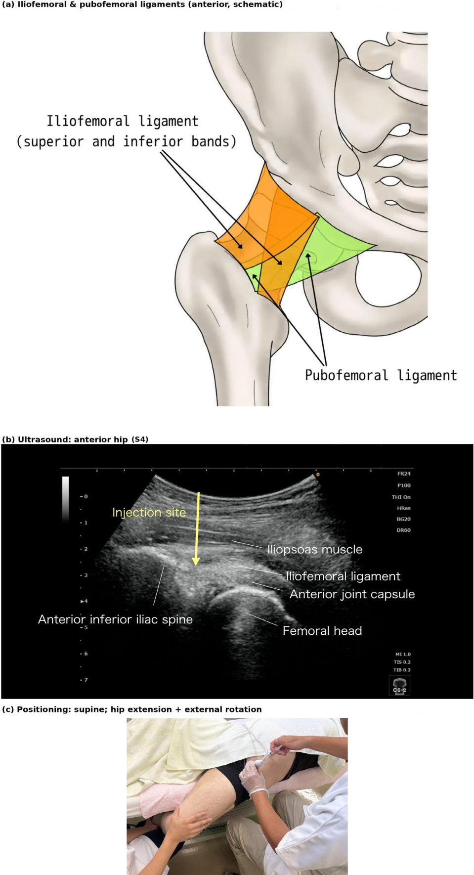
- Position: supine; hip extension + external rotation (positioning photograph: anterior hip).
- Ultrasound (convex probe, C5-2): over the anterior hip, the iliofemoral ligament and the anterior joint capsule are shown overlying the femoral head, with the iliopsoas superficial and the anterior inferior iliac spine as a bony landmark. The femoral nerve and femoral vessels lie medially and are identified; the procedure is performed lateral to them, over the femoral head (Figure 4b).
- Release: hydrorelease is delivered to the anterior capsule and iliofemoral ligament while the position is maintained under stretch.
- Video: S4.
Figure 2.
Shoulder, superior joint capsule (P2, S2). (a-1) posterior view with the supraspinatus in place; (a-2) supraspinatus removed to expose the superior joint capsule; (b) annotated ultrasound; (c) positioning photograph (shoulder; superior joint capsule). Position: side-lying. Passive movement: scapular elevation and upward rotation, with longitudinal traction and adduction of the humerus. The superior capsule is tensioned in the direction of glenohumeral adduction; however, with the arm resting at the side in the lying position, further glenohumeral adduction is difficult to obtain. Therefore, with the humerus held under longitudinal traction, the scapula is elevated and rotated upward so that the humerus is guided into relative adduction with respect to the scapula, thereby tensioning the superior capsule. Strictly, the scapular movement that produces relative glenohumeral adduction is upward rotation; in practice, elevation is combined to facilitate upward rotation. Illustration: Hidenori Horigome (Kimura Pain Clinic).
Figure 2.
Shoulder, superior joint capsule (P2, S2). (a-1) posterior view with the supraspinatus in place; (a-2) supraspinatus removed to expose the superior joint capsule; (b) annotated ultrasound; (c) positioning photograph (shoulder; superior joint capsule). Position: side-lying. Passive movement: scapular elevation and upward rotation, with longitudinal traction and adduction of the humerus. The superior capsule is tensioned in the direction of glenohumeral adduction; however, with the arm resting at the side in the lying position, further glenohumeral adduction is difficult to obtain. Therefore, with the humerus held under longitudinal traction, the scapula is elevated and rotated upward so that the humerus is guided into relative adduction with respect to the scapula, thereby tensioning the superior capsule. Strictly, the scapular movement that produces relative glenohumeral adduction is upward rotation; in practice, elevation is combined to facilitate upward rotation. Illustration: Hidenori Horigome (Kimura Pain Clinic).
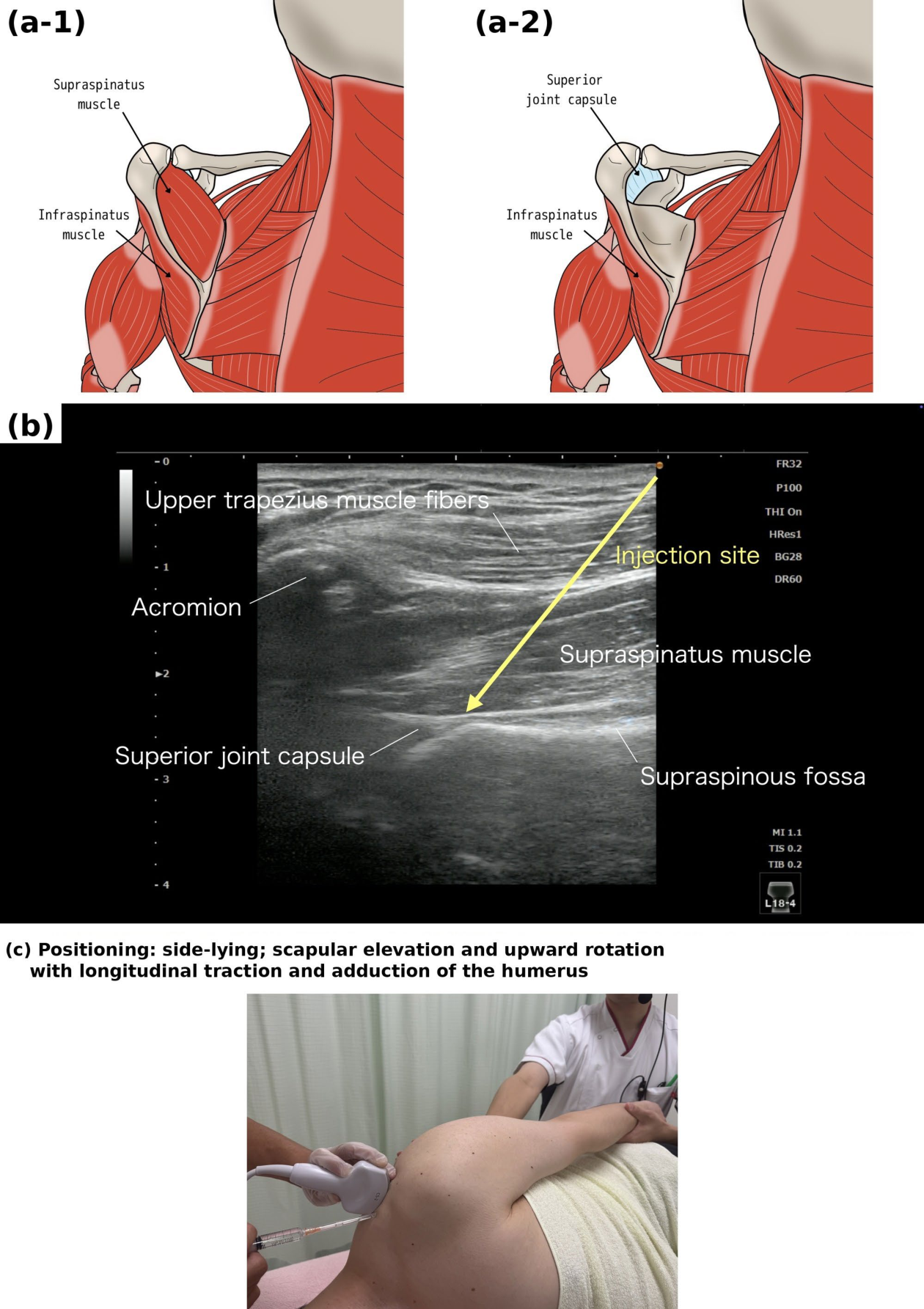
Figure 3.
Shoulder, coracohumeral ligament (P3, S3). (a) anatomical schematic; (b) annotated ultrasound; (c) positioning photograph; (d) dynamic ultrasound during passive external rotation: left, the tight fascia sign (restricted fascial extensibility and gliding impair humeral movement); right, after release, the restriction is reduced and external rotation improves (Video S3, part 2). Illustration: Ayato Kurosawa (formerly Trigger Point Therapy Clinic), redrawn by Hidenori Horigome (Kimura Pain Clinic).
Figure 3.
Shoulder, coracohumeral ligament (P3, S3). (a) anatomical schematic; (b) annotated ultrasound; (c) positioning photograph; (d) dynamic ultrasound during passive external rotation: left, the tight fascia sign (restricted fascial extensibility and gliding impair humeral movement); right, after release, the restriction is reduced and external rotation improves (Video S3, part 2). Illustration: Ayato Kurosawa (formerly Trigger Point Therapy Clinic), redrawn by Hidenori Horigome (Kimura Pain Clinic).
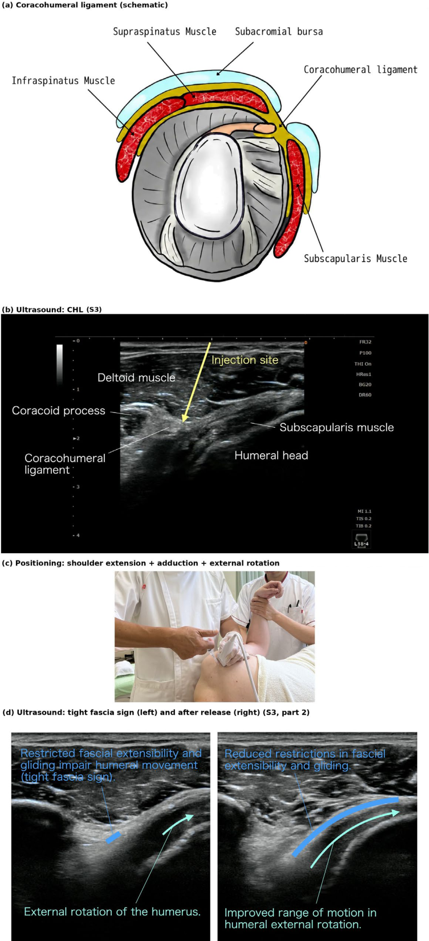
Figure 4.
Anterior hip: iliofemoral ligament and anterior capsule (P4, S4). (a) anatomical schematic (the adjacent pubofemoral ligament is also shown); (b) annotated ultrasound; (c) positioning photograph. Illustration: Hidenori Horigome (Kimura Pain Clinic).
Figure 4.
Anterior hip: iliofemoral ligament and anterior capsule (P4, S4). (a) anatomical schematic (the adjacent pubofemoral ligament is also shown); (b) annotated ultrasound; (c) positioning photograph. Illustration: Hidenori Horigome (Kimura Pain Clinic).
3.5. P5 — Hip, Posterior: Posterior Capsule
- Position: side-lying; hip extension + internal rotation (positioning photograph: posterior hip).
- Ultrasound (convex probe, C5-2): from a posterior approach, the posterior joint capsule and the ischiofemoral ligament are shown between the ischium and the femoral head, deep to the gluteus maximus. The sciatic nerve, which runs nearby, is identified and avoided (Figure 5b).
- Release: hydrorelease is delivered to the posterior capsule under passive stretch (extension with internal rotation).
- Video: S5.
- Anatomical rationale: the posterior capsule and the ischiofemoral ligament form a continuous capsulo-ligamentous complex — the ligaments being thickenings of the fibrous capsule, as shown for the iliofemoral ligament [28] — that is tensioned in extension and internal rotation [29,31]. Histological analysis demonstrates that the hip capsule in osteoarthritis exhibits greater thickness and a higher frequency of severe fibrosis compared to that in femoral neck fracture [32]. The mapping of pain sources in the hip region (including the posterior compartment) is treated in the separate protocol for groin and hip-related pain (in preparation); the present article provides the technique.
3.6. P6 — Gluteal Region (Deep Gluteal Syndrome): Peri-Sciatic
- Position: side-lying; hip flexion + adduction + internal rotation (positioning photograph: gluteal region).
- Ultrasound (convex probe, C5-2): from superficial to deep, the gluteus maximus, the piriformis and the sciatic nerve are shown, with the ischium as a bony landmark (Figure 6b).
- Release: with the stretched position (flexion + adduction + internal rotation) maintained, hydrorelease is delivered around the sciatic nerve to free the perineural fascia. As a technical point, once the needle tip approaches the sciatic nerve, the fluid is injected continuously while the needle is advanced slowly; the advancing injectate opens the space ahead of the tip, keeping the needle away from the nerve, and its spread on ultrasound helps confirm the position of the needle tip. This is intended to minimize the risk of nerve injury.
- Video: S6.
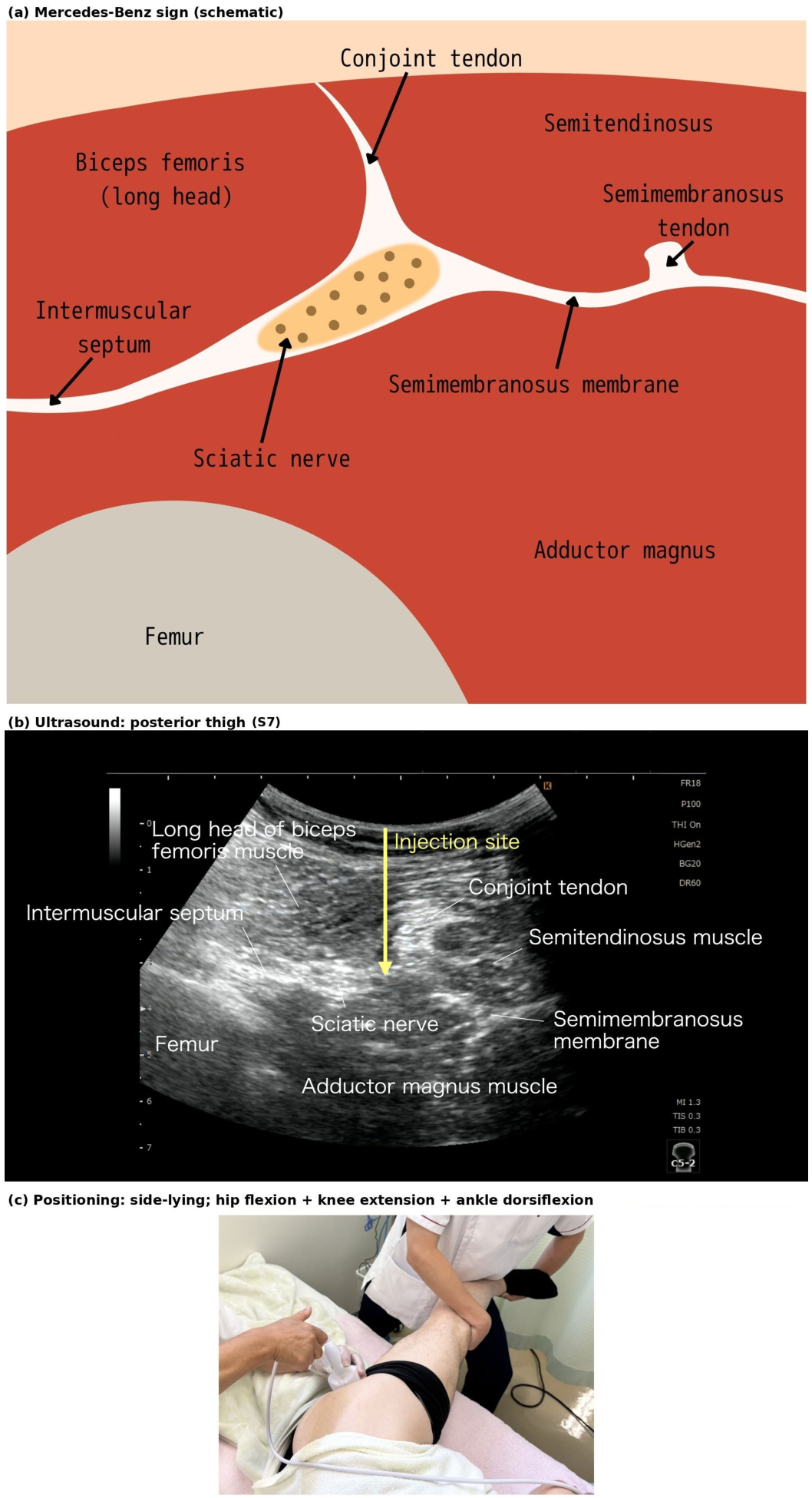
3.7. P7 — Posterior Thigh: Sciatic Nerve Short Axis (the “Mercedes-Benz Sign”)
- Position: side-lying; hip flexion with the knee extended (straight-leg-raise-like) + ankle dorsiflexion (positioning photograph: posterior thigh).
- Ultrasound (convex probe, C5-2): in the short axis over the posterior thigh, the sciatic nerve is seen centrally, with the long head of biceps femoris, the semitendinosus and semimembranosus, and the adductor magnus (over the femur) forming a trident that resembles the Mercedes-Benz emblem (Figure 7b).
- Release: with the stretched position (straight-leg-raise-like hip flexion + ankle dorsiflexion) maintained, hydrorelease is delivered around the sciatic nerve. Once the needle tip approaches the sciatic nerve, the fluid is injected continuously while the needle is advanced slowly, as for P6.
- Video: S7.
- Anatomical rationale: the so-called “Mercedes-Benz sign” — the sciatic nerve centered among the hamstring muscles and the adductor magnus [36] — serves as a landmark for the sciatic nerve in the posterior thigh. In the posterior thigh, a case has also been reported in which the sciatic nerve was tethered to the hamstrings by scar tissue, causing neuropathy [37]. The straight-leg-raise-like maneuver with dorsiflexion tensions the nerve along its course.
Figure 5.
Posterior hip: posterior capsule and ischiofemoral ligament (P5, S5). (a) anatomical schematic; (b) annotated ultrasound; (c) positioning photograph (side-lying; hip extension + internal rotation). Illustration: Hidenori Horigome (Kimura Pain Clinic).
Figure 5.
Posterior hip: posterior capsule and ischiofemoral ligament (P5, S5). (a) anatomical schematic; (b) annotated ultrasound; (c) positioning photograph (side-lying; hip extension + internal rotation). Illustration: Hidenori Horigome (Kimura Pain Clinic).
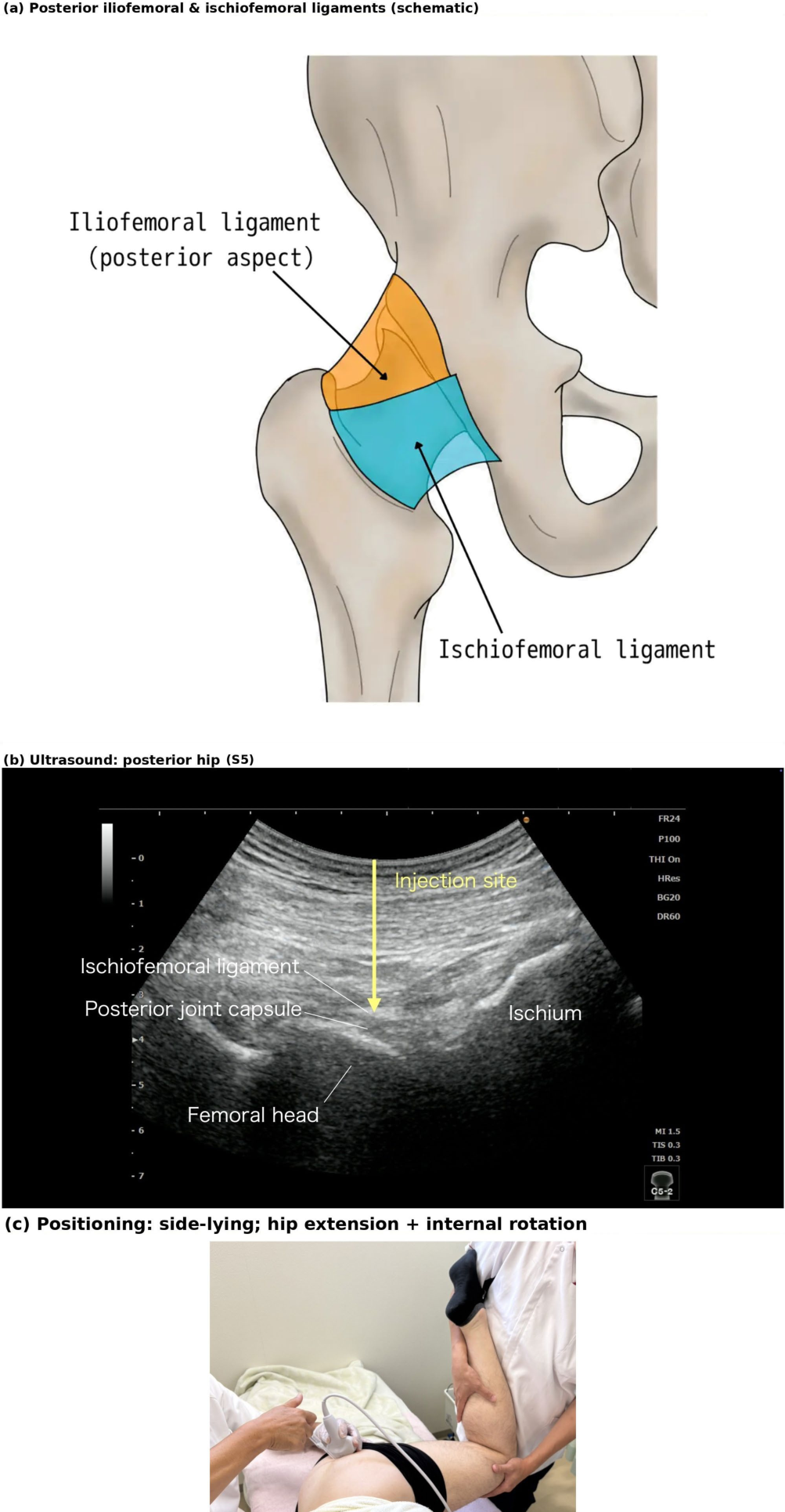
Figure 6.
Gluteal region / deep gluteal syndrome (P6, S6). (a) anatomical schematic; (b) annotated ultrasound; (c) positioning photograph. Illustration: Ayato Kurosawa (formerly Trigger Point Therapy Clinic), labels modified by Hidenori Horigome (Kimura Pain Clinic).
Figure 6.
Gluteal region / deep gluteal syndrome (P6, S6). (a) anatomical schematic; (b) annotated ultrasound; (c) positioning photograph. Illustration: Ayato Kurosawa (formerly Trigger Point Therapy Clinic), labels modified by Hidenori Horigome (Kimura Pain Clinic).
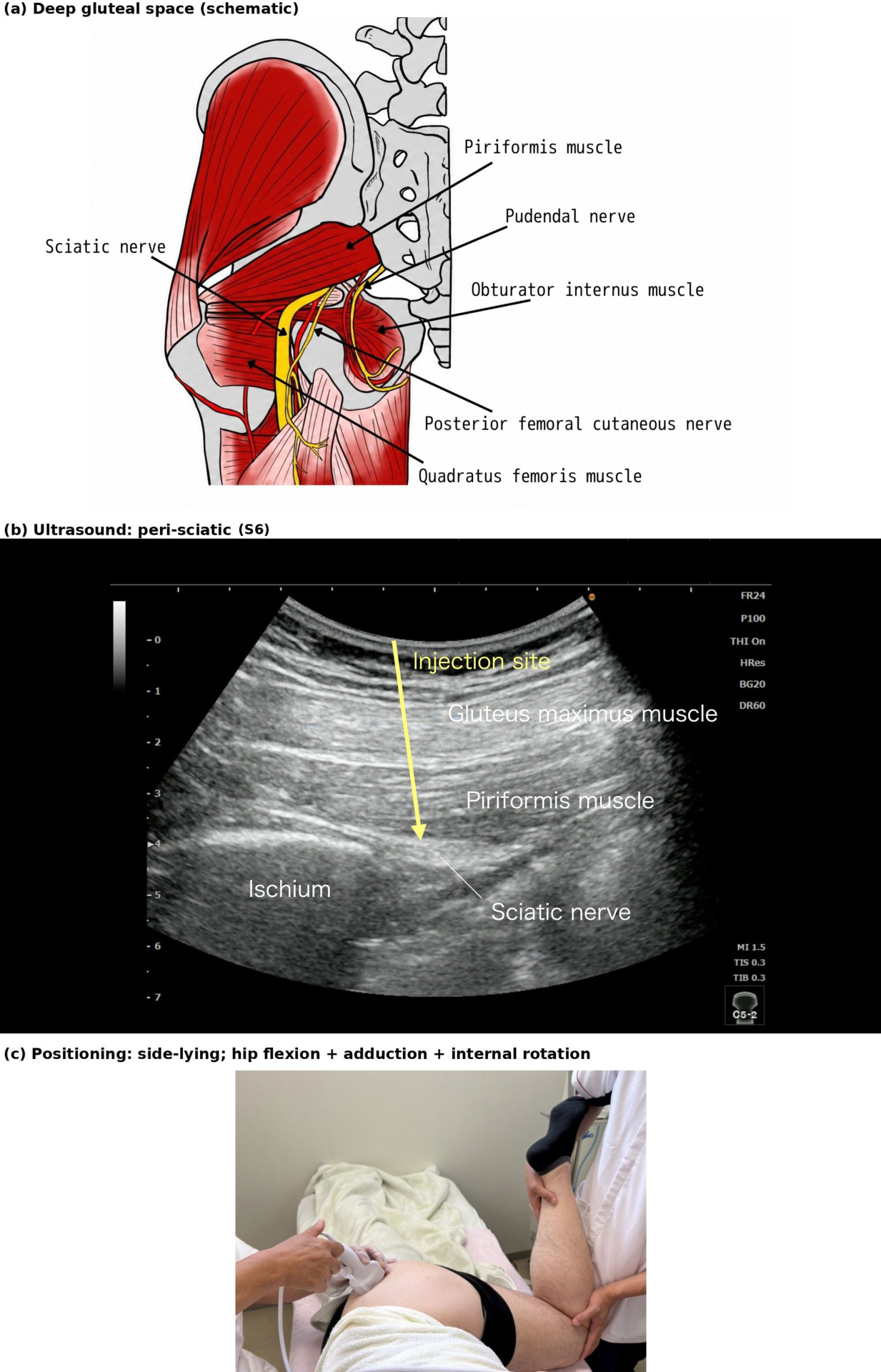
Figure 7.
Posterior thigh, “Mercedes-Benz sign” (P7, S7). (a) anatomical schematic; (b) annotated ultrasound; (c) positioning photograph. Illustration: Hidenori Horigome (Kimura Pain Clinic).
Figure 7.
Posterior thigh, “Mercedes-Benz sign” (P7, S7). (a) anatomical schematic; (b) annotated ultrasound; (c) positioning photograph. Illustration: Hidenori Horigome (Kimura Pain Clinic).
3.8. P8 — Posterior Knee: Oblique Popliteal Ligament and Capsule
- Position: prone; knee extension (positioning photograph: posterior knee).
- Ultrasound (linear probe, L18-4): in the popliteal fossa, the oblique popliteal ligament is shown near the medial femoral condyle, with the medial head of gastrocnemius, the popliteal artery, and the tibial nerve as landmarks (Figure 8b).
- Release: with the stretched position (knee extension) maintained, hydrorelease is delivered to the oblique popliteal ligament and the posterior capsule. As a technical point, because the popliteal artery and the tibial nerve lie close by, once the needle tip approaches them the fluid is injected continuously while the needle is advanced slowly; the advancing injectate opens the space ahead of the tip, keeping the needle away from the artery and nerve, and its spread on ultrasound helps confirm the position of the needle tip. This is intended to minimize the risk of vascular or nerve injury.
- Video: S8.
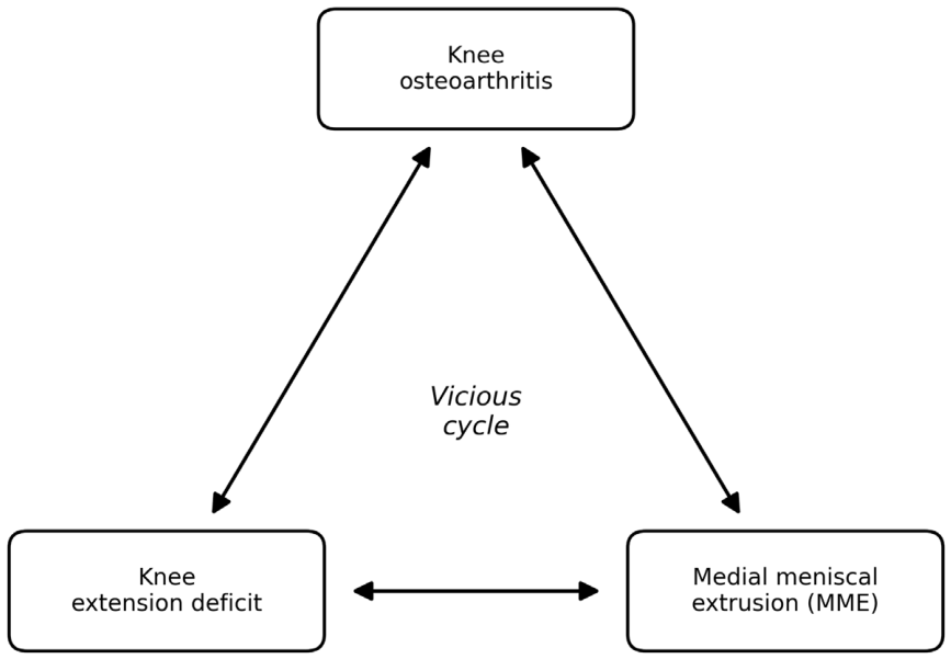
- Anatomical rationale: as noted in the Introduction, findings suggestive of fibrosis have been reported in the posterior capsule of knees with flexion contracture [9,10], and a flexion contracture is itself associated with progression of osteoarthritis [11]; the oblique popliteal ligament is a lateral expansion of the semimembranosus tendon that reinforces and blends with the posterior knee capsule to form a continuous capsulo-ligamentous complex [38,39,40]. Part of the posteromedial joint capsule forms the tendon sheath of the semimembranosus and is continuous with the fibrous layer superficial to it [41], and in vivo ultrasound in healthy volunteers has shown that the medial meniscus, joint capsule, and semimembranosus move in a coordinated manner, suggesting that the capsule may transmit semimembranosus action to the meniscus [42]. We propose that knee osteoarthritis, knee extension deficit, and medial meniscal extrusion (MME) interact mechanically and may form a vicious cycle (Figure 9). In a 4-year analysis of 1,018 participants from the Osteoarthritis Initiative, a large US cohort, baseline loss of knee extension was associated with subsequent worsening of MME on magnetic resonance imaging [43]. In a retrospective series, improvement of knee extension range by semimembranosus stretching and passive ROM exercise was accompanied by a decrease in MME measured on ultrasound [44], although the changes in ROM and MME were not significantly correlated. Within this framework, P8 targets the extension-deficit limb of the cycle; evaluation of MME and the remaining limbs will be addressed in a separate protocol for knee osteoarthritis. This cycle was conceived in October 2023 and first presented at a scientific meeting in March 2026 (the 6th Kita-Kanto and Koshinetsu Regional Meeting of the Japan Society of Pain Clinicians, Matsumoto, 7 March 2026).
Figure 8.
Posterior knee: oblique popliteal ligament and capsule (P8, S8). (a) cadaveric specimen (Nihon University School of Medicine Ethics Committee, approval no. 28-8-0, 20 December 2016); (b) annotated ultrasound; (c) positioning photograph.
Figure 8.
Posterior knee: oblique popliteal ligament and capsule (P8, S8). (a) cadaveric specimen (Nihon University School of Medicine Ethics Committee, approval no. 28-8-0, 20 December 2016); (b) annotated ultrasound; (c) positioning photograph.
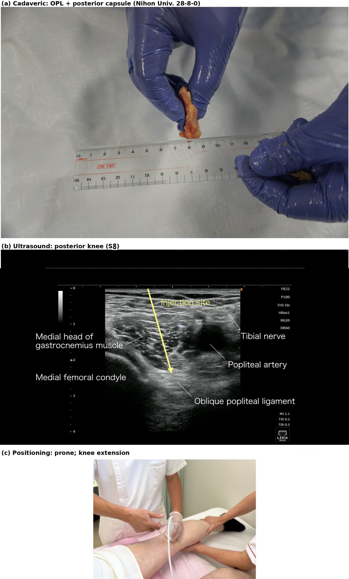
Figure 9.
Proposed mechanical interaction among knee osteoarthritis, knee extension deficit and medial meniscal extrusion (MME), forming a vicious cycle. Passive release of the oblique popliteal ligament and capsule (P8) addresses the extension-deficit limb of this cycle. Schematic.
Figure 9.
Proposed mechanical interaction among knee osteoarthritis, knee extension deficit and medial meniscal extrusion (MME), forming a vicious cycle. Passive release of the oblique popliteal ligament and capsule (P8) addresses the extension-deficit limb of this cycle. Schematic.
4. Discussion
Passive release is positioned as one form of combination release [6]. It belongs to a series of protocols on US-FHR: multi-point protocols have been described for fascia-derived low back pain [45] and for non-odontogenic toothache and orofacial pain [46]; furthermore, US-FHR around the artery has been clinically evaluated for myofascial neck pain [47]. The present article describes it across multiple regions in a reproducible form, pairing an assessment based on identification of stacking fascia under passive stretch — supplemented where visible by the tight fascia sign — with a release performed under a passively stretched position.
The forearm point (P1) shows the principle in its simplest form: two muscle–tendon layers that must glide on each other are placed under tension by passive finger extension, and the stacking fascia between them is released at a site (the forearm) remote from the site of symptoms (the fingers). At the joints (P2–P5 and P8), the joint capsule and its reinforcing ligaments are the common targets, so the protocol connects conditions that share a mechanism of reduced capsular and ligamentous extensibility, such as stiff shoulder, stiff hip, and knee flexion contracture. At the gluteal region and posterior thigh (P6–P7), the target is instead the perineural fascia around the sciatic nerve. The same technical principle — maintaining tissue tension by controlling the joint through its increasing range while hydrorelease dissects the target fascia — is applied at each site. The clinical relevance of this end-range target is best documented in the knee, where a flexion contracture is associated with osteoarthritis incidence, progression, and earlier arthroplasty [11]. Whether posterior capsule release alters this clinical course (the proposed cycle in Figure 9) remains unexamined and is beyond the scope of this study. In our clinical experience, the majority of patients exhibit improved knee extension during the injection (Section 2.4).
Relative to standard US-FHR, the distinguishing feature of passive release is the manual control of a passive-stretch position that accompanies the injection; the two may be used selectively or in combination according to the target tissue. Injection procedures at the same sites have been reported, and the differences from passive release should be made explicit. Hydrodilatation for adhesive capsulitis of the shoulder [48,49] involves injecting fluid (commonly with corticosteroid and local anesthetic) into the joint cavity to distend the capsule from within, without deliberate stretching of the joint; its pooled effect on pain and ROM is small [49]. Passive release, by contrast, does not enter the joint. Instead, it targets the stacking fascia on the outer surface of the capsule and ligaments (P2–P5, P8) with normal saline or bicarbonate Ringer’s solution while the assistant holds the joint at the end of its range, and the change in ROM and tissue resistance is confirmed during the injection. Nerve hydrodissection for entrapment neuropathies [50,51,52] involves injecting saline, dextrose, local anesthetic or other solutions around the nerve at the site of entrapment to separate it from the surrounding tissue, typically without deliberate tensioning of the nerve; the optimal volume has not been defined [51]. Passive release at the sciatic nerve (P6, P7) shares the perineural target but differs in that the nerve is first placed under tension using a passive-stretch position (hip flexion, adduction, and internal rotation at P6; a straight-leg-raise-like maneuver with ankle dorsiflexion at P7). Once the stacking fascia that restricts its gliding becomes visible, the injectate is delivered directly to it, and the release is continued while maintaining the stretch. To our knowledge, a multi-region protocol performed under passive stretch has not been published.
The rationale for injecting with the target tissue under passive tension is currently theoretical. Loose (areolar) connective tissue behaves as a linear viscoelastic material under uniaxial tension, with stress relaxation over seconds to minutes [53]; its fibroblasts remodel their cytoskeleton within minutes of sustained stretch ex vivo and in vivo [54] and thereby actively modulate whole-tissue tension [55]. On this basis, Langevin et al. proposed that sustained stretch of areolar connective tissue may decrease matrix compaction and transiently lower interstitial fluid pressure, allowing fluid to enter the tissue [56]; release of fibroblast tension on the collagen network also lowers interstitial fluid pressure and permits fluid uptake [57,58]. Passive elongation could therefore alter the resistance of interfascial loose connective tissue to injectate. However, this has not been measured directly: in rat skin in vitro, tensile strain transiently raised interstitial fluid pressure [59], and in rat and rabbit muscle, intramuscular pressure rises with passive stretch [60,61]. Patient position has been shown to influence injectate spread in the erector spinae plane block [62], but the effect of tissue tension itself remains to be examined with imaging of spread in future studies. Preclinical support comes from a rat sciatic-nerve adhesion model, in which Miyatake et al. reported that ultrasound-guided hydrorelease with 2 mL of saline lowered the traction force needed to separate the nerve from its bed on the following day from 3.10 N to 2.05 N (without returning to control values), suggesting that fluid injection can reduce mechanical inter-tissue constraint [63]. (In that report the terms hydrorelease and hydrodissection were used interchangeably.)
The technique of the present protocol differs from that of our earlier pilot study of the coracohumeral ligament [27]: in that study, saline was injected with the shoulder held at maximal external rotation, whereas in passive release the assistant holds the joint at the end of its range and, as the range increases during the injection, increases the stretch to keep the tissue under continuous tension.
Place of passive release within multimodal care. Passive release is not meant to stand alone. We use the interval of easier motion and less pain that follows the procedure to get patients moving again — including walking and Radio Taiso (the calisthenics routine familiar throughout Japan) — with referral to exercise therapy when indicated. How long this interval lasts, and whether the regained motion prevents stacking fascia from returning, has not yet been examined.
4.1. Limitations
This protocol has several limitations. First, it is based on our clinical experience; no formal consensus process was used, and the literature was selected rather than systematically retrieved (Section 2.1). The article describes a technique and does not study its clinical effectiveness; therefore no claim of efficacy is made.
Second, the components of the protocol rest on evidence of unequal strength. Clinical or experimental studies suggest fibrotic change of the posterior capsule in knee flexion contracture and its association with the course of osteoarthritis [9,10,11], strain behaviour of the coracohumeral ligament under passive motion [12], differential excursion of the flexor tendons [18,19], viscoelastic and fibroblast responses of areolar connective tissue to stretch [53,54,55,56], cellular control of interstitial fluid pressure [57,58], and reduction of gliding resistance after hydrorelease in an animal model [63]. The location of each treatment point and its relationship to nerves and vessels (Section 3) rest on anatomical reasoning. The positions and passive maneuvers (Table 1), the tendency toward greater improvement in range of motion than with hydrorelease in a relaxed position [5], the injection volumes and the safety observations (Section 2.8) rest on our clinical experience. Whether restoring hip extension at the anterior hip point (P4) influences low back pain — plausible because limited hip extension has been proposed to contribute to altered lumbopelvic mechanics and low back pain (the hip–spine syndrome) [64] — was not evaluated and remains a question for future study.
Third, passive release, stacking fascia, the tight fascia sign and the fascia stretch test were proposed by our group and by JNOS, with which we are affiliated, and have not been validated externally; the fascia stretch test in particular is presented only as an introductory concept, and its procedure and positivity criteria are not standardized here.
Fourth, the effect of the procedure is difficult to show on imaging. Although a change in ROM has been observed, it is rarely accompanied by a sonographically identifiable change at most sites; the coracohumeral ligament is an exception, where a change in the tight fascia sign can sometimes be observed (Figure 3d; Video S3, part 2). In contrast, the assistant maintaining the passive stretch can perceive a change in tissue resistance in most cases. The effect is therefore currently confirmed by the improvement in ROM during the injection, as well as the operator’s and assistant’s tactile perception, neither of which is blinded or controlled, and quantitative imaging correlates remain to be determined. The conspicuity of the tight fascia sign varies by site; it is usually clear at the coracohumeral ligament but often indistinct at other points, and the release target throughout this protocol is therefore defined as stacking fascia rather than the sign itself.
Fifth, the safety data are clinical observations rather than a prospective adverse-event registry (Section 2.8), and long-term outcomes have not been recorded. We intend to validate the protocol in a stepwise manner through multicentre studies within JNOS: first, by assessing inter-examiner reliability regarding stacking fascia under passive stretch and the tight fascia sign; and second, through controlled trials evaluating clinical efficacy, including comparison with hydrorelease in a relaxed position and with active exercise alone. Independent validation by other groups is highly encouraged.
5. Conclusions
This article describes passive release in a reproducible form as a multi-region clinical protocol, pairing an assessment based on identification of stacking fascia under passive stretch, supplemented where visible by the tight fascia sign, with a release performed while the target tissue is held in a passively stretched position by an assistant. Eight treatment points in six regions are described with their positioning, ultrasound visualization, release procedure, safety considerations, anatomical rationale, and demonstration videos. The protocol describes a technique based on our clinical experience; no claims regarding clinical effectiveness are made. The effect of the procedure is currently difficult to show on imaging and is confirmed by the improvement in range of motion during the injection and by the operator’s and assistant’s tactile perception. Passive release is proposed as one component of multimodal management rather than as a treatment on its own; the interval of improved motion is used to resume active exercise. The inter-examiner reliability of the ultrasound findings, the clinical outcomes and the safety of the protocol remain to be evaluated in the prospective studies planned through JNOS. The eight points serve as representative examples rather than an exhaustive list. Other sites where stacking fascia develops under passive stretch may be approached on the same principles.
Figure Legends
Figure 1, Figure 2, Figure 3, Figure 4, Figure 5, Figure 6, Figure 7 and Figure 8 each comprise vertically arranged panels: (a) an anatomical schematic (Figure 2: a-1 and a-2; Figure 8: a cadaveric photograph), (b) the annotated ultrasound image and (c) the positioning (external) photograph; Figure 3 additionally includes (d) a dynamic ultrasound image. Figure 9 is a schematic diagram. The ultrasound image does not embed the positioning photograph. Illustration credits are given in each caption; illustrator names are not printed inside the images, and copyright is held by Kimura Pain Clinic.
Supplementary Materials
The following supporting information can be downloaded at the website of this paper posted on Preprints.org. The following are available: Video S1 (forearm, flexor digitorum superficialis / profundus interface), Video S2 (shoulder, superior capsule), Video S3 (shoulder, coracohumeral ligament; part 1, release; part 2, tight fascia sign), Video S4 (anterior hip), Video S5 (posterior hip), Video S6 (gluteal region, peri-sciatic), Video S7 (posterior thigh), Video S8 (posterior knee). The videos are archived in Zenodo under CC BY 4.0 (DOI: 10.5281/zenodo.23408017).
Author Contributions
Conceptualization, H.K. and T.K.; methodology, H.K. and T.K.; technique demonstration and investigation, H.K., R.A. and H.H.; resources, illustration and ultrasound annotation, H.H. and R.A.; writing—original draft, H.K.; ethical review coordination, T.H.; validation, H.O.; writing—review and editing, T.H., H.O. and all other authors; supervision, H.K. and T.K. All authors have read and agreed to the published version of the manuscript.
Funding
This research received no external funding.
Institutional Review Board Statement
This article is a clinical protocol based on the authors’ clinical experience and published literature; it did not involve a new prospective study on human participants, and institutional review board approval for a clinical study is therefore not applicable. The cadaveric image in Figure 8 was obtained during an anatomy course at Nihon University School of Medicine and approved by the Ethics Committee of Nihon University School of Medicine (approval no. 28-8-0, 20 December 2016). The Ethics Committee of Isesaki Municipal Hospital confirmed that this manuscript series does not constitute research under the Japanese Ethical Guidelines for Life Science and Medical Research Involving Human Subjects and that ethics review is not required (Framework Confirmation of Non-Applicability of Ethics Review, Ref. No. 2026-14-2, 6 October 2026).
Informed Consent Statement
Written informed consent for the anonymized publication of ultrasound images and videos was obtained from the patients in accordance with the consent policy of Kimura Pain Clinic (effective 1 October 2016). All persons shown in the positioning photographs (three authors and one volunteer) gave written consent for publication.
Data Availability Statement
The demonstration videos (Videos S1–S8) are deposited in the Zenodo repository under a Creative Commons Attribution 4.0 International (CC BY 4.0) license; the dataset DOI is https://doi.org/10.5281/zenodo.23408017 (Concept DOI, resolving to the latest version). Deposition was performed by H.K.
Acknowledgments
The authors thank Ayato Kurosawa (formerly Trigger Point Therapy Clinic) for the original anatomical illustrations used in Figures 3 and 6; copyright of the original illustrations is held by Kimura Pain Clinic. AI-assisted tools (Claude, Anthropic) were used for language editing, Japanese–English translation, and manuscript formatting. The authors reviewed and take full responsibility for the content.
Conflicts of Interest
H.K. is the chairman of Medical Corporation Fascia Research Association, which holds the Japanese registered trademark “Passive release” (Registration No. 6736285). H.K. and T.K. are editors of a book cited in this article [5] and receive royalties from it. T.K. is the president of the Japanese Non-surgical Orthopedics Society (JNOS), and H.K. is its founding president and current head of its public relations bureau. T.K. is also the representative director of Academia R&D Support, Inc., a certified university-launched venture of Hirosaki University providing consulting services; this role is unrelated to the present work, and the company had no involvement in the conception, preparation, or writing of this article. The other authors declare no conflicts of interest.
| ‡ | “Passive release” is a registered trademark in Japan (Registration No. 6736285, registered 14 September 2023; holder: Medical Corporation Fascia Research Association). |
References
- Stecco, C.; Pratt, R.; Nemetz, L.D.; Schleip, R.; Stecco, A.; Theise, N.D. Towards a comprehensive definition of the human fascial system. J. Anat. 2025, 246(6), 1084–1098. [Google Scholar] [CrossRef] [PubMed]
- Adstrum, S.; Hedley, G.; Schleip, R.; Stecco, C.; Yucesoy, C.A. Defining the fascial system. J. Bodyw. Mov. Ther. 2017, 21(1), 173–177. [Google Scholar] [CrossRef] [PubMed]
- Bordoni, B.; Escher, A.R.; Castellini, F.; et al. Fascial nomenclature: update 2024. Cureus 2024, 16(2), e53995. [Google Scholar] [CrossRef] [PubMed]
- Stecco, C.; Stern, R.; Porzionato, A.; Macchi, V.; Masiero, S.; Stecco, A.; De Caro, R. Hyaluronan within fascia in the etiology of myofascial pain. Surg. Radiol. Anat. 2011, 33(10), 891–896. [Google Scholar] [CrossRef] [PubMed]
- Kimura, H.; Kobayashi, T.; Namiki, H. (Eds.) Kaibō, Dōsa, Ekō de Michibiku Fascia Rirīsu no Kihon to Rinshō [Fundamentals and Clinical Practice of Fascia Release Guided by Anatomy, Movement and Ultrasound]. In Hydrorelease no Subete [All About Hydrorelease], (in Japanese), 2nd ed.; Bunkodo: Tokyo, 2021; pp. 111–112 and ch. 6. [Google Scholar]
- Japanese Non-surgical Orthopedics Society (JNOS). For medical professionals: 5-1-2 Combination release techniques: e.g., Fascia stretch test → Passive Release (with Hydrorelease). https://www.jnos-global.org/for-medical (updated 13 July 2024; accessed 7 September 2026). Japanese version: https://www.jnos.or.jp/for_medical#4-2-2_Combination_release.
- Kimura, H.; Kobayashi, T.; Imagita, H. Hydrorelease as a new injection technique targeting fascia [invited video presentation]. International Forum: Recent Advancement in Myofascial Pain. 8th Asia-Oceanian Conference of Physical and Rehabilitation Medicine (AOCPRM 2023), Chengdu, China, 6–9 October 2023. [Google Scholar]
- Kobayashi, T.; Kimura, H.; Zenita, Y.; Imagita, H. Hydrorelease of fascia. In Fascia: The Tensional Network of the Human Body, 2nd ed.; Schleip, R., Stecco, C., Driscoll, M., Huijing, P.A., Eds.; Elsevier: Edinburgh, 2022; pp. 609–617. [Google Scholar]
- Campbell, T.M.; Trudel, G.; Laneuville, O. Knee flexion contractures in patients with osteoarthritis: clinical features and histologic characterization of the posterior capsule. PM R 2015, 7(5), 466–473. [Google Scholar] [CrossRef] [PubMed]
- Campbell, T.M.; Trudel, G.; Wong, K.K.; Laneuville, O. Genome wide gene expression analysis of the posterior capsule in patients with osteoarthritis and knee flexion contracture. J. Rheumatol. 2014, 41(11), 2232–2239. [Google Scholar] [CrossRef] [PubMed]
- Campbell, T.M.; McGonagle, D. Flexion contracture is a risk factor for knee osteoarthritis incidence, progression and earlier arthroplasty: data from the Osteoarthritis Initiative. Ann. Phys. Rehabil. Med. 2021, 64(2), 101439. [Google Scholar] [CrossRef] [PubMed]
- Izumi, T.; Aoki, M.; Tanaka, Y.; et al. Stretching positions for the coracohumeral ligament: strain measurement during passive motion using fresh/frozen cadaver shoulders. Sports Med. Arthrosc. Rehabil. Ther. Technol. 2011, 3(1), 2. [Google Scholar] [CrossRef] [PubMed]
- Kimura, H.; Hiroki, T.; Kobayashi, T.; Obata, H. Local twitches during ultrasound-guided fascial hydrorelease occur within stacking fascia: a retrospective analysis of a large video archive. Med. Sci. 2026, 14(3), 350. [Google Scholar] [CrossRef] [PubMed]
- Auroy, Y.; Benhamou, D.; Bargues, L.; et al. Major complications of regional anesthesia in France: the SOS Regional Anesthesia Hotline Service. Anesthesiology 2002, 97(5), 1274–1280. [Google Scholar] [CrossRef] [PubMed]
- Lewis, S.R.; Price, A.; Walker, K.J.; McGrattan, K.; Smith, A.F. Ultrasound guidance for upper and lower limb blocks. Cochrane Database Syst. Rev. 2015, 2015(9), CD006459. [Google Scholar] [CrossRef] [PubMed]
- Standring, S. (Ed.) Gray’s Anatomy: The Anatomical Basis of Clinical Practice, 42nd ed.; Elsevier: Edinburgh, 2021. [Google Scholar]
- Tee, Q.X.; Stuckey, S.; Lau, K.K. Persistent median artery on computed tomographic arteriovenous fistulograms in patients on haemodialysis. J. Med. Imaging Radiat. Oncol. 2023, 67(6), 612–618. [Google Scholar] [CrossRef] [PubMed]
- Wehbé, M.A.; Hunter, J.M. Flexor tendon gliding in the hand. Part I. In vivo excursions. J. Hand Surg. Am. 1985, 10(4), 570–574. [Google Scholar] [CrossRef] [PubMed]
- Wehbé, M.A.; Hunter, J.M. Flexor tendon gliding in the hand. Part II. Differential gliding. J. Hand Surg. Am. 1985, 10(4), 575–579. [Google Scholar] [CrossRef] [PubMed]
- Sampson, S.P.; Badalamente, M.A.; Hurst, L.C.; Seidman, J. Pathobiology of the human A1 pulley in trigger finger. J. Hand Surg. Am. 1991, 16(4), 714–721. [Google Scholar] [CrossRef] [PubMed]
- Yoon, Y.; Lam, K.H.S.; Suryadi, T.; Suhaimi, A.; Su, D.C. Atypical trigger finger associated with proximal flexor digitorum superficialis dynamic instability diagnosed with ultrasound and treated with hydrodissection: a case report. Cureus 2026, 18(7), e113614. [Google Scholar] [CrossRef] [PubMed]
- Kimura, H.; Asaka, R.; Kobayashi, T.; Obata, H. Common fascial abnormalities in five thumb pain conditions: a fifteen-point ultrasound-guided fascia hydrorelease clinical protocol. J. Funct. Morphol. Kinesiol 2026, 11(3), 308. [Google Scholar] [CrossRef] [PubMed]
- Petchprapa, C.N.; Beltran, L.S.; Jazrawi, L.M.; Kwon, Y.W.; Babb, J.S.; Recht, M.P. The rotator interval: a review of anatomy, function, and normal and abnormal MRI appearance. AJR Am. J. Roentgenol. 2010, 195(3), 567–576. [Google Scholar] [CrossRef] [PubMed]
- Kask, K.; Põldoja, E.; Lont, T.; et al. Anatomy of the superior glenohumeral ligament. J. Shoulder Elb. Surg. 2010, 19(6), 908–916. [Google Scholar] [CrossRef] [PubMed]
- Yang, H.F.; Tang, K.L.; Chen, W.; et al. An anatomic and histologic study of the coracohumeral ligament. J. Shoulder Elb. Surg. 2009, 18(2), 305–310. [Google Scholar] [CrossRef] [PubMed]
- Wu, C.H.; Chen, W.S.; Wang, T.G. Elasticity of the coracohumeral ligament in patients with adhesive capsulitis of the shoulder. Radiology 2016, 278(2), 458–464. [Google Scholar] [CrossRef] [PubMed]
- Kimura, H.; Suda, M.; Kobayashi, T.; Suzuki, S.; Fukui, S.; Obata, H. Effectiveness of ultrasound-guided fascia hydrorelease on the coracohumeral ligament in patients with global limitation of the shoulder range of motion: a pilot study. Sci. Rep. 2022, 12(1), 19782. [Google Scholar] [CrossRef] [PubMed]
- Tsutsumi, M.; Nimura, A.; Akita, K. New insight into the iliofemoral ligament based on the anatomical study of the hip joint capsule. J. Anat. 2020, 236(5), 946–953. [Google Scholar] [CrossRef] [PubMed]
- Wagner, F.V.; Negrão, J.R.; Campos, J.; et al. Capsular ligaments of the hip: anatomic, histologic, and positional study in cadaveric specimens with MR arthrography. Radiology 2012, 263(1), 189–198. [Google Scholar] [CrossRef] [PubMed]
- D’Ambrosi, R.; Ursino, N.; Messina, C.; Della Rocca, F.; Hirschmann, M.T. The role of the iliofemoral ligament as a stabilizer of the hip joint. EFORT Open Rev. 2021, 6(7), 545–555. [Google Scholar] [CrossRef] [PubMed]
- Martin, H.D.; Savage, A.; Braly, B.A.; Palmer, I.J.; Beall, D.P.; Kelly, B. The function of the hip capsular ligaments: a quantitative report. Arthroscopy 2008, 24(2), 188–195. [Google Scholar] [CrossRef] [PubMed]
- Teves, J.I.; Latorre, M.R.; Albani, A.; et al. Histopathological characterisation of the hip joint capsule: comparative study of patients with hip osteoarthritis versus patients with femoral neck fracture. Hip Int. 2026, 36(4), 575–581. [Google Scholar] [CrossRef] [PubMed]
- Martin, H.D.; Reddy, M.; Gómez-Hoyos, J. Deep gluteal syndrome. J. Hip Preserv Surg. 2015, 2(2), 99–107. [Google Scholar] [CrossRef] [PubMed]
- Yoon, Y.H.; Hwang, J.H.; Lee, H.W.; et al. Beyond nerve entrapment: a narrative review of muscle–tendon pathologies in deep gluteal syndrome. Diagnostics 2025, 15(19), 2531. [Google Scholar] [CrossRef] [PubMed]
- Yen, Y.S.; Lin, C.H.; Chiang, C.H.; Wu, C.Y. Ultrasound-guided sciatic nerve hydrodissection can improve the clinical outcomes of patients with deep gluteal syndrome: a case-series study. Diagnostics 2024, 14(7), 757. [Google Scholar] [CrossRef] [PubMed]
- Balius, R.; Pedret, C.; Estrada-Alarcón, P.; Corominas, H. Overview of thigh and leg anatomical and sonographic landmarks in rheumatic patients. Eur. J. Rheumatol. 2024, 11 (Suppl 3), S305–S315. [Google Scholar] [CrossRef] [PubMed]
- Solankee, J.; Srinivasan, S.; Payne, R.; Cai, H. Neuromuscular ultrasound in delayed sciatic neuropathy following hamstring tear. PM R 2025, 17(5), 582–584. [Google Scholar] [CrossRef] [PubMed]
- Hedderwick, M.; Stringer, M.D.; McRedmond, L.; Meikle, G.R.; Woodley, S.J. The oblique popliteal ligament: an anatomic and MRI investigation. Surg. Radiol. Anat. 2017, 39(9), 1017–1027. [Google Scholar] [CrossRef] [PubMed]
- Wu, X.D.; Yu, J.H.; Zou, T.; et al. Anatomical characteristics and biomechanical properties of the oblique popliteal ligament. Sci. Rep. 2017, 7, 42698. [Google Scholar] [CrossRef] [PubMed]
- De Maeseneer, M.; Van Roy, P.; Shahabpour, M.; Gosselin, R.; De Ridder, F.; Osteaux, M. Normal anatomy and pathology of the posterior capsular area of the knee: findings in cadaveric specimens and in patients. AJR Am. J. Roentgenol. 2004, 182(4), 955–962. [Google Scholar] [CrossRef] [PubMed]
- Tsutsumi, M.; Nimura, A.; Tharnmanularp, S.; Kudo, S.; Akita, K. Posteromedial capsular anatomy of the tibia for consideration of the medial meniscal support structure using a multidimensional analysis. Sci. Rep. 2023, 13, 12030. [Google Scholar] [CrossRef] [PubMed]
- Tsutsumi, M.; Kanazawa, S.; Nimura, A.; Akita, K.; Kudo, S. In vivo movement interrelationships among the medial meniscus, joint capsule, and semimembranosus during tibial rotation. Sci. Rep. 2024, 14, 17022. [Google Scholar] [CrossRef] [PubMed]
- Chan Chun Kong, D.; Conaghan, P.G.; Campbell, T.M. Baseline loss of knee extension is associated with regional MRI progression in knee osteoarthritis: a retrospective longitudinal cohort study—data from the osteoarthritis initiative. Rheumatol. Int. 2025, 45(11), 257. [Google Scholar] [CrossRef] [PubMed]
- Yoshizuka, H.; Taniguchi, T.; Fukuta, K.; Mitsutake, T.; Honda, S. Decrease in medial meniscal extrusion after physical therapy to improve knee pain and range of motion in patients with knee osteoarthritis: a retrospective study. PLoS ONE 2022, 17(11), 36449464. [Google Scholar] [CrossRef] [PubMed]
- Kimura, H.; Asaka, R.; Horigome, H.; Kobayashi, T.; Obata, H. A Multi-Point Ultrasound-Guided Fascia Hydrorelease Protocol for Fascia-Derived Low Back Pain: A Clinical Protocol. Preprints 2026, 2026090625. [Google Scholar] [CrossRef]
- Kimura, H.; Kobayashi, T.; Asaka, R.; Obata, H. An Eleven-Point Ultrasound-Guided Fascia Hydrorelease Protocol for Non-Odontogenic Toothache and Orofacial Pain: A Clinical Protocol. Medicina 2026, 62(9), 1808. [Google Scholar] [CrossRef] [PubMed]
- Hiroki, T.; Kimura, H.; Kobayashi, T.; Horigome, H.; Suda, M.; Fukui, S.; et al. The analgesic effect of ultrasound-guided fascia hydrorelease around the artery for myofascial neck pain: a prospective single-arm interventional study. PLoS ONE 2026, 21(7), 42536624. [Google Scholar] [CrossRef] [PubMed]
- Elnady, B.; Rageh, E.M.; Hussein, M.S.; et al. In shoulder adhesive capsulitis, ultrasound-guided anterior hydrodilatation in rotator interval is more effective than posterior approach: a randomized controlled study. Clin. Rheumatol. 2020, 39(12), 3805–3814. [Google Scholar] [CrossRef] [PubMed]
- Saltychev, M.; Laimi, K.; Virolainen, P.; Fredericson, M. Effectiveness of hydrodilatation in adhesive capsulitis of shoulder: a systematic review and meta-analysis. Scand. J. Surg. 2018, 107(4), 285–293. [Google Scholar] [CrossRef] [PubMed]
- Buntragulpoontawee, M.; Chang, K.V.; Vitoonpong, T.; et al. The effectiveness and safety of commonly used injectates for ultrasound-guided hydrodissection treatment of peripheral nerve entrapment syndromes: a systematic review. Front Pharmacol. 2021, 11, 33746745. [Google Scholar] [CrossRef] [PubMed]
- Colorado, B.; McNeill, D.; Norbury, J. Ultrasound-guided nerve hydrodissection for peripheral entrapment neuropathies. Muscle Nerve 2025, 72(5), 1052–1059. [Google Scholar] [CrossRef] [PubMed]
- Chang, K.V.; Wu, W.T.; Özçakar, L. Ultrasound imaging and guidance in peripheral nerve entrapment: hydrodissection highlighted. Pain Manag. 2020, 10(2), 97–106. [Google Scholar] [CrossRef] [PubMed]
- Iatridis, J.C.; Wu, J.; Yandow, J.A.; Langevin, H.M. Subcutaneous tissue mechanical behavior is linear and viscoelastic under uniaxial tension. Connect Tissue Res. 2003, 44(5), 208–217. [Google Scholar] [CrossRef] [PubMed]
- Langevin, H.M.; Bouffard, N.A.; Badger, G.J.; Iatridis, J.C.; Howe, A.K. Dynamic fibroblast cytoskeletal response to subcutaneous tissue stretch ex vivo and in vivo. Am. J. Physiol. Cell Physiol. 2005, 288(3), C747–C756. [Google Scholar] [CrossRef] [PubMed]
- Langevin, H.M.; Bouffard, N.A.; Fox, J.R.; et al. Fibroblast cytoskeletal remodeling contributes to connective tissue tension. J. Cell Physiol. 2011, 226(5), 1166–1175. [Google Scholar] [CrossRef] [PubMed]
- Langevin, H.M.; Nedergaard, M.; Howe, A.K. Cellular control of connective tissue matrix tension. J. Cell Biochem. 2013, 114(8), 1714–1719. [Google Scholar] [CrossRef] [PubMed]
- Reed, R.K.; Rubin, K.; Wiig, H.; Rodt, S.Å. Blockade of β1-integrins in skin causes edema through lowering of interstitial fluid pressure. Circ. Res. 1992, 71(4), 978–983. [Google Scholar] [CrossRef] [PubMed]
- Reed, R.K.; Rubin, K. Transcapillary exchange: role and importance of the interstitial fluid pressure and the extracellular matrix. Cardiovasc Res. 2010, 87(2), 211–217. [Google Scholar] [CrossRef] [PubMed]
- Wright, D.M.; Wiig, H.; Winlove, C.P.; Bert, J.L.; Reed, R.K. Simultaneous measurement of interstitial fluid pressure and load in rat skin after strain application in vitro. Ann. BioMed Eng. 2003, 31(10), 1246–1254. [Google Scholar] [CrossRef] [PubMed]
- Kirkebø, A.; Wisnes, A. Regional tissue fluid pressure in rat calf muscle during sustained contraction or stretch. Acta Physiol. Scand. 1982, 114(4), 551–556. [Google Scholar] [CrossRef] [PubMed]
- Davis, J.; Kaufman, K.R.; Lieber, R.L. Correlation between active and passive isometric force and intramuscular pressure in the isolated rabbit tibialis anterior muscle. J. Biomech. 2003, 36(4), 505–512. [Google Scholar] [CrossRef] [PubMed]
- Shan, T.; Zhang, X.; Zhao, Z.; et al. Spread of local anaesthetic after erector spinae plane block: a randomised, three-dimensional reconstruction, imaging study. Br. J. Anaesth. 2025, 134(3), 830–838. [Google Scholar] [CrossRef] [PubMed]
- Miyatake, K.; Tsujiku, S.; Choe, H.; et al. Ultrasound-guided hydrorelease/hydrodissection reduces gliding resistance in a rat sciatic nerve adhesion model: a preclinical experimental study. J. Orthop. Surg. Res. 2026, 21(1), 329. [Google Scholar] [CrossRef] [PubMed]
- Khoury, A.N.; Hatem, M.; Bowler, J.; Martin, H.D. Hip–spine syndrome: rationale for ischiofemoral impingement, femoroacetabular impingement and abnormal femoral torsion leading to low back pain. J. Hip Preserv Surg. 2020, 7(3), 390–400. [Google Scholar] [CrossRef] [PubMed]
Figure 1.
Forearm: flexor digitorum superficialis / profundus interface (P1, S1). (a) schematic of the finger (digits 2–5) flexor mechanism showing the flexor digitorum superficialis and profundus tendons and Camper’s chiasma (chiasma tendinum) (illustration: Hidenori Horigome, Kimura Pain Clinic); (b) annotated ultrasound image of the forearm (in-house image, linear probe L18-4; annotation by R. Asaka, physical therapist): flexor digitorum superficialis, median nerve, flexor digitorum profundus; yellow arrow = injection site between the flexor digitorum superficialis and profundus; (c) positioning photograph (supine, arm at the side, forearm supinated; passive extension of the fingers (digits 2–5) at the metacarpophalangeal, proximal and distal interphalangeal joints, with wrist dorsiflexion).
Figure 1.
Forearm: flexor digitorum superficialis / profundus interface (P1, S1). (a) schematic of the finger (digits 2–5) flexor mechanism showing the flexor digitorum superficialis and profundus tendons and Camper’s chiasma (chiasma tendinum) (illustration: Hidenori Horigome, Kimura Pain Clinic); (b) annotated ultrasound image of the forearm (in-house image, linear probe L18-4; annotation by R. Asaka, physical therapist): flexor digitorum superficialis, median nerve, flexor digitorum profundus; yellow arrow = injection site between the flexor digitorum superficialis and profundus; (c) positioning photograph (supine, arm at the side, forearm supinated; passive extension of the fingers (digits 2–5) at the metacarpophalangeal, proximal and distal interphalangeal joints, with wrist dorsiflexion).
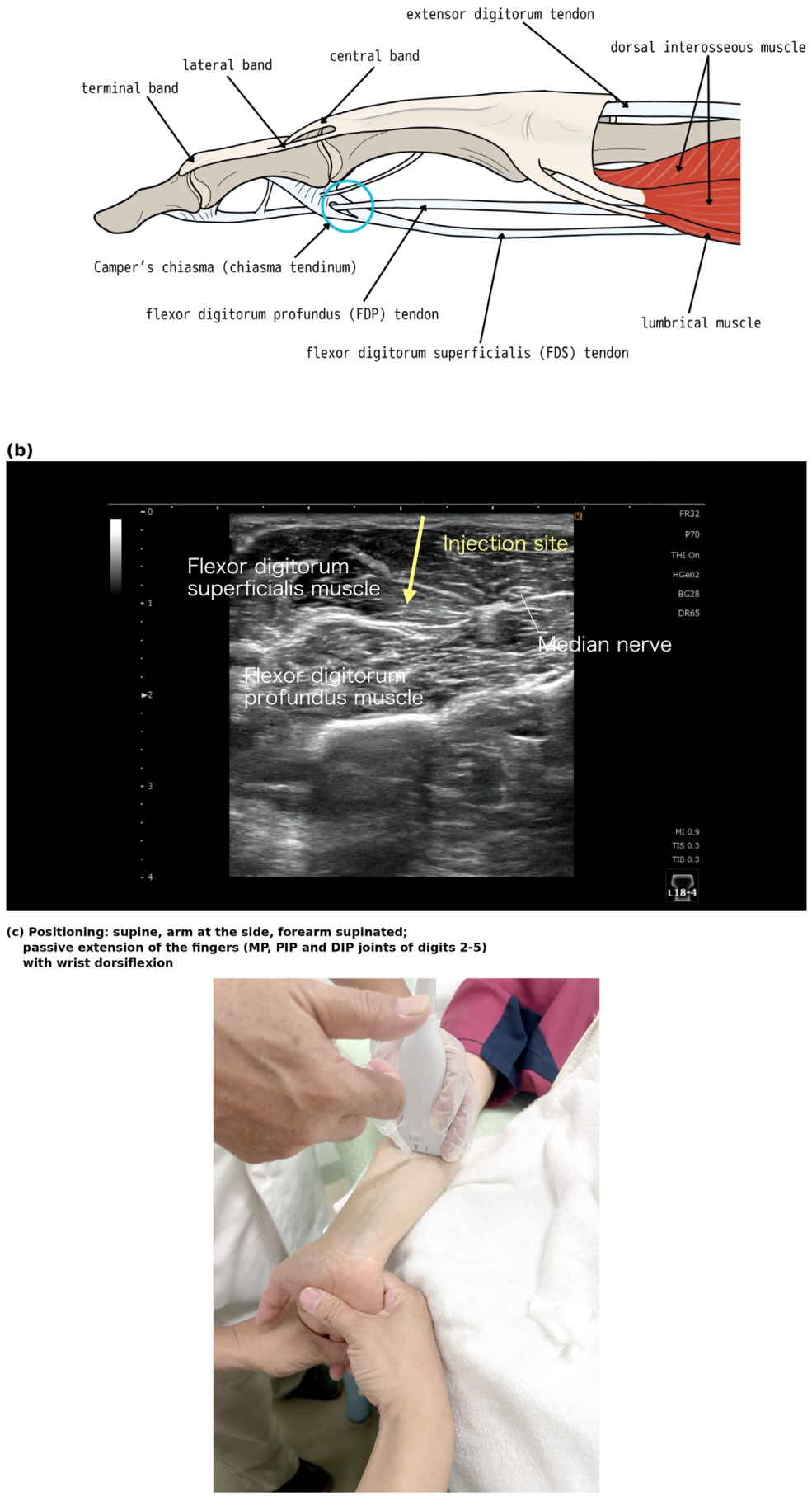

Table 1.
Treatment points, body positions and passive maneuvers.
| Point | Region / target | Body position | Passive maneuver | Video |
|---|---|---|---|---|
| P1 | Forearm: flexor digitorum superficialis / profundus interface (trigger finger, finger contracture) | Supine (forearm supinated) | Passive extension of the wrist and fingers (digits 2–5), held at end range by the assistant | S1 |
| P2 | Shoulder: superior capsule | Side-lying | Longitudinal traction of the humerus with glenohumeral adduction, combined with scapular elevation and upward rotation (relative adduction of the humerus to the scapula) | S2 |
| P3 | Shoulder: coracohumeral ligament (CHL) | Side-lying | Shoulder extension + adduction + external rotation | S3 |
| P4 | Hip, anterior: iliofemoral ligament + anterior capsule | Supine | Hip extension + external rotation | S4 |
| P5 | Hip, posterior: posterior capsule | Side-lying | Hip extension + internal rotation | S5 |
| P6 | Gluteal region (deep gluteal syndrome): peri-sciatic | Side-lying | Hip flexion + adduction + internal rotation | S6 |
| P7 | Posterior thigh: sciatic nerve short axis (“Mercedes-Benz sign”) | Side-lying | Hip flexion with knee extended (straight-leg-raise-like) + ankle dorsiflexion | S7 |
| P8 | Posterior knee: oblique popliteal ligament + capsule | Prone | Knee extension | S8 |
Disclaimer/Publisher’s Note: The statements, opinions and data contained in all publications are solely those of the individual author(s) and contributor(s) and not of MDPI and/or the editor(s). MDPI and/or the editor(s) disclaim responsibility for any injury to people or property resulting from any ideas, methods, instructions or products referred to in the content. |
© 2026 by the authors. Licensee MDPI, Basel, Switzerland. This article is an open access article distributed under the terms and conditions of the Creative Commons Attribution (CC BY) license (http://creativecommons.org/licenses/by/4.0/).
Copyright: This open access article is published under a Creative Commons CC BY 4.0 license, which permit the free download, distribution, and reuse, provided that the author and preprint are cited in any reuse.