

Submitted:
26 October 2026
Posted:
27 October 2026
You are already at the latest version
Abstract
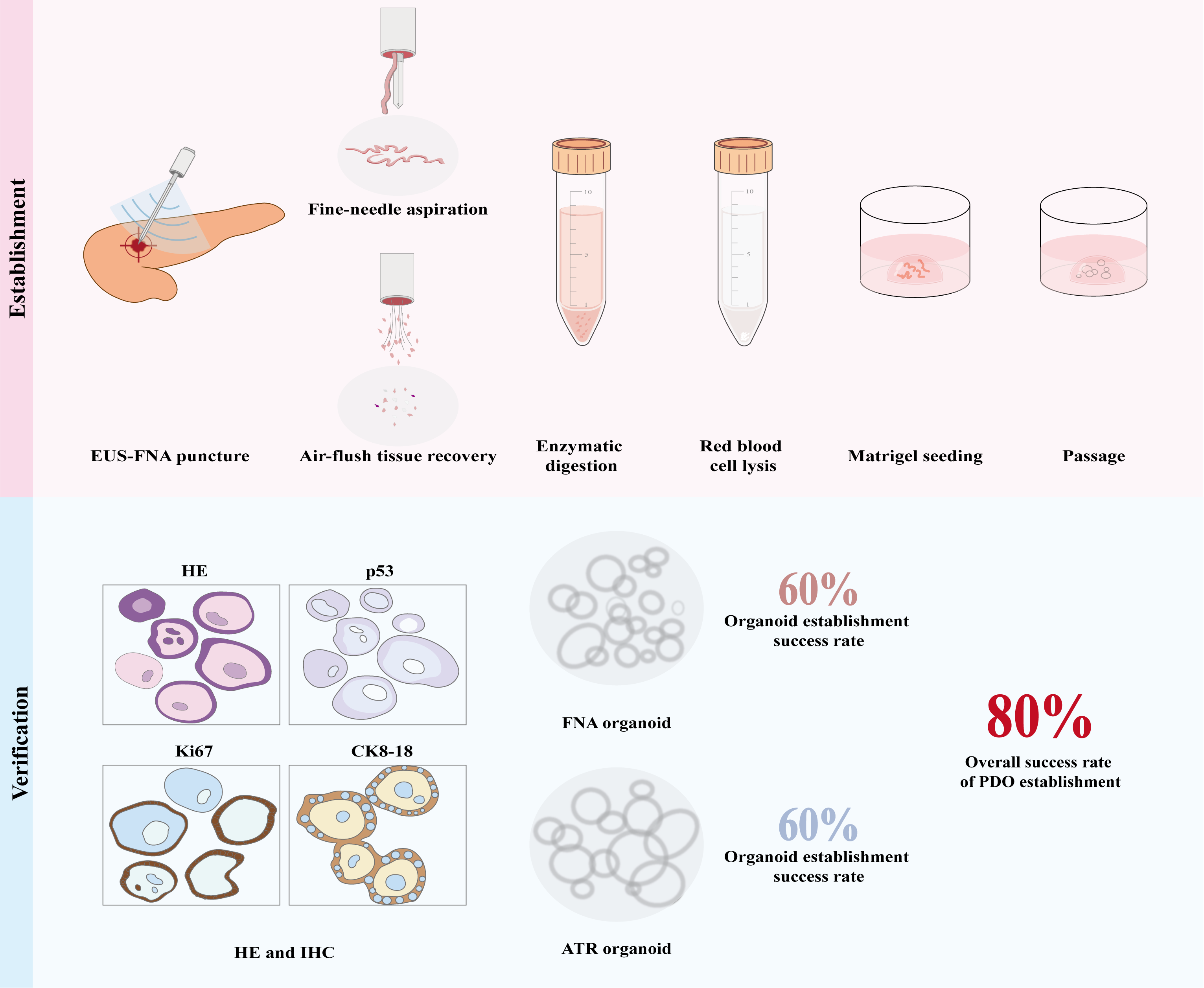
Background: Successful generation of patient-derived organoids (PDOs) from pancreatic ductal adenocarcinoma (PDAC) depends on an adequate amount of viable tissue. After routine endoscopic ultrasound-guided fine-needle aspiration (EUS-FNA) yields tissue for pathological diagnosis, additional needle passes are typically required to obtain organoid-culture material. This study examined whether air-flush tissue recovery (ATR) of residual needle-tract tissue can derive pancreatic cancer organoids without extra passes. Methods: We enrolled 10 PDAC patients who underwent EUS-FNA at two centers (The Ninth Medical Center of Chinese PLA General Hospital and the Second Hospital of Hebei Medical University) between February and June 2026. Using a self-controlled design, conventional FNA tissue strips (FNA group) and air-flush-recovered needle-tract residuals (ATR group) were collected from each patient and used to establish organoids. The two groups were compared for P3-generation passage rate, histological features, and molecular characteristics. Results: The ≥P3 success rate was 60.0% (6/10) in both the FNA and ATR groups. The overall organoid establishment rate (at least one method) was 80.0% (8/10). Hematoxylin and eosin (HE) staining and immunohistochemistry showed that organoids from both sources retained the histological morphology and key molecular features of the primary tumor, with consistent expression patterns. Conclusions: Harvesting residual tissue from the EUS-FNA needle tract by air flushing enables successful establishment of pancreatic cancer PDOs. The establishment success rate is comparable to that of organoids derived from conventional FNA specimens. In high-risk patients who cannot tolerate additional passes, a single ATR sample can still provide material for organoid generation; simultaneously culturing FNA tissue strips together with ATR residuals improved the overall organoid establishment rate from one puncture. This minimally invasive protocol is straightforward to perform and merits validation in larger cohorts.
Keywords:
pancreatic ductal adenocarcinoma
; endoscopic ultrasound-guided fine-needle aspiration
; tissue acquisition
; air-flush tissue recovery
; organoids
1. Introduction
Pancreatic ductal adenocarcinoma (PDAC) is a highly malignant digestive-system tumor. Its insidious onset, limited treatment options, and propensity for drug resistance result in an extremely low 5-year survival rate[1,2]. Patient-derived organoids (PDOs) have emerged as a promising technology that offers hope for personalized therapy. Organoids are three-dimensional structures formed by cellular self-assembly and self-renewal within a matrix gel, and they preserve the histological and molecular characteristics of the parental tumor[3,4]. PDOs can be used to predict tumor drug sensitivity and to guide individualized treatment planning[5].
However, successful PDO establishment depends on acquiring sufficient viable tumor tissue, which remains a bottleneck in translating the technology from bench to clinic. Most early studies used surgically resected specimens[6,7,8], yet 80% of PDAC patients are inoperable at diagnosis[9,10]. Obtaining viable tumor tissue minimally invasively has therefore become central to clinical translation. Endoscopic ultrasound-guided fine-needle aspiration (EUS-FNA) is now the primary minimally invasive approach for PDAC diagnosis and tissue acquisition. Several groups have established pancreatic cancer PDOs from EUS-FNA-derived tissue strands[10,11,12,13,14]. In 2024, Grützmeier et al. systematically compared organoid success rates between EUS-FNA and surgical specimens and found them comparable[15]. Obtaining extra material for organoids, however, usually requires 1-2 additional needle passes, which prolongs the procedure and raises the risk of complications[16,17].
Generating organoids from existing FNA specimens without extra punctures is thus an unmet clinical need. We propose an air-flush tissue recovery (ATR) technique that rinses the biopsy needle tract with sterile air to retrieve residual fragments for organoid culture. Using a self-controlled design, we systematically compared the efficacy of traditional FNA tissue strips versus ATR residuals from the same patient, aiming to identify a safer, more convenient tissue-acquisition strategy for pancreatic cancer organoids.
2. Materials and Methods
2.1. Patients and Tissue Samples
This study was approved by the Ethics Review Committee of the Ninth Medical Center of Chinese PLA General Hospital (Approval No. LL-LCSY-2025-37). From February to June 2026, a cohort of 10 consecutive PDAC patients meeting the inclusion and exclusion criteria was enrolled.
All patients provided written informed consent. Inclusion criteria: (1) strong clinical or imaging suspicion of PDAC scheduled for EUS-FNA for pathological diagnosis; (2) primary or metastatic pancreatic lesion ≥ 1.0 cm with a safe puncture path on EUS or CT and no obvious major-vessel encasement; (3) age > 18 years; (4) planned 2-3 needle insertions, with at least one insertion able to yield tissue strips and air-flushed material without compromising the volume required for routine pathological diagnosis. Exclusion criteria: (1) contraindications to puncture (severe ascites, bowel or vascular obstruction of the path, extensive peripancreatic infection/necrosis, or inability to cooperate); (2) specimens yielding only blood, necrosis, or macroscopically invisible strips on gross inspection and rapid on-site evaluation (ROSE); (3) prior radiotherapy, chemotherapy, or targeted therapy for pancreatic cancer. Tumor tissue was obtained from the two participating centers via EUS-FNA and air flushing of the puncture tract.
2.2. Puncture Procedure
EUS-FNA was performed by experienced endoscopists. The 22G or 25G needle was selected at the operator's discretion based on preoperative findings and patient condition. After localizing the lesion by EUS, the needle was inserted into the tumor, and, depending on tissue consistency, mild negative pressure with repeated in-and-out movements was applied; the lesion was sampled 2-3 times to obtain sufficient tissue strips. Diff-Quik rapid staining with ROSE was then used to confirm malignant cells on the smear.
The tissue strips were expelled directly into pre-chilled (4 °C) basal medium(Basal medium: DMEM/F-12, GlutaMax, Pen Strep, HEPES). A sterile air-filled syringe was then connected to the needle hub, and air was injected gently and steadily to flush residual tissue from the lumen into the basal medium. All samples were stored and transported on ice (Figure 1).
2.3. Generation and Culture of Organoids
All specimens were delivered to the Institute of Microbiology, Chinese Academy of Sciences, within 6 hours for processing and organoid culture. FNA and ATR samples were handled simultaneously in a biosafety cabinet. ATR samples were first pelleted by centrifugation; both groups were washed three times with pre-chilled wash buffer(Wash buffer: DMEM/F-12 with 15mM HEPES, BSA, Pen Strep). Enzymatic digestion(digestive enzymes: DMEM+GlutaMax-1, FBS, Pen Strep, Sodium Pyruvate, Collagenas, Dnase I, Dispase II) was performed for 20 min in a 37 °C shaking incubator, after which digestion was stopped and the suspension centrifuged for 5 min. When the pellet contained many red blood cells, red-blood-cell lysis buffer was added, incubated on ice for 3 min, and centrifuged to recover the tissue pellet. The pellet was mixed with Matrigel and seeded into preheated 24-well plates. After solidification, growth-factor-supplemented basal medium was added, and plates were cultured at 37 °C with 5% CO2 and medium changed every 3-4 days. Organoids with stable morphology and diameters > 200 μm were dissociated by gentle pipetting and passaged (Figure 1). Successful establishment was defined as stable passage for ≥ 3 generations with recovery after cryopreservation[18]. Representative mature PDOs derived from both sampling groups were subjected to histological and biomarker validation via hematoxylin-eosin (HE) staining and immunohistochemistry (IHC) targeting pancreatic ductal adenocarcinoma markers CK8-18, Ki-67 and p53, to verify whether organoids recapitulated the histologic architecture and molecular signature of matched primary tumor tissue.
2.4. Statistical Analysis
Data were analyzed with GraphPad Prism 10.6.0. Continuous variables were first tested for normality by Shapiro-Wilk. Normally distributed data are reported as mean ± SD and compared by paired Student's t-test; non-normal data by Wilcoxon signed-rank test. Categorical variables are reported as frequencies (percentages). Two-sided P < 0.05 was considered significant.
3. Results
3.1. Clinical Characteristics of Patients
Twenty samples were collected from 10 patients (one EUS-FNA and one air-flush sample each). Clinical details are in Table 1. The cohort comprised 1 male and 9 females, mean age 69.2 ± 12.3 years. Primary tumors measured 3.6 ± 1.0 cm, all pathologically confirmed as PDAC; 6 patients had metastases at diagnosis (Table 2).
3.2. Organoid Establishment
Among the 10 paired samples, the overall establishment rate (at least one method) was 80.0% (8/10). The ≥P3 success rates were 60.0% (6/10) in both groups. Digestion times were 24.30 ± 4.30 min (FNA) and 13.50 ± 6.08 min (ATR; P = 0.002) (Table 3).
The two methods were not interchangeable: in some patients only FNA succeeded, whereas in others only ATR did (Figure 2 and Table 4). Because their failures were largely non-overlapping, pooling both sources from a single puncture yielded at least one ≥P3 line in 8/10 patients (80%). McNemar's exact test showed no significant difference between the two methods in ≥P3 attainment (P > 0.05).
3.3. Morphological and Molecular Phenotype Validation
PDOs from both sources formed capsule-like, translucent spheres with defined edges, a central lumen, and good refractility (Figure 3). HE staining showed that the ductal architecture and differentiation of 3D-cultured PDOs resembled those of the primary tumors.
Immunohistochemistry for p53, Ki-67, and CK8-18 gave consistent expression patterns between FNA- and ATR-derived PDOs (Figure 4). Intergroup comparison confirmed similar histological and molecular features that stably preserved the key characteristics of the primary tumor.
4. Discussion
In conventional FNA, tissue is aspirated into the needle lumen and expelled by reinserting the stylet for diagnosis[15]. Single-pass EUS-FNA, however, yields limited tissue[19]. Extra passes for organoids prolong the procedure and increase risks of hemorrhage, pancreatitis, and needle-tract seeding[16,20,21]. After FNA, certain amount of tissue fragments remains in the needle tract and are usually discarded. As early as 2013, Lee et al. [22] showed that air flushing reduces blood contamination versus stylet re-expulsion, is simpler and safer, and preserves cellularity and diagnostic accuracy. However, its value for organoid establishment was unexplored.
We recovered these needle-tract "byproducts" with air and successfully established pancreatic cancer PDOs. The ≥P3 rate was identical (60%) between ATR and FNA. Tiriac et al.[10] reported a 66% EUS-FNA organoid success rate, and Grützmeier et al.16 pooled 11 studies to estimate 60%. Our result aligns with these cohorts. Histological and molecular comparisons with the primary tumor likewise matched prior reports[3,23].
Ikezawa et al.[24] first attempted organoids from saline-flushed needle tracts, but their small pilot lacked paired FNA controls and could not show whether lavage reduced passes. Saline introduces cell dilution and contamination risk that may disturb culture, and their study had no internal FNA comparison. Air, by contrast, is faster, avoids liquid shear and osmotic stress on cells, and requires no extra passes, providing a safe tissue source for organoids.
Culture outcomes were inconsistent between the two methods within the same patient; sometimes only FNA succeeded, sometimes only ATR. This reflects two distinct, non-overlapping technical constraints that fail independently. Standard FNA yields an ample solid-tissue mass, so tumor-cell quantity is not limiting. We observed, however, that FNA samples more often showed early epithelioid growth, plausibly reflecting contamination with normal pancreatic epithelium in the intact strip. Prior work links such normal epithelium to competitive inhibition of pancreatic cancer organoid expansion[25]. We did not directly quantify epithelial composition, so this interpretation remains provisional and warrants validation. ATR yields only sparse needle-lumen fragments with minimal benign epithelial contamination, which may weaken competitive inhibition and favor tumor-organoid expansion. Its yield, however, depends on how much tumor debris remains in the lumen; in highly desmoplastic lesions with little shed material, ATR may recover mostly stroma, leaving insufficient viable tumor cells for even P0 formation. This explains cases where only ATR succeeded within a patient.
Given the independent nature of assay failures, a stratified sampling strategy can be adopted. High-risk patients presenting with major vascular encasement, coagulopathy, or advanced age and frailty cannot tolerate additional needle passes; for these individuals, a single-puncture ATR sample enables organoid generation with no incremental trauma. For standard-risk candidates, parallel culture of FNA strips and ATR residuals offsets the limitations of each technique, elevates the overall organoid establishment rate from a single puncture, and optimises EUS-FNA utilisation.
This pilot study presents notable limitations. First, enrolment was restricted to 10 patients. The cohort exhibited substantial gender imbalance, as 90 % of participants were female, and disease was nearly exclusively confined to the pancreatic head, constraining broader generalizability. Second, systematic cell weighing and counting were not performed, precluding quantitative comparison of cellular abundance between sampling approaches. Third, our evaluation was limited to organoid establishment, without validation of downstream applications such as functional assays and drug-sensitivity testing.
In summary, air-flush recovery of residual FNA needle-tract tissue represents a safe and feasible supplementary strategy for pancreatic cancer organoid generation. Requiring no additional needle passes and inflicting no extra procedural trauma, this readily deployable clinical approach enables successful establishment of patient-derived organoids, with success rates comparable to those of organoids derived from conventional FNA specimens. It can be implemented in a tiered manner alongside standard FNA workflows: for high-risk patients unable to tolerate extra needle passes, a single ATR sample alone can yield organoid-forming material, whereas parallel culture of FNA tissue strips and ATR residuals improves the overall organoid establishment rate obtained from a single puncture. This minimally invasive protocol is straightforward to perform and merits further validation in larger patient cohorts.
5. Conclusions
This study demonstrates that the air-flush tissue recovery (ATR) technique provides a safe and effective alternative strategy for tissue acquisition in the generation of pancreatic cancer organoids. This method does not require an increase in the number of biopsy samples and can be readily applied in clinical practice. Organoids derived from ATR residual samples reproduce the histological and biomarker characteristics of the primary tumor, and their stable passage success rate is comparable to that of conventional EUS-FNA samples. Co-culturing conventional FNA tissue with ATR residual samples can significantly improve the overall success rate of organoid establishment from a single puncture, which is particularly important for patients who are not suitable for multiple punctures. However, the results of this study are limited by the small sample size. Future large-scale, multicenter studies are needed to validate its functionality and promote its application in personalized drug sensitivity testing.
Author Contributions
Yujiao Xu and Lichao Zhang are co-first authors and contributed to experimental design, clinical specimen collection, organoid culture, statistical analysis, figure preparation, and manuscript drafting and revision. Meijuan Wang contributed to clinical specimen collection, data sorting, manuscript revision. Lianyong Li and Ningning Liu are corresponding authors and led conceptualization, project coordination, and funding acquisition; they guided experimental procedures, reviewed results, and finalized the manuscript. Xin Huang, Shaojie Zheng, and Senlin Hou contributed to clinical sample collection, case selection, and clinical data organization. Kaixin Zhou offered valuable technical assistance and support during the experimental work.
Funding
This research received no external funding.
Institutional Review Board Statement
The study was approved by the Ethics Review Committee of the Ninth Medical Center of Chinese PLA General Hospital (No. LL-LCSY-2025-37, approval date: 27 October 2025) and conducted in accordance with the Declaration of Helsinki.
Informed Consent Statement
Informed consent was obtained from all subjects involved in the study.
Data Availability Statement
All data generated or analyzed during this study are included in this article and its supplementary material files. Further enquiries can be directed to the corresponding author.
Acknowledgments
We appreciate the Institute of Microbiology, Chinese Academy of Sciences for the provision of experimental platforms and clinical staff for their support in specimen acquisition.
Conflicts of Interest
The authors declare no conflicts of interest.
Abbreviations
The following abbreviations are used in this manuscript:
| PDOs | Patient-derived organoids |
| PDAC | Pancreatic ductal adenocarcinoma |
| EUS-FNA | EUS-guided fine-needle aspiration |
| ATR | Air-flush tissue recovery |
| ROSE | Rapid On-Site Evaluation |
| IHC | immunohistochemistry |
| HE | Hematoxylin and eosin |
References
- Siegel, R.L.; Giaquinto, A.N.; Jemal, A. Cancer Statistics, 2024. Ca. Cancer J. Clin. 2024, 74, 12–49. [Google Scholar] [CrossRef] [PubMed]
- Yang, G.; Guan, W.; Cao, Z.; Guo, W.; Xiong, G.; Zhao, F.; Feng, M.; Qiu, J.; Liu, Y.; Zhang, M.Q.; et al. Integrative Genomic Analysis of Gemcitabine Resistance in Pancreatic Cancer by Patient-Derived Xenograft Models. Clin. Cancer Res. Off. J. Am. Assoc. Cancer Res. 2021, 27, 3383–3396. [Google Scholar] [CrossRef] [PubMed]
- Tiriac, H.; Belleau, P.; Engle, D.D.; Plenker, D.; Deschênes, A.; Somerville, T.D.D.; Froeling, F.E.M.; Burkhart, R.A.; Denroche, R.E.; Jang, G.-H.; et al. Organoid Profiling Identifies Common Responders to Chemotherapy in Pancreatic Cancer. Cancer Discov. 2018, 8, 1112–1129. [Google Scholar] [CrossRef] [PubMed]
- Iovanna, J.; Dusetti, N. Speeding towards Individualized Treatment for Pancreatic Cancer by Taking an Alternative Road. Cancer Lett. 2017, 410, 63–67. [Google Scholar] [CrossRef]
- Drost, J.; Clevers, H. Organoids in Cancer Research. Nat. Rev. Cancer 2018, 18, 407–418. [Google Scholar] [CrossRef] [PubMed]
- Boj, S.F.; Hwang, C.-I.; Baker, L.A.; Chio, I.I.C.; Engle, D.D.; Corbo, V.; Jager, M.; Ponz-Sarvise, M.; Tiriac, H.; Spector, M.S.; et al. Organoid Models of Human and Mouse Ductal Pancreatic Cancer. Cell 2015, 160, 324–338. [Google Scholar] [CrossRef] [PubMed]
- Hirt, C.K.; Booij, T.H.; Grob, L.; Simmler, P.; Toussaint, N.C.; Keller, D.; Taube, D.; Ludwig, V.; Goryachkin, A.; Pauli, C.; et al. Drug Screening and Genome Editing in Human Pancreatic Cancer Organoids Identifies Drug-Gene Interactions and Candidates for off-Label Therapy. Cell Genom. 2022, 2, 100095. [Google Scholar] [CrossRef] [PubMed]
- Seppälä, T.T.; Zimmerman, J.W.; Suri, R.; Zlomke, H.; Ivey, G.D.; Szabolcs, A.; Shubert, C.R.; Cameron, J.L.; Burns, W.R.; Lafaro, K.J.; et al. Precision Medicine in Pancreatic Cancer: Patient-Derived Organoid Pharmacotyping Is a Predictive Biomarker of Clinical Treatment Response. Clin. Cancer Res. 2022, 28, 3296–3307. [Google Scholar] [CrossRef] [PubMed]
- Mucciolo, G.; Araos Henríquez, J.; Jihad, M.; Pinto Teles, S.; Manansala, J.S.; Li, W.; Ashworth, S.; Lloyd, E.G.; Cheng, P.S.W.; Luo, W.; et al. EGFR-Activated Myofibroblasts Promote Metastasis of Pancreatic Cancer. Cancer Cell 2024, 42, 101–118.e11. [Google Scholar] [CrossRef] [PubMed]
- Tiriac, H.; Carlos Bucobo, J.; Tzimas, D.; Grewel, S.; Lacomb, J.F.; Rowehl, L.M.; Nagula, S.; Wu, M.; Kim, J.; Sasson, A.; et al. Successful Creation of Pancreatic Cancer Organoids by Means of EUS-Guided Fine-Needle Biopsy for Personalized Cancer Treatment. Gastrointest. Endosc. 2018, 87, 1474–1480. [Google Scholar] [CrossRef] [PubMed]
- Bian, B.; Juiz, N.A.; Gayet, O.; Bigonnet, M.; Brandone, N.; Roques, J.; Cros, J.; Wang, N.; Dusetti, N.; Iovanna, J. Pancreatic Cancer Organoids for Determining Sensitivity to Bromodomain and Extra-Terminal Inhibitors (BETi). Front. Oncol. 2019, 9, 475. [Google Scholar] [CrossRef] [PubMed]
- Seppälä, T.T.; Zimmerman, J.W.; Sereni, E.; Plenker, D.; Suri, R.; Rozich, N.; Blair, A.; Thomas, D.L.; Teinor, J.; Javed, A.; et al. Patient-Derived Organoid Pharmacotyping Is a Clinically Tractable Strategy for Precision Medicine in Pancreatic Cancer. Ann. Surg. 2020, 272, 427–435. [Google Scholar] [CrossRef] [PubMed]
- Oyama, S.; Matsuda, A.; Murakami, R.; Kakizaki, Y.; Ishizawa, T.; Kobayashi, T.; Nakamura, H.; Nawa, Y.; Otaki, Y.; Nagata, Y.; et al. Pancreatic Cancer Organoids Derived from EUS-Guided Fine Needle Aspiration Specimens Can Be Used to Predict Chemotherapy Resistance. Sci. Rep. 2025, 15, 23818. [Google Scholar] [CrossRef] [PubMed]
- Klauss, S.; Xue, Y.; Alnatsha, A.; Allawadhi, P.; Mahajan, U.M.; Goni, E.; Vornhülz, M.; Sirtl, S.; Schulz, C.; Op Den Winkel, M.; et al. Diagnostic Accuracy and Patient Derived Organoid (PDO) Yield of 19G vs 22G Franseen Needles for EUS-Guided Biopsy in Solid Pancreatic Lesions: A Prospective Comparative Study. Pancreatology 2025, S1424390325005952. [Google Scholar] [CrossRef] [PubMed]
- Grützmeier, S.E.; Sodal, H.M.M.; Kovacevic, B.; Vilmann, P.; Karstensen, J.G.; Klausen, P. EUS-Guided Biopsies versus Surgical Specimens for Establishing Patient-Derived Pancreatic Cancer Organoids: A Systematic Review and Meta-Analysis. Gastrointest. Endosc. 2024, 100, 750–755. [Google Scholar] [CrossRef] [PubMed]
- Itonaga, M.; Ashida, R.; Kitano, M. Adverse Events of EUS-Guided Needle Aspiration/Needle Biopsy for Pancreatic Lesions. Suizo 2024, 39, 297–304. [Google Scholar] [CrossRef]
- Hébert-Magee, S.; Hawes, R.H.; Varadarajulu, S. Is It Time to Take a Pass on the Increased Number of Passes in EUS-FNA? Dig. Dis. Sci. 2013, 58, 3068–3069. [Google Scholar] [CrossRef] [PubMed]
- Kim, H.; Jang, J.; Choi, J.H.; Song, J.H.; Lee, S.H.; Park, J.; Ryoo, S.K.; Lee, E.M.; Jeong, H.; Kim, S.; et al. Establishment of a Patient-Specific Avatar Organoid Model Derived from EUS-Guided Fine-Needle Biopsy for Timely Clinical Application in Pancreatic Ductal Adenocarcinoma (with Video). Gastrointest. Endosc. 2024, 100, 85–96.e9. [Google Scholar] [CrossRef]
- Matsumoto, K.; Kato, H.; Nouso, K.; Ako, S.; Kinugasa, H.; Horiguchi, S.; Saragai, Y.; Takada, S.; Yabe, S.; Muro, S.; et al. Evaluation of Local Recurrence of Pancreatic Cancer by KRAS Mutation Analysis Using Washes from Endoscopic Ultrasound-Guided Fine-Needle Aspiration. Dig. Dis. Sci. 2020, 65, 2907–2913. [Google Scholar] [CrossRef] [PubMed]
- Kanno, A.; Yasuda, I.; Irisawa, A.; Hara, K.; Ashida, R.; Iwashita, T.; Takenaka, M.; Katanuma, A.; Takikawa, T.; Kubota, K.; et al. Adverse Events of Endoscopic Ultrasound-Guided Fine-Needle Aspiration for Histologic Diagnosis in Japanese Tertiary Centers: Multicenter Retrospective Study. Dig. Endosc. 2021, 33, 1146–1157. [Google Scholar] [CrossRef] [PubMed]
- Yane, K.; Kuwatani, M.; Yoshida, M.; Goto, T.; Matsumoto, R.; Ihara, H.; Okuda, T.; Taya, Y.; Ehira, N.; Kudo, T.; et al. Non-Negligible Rate of Needle Tract Seeding after Endoscopic Ultrasound-Guided Fine-Needle Aspiration for Patients Undergoing Distal Pancreatectomy for Pancreatic Cancer. Dig. Endosc. 2020, 32, 801–811. [Google Scholar] [CrossRef] [PubMed]
- Lee, J.K.; Choi, J.H.; Lee, K.H.; Kim, K.M.; Shin, J.U.; Lee, J.K.; Lee, K.T.; Jang, K.-T. A Prospective, Comparative Trial to Optimize Sampling Techniques in EUS-Guided FNA of Solid Pancreatic Masses. Gastrointest. Endosc. 2013, 77, 745–751. [Google Scholar] [CrossRef] [PubMed]
- Boilève, A.; Cartry, J.; Goudarzi, N.; Bedja, S.; Mathieu, J.R.R.; Bani, M.-A.; Nicolle, R.; Mouawia, A.; Bouyakoub, R.; Nicotra, C.; et al. Organoids for Functional Precision Medicine in Advanced Pancreatic Cancer. Gastroenterology 2024, 167, 961–976.e13. [Google Scholar] [CrossRef]
- Ikezawa, K.; Ekawa, T.; Hasegawa, S.; Kai, Y.; Takada, R.; Yamai, T.; Fukutake, N.; Ogawa, H.; Akazawa, T.; Mizote, Y.; et al. Establishment of Organoids Using Residual Samples from Saline Flushes during Endoscopic Ultrasound-Guided Fine-Needle Aspiration in Patients with Pancreatic Cancer. Endosc. Int. Open 2022, 10, E82–E87. [Google Scholar] [CrossRef]
- Du, C.; Liu, Y.; Lu, X.; Zhang, M.; Jiang, S.; Cao, Z.; Yang, G.; Qiu, J.; Zhang, T.; Zhao, Y. Organoids in the Research of Pancreatic Neoplasms: Cultivation, Applications, and Limitations. Cancer Lett. 2025, 632, 217968. [Google Scholar] [CrossRef] [PubMed]
Figure 1.
Schematic workflow of tissue acquisition via EUS-FNA and ATR, followed by pancreatic cancer PDO culture and serial passaging. All tissue specimens were transported on ice within 6–7 h post-procedure for enzymatic digestion, red blood cell lysis, Matrigel seeding, and subsequent passaging once organoids reached diameters >200 μm.
Figure 1.
Schematic workflow of tissue acquisition via EUS-FNA and ATR, followed by pancreatic cancer PDO culture and serial passaging. All tissue specimens were transported on ice within 6–7 h post-procedure for enzymatic digestion, red blood cell lysis, Matrigel seeding, and subsequent passaging once organoids reached diameters >200 μm.
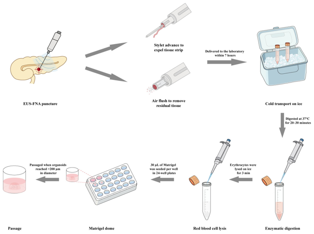
Figure 2.
Representative brightfield images showing failed organoid establishment in paired specimens. (A, B) Specimen Pan 10 at passage 1 day 3: no viable organoids derived from FNA tissue strips, whereas ATR residuals yielded successful organoids. (C, D) Specimen Pan 8 at passage 1 day 7: robust organoid growth from FNA samples and culture failure in matched ATR specimens. Scale bar: 1000 μm.
Figure 2.
Representative brightfield images showing failed organoid establishment in paired specimens. (A, B) Specimen Pan 10 at passage 1 day 3: no viable organoids derived from FNA tissue strips, whereas ATR residuals yielded successful organoids. (C, D) Specimen Pan 8 at passage 1 day 7: robust organoid growth from FNA samples and culture failure in matched ATR specimens. Scale bar: 1000 μm.
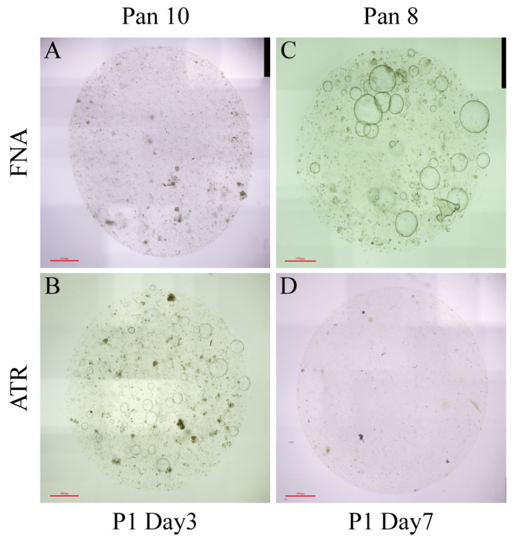
Figure 3.
Brightfield images displaying typical vesicular morphology of successfully established pancreatic cancer PDOs derived from paired FNA and ATR samples. (A) PDOs cultured from routine EUS-FNA tissue fragments; (B) PDOs generated from residual needle-tract tissue collected via air-flush recovery.Scale bar: 1000 μm.
Figure 3.
Brightfield images displaying typical vesicular morphology of successfully established pancreatic cancer PDOs derived from paired FNA and ATR samples. (A) PDOs cultured from routine EUS-FNA tissue fragments; (B) PDOs generated from residual needle-tract tissue collected via air-flush recovery.Scale bar: 1000 μm.
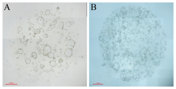
Figure 4.
Histological and molecular phenotype validation via HE staining and IHC for pancreatic ductal adenocarcinoma biomarkers CK8-18, Ki-67, and p53. Matched primary tumor biopsy tissue, FNA-derived PDOs, and ATR-derived PDOs displayed highly concordant histological architecture and biomarker expression profiles. Scale bar: 100 μm.
Figure 4.
Histological and molecular phenotype validation via HE staining and IHC for pancreatic ductal adenocarcinoma biomarkers CK8-18, Ki-67, and p53. Matched primary tumor biopsy tissue, FNA-derived PDOs, and ATR-derived PDOs displayed highly concordant histological architecture and biomarker expression profiles. Scale bar: 100 μm.
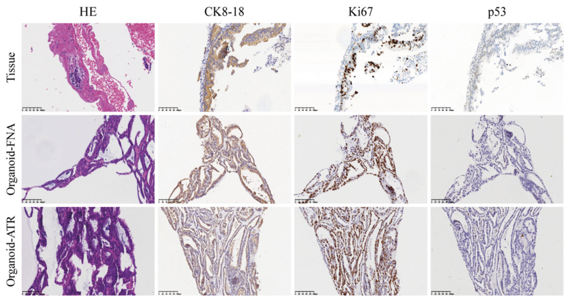

Table 1.
Clinical information of enrolled patients.
| No. | Sex | Age (years) | Tumor type | Tumor size (cm) | Location | TNM | Metastasis | Needle | Passes | Passes for organoid | Adverse events | FNA outcome | ATR outcome | Source |
|---|---|---|---|---|---|---|---|---|---|---|---|---|---|---|
| Pan 1 | F | 90 | PDAC | 3.70×2.41 | Head | III | PV, celiac LN | 22G | 2 | 1 | Acute pancreatitis | Success | Success | a |
| Pan 2 | F | 75 | PDAC | 3.50×4.00 | Head | >=IIA | None | 22G | 2 | 1 | None | Success | Insufficient cellularity | a |
| Pan 3 | F | 71 | PDAC | 2.55×2.70 | Head | IB | None | 22G | 2 | 1 | None | Success | Success | a |
| Pan 4 | M | 61 | PDAC | 4.50×4.50 | Head | III | Retroperitoneal LN | 22G | 2 | 1 | None | Insufficient cellularity | Success | b |
| Pan 5 | F | 76 | PDAC | 2.10×1.80 | Body | >=IIA | L adrenal | 22G | 2 | 1 | None | Success | Success | b |
| Pan 6 | F | 74 | PDAC | 4.80×3.08 | Head | IIA | PV, liver | 22G | 2 | 1 | None | Success | Success | a |
| Pan 7 | F | 67 | PDAC | 2.47×2.08 | Head | IB | None | 25G | 3 | 1 | None | Microbial contamination | Insufficient cellularity | a |
| Pan 8 | F | 66 | PDAC | 4.06×3.60 | Head | >=IIA | None | 22G | 3 | 1 | None | Success | Insufficient cellularity | b |
| Pan 9 | F | 42 | PDAC | 3.10×2.40 | Head | IV | Liver | 22G | 2 | 1 | None | Microbial contamination | Microbial contamination | b |
| Pan 10 | F | 70 | PDAC | 5.00×5.00 | Head | IV | Liver | 22G | 2 | 1 | None | Insufficient cellularity | Success | b |
*a: The Ninth Medical Center of Chinese PLA General Hospital; b: Second Hospital of Hebei Medical University.
Table 2.
Baseline clinical characteristics and EUS-FNA procedural parameters for enrolled patients.
| Characteristi | Data |
|---|---|
| Sex, male/female | 1/9 |
| Age, mean ± SD (yr) | 69.2 ± 12.3 |
| Tumor type, PDAC/other | 10/0 |
| Max size, mean ± SD (cm) | 3.6 ± 1.0 |
| Location, head/body/tail | 9/1/0 |
| Needle, 22G/25G | 10/1 |
| Metastasis, yes/no | 6/4 |
| Transit time, mean ± SD (min) | 180.3 ± 106.7 |
| Puncture times, mean ± SD | 2.0 ± 0.7 |
| Organoid passes, mean ± SD | 1.0 ± 0.0 |
| TNM stage, I/II/III/IV | 2/4/2/2 |
Table 3.
Summary of quality-control parameters and establishment outcomes of pancreatic cancer-derived organoids.
Table 3.
Summary of quality-control parameters and establishment outcomes of pancreatic cancer-derived organoids.
| Parameter | FNA (n=10) | ATR (n=10) | P |
|---|---|---|---|
| Digestion time, mean ± SD (min) | 24.30 ± 4.30 | 13.50 ± 6.08 | 0.002 |
| RBC lysis cycles, mean ± SD | 1.50 ± 0.53 | 1.50 ± 0.71 | >0.999 |
| Establishment (P0) | 8 (80.0%) | 8 (80.0%) | - |
| Passaging ≥ P3 | 6 (60.0%) | 6 (60.0%) | - |
| Failure: insufficient cellularity | 2 (20.0%) | 3 (30.0%) | - |
| Failure: microbial contamination | 2 (20.0%) | 1 (10.0%) | - |
Table 4.
Paired outcomes of organoid establishment by ATR and FNA sampling methods.
| FNA success | FNA failure | |
|---|---|---|
| ATR success | 4 | 2 |
| ATR failure | 2 | 2 |
Disclaimer/Publisher’s Note: The statements, opinions and data contained in all publications are solely those of the individual author(s) and contributor(s) and not of MDPI and/or the editor(s). MDPI and/or the editor(s) disclaim responsibility for any injury to people or property resulting from any ideas, methods, instructions or products referred to in the content. |
© 2026 by the authors. Licensee MDPI, Basel, Switzerland. This article is an open access article distributed under the terms and conditions of the Creative Commons Attribution (CC BY) license (http://creativecommons.org/licenses/by/4.0/).
Copyright: This open access article is published under a Creative Commons CC BY 4.0 license, which permit the free download, distribution, and reuse, provided that the author and preprint are cited in any reuse.