

Submitted:
24 October 2026
Posted:
26 October 2026
You are already at the latest version
Abstract
This article provides a comprehensive overview of recent advances in liposomal nanoformulations encapsulating whole-plant extracts for topical skin applications, with a focus on research published between 2016 and 2026. Literature searches were conducted using the Web of Science, Scopus and PubMed databases. The evolution from conventional liposomes to advanced nanovesicles (ethosomes, transfersomes, transethosomes, propylene glycol- and glycerol-enriched liposomes, and (bio)polymer-coated nanovesicles) is analyzed alongside their integration into hybrid liposome-in-vehicle delivery systems. Liposome preparation methods, bilayer constituents, and semisolid vehicles used to increase viscosity of liquid liposomal dispersions were evaluated and discussed with respect to their physicochemical properties as well as the botanical, phytochemical and biological activity profiles and skin-related effects of the plant-extract-loaded liposomal nanocarriers. Encapsulation into liposomes was found to significantly enhance the stability of botanical extracts, their cutaneous deposition, as well as therapeutic efficacy and dermocosmetic potential. Reported biological effects were categorized according to their pharmacological activities, such as antioxidant, anti-melanogenic, anti-inflammatory, antimicrobial, cytotoxic, wound-healing, and anti-aging activities. In response to the growing demand for clean beauty and plant-based dermatological solutions, this article summarizes current knowledge on plant-extract-loaded liposomal nanoformulations to guide future research toward their potential applications in dermatology and cosmetology.
Keywords:
herbal extracts
; liposomes
; phospholipid nanovesicles
; nanoformulation
; nanovesicular systems
; skin
; dermal delivery
; biological activity
; phytopharmaceutical
; phytocosmeceutical
1. Introduction
Treating skin disorders and maintaining skin health and youthfulness represent important research areas in the medical and beauty industries [1]. Human skin is a large and complex organ that serves as the body’s first line of defense against external environmental damage. Anatomically, skin is composed of three primary layers: subcutaneous tissue (hypodermis), dermis, and epidermis. The outermost layer of the epidermis, stratum corneum, determines the rate of permeation and acts as the major obstacle to the permeation of active compounds through the skin. Despite this barrier, topical (trans)dermal drug delivery offers significant advantages over oral administration, including avoidance of gastrointestinal tract irritation and first-pass hepatic metabolism. Furthermore, topical formulations allow direct delivery to the desired site of action, ensuring higher local drug concentrations, while minimizing systemic side effects [2,3]. Depending on the desired pharmacological outcome, topical dermal formulations can be designed for: (i) strictly superficial effects avoiding any systemic absorption (e.g., sunscreens, repellents, metal-based protectants); (ii) targeting the epidermis (e.g., acne or eczema); (iii) aiming the deeper tissues of the skin (e.g., analgesics and anti-inflammatories); and (iv) transdermal systemic absorption (e.g., hormone or insulin patches) [4].
Since ancient times, across different cultures worldwide, botanical extracts have been used for their diverse pharmacological properties. To date, they remain essential ingredients in a wide range of nutraceutical, pharmaceutical and dermocosmetic applications. According to numerous scientific studies, the therapeutic value of medicinal plants and their extracts is attributed to biologically active specialized metabolites, such as polyphenols, flavonoids, alkaloids, tannins, saponins, lignins, glycosides, and terpenoids, which have been proven to exhibit antioxidant, antimicrobial, anti-inflammatory, analgesic, antitumor and many other pharmacological effects [5]. Among these compounds, polyphenols, particularly flavonoids, play essential roles in scavenging free radicals, providing UV protection, regulating cell growth, suppressing pro-inflammatory cytokines, preserving mitochondrial function, and promoting tissue repair [6,7,8,9,10,11]. Such a vast and versatile repertoire of phytoactives, which often possess complementary mechanisms of action and potential synergistic interactions, is the main reason why botanical extracts can offer multi-target therapeutic efficacy [12]. Plant-derived medicines are often perceived as safer alternatives to conventional synthetic drugs. However, their potential toxicity and adverse effects should not be overlooked [13]. Moreover, due to the relatively large molecular sizes of their phytoconstituents, direct application of botanical extracts faces major challenges due to their chemical instability, sensitivity to light, pH and enzymes, low water solubility, as well as poor diffusion across lipid membranes [7,14,15]. Furthermore, botanical extracts are complex mixtures that frequently contain plant debris, inactive components, or organic solvent residues, thereby requiring purification, standardization, and appropriate quality control measures [16]. Because plant characteristics vary naturally, depending on environmental and genetic factors, herbal preparations often exhibit inconsistent concentrations of bioactive phytoconstituents, which directly undermines dosage uniformity and therapeutic predictability [17].
Nanoparticle encapsulation has emerged as a promising strategy for overcoming biopharmaceutical limitations of botanical extracts, bridging traditional phytotherapy and modern pharmacology. Nanotechnology-based approaches have been shown to increase the effective surface area of the particles and enhance solubility and stability of poorly water-soluble and volatile phytochemicals, at the same time protecting them from chemical or enzymatic degradation and ensuring their controlled release at the site of action [18,19]. In other words, nanotechnology has the potential to convert botanical extracts into standardized, stabilized, and more effective therapeutic agents. Examples of such nanocarriers include lipid-based nanoparticles such as solid lipid nanoparticles (SLNs), nanostructured lipid carriers (NLCs), phytosomes, and different types of liposomes [19]. First described by Dr. Bangham and Dr. Horne in 1964 [20], liposomes are spherical vesicles composed of one or more phospholipid bilayers surrounding inner aqueous compartment(s). Due to their amphiphilic nature, liposomes can encapsulate hydrophilic compounds within their aqueous core, hydrophobic substances within their lipid bilayer(s) and amphiphilic compounds between these two regions. They are typically categorized by their size and lamellarity into multilamellar vesicles (MLVs), small unilamellar vesicles (SUVs), large unilamellar vesicles (LUVs), and multivesicular vesicles (MVVs) [21,22].
Conventional liposomes are generally composed of neutral or a combination of neutral and cationic or anionic charged phospholipids, with or without cholesterol [21]. Further compositional modifications of the phospholipid bilayer with solvents such as ethanol, glycerol or surfactants (e.g., ethosomes, glycerosomes, transfersomes) may enhance skin penetration and (trans)dermal delivery of encapsulated substances [23,24]. Since liposomal components such as phosphatidylcholine (PC) and phosphatidylserine (PS) are naturally occurring phospholipids in mammalian cell membranes, they exhibit biocompatibility and biodegradability, as well as the ability to deposit their payload into deeper skin layers [6,25,26,27]. Liposomes have been widely studied for topical applications and are generally recognized as non-toxic and non-immunogenic delivery systems. In addition to acting as drug nanocarriers, liposomes themselves can also serve as therapeutic agents because their richness in phospholipids, cholesterol and omega fatty acids can help restore dry or damaged skin through lipid deposition inside the epidermis [27]. Additionally, liposomal vesicles form an occlusive surface barrier that helps retain skin moisture, reduces transepidermal water loss (TEWL), and protects skin against environmental stressors, including UV radiation [6]. Recent studies have suggested the effectiveness of liposomes for cosmetic and dermatological applications, indicating they may stabilize and protect active ingredients including the ones of plant origin [28]. The potential advantages of incorporating botanical extracts into liposomal nanoformulations for topical use over non-encapsulated extracts are illustrated in Figure 1.
While a few scientific papers review liposomal delivery of isolated phytoactives for human health applications [7,19,29,30,31], literature synthesizing research on liposomal delivery of whole plant extracts for potential skin applications remains limited, with plant-extract-loaded liposomal nanoformulations being sporadically addressed within broader reviews of phytochemical delivery and lipid-based nanocarriers, including the recent reviews on lipid nanocosmeceuticals [28] liposome-based cosmeceuticals [32], and novel cosmetic delivery systems [33]. Studies investigating other promising nanocarrier systems for enhanced bioavailability and therapeutic efficacy of herbal products such as plant-derived nanovesicles [34], phytosomes (phytophospholipid complexes) [35] and niosomes (non-ionic surfactant-based vesicles) [36] have been recently published and were out of the scope of the present review. Unlike individually isolated phytoactives (e.g., quercetin, arbutin, catechins, retinol, betanin, resveratrol, etc.), whole botanical extracts represent complex mixtures of multiple active components that may act synergistically. Accordingly, this review focuses on studies investigating liposomal formulations containing whole plant extracts for topical skin applications. It aims to characterize the phospholipid composition, preparation methods, physicochemical properties, botanical characteristics (including plant species and families, geographical origins, and natural biogeographical distributions), phytochemical profiles, biological activities, and skin-related effects of these formulations, as well as the challenges associated with their development. Given the objective of mapping key concepts, identifying emerging evidence and research gaps, and informing future investigations, a scoping review was selected as the most appropriate approach [37].
2. Materials and Methods
This scoping review was conducted and reported in accordance with the Preferred Reporting Items for Systematic Reviews and Meta-Analyses for scoping reviews (PRISMA-ScR) guidelines [38]. The review methodology was predefined a priori, including the search strategy, database selection, publication filters, eligibility criteria, and predefined variables for data extraction. To identify relevant studies, a comprehensive literature search was executed independently across three major databases: Scopus, Web of Science, and PubMed. The timeframe was restricted to an approximate period of the last 10 years, covering publications from 2016 to July 6, 2026. The exact search string applied across all databases was: (liposome* OR vesic*) AND (botanical OR herbal OR plant) AND extract AND (skin OR dermal OR topical*). The search syntax was constructed using specific keywords, truncation symbols (*), and Boolean operators (AND, OR) to capture variations in terminology regarding vesicular delivery systems and plant extracts for skin applications. Before screening, when possible, database-specific filters were applied to restrict the retrieved records by publication year (2016–2026) and document type (Article).
To ensure a precise synthesis of original data, strict inclusion and exclusion criteria were defined prior to the screening process: (i) article type: original, peer-reviewed primary research articles; (ii) formulation: liposomes or related artificially prepared vesicular carrier systems with additional components incorporated into the phospholipid bilayer and with the active compound encapsulated within the aqueous core or within the bilayer (e.g., ethosomes, transfersomes, hyalurosomes, etc.), not plant-derived nanovesicles (naturally occurring vesicular structures isolated from plants or plant-derived materials), phytosomes (phytophospholipid complexes in which a phytoconstituent is molecularly associated with phospholipids), or non-ionic surfactant-based vesicles (niosomes); (iii) active compounds: whole plant extracts, not (exclusively) individual phytoactives; (iv) aimed route of administration: dermal, topical or localized skin-related applications for humans; (v) language: articles available in English.
Records (649 in total) retrieved from the database search were compiled, and duplicate entries were manually removed, resulting in 427 unique records. The records were screened by four independent reviewers by title and abstract against the eligibility criteria. The full texts of 104 potentially relevant articles were retrieved and reviewed. Review articles, systematic reviews, meta-analyses, book chapters, conference abstracts, editorials, and patents were excluded during title and abstract screening or subsequent full-text review. Ultimately, 103 studies met the inclusion criteria and were included in this review (Figure 2).
To ensure consistency in the botanical characterization of the plant taxa represented in the included studies, the nomenclature of plant taxa, including species, subspecies, varieties, and hybrids, as well as plant families, was standardized according to Plants of the World Online (POWO), provided by the Royal Botanic Gardens, Kew [39]. Geographical origin and natural distribution of the evaluated taxa were likewise verified and standardized according to POWO, and these data were subsequently used to assign the taxa to the corresponding biogeographical regions. The resulting botanical dataset, provided in Supplementary Table (Table S1), served as the reference for the presentation and analysis of botanical information throughout the review; accordingly, all tables and figures reporting taxonomic, family-level, geographical, or biogeographical information follow the nomenclature and classifications established therein.
The included studies were grouped by classes of liposomal nanoformulations and investigated biological activities and skin-related effects of botanical-extracts-loaded liposomes, and their main characteristics and findings were summarized in tables, figures, and a narrative format. Liposomal nanoformulations were described according to the botanical extract source of their encapsulated substances, preparation methods, liposomal phospholipid bilayer constituents, and incorporation into topical vehicles (if applicable). Botanical and phytochemical profiles were summarized according to plant taxa (usually species) and families, plant parts, extraction solvent and methods, and main constituents together with the analytical techniques used for their detection/quantification reported in the respective studies. Biological activities were organized according to the investigated in vitro and in vivo activities (e.g., antioxidant, anti-inflammatory, antimicrobial, anti-melanogenic activities) and skin-related outcomes (e.g., cytoprotective activity, wound healing assays with cell and animal models, and human studies) to map the available evidence on the potential topical applications of botanical-extract-loaded liposomal systems.
3. Classes of Liposomal Nanoformulations As Botanical Extract Carriers
To overcome physicochemical and biopharmaceutical limitations of crude botanical extracts, researchers have developed various phospholipid-based vesicular nanocarriers. By modifying the phospholipid bilayer composition, vesicle size and surface charge, or by adding elasticizers, penetration enhancers, polyols, and biopolymers, different types of liposomes can be tailored for pharmaceutical and/or cosmeceutical dermal applications (Figure 3).
3.1. Conventional Liposomes
Over the past decade, most studies investigating liposomal delivery of botanical extracts have focused on conventional liposomes, utilizing them either as primary delivery platforms or as benchmarks for comparison against next-generation phospholipid nanocarriers (Table 1). Conventional liposomes typically consist of PC, sourced either as crude or refined soy/egg lecithin or purified PC fractions (e.g., Phospholipon® 90 H, Lipoid® S 100, Lipoid® S 75, Phosal® 75 SA, Phosal® 53 MCT, etc.). The structural stability of phospholipid vesicles is critically dictated by the constituent phospholipids and the incorporation of sterols. For instance, selecting phospholipids with saturated acyl chains, such as hydrogenated soybean phosphatidylcholine (HSPC) or distearoylphosphatidylcholine (DSPC), enhances bilayer stability as these lipids establish stronger intermolecular van der Waals interactions, yielding rigid and mechanically stable bilayer membranes [40,41,42,43,44]. On the other hand, incorporating phospholipids containing unsaturated acyl chains increases fluidity and elasticity, although it may also render the vesicular system more susceptible to environmental or thermal destabilization under specific conditions [45,46].
Bilayer composition determines the surface charge of liposomes, as reflected by their zeta potentials, which can be neutral, anionic or cationic. Although the vast majority of botanical extract-loaded liposomal formulations rely on PC, either alone or in combination with anionic phospholipids, typically yielding neutral or negative surface charge, some formulations employ cationic conventional liposomes by incorporating certain portions of cationic lipids into the phospholipid bilayer. For instance, Amalu et al. [47] used the cationic stearylamine alongside PC and cholesterol to prepare positively charged liposomes (+33.0 mV) encapsulating orange peel (Citrus × aurantium f. aurantium) and liquorice (Glycyrrhiza glabra) extracts. The primary rationale for employing cationic conventional liposomes in topical delivery lies in the strong electrostatic attraction between the positively charged nanovesicles and the net negatively charged biological surface of the skin [48]. This electrostatically driven affinity promotes bioadhesion and prolonged retention on the skin. Furthermore, positive surface charges may facilitate closer contact with lipid membranes, promote penetration across the stratum corneum, and enhance cellular internalization via endocytosis or membrane fusion, ultimately maximizing dermal deposition and the biological efficacy of encapsulated phytoconstituents [49,50]. Indeed, Bonechi et al. [45] incorporated synthetic cationic lipids 1,2-dioleoyl-3-trimethylammonium-propane (DOTAP) or dimethyldioctadecylammonium bromide (DDAB) into dioleoylphosphatidylcholine (DOPC) bilayers, producing stable monodisperse nanovesicles loaded with Coffea arabica leaf extract, demonstrating exceptionally high positive surface charges (zeta potential up to +94.5 mV), which significantly enhanced cellular interactions and uptake in human dermal fibroblasts without inducing cytotoxicity.
In addition to phospholipids, cholesterol is almost routinely used in the preparation of conventional liposomes. While phospholipids form the structural backbone of nanovesicles, cholesterol intercalates between phospholipid acyl chains, thereby modulating membrane fluidity, mechanical strength, and overall stability. Specifically, above the main phase transition temperature (Tm) of the lipid bilayer, cholesterol restricts the conformational mobility of fluid lipid chains, increasing the vertical packing density and membrane rigidity. This mechanical reinforcement enables the vesicles to better withstand shear forces and physical stress. Conversely, below the Tm, cholesterol disrupts the highly ordered, crystalline gel phase of the phospholipids, preventing the membrane from becoming overly brittle. The described effects of cholesterol intercalation result in higher colloidal stability, prevention of the premature leakage of encapsulated compounds and longer shelf life of the formulation [51,52,53]. When present in liposomes, cholesterol content typically ranges between 10% and 33% (w/w) of total lipid mass. Ratios exceeding 30% (w/w) often result in rigidification and reduced entrapment of the botanical extracts [54], as well as increased particle size heterogeneity, indicated by an increased polydispersity index (PDI), compromising overall formulation homogeneity [26]. Although cholesterol remains the most frequently used sterol, a recent study has highlighted the potential of phytosterols as functional alternatives. In this context, Ahmoda et al. [55] investigated plant-derived sterol β-sitosterol and fungus-derived sterol ergosterol, which provide some biophysical and pharmacological advantages. β-Sitosterol closely resembled the molecular architecture of cholesterol, enhancing bilayer rigidity and stabilizing delicate encapsulated phytoconstituents. It also contributed to the intrinsic anti-aging and anti-inflammatory activity of the formulation. Ergosterol, in contrast, promoted greater membrane fluidity and more homogeneous bilayer packing than cholesterol, while helping to protect phytoactive compounds against oxidative and photolytic degradation.
In comparison to the non-encapsulated botanical extracts, conventional liposomes offer more pronounced skin deposition of their payload, convenient for localized dermal delivery [14,46,55,56,57,58]. However, despite their ability to serve as sustained-release reservoirs, protect sensitive phytochemicals from oxidation, amplify their biological activities, and exhibit high biocompatibility, conventional liposomes face a major biopharmaceutical limitation: their penetration into the skin is largely restricted to the superficial layers of the stratum corneum, resulting in their predominant retention within the uppermost skin layers [11]. To achieve even deeper dermal deposition and enhanced transdermal flux, advanced skin-targeting nanovesicles have been engineered, including ethosomes, transfersomes, transethosomes, propylene glycol-enriched liposomes (PGLs), glycerosomes, and hyalurosomes (Table 1).
3.2. Ethosomes
Ethosomes, pioneeringly designed by Touitou et al. [138], are soft, malleable nanovesicles composed of phospholipids and high concentrations of ethanol (20–40%, w/w). The coexistence of these molecules in ethosomes results in higher entrapment efficiencies and smaller vesicle sizes compared with conventional liposomes [103]. Ethosomes, enable skin penetration of the encapsulated active ingredients through two synergistic mechanisms: (i) ethanol from the outer water phase of ethosome dispersion fluidizes and disrupts the tightly packed intercellular lipid domains of the stratum corneum; (ii) ethanol in the phospholipid bilayer imparts high flexibility and elasticity of the vesicles, facilitating vesicle penetration into the deeper layers of the skin, including the viable epidermis and dermis. One-fifth of the studies surveyed in this review investigated ethosomes for diverse dermatological indications (Table 1), highlighting their potential as promising nanocarriers for complex botanical extracts [87,88,90,94,96,103,105,106,107,136].
3.3. Transfersomes
Transfersomes, in the literature frequently referred to as deformable, ultra-deformable, or elastic liposomes, are vesicles that combine phospholipids with single-chain surfactants, commonly termed "edge activators" (e.g., Tween 80, Tween 20, Span 80, Span 20, sodium cholate, sodium deoxycholate, etc.). The incorporation of edge activators subtly destabilizes the phospholipid bilayer, lowering its interfacial tension and conferring high membrane elasticity and deformability [123]. Driven by the osmotic force of the natural transdermal hydration gradient across the skin, transfersomes navigate through narrow inter-corneocyte pathways that are 5 to 10 times smaller than their own hydrodynamic diameter (e.g., passing through 30–50 nm intercellular pores), without undergoing rupture or premature leakage of their encapsulated cargo. Transfersomes serve both as drug-releasing reservoirs and localized penetration enhancers by temporarily lowering skin lipid resistance [139].
When designing transfersomes, critical formulation parameters are phospholipid-to-surfactant ratio and hydrophilic-lipophilic balance (HLB) of the edge activator. Optimal surfactant concentrations (typically 10–20%, w/w) maximize membrane flexibility and entrapment efficiency (EE, %). However, excessive surfactant incorporation (> 20–25%) leads to bilayer solubilization and the formation of mixed micelles, which possess reduced drug-loading capacity, lower deformability, and lower transdermal flux. Non-ionic surfactants with higher HLB values (e.g., Tween 80), interact strongly with the aqueous phase. They also lower interfacial tension, increase membrane curvature, and yield smaller, highly flexible nanovesicles compared to low-HLB surfactants (e.g., Span 80) [43,44,109,116]. Moreover, surfactants with flexible hydrocarbon chains, such as Tween 80, generally provide greater membrane deformability than bulky bile salts with steroidal structure, such as sodium (deoxy)cholate [44]. To mitigate long-term physical instability (e.g., aggregation or vesicle fusion due to low zeta potential) and further potentiate intracutaneous performance, transfersomes have also been functionalized with hydrophilic biopolymers, which tend to form protective polymeric shells around the elastic lipid vesicles [8,123]. These polymer coatings provide steric stabilization, enhance formulation viscosity and skin adhesion as further discussed in section 3.5.
3.4. Transethosomes
Aspiring to further enhance cutaneous flux and bypass skin barrier constraints, transethosomes integrate the structural features of both ethosomes and transfersomes by incorporating high ethanol concentrations alongside membrane-destabilizing edge activators. This synergistic combination provides superior EE for poorly soluble active ingredients, enhanced stability, and deeper dermal penetration for difficult-to-deliver phytocomplexes, as demonstrated for botanical extracts of Brassica oleracea [92], Curcuma longa [126], Rubia cordifolia [128], and Prinsepia utilis seed oil [136].
Vera Pérez et al. [125] developed transethosomes loaded with Chenopodiastrum murale extract, confirming that the combination of 30% ethanol, the non-ionic surfactant Tween 80, and cholesterol in the formulation yielded highly elastic nanovesicles with optimal EE and enhanced skin permeability. Alternatively, Manca et al. [122] employed a non-ionic surfactant Labrasol® (caprylocaproyl polyoxyl-8 glycerides), relying on its ability to fluidize the stratum corneum lipids and reversibly modulate epidermal tight junctions to allow deep skin penetration. In this study, three types of phospholipid vesicles loaded with 50 mg/mL of grape extract were formulated and directly compared: conventional liposomes (composed solely of SC), Lab-PEVs (transfersomes obtained by incorporating Labrasol® as the edge activator), and LabEt-PEVs (transethosomes containing both Labrasol® and ethanol). Conventional liposomes were exceptionally large (775–927 nm) and polydisperse (PDI = 0,52), whereas the addition of Labrasol® drastically reduced vesicle diameter into the nanometer range (150 nm for Lab-PEVs and 128 nm for LabEt-PEVs), paired with high monodispersity and size homogeneity (PDI values of 0.26 and 0.28, respectively). Also, transethosomes achieved near-complete incorporation of the polyphenolic phytocomplex from the extract (EE of 98%), significantly outperforming conventional liposomes (EE of 79%). Even though all nanovesicles possessed strongly negative surface charge (−53 to −62 mV), providing robust electrostatic repulsion, transfersomes (Lab-PEVs) and transethosomes (LabEt-PEVs) maintained their hydrodynamic particle sizes, PDIs, and zeta potentials virtually unchanged throughout 90 days of storage at room temperature. On the other hand, conventional liposomes exhibited marked physical instability, undergoing progressive vesicle aggregation, membrane fusion, and size growth after just 30 days of storage.
3.5. Propylene Glycol- or Glycerol-Enriched Liposomes
Propylene glycol (PG)-enriched liposomes (PG-liposomes, PGLs) or propylene glycol-penetration enhancer-containing vesicles (PG-PEVs) represent a class of phospholipid-based vesicular nanocarriers incorporating PG as a humectant and membrane elasticizer. Upon cutaneous application, PG acts by temporarily hydrating and altering inter-corneocyte lipid packing, thereby promoting skin flux without causing irreversible structural or tissue damage [140]. Studies investigating botanical extracts such as those of Prunus spinosa [14], Onopordum illyricum [15], and grape pomace extract [131], confirmed that PGLs containing 10–20% of PG (v/v) exhibited high entrapment efficiencies (often exceeding 80–90% for major polyphenolic compounds) and significantly enhanced the localization of phytoactives within the deep epidermal and dermal layers.
Glycol-enriched vesicles or glycerosomes also belong to novel elastic phospholipid nanovesicles. They are obtained by adding high amounts of glycerol (10–50%, v/v) into the aqueous phase during the hydration step in liposome preparation. In the same manner as PG, glycerol acts as a biocompatible, non-toxic humectant that simultaneously fluidizes the lipids in the stratum corneum and hydrates the skin. Furthermore, the presence of high concentration of glycerol shields vesicles against thermal aggregation and increases vesicular bilayer elasticity. The concept of glycerosomes as botanical-extract nanocarriers was exploited in the studies of Firoznezhad et al. [120] and Manconi et al. [132].
Allaw et al. [134] focused on both glycerosomes and modified glycerosomal vesicles enriched with soluble dextrins, such as Glucidex, to reinforce inter-lamellar mechanics and enhance physical stability of the obtained vesicles (gluglycerosomes). Gluglycerosomes were further modified by adding biopolymers like gelatin or hyaluronan to form structured networks that promote cellular migration and accelerate wound closure (gel-gluglycerosomes and hyal-gluglycerosomes), whereas glycerohyalurosomes combined glycerol, hyaluronan, and nonionic surfactant (Tween 80) to form close-packed, oligolamellar, and multicompartment nanostructures [120]. These glycerol-comprising formulations containing Hypericum scruglii extract, showed delayed and prolonged cellular uptake, minimal cytotoxicity, and superior protection against H₂O₂-induced oxidative cell damage compared to conventional liposomes. Perra et al. [124] investigated sustainable loading of grape pomace extract in glycerosomes and glycerosomes containing non-ionic emulsifier Montanov® 82, introducing montanov-glycerosomes, as hybrid glycerotransfersomes. The addition of Montanov® 82 enhanced particle homogeneity (PDI ~ 0.28) while allowing almost complete encapsulation of the extract (~ 100%). Along the same line of advanced hybrid platforms, Rossi et al. [101] developed hybrid glycethosomes by combining phospholipids hydrated with a mixture of water, glycerol, and ethanol, which were loaded onto a Spanish broom fiber wound dressing.
3.6. (Bio)Polymer-Coated Liposomes
To prolong cutaneous retention, enable interactions with specific cellular receptors, and promote tissue repair, biopolymers can be embedded into liposomal formulations to produce specialized (bio)polymer-coated nanovesicles for advanced dermatological drug delivery. Among these hybrid vesicles, hyalurosomes are particularly prominent for dermal applications. They are engineered by non-covalent immobilization of sodium hyaluronate (hyaluronic acid) onto phospholipid bilayers, coating both the internal aqueous compartments and the external vesicular surfaces to form densely packed, oligolamellar structures. Unlike the previously described penetration-enhancing vesicles (PEVs) that rely on the fluidization of the stratum corneum lipid domain, hyalurosomes mediate skin delivery through the intrinsic physical and biological attributes of hyaluronic acid. Namely, surface-bound hyaluronan extensively binds water molecules, inducing localized hydration of the stratum corneum, which may increase tissue permeability to nanocarriers. Furthermore, since epidermal keratinocytes express high levels of CD44 transmembrane receptors, hyalurosomes facilitate CD44-mediated binding and endocytosis, promoting active uptake into deeper epidermal layers. In other words, skin actively attracts and "absorbs" hyalurosomes deeper into the lower tissue. Beyond serving as structural stabilizers and delivery vehicles, hyalurosomes actively participate in tissue remodeling, attenuation of inflammation, and accelerated cutaneous wound repair by promoting keratinocyte and fibroblast proliferation as well as rapid re-epithelialization [10,102,120,123,131].
Building on this approach, Castangia et al. [8] functionalized elastic transfersomes composed of PC and Tween 80 with biopolymers sodium hyaluronate or hydroxyethylcellulose (HEC), yielding hyaluronan- and HEC-transfersomes. The formation of the biopolymer shells increased the vesicles’ hydrodynamic diameter, improved their storage stability, and enhanced cytoprotection against H₂O₂-induced oxidative toxicity. Furthermore, loaded with Plinia cauliflora (jabuticaba) peel extract, hyalurotransfersomes exhibited a superior capacity to scavenge reactive oxygen species (ROS) and accelerate keratinocyte monolayer wound closure compared to unencapsulated reference solutions. Likewise, hyaluronan-coated transfersomes loaded with grape pomace extracts have shown enhanced skin retention, high antioxidant capacity and cellular cytoprotection [123]. Similarly, hyaluronan-modified glycerosomes encapsulating Hypericum scruglii extract confirmed that the structural integration of sodium hyaluronate into glycerosomal matrices significantly promoted cellular migration, neutralization of reactive oxygen species (ROS), and rapid cutaneous lesion closure [134].
On the other hand, chitosan-coated liposomes (chitosomes) have gained substantial traction due to their positive surface charge, bioadhesiveness, and superior colloidal stability. Chen et al. [137] conducted molecular dynamics simulations and experimental characterization of chitosan-coated liposomes loaded with Sapindus saponaria saponin extract. Their results revealed that chitosan coating is governed by strong electrostatic anchoring between lipid phosphate groups and chitosan amino groups, complemented by an extensive, dynamic hydrogen-bonding network involving water molecules, chitosan hydroxyls, and saponin glycosidic chains. The chitosan coating was responsible for positive surface charge of the vesicles (+39.9 mV) and conferred a 5-fold increase in skin retention compared to conventional liposomes, establishing a localized bioactive reservoir within the skin.
In contrast to biopolymer coatings, synthetic polymer functionalization of liposomes has also been studied. PEG-modified liposomes comprise amphiphilic polyethylene glycol (PEG) lipid derivatives, such as 1,2-distearoyl-sn-glycero-3-phosphatidylethanolamine-N-[methoxy(polyethylene glycol)-2000] (DSPE-PEG 2000), as phopsholipid bilayer constituents which project flexible, hydrophilic PEG chains outward into the aqueous medium. Eom et al. [41] designed and compared PEG-modified liposomes and PEG-bicelles encapsulating the extract of Centella asiatica. The steric hydration shell imparted by the PEG-lipid moieties effectively shielded these nanovesicles against coalescence, phase separation, and cargo leakage over extended storage across broad temperature ranges. Furthermore, ex vivo human cadaver skin permeation studies revealed that PEGylated lipid nanostructures achieved deeper, more uniform dermal deposition and higher retention of bioactive triterpenes compared to un-PEGylated conventional vesicles, demonstrating that the synthetic polymer modification provided both superior colloidal stability and enhanced transdermal flux.
4. Critical Quality Attributes of Liposomes
Critical quality attributes of liposomes include their particle size, PDI, bilayer fluidity, surface charge, and EE. Regarding the dermal deposition of nanoencapsulated botanical extracts, the conducted literature search indicated that the mean hydrodynamic vesicle size should ideally remain below 200 nm to facilitate effective penetration into deeper viable epidermal and dermal strata. Complementarily, PDI, which quantifies particle size heterogeneity and size distribution breadth, is typically expected to be below 0.5, with values approaching zero reflecting narrow, monodisperse populations essential for reproducible skin flux and physical stability. The zeta potential serves as a crucial indicator of long-term colloidal stability, reflecting the surface charge of nanovesicles and the resulting interparticle electrostatic repulsion. Absolute zeta potential values exceeding +30 mV or falling below -30 mV are generally associated with sufficient repulsive forces to prevent vesicle coalescence, physical aggregation, and phase separation or flocculation during storage [110]. Furthermore, the encapsulation of complex botanical extracts, alongside excipients such as residual ethanol, edge activators, glycols, glycerol, or biopolymers, significantly modulates the mean particle diameter, net surface charge, steric stabilization, and EE. For botanical extracts, EE, which typically ranges between 25% and 90%, is primarily governed by the polarity of bioactive compounds, the lipid-to-drug mass ratio, and the overall bilayer composition [14,57,68,115].
Over the last decade, Quality by Design (QbD) and Design of Experiments (DoE) methodologies have been increasingly implemented in the development and optimization of liposomal nanocarriers of botanical extracts, by systematically screening impact of lipid-to-surfactant ratio, lipid-to-cholesterol ratio, ethanol concentration, temperature, sonication duration and other process parameters [1,44,54,71,94,95,108,128,136,137].
5. Liposome-in-Vehicle Formulations
Despite the numerous biopharmaceutical advantages that liposomes can offer, the fluid nature and low viscosity of liposomal dispersions make them unsuitable for direct topical or transdermal application, resulting in vehicle runoff, poor cutaneous adhesion, and short localized contact time [1,61,102,114,127]. To overcome these rheological limitations, liposomal dispersions are frequently incorporated into semisolid vehicles, primarily polymeric hydrogel networks, resulting in liposome-in-vehicle formulations. These semisolid formulations are broadly referred to as liposomal gels, ethosomal gels, or vesicular hybrid hydrogels (Table 1).
Hydrogel vehicles are commonly formulated using polymers such as Carbopol (940, 934K, 974P, ultrez 21), cellulose and its derivatives, hyaluronic acid, gelatin, xanthan gum, chitosan, poloxamer, or different co-polymers, which are stabilized via covalent, hydrogen, or ionic cross-linking. Hydrogels can absorb and retain substantial quantities of water without structural dissolving, making them suitable for the incorporation of liposomal dispersions [141]. Incorporation of liposomes within hydrogel matrices results in semisolid biocompatible preparations with extended bioadhesion, optimal spreadability, desirable pseudoplastic rheological behaviour, and prolonged and/or controlled release of bioactive compounds [66,102,127,142]. Furthermore, hybrid liposomal hydrogel platforms maintain a physiologically moist microenvironment at the skin surface, which is critical for promoting the stratum corneum hydration, preserving the cutaneous barrier function, establishing microbial control, and accelerating tissue regeneration and wound closure [1,63,114].
Even though hydrogels represent the primary vehicle for incorporating botanical-extract-loaded nanovesicles (Table 1), alternative topical bases such as creams have also been successfully employed. For instance, Djawad et al. [72] incorporated a nanoliposomal mangosteen pericarp extract into an oil-in-water (O/W) cream base enriched with titanium dioxide and dimethicone to develop a photoprotective formulation against UVB-induced erythema. Likewise, Ramadon et al. [110] formulated a transfersomal cream loaded with Camellia sinensis extract, which demonstrated significantly enhanced in vitro skin flux and transdermal permeation compared with its non-transfersomal cream counterpart.
Giacomelli et al. [27] provided a valuable insight into the biophysical characteristics and emerging clinical evidence surrounding Capilen® (Capietal Italia S.r.l., Bergamo, Italy), a commercially available liposomal cream. Capilen® is manufactured using a proprietary high-concentration frozen phospholipid method, which encapsulates fresh botanical extracts alongside plant-derived omega-3, omega-6, omega-7, and omega-9 fatty acids, phospholipids, and ceramide precursors into a jelly-like liposomal matrix. While randomized controlled trials are currently lacking, published patient cohorts and clinical case series, supported by over a decade of widespread commercial use across multiple countries, provide supporting evidence for its clinical utility. Capilen® is considered to represent an effective treatment alone or as an adjunctive treatment for dermatological conditions characterized by stratum corneum barrier disruption (e.g., atopic dermatitis, inflammatory lessions, xerosis, bedsores).
6. Phytochemical and Biological Activity Profiles of Plant-Extract-Loaded Liposomal Nanoformulations
The studies covered in this review included taxonomically diverse plant species and a wide range of plant materials, including aerial parts, leaves, flowers, fruits, peels, roots, rhizomes, seeds, stalks, exudates, and agro-industrial by-products. Interpretation was done based on the following elements: botanical identity, plant part, extract or preparation type, main compounds, and recorded biological activities and skin-related effects. Compounds known from the general phytochemical literature were not attributed to the extracts unless the respective study confirmed their presence. Detailed information for each extract type, together with the recorded biological activities and skin-related effects discussed in this review, is summarized in Table 2.
Water-based and hydroethanolic extracts (ranging from 50% to 100% ethanol) represented the most common extract types in the selected studies. Conventional maceration was frequently used alone or in combination with ultrasound, microwave irradiation, reflux, Soxhlet extraction, or pressurized liquid extraction. The choice of solvent used for the extractions and the extraction techniques reflected the polarity of the target constituents: water-based and hydroethanolic extracts primarily yielded phenolic-rich phytocomplexes, whereas n-hexane extraction, steam distillation, and commercial oil preparations were used for fatty acids, phytosterols, and volatile terpenes. Aqueous and hydroethanolic extracts of Stellaria media differed in their flavonoid profiles [85,119], while different extracts of Centella asiatica ranged from an incompletely characterized ethanolic extract to a triterpene-standardized extract rich in madecassoside and asiaticoside [67,112].
The quality of phytochemical characterization varied substantially across studies, ranging from basic total phenolic/flavonoid assays or non-specific profiles to advanced chromatographic and spectrometric techniques (e.g., HPLC-DAD, LC–MS/MS, UHPLC-ESI-QqTOF-MS/MS, and NMR). Phenolic compounds—predominantly flavonoids, such as rutin, quercetin, apigenin, luteolin, and catechin derivatives, and phenolic acids—represented the primary bioactive compounds associated with antioxidant, cytoprotective, and tissue-reparative effects. Specific chromatographic profiling identified distinct bioactive markers across the botanical extracts of interest, including apigenin/luteolin glycosides, rutin, and naringin in Stellaria media [85,119]; luteolin glucuronide, 1,5-dicaffeoylquinic acid, and related compounds in Onopordum illyricum [15]; verbascoside, chlorogenic acid, and luteolin derivatives in Teucrium marum [120]; isoquercitrin and catechin in Aristolochia clematitis [11] and Rosa canina [102]; as well as broad profiles of flavanone/flavone glycosides and phenolic acids in Citrus × limon [132]. Other key phenolic markers included rosmarinic acid in Lavandula angustifolia [10]; gallic acid derivatives and quercetin rhamnoside in Ceratonia siliqua [68]; as well as catechin, quercetin, fisetin, and anthocyanins for Vitis vinifera pomace, seed, and stalk extracts [123,124].
Anthocyanin- and tannin-rich extracts from the Myrtaceae and the Rosaceae species, including Myrtus communis, Plinia cauliflora, Prunus spinosa, Rosa canina, and Vaccinium myrtillus, demonstrated potent antioxidant, photoprotective, and tissue-reparative effects [8,14,76,101,102,121]. While these findings highlight the synergistic therapeutic value of the full phytocomplexes, the contribution of individual constituents remains to be fully elucidated. Sequential extraction of Lavandula austroapennina yielded polar fractions rich in hydroxycinnamoyl and yunnaneic acid derivatives, and apolar fractions containing oleanane- and ursane-type triterpenes (predominantly oleanolic acid) alongside polyunsaturated fatty acids [9]. Furthermore, triterpene saponins—specifically madecassoside, asiaticoside, and their aglycones in Centella asiatica [112], and ginsenosides (Rg1, Rg3, Rb1, Re) in ginseng extract [1] were associated with anti-inflammatory, anti-melanogenic, and enhanced wound-healing activities.
Specific analytical biomarkers characterized across various botanical extracts included curcuminoids (Curcuma longa), zerumbone (Zingiber zerumbet), piperine and piperlonguminine (Piper longum), plumbagin (Plumbago zeylanica), rhodomyrtone (Rhodomyrtus tomentosa), berberine (Berberis aristata), and α-pinene-rich essential oils (Pistacia vera), which were associated with antioxidant, anti-inflammatory, antimicrobial, or skin-repair activities [56,98,107,109,115,117,126]. However, the studies that relied solely on qualitative screening or that lacked post-formulation re-evaluation could not establish compound-specific contributions. This limitation is particularly evident in multicomponent and polyherbal systems, where the complexity of the formulations can make it difficult to attribute biological effects to individual constituents [46,61,94,114]. Since the evaluations presented in this review investigated complete extracts rather than their isolated phytocomponents, identified biomarkers should be viewed as potential activity contributors and quality markers rather than independently proven active principles.
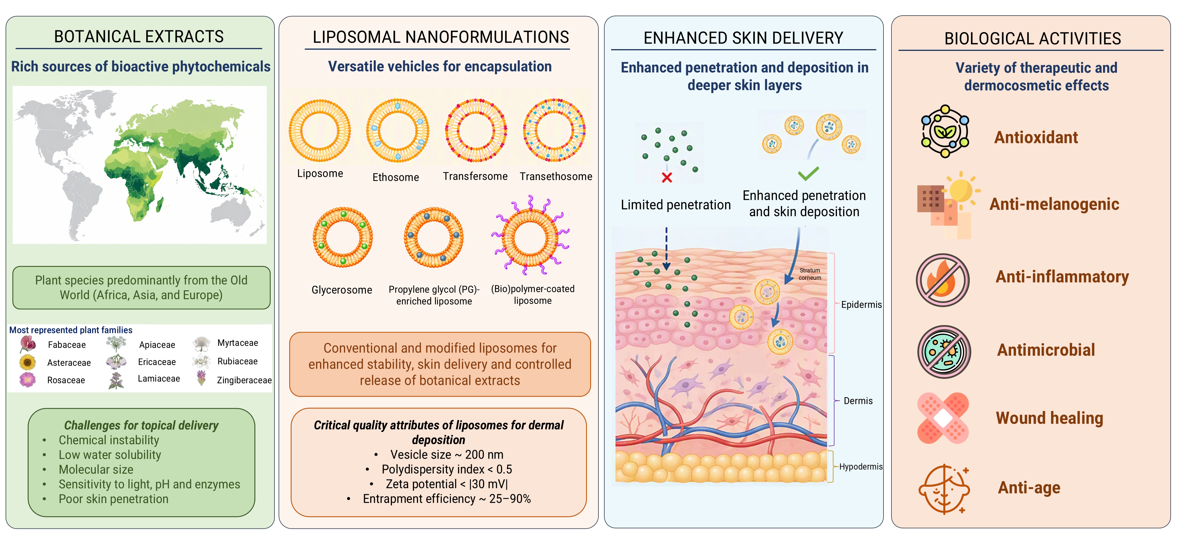
The studies included in this review employed a heterogeneous range of conventional and modified liposomal nanoformulations. As their technological characteristics were discussed in section 3, the present section organizes the available evidence primarily according to the investigated biological activities and therapeutic outcomes (Figure 4).
6.1. Antioxidant Potential of Plant-Extract-Loaded Liposomal Nanoformulations: Cell-Free Assays
Oxidative stress plays a significant role in accelerating the skin aging process. Although effective, endogenous antioxidant protection is sometimes insufficient because the influence of external factors may create an imbalance that leads to cellular damage [143]. Antioxidants in cosmetic formulations are known to play a dual role: they both protect the skin from oxidative stress and stabilize the cosmetic product by preventing pro-oxidative reactions. In this way, phenolic compounds of plant extracts can also act as preservatives, protecting the formulation against oxidation during storage and use [144].
The majority of herein reviewed studies focused on liposomal formulations delivering botanical extracts with inherent antioxidant properties. While taking into consideration that cell-free assays (e.g., DPPH, ABTS, FRAP, TEAC) measure the chemical radical-scavenging capacity rather than the direct antioxidative cellular protection, studies confirm that liposomal encapsulation generally preserves or enhances the antioxidant power of extracts. For instance, liposomal encapsulation of Fumaria officinalis extract maintained the level of DPPH scavenging (46.37–55.37% vs. 47.73% for the free extract) and significantly boosted ABTS scavenging activity (88–95% vs. 72% for the free extract), which remained stable even after UV irradiation [55]. Similarly, Myrtus communis extract-loaded liposomes largely retained antioxidant capacity of the free extract (DPPH scavenging 91% vs. 96%, respectively), while empty carrier vehicles also showed modest intrinsic antioxidant activity (~ 39% DPPH scavenging), highlighting the contribution of the phospholipid constituents of the formulation [76]. Furthermore, loading Teucrium marum extract into different types of vesicles increased DPPH activity from 90% (free extract) up to 136% for transfersomes which outperformed hyalurosomes (100%), glycerosomes (97%), and conventional liposomes (95%), presumably due to the phospholipid–surfactant synergy [120].
Some studies assessed the free-radical-scavenging activity of the plant extracts solely prior to liposomal encapsulation and subsequently focused on functional cellular outcomes, such as in vitro wound-healing potential of liposomal gel containing Stellaria media extract, without directly reassessing antioxidant activity after encapsulation [85]. In another study, Stellaria media extract demonstrated potent ABTS radical-scavenging activity, and its liposomal encapsulation doubled skin permeation flux across excised human stratum corneum and epidermis (SCE) membranes as compared to the unencapsulated extract [119].
Evaluating Onopordum illyricum extract, Caddeo et al. [15] showed that vesicular incorporation (conventional liposomes and PGLs) preserved high DPPH radical scavenging activity (~ 99%) and significantly enhanced FRAP reducing capacity (from ~ 450 to > 550 µg/mL Fe²⁺ equivalents). Notably, empty liposomes also exhibited intrinsic antioxidant capacity (39% DPPH, ~ 190 µg/mL Fe²⁺ equivalents), confirming that lipid carrier components actively contribute to the overall formulation response. Likewise, Manca et al. [123] reported an increase in DPPH scavenging for grape seed and stalk extracts upon loading into various types of liposomes (conventional liposomes, transfersomes, hyalurosomes, hyalotransfersomes). Moreover, vesicular carriers substantially improved bioactive retention and stability during cutaneous permeation - ethosomes loaded with Rosa canina extract maintained 81% DPPH inhibition in porcine skin donor compartments after 6 h (vs. 16% for free extract) and increased 24-hour phenolic skin retention to 49% (vs. 41% for free extract) [102].
6.2. Cellular Antioxidant and Cytoprotective Effects of Plant-Extract-Loaded Liposomal Nanoformulations
It is important to accentuate that cell-free antioxidant potential does not automatically translate into intracellular cytoprotection, as vesicular encapsulation fundamentally modulates bioactive stability, release kinetics, cellular uptake, and carrier synergy, which all affect cellular response. Nevertheless, comparative cell-based evaluations under H₂O₂-induced oxidative stress also consistently demonstrate that nanovesicular incorporation significantly enhances intracellular ROS scavenging and cell survival relative to unencapsulated extracts. For instance, while unencapsulated Onopordum illyricum extract required 10 µg/mL to reduce intracellular ROS by ~ 35% in dermal cells, PGLs achieved a comparable reduction at a 100-fold lower concentration (0.1 µg/mL), whereas at the same low dose conventional liposomes suppressed ROS by ~ 50% [15]. Citrus × limon extract-loaded nanovesicles superiorly protected skin cells against H₂O₂ cytotoxicity, with gly-hyalurosomes restoring keratinocyte viability to ~ 105%, and glycerosomes/hyalurosomes boosting fibroblast viability up to ~ 130% (vs. 107% for conventional liposomes) [132]. Likewise, grape seed and stalk extract-loaded vesicles completely counteracted H₂O₂-toxicity in both keratinocytes and fibroblasts, raising viability from ~ 70% (H₂O₂ control) and ~ 90% (free extract) to > 100% across all nanovesicular platforms [123]. Extending these benefits to tissue repair, polymer-functionalized transfersomes (HEC- and hyaluronan-modified) loading jabuticaba (Plinia cauliflora) peel extract restored keratinocyte viability under oxidative stress to ~ 104% (vs. ~ 78% for free extract) [8]. Conventional liposomes and glycerosomes loaded with Vitis vinifera grape pomace extract both raised H₂O₂-damaged fibroblast viability to ≥ 83% (vs. ~ 70% for free extract and ~ 50% for H₂O₂ control) [124].
Cellular cytoprotective capacity can also vary significantly depending on the target cell line. Liposomal encapsulation of Ceratonia siliqua extract enhanced antioxidant protection in 3T3 fibroblasts under H₂O₂ stress (increasing protective capacity from 6.9% to 31.2%) yet showed lower efficacy than the free extract in HaCaT keratinocytes (7.7% vs. 34.1%) and A431 cells (8.4% vs. 35.1%), underscoring that cell-free antioxidant activity does not uniformly translate across different cell types [68]. Carrier composition further dictates cell-protective responses under oxidative stress. In 3T3 fibroblasts, while unencapsulated Teucrium marum extract restored viability from ~ 42% to ~ 90%, hyalurosomes and glycerohyalurosomes elevated viability to ~ 110% through synergistic sodium hyaluronate–cell interactions [120]. Even more pronounced effect of hyaluronic acid liposome coating was observed in the study of Perra et al. [10], in which unencapsulated rosmarinic acid-rich Lavandula angustifolia extract provided minimal protection against H₂O₂-induced cytotoxicity (~ 60–70% viability), whereas its incorporation into hyalurosomes elevated fibroblast viability to ~ 120% through enhanced membrane interactions and intracellular bioactive delivery. Direct intracellular ROS measurements in TBH-stressed WS1 fibroblasts further confirm elevating effects of liposomal encapsulation: whereas free extracts offered only transient protection, allanthosomes carrying Rosa canina extract produced sustained (~ 50%) reductions in intracellular ROS [101]. This prolonged antioxidant protection directly correlated with accelerated tissue repair in the scratch assay, whereas the respective formulation achieved complete fibroblast wound closure, effectively bridging intracellular cytoprotection with cutaneous tissue regeneration, as further discussed in subsection 6.7.
6.3. Regulation of Melanogenesis and Pigmentation of Plant-Extract-Loaded Liposomal Nanoformulations
Melanin overproduction underlies both post-inflammatory and UV-induced hyperpigmentation, with tyrosinase serving as the rate-limiting enzyme in melanogenesis. To attenuate melanogenesis, bioactive botanical extracts incorporated into vesicular carriers have been evaluated across cell-free in vitro and very few clinical models. Citrus × junos callus extract exhibited direct L-DOPA oxidation inhibition (25.2% at 500 µg/mL) and suppressed cellular melanin production by 59.3% at 1000 µg/mL. L-DOPA (L-3,4-dihydroxyphenylalanine) is a naturally occurring amino acid compound and a key intermediate in the metabolic pathway of melanin synthesis (melanogenesis) in the skin [40]. Rhodomyrtone-rich Rhodomyrtus tomentosa extracts also demonstrated complementary anti-tyrosinase capacity [117], whereas ginsenoside-rich ginseng extract embedded into a liposomal hybrid hydrogel reduced tyrosinase/melanin expression and accelerated repair in UV-damaged cutaneous tissue [1]. Clinical evaluation by Khan et al. [92] showed that an ethosomal gel containing red cabbage (Brassica oleracea)-extract significantly outperformed the free-extract gel, achieving a 20.1% reduction in skin melanin index after 12 weeks versus 7.6% for the unencapsulated control.
Overall, vesicular platforms primarily enhance transdermal permeation, retention, and cellular availability rather than altering the intrinsic enzyme-inhibitory potency of phytoconstituents, presenting promising cosmeceutical strategies for topical hyperpigmentation management.
6.4. Anti-inflammatory Effects of Plant-Extract-Loaded Liposomal Nanoformulations
Inflammation is closely associated with oxidative stress and represents an important component of numerous acute and chronic dermatological disorders. Accordingly, the anti-inflammatory activity of plant extract-loaded vesicular systems has been investigated using cellular models, acute animal models of inflammation and disease-specific models, including psoriasis, atopic dermatitis and radiation-induced skin damage. In activated macrophages, transfersomal delivery of Pistacia vera essential oil modulated immunometabolism by suppressing ATP-citrate lyase (ACLY) expression and reducing ROS and nitric oxide production by ~ 10% and ~ 20%, respectively, while simultaneously eliminating cytotoxicity of the free essential oil. Notably, since the nanovesicle carrier contributed to the baseline activity at lower concentrations, this efficacy reflects a formulation-level synergy rather than an extract-isolated response [115].
Disease-specific in vivo and clinical models provide preliminar evidence regarding the therapeutic efficacy of botanical extract-loaded nanovesicles in inflammatory skin disorders. In an atopic dermatitis model, a liposomal gel loaded with Vaccinium vitis-idaea extract significantly mitigated skin lesions and histological damage while suppressing pro-inflammatory cytokines (TNF-α, IL-4, IL-13), reducing lipid peroxidation (MDA), and enhancing SOD (superoxide dismutase) activity and collagen organization [53]. Translating to clinical evaluation, randomized double-blind trials of liposomal chamomile extract gel in breast cancer (100 patients) [74] and head/neck cancer (60 patients) [75] radiotherapy cohorts demonstrated reduced dry skin desquamation rates and significant relief from radiation-induced pruritus. Interestingly, no statistically significant difference between the chamomile extract-loaded liposomal gel and the placebo liposomal gel (without chamomile extract) in either trial was observed, indicating the intrinsic inflammation-protective contribution of the liposomal vehicle itself [74, 75].
Beyond described anti-inflammatory effects, several studies link immunomodulation with tissue repair. Liposome-encapsulated Centella asiatica extract markedly reduced TNF-α and IL-1β expression (even at low concentrations, 6.5–12.5 µg/mL) and promoted fibroblast migration/proliferation in vitro, as well as wound closure in vivo [67]. C. asiatica-loaded transfersomes suppressed nitric oxide production in macrophages in vitro, while also stimulating fibroblast migration/proliferation in vitro, and enhanced collagen deposition and wound closure in vivo [112]. Similarly, liposomal stem cell extract cream from Coffea canephora modulated TGF-β/VEGF signaling, accelerated wound closure, and promoted vascularization and collagenesis in rats [25]. In acute in vivo animal model, ethosomal gel co-delivering Dalbergia sissoo and Curcuma longa extracts achieved up to 69% inhibition of carrageenan-induced paw edema in rats after 360 minutes, confirming that vesicular co-encapsulation can effectively maximize anti-inflammatory outcomes in cutaneous tissue, while simultaneously accelerating wound healing and tissue regeneration [94]. These findings confirm that anti-inflammatory effects displayed by nanocarriers support cell migration/proliferation and cutaneous tissue remodeling.
Psoriasis is a chronic inflammatory dermatosis characterized by epidermal hyperproliferation, leukocyte infiltration, and dysregulated cytokine cascades. To overcome the barrier resistance of psoriatic skin, botanical extracts incorporated into a transfersomal gel have been evaluated in preclinical models. In a mouse model of imiquimod induced psoriasis, a transfersomal gel containing Berberis aristata extract reduced paw edema, markedly decreased epidermal thickness and rete ridge elongation, while cutaneous irritation was negligible [109]. Similarly, liposomal Boswellia serrata suppressed IL-23 production, elevated anti-inflammatory IL-10 in RAW 264.7 macrophages, and attenuated ear swelling and hyperplasia in oxazolone-challenged mice model, though some observed reporting inconsistencies in this study warrant cautious interpretation [58]. Transethosomal gels carrying Rubia cordifolia extract demonstrated enhanced ex vivo skin permeation through goat skin, establishing dermal delivery feasibility rather than direct antipsoriatic efficacy due to a lack of disease-specific model validation in the study [128]. Overall, antipsoriatic evidence for botanical nanovesicles remains preliminary, underscoring the need for rigorous methodological standardization and clinical trials.
6.5. Antimicrobial Activity of Plant-Extract-Loaded Liposomal Nanoformulations
Cutaneous infections caused by bacterial, fungal, or viral pathogens exacerbate skin inflammation, necessitating effective topical antimicrobial strategies. To overcome the biopharmaceutical limitations of whole botanical extracts, nanovesicular delivery platforms have been evaluated against various skin infection-relevant microorganisms. A hydroalcoholic Neolamarckia cadamba leaf extract incorporated into liposomal Carbopol hydrogel exhibited moderate antimicrobial activity against Candida albicans, Aspergillus niger, Staphylococcus aureus, and Escherichia coli (inhibition zones: 4.8–6.1 mm), comparable to commercial formulations [114]. Conversely, ethosomes loaded with Zingiber zerumbet extract displayed higher MIC/MFC values against C. albicans than the free extract (312.5 µg/mL), indicating that delayed release temporarily reduced immediate antifungal potency despite significantly enhancing skin penetration and retention [107]. Similarly, liposomal encapsulation of ethyl-acetate fraction of Petroselinum crispum leaves produced modest antifungal activity against Malassezia furfur without demonstrating clear superiority over unencapsulated control fractions [78]. In contrast, transfersomes carrying a Rhodomyrtus tomentosa leaf extract exhibited superior antibacterial effect against S. aureus and Staphylococcus epidermidis, lowering MIC to 8 µg/mL (vs. 32 µg/mL for the free extract) and achieving hydrogel inhibition zones up to 19 mm [117]. Finally, liposomes loading Paeonia tenuifolia petal extract exhibited potent antibacterial (MIC/MBC 0.5–1 mg/mL against Staphylococcus aureus and Proteus vulgaris) and antifungal activity (MIC/MFC 0.25/0.5 mg/mL against Candida krusei), alongside 50.5% Staphylococcus lugdunensis biofilm inhibition; however, subsequent incorporation into carboxymethyl cellulose (CMC) films attenuated active release and antimicrobial potency [77].
In contrast, PC and PS liposomes carrying Aristolochia clematitis extract maintained, but did not enhance, the intrinsic antibacterial activity of the extract against S. aureus, Streptococcus pneumoniae, Escherichia coli, and Pseudomonas aeruginosa, as observed by disk-diffusion assays [11]. Addressing acne-associated pathogens (Cutibacterium acnes and S. epidermidis), ethosomal gels loaded with Plumbago zeylanica root extract yielded smaller inhibition zones than the free extract [98], while nanoliposomal Sonneratia caseolaris leaf fractions showed moderate bacterial inhibition (11.8–13.2 mm) but lacked unencapsulated controls and MIC/MBC determinations [46]. Translating to clinical evaluation, a 1% liposomal Rhodomyrtus tomentosa serum, evaluated over 8 weeks in 60 mild-to-moderate acne patients, achieved a 36.4% reduction in inflammatory lesions, demonstrating comparable efficacy to 1% clindamycin gel (34.7%) without statistical superiority [145].
Collectively, vesicular encapsulation can preserve or modulate antibacterial/antifungal activity depending on the carrier composition and release kinetics, yet direct evidence establishing superior antimicrobial efficacy over free extracts remains constrained. Beyond antibacterial and antifungal properties, liposomal nanovesicles have demonstrated extraordinary potential in topical antiviral therapy against Herpes simplex virus type 1 (HSV-1). In a recent study, Fulgheri et al. [135] incorporated a biotechnological rosmarinic acid-enriched extract of Lavandula angustifolia into glycerosomes, hyalurosomes, and glycerohyalurosomes. These specialized vesicles exhibited potent antiviral protection in Vero cells during simultaneous infection (up to ~ 100% protection) and post-infection (~ 80% protection), demonstrating antiviral potency superior to acyclovir at significantly lower effective concentrations.
6.6. Cytotoxic Effect of Plant-Extract-Loaded Liposomal Nanoformulations
Vesicular encapsulation of botanical extracts has demonstrated enhanced in vitro cytotoxicity against melanoma cell lines, which is primarily attributed to improved intracellular uptake. Liposomes loaded with standardized Piper longum fruit extract (rich in piperine and piperlonguminine) exhibited selective anti-melanoma activity against B16F10 cells, lowering the IC₅₀ from 89 µg/mL (free extract) to 60 µg/mL, without inducing toxicity in non-cancerous HEK293 cells [56]. Similarly, nanotransfersomes carrying Rotheca serrata root extract enhanced cytotoxicity against B10F16 melanoma cells (60% inhibition at 100 µg/mL vs. 50% for free extract) [118], while elastic lipid nanovesicles loaded with Lantana camara leaf extract markedly suppressed SK-MEL-37 cell viability (vs. minimal response for unencapsulated extract) [42]. Although these findings indicate that liposomal nanocarriers augment the antiproliferative potency of botanical extracts, evidence remain restricted to short-term in vitro assays. On the other hand, Mohapatra et al. [56] reported in vivo validation of tumor regression and synergy with dacarbazine in melanoma-bearing mice, when applying topical ultradeformable transfersomal hydrogel loaded with P. longum extract.
6.7. Wound-Healing and Regenerative Activity of Plant-Extract-Loaded Liposomal Nanoformulations
Wound healing is a multi-stage process requiring coordinated inflammatory resolution, keratinocyte and fibroblast migration, matrix deposition, angiogenesis, and tissue remodeling. Although frequently employed in vitro scratch assays effectively evaluate cellular migratory potential [8,9,26,40,67,85,101,112,120,121,123,133,134,135], in vivo wound models are a necessary follow-up to confirm comprehensive cutaneous tissue repair by botanical nanovesicles.
When tested on HaCaT keratinocytes, liposomes loaded with Hypericum scruglii extract accelerated wound closure, with gel-gluglycerosomes achieving up to 93% scratch closure after 48 h [134]. Similarly, transfersomes carrying Plinia cauliflora peel extract yielded ~ 90% keratinocyte migration, whereas hyaluronan-modified transfersomes enabled virtually complete closure via synergistic carrier-cell interactions [8]. Nanovesicles containing Vitis vinifera seed and stalk extracts likewise outperformed free extracts on both HaCaT keratinocytes and 3T3 fibroblasts, with stalk extract-loaded hyalurosomes and hyalo-transfersomes driving complete fibroblast wound closure within 48 h [123]. Furthermore, liposomal encapsulation of Platanus orientalis extract enhanced dermal fibroblast migration, achieving ~ 50% wound closure at 24 h [26]. Collectively, these findings demonstrate that vesicular encapsulation can enhance wound closure kinetics in vitro compared with unencapsulated botanical extracts. Dermal fibroblasts treated with glycethosomes or allantosomes containing Rosa canina extract achieved complete scratch closure within approximately 30 h, with sustained ROS suppression potentially contributing to enhanced cellular migration [101]. Similarly, In HaCaT keratinocytes, liposomes containing Lavandula austroapennina extract achieved complete scratch closure within 12 h, reducing the time required by half compared with the unencapsulated extract, whereas empty liposomes achieved only approximately 10% closure [9].
In vivo animal wound models provide validation of cutaneous tissue repair by evaluating re-epithelialization, granulation, collagen deposition, and matrix remodeling. In a rat full-thickness wound model, thermosensitive liposomal hydrogels loaded with Danggui Buxue extract achieved 93% wound contraction by day 14 through activation of VEGF/PI3K/Akt and TGF-β/Smads signaling cascades [61]. Similarly, liposomal Coffea canephora stem cell extract reduced wound area by 88% at day 14 while downregulating pro-inflammatory cytokines (TNF-α, IL-6) and upregulating IL-10 [25]. In rat incisional and excisional models, liposomal Carpobrotus edulis improved macroscopic and histological healing, though tensile strength did not surpass the Centella asiatica reference [65]. Furthermore, ethosomal Curcuma longa extract accelerated tissue resolution over crude extracts [126], while C. asiatica transfersomal gels suppressed macrophage nitric oxide production and promoted collagen deposition and epithelial regeneration [112].
In acute full-thickness rat wound models, liposome-encapsulated C. asiatica ethanolic extract achieved 100% wound closure by day 12, significantly outperforming vitamin E, saline, and blank liposomes [67]. Likewise, a polyherbal ethosomal gel co-delivering Dalbergia sissoo and Curcuma longa extracts reached 98% wound contraction at day 8 and completed closure by day 12, while concurrently suppressing carrageenan-induced paw edema by 69% to demonstrate coupled anti-inflammatory and tissue-remodeling effects [94]. Although these preclinical findings confirm cell migration, wound contraction, re-epithelialization, collagen deposition, and neo-vascularization, frequent omission of unencapsulated extract or empty liposomes controls limits definitive conclusions regarding the observed therapeutic gains. Moreover, therapeutic gains should probably be attributed to the multi-component hydrogel formulation rather than vesicle encapsulation alone.
6.8. Anti-Aging Activity of Plant-Extract-Loaded Liposomal Nanoformulations
Clinical evidence evaluating botanical extract-loaded nanovesicles in human subjects remains limited predominantly to short-term cosmetic outcomes in healthy volunteers. For example, in an evaluation of a commercial liposomal Rhododendron ferrugineum leaf cell-culture formulation, it was observed that the stratum corneum hydration increased. However, improvements in skin elasticity were absent, and TEWL barrier recovery on irritated skin was comparable to placebo, supporting general moisturizing performance rather than specific anti-aging activity [82]. In a split-face study involving ten healthy women, a hydrogel containing freeze-dried liposomes loaded with Caryocar brasiliense fruit oil improved stratum corneum hydration, TEWL, and skin microrelief 2 h post-application relative to the empty liposomes. Yet, the acute evaluation period, small cohort, and lack of unencapsulated oil control preclude conclusions regarding sustained anti-aging efficacy or carrier-mediated advantages in this study [66]. A 4-week split-face trial in twenty women using multilayered liposomes incorporating Eryngium maritimum callus culture medium demonstrated improvements in skin density, hydration, elasticity, and skin sagging and wrinkle parameters [133], though the absence of unencapsulated extract control limits efficacy attribution to the complete cosmetic product. In conclusion, while extract-loaded nanovesicles show clear potential to enhance skin hydration and skin barrier metrics, clinical proof of targeted anti-aging superiority remains preliminary.
7. Botanical Aspects of Investigated Plants – Taxonomic and Biogeographical Patterns
The analysis of 103 publications included in this review identified a total of 123 taxon records, including species, subspecies, varieties, and hybrids. Several taxa, however, were investigated in more than one publication. For example, Vitis vinifera was examined in five publications, Rosa canina in four publications, and Centella asiatica, Curcuma longa and Piper longum in three publications each, while eleven taxa were reported in two publications. After accounting for repeated records, the number of distinct plant taxa represented in the reviewed literature was 99, belonging to 53 plant families (Table S1).
At the family level, Fabaceae, Asteraceae, and Rosaceae were the most frequently represented, each accounting for ≥ 5% of the distinct taxa, followed by Apiaceae, Ericaceae, Lamiaceae, Myrtaceae, Rubiaceae, and Zingiberaceae (Figure 5). Similar observations were reported in an electronic exploratory survey of novel plant based cosmeceutical natural ingredients in which the greatest number of plant species belonged to the botanical families Asteraceae, Lamiaceae, Fabaceae, Poaceae, Malvaceae, and Rosaceae, each accounting for ≥ 5% taxa, with around 68 plant species belonging to these plant families that were utilized for various cosmeceutical applications such as anti-aging, UV protection, antioxidant, anti-inflammatory, anti-hyperpigmentation, etc. [146]. Similarly, a review focused on the detailed study of 128 ethnobotanical surveys conducted in Albania, Cyprus, Greece, and Turkey, with the aim to provide an overview of the ethnobotanical knowledge of medicinal plants and traditional medical practices for the treatment of skin disorders, established that the majority of the plants belonged to the families of Asteraceae (11.7%), Lamiaceae (7.4%), Rosaceae (6.7%), Plantaginaceae (5.4%), and Malvaceae (3.8%) [147].
Most of the families that were found to be highly represented in the present review article are also among the twenty largest angiosperm families globally, although Rosaceae and Zingiberaceae are exceptions to this pattern [148]. Globally, Fabaceae and Lamiaceae are among the plant families with the highest absolute numbers of medicinal species. When medicinal species are considered relative to the total number of species within each family, four of the 12 families identified by Nikolić [149] as having the highest relative representation of medicinal species are also represented in the present dataset: Apiaceae, Lamiaceae, Rutaceae, and Fabaceae. Given that the publications included in the present review are predominantly recent studies identified through major bibliographic databases, including PubMed, Scopus, and Web of Science, some degree of divergence between the family-level patterns observed here and those reported at the global level is not unexpected. Moreover, the representation of individual families should be interpreted in relation to their overall species richness. In this context, the relatively high representation of Berberidaceae and Zingiberaceae is noteworthy despite their comparatively smaller global species pools, estimated at fewer than 750 and 1750 species, respectively [147].
The analysis of the standardized geographical data revealed a predominance of taxa native to the Old World. Most taxa included in the review have distributions extending across regions characterized predominantly by temperate climates in Europe, Asia, and Africa, corresponding broadly to the Palearctic biogeographical realm, as well as the subtropical and tropical regions of Southeast Asia, corresponding largely to the Indomalayan realm (Figure 6). In contrast, Neotropical taxa were relatively poorly represented in the reviewed literature, despite the exceptional plant species richness of the Neotropics at the global scale [150].
This pattern should not, however, be interpreted as evidence of lower dermatological relevance or therapeutic potential among Neotropical or Afrotropical plants. Medicinal plants from both regions have been documented for the treatment and management of various skin conditions [151,152,153]. The observed geographical distribution in the present review may instead reflect a combination of historical, scientific, socioeconomic, and bibliometric factors. These may include the historical development of ethnobotanical research, the availability of research infrastructure and funding, access to plant material, established traditions in pharmacognosy and phytochemistry, the language of scientific communication, journal indexing practices, opportunities for patenting and commercialization, regulatory frameworks, and historical colonial and scientific networks.
Accordingly, the geographical representation of medicinal plants in scientific literature should not necessarily be regarded as a direct proxy for their therapeutic potential. Rather, it may also reflect differences in research intensity, scientific infrastructure, research funding, accessibility of biological resources, and patterns of scientific dissemination. This interpretation is consistent with the findings of Jiang et al. [154], who reported that 48.1% of the regional medicinal plant databases that were included in their analysis focused on Asian medicinal plants, with China and India accounting for 22.2% and 8.6% of the databases, respectively. Similarly, Sood et al. [155] demonstrated that geographical factors may be associated with patterns of publication bias in the field of complementary and alternative medicine. Nevertheless, these observations do not provide sufficient evidence to establish that differences in research infrastructure, publication practices, or other geographical factors are directly responsible for the greater representation of Old-World plants in the literature concerning dermatological applications. Rather, they indicate that such factors should be considered when interpreting the geographical patterns identified in the present review.
8. Discussion and Future Perspectives
The present work systematically represents the results presented in 103 original research articles related to liposomal nanoformulations for skin delivery of bioactive botanical extracts that have been published since 2016 to 2026. To keep this review concise, we chose to summarize the general information extracted from 103 studies rather than exhaustively list the exact conditions used and values obtained by the authors of the publications included. Therefore, specific formulation details (e.g., lipid-to-surfactant ratios, additive percentages) and precise biological activity results are not fully presented in the tables, and should be searched for in the original papers cited. As per standard scoping review methodology (PRISMA-ScR checklist), a systematic evaluation of methodological quality or risk of bias of individual studies was omitted and the reported efficacy of individual nanocarriers reflects primarily findings published by the respective authors without critical evaluation of potential experimental bias.
A major issue identified in the literature are terminological inconsistencies concerning different types of liposomal nanocarriers. Authors frequently use the general term liposomes to describe both conventional and non-conventional nanovesicles. For instance, formulations containing high proportions of propylene glycol or surfactants are often reported simply as liposomes rather than propylene glycol-enriched liposomes (PGLs/PG-PEVs) or transfersomes. Likewise, penetration-enhancer-containing vesicles (PEVs, PG-PEVs, LabEt-PEVs) are in some studies treated as a distinct liposome class, whereas functionally they overlap with transfersomes, ethosomes or propylene glycol-enriched liposomes. Additionally, authors frequently designate seemingly novel liposome categories based on commercial surfactant or additive names (e.g. Lab-PEVs, montanov-liposomes, gluglycerosomes), whereas they actually represent subvariants of transfersomes or glycerosomes. Future research should adopt more precise terminology reflective of vesicle composition based on the presence and concentration of penetration enhancers or edge activators.
A significant subset of published studies report botanical extracts without specifying exact solvent ratios or extraction method used, and rely solely on non-specific spectrophotometric assays (e.g., UV-Vis or FTIR) without chromatographic profiling (e.g., HPLC-DAD, UPLC, LC-MS/MS) or quantification of specific bioactive marker compounds. Similarly, the exact composition of semisolid vehicles used as the base for the preparation of liposome-in-vehicle formulations is occasionally incompletely reported (without specifying polymer type or concentration), whereas certain clinical studies lack details about the tested nanovesicles (e.g., formulation composition, preparation method, physicochemical parameters of the vesicles).
To facilitate clinical and commercial translation of plant extract-loaded nanovesicles into reproducible and validated formulations, several methodological guidelines are proposed. First, taxonomic authentication of raw plant materials used for the extract preparation, full documentation of provenance and extraction conditions, coupled with chromatographic and spectroscopic profiling is essential to quantify chemical markers and ensure reproducibility. Second, in vitro and ex vivo skin permeation assays should be performed according to uniform experimental conditions and protocols to enable valid cross-study comparisons. Third, bioactivity assays should always include appropriate formulation controls (e.g., unencapsulated extracts, blank vehicle, and blank nanocarrier) to distinguish vesicle-mediated gains from intrinsic plant extract activity or synergistic effects. Furthermore, implementation of systematic experimental design (DoE) and QbD principles can be of great use to achieve more time- and resource-efficient optimization of the processing parameters for liposomal formulations. Importantly, to confirm therapeutic efficacy and safety in humans, short-term cytocompatibility screening should not be considered reliable without irritation testing and randomized clinical trials. Ultimately, potential commercial realization of the developed formulations depends on technological scalability and regulatory clarity. Transitioning from bench-scale protocols to continuous scalable manufacturing (e.g., microfluidics or high-pressure homogenization) remains a critical engineering challenge. Overcoming these processing obstacles, as well as defining clear and harmonized regulatory standards specific to botanical nanocosmeceuticals and phytomedicines, will be crucial in translating versatile liposomal nanocarriers from laboratory models into marketed formulations.
9. Conclusions
Liposomal nanovesicular formulations represent a transformative platform for topical and (trans)dermal delivery of bioactive botanical extracts. Research publications consolidated in this scoping review demonstrate gradual shift of scientific focus from conventional liposomes toward advanced phospholipid-based nanovesicles, such as transfersomes, ethosomes, transethosomes, glycerosomes, and biopolymer-coated vesicles. By overcoming skin barrier restrictions and protecting delicate phytochemical complexes, these systems significantly elevate the cosmeceutical and dermatological potential of natural products. Addressing current gaps and inconsistencies in nomenclature, analytical standardization, and complete methodological reporting is essential to advance this field from laboratory experimentation to standardised and reproducible dermatological therapies.
Supplementary Materials
The following supporting information can be downloaded at the website of this paper posted on Preprints.org, Table S1: Botanical profiles of plant-extract-loaded liposomal nanoformulations included in this review.
Author Contributions
Conceptualization, Z.R. and M.F.; methodology, Z.R. and M.F.; formal analysis, Z.R., L.J., D.H., and M.F.; investigation, Z.R., L.J., D.H., B.M., and M.F.; resources, Z.R., L.J., D.H., B.M., and M.F..; data curation, Z.R., L.J., and D.H..; writing—original draft preparation, Z.R., L.J., D.H., and M.F.; writing—review and editing, Z.R., L.J., T.M., M.P., D.H., B.M., Z.V., and M.F..; visualization, Z.R., L.J., M.P., D.H., and M.F..; supervision, B.M., Z.V., Z.M., and M.F..; project administration, M.F..; funding acquisition, M.F.. All authors have read and agreed to the published version of the manuscript.
Funding
This work has been supported by the Croatian Science Foundation under the project UIP-2025-02-7092.
Institutional Review Board Statement
Not applicable.
Informed Consent Statement
Not applicable.
Data Availability Statement
The data presented in this review are available within the article and its supporting materials.
Acknowledgments
The authors would like to thank Maja Fehir and Maja Špac for their assistance and prompt responses provided in retrieving all eligible articles. During the preparation of the manuscript, the authors used Illustrae.co (UK) for the preparation of Figures 1 and 3, and Microsoft Copilot (Microsoft Corporation, Redmond, WA, USA) to generate Figure 4 based on author-developed prompts as well as to further refine Figures 5 and 6. The authors used Gemini Notebook (Google LLC, Mountain View, CA, USA) as an assistive tool to organize the author-curated literature collection and refine the readability of author-drafted text. All literature screening, data extraction, synthesis, and interpretations were performed by the authors, who take full responsibility for the content of this publication.
Conflicts of Interest
The authors declare no conflicts of interest.
Abbreviations
The following abbreviations are used in this manuscript:
| 3T3 | mouse embryonic fibroblast cell line (3T3 fibroblasts) |
| A431 | human epidermoid carcinoma cell line (A431) |
| ABTS | 2,2′-azino-bis(3-ethylbenzothiazoline-6-sulfonic acid) |
| ACLY | ATP-citrate lyase |
| Akt | protein kinase B (AKT), a serine/threonine kinase |
| ATP | adenosine triphosphate |
| B16F10 | murine melanoma cell line (B16F10) |
| BALB/c | inbred laboratory mouse strain (BALB/c) |
| chol | cholesterol |
| CMC | carboxymethyl cellulose |
| CMCh | carboxymethyl chitosan |
| DCP | dicetyl phosphate |
| DDAB | dimethyldioctadecylammonium bromide |
| DoE | Design of Experiments |
| DOPC | 1,2-dioleoyl-sn-glycero-3-phosphocholine |
| DOTAP | 1,2-dioleoyl-3-trimethylammonium-propane |
| DPPC | 1,2-dipalmitoyl-sn-glycero-3-phosphocholine |
| DPPH | 2,2-diphenyl-1-picrylhydrazyl |
| DSPC | 1,2-distearoyl-sn-glycero-3-phosphocholine |
| DSPE | 1,2-distearoyl-sn-glycero-3-phosphatidylethanolamine |
| DSPE-PEG 2000 | 1,2-distearoyl-sn-glycero-3-phosphatidylethanolamine-N-[methoxy(polyethylene glycol)-2000] |
| EE | entrapment efficiency |
| FRAP | Ferric reducing antioxidant power |
| FTIR | Fourier-transform infrared spectroscopy |
| GC–MS | gas chromatography–mass spectrometry |
| HaCaT | immortalized human keratinocyte cell line (HaCaT) |
| HEC | hydroxyethylcellulose |
| HEK293 | human embryonic kidney 293 cell line |
| HLB | hydrophilic-lipophilic balance |
| HPC | hydrogenated phosphatidylcholine |
| HPLC | high-performance liquid chromatography |
| HPLC-DAD | high-performance liquid chromatography with diode-array detection |
| HPLC-DAD-ESI | high-performance liquid chromatography with diode-array detection and electrospray ionization |
| HPLC-ESI-TOF-MS | high-performance liquid chromatography–electrospray ionization–time-of-flight mass spectrometry |
| HPLC-ESI-QToF-MS/MS | high-performance liquid chromatography–electrospray ionization–quadrupole time-of-flight tandem mass spectrometry |
| HPLC-MS | high-performance liquid chromatography–mass spectrometry |
| HPLC-PDA | high-performance liquid chromatography with photodiode-array detection |
| HPLC-UV | high-performance liquid chromatography with ultraviolet detection |
| HPMC | hydroxypropyl methylcellulose |
| HPTLC | high-performance thin-layer chromatography |
| HRLC-MS/MS | high-resolution liquid chromatography–tandem mass spectrometry |
| HSPC | hidrogenated soybean phosphatidylcholine |
| HSV-1 | Herpes simplex virus type 1 |
| IC₅₀ | half-maximal inhibitory concentration |
| IFN-γ | interferon gamma |
| IL | interleukin |
| Lab-PEVs | Labrasol®-containing penetration-enhancing vesicles |
| LabEt-PEVs | Labrasol®- and ethanol-containing penetration-enhancing vesicles |
| L-DOPA | L-3,4-dihydroxyphenylalanine (levodopa) |
| LC-DAD | liquid chromatography with diode-array detection |
| LC-HR-ESI-MS | liquid chromatography–high-resolution electrospray ionization mass spectrometry |
| LC–MS | liquid chromatography–mass spectrometry |
| LC-Orbitrap | liquid chromatography–Orbitrap mass spectrometry |
| MBC | minimum bactericidal concentration |
| MFC | minimum fungicidal concentration |
| MIC | minimum inhibitory concentration |
| NMR | nuclear magnetic resonance spectroscopy |
| PC | phosphatidylcholine |
| PDI | polydispersity index |
| PEG | polyethylene glycol |
| PG | propylene glycol |
| PGLs | propylene glycol-enriched liposomes |
| PG-PEVs | propylene glycol-penetration enhancer-containing vesicles |
| PI3K | phosphoinositide 3-kinase |
| POWO | Plants of the World Online |
| PS | phosphatidylserine |
| PVA | polyvinyl alcohol |
| QbD | Quality by Design |
| RAW 264.7 | murine macrophage cell line (RAW 264.7) |
| ROS | reactive oxygen species |
| RP-HPLC | reversed-phase high-performance liquid chromatography |
| SCh | sodium cholate |
| SDCh | sodium deoxycholate |
| SK-MEL-37 | human melanoma cell line (SK-MEL-37) |
| Smads | intracellular signal-transducing proteins of the TGF-β pathway |
| STDCh | sodium taurodeoxycholate |
| TBH | tert-butyl hydroperoxide |
| TEAC | Trolox equivalent antioxidant capacity |
| TEWL | transepidermal water loss |
| TGF-β | transforming growth factor beta |
| TLFH | thin lipid film hydration |
| Tm | main phase transition temperature |
| TNF-α | tumor necrosis factor alpha |
| UHPLC | ultra-high-performance liquid chromatography |
| UHPLC-ESI-QqTOF-MS/MS | ultra-high-performance liquid chromatography–electrospray ionization–quadrupole time-of-flight tandem mass spectrometry |
| UV | ultraviolet |
| UV-Vis | ultraviolet–visible spectroscopy |
| VEGF | vascular endothelial growth factor |
| WS1 | human skin fibroblast cell line (WS1) |
References
- Zhang, D.; Tian, N.; Guo, Y.; Liu, Y.; Sun, G.; Jiang, R.; He, X.; Xiao, L.; Mi, J.; Liu, Y.; Zheng, W.; Jiang, Y. Multifunctional natural herbal extracts liposomal loaded hybrid hydrogel for skin improvement and repair. Nat. Prod. Commun. 2025, 20, 1–17. [Google Scholar] [CrossRef]
- Alkilani, A.Z.; McCrudden, M.T.C.; Donnelly, R.F. Transdermal drug delivery: Innovative pharmaceutical developments based on disruption of the barrier properties of the stratum corneum. Pharmaceutics 2015, 7, 438–470. [Google Scholar] [CrossRef] [PubMed]
- Secci, D.; Urquhart, A.; Bekiaris, V.; Qvortrup, K. Biomaterials for improving skin penetration in treatment of skin cancer. J. Funct. Biomater. 2026, 17, 39. [Google Scholar] [CrossRef] [PubMed]
- Chatterjee, B.; Reddy, A.; Santra, M.; Khamanga, S. Amorphization of drugs for transdermal delivery-a recent update. Pharmaceutics 2022, 14, 983. [Google Scholar] [CrossRef] [PubMed]
- Hussain, M.S.; Fareed, S.; Ansari, S.; Rahman, M.A.; Ahmad, I.Z.; Saeed, M. Current approaches toward production of secondary plant metabolites. J. Pharm. Bioallied Sci. 2012, 4, 10–20. [Google Scholar] [CrossRef] [PubMed]
- Mondéjar-López, M.; García-Simarro, M.P.; Vega, J.; Fajardo, C.M.; López-López, S.; Ahrazem, O.; Gómez-Gómez, L.; Figueroa, F.L.; Niza, E. Liposomal encapsulation of crocin-rich tomato extract (Tomafran) and its in-depth evaluation as a cosmeceutical ingredient. Colloids Surf. B Biointerfaces 2025, 253, 114766. [Google Scholar] [CrossRef] [PubMed]
- Panza, S.; Pellegrini, B.; Fiore, D.; Tarsitano, M.; Mancuso, A.; Cristiano, M.C.; Paolino, D. Botanical and upcycled bioactives for advanced topical formulations: Mechanistic pathways, cutaneous delivery, and sustainability-by-design. Pharmaceutics 2026, 18, 375. [Google Scholar] [CrossRef] [PubMed]
- Castangia, I.; Manca, M.L.; Allaw, M.; Hellström, J.; Granato, D.; Manconi, M. Jabuticaba (Myrciaria jaboticaba) peel as a sustainable source of anthocyanins and ellagitannins delivered by phospholipid vesicles for alleviating oxidative stress in human keratinocytes. Molecules 2021, 26, 6697. [Google Scholar] [CrossRef] [PubMed]
- Gravina, C.; Piccolella, S.; Alonso, C.; Martí, M.; Formato, M.; Pacifico, S.; Coderch, L.; Esposito, A. Encapsulation of Lavandula austroapennina N.G. Passal., Tundis & Upson extracts: Focus on leaf and stem enriched liposome for cosmeceutical innovation. Ind. Crops Prod. 2024, 213, 118362. [Google Scholar] [CrossRef]
- Perra, M.; Fancello, L.; Castangia, I.; Allaw, M.; Escribano-Ferrer, E.; Peris, J.E.; Usach, I.; Manca, M.L.; Koycheva, I.K.; Georgiev, M.I.; Manconi, M. Formulation and testing of antioxidant and protective effect of hyalurosomes loading extract rich in rosmarinic acid biotechnologically produced from Lavandula angustifolia Miller. Molecules 2022, 27, 2423. [Google Scholar] [CrossRef] [PubMed]
- Vicaș, L.G.; Marian, N.A.; Ali, D.H.; Duteanu, N.; Svera, P.; Dehelean, C.; Vlase, A.-M.; Frenț, O.-D.; Dejeu, I.-L.; Negrean, R.A.; Oros, R.M.; Fritea, L.; Smeu, A.; Mureșan, M.E. Potentiation of the pharmacological effects of an Aristolochia clematitis L. extract by loading into liposomes facilitating release to HaCaT cells. Pharmaceutics 2026, 18, 89. [Google Scholar] [CrossRef] [PubMed]
- Vaou, N.; Stavropoulou, E.; Voidarou, C.; Tsakris, Z.; Rozos, G.; Tsigalou, C.; Bezirtzoglou, E. Interactions between medical plant-derived bioactive compounds: Focus on antimicrobial combination effects. Antibiotics 2022, 11, 1014. [Google Scholar] [CrossRef] [PubMed]
- Karimi, A.; Majlesi, M.; Rafieian-Kopaei, M. Herbal versus synthetic drugs; beliefs and facts. J. Nephropharmacol. 2015, 4, 27–30. [Google Scholar] [PubMed]
- De Luca, M.; Tuberoso, C.I.G.; Pons, R.; García, M.T.; Morán, M.D.C.; Ferino, G.; Vassallo, A.; Martelli, G.; Caddeo, C. Phenolic fingerprint, bioactivity and nanoformulation of Prunus spinosa L. fruit extract for skin delivery. Pharmaceutics 2023, 15, 1063. [Google Scholar] [CrossRef] [PubMed]
- Caddeo, C.; Tuberoso, C.I.G.; Floris, S.; Masala, V.; Sanna, C.; Pintus, F. A nanotechnological approach to exploit and enhance the bioactivity of an extract from Onopordum illyricum L. leaves. Plants 2023, 12, 1453. [Google Scholar] [CrossRef] [PubMed]
- Ekor, M. The growing use of herbal medicines: issues relating to adverse reactions and challenges in monitoring safety. Front. Pharmacol. 2014, 4, 177. [Google Scholar] [CrossRef] [PubMed]
- Atanasov, A.G.; Zotchev, S.B.; Dirsch, V.M.; International Natural Product Sciences Taskforce; Supuran, C.T. Natural products in drug discovery: advances and opportunities. Nat. Rev. Drug Discov. 2021, 20, 200–216. [Google Scholar] [CrossRef] [PubMed]
- Antonelli, A.; Palma, F. Nanocarrier-based delivery systems for natural compounds across research stages. Materials 2025, 18, 4960. [Google Scholar] [CrossRef] [PubMed]
- Parvin, N.; Aslam, M.; Joo, S.W.; Mandal, T.K. Nano-phytomedicine: Harnessing plant-derived phytochemicals in nanocarriers for targeted human health applications. Molecules 2025, 30, 3177. [Google Scholar] [CrossRef] [PubMed]
- Bangham, A.D.; Horne, R.W. Negative staining of phospholipids and their structural modification by surface-active agents as observed in the electron microscope. J. Mol. Biol. 1964, 8, 660–668. [Google Scholar] [CrossRef] [PubMed]
- Guimarães, D.; Cavaco-Paulo, A.; Nogueira, E. Design of liposomes as drug delivery system for therapeutic applications. Int. J. Pharm. 2021, 601, 120571. [Google Scholar] [CrossRef] [PubMed]
- Torchilin, V.P. Recent advances with liposomes as pharmaceutical carriers. Nat. Rev. Drug Discov. 2005, 4, 145–160. [Google Scholar] [CrossRef] [PubMed]
- Guillot, A.J.; Martínez-Navarrete, M.; Garrigues, T.M.; Melero, A. Skin drug delivery using lipid vesicles: A starting guideline for their development. J. Control. Release 2023, 355, 624–654. [Google Scholar] [CrossRef] [PubMed]
- El Maghraby, G.M.; Fayed, N.D. Soft and solid lipid-based nanostructures for dermal and transdermal drug delivery. J. Drug Deliv. Sci. Technol. 2026, 122, 108459. [Google Scholar] [CrossRef]
- Guidoni, M.; De Sousa Júnior, A.D.; Aragão, V.P.M.; De Melo Costa Pereira, T.; Dos Santos, W.C.; Monteiro, F.C.; Guimarães, M.C.C.; Fronza, M. Liposomal stem cell extract formulation from Coffea canephora shows outstanding anti-inflammatory activity, increased tissue repair, neocollagenesis and neoangiogenesis. Arch. Dermatol. Res. 2023, 315, 491–503. [Google Scholar] [CrossRef] [PubMed]
- Demirel, F.; Sağıroğlu, A.A.; Toraman, G.Ö.A.; Gunaydin-Akyildiz, A.; Keskin, Z.; Aydın, B.S.; Topçu, G. Development and evaluation of Platanus orientalis L. extract-loaded liposomes for enhanced wound healing. Pharmaceuticals 2026, 19, 32. [Google Scholar] [CrossRef] [PubMed]
- Giacomelli, L.; Moglia, A.; Losa, G.; Quaglino, P. Clinical use of Capilen, a liposomal cream based on fresh plant extracts enriched with omega fatty acids. Drugs Context 2020, 9, 2019-10-1. [Google Scholar] [CrossRef] [PubMed]
- Venkatesan, K.; Dutta, G.; Bandyopadhyay, R.; Guha, N.; Debnath, B.; Manickam, S.; Sugumaran, A. Lipid nanocosmeceuticals: a novel approach to skin therapy for anti-aging and skin disorders. Beni-Suef Univ. J. Basic Appl. Sci. 2025, 14, 114. [Google Scholar] [CrossRef]
- Sogut, O.; Aydemir Sezer, U.; Sezer, S. Liposomal delivery systems for herbal extracts. J. Drug Deliv. Sci. Technol. 2021, 61, 102147. [Google Scholar] [CrossRef]
- Jacob, S.; Kather, F.S.; Boddu, S.H.S.; Rao, R.; Nair, A.B. Vesicular carriers for phytochemical delivery: A comprehensive review of techniques and applications. Pharmaceutics 2025, 17, 464. [Google Scholar] [CrossRef] [PubMed]
- Kothapalli, P.; Vasanthan, M. Lipid-based nanocarriers for enhanced delivery of plant-derived bioactive molecules: A comprehensive review. Ther. Deliv. 2024, 15, 135–155. [Google Scholar] [CrossRef] [PubMed]
- Shaw, T.K.; Paul, P.; Chatterjee, B. Research-based findings on scope of liposome-based cosmeceuticals: an updated review. Future J. Pharm. Sci. 2022, 8, 46. [Google Scholar] [CrossRef]
- Yang, S.; Liu, L.; Han, J.; Tang, Y. Encapsulating plant ingredients for dermocosmetic application: an updated review of delivery systems and characterization techniques. Int. J. Cosmet. Sci. 2020, 42, 16–28. [Google Scholar] [CrossRef] [PubMed]
- Coeurvolan, A.; Koli, H.; Lousa, C.D.M.; Villeneuve, P.; Dorandeu, C.; Sanchez-Ballester, N.M.; Durand, E.; Begu, S.; Simon, L. Medicinal plant-derived nanovesicles for skin delivery: A natural biovector for therapeutics and cosmetics. J. Control. Release 2026, 394, 114926. [Google Scholar] [CrossRef] [PubMed]
- Talebi, M.; Shahbazi, K.; Dakkali, M.S.; Akbari, M.; Almasi Ghale, R.; Hashemi, S.; Sashourpour, M.; Mojab, F.; Aminzadeh, S. Phytosomes: A promising nanocarrier system for enhanced bioavailability and therapeutic efficacy of herbal products. Phytomed. Plus 2025, 5, 100779. [Google Scholar] [CrossRef]
- Lens, M. Niosomes as vesicular nanocarriers in cosmetics: Characterisation, development and efficacy. Pharmaceutics 2025, 17, 287. [Google Scholar] [CrossRef] [PubMed]
- Ghosh, A.; Choudhury, S. Understanding different types of review articles: A primer for early career researchers. Indian J. Psychiatry 2025, 67, 535–541. [Google Scholar] [CrossRef] [PubMed]
- Tricco, A.C.; Lillie, E.; Zarin, W.; O’Brien, K.K.; Colquhoun, H.; Levac, D.; Moher, D.; Peters, M.D.J.; Horsley, T.; Weeks, L.; Hempel, S.; Akl, E.A.; Chang, C.; McGowan, J.; Stewart, L.; Hartling, L.; Aldcroft, A.; Wilson, M.G.; Garritty, C.; Lewin, S.; Godfrey, C.M.; Macdonald, M.T.; Langlois, E.V.; Soares-Weiser, K.; Moriarty, J.; Clifford, T.; Tunçalp, Ö.; Straus, S.E. PRISMA extension for scoping reviews (PRISMA-ScR): Checklist and explanation. Ann. Intern. Med. 2018, 169, 467–473. [Google Scholar] [CrossRef] [PubMed]
- Plants of the World Online, provided by the Royal Botanic Gardens, Kew (POWO. Plants of the World Online. Facilitated by the Royal Botanic Gardens, Kew. 2026. Available online: https://powo.science.kew.org/ (accessed on 3 October 2026).
- Adhikari, D.; Panthi, V.; Pangeni, R.; Kim, H.; Park, J. Preparation, characterization, and biological activities of topical anti-aging ingredients in a Citrus junos callus extract. Molecules 2017, 22, 2198. [Google Scholar] [CrossRef] [PubMed]
- Eom, S.; Kim, E.R.; Jung, H.; Lee, J.; Song, C.; Yoo, J.W.; Baek, H.S.; Lee, J.; Jung, D.; Kwak, M.K.; Jeong, H.E.; Jung, H. Thermally stable nanoarchitectonics of bicellar nanocarriers for superior transdermal delivery of Centella asiatica extracts. ACS Appl. Mater. Interfaces 2026, 18, 11088–11101. [Google Scholar] [CrossRef] [PubMed]
- Maheshwari, R.; Sharma, M.; Bhattacharya, S. Exploring elastic lipid nanovesicles for enhanced skin permeation of whole plant extract: A comprehensive investigation. J. Biomed. Mater. Res. B Appl. Biomater. 2025, 113, e35527. [Google Scholar] [CrossRef] [PubMed]
- Potisuwan, S.; Apichatwatana, N.; Rujivipat, S. Improved skin permeation of transferosomes containing Eulophia macrobulbon extract. Colloids Surf. B Biointerfaces 2023, 229, 113474. [Google Scholar] [CrossRef] [PubMed]
- Mohapatra, D.; Kumar, D.N.; Shreya, S.; Panigrahi, D.; Agrawal, A.K.; Sahu, A.N. Quality-by-Design-based development of ultradeformable nanovesicular transgelosome of standardized Piper longum extract for melanoma. Nanomedicine 2023, 18, 963–985. [Google Scholar] [CrossRef] [PubMed]
- Bonechi, C.; Tamasi, G.; Donati, A.; Bisozzi, F.; Baglioni, M.; Andreassi, M.; Ietta, F.; Leone, G.; Magnani, A.; Rossi, C. Cationic liposomes as carriers of natural compounds from plant extract. Biophys. Chem. 2025, 318, 107381. [Google Scholar] [CrossRef] [PubMed]
- Suryani, A.; Istiqhfarin, H.A.S.; Hidayah, N.; Nitisara, N.N.; Aryanti, A.M.; Ardana, R.; Kartika, E.R. Synergistic modification of polyvinyl alcohol and natural phospholipids: Nanoliposomal carrier for Sonneratia caseolaris L. delivery and therapeutic care. J. Appl. Pharm. Sci. 2025, 15, 251–265. [Google Scholar] [CrossRef]
- Amalu, M.; Roshita, M.B.; Honeymol, G.; J, T.; Gifty, J.M. Formulation and optimization of novel antiaging polyherbal liposal gel using central composite design. Int. J. Pharm. Compd. 2025, 29, 210–221. [Google Scholar] [PubMed]
- Wang, D.-Y.; van der Mei, H.C.; Ren, Y.; Busscher, H.J.; Shi, L. Lipid-based antimicrobial delivery-systems for the treatment of bacterial infections. Front. Chem. 2020, 7, 872. [Google Scholar] [CrossRef] [PubMed]
- Akombaetwa, N.; Ilangala, A.B.; Thom, L.; Memvanga, P.B.; Witika, B.A.; Buya, A.B. Current advances in lipid nanosystems intended for topical and transdermal drug delivery applications. Pharmaceutics 2023, 15, 656. [Google Scholar] [CrossRef] [PubMed]
- Lee, M.S.; Lee, J.W.; Kim, S.J.; Pham-Nguyen, O.-V.; Park, J.; Park, J.H.; Jung, Y.M.; Lee, J.B.; Yoo, H.S. Comparison study of the effects of cationic liposomes on delivery across 3D skin tissue and whitening effects in pigmented 3D skin. Macromol. Biosci. 2021, 21, e2000413. [Google Scholar] [CrossRef] [PubMed]
- Briuglia, M.-L.; Rotella, C.; McFarlane, A.; Lamprou, D.A. Influence of cholesterol on liposome stability and on in vitro drug release. Drug Deliv. Transl. Res. 2015, 5, 231–242. [Google Scholar] [CrossRef] [PubMed]
- Nsairat, H.; Ibrahim, A.A.; Jaber, A.M.; Abdelghany, S.; Atwan, R.; Shalan, N.; Abdelnabi, H.; Odeh, F.; El-Tanani, M.; Alshaer, W. Liposome bilayer stability: emphasis on cholesterol and its alternatives. J. Liposome Res. 2024, 34, 178–202. [Google Scholar] [CrossRef] [PubMed]
- Zhang, Z.; Huo, J.; Feng, L.; Wang, J.; Fan, X.; Wang, W. Development and characterization of a Vaccinium vitis-idaea liposomal gel for the treatment of atopic dermatitis. Drug Dev. Ind. Pharm. 2025, 51, 273–283. [Google Scholar] [CrossRef] [PubMed]
- Guo, D.; Liu, J.; Fan, Y.; Cheng, J.; Shi, Y.; Zou, J.; Zhang, X. Optimization, characterization and evaluation of liposomes from Malus hupehensis (Pamp.) Rehd. extracts. J. Liposome Res. 2020, 30, 366–376. [Google Scholar] [CrossRef] [PubMed]
- Ahmoda, R.A.; Milošević, M.; Marinković, A.; Jovanović, A.A. Nano-liposomal carrier as promising dermal delivery platform for Fumaria officinalis L. bioactives. Pharmaceutics 2025, 17, 782. [Google Scholar] [CrossRef] [PubMed]
- Himani; Mohapatra, D.; Shreya, S.; Biswal, S.; Naik, G.G.; Malang, S.D.; Kathait, P.; Patel, P.K.; Shambhavi; Senapati, P.C.; Acharya, P.C.; Sahu, A.N. Development and characterization of liposomal phytoformulation of Piper longum L. for melanoma therapy. Pharm. Sci. 2025, 31, 237–250. [Google Scholar] [CrossRef]
- Jovanović, A.A.; Balanč, B.; Volić, M.; Pećinar, I.; Živković, J.; Šavikin, K.P. Rosehip extract-loaded liposomes for potential skin application: Physicochemical properties of non- and UV-irradiated liposomes. Plants 2023, 12, 3063. [Google Scholar] [CrossRef] [PubMed]
- Quazi, R.S.; Quazi, A.I. Development of a novel liposomal herbal nanocarrier of Boswellia serrata: In vitro and in vivo evaluation for psoriasis management. Genet. Mol. Res. 2026, 25, 1–13. [Google Scholar] [CrossRef]
- Pankaew, P.; Chokboribal, J.; Kumklam, P.; Anurukvorakun, O. Vesicular encapsulation of biotransformed Abelmoschus esculentus seed oligopeptides in novel sleeping mask formulations for enhanced anti-aging efficacy. J. Drug Deliv. Sci. Technol. 2026, 121, 108264. [Google Scholar] [CrossRef]
- Ghafarzadeh, M.; Eatemadi, A. Clinical efficacy of liposome-encapsulated Aloe vera on melasma treatment during pregnancy. J. Cosmet. Laser Ther. 2017, 19, 181–187. [Google Scholar] [CrossRef] [PubMed]
- Cui, M.-D.; Pan, Z.-H.; Pan, L.-Q. Danggui Buxue extract-loaded liposomes in thermosensitive gel enhance in vivo dermal wound healing via activation of the VEGF/PI3K/Akt and TGF-β/Smads signaling pathway. Evid. Based Complement. Altern. Med. 2017, 2017, 8407249. [Google Scholar] [CrossRef] [PubMed]
- Mohamad, E.A.; Fahmy, H.M. Niosomes and liposomes as promising carriers for dermal delivery of Annona squamosa extract. Braz. J. Pharm. Sci. 2020, 56, e18096. [Google Scholar] [CrossRef]
- Dave, V.; Sharma, S.; Yadav, R.B.; Agarwal, U. Herbal liposome for the topical delivery of ketoconazole for the effective treatment of seborrheic dermatitis. Appl. Nanosci. 2017, 7, 973–987. [Google Scholar] [CrossRef]
- Naseriyeh, T.; Kahrizi, D.; Alvandi, H.; Rajati, H.; Behbood, L.; Shahraky, M.K.; Arkan, E. Preparation of liposomal hydrogel containing Calendula and application as a wound dressing. Cell. Mol. Biol. 2022, 68, 1–7. [Google Scholar] [CrossRef] [PubMed]
- Gunal, M.Y.; Ayla, S.; Caglayan, B.; Beker, M.C.; Bedri, N.; Aslan, I.; Ozdemir, E.M.; Kilic, E.; Yesilada, E. Can Carpobrotus edulis, heal incisional and excisional wounds on the skin? Bratisl. Lek. Listy 2021, 122, 912–917. [Google Scholar] [CrossRef] [PubMed]
- Kakuda, L.; Maia Campos, P.M.B.G.; Oliveira, W.P. Development and efficacy evaluation of innovative cosmetic formulations with Caryocar brasiliense fruit pulp oil encapsulated in freeze-dried liposomes. Pharmaceutics 2024, 16, 595. [Google Scholar] [CrossRef] [PubMed]
- Chonsut, P.; Tawanwongsri, W.; Naphatthalung, J.; Chokpaisarn, J.; Charoensup, R.; Puttarak, P.; Mah, S.H.; Roumwong, A.; Goodla, L.; Mordmuang, A. Evaluation of liposome-encapsulated Centella asiatica ethanolic extract for enhanced in vitro and in vivo wound healing. Front. Med. Technol. 2026, 8, 1740835. [Google Scholar] [CrossRef] [PubMed]
- De Luca, M.; Tuberoso, C.I.G.; Pons, R.; García, M.T.; Morán, M.D.C.; Martelli, G.; Vassallo, A.; Caddeo, C. Ceratonia siliqua L. pod extract: From phytochemical characterization to liposomal formulation and evaluation of behaviour in cells. Antioxidants 2023, 12, 1209. [Google Scholar] [CrossRef] [PubMed]
- Elwakil, B.H.; Awad, D.; Hussein, A.A.; Harfoush, R.A.; Gohar, Y.M. Chitosan and liposomes nanoparticles encapsulated cinnamon extract: Antiproteolytic activity and wound healing efficiency of diabetic rats. Chiang Mai Univ. J. Nat. Sci. 2020, 19, 595–611. [Google Scholar] [CrossRef]
- Choochuen, N.; Jimtaisong, A. Physical stability and subjective efficacy study of liposome loaded with Clitoria ternatea (butterfly pea) flower extract and Eugenia caryophyllus (clove) oil. Pharm. Sci. Asia 2022, 49, 51–58. [Google Scholar] [CrossRef]
- BalaYadav, R.; Pathak, D.P.; Varshney, R.; Arora, R. Design and optimization of a novel herbosomal-loaded PEG–poloxamer topical formulation for the treatment of cold injuries: a Quality-by-Design approach. Drug Deliv. Transl. Res. 2022, 12, 2793–2823. [Google Scholar] [CrossRef] [PubMed]
- Djawad, K.; Wahab, S.; Wibisono, O.; Avanti, C.; Putranti, A.R.; Bukhari, A.; Patellongi, I.J. Protective effect of mangosteen pericarp extract cream against UVB-induced erythema. J. Clin. Aesthet. Dermatol. 2020, 13, E59–E65. [Google Scholar]
- Roslan, N.Z.I.; Abdul Ghani, S.M.; Yusof, N.B.; Abd. Aziz, A. Liposome as transdermal carrier for Labisia pumila and Ficus deltoidea water extracts. J. Teknol. 2017, 79, 161–166. [Google Scholar] [CrossRef]
- Meneses, A.G.D.; Ferreira, E.B.; Vieira, L.A.C.; de Souza Maggi Bontempo, P.; Guerra, E.N.S.; Ciol, M.A.; Reis, P.E.D.D. Comparison of liposomal gel with and without chamomile to prevent radiation dermatitis in breast cancer patients: A randomized controlled trial. Strahlenther. Onkol. 2025, 201, 115–125. [Google Scholar] [CrossRef] [PubMed]
- Meneses, A.G.; Ferreira, E.B.; Vieira, L.A.C.; de Souza Maggi Bontempo, P.; Guerra, E.N.S.; Ciol, M.A.; Reis, P.E.D. Comparison of liposomal gel with and without addition of chamomile for prevention of radiation dermatitis in head and neck cancer patients: A randomized controlled trial. Radiother. Oncol. 2024, 199, 110440. [Google Scholar] [CrossRef] [PubMed]
- De Luca, M.; Lucchesi, D.; Tuberoso, C.I.G.; Fernàndez-Busquets, X.; Vassallo, A.; Martelli, G.; Fadda, A.M.; Pucci, L.; Caddeo, C. Liposomal formulations to improve antioxidant power of myrtle berry extract for potential skin application. Pharmaceutics 2022, 14, 910. [Google Scholar] [CrossRef] [PubMed]
- Čutović, N.; Marković, T.; Carević, T.; Stojković, D.; Bugarski, B.; Jovanović, A.A. Liposomal and liposomes-film systems as carriers for bioactives from Paeonia tenuifolia L. petals: Physicochemical characterization and biological potential. Pharmaceutics 2023, 15, 2742. [Google Scholar] [CrossRef] [PubMed]
- Hasanuddin, S.; Ridwan, B.A.; Trisnaputri, D.R. Formulation and evaluation of ethyl acetate fraction of parsley leaves as liposomes: In vitro and in silico activity against Malassezia furfur. Res. J. Pharm. Technol. 2025, 18, 6091–6100. [Google Scholar] [CrossRef]
- Oskoueian, E.; Karimi, E.; Noura, R.; Ebrahimi, M.; Shafaei, N.; Karimi, E. Nanoliposomes encapsulation of enriched phenolic fraction from pistachio hulls and its antioxidant, anti-inflammatory, and anti-melanogenic activities. J. Microencapsul. 2020, 37, 1–13. [Google Scholar] [CrossRef] [PubMed]
- Erdal Altintas, O.; Aytar Celik, P. A liposomal controlled delivery system for Plantago major L. extracts and their biological activity for wound healing. J. Microbiol. Biotechnol. Food Sci. 2024, 14, e9929. [Google Scholar] [CrossRef]
- Rezadoost, M.H.; Kumleh, H.H.; Ghasempour, A. Cytotoxicity and apoptosis induction in breast cancer, skin cancer and glioblastoma cells by plant extracts. Mol. Biol. Rep. 2019, 46, 5131–5142. [Google Scholar] [CrossRef] [PubMed]
- Filipovic, M.; Gledovic, A.; Lukic, M.; Tasic-Kostov, M.; Isailovic, T.; Pantelic, I.; Vuleta, G.; Savic, S. Alp Rose stem cells, olive oil squalene and a natural alkyl polyglucoside emulsifier: Are they appropriate ingredients of skin moisturizers - in vivo efficacy on normal and sodium lauryl sulfate - irritated skin? Vojnosanit. Pregl. 2016, 73, 991–1002. [Google Scholar] [CrossRef] [PubMed]
- Ghafarzadegan, R.; Hajiaghaee, H.; Yekefallah, M.; Bafkary, R.; Malakootikhah, J.; Aliabad, S.M.; Vaseghi, S.; Zarinpanjeh, N. From plant to nano-clinic: Advancing wound healing with nano-encapsulated Rosa damascena callus extract. Pharm. Nanotechnol. 2026, e22117385417799. [Google Scholar] [CrossRef] [PubMed]
- Vanti, G.; Bergonzi, M.C.; Bilia, A.R. Development of nanoliposomes loaded with carbon dioxide Serenoa repens (saw palmetto) extract. J. Nanosci. Nanotechnol. 2021, 21, 2943–2945. [Google Scholar] [CrossRef] [PubMed]
- Miere (Groza), F.; Ganea, M.; Teodorescu, A.G.; Horvath, T.; Hanga-Farcas, A.; Csaba, N.; Zdinca, M.; Zdinca, M.; Dobjanschi, L. Characterization in terms of phytochemical content and medicinal potential of the Stellaria media plant extract. Pharmacophore 2023, 14, 45–55. [Google Scholar] [CrossRef]
- Andleeb, M.; Khan, H.M.S.; Daniyal, M. Development, characterization and stability evaluation of topical gel loaded with ethosomes containing Achillea millefolium L. extract. Front. Pharmacol. 2021, 12, 603227. [Google Scholar] [CrossRef] [PubMed]
- Javed, N.; Ijaz, S.; Akhtar, N.; Khan, H.M.S. Nanostructured ethosomal gel loaded with Arctostaphylos uva-ursi extract; In-vitro/in-vivo evaluation as a cosmeceutical product for skin rejuvenation. Curr. Drug Deliv. 2022, 19, 706–720. [Google Scholar] [CrossRef] [PubMed]
- De Luca, M.; Casula, L.; Tuberoso, C.I.G.; Pons, R.; Morán, M.D.C.; García, M.T.; Martelli, G.; Vassallo, A.; Caddeo, C. Formulating a horseradish extract in phospholipid vesicles to target the skin. Pharmaceutics 2024, 16, 1507. [Google Scholar] [CrossRef] [PubMed]
- Dave, V.; Bhardwaj, N.; Gupta, N.; Tak, K. Herbal ethosomal gel containing luliconazole for productive relevance in the field of biomedicine. 3 Biotech 2020, 10, 97. [Google Scholar] [CrossRef] [PubMed]
- Dwivedi, D.; Nimisha; Mishra, N.; Singh, S.P.; Shukla, A.K. Performance evaluation of Bacopa monneri-loaded ethosomes for topical delivery. J. Appl. Nat. Sci. 2024, 16, 1040–1048. [Google Scholar] [CrossRef]
- Nimisha; Fatima, Z.; Kaur, C.D. Formulation and performance evaluation of Berberis aristata extract loaded ethosomal gel. Asian J. Pharm. 2017, 11, 176–183. [Google Scholar] [CrossRef]
- Khan, P.; Akhtar, N.; Khan, H.M.S.; Tasneem, R.; Zaka, H.S.; Akhtar, N.; Sharif, A. Assessment of Brassica oleraceae L. (Brassicaceae) extract loaded ethosomal gel as a versatile vesicular carrier system for dermocosmetic application: A noninvasive split-faced study. J. Cosmet. Dermatol. 2022, 21, 7153–7162. [Google Scholar] [CrossRef] [PubMed]
- Kusumawati, I.; Kurniawan, K.O.; Rohmania, R.; Pratama, B.A.; Pratama, Y.A.; Rullyansyah, S.; Warsito, M.F.; Widyowati, R.; Hestianah, E.P.; Matsunami, K. Comparative study of liposomal and ethosomal formulations of Curcuma heyneana rhizome extract in a transdermal delivery system. Pharm. Nanotechnol. 2025, 13, 303–312. [Google Scholar] [CrossRef] [PubMed]
- Das, V.; Pradhan, J. Innovative transdermal delivery of synergistic phytoconstituents from Dalbergia sissoo and Curcuma longa: Formulation, characterization, and biological efficacy. J. Pharm. Innov. 2025, 20, 194. [Google Scholar] [CrossRef]
- Singh, V.; Bajpai, M.; Bansal, K. Development, optimization, and in vitro evaluation of ethosomal gel of Dalbergia sissoo leaves extract. Asian J. Pharm. 2024, 18, 855–865. [Google Scholar] [CrossRef]
- Pintus, F.; Floris, S.; Fais, A.; Era, B.; Porcedda, C.; Tuberoso, C.I.G.; Caddeo, C. Euphorbia characias extract: Inhibition of skin aging-related enzymes and nanoformulation. Plants 2022, 11, 1849. [Google Scholar] [CrossRef] [PubMed]
- Sasindran, S.; Easwaran, M.; Shyamala, G.; Karuppaiah, A.; Siram, K.; Veintramuthu, S. Phytochemical screening and cytotoxicity evaluation of crude extracts: Toxicity comparison of crude extracts and its ethosomal formulations. J. Cosmet. Dermatol. 2020, 19, 1794–1803. [Google Scholar] [CrossRef] [PubMed]
- Sandhya, P.; Snehalata, J. Formulation development and evaluation of antiacne activity of ethosomal gel prepared using Plumbago zeylanica root extract. Int. J. Res. Pharm. Sci. 2020, 11, 5511–5516. [Google Scholar] [CrossRef]
- Mekala, K.; Shaheedha, S.M.; Prakash, R. Impact of nanoethosomes loaded with polyherbal extracts on the mechanisms involved in wound healing in-vitro research. Int. J. Drug Deliv. Technol. 2024, 14, 1334–1343. [Google Scholar] [CrossRef]
- Jufri, M.; Amelia, L.; Munim, A. Hair growth activity and safety test of ethosomal gel ethyl acetate fraction of Nothopanax leaves (Nothopanax scutellarium Merr.). Asian J. Pharm. Clin. Res. 2017, 10, 134–138. [Google Scholar] [CrossRef]
- Rossi, M.; Rossello, S.; Sallustio, V.; Mandrone, M.; Cerchiara, T.; Chiocchio, I.; Chidichimo, G.; Protti, M.; Mercolini, L.; Luppi, B.; Bigucci, F.; Abruzzo, A.; Cappadone, C. GinExtraMed: Focus on Rosa canina L. extract encapsulated into glycethosomes and allanthosomes for accelerating skin wound healing. Pharmaceutics 2025, 17, 632. [Google Scholar] [CrossRef] [PubMed]
- Sallustio, V.; Chiocchio, I.; Mandrone, M.; Cirrincione, M.; Protti, M.; Farruggia, G.; Abruzzo, A.; Luppi, B.; Bigucci, F.; Mercolini, L.; Poli, F.; Cerchiara, T. Extraction, encapsulation into lipid vesicular systems, and biological activity of Rosa canina L. bioactive compounds for dermocosmetic use. Molecules 2022, 27, 3025. [Google Scholar] [CrossRef] [PubMed]
- Sallustio, V.; Farruggia, G.; Di Cagno, M.P.; Tzanova, M.M.; Marto, J.; Ribeiro, H.; Goncalves, L.M.; Mandrone, M.; Chiocchio, I.; Cerchiara, T.; Abruzzo, A.; Bigucci, F.; Luppi, B. Design and characterization of an ethosomal gel encapsulating rosehip extract. Gels 2023, 9, 362. [Google Scholar] [CrossRef] [PubMed]
- Somwanshi, S.B.; Hiremath, S.N. Development and evaluation of novel ethosomal vesicular drug delivery system of Sesamum indicum L. seed extract. Asian J. Pharm. 2018, 12, S1282–S1290. [Google Scholar] [CrossRef]
- Patekar, R.R.; Choudhary, H.B.; Rede, S.D. Formulation and evaluation of ethosomal gel and non-ethosomal gel of S. grandiflora leaves. Res. J. Pharm. Technol. 2022, 15, 1029–1036. [Google Scholar] [CrossRef]
- Pratap, B.; Shamim, S.; Ali, S. Formulation and characterisation of herbal ethosomal gel of luliconazole and clove oil for modified drug diffusion to the skin. Res. J. Pharm. Technol. 2025, 18, 3501–3508. [Google Scholar] [CrossRef]
- Huanbutta, K.; Rattanachitthawat, N.; Luangpraditkun, K.; Sriamornsak, P.; Puri, V.; Singh, I.; Sangnim, T. Development and evaluation of ethosomes loaded with Zingiber zerumbet Linn rhizome extract for antifungal skin infection in deep layer skin. Pharmaceutics 2022, 14, 2765. [Google Scholar] [CrossRef] [PubMed]
- Albratty, M. Design, optimization, and characterization of Althaea officinalis-loaded transliposomes for the treatment of atopic dermatitis: a Box Behnken design, in vitro, and ex vivo study. J. Biomater. Sci. Polym. Ed. 2023, 34, 2356–2375. [Google Scholar] [CrossRef] [PubMed]
- Nimisha; Rizvi, D.A.; Fatima, Z.; Neema; Kaur, C.D. Antipsoriatic and anti-inflammatory studies of Berberis aristata extract loaded nanovesicular gels. Pharmacogn. Mag. 2017, 13, S587–S594. [Google Scholar] [CrossRef] [PubMed]
- Ramadon, D.; Harme, M.; Anwar, E. Formulation of transfersomal green tea (Camellia sinensis L. Kuntze) leaves extract cream and in vitro penetration study using Franz difussion cell. J. Young Pharm. 2018, 10, S63–S68. [Google Scholar] [CrossRef]
- Sarwa, K.; Mazumder, B.; Suresh, P.; Kaur, C. Topical analgesic nanolipid vesicles formulation of capsaicinoids extract of Bhut Jolokia (Capsicum chinense Jacq): Pharmacodynamic evaluation in rat models and acceptability studies in human volunteers. Curr. Drug Deliv. 2016, 13, 1325–1338. [Google Scholar] [CrossRef] [PubMed]
- Lapmanee, S.; Bunwatcharaphansakun, P.; Phongsupa, W.; Namdee, K.; Suttisintong, K.; Asawapirom, U.; Ruktanonchai, U.; Wongchitrat, P.; Bhubhanil, S.; Maitarad, P.; Khongkow, M. Transfersomal delivery of Centella asiatica promotes efficient excision wound healing in rats. Drug Deliv. 2025, 32, 2563649. [Google Scholar] [CrossRef] [PubMed]
- Nangare, S.; Bhatane, D.; Mali, R.; Shitole, M. Development of a novel freeze-dried mulberry leaf extract-based transfersome gel. Turk. J. Pharm. Sci. 2021, 18, 44–55. [Google Scholar] [CrossRef] [PubMed]
- Dravidamani, M.; Yalavarthi, P.R.; A, G.L.; N, S.; D, A.; R, T.; SP, S.K.; M, N.B. Antimicrobial potentials of kadamba extract-loaded liposomal hydrogels. Int. J. Ayurvedic Med. 2025, 16, 746–750. [Google Scholar] [CrossRef]
- Pani, S.; Pappalardo, I.; Santarsiero, A.; Vassallo, A.; Radice, R.P.; Martelli, G.; Siano, F.; Todisco, S.; Convertini, P.; Caddeo, C.; Infantino, V. Immunometabolism modulation by extracts from pistachio stalks formulated in phospholipid vesicles. Pharmaceutics 2023, 15, 1540. [Google Scholar] [CrossRef] [PubMed]
- Chamsai, B.; Rangsimawong, W.; Suriyaamporn, P.; Opanasopit, P.; Samprasit, W. Development of radish extract-loaded transfersomes blended sunscreen formulation for tyrosinase melanin and photoprotective sunscreening effect. J. Drug Deliv. Sci. Technol. 2024, 101, 106230. [Google Scholar] [CrossRef]
- Ontong, J.C.; Singh, S.; Siriyong, T.; Voravuthikunchai, S.P. Transferosomes stabilized hydrogel incorporated rhodomyrtone-rich extract from Rhodomyrtus tomentosa leaf fortified with phosphatidylcholine for the management of skin and soft-tissue infections. Biotechnol. Lett. 2024, 46, 127–142. [Google Scholar] [CrossRef] [PubMed]
- Bhinge, S.D.; Jadhav, S.; Lade, P.; Bhutkar, M.A.; Gurav, S.; Jadhav, N.; Patil, A.; Upmanyu, N. Biogenic nanotransferosomal vesicular system of Clerodendrum serratum L. for skin cancer therapy: formulation, characterization, and efficacy evaluation. Future J. Pharm. Sci. 2025, 11, 5. [Google Scholar] [CrossRef]
- Ciriolo, L.; d’Avanzo, N.; Mancuso, A.; Cristiano, M.C.; Barone, A.; Mare, R.; Tolomeo, A.M.; Comaniciu, A.I.; Nitulescu, G.; Olaru, O.T.; Cilurzo, F.; Paolino, D.; Fresta, M. Microfluidic design of ultradeformable liposomes for advanced skin delivery of Stellaria media phytocomplex. Pharmaceutics 2025, 17, 1390. [Google Scholar] [CrossRef] [PubMed]
- Firoznezhad, M.; Castangia, I.; Tuberoso, C.; Cottiglia, F.; Marongiu, F.; Porceddu, M.; Usach, I.; Escribano-Ferrer, E.; Manca, M.; Manconi, M. Formulation and in vitro efficacy assessment of Teucrium marum extract loading hyalurosomes enriched with Tween 80 and glycerol. Nanomaterials 2022, 12, 1096. [Google Scholar] [CrossRef] [PubMed]
- Bucci, P.; Prieto, M.J.; Milla, L.; Calienni, M.N.; Martinez, L.; Rivarola, V.; Alonso, S.; Montanari, J. Skin penetration and UV-damage prevention by nanoberries. J. Cosmet. Dermatol. 2018, 17, 889–899. [Google Scholar] [CrossRef] [PubMed]
- Manca, M.L.; Marongiu, F.; Castangia, I.; Catalán-Latorre, A.; Caddeo, C.; Bacchetta, G.; Ennas, G.; Zaru, M.; Fadda, A.M.; Manconi, M. Protective effect of grape extract phospholipid vesicles against oxidative stress skin damages. Ind. Crops Prod. 2016, 83, 561–567. [Google Scholar] [CrossRef]
- Manca, M.L.; Firoznezhad, M.; Caddeo, C.; Marongiu, F.; Escribano-Ferrer, E.; Sarais, G.; Peris, J.E.; Usach, I.; Zaru, M.; Manconi, M.; Fadda, A.M. Phytocomplexes extracted from grape seeds and stalks delivered in phospholipid vesicles tailored for the treatment of skin damages. Ind. Crops Prod. 2019, 128, 471–478. [Google Scholar] [CrossRef]
- Perra, M.; Lozano-Sánchez, J.; Leyva-Jiménez, F.-J.; Segura-Carretero, A.; Pedraz, J.L.; Bacchetta, G.; Muntoni, A.; De Gioannis, G.; Manca, M.L.; Manconi, M. Extraction of the antioxidant phytocomplex from wine-making by-products and sustainable loading in phospholipid vesicles specifically tailored for skin protection. Biomed. Pharmacother. 2021, 142, 111959. [Google Scholar] [CrossRef] [PubMed]
- Vera Pérez, J.; Martínez Cortés, D.M.; Gómez Y Gómez, Y. Potential use of transethosomes as a transdermal delivery system for metabolites from Chenopodium murale. Mater. Today Commun. 2022, 30, 103165. [Google Scholar] [CrossRef]
- Kumar, S.; Kumar, A.; Kumar, N.; Singh, P.; Singh, T.U.; Singh, B.R.; Gupta, P.K.; Thakur, V.K. In vivo therapeutic efficacy of Curcuma longa extract loaded ethosomes on wound healing. Vet. Res. Commun. 2022, 46, 1033–1049. [Google Scholar] [CrossRef] [PubMed]
- Yalavarthi, P.R.; Allagadda, R.D.; D, Y.; S, P.; Ponna M, R.; S, M.; KVSS, H.S. Liposomal hydrogel of Piper longum for enhanced antifungal therapy: Formulation and evaluation. Int. J. Ayurvedic Med. 2025, 16, 735–740. [Google Scholar] [CrossRef]
- Akoshkar, P.G.; Khaire, R.D.; Kunde, V.D. Development and optimizations of transethosomal gel loaded with Rubia cordifolia extract for treatment of psoriasis. Int. J. Appl. Pharm. 2025, 17, 241–254. [Google Scholar] [CrossRef]
- Kusumawati, I.; Rohmania; Warsita, M.F.; Hestianah, E.P. Assessment of topical and transdermal penetration of Curcuma heyneana rhizome extract in rat skin: Histological analysis. Methods Mol. Biol. 2022, 2343, 299–307. [Google Scholar] [CrossRef] [PubMed]
- Castangia, I.; Marongiu, F.; Manca, M.L.; Pompei, R.; Angius, F.; Ardu, A.; Fadda, A.M.; Manconi, M.; Ennas, G. Combination of grape extract-silver nanoparticles and liposomes: A totally green approach. Eur. J. Pharm. Sci. 2017, 97, 62–69. [Google Scholar] [CrossRef] [PubMed]
- Perra, M.; Abi Rached, R.; Fancello, L.; Nacher, A.; Diez-Sales, O.; Ruiz-Sauri, A.; Duque-Soto, C.; Lozano-Sanchez, J.; Escobedo-Lucea, C.; Matricardi, P.; Del Giudice, A.; Castangia, I.; Manconi, M.; Manca, M.L. Grape pomace and quercetin: a synergistic approach using PG-PEVs for psoriasis treatment. J. Drug Deliv. Sci. Technol. 2025, 114, 107544. [Google Scholar] [CrossRef]
- Manconi, M.; Manca, M.L.; Marongiu, F.; Caddeo, C.; Castangia, I.; Petretto, G.L.; Pintore, G.; Sarais, G.; D’hallewin, G.; Zaru, M.; Bacchetta, G.; Fadda, A.M. Chemical characterization of Citrus limon var. pompia and incorporation in phospholipid vesicles for skin delivery. Int. J. Pharm. 2016, 506, 449–457. [Google Scholar] [CrossRef] [PubMed]
- Kim, E.; Jang, J.; Lim, M.-J.; Kim, S.-Y.; Yun, S.K.; Song, J.; Seo, H.H.; Lee, J.H.; Moh, S.H. Effective release of Eryngium maritimum L. callus extract via encapsulation in multilayered liposomes for skin delivery. Ther. Deliv. 2025, 16, 459–473. [Google Scholar] [CrossRef] [PubMed]
- Allaw, M.; Manconi, M.; Aroffu, M.; Marongiu, F.; Porceddu, M.; Bacchetta, G.; Usach, I.; Rached, R.A.; Rajha, H.N.; Maroun, R.G.; Pedraz, J.L.; Lopez-Mendez, T.B.; Fadda, A.M.; Manca, M.L. Extraction, characterization and incorporation of Hypericum scruglii extract in ad hoc formulated phospholipid vesicles designed for the treatment of skin diseases connected with oxidative stress. Pharmaceutics 2020, 12, 1010. [Google Scholar] [CrossRef] [PubMed]
- Fulgheri, F.; Angius, F.; Perra, M.; Delogu, I.; Puxeddu, S.; Georgiev, M.I.; Novotná, R.; Franková, J.; Lobina, M.; Manzin, A.; Manconi, M.; Manca, M.L. Liposomal delivery of a biotechnological Lavandula angustifolia Miller extract rich in rosmarinic acid for topical Herpes simplex therapy. Antioxidants 2025, 14, 811. [Google Scholar] [CrossRef] [PubMed]
- Fang, Q.; Li, Y.; Xiong, F.; Hu, X.; Song, Y.; Shen, W.; Dong, H.; Shi, X.; Wang, H. Formulation and characterization of ethosomes for transdermal delivery of Prinsepia utilis Rogle seed oil with ameliorative effects against UVB-induced skin damage. AAPS PharmSciTech 2024, 25, 122. [Google Scholar] [CrossRef] [PubMed]
- Chen, Q.; Sun, Y.; Zhang, S. Transdermal behaviour and molecular dynamics simulation of chitosan-cross-linked Sapindus extract liposomes. Langmuir 2026, 42, 8534–8547. [Google Scholar] [CrossRef] [PubMed]
- Touitou, E.; Dayan, N.; Bergelson, L.; Godin, B.; Eliaz, M. Ethosomes — novel vesicular carriers for enhanced delivery: characterization and skin penetration properties. J. Control. Release 2000, 65, 403–418. [Google Scholar] [CrossRef] [PubMed]
- Fernández-García, R.; Lalatsa, A.; Statts, L.; Bolás-Fernández, F.; Ballesteros, M.P.; Serrano, D.R. Transferosomes as nanocarriers for drugs across the skin: Quality by Design from lab to industrial scale. Int. J. Pharm. 2020, 573, 118817. [Google Scholar] [CrossRef] [PubMed]
- Elsayed, M.M.A.; Abdallah, O.Y.; Naggar, V.F.; Khalafallah, N.M. PG-liposomes: novel lipid vesicles for skin delivery of drugs. J. Pharm. Pharmacol. 2007, 59, 1447–1450. [Google Scholar] [CrossRef] [PubMed]
- Rukavina, Z.; Jøraholmen, M.W.; Božić, D.; Frankol, I.; Golja Gašparović, P.; Škalko-Basnet, N.; Šegvić Klarić, M.; Vanić, Ž. Azithromycin-loaded liposomal hydrogel: a step forward for enhanced treatment of MRSA-related skin infections. Acta Pharm. 2023, 73, 559–579. [Google Scholar] [CrossRef] [PubMed]
- Braido, B.; Rukavina, Z.; Grimstad, Ø.; Franzè, S.; Cilurzo, F.; Vanić, Ž.; Škalko-Basnet, N.; Hemmingsen, L.M. Liposomes-in-hydrogel for topical drug delivery: Mechanical, kinetic, and biological insights. J. Drug Deliv. Sci. Technol. 2025, 113, 107380. [Google Scholar] [CrossRef]
- Chaiyana, W.; Charoensup, W.; Sriyab, S.; Punyoyai, C.; Neimkhum, W. Herbal extracts as potential antioxidant, anti-aging, anti-inflammatory, and whitening cosmeceutical ingredients. Chem. Biodivers. 2021, 18, e2100245. [Google Scholar] [CrossRef] [PubMed]
- Barel, A.O.; Paye, M.; Maibach, H.I. (Eds.) Handbook of Cosmetic Science and Technology, 4th ed.; CRC Press: Boca Raton, FL, USA, 2014. [Google Scholar] [CrossRef]
- Wunnoo, S.; Bilhman, S.; Amnuaikit, T.; Ontong, J.C.; Singh, S.; Auepemkiate, S.; Voravuthikunchai, S.P. Rhodomyrtone as a new natural antibiotic isolated from Rhodomyrtus tomentosa leaf extract: A clinical application in the management of acne vulgaris. Antibiotics 2021, 10, 108. [Google Scholar] [CrossRef]
- Charles Dorni, A.I.; Amalraj, A.; Gopi, S.; Varma, K.; Anjana, S.N. Novel cosmeceuticals from plants—An industry guided review. J. Appl. Res. Med. Aromat. Plants 2017, 7, 1–26. [Google Scholar] [CrossRef]
- Tsioutsiou, E.E.; Amountzias, V.; Vontzalidou, A.; Dina, E.; Stevanović, Z.D.; Cheilari, A.; Aligiannis, N. Medicinal plants used traditionally for skin related problems in the South Balkan and East Mediterranean region—A review. Front. Pharmacol. 2022, 13, 936047. [Google Scholar] [CrossRef] [PubMed]
- Christenhusz, M.J.M.; Fay, M.F.; Chase, M.W. Plants of the World: An Illustrated Encyclopedia of Vascular Plants; University of Chicago Press: Chicago, IL, USA, 2017. [Google Scholar] [CrossRef]
- Nikolić, T. Biljke i ljudi: Ekonomska botanika; Alfa: Zagreb, Croatia, 2026. [Google Scholar]
- Ulloa Ulloa, C.; Acevedo-Rodríguez, P.; Beck, S.; Belgrano, M.J.; Bernal, R.; Berry, P.E.; Brako, L.; Celis, M.; Davidse, G.; Forzza, R.C.; Gradstein, S.R.; Hokche, O.; León, B.; León-Yánez, S.; Magill, R.E.; Neill, D.A.; Nee, M.; Raven, P.H.; Stimmel, H.; Strong, M.T.; Villaseñor, J.L.; Zarucchi, J.L.; Zuloaga, F.O.; Jørgensen, P.M. An integrated assessment of the vascular plant species of the Americas. Science 2017, 358, 1614–1617. [Google Scholar] [CrossRef]
- Van Wyk, B.-E.; Van Oudtshoorn, B.; Gericke, N. Medicinal Plants of South Africa, 2nd revised and expanded ed.; Briza Publications: Pretoria, South Africa, 2009. [Google Scholar]
- Mabona, U.; Van Vuuren, S.F. Southern African medicinal plants used to treat skin diseases. S. Afr. J. Bot. 2013, 87, 175–193. [Google Scholar] [CrossRef]
- Elmahaishi, L.M.S.; Fisher, F.; Hussein, A.; Africa, C.W.J. The role of African medicinal plants in dermatological treatments: A systematic review of antimicrobial, wound-healing and melanogenesis inhibition. Cosmetics 2025, 12, 132. [Google Scholar] [CrossRef]
- Jiang, Y.; Jia, X.; Yang, Z. Exploring the global mosaic of medicinal plant databases: unveiling nature’s pharmacopoeia. Nat. Prod. Bioprospect. 2026, 16, 67. [Google Scholar] [CrossRef] [PubMed]
- Sood, A.; Knudsen, K.; Sood, R.; Wahner-Roedler, D.L.; Barnes, S.A.; Bardia, A.; Bauer, B.A. Publication bias for CAM trials in the highest impact factor medicine journals is partly due to geographical bias. J. Clin. Epidemiol. 2007, 60, 1123–1126. [Google Scholar] [CrossRef] [PubMed]
Figure 1.
Potential advantages of incorporating botanical extracts into liposomal nanoformulations for topical use.
Figure 1.
Potential advantages of incorporating botanical extracts into liposomal nanoformulations for topical use.
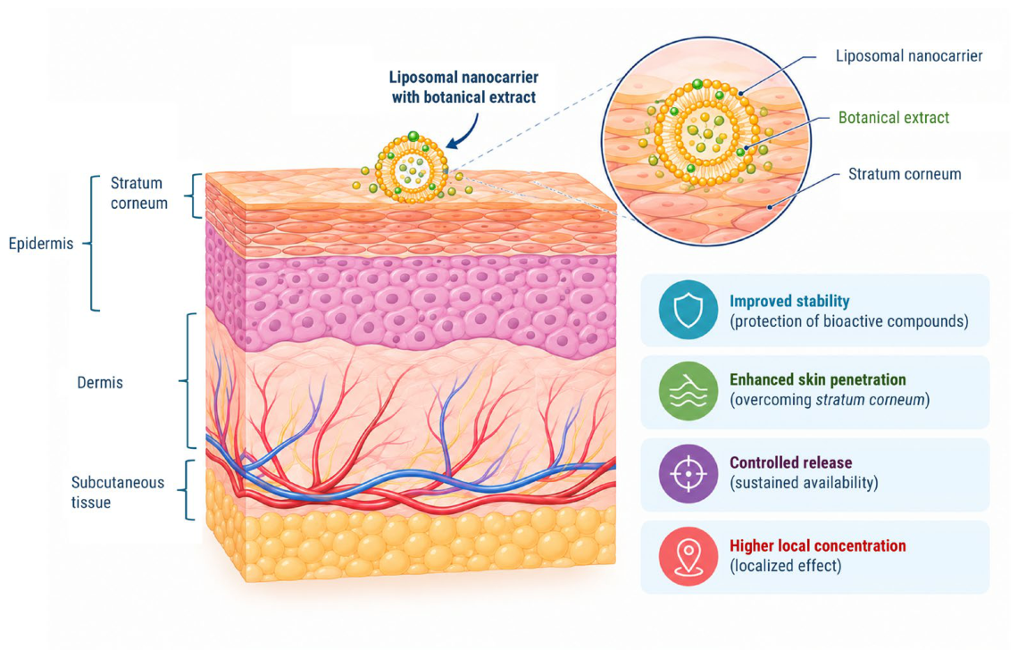
Figure 2.
PRISMA flow diagram showing the number of studies included in the present review.
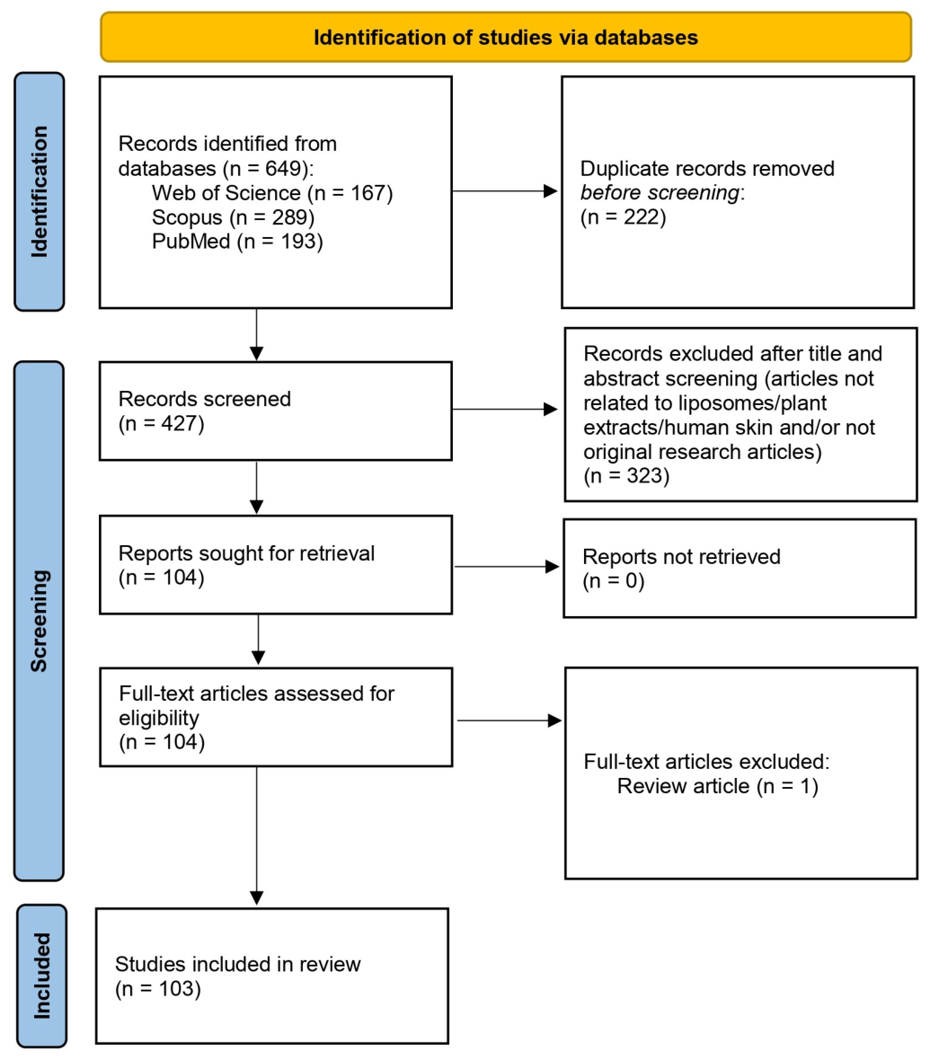
Figure 3.
Liposomal nanoformulations used for botanical extract encapsulation covered in this review.
Figure 3.
Liposomal nanoformulations used for botanical extract encapsulation covered in this review.
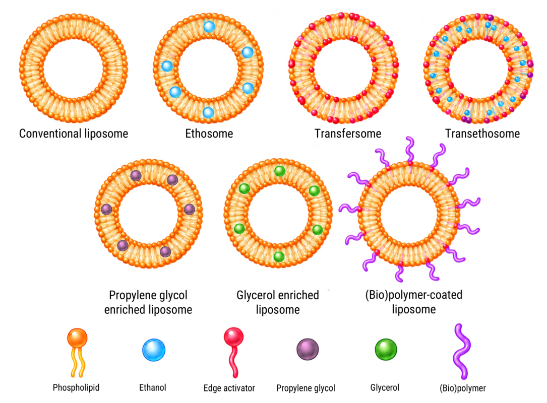
Figure 4.
Reported skin-related biological activities associated with the use of botanical-extract-loaded liposomes.
Figure 4.
Reported skin-related biological activities associated with the use of botanical-extract-loaded liposomes.
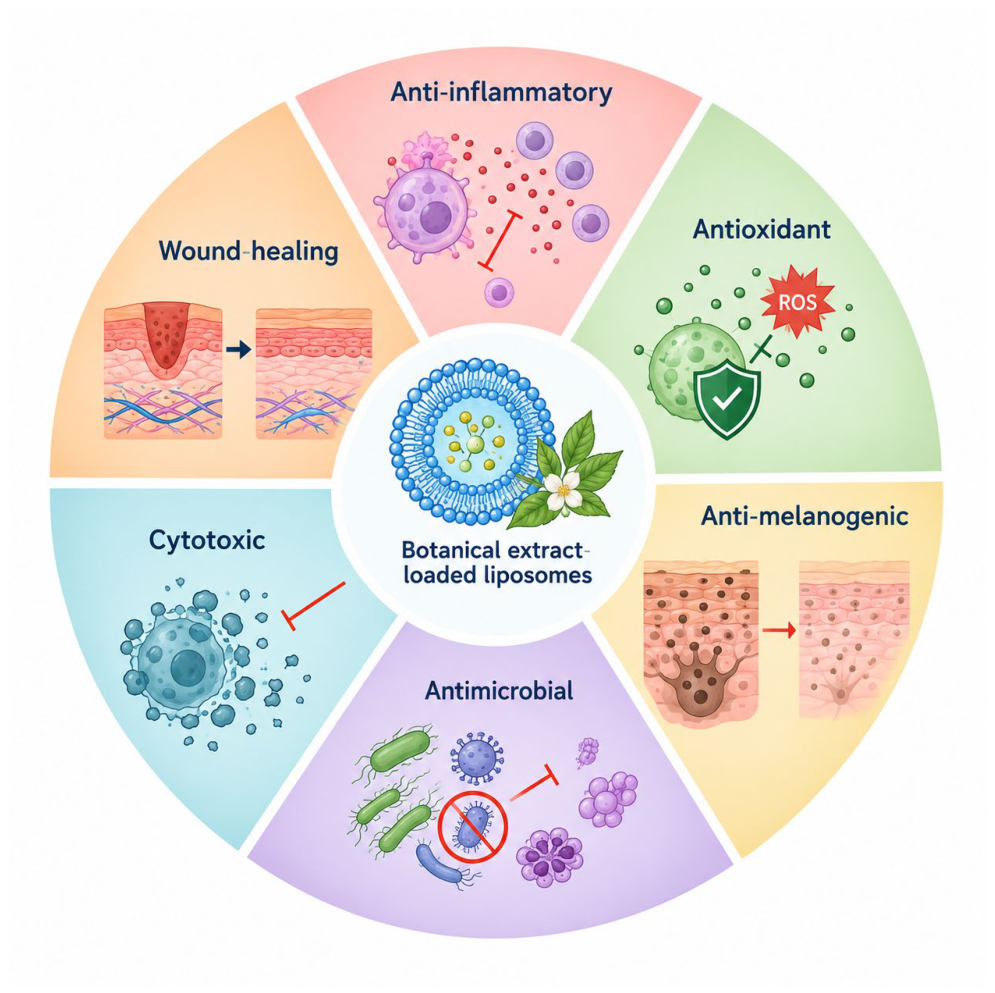
Figure 5.
Most represented plant families based on the number of individual taxa that were used in the preparation of botanical-extract-loaded liposomal nanoformulations reported in the articles included in this review.
Figure 5.
Most represented plant families based on the number of individual taxa that were used in the preparation of botanical-extract-loaded liposomal nanoformulations reported in the articles included in this review.
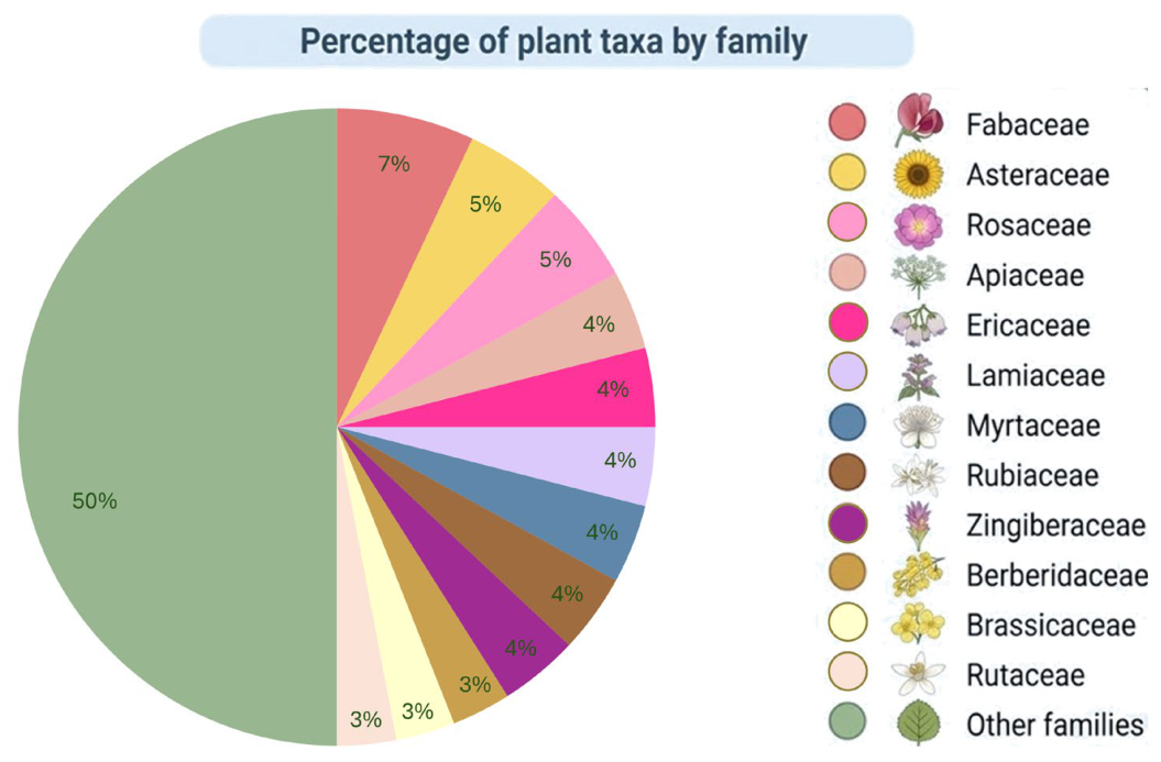
Figure 6.
Representation of geographical origin and natural biogeographical regions based on the number of individual taxa that were used in the preparation of botanical-extract-loaded liposomal nanoformulations reported in the articles included in this review.
Figure 6.
Representation of geographical origin and natural biogeographical regions based on the number of individual taxa that were used in the preparation of botanical-extract-loaded liposomal nanoformulations reported in the articles included in this review.
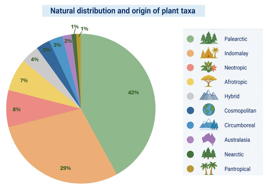

Table 1.
Liposomal nanoformulations used as botanical extract carriers.
|
Liposome Class |
Botanical Extract Source |
Liposome Preparation Method |
Liposome Bilayer Constituents |
Semisolid Vehicle |
Refs |
|
Conventional Liposomes |
Abelmoschus esculentus | TLFH method | PC, chol | cream mask | [59] |
| Aloe vera | microfluidization | SLP-WHITE | gel (Aloe vera) | [60] | |
|
Conventional Liposomes |
Angelica sinensis, Astragalus mongholicus |
TLFH method | PC, chol | gel (Poloxamer P407, Poloxamer P188, HPMC) | [61] |
| Annona squamosa | TLFH method | DPPC | − | [62] | |
| Aristolochia clematitis | TLFH method | PC or PS, chol | − | [11] | |
| Azadirachta indica | TLFH method | PC, chol | gel (Carbopol 934K) | [63] | |
| Boswellia serrata | ethanol injection or reverse-phase evaporation method |
PC, chol | − | [58] | |
| Calendula officinalis | TLFH method | PC | gel (chitosan/Aloe vera) |
[64] | |
| Carpobrotus edulis | TLFH method | HPC, DCP, chol | − | [65] | |
| Caryocar brasiliense | microfluidization sonication |
PC, chol | gel (ammonium/ acryloyldimethyl- taurate/VP copolymer) |
[66] | |
| Centella asiatica | TLFH method | PC, chol | − | [67] | |
| Ceratonia siliqua | ultrasonication method | PC | − | [68] | |
| Cinnamomum verum | TLFH method | PC, chol | gel (xanthan gum, hyaluronan) |
[69] | |
|
Citrus × aurantium f. aurantium, Glycyrrhiza glabra |
TLFH method | PC, chol, stearylamine | gel (Carbopol 934) | [47] | |
| Citrus × junos | microfluidization | PC, chol, squalene, caprylic/capric triglyceride, glycerol |
− | [40] | |
|
Clitoria ternatea, Syzygium aromaticum |
proliposome method | commercial Pro-liposome (Lipobelle® ST) |
− | [70] | |
| Coffea arabica | TLFH method | DOPC, DOTAP or DOPC, DDAB |
− | [45] | |
| Coffea canephora | TLFH method | PC, chol, PEG 400 | cream base | [25] | |
|
Curcuma longa, Arnica montana |
TLFH method | PC, chol, stearic acid | cream base | [71] | |
| Fumaria officinalis | proliposome method | PC, β-sitosterol or ergosterol | − | [55] | |
| Garcinia mangostana | TLFH method | PC, chol | TiO2 cream base | [72] | |
|
Labisia pumila, Ficus deltoidea |
TLFH method | PC | − | [73] | |
|
Lavandula austroapennina |
TLFH method | PC, chol | − | [9] | |
|
Conventional Liposomes |
Malus hupehensis | TLFH method | PC, chol | − | [54] |
| Matricaria chamomilla | n.d. | PC | gel (n.d.) | [74] | |
| Matricaria chamomilla | n.d. | PC | gel (n.d.) | [75] | |
| Myrtus communis | ultrasonication method | PC | − | [76] | |
| Paeonia tenuifolia | proliposome method | Phosal SA, Phosal MCT 53, PC | film (CMC) | [77] | |
| Panax sp. | solvent injection method | PC, chol | gel (gelatine, PVA, hyaluronic acid, CMCh) |
[1] | |
| Petroselinum crispum | TLFH method | PC, chol | − | [78] | |
| Piper longum | TLFH method | EPC, chol | − | [56] | |
| Pistacia vera | ultrasonication method | PC | − | [79] | |
| Plantago major | ethanol injection method | PC | − | [80] | |
| Platanus orientalis | TLFH method | PC, chol | − | [26] | |
| polyherbal extract * | TLFH method | PC | − | [81] | |
|
Rhododendron ferrugineum |
n.d. |
commercial PhytoCellTec™ Alp Rose |
cream | [82] | |
| Rosa canina | proliposome method | PC | − | [57] | |
| Rosa × damascena | ultrasonication method | PC | gel (n.d.) | [83] | |
| Serenoa repens | TLFH method | PC | − | [84] | |
| Solanum lycopersicum | TLFH method | PC, chol | − | [6] | |
| Stellaria media | TLFH method | PS, chol | − | [85] | |
|
Vaccinium vitis-idaea |
TLFH method | PC, chol | gel (Poloxamer 188, Poloxamer 407) |
[53] | |
| Ethosomes | Achillea millefolium | classical cold method | PC, ethanol, glycerol, PG |
gel (Carbopol 940) | [86] |
|
Arctostaphylos uva-ursi |
modified cold method | PC, ethanol, PG | gel (Carbopol 940) | [87] | |
| Armoracia rusticana | ultrasonication method | PC, ethanol | − | [88] | |
| Azadirachta indica | classical cold method | PC, ethanol, PG | gel (Carbopol 934K) | [89] | |
| Bacopa monnieri | classical cold method | PC, ethanol | − | [90] | |
| Ethosomes | Berberis aristata | modified cold method | PC, ethanol | gel (Carbopol 934P) | [91] |
| Brassica oleracea | modified cold method | PC, chol, ethanol, PG | gel (Carbopol 940) | [92] | |
| Curcuma heyneana | ethanol injection method | PC, chol, ethanol | − | [93] | |
|
Curcuma longa, Dalbergia sissoo |
TLFH method | PC, ethanol | gel (Carbopol 934K) | [94] | |
| Dalbergia sissoo | modified cold method | PC, ethanol, PG | gel (Carbopol 934K) | [95] | |
| Euphorbia characias | ultrasonication method | PC, ethanol | − | [96] | |
|
Croton tiglium, Phyllanthus niruri, Zingiber officinale |
classical cold method | PC, ethanol | − | [97] | |
| Plumbago zeylanica | TLFH method | PC, ethanol | gel (Carbopol 974P) | [98] | |
| polyherbal extract ** | ultrasonication method | PC, ethanol | gel (Carbopol 940) | [99] | |
| Polyscias scutellaria | TLFH method | PC, ethanol, PG | gel (Carbomer) | [100] | |
| Rosa canina | ethanol injection method | HPC, ethanol,glycerol or allantoin | − | [101] | |
| Rosa canina | ethanol injection method | PC, ethanol | − | [102] | |
| Rosa canina | ethanol injection method | PC, ethanol | gel (hyaluronic acid) | [103] | |
| Sesamum indicum | classical cold method | PC, ethanol | gel (Carbopol 934) | [104] | |
| Sesbania grandiflora | modified cold method | PC, ethanol, glycerol, PEG, castor oil |
gel (Carbomer) | [105] | |
| Syzygium aromaticum | modified cold method | PC, ethanol, PG | gel (Carbopol 934K) | [106] | |
| Zingiber zerumbet | classical cold method | PC, ethanol, PEG 4000 |
− |
[107] | |
| Transfersomes | Althaea officinalis | TLFH method | PC, SCh | gel (Carbopol 934) | [108] |
| Berberis aristata | TLFH method | PC, Tween 80 or Span 80 or SDCh | gel (Carbopol 934P) | [109] | |
| Camellia sinensis | TLFH method | PC, Span 80 | cream base | [110] | |
| Capsicum chinense | TLFH method | PC, Tween 80 | − | [111] | |
| Centella asiatica | hot high-speed homogenization method (Mozafari method) |
PC, chol, Span 80, Tween 80, PG, tocopheryl acetate |
gel (ammonium/ acryloyldimethyl- taurate/VP copolymer) |
[112] | |
| Eulophia macrobulbon | TLFH method | PC, Tween 80, Span 80 | gel (Carbopol Ultrez-21) |
[43] | |
| Transfersomes | Lantana camara | TLFH method | DSPC, chol, Span 80, Tween 80 | − | [42] |
| Morus alba | TLFH method | PC, Tween 80 | gel (Carbopol 940) | [113] | |
| Neolamarckia cadamba | TLFH method | PC, chol, Tween 80 | gel (Carbopol 940) | [114] | |
| Piper longum | TLFH method | PC, Tween 80 | gel (xanthan gum) | [44] | |
| Pistacia vera | ultrasonication method | PC, Tween 80 | − | [115] | |
| Raphanus raphanistrum subsp. sativus | TLFH method | PC, chol, Tween 80 | emulgel (Carbopol 940) |
[116] | |
| Rhodomyrtus tomentosa | TLFH method | PC, SDCh | gel (Carbopol Ultrez-21) |
[117] | |
| Rotheca serrata | TLFH method | PC, SDCh/Tween 80 | − | [118] | |
| Sonneratia caseolaris | TLFH method | Lipoid Ultraspheres®, Lipoid Phytosolve®, Tween 20 |
− | [46] | |
| Stellaria media | microfluidization | PC, STDCh | − | [119] | |
| Teucrium marum | ultrasonication method | PC, Tween 80 | − | [120] | |
| Vaccinium myrtillus | TLFH method | SPC, NaCh | gel (Carbopol 940) | [121] | |
| Vaccinium vitis-idaea | TLFH method | PC, chol, Tween 80 | gel (Poloxamer 407, Poloxamer 88) | [53] | |
| Vitis vinifera | ultrasonication method | PC, Labrasol® | − | [122] | |
| Vitis vinifera | ultrasonication method | PC, Tween 80 | − | [123] | |
| Vitis vinifera | ultrasonication method | PC, Montanov® 82 | − | [124] | |
| Trans-ethosomes |
Chenopodiastrum murale |
ethanol injection method | PC, chol, ethanol, Tween 80 | − | [125] |
| Curcuma longa | modified cold method | PC, ethanol, Tween 80, SLS | − | [126] | |
| Piper longum | TLFH method | PC, chol, ethanol, Tween 80 | gel (Carbopol 940) | [127] | |
| Rubia cordifolia | modified cold method | PC, SDCh, ethanol | gel (Carbopol 940, gellan gum) |
[128] | |
| Vitis vinifera | ultrasonication method | PC, Labrasol®, ethanol |
− | [122] | |
|
Propylene Glycol-Enriched Liposomes (PGLs) |
Curcuma heyneana | high-speed homogenization method |
PC, PG | gel (n.d.) | [129] |
| Onopordum illyricum | ultrasonication method | PC, PG | − | [15] | |
|
Propylene Glycol-Enriched Liposomes (PGLs) |
Prunus spinosa | ultrasonication method | PC, PG | − | [14] |
| Vitis vinifera | ultrasonication method | PC, PG | − | [130] | |
| Vitis vinifera | ultrasonication method | PC, PG | − | [131] | |
| Glycerosomes | Citrus × limon | ultrasonication method | PC, glycerol | − | [132] |
| Eryngium maritimum | microfluidization | HPC, squalene, glycerol, caprylic/ capric triglycerides |
− | [133] | |
| Hypericum scruglii | ultrasonication method | PC, glycerol, dextrin (Glucidex) |
− | [134] | |
| Lavandula angustifolia | ultrasonication method | PC, glycerol | − | [135] | |
| Prinsepia utilis | modified cold method | HSPC, glycerol, PG | − | [136] | |
| Teucrium marum | ultrasonication method | PC, glycerol | − | [120] | |
| Vitis vinifera | ultrasonication method | PC, Montanov® 82, glycerol | − | [124] | |
|
(Bio)polymer-Coated Liposomes |
Centella asiatica | TLFH method | HSPC, chol, DSPE-PEG 2000, butylene glycol | − | [41] |
| Citrus × limon | ultrasonication method | PC, sodium hyaluronate | − | [132] | |
| Lavandula angustifolia | ultrasonication method | PC, sodium hyaluronate | − | [135] | |
| Lavandula angustifolia | ultrasonication method | PC, sodium hyaluronate | − | [10] | |
| Plinia cauliflora | ultrasonication method | PC, Tween 80, HEC or sodium hyaluronate | − | [8] | |
| Prunus spinosa | TLFH method | PC, sodium hyaluronate | − | [14] | |
| Sapindus saponaria | TLFH method, chitosan coating |
SP, stigmasterol, chitosan | − | [137] | |
| Teucrium marum | ultrasonication method | PC, sodium hyaluronate | − | [120] | |
| Vitis vinifera | ultrasonication method | PC, Tween 80, sodium hyaluronate |
− | [123] | |
|
Glycero- hyalurosomes |
Citrus × limon | ultrasonication method | PC, glycerol, sodium hyaluronate |
− | [132] |
| Hypericum scruglii | ultrasonication method | PC, glycerol, dextrin (Glucidex), sodium hyaluronate or gelatine | − | [134] | |
| Lavandula angustifolia | ultrasonication method | PC, glycerol, sodium hyaluronate |
− | [135] | |
| Teucrium marum | ultrasonication method | PC, Tween 80, glycerol, sodium hyaluronate |
− | [120] |
* Berberis fortunei, Berberis integerrima, Betula pendula, Catharanthus roseus, Convolvulus sepium, Melia azedarach, Narcissus tazetta, Plantago major; **Basella alba, Dicliptera paniculata, Lawsonia inermis, Portulaca oleracea, Trigonella foenum-graecum; The abbreviations stand for: chol – cholesterol, CMC – carboxymethyl cellulose, CMCh – carboxymethyl chitosan, DCP – dicetyl phosphate, DDAB – dimethyldioctadecylammonium bromide, DOPC – 1,2-dioleoyl-sn-glycero-3-phosphocholine, DOTAP – 1,2-dioleoyl-3-trimethylammonium-propane, DPPC – 1,2-dipalmitoyl-sn-glycero-3-phosphocholine, DSPC – 1,2-distearoyl-sn-glycero-3-phosphocholine, DSPE – distearoylphosphatidylethanolamine, DSPE-PEG 2000 – 1,2-distearoyl-sn-glycero-3-phosphatidylethanolamine-N-[methoxy(polyethylene glycol)-2000], HEC – hydroxyethyl cellulose, HPC – hydrogenated phosphatidylcholine, HPMC – hydroxypropyl methylcellulose, HSPC – hydrogenated soybean phosphatidylcholine, n.d. – not defined, PC – phosphatidylcholine, PG – proylene glycol, PS – phosphatidylserine, PVA – polyvinyl alcohol, SCh – sodium cholate, SDCh – sodium deoxycholate, STDCh – sodium taurodeoxycholate, TLFH – thin lipid film hydration.
Table 2.
Phytochemical and biological activity profiles and skin-related effects of plant-extract-loaded liposomal nanoformulations.
Table 2.
Phytochemical and biological activity profiles and skin-related effects of plant-extract-loaded liposomal nanoformulations.
|
Plant Taxa and Plant Parts |
Extract Type and Extraction Method |
Analytical Characterization and Main Compounds |
Biological Activities and Skin-Related Effects |
Refs |
|
Abelmoschus esculentus seeds |
commercial biotransformed seed oligopeptide complex (MYOXINOL LS 9736) |
no chromatographic analysis | in vitro inhibition of aging-related enzymes (collagenase, elastase, hyluronidase) | [59] |
|
Achillea millefolium aerial parts |
70% hydroethanolic extract; maceration | UV-Vis: total phenolic content, UV/FTIR functional-group characterization; no chromatographic analysis |
in vitro antioxidant activity; ex vivo rat skin permeation |
[86] |
|
Aloe vera inner leaf gel |
mechanical separation from leaves | no chromatographic analysis |
in vivo depigmenting and anti-melanogenic effect in pregnant women (clinical trial) |
[60] |
|
Aloe vera gel, Calendula officinalis unknown plant part |
commercial calendula extract plus aloe gel | UV/FTIR functional-group characterization; no chromatographic analysis |
ex vivo rat skin permeation; in vitro skin cell biocompatibility |
[64] |
|
Althaea officinalis roots |
lyophilization | no chromatographic analysis |
in vitro antioxidant activity; ex vivo rat skin permeation |
[108] |
|
Angelica sinensis and Astragalus mongholicus roots |
aqueous extract; reflux extraction and alcohol precipitation |
HPLC-DAD: ferulic acid and calycosin-7-O-glucoside |
in vivo wound healing in rats | [61] |
|
Annona squamosa leaves |
80% methanolic extract; solvent extraction |
GC–MS: limonene and R-carvone |
ex vivo rat skin permeation | [62] |
|
Arctostaphylos uva-ursi leaves |
50% hydroetanolic extract; maceration |
no chromatographic analysis |
in vitro antioxidant activity; in vitro tyrosinase inhibiton; in vitro skin cell biocompatibility; ex vivo rat skin permeation; in vivo anti-melanogenic, anti-erythema, skin hydration, skin rejuvenation and sebum reduction effect in humans (clinical trial) |
[87] |
|
Aristolochia clematitis leaves |
70% hydroethanolic extract; maceration | HPLC-MS: isoquercetin, ferulic acid, rutin, p-coumaric acid, protocatechuic acid, epigallocatechin gallate, vanillic acid, epicatechin |
in vitro antioxidant and antimicrobial activity |
[11] |
|
Armoracia rusticana roots |
70% hydroethanolic extract; maceration and ultrasonication | UPLC-MS, HPLC-PDA: glucosinolates, phenolic acids, kaempferol |
in vitro antioxidant activity; in vitro skin cell biocompatibility |
[88] |
| Arnica montana flowers and Curcuma longa rhizomes | 30–50% hydro- ethanolic extracts; homogenizer-assisted extraction |
FTIR functional group characterization; UV-Vis: total phenolic and flavonoid content |
ex vivo goat skin permeability; in vitro antioxidant activity; in vivo cold injury healing and skin irritancy in rats |
[71] |
|
Azadirachta indica leaves |
ethanolic extract; maceration |
FTIR functional-group characterization |
in vitro antifungal activity against Aspergillus niger and Candida tropicalis |
[63] |
|
Azadirachta indica leaves |
ethanolic extract; Soxhlet extraction | FTIR functional-group characterization |
in vitro antifungal activity against Candida parapsilosis and A. niger; ex vivo rat skin permeation |
[89] |
|
Bacopa monnieri unknown plant part |
commercial extract; method not reported |
HPLC: bacoside A and FTIR functional-group characterization |
ex vivo goat skin permeation |
[90] |
|
Basella alba, Dicliptera paniculata, Lawsonia inermis, Portulaca oleracea, Trigonella foenum-graecum unknown plant part |
70% ethanolic extracts; maceration |
UV-Vis: total phenolic, flavonoid, alkaloid and tannin content, UV/FTIR functional-group characterization |
in vitro skin cell biocompatibility and skin cell proliferation stimulation |
[99] |
|
Berberis aristata roots |
70% hydroethanolic extract; maceration | HPTLC: berberine | none reported | [91] |
|
Berberis aristata roots |
ethanolic extract; method not reported |
HPTLC: berberine |
in vivo anti-psoriatic activity on imiquimod-induced psoriasis-like mouse skin; in vivo anti-inflammatory activity in carrageenan-induced paw edema |
[109] |
|
Berberis fortunei, Berberis integerrima, Betula pendula, Convolvulus sepium, Catharanthus roseus, Melia azedarach and Plantago major leaves and Narcissus tazetta bulbs |
80% ethanolic and methanolic extracts; maceration |
no chromatographic analysis | in vitro skin cell biocompatibility | [81] |
|
Boswellia serrata unknown plant part |
frankincense oil (extract); method not reported |
HPLC: 3-acetyl-11-keto-β-boswellic acid |
in vitro anti-inflammatory activity; in vitro skin cell biocompatibility; in vivo anti-psoriatic activity in mice |
[58] |
|
Brassica oleracea red-cabbage leaves |
80% hydroethanolic extract; maceration |
HPLC: ascorbic acid |
in vitro antioxidant and in vivo melanin, sebum, erythema, pore, and wrinkle reduction, and increase in skin hydration, elasticity, and smoothness in humans (clinical trial) |
[92] |
|
Camellia sinensis leaves |
commercial extract; method not reported |
RP-HPLC: epigallocatechin gallate |
ex vivo rat-skin penetration in Franz cells |
[110] |
|
Capsicum chinense fruits |
ethanolic extract; sonication and maceration |
HPLC: capsaicin | in vivo anti-inflammatory activity in rats | [111] |
|
Carpobrotus edulis fresh leaves |
aqueous extract; homogenization and lyophilization |
no chromatographic analysis | in vivo incisional and excisional wound healing in rats | [65] |
|
Caryocar brasiliense fruit pulp |
cold extraction method | no chromatographic analysis | split-face study in ten healthy women: hydration, TEWL (clinical trial) |
[66] |
|
Centella asiatica unknown plant part |
commercial extract; method not reported |
HPLC-UV: asiaticoside; asiatic and madecassic acids |
ex vivo human cadaver-skin penetration |
[41] |
|
Centella asiatica unknown plant part |
95% ethanolic extract; maceration |
individual triterpenoid constituents not quantitatively standardized in the reported extract methods |
in vitro anti-inflammatory activity; in vitro skin cell biocompatibility, migration and proliferation; in vivo wound healing in rats |
[67] |
|
Centella asiatica leaf |
methanolic extract; maceration | UHPLC: pentacyclic triterpenes (asiaticoside, madecasoside) |
ex vivo pig skin permeation; in vitro skin cell biocompatibility and migration/proliferation; in vitro collagen production promotion; in vitro anti-inflammatory; in vivo wound healing in rats |
[112] |
|
Ceratonia siliqua ripe pods |
70% hydroethanolic extract; sonication and maceration |
HPLC-ESI-QToF-MS/MS and HPLC-PDA: gallic-acid derivatives (gallotannins) and quercetin-3-O-rhamnoside |
in vitro cell-free and cellular antioxidant activity |
[68] |
|
Chenopodiastrum murale aerial parts |
ethanolic extract; maceration |
UV-Vis: total phenolic, flavonoid, and steroid content; UPLC, UV/FTIR functional group characterization; quercetin and gallic acid |
ex vivo rat skin permeation | [125] |
|
Cinnamomum verum unknown plant part |
99% ethanolic extract; maceration |
GC–MS: cinnamaldehyde |
in vitro antibacterial activity; in vivo wound healing in rats |
[69] |
|
Citrus × aurantium f. aurantium fruit peel and Glycyrrhiza sp. roots |
methanolic extracts; maceration |
no chromatographic analysis | in vitro antioxidant activity | [47] |
|
Citrus × junos callus derived from leaf, flower and seed explants |
aqueous extract; decoction |
HPLC-PDA/LC-MS/NMR: p-hydroxycinnamoylmalic acid |
in vitro anti-tyrosinase and anti-melanogenic activity, in vitro fibroblast proliferation, in vitro anti-wrinkle wound healing activity; in vitro permeation (artificial biomimetic membrane Strat M®) |
[40] |
|
Citrus × limon fresh fruits |
50% hydroethanolic extracts; ultrasound-assisted extraction |
UV-Vis: total phenolic content; HPLC-DAD, LC-MS/MS: naringin, (neo)eriocitrin, neohesperidin, isoquercetin, qunic acid, robinin, rutin, sinapic acid, myricitrin, ferulic acid, phlorizin |
in vitro antioxidant activity; in vitro skin cell biocompatibility |
[132] |
| Clitoria ternatea flower and Syzygium aromaticum flower buds | 70% hydroethanolic extract (Clitoria ternatea); maceration; commercial S. aromaticum clove oil |
no chromatographic analysis | in vivo eyebrow growth and skin irritation test in humans | [70] |
|
Coffea arabica leaves |
freeze-dried extract; extraction conditions not reported |
HPLC-DAD-UV/ESI-LIT-MS: chlorogenic acids, 5-caffeoylquinic acid, 3,5-dicaffeoylquinic acid; mangiferin, trigonelline and caffeine |
in vitro skin cell biocompatibility | [45] |
|
Coffea canephora plant-cell culture |
aqueous extract; ultrasonication and lyophilization |
no chromatographic analysis | in vivo anti-inflammatory activity, wound healing, neoangiogenesis and necollagenesis in rats | [25] |
|
Croton tiglium seeds; Phyllanthus niruri aerial parts; Zingiber officinale rhizomes |
hydroethanolic extracts; Soxhlet extraction/maceration |
GC-MS, HPTLC, UV-Vis: total flavonoid content; saponins, flavonoids, terpenoids, alkaloids, steroids | in vitro skin cell biocompatibility; ex vivo safety histopathological study in rats | [97] |
|
Curcuma heyneana rhizomes |
ethanolic extract, microwave-assisted extraction |
no chromatographic analysis | in vivo skin penetration and UV exposure in rats | [93] |
|
Curcuma heyneana rhizomes |
extraction procedure not reported | no phytochemical analysis. | in vivo skin penetration in rats | [129] |
|
Curcuma longa rhizomes |
aqueous, 70% methanolic and ethanolic extract; cold percolation |
RP-HPLC: curcumin, demethoxycurcumin, bisdemethoxycurcumin, total curcuminoid content |
in vivo wound healing in rats | [126] |
|
Curcuma longa and Dalbergia sissoo unknown plant part |
commercial extracts; methods not reported |
UV-Vis: dalbergin and curcumin | in vivo anti-inflammatory activity and wound healing in rats | [94] |
|
Dalbergia sissoo leaves |
commercial extract; solvent and extraction procedure not reported |
UV-Vis: total phenolic and flavonoid content; FTIR functional-group characterization |
in vitro antioxidant activity | [95] |
| Eryngium maritimum seed-derived callus culture | concentrated callus medium from a bioreactor culture; filtration |
untargeted LC-MS: lipids, organoheterocyclic compounds, organic acids and benzenoids |
in vitro skin cell migration/ proliferation; in vitro collagenesis; in vitro antioxidant activity and cytoprotection against UVB and oxidative stress; in vitro antimicrobial activity; in vivo improved dermal density, hydration, elasticity and reduction in skin sagging and wrinkles in humans (clinical trial) |
[133] |
|
Eulophia macrobulbon rhizomes |
extraction procedure not reported | HPLC: phenanthrene compounds | in vitro permeation (artificial biomimetic membrane Strat M®) | [43] |
|
Euphorbia characias aerial parts |
methanolic and hydroethanolic extracts; maceration |
HPLC-DAD: quercetin glucosides |
in vitro inhibition of aging-related enzymes (collagenase, elastase, hyluronidase); in vitro sun protection factor; in vitro antioxidant activity; in vitro anti-melanogenic activity; in vitro skin cell biocompatibility |
[96] |
| Ficus deltoidea leaves; Labisia pumila aerial parts; | extraction procedure not reported | no chromatographic analysis | ex vivo rat skin permeation | [73] |
|
Fumaria officinalis unknown plant part |
70% ethanolic extract; microwave-assisted extraction |
UV-Vis: total phenolic content |
in vitro antioxidant activity |
[55] |
|
Garcinia mangostana fruit pericarp |
extraction procedure not reported | no chromatographic analysis |
in vivo photoprotective effect against UVB-induced erythema in humans (clinical trial) |
[72] |
|
Hypericum scruglii aerial parts |
70% ethanolic extract; maceration |
HPLC-DAD: chlorogenic acid and quercetin; UV-Vis: total phenolic content |
in vitro cytoprotection against oxidative stress; in vitro skin cell migration/proliferation |
[134] |
|
Lantana camara leaves |
ethanolic extract; orbital-shaker- assisted extraction |
FTIR functional-group characterization |
ex vivo goat skin permeation; in vitro cytotoxic activity against a skin-cancer cell line |
[42] |
|
Lavandula angustifolia cell-suspension biomass |
50% methanolic extract; ultrasound- assisted extraction |
NMR/HPLC: rosmarinic acid |
in vitro cell-free and cellular antioxidant protection, cellular proliferation |
[10] |
|
Lavandula angustifolia cell-suspension biomass |
50% methanolic extract; ultrasound- assisted extraction |
NMR/HPLC: rosmarinic acid |
in vitro skin cell biocompatibility; in vitro cytoprotection against oxidative damage; in vitro skin cell migration/ proliferation stimulation; in vitro antiviral cytoprotection |
[135] |
| Lavandula austroapennina leaves and stems | methanolic extract; ultrasound-assisted extraction |
UHPLC-ESI-QqTOF-MS/MS: ferulic acid, oleanolic acid, linoleic acid, oleic acid, palmitic acid |
in vitro skin cell biocompatibility; in vitro skin cell migration/ proliferation stimulation; ex vivo pig skin permeation |
[9] |
|
Malus hupehensis leaves |
70% ethanolic extract; reflux extraction |
UV-Vis: total flavonoid content | ex vivo rat skin permeation | [54] |
|
Matricaria chamomilla flowers |
glycolic extract; method not reported |
no phytochemical analysis | in vivo prevention of radiation dermatitis in breast cancer patients (clinical trial) | [74] |
|
Matricaria chamomilla unknown plant part |
glycolic extract; method not reported |
no phytochemical analysis | in vivo prevention of radiation dermatitis in head and neck cancer patients (clinical trial) | [75] |
|
Morus alba leaves |
70% ethanolic extract; Soxhlet extraction followed by freeze-drying |
HPTLC: quercetin; formulation compatibility analysis |
in vitro antioxidant activity; ex vivo skin delivery |
[113] |
|
Myrtus communis purple berries |
96% ethanolic extract; ultrasound-assisted extraction |
HPLC-DAD: anthocyanins (delphinidin, cyanidin, malvidin 3-O-glucosides, petunidin-3-O-glucoside, and peonidin-3-O-glucoside), myricetin glycosides, gallic acid and ellagic acid |
in vitro antioxidant activity; in vitro cytoprotection against oxidative damage |
[76] |
|
Neolamarckia cadamba leaves |
75% hydroethanolic extract; maceration |
qualitative screening: alkaloids, flavonoids, terpenoids, tannins, cardiac glycosides and saponins |
in vitro antibacterial and antifungal activity |
[114] |
|
Onopordum illyricum leaves |
80% ethanolic extract; maceration |
HRLC-ESI-QTOF-MS/MS and HPLC-DAD: luteolin glucuronide, 1,5-dicaffeoylquinic acid and other caffeoylquinic acid derivatives; hydroxycinnamic acids and flavonols |
in vitro antioxidant activity; in vitro cytoprotection against oxidative damage |
[15] |
|
Paeonia tenuifolia petals |
methanolic extract; maceration |
UV-Vis: total phenolic content, FTIR functional-group characterization; no chromatographic analysis |
in vitro antimicrobial activity | [77] |
| Panax sp. | aqueous extract; decoction |
TLC; FTIR functional-group characterization; HPLC: ginsenosids |
in vitro skin cell proliferation stimulation; in vitro depigmentation and melanogenesis inhibition; in vitro antioxidant activity; in vitro antibacterial activity; in vivo tissue repair, anti-erythema, dermal collagen regeneration and anti-inflammatory effect in mice |
[1] |
|
Petroselinum crispum leaves |
absolute ethanolic extract; maceration followed by fractionation with ethyl acetate |
LC-MS: furanocoumarins (xanthotoxin and aviprin) |
in vitro antifungal activity | [78] |
|
Piper longum fruits |
ethanolic extract | no chromatographic analysis | in vitro cytotoxic activity against skin-cancer cell lines; ex vivo mouse skin permeation; in vivo anti-melanoma effect in mice | [44] |
|
Piper longum fruits |
absolute ethanolic extract; microwave-assisted extraction followed by triple maceration |
validated HPLC: piperine and piperlonguminine; HPTLC: piperine |
in vitro cytotoxic activity against skin-cancer cell lines | [56] |
|
Piper longum fruits |
aqueous ethanolic extract; maceration |
no chromatographic analysis | in vitro antimicrobial activity | [127] |
|
Pistacia vera fresh fruits |
methanolic extract; extraction and lyophilization |
HPLC-DAD: gallic acid, pyrogallol, catechin, myricetin, naringin, quercetin and rutin |
in vitro antioxidant activity; in vitro anti-inflammatory activity; in vitro anti-tyrosinase activity |
[79] |
|
Pistacia vera fresh stalks |
essential oil and hydrolate; steam distillation |
LC-Orbitrap/GC–MS: naringenin, catechin, myristic acid and α-pinene-rich essential oil |
in vitro antioxidant activity; in vitro cytoprotection against oxidative damage |
[115] |
|
Plantago major leaves |
aqueous and aqueous ethanolic extracts; ultrasound-assisted extraction |
no chromatographic analysis |
in vitro antioxidant activitiy; in vitro skin cell biocompatibility; in vitro antimicrobial activity |
[80] |
|
Platanus orientalis leaves |
ethanolic extract; maceration |
LC-HRMS: chlorogenic acid, hyperoside, quercitrin and quercetin |
in vitro skin cell biocompatibility; in vitro skin cell migration/ proliferation |
[26] |
|
Plinia cauliflora fruit peel |
acidified aqueous extract; pressurized liquid extraction |
UHPLC-QTOF-MS, HPLC-DAD: delphinidin-3-glucoside, cyanidin-3-glucoside, gallic acid, ellagitannins |
in vitro antioxidant activity; in vitro skin cell biocompatibility; in vitro skin cell migration/proliferation |
[8] |
|
Plumbago zeylanica roots |
70% hydroethanolic extract; Soxhlet extraction |
UV-Vis: plumbagin (9.6% w/w) | in vitro antimicrobial activity against acne-associated bacteria | [98] |
|
Polyscias scutellaria leaves |
chloroformic extract, ethyl acetate fraction; maceration | no phytochemical analysis |
ex vivo mouse skin permeation; in vivo hair growth activity in rabbits |
[100] |
|
Prinsepia utilis seeds |
oil; extraction procedure not reported | HPLC: oleic acid |
in vivo protection against UVB-induced skin damage in mice |
[136] |
|
Prunus spinosa fruits |
70% hydroethanolic extract; maceration | LC-MS, HPLC-DAD: quercetin, vanillic acid, caffeine acid derivatives, cyanidin glucosides, peonidin glucosides |
in vitro antioxidant activity; in vitro antibacterial activity; in vitro skin cell biocompatibility |
[14] |
|
Raphanus raphanistrum subsp. sativus sprouts |
extraction procedure not reported | UV-Vis: total phenolic content |
in vitro anti-tyrosinase, in vitro sun protection factor and UVA photoprotection |
[116] |
|
Rhododendron ferrugineum leaf-derived cell culture |
commercial liposomal cell-culture preparation |
no chromatographic analysis |
in vivo skin hydration/skin barrier preservation and accelerated barrier recovery/rehydration of irritated skin in humans (clinical trial) |
[82] |
| Rhodomyrtus tomentosa leaves | 95% ethanolic extract; maceration |
HPLC-PDA: rhodomyrtone |
in vitro antibacterial activity; in vitro anti-inflammatory and anti-melanogenic (tyrosinase inhibition) activity; in vitro skin cell biocompatibility |
[117] |
|
Rosa canina dried rosehip shells |
70% ethanolic extract; percolation |
HPLC, UV-Vis: total phenolic content; chlorogenic acid, rutin, hyperoside, isoquercetin and quercitrin |
in vitro antioxidant activity before and after UV treatment | [57] |
|
Rosa canina fresh rosehip pulp |
50% hydroethanolic extract; ultrasound- assisted extraction |
HPLC-DAD-MS/MS: cyanidin, catechin, quercetin, ascorbic acid and gallic acid |
in vivo antioxidant activity; wound closure |
[101] |
|
Rosa canina lyophilized rosehip pseudofruit pulp |
50% hydroethanolic extract; ultrasound-assisted extraction |
LC-DAD-MS/MS: catechin, cyanidin, ascorbic acid, quercetin and gallic acid |
in vitro antioxidant protection; release and skin retention |
[102] |
|
Rosa canina pseudofruit pulp |
50% hydroethanolicextract; ultrasound- assisted extraction (reported in the linked preceding study [100]) |
reported in the linked preceding study [102] |
in vitro antioxidant activity; skin cell biocompatibility; in vitro permeation (artificial biomimetic membrane PermeaPad®); ex vivo pig skin permeation |
[103] |
|
Rosa × damascena callus |
70% hydroethanolic extract; Hidolf mechanicistic method | UV-Vis: total flavonoid and phenol content; HPLC MS/MS: quercetin and kaempferol derivatives | in vitro antioxidant activity; in vivo wound healing and acute dermal toxicity assessment in rats; in vivo skin irritation evaluation in rabbits | [83] |
|
Rotheca serrata roots |
65% hydroethanolic extract; Soxhlet extraction |
UV-Vis: total phenolic content, UV/FTIR functional-group characterization |
in vitro cytotoxicity against skin-cancer cell line |
[118] |
|
Rubia cordifolia unknown plant part |
70% hydroethanolic extract; Soxhlet extraction |
UV/FTIR functional-group characterization; FTIR bands attributed to phenolic and anthraquinone constituents |
ex vivo goat skin permeation | [128] |
|
Sapindus saponaria fruit pericarp |
aqueous extract | HPLC: saponin B | ex vivo minipig skin permeation | [137] |
|
Serenoa repens unknown plant part |
carbon dioxide (CO2) extract; extraction by supercritical CO2 | no chromatographic analysis | none reported | [84] |
|
Sesamum indicum seeds |
ethanolic oil; Soxhlet extraction |
UV/FTIR functional-group characterization |
none reported | [104] |
|
Sesbania grandiflora leaves |
ethyl acetate extract; Soxhlet extraction | no chromatographic analysis |
in vivo skin irritation test in humans (clinical trial) |
[105] |
|
Solanum lycopersicum fruits |
50% methanolic extract; ultrasound- assisted extraction |
HPLC-DAD: crocins |
in vitro and in vivo antioxidant activity; UV protection |
[6] |
|
Sonneratia caseolaris leaves |
ethyl acetate fraction; maceration and liquid-liquid fractionation |
qualitative screening: alkaloids, polyphenols, flavonoids, saponins, tannins, quinones, steroids and terpenoids |
antimicrobial activity against acne-associated bacteria | [46] |
|
Stellaria media whole flowering plant |
70% ethanolic and aqueous extracts; maceration |
HPLC-DAD-ESI: apigenin- apiosyl-glucoside, apigenin- diglucoside and luteolin-rutinoside |
in vitro antioxidant activity; wound closure | [85] |
|
Stellaria media flowering aerial parts |
aqueous extract; reflux extraction |
HPLC-UV: rutin, naringin, hesperidin and apigenin |
in vitro antioxidant activity; ex vivo human skin permeation; in vitro skin cell biocompatibility |
[119] |
|
Syzygium aromaticum cloves |
essential oil | no chromatographic analysis | none reported | [106] |
|
Teucrium marum aerial parts |
70% hydroethanolic extract; stirring and sonication | HPLC-DAD: verbascoside, chlorogenic acid, luteolin-7-O- glucoside, luteolin and apigenin |
in vivo antioxidant protection; wound closure | [120] |
|
Vaccinium myrtillus fresh fruits |
96% ethanolic extract; solvent extraction and evaporation |
UV-Vis: total phenolics and total anthocyanins |
in vitro photoprotective activity in skin cells; in vitro skin cell migration/ proliferation |
[121] |
|
Vaccinium vitis-idaea fresh fruits |
purified hydro- ethanolic extract; ultrasound-assisted extraction |
UV-Vis: total polyphenols |
in vivo antioxidant and anti-inflammatory activity in atopic dermatitis in mice |
[53] |
|
Vitis vinifera fruit pomace |
50% hydroethanolic extract; maceration and ultrasonication | UV-Vis: total phenolic content; polyphenols, anthocyanins, catechins, procyanidins, flavonol glycosides, stilbenes |
in vitro skin cell biocompatibility; in vitro cytoprotection against oxidative damage |
[122] |
|
Vitis vinifera seeds and stalks |
ethanolic extract; maceration and lyophilization |
HPLC: catechin, epicatehin, epicatechin gallate (seeds); gallic acid, epigallocatechin gallate, quercetin, quercetin-3-glucoside, malvidin-3-glucoside (stalks) |
in vitro skin cell biocompatibility; in vitro skin cell migration/ proliferation; in vitro antioxidant activity; in vitro cytoprotection against oxidative damage |
[123] |
|
Vitis vinifera fruit pomace |
70% ethanolic extract; maceration with sonication |
HPLC-ESI-TOF-MS; catechin, quercetin, fisetin and gallic acid among 55 identified compounds |
in vitro antioxidant protection; in vitro cytoprotection against oxidative damage; in vitro skin cell biocompatibility |
[124] |
|
Vitis vinifera fruit pomace |
50% hydroethanolic extract; ultrasound- assisted maceration |
UV-Vis: total phenolic content; polyphenols (anthocyanins, catechins, procyanidins, flavonol glycosides, stilbenes) |
in vitro antibacterial activity; in vitro skin cell biocompatibility; in vitro antioxidant activity; in vitro cytoprotection against oxidative damage |
[130] |
|
Vitis vinifera fruit pomace |
70% hydroethanolic extract; maceration | no chromatographic analysis |
in vitro cytoprotection against oxidative damage; in vitro skin cell biocompatibility; in vivo anti-inflammatory and antipsoriatic effect in mice |
[131] |
|
Zingiber zerumbet rhizomes |
n-hexane extract; maceration |
HPLC: zerumbone |
in vitro antifungal activity; ex vivo pig skin permeation |
[107] |
The abbreviations stand for: FTIR – Fourier-transform infrared spectroscopy, GC–MS – gas chromatography–mass spectrometry, HPLC – high-performance liquid chromatography, HPLC-DAD – high-performance liquid chromatography with diode-array detection, HPLC-DAD-ESI – high-performance liquid chromatography with diode-array detection and electrospray ionization, HPLC-ESI-QToF-MS/MS – high-performance liquid chromatography–electrospray ionization–quadrupole time-of-flight tandem mass spectrometry, HPLC-ESI-TOF-MS – high-performance liquid chromatography–electrospray ionization–time-of-flight mass spectrometry, HPLC-MS – high-performance liquid chromatography–mass spectrometry, HPLC-PDA – high-performance liquid chromatography with photodiode-array detection, HPLC-UV – high-performance liquid chromatography with ultraviolet detection, HPTLC – high-performance thin-layer chromatography, HRLC-MS/MS – high-resolution liquid chromatography–tandem mass spectrometry, LC-DAD – liquid chromatography with diode-array detection, LC-HR-ESI-MS – liquid chromatography–high-resolution electrospray ionization mass spectrometry, LC–MS – liquid chromatography–mass spectrometry, LC–Orbitrap – liquid chromatography–Orbitrap mass spectrometry, NMR – nuclear magnetic resonance spectroscopy, RP-HPLC - reversed-phase high-performance liquid chromatography, TEWL – transdermal water loss, UHPLC – ultra-high-performance liquid chromatography, UHPLC-ESI-QqTOF-MS/MS – ultra-high-performance liquid chromatography–electrospray ionization–quadrupole time-of-flight tandem mass spectrometry, UHPLC-QTOF-MS/HPLC-DAD – ultra-high-performance liquid chromatography–quadrupole time-of-flight mass spectrometry/high-performance liquid chromatography with diode-array detection; UV-Vis – ultraviolet; UV-Vis – ultraviolet–visible spectroscopy.
Disclaimer/Publisher’s Note: The statements, opinions and data contained in all publications are solely those of the individual author(s) and contributor(s) and not of MDPI and/or the editor(s). MDPI and/or the editor(s) disclaim responsibility for any injury to people or property resulting from any ideas, methods, instructions or products referred to in the content. |
© 2026 by the authors. Licensee MDPI, Basel, Switzerland. This article is an open access article distributed under the terms and conditions of the Creative Commons Attribution (CC BY) license (http://creativecommons.org/licenses/by/4.0/).
Copyright: This open access article is published under a Creative Commons CC BY 4.0 license, which permit the free download, distribution, and reuse, provided that the author and preprint are cited in any reuse.