

Submitted:
26 October 2026
Posted:
26 October 2026
You are already at the latest version
Abstract
Background: Robot-assisted thoracic surgery (RATS) for mediastinal tumors is a minimally invasive approach that offers three-dimensional magnified visualization, tremor filtration, and articulated instruments. Although it is expected to reduce postoperative pain compared with open surgery, pain may still arise from intercostal ports, subxiphoid incisions and chest tubes. Therefore, a consistent multimodal analgesic strategy is needed from the intraoperative to the postoperative period, tailored to the surgical approach and port placement. Objective: This review aims to characterize the mechanisms underlying postoperative pain after RATS for mediastinal tumors and to examine the current evidence and clinical indications for systemic analgesics and regional analgesic techniques, including thoracic epidural analgesia (TEA), peripheral nerve blocks, and local wound infiltration. Conclusions: Routine use of TEA may not be necessary for all patients undergoing RATS for medi-astinal tumors. In most cases, a multimodal analgesic regimen based on acetaminophen, nonsteroidal anti-inflammatory drugs, or cyclooxygenase-2 selective inhibitors, supplemented with a single-shot regional block or local wound infiltration according to the surgical approach and port placement, appears reasonable. However, individualized analgesic strategies incorporating continuous regional analgesia should be considered in patients undergoing extensive thymectomy, those requiring ports across multiple intercostal spaces, those at risk of conversion to open surgery, and those with severe preoperative pain or an increased risk of chronic postoperative pain.
Keywords:
robot-assisted thoracic surgery
; mediastinal tumors
; regional anesthesia
; nerve block
; multimodal analgesia
; postoperative pain
Introduction
The surgical approach for mediastinal tumors is selected based on tumor location, invasion of adjacent organs, tumor size, and patient habitus. Available approaches include median sternotomy, conventional thoracoscopic surgery, the subxiphoid approach, and robot-assisted thoracic surgery (RATS) [1,2,3]. RATS has evolved as a technique that provides smaller incisions than median sternotomy while complementing the visualization and maneuverability of conventional thoracoscopic surgery. The three-dimensional, high-definition view, articulating instruments, and tremor filtration of robotic systems are particularly advantageous when operating within the confined mediastinal space. However, postoperative pain after RATS is multifactorial and may result from intercostal nerve irritation or injury associated with port placement, localized pain caused by chest tubes, and abdominal wall pain related to the subxiphoid incision, consequently, effective perioperative pain management is essential. Although routine thoracic epidural analgesia (TEA) may not be necessary for all patients undergoing RATS for mediastinal tumors, postoperative pain should not be regarded as a single entity. Instead, somatic, visceral, and referred pain should be assessed separately, and a multimodal analgesic strategy should be implemented [4,5,6,7]. This review examines postoperative pain mechanisms and current analgesic strategies after RATS for mediastinal tumors, including systemic analgesics, TEA, peripheral nerve blocks, and local wound infiltration.
1. Types of Mediastinal Tumors
Mediastinal tumors are broadly classified according to their anatomical location as anterior, middle, or posterior mediastinal tumors [8,9]. From the perspective of preoperative imaging assessment, a classification that divides the mediastinum into four compartments—superior mediastinum, anterior mediastinum, middle mediastinum, and posterior mediastinum—based on the JART classification is useful [10]. These systems may include superior, prevascular, visceral, and paravertebral regions, although the specific terminology varies among classification systems. Because the organs and tissues contained within each mediastinal compartment differ, the types of tumors, their relationships with adjacent structures, the choice of surgical approach, and the characteristics of postoperative pain may also vary according to tumor location [9]. If the mediastinal tumor is localized, surgery can generally be performed using three robotic arm ports and, if necessary, one assist port. Key points to consider in each of the four compartments are listed below.
1.1. Superior Mediastinum
In the superior mediastinum, when viewed from within the thoracic cavity, nerves and blood vessels are located on the blind side of the tumor and are difficult to visualize; therefore, it is necessary to thoroughly assess their positional relationships to the brachiocephalic vessels, subclavian vessels, and brachial plexus preoperatively. If invasion is suspected, an approach to mobilize the sternoclavicular joint from the ventral side is required [11]. Additionally, it may be necessary to make an auxiliary incision in the neck or supraclavicular fossa.
1.2. Anterior Mediastinal Tumors
Anterior mediastinal tumors include thymic epithelial tumors, germ cell tumors, lymphoid malignancies, thymic cysts, and intramediastinal thyroid tumors [9]. Representative entities include thymoma, thymic carcinoma, thymic cysts, germ cell tumors such as mature and immature teratomas and seminomas, and malignant lymphoma [12]. The extent of surgical invasiveness in patients with thymoma depends on tumor size, capsular invasion, and involvement of adjacent structures, including the pericardium and lungs. If the left brachiocephalic vein is resected without reconstruction, establishing intravenous access via the left arm is contraindicated. For localized tumors, RATS may be performed through a subxiphoid approach or via a unilateral thoracic approach [13]. The basic position is supine. However, when invasion of adjacent organs is suspected, conversion to median sternotomy, thoracotomy, or a combined approach should be considered. From an analgesic perspective, thymectomy may cause pain at the intercostal port sites, subxiphoid incision, chest-tube insertion site, and anterior chest wall. With the subxiphoid approach, pain may involve not only the intercostal nerve territories of the lateral chest wall but also the anterior chest wall and abdominal wall. Accordingly, local wound infiltration should be combined, as appropriate to the port configuration, with techniques such as the rectus sheath block, parasternal intercostal plane block or serratus anterior plane block (SAPB) [14,15].
1.3. Middle Mediastinal Tumors
Middle mediastinal tumors include bronchogenic cysts, pericardial cysts, esophageal cysts, lymph node lesions, and lymphoid neoplasms [9,16]. The middle mediastinum contains the trachea, main bronchi, esophagus, pericardium, major blood vessels, and lymph nodes. Therefore, computed tomography is essential for evaluating the anatomical relationship between the tumor and adjacent structures. Basically, the positioning is lateral decubitus, as with pulmonary resection. Robot-assisted resection may require meticulous dissection around the bronchi, esophagus, major vessels, and vagus nerve. Postoperative pain is generally dominated by somatic pain associated with intercostal port sites and chest-tube insertion. However, deep thoracic pain and pain on coughing may also occur as a result of manipulation around the bronchi and esophagus.
1.4. Posterior Mediastinal Tumors
Most posterior mediastinal tumors are neurogenic tumors, including schwannomas, neurofibromas, ganglioneuromas, ganglioneuroblastomas, and neuroblastomas [16,17]. Esophageal cysts, bronchogenic cysts, and meningocele-related lesions may also occur in the posterior mediastinum. Basically, the positioning is lateral decubitus, as with pulmonary resection. Robot-assisted resection of posterior mediastinal schwannomas requires surgical manipulation within the narrow paravertebral space bounded by the vertebral column and ribs, necessitating meticulous preservation of critical neural structures and adjacent vessels. Accordingly, multiple ports are often placed along the lateral chest wall. In many cases, multiple ports and a mini-thoracotomy are placed at the 7th to 9th intercostal spaces, and postoperative pain is predominantly intercostal in origin, arising from the port and chest tube. The posterior mediastinum is a common site for neurogenic tumors, most of which originate from the intercostal nerves or the sympathetic nerves. Postoperative attention should be paid to intercostal neuralgia and Horner's syndrome. When a tumor extends into the intervertebral foramen, a posterior approach in the prone position must be combined due to the risk of spinal cord injury.
2. Characteristics of Postoperative Pain
2.1. Port–Related Pain
RATS requires the placement of multiple intercostal ports to provide an adequate range of motion for the robotic instruments (Figure 1) [18]. Intercostal nerve compression, traction, or localized inflammation may occur, particularly when relatively large-diameter ports are used or when the instruments are manipulated in a fixed direction for prolonged periods. Port-related pain commonly manifests as pain during movement, deep inspiration, or coughing. Although somatic pain predominates during the early postoperative period, persistent burning pain, electric shock–like pain, or hyperesthesia lasting several weeks to months should raise concern for intercostal neuropathic pain [19]. In robotic-assisted mediastinal tumor resection, port trajectories are generally less acute than in robotic-assisted lung cancer surgery, and visceral pain is presumed to be less frequent owing to the absence of pulmonary parenchymal resection and hilar manipulation.
2.2. Chest-Tube–Related Pain
Chest tubes are a major source of postoperative pain, both before and after their removal. At the insertion site, the skin, intercostal muscles, parietal pleura, and intercostal nerves may be stimulated or injured [20]. In patients who report unexpectedly severe pain after RATS, the chest-tube site, rather than the surgical incision, is often the primary source of pain. Recent reports of single-port RATS have also described severe chest-tube–related pain immediately after emergence from anesthesia, underscoring the importance of local anesthetic infiltration and regional analgesic techniques targeting the chest-tube insertion site.
2.3. Pain Associated with the Subxiphoid Approach
The subxiphoid approach involves tissue injury not only at the skin incision but also in the anterior chest wall, linea alba, perirectus tissues, and tissues adjacent to the diaphragm. Because this approach avoids direct intercostal incision, the distribution and mechanisms of postoperative pain differ from those associated with procedures performed primarily through intercostal ports. Consequently, intercostal nerve block (ICB) alone may not provide adequate analgesia when the subxiphoid incision, anterior chest wall, or abdominal wall represents a major source of pain. For this approach, a combination of local wound infiltration, a rectus sheath block, parasternal intercostal plane block and posterior or lateral chest-wall blocks may be considered according to the incision and port configuration. Recent randomized data support adding a bilateral external oblique–intercostal (EOI) fascial plane block at the level of approximately the 6th rib to cover the T5–T11 dermatomes, with the main sensory coverage around T6–T9 [21]. In a randomized controlled trial of subxiphoid video-assisted thoracoscopic thymectomy, bilateral ultrasound-guided EOI block (20 mL 0.375% ropivacaine per side, with dose adjusted for weight) improved QoR-15 scores and reduced early postoperative visual analog scale scores and opioid consumption compared with control. The addition of a rectus sheath block may be particularly useful when pain arises from the epigastric or subxiphoid region, whereas SAPB may provide analgesia for intercostal port and chest-tube sites. Particular attention should be paid to the cumulative dose of local anesthetic and the risk of local anesthetic systemic toxicity (LAST) when simultaneous coverage of the subxiphoid incision and bilateral chest-wall ports is required.
3. Analgesic Method
3.1. Systemic Analgesics
3.1.1. Acetaminophen
Acetaminophen is a suitable component of baseline analgesia after RATS because it has relatively limited effects on renal function, gastrointestinal bleeding risk, and platelet function. Regular around-the-clock dosing of acetaminophen is recommended as part of a multimodal analgesic regimen, unless contraindicated [22]. However, the dose and dosing interval should be determined cautiously in patients with hepatic dysfunction, malnutrition, low body weight, or a history of heavy chronic alcohol consumption [23].
3.1.2. Nonsteroidal Anti-Inflammatory Drugs
Nonsteroidal anti-inflammatory drugs (NSAIDs) are effective for incisional pain and pain associated with pleural inflammation, and their combination with acetaminophen may provide additive analgesic effects [22]. Before administration, renal function, hydration status, gastrointestinal bleeding risk, concomitant anticoagulant therapy, and concerns regarding perioperative bleeding should be assessed. Nonselective NSAIDs should be avoided in patients with a history of aspirin-exacerbated respiratory disease or respiratory reactions to aspirin or other NSAIDs, because cross-reactive bronchospasm may occur.
3.1.3. Opioids
Opioids should be reserved as rescue analgesics when regional anesthesia and nonopioid analgesics are insufficient. In a propensity score-matched study of patients undergoing RATS for early-stage non-small cell lung cancer, RATS was associated with a shorter hospital stay than video-assisted thoracic surgery (VATS) but also with greater early postoperative pain and higher inpatient opioid consumption during the initial recovery period [24]. These findings suggest that, even after minimally invasive RATS, opioid requirements may remain substantial in the early postoperative phase and should be managed as part of a multimodal analgesic strategy.
Intravenous patient-controlled analgesia (IV-PCA) may be considered when rescue opioid administration is anticipated to be required repeatedly, when regional analgesia is contraindicated, not performed, or provides insufficient analgesia, or when oral analgesics cannot be administered reliably during the immediate postoperative period. IV-PCA enables cognitively intact patients to self-administer small, predetermined intravenous opioid boluses in response to breakthrough pain, thereby permitting individualized titration while avoiding delays associated with intermittent clinician-administered rescue doses.
When IV-PCA is used, regular assessment of pain, level of consciousness, sedation score, respiratory rate, oxygen saturation, nausea and vomiting, and pruritus is required. Enhanced respiratory monitoring, including continuous pulse oximetry and, when feasible or indicated, capnography, should be considered in patients at increased risk of opioid-induced respiratory depression, such as older or frail patients, patients with obstructive sleep apnea, obesity hypoventilation syndrome, chronic pulmonary disease, renal or hepatic dysfunction, concomitant sedative use, or ongoing postoperative respiratory compromise [25]. Naloxone and protocols for the prompt management of opioid-related oversedation or respiratory depression should be readily available.
However, postoperative opioid requirements may increase in patients who undergo conversion to thoracotomy, extensive pleural dissection, prolonged postoperative mechanical ventilation, or those who have severe preoperative pain. When TEA is used, the addition of an opioid to the local anesthetic solution may reduce the required local anesthetic dose and improve hemodynamic stability, with appropriate monitoring for opioid-related adverse effects, including sedation and respiratory depression.
3.1.4. Gabapentinoids
Gabapentinoids may be considered in selected patients with neuropathic pain or a high risk of persistent postoperative pain. However, the risks of sedation and respiratory depression may be increased in older patients, those with respiratory disease or renal impairment, and those receiving concomitant opioids. Therefore, gabapentinoids should be administered selectively according to individual patient characteristics. In the PROSPECT recommendations of the European Society of Regional Anaesthesia and Pain Therapy, the recommendation for gabapentinoids in VATS and thoracotomy is limited, further supporting a selective rather than routine approach [26].
3.1.5. Dexmedetomidine
Dexmedetomidine may be considered as an adjunct to multimodal analgesia after RATS for mediastinal tumors because of its potential opioid-sparing effects. However, its routine use as a sole analgesic is not supported by current evidence. The principal concerns associated with dexmedetomidine are bradycardia, hypotension, and excessive sedation. Although dexmedetomidine is generally associated with less respiratory depression than opioids, respiratory status and level of consciousness should be carefully monitored when it is administered concomitantly with opioids, sedatives, or hypnotic agents.
3.1.6. Mirogabalin
Mirogabalin, a selective ligand of the α2δ subunit of voltage-gated calcium channels, may be a therapeutic option for neuropathic pain following thoracic surgery. Miyazaki et al. conducted a multicenter, randomized, open-label trial (ADMIT-NeP study) involving 128 patients with peripheral neuropathic pain after lung resection to evaluate the efficacy and safety of mirogabalin added to conventional analgesic therapy with nonsteroidal anti-inflammatory drugs and/or acetaminophen [27]. Although the addition of mirogabalin did not significantly improve pain intensity at rest at 8 weeks compared with conventional therapy alone, significant improvements in activities of daily living and quality of life were observed. The most common adverse events were dizziness (12.7%) and somnolence (7.9%). These findings suggest that mirogabalin may be considered as an adjunctive treatment in selected patients with neuropathic pain following thoracic surgery. However, because the study primarily involved patients undergoing lung resection rather than robot-assisted mediastinal tumor surgery, further studies are needed to establish its efficacy in this specific surgical population.
3.2. Regional Anesthesia
3.2.1. Thoracic Epidural Analgesia
TEA is an established analgesic technique for thoracotomy and procedures involving extensive chest-wall trauma. However, its benefits may be limited in RATS for mediastinal tumors when only a small number of ports are used and chest-wall invasion is minimal [4]. We conducted a retrospective study comparing the effects of general anesthesia alone, general anesthesia combined with TEA, and general anesthesia combined with ultrasound-guided thoracic block; the results demonstrated that TEA provided superior analgesic efficacy following RATS for mediastinal disease compared to general anesthesia alone, as evidenced by the lowest pain scores and reduced requirements for rescue analgesics. However, TEA is associated with potential complications, including hypotension, urinary retention, motor block, pruritus, epidural hematoma, and increased management complexity [4]. Therefore, in patients undergoing RATS, its use should be considered selectively, particularly in those at high risk of conversion to thoracotomy, those expected to require extensive pleural dissection or intercostal manipulation, those with large tumors or invasion of adjacent tissues, those presenting with severe preoperative chest pain and those with a history of chronic pain or opioid use; and those expected to require prolonged postoperative mechanical ventilation or intensive care. Although the current PROSPECT guidelines no longer recommend TEA for thoracoscopic surgery, its role in robot-assisted thoracic surgery remains unclear [26]. The potential for a higher failure rate with TEA than with regional nerve blocks should also be considered [28].
3.2.2. Thoracic Paravertebral Block
Thoracic paravertebral block (TPVB) can reduce not only somatic pain arising from the unilateral chest wall but also, to some extent, pleural and visceral pain. Accordingly, it may serve as an alternative to TEA in procedures involving unilateral intercostal port placement. Because TPVB produces a unilateral, segmental block, it is less likely to cause hypotension than TEA and may provide greater hemodynamic stability [24]. A single-injection technique is suitable for early postoperative analgesia, whereas multilevel injections or continuous catheter-based administration may provide broader and more prolonged analgesia. Although ultrasound guidance is associated with a low risk of complications such as pneumothorax, TPVB is a deep regional anesthetic technique, therefore, preoperative assessment of anticoagulant or antiplatelet therapy is essential. Because its effect on visceral pain is limited and may be less predictable than that of TEA, TPVB should be used as one component of a multimodal analgesic regimen rather than as a sole analgesic technique in patients undergoing RATS for mediastinal tumors.
3.2.3. Erector Spinae Plane Block
The erector spinae plane block (ESPB), first described by Forero et al. in 2016, involves injection of local anaesthetic into the fascial plane between the erector spinae muscle and the transverse process. Owing to its technical simplicity and the potential for extensive thoracic wall analgesia, ESPB is increasingly used in RATS. The PROSPECT Working Group of the European Society of Regional Anaesthesia and Pain Therapy also recommends ESPB as one of the analgesic options after VATS [26]. To ensure sufficient analgesic coverage, a volume of approximately 3.4 mL per vertebral level (total ~30 mL across nine levels) has been described [29]. Nevertheless, the final volume and concentration should be determined according to local anaesthetic formulation, patient characteristics, and institutional protocols. The mechanism of ESPB was initially attributed to cranio-caudal spread of local anaesthetic into the paravertebral space. However, recent imaging and anatomical studies have increasingly challenged this view, with many reports failing to demonstrate consistent paravertebral spread. It has been suggested that some of the clinical effects originally ascribed to ESPB may have resulted from inadvertent deeper needle placement, effectively producing a midpoint transverse process-to-pleura (MTP) block rather than a true plane block confined to the erector spinae compartment.
3.2.4. Intertransverse Process Block
The intertransverse process block (ITPB) is a relatively novel regional analgesic technique in which local anesthetic is injected into the intertransverse tissue plane adjacent to the thoracic paravertebral space, typically posterior to the superior costotransverse ligament (SCTL). The MTP block, a representative technique within this category, involves advancing the needle to a point midway between the transverse process and the pleura. This approach is intended to facilitate spread of local anesthetic toward the paravertebral and intercostal spaces without direct needle insertion into the paravertebral space. Anatomical investigations have identified two slit-like openings within the SCTL, providing a potential anatomical route through which local anesthetic deposited posterior to the ligament may reach the thoracic paravertebral space [30]. The block may affect both the dorsal and ventral rami of the thoracic spinal nerves and may also provide visceral analgesia. Therefore, MTP block may provide a broader analgesic distribution than more superficial fascial plane blocks, such as the ESPB. Although avoidance of direct needle advancement into the paravertebral space may reduce the risk of pleural puncture, neuraxial spread, and vascular injury, these complications cannot be completely excluded. Recent studies describe MTP block as a potentially safer alternative to thoracic PVB while retaining paravertebral spread; however, direct comparative evidence remains limited. In RATS, the available clinical evidence remains limited and heterogeneous, particularly in thoracic robotic surgery. The optimal injection volume, concentration, and number of injection levels for MTP block in patients undergoing RATS have therefore not been established, and further prospective clinical studies are warranted. Evidence cited in recent trial protocols suggests that ITPB may be non-inferior to thoracic paravertebral block during the first 24 postoperative hours after VATS, but this finding should not yet be extrapolated uncritically to RATS [26].
3.2.5. Serratus Anterior Plane Block
SAPB is a regional anesthetic technique in which local anesthetic is injected either superficial or deep to the serratus anterior muscle. It is particularly suitable for managing somatic pain arising from lateral chest-wall ports and chest-tube insertion sites. The serratus anterior muscle extends from the lateral aspects of the first through ninth ribs to the medial border of the scapula, with the lateral cutaneous branches of the intercostal nerves, the long thoracic nerve, and the thoracodorsal nerve coursing in its vicinity [31]. The efficacy of SAPB has been evaluated in a randomized, placebo-controlled trial involving patients undergoing trans-subxiphoid robotic thymectomy [32]. In that study, bilateral low thoracic deep SAPB was associated with reduced early postoperative pain scores and opioid consumption, as well as improved quality of recovery. SAPB has also been investigated in patients undergoing VATS. A 2023 systematic review and meta-analysis by Li et al. included 12 studies involving 837 patients and compared SAPB with no regional block after VATS [33]. The SAPB group had lower pain scores and reduced opioid consumption during the first 24 postoperative hours, accompanied by a lower incidence of postoperative nausea and vomiting, presumably owing to reduced opioid requirements. Whether local anesthetic should be injected into the superficial or deep plane of the serratus anterior muscle remains controversial.
3.2.6. Intercostal Nerve Block
ICB provides direct analgesia at the intercostal regions corresponding to individual port sites and chest-tube insertion sites. It may be performed by the surgeon as local anesthetic infiltration at the end of the procedure or by the anesthesiologist as an ultrasound-guided block. However, when local anesthetic is administered across multiple intercostal spaces or to bilateral port sites and a subxiphoid incision, the cumulative dose may increase substantially. Accordingly, the concentration and total dose of local anesthetic should be carefully documented, and appropriate precautions should be taken to minimize the risk of LAST. Although not limited to patients with mediastinal tumors, our previous retrospective study compared surgeon-administered ICB with TEA in patients undergoing RATS [6]. The study suggested that TEA with morphine and levobupivacaine provided superior postoperative analgesia after RATS compared with ICB combined with IV-PCA.
3.2.7. Parasternal Intercostal Plane Blocks
In robotic-assisted mediastinal tumor resection performed through a subxiphoid approach, parasternal intercostal plane blocks may be useful adjuncts to multimodal analgesia. The subxiphoid incision and manipulation of the anterior mediastinum may cause pain in the lower parasternal and anterior chest-wall regions, although the dermatomal extent of pain may vary according to the incision site, port configuration, and extent of mediastinal dissection. Because parasternal intercostal plane blocks target the anterior cutaneous branches of the intercostal nerves, they may provide analgesia for the somatic component of pain arising from the subxiphoid incision and the adjacent anterior chest wall. Evidence from anterior thoracic and cardiac surgery suggests that both superficial and deep parasternal intercostal plane blocks may improve postoperative analgesia and reduce opioid requirements [34]. However, direct evidence supporting their use in subxiphoid RATS for mediastinal tumors remains limited. Parasternal intercostal plane blocks primarily provide somatic analgesia, and their ability to relieve visceral pain associated with mediastinal manipulation may be limited; they should therefore be used as components of a multimodal analgesic strategy rather than as sole analgesic techniques. The subxiphoid robotic approach itself is designed to minimize intercostal nerve trauma, but incision-related and anterior chest-wall pain may nonetheless warrant targeted regional analgesia.
Recent anatomical and clinical studies suggest that deep parasternal intercostal plane block (DPIP) may achieve broader spread and greater opioid-sparing effects than superficial parasternal intercostal plane block (SPIP) in some surgical settings [35]. In contrast, other studies have reported broadly comparable postoperative analgesia between the two techniques. Thus, neither approach can currently be considered universally superior. The choice between SPIP and DPIP should be individualized according to the incision and port configuration, the required dermatomal coverage, patient anatomy, planned local anesthetic dose, and operator expertise. In particular, although DPIP may offer more extensive coverage, this potential benefit must be balanced against its deeper target and proximity to vulnerable intrathoracic structures.
4. Local Wound Infiltration
Local wound infiltration is one of the simplest and most reproducible analgesic techniques [5]. Local anesthetic can be administered to port sites, subxiphoid incisions, and chest-tube insertion sites before wound closure. Because postoperative pain is often confined to specific areas after RATS for mediastinal tumors, local wound infiltration can serve as a core component of multimodal analgesia. To optimize its efficacy, local anesthetic should be deposited not only subcutaneously but also around the relevant pain-generating structures, including the fascia, intercostal muscles, and chest-tube insertion site.
5. Proposed Clinical Protocol
5.1. Patients at Standard Risk
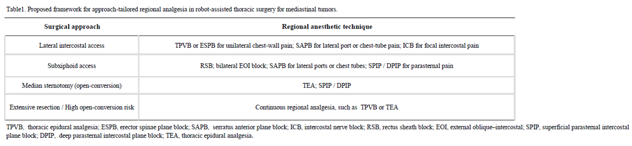
In patients undergoing RATS primarily through intercostal ports and at low risk of conversion to thoracotomy, a practical analgesic strategy is a combination of peripheral nerve block, intraoperative acetaminophen administration, and scheduled postoperative NSAIDs or cyclooxygenase-2 (COX-2) inhibitors, when not contraindicated (Table1). If peripheral nerve block is not performed, local anesthetic infiltration should be applied around all port sites and chest-tube insertion sites. Postoperatively, nonopioid analgesics should be administered on a scheduled basis, with low-dose opioids (IV-PCA) reserved as rescue medication for patients with higher pain scores on the Numerical Rating Scale.
5.2. Patients at High Risk of Postoperative Pain
In cases involving a large tumor, extensive pleural dissection, a bilateral surgical approach, or a high risk of conversion to thoracotomy, the indication for preoperative TPVB or TEA should be carefully considered [36]. Postoperatively, continuous local analgesia via an indwelling catheter may also be considered. If neuropathic pain is suspected, early assessment and therapeutic intervention, including the use of gabapentinoids such as mirogabalin, should be initiated.
6. Future Directions
Prospective studies are needed to compare regional analgesic techniques according to the surgical approach and distribution of postoperative pain in patients undergoing RATS for mediastinal tumors. In particular, optimal analgesic strategies for single-port procedures and the subxiphoid approach remain to be established. Further research is also required to define safe and effective perioperative analgesic protocols for pediatric and adolescent patients.
7. Conclusions
RATS for mediastinal tumors may reduce surgical trauma compared with conventional thoracotomy or median sternotomy. Analgesic management should be individualized according to the surgical approach, port configuration, chest-tube location, and extent of resection. For standard-risk patients, a multimodal analgesic regimen comprising acetaminophen and a nonsteroidal anti-inflammatory drug or cyclooxygenase-2 inhibitor, combined with local wound infiltration or a single-shot regional block, appears appropriate. TEA and TPVB may be reserved for patients anticipated to require extensive chest-wall or pleural dissection, as well as for those at increased risk of conversion to thoracotomy.
Availability of data and materials
The datasets related to this report are available from the corresponding author on reasonable request.
Competing Interests
The authors declare that they have no conflicts of interest.
Funding
None.
Authors’ Contributions
MF, IW, and IK drafted the manuscript. YK, RU, SS and YY conducted the literature investigation. IK and KS edited the manuscript. All authors reviewed and approved the final version of the manuscript.
Abbreviations
| EOI: external oblique–intercostal |
| ESPB: erector spinae plane block |
| DPIP: deep parasternal intercostal plane block |
| ICB: intercostal nerve block |
| IV-PCA: intravenous-patient controlled analgesia |
| LAST: local anesthetic systemic toxicity |
| MTP: midpoint transverse process-to-pleura |
| NSAIDs: nonsteroidal anti-inflammatory drugs |
| RATS: robot-assisted thoracic surgery |
| SAPB: serratus anterior plane block |
| SCTL: superior costotransverse ligament |
| SPIP: superficial parasternal intercostal plane block |
| TEA: thoracic epidural analgesia |
| TPVB: thoracic epidural analgesia |
| VATS: video-assisted thoracic surgery |
References
- Augustin, F.; Schmid, T.; Bodner, J. Robotic surgery for mediastinal tumors. Eur. Surg. 2011, 43, 218–223. [Google Scholar] [CrossRef]
- Yoo, D.G.; et al. Robot-assisted thoracoscopic surgery for mediastinal masses: a single-institution experience. J. Thorac. Dis. 2020, 12, 474–82. [Google Scholar] [CrossRef]
- Watanabe, H.; et al. Uniportal robotic-assisted thoracic surgery for mediastinal tumors: initial experience and perioperative outcomes. J. Vis. Surg. 2024. [Google Scholar]
- Kusano, T.; et al. Postoperative analgesia following robot-assisted thoracic surgery for mediastinal disease: retrospective comparative study of general anesthesia alone, combined with epidural analgesia, and with ultrasound-guided thoracic paraspinal block. Ann. Transl. Med. 2023, 11, 206. [Google Scholar] [CrossRef]
- La Via, L.; et al. Loco-regional anesthesia for pain management in robotic thoracic surgery. J. Clin. Med. 2024, 13, 3141. [Google Scholar] [CrossRef]
- Kawagoe, I.; et al. Postoperative analgesia in patients undergoing robot-assisted thoracic surgery: a comparison between thoracic epidural analgesia and intercostal nerve block combined with intravenous patient-controlled analgesia. Ann. Palliat. Med. 2021, 10, 1985–93. [Google Scholar] [CrossRef]
- Elsolh, B.; et al. Special anesthesiologic considerations in robot-assisted thoracic surgery. Curr. Anesthesiol. Rep. 2021, 11, 429–436. [Google Scholar] [CrossRef]
- Strollo, D.C.; Rosado-de-Chirtenson, M.L.; Jett, J.R. Primary mediastinal tumors: part1-tumors of the anterior mediastinum. Chest 1997, 112, 259S–272S. [Google Scholar]
- Carter, B.W.; Benaveniste, M.F.; Madan, R.; et al. ITMIG classification of mediastinal compartments and multidisciplinary approach to mediastinal masses. Radiographics 2017, 37, 413–36. [Google Scholar] [CrossRef]
- Fujimoto, K.; et al. Proposal for a new medias- tinal compartment classification of transverse plane images according to the Japanese Association for Research on the Thymus(JART) general rules for the study of mediastinal tumors. Oncol. Rep. 2014, 31, 565–72. [Google Scholar] [CrossRef]
- Grunenwald, D.; Spaggiari, L. Transmanubrial osteomuscularn sparing approach for apical chest tumors. Ann. Thorac. Surg. 1997, 63(2), 563–6. [Google Scholar] [CrossRef]
- Ackman, J.B. A review of the CT findings in four common types of mediastinal tumors. Radiol. Clin. North Am. 2019, 57, 113–25. [Google Scholar]
- Deckarm, R.; Flury, D.V.; Deckarm, S.; Ott, S.; Kocher, G.J. Surgical management of thymic tumors: a narrative review with focus on robotic-assisted surgery. Mediastinum 2024, 8, 48. [Google Scholar] [CrossRef]
- Li, X.; et al. Serratus anterior plane block for postoperative analgesia after video-assisted thoracoscopic surgery: a systematic review and meta-analysis. 2023. [Google Scholar] [CrossRef]
- Wang, L.; et al. Serratus anterior plane block versus thoracic paravertebral block for postoperative analgesia after video-assisted thoracoscopic surgery: a prospective randomized double-blind non-inferiority trial. 2022. [Google Scholar] [CrossRef]
- Ghigna, M.R.; Guinde, J.; Rosti, V.; et al. Mediastinal tumours and pseudo-tumours: a comprehensive review with emphasis on multidisciplinary approach. Eur. Respir. Rev. 2021, 30, 200309. [Google Scholar] [CrossRef]
- den Bakker, M.A.; et al. Neurogenic tumours of the posterior mediastinum and differential diagnosis: a review. Histopathology 2024. [Google Scholar] [CrossRef]
- Streit, A.; Walsh, L.C.; Stasiak, F.; Vautrin, N.; Guerci, P.; Seitlinger, J.; Renaud, S. Pain outcomes of outside-the-cage robotic thoracic surgery: a prospective matched-cohort study. J. Robot Surg. 2024, 18(1), 351. [Google Scholar] [CrossRef]
- Tokuishi, K.; Wakahara, J.I.; Ueda, Y.; Miyahara, S.; Nakashima, H.; Masuda, Y.; Waseda, R.; Shiraishi, T.; Sato, T. Factors related to post-thoracotomy pain following robotic-assisted thoracic surgery. Asian J. Endosc. Surg. 2024, 17(2), e13302. [Google Scholar] [CrossRef]
- Miyazaki, T.; Doi, R.; Matsumoto, K. Post-thoracotomy pain syndrome in the era of minimally invasive thoracic surgery. J. Thorac. Dis. 2024, 16(5), 3422–30. [Google Scholar] [CrossRef]
- Li, Z.; Hu, L.; Xi, Y.; Wang, L.; Zhang, X.; Mugaanyi, J. Effect of Ultrasound-Guided External Oblique Intercostal Block on Postoperative Recovery After Subxiphoid Video-Assisted Thoracoscopic Thymectomy: A Randomized Controlled Trial. J. Ultrasound Med. 2025, 44(1), 137–45. [Google Scholar] [CrossRef]
- Chou, R.; Gordon, D.B.; de Leon-Casasola, O.A.; Rosenberg, J.M.; Bickler, S.; Brennan, T.; Carter, T.; Cassidy, C.L.; Chittenden, E.H.; Degenhardt, E.; Griffith, S.; Manworren, R.; McCarberg, B.; Montgomery, R.; Murphy, J.; Perkal, M.F.; Suresh, S.; Sluka, K.; Strassels, S.; Thirlby, R.; Viscusi, E.; Walco, G.A.; Warner, L.; Weisman, S.J.; Wu, C.L. Management of Postoperative Pain: A Clinical Practice Guideline From the American Pain Society, the American Society of Regional Anesthesia and Pain Medicine, and the American Society of Anesthesiologists' Committee on Regional Anesthesia, Executive Committee, and Administrative Council. J. Pain 2016, 17(2), 131–57. [Google Scholar] [CrossRef]
- Chandok, N.; Watt, K.D. Pain management in the cirrhotic patient: the clinical challenge. Mayo Clin. Proc. 2010, 85(5), 451–8. [Google Scholar] [CrossRef]
- Chen, S.J.; Albano, D.A.; Ashamalla, M.; McLarty, A.J.; Plank, A.I.; Yu, A.; Dhamija, A. Postoperative pain management and hospital outcomes following roboticassisted thoracoscopic surgery for non-small cell lung cancer: a propensity score-matched study. Curr. Chall. Thorac. Surg. 2025, 7, 22. [Google Scholar] [CrossRef]
- Dahan, A.; Aarts, L.; Smith, T.W. Incidence, Reversal, and Prevention of Opioid-induced Respiratory Depression. Anesthesiology 2010, 112(1), 226–38. [Google Scholar] [CrossRef]
- Feray, S.; Lubach, J.; Joshi, G.P.; Bonnet, F.; Van de Velde, M. PROSPECT Working Group *of the European Society of Regional Anaesthesia and Pain Therapy. PROSPECT guidelines for video-assisted thoracoscopic surgery: a systematic review and procedure-specific postoperative pain management recommendations. Anaesthesia 2022, 77(3), 311–25. [Google Scholar]
- Miyazaki, T.; Matsumoto, K.; Sato, T.; Sano, I.; Furukawa, K.; Shimoyama, K.; Kamohara, R.; Suzuki, M.; Kondou, M.; Ikeda, N.; Tabata, S.; Shiosakai, K.; Nagayasu, T. ADMIT-NeP Study group. Efficacy and safety of add-on mirogabalin to conventional therapy for the treatment of peripheral neuropathic pain after thoracic surgery: the multicenter, randomized, open-label ADMIT-NeP study. BMC Cancer 2024, 24(1), 80. [Google Scholar] [CrossRef]
- Perez, J.J.; DeFaccio, R.J.; Heintschel, A.; Warren, D.; Strunk, J.D. Thoracic epidural catheter placement failure rate by contrast epidurography: a single-center retrospective study. Reg. Anesth. Pain Med. 2026, rapm-2025-107456. [Google Scholar] [CrossRef]
- Mehta, S.; Jen, T.T.H.; Hamilton, D.L. Regional analgesia for acute pain relief after open thoracotomy and video-assisted thoracoscopic surgery. BJA Educ. 2023, 23(8), 295–303. [Google Scholar] [CrossRef]
- Kim, S.H. Anatomical classification and clinical application of thoracic paraspinal blocks. Korean J. Anesthesiol. 2022, 75(4), 295–306. [Google Scholar] [CrossRef]
- Deshmukh, S.; Fayad, L.M.; Ahlawat, S. MR neurography (MRN) of the long thoracic nerve: retrospective review of clinical findings and imaging results at our institution over 4 years. Skelet. Radiol. 2017, 46(11), 1531–40. [Google Scholar] [CrossRef]
- Fu, Y.; Fu, H.; Wei, W.; Liu, H.; Wen, Z.; Lv, X.; Lu, Y. Effect of bilateral low serratus anterior plane block on quality of recovery after trans-subxiphoid robotic thymectomy: Results of a randomized placebo-controlled trial. Int. J. Med. Sci. 2024, 21(7), 1241–9. [Google Scholar] [CrossRef]
- Li, J.; Wang, X.; Wang, Y.; Zhang, W. Analgesic effectiveness of serratus anterior plane block in patients undergoing video-assisted thoracoscopic surgery: a systematic review and updated meta-analysis of randomized controlled trials. BMC Anesthesiol. 2023, 23(1), 235. [Google Scholar] [CrossRef]
- Kaya, C.; Dost, B.; Dokmeci, O.; Yucel, S.M.; Karakaya, D. Comparison of Ultrasound-Guided Pecto-intercostal Fascial Block and Transversus Thoracic Muscle Plane Block for Acute Poststernotomy Pain Management After Cardiac Surgery: A Prospective, Randomized, Double-Blind Pilot Study. J. Cardiothorac. Vasc. Anesth. 2022, 36, 2313–21. [Google Scholar] [CrossRef]
- Douglas, R.N.; Kattil, P.; Lachman, N.; Johnson, R.L.; Niesen, A.D.; Martin, D.P.; Ritter, M.J. Superficial versus deep parasternal intercostal plane blocks: cadaveric evaluation of injectate spread. Br. J. Anaesth. 2024, 132(5), 1153–9. [Google Scholar] [CrossRef]
- Neskovic, V.; Hofmeyr, R.; Kawagoe, I.; Guclu, C.Y.; Jiménez, M.J. Pain management in thoracic surgery: bridging evidence and practice. Curr. Opin. Anaesthesiol. 2026, 39(1), 92–9. [Google Scholar] [CrossRef]
Figure 1.
Port placement for robot-assisted thoracoscopic surgery for mediastinal tumors (unilateral transthoracic approach). The camera port was placed at the fifth anterior axillary line through a 10-mm incision. The first and second robotic ports were placed at the third and sixth anterior axillary line respectively, each through a 10-mm incision. The assistant port was placed at the seventh midaxillary line through a 15-mm incision.
Figure 1.
Port placement for robot-assisted thoracoscopic surgery for mediastinal tumors (unilateral transthoracic approach). The camera port was placed at the fifth anterior axillary line through a 10-mm incision. The first and second robotic ports were placed at the third and sixth anterior axillary line respectively, each through a 10-mm incision. The assistant port was placed at the seventh midaxillary line through a 15-mm incision.
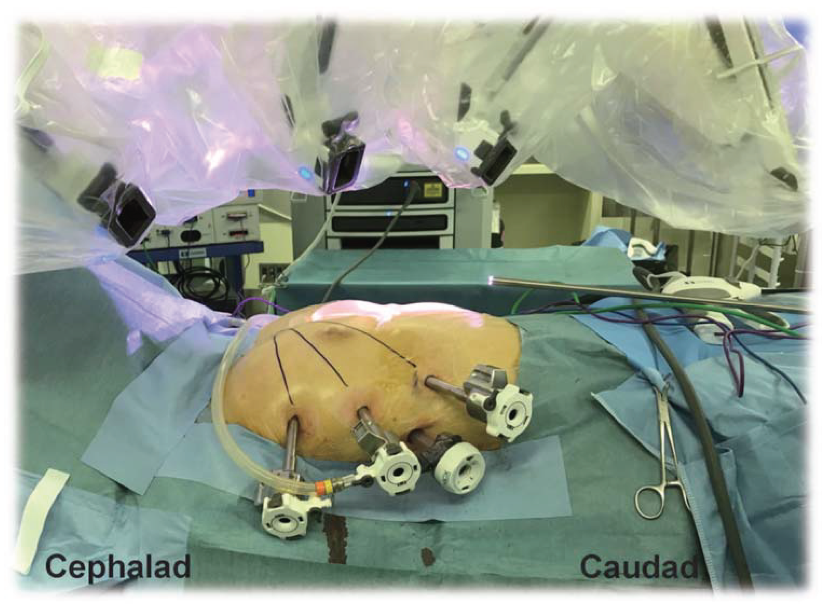

Table 1.
Proposed framework for approach-tailored regional analgesia in robot-assisted thoracic surgery for mediastinal tumors.
Table 1.
Proposed framework for approach-tailored regional analgesia in robot-assisted thoracic surgery for mediastinal tumors.
 |
Disclaimer/Publisher’s Note: The statements, opinions and data contained in all publications are solely those of the individual author(s) and contributor(s) and not of MDPI and/or the editor(s). MDPI and/or the editor(s) disclaim responsibility for any injury to people or property resulting from any ideas, methods, instructions or products referred to in the content. |
© 2026 by the authors. Licensee MDPI, Basel, Switzerland. This article is an open access article distributed under the terms and conditions of the Creative Commons Attribution (CC BY) license (http://creativecommons.org/licenses/by/4.0/).
Copyright: This open access article is published under a Creative Commons CC BY 4.0 license, which permit the free download, distribution, and reuse, provided that the author and preprint are cited in any reuse.