

Submitted:
23 October 2026
Posted:
26 October 2026
You are already at the latest version
Abstract
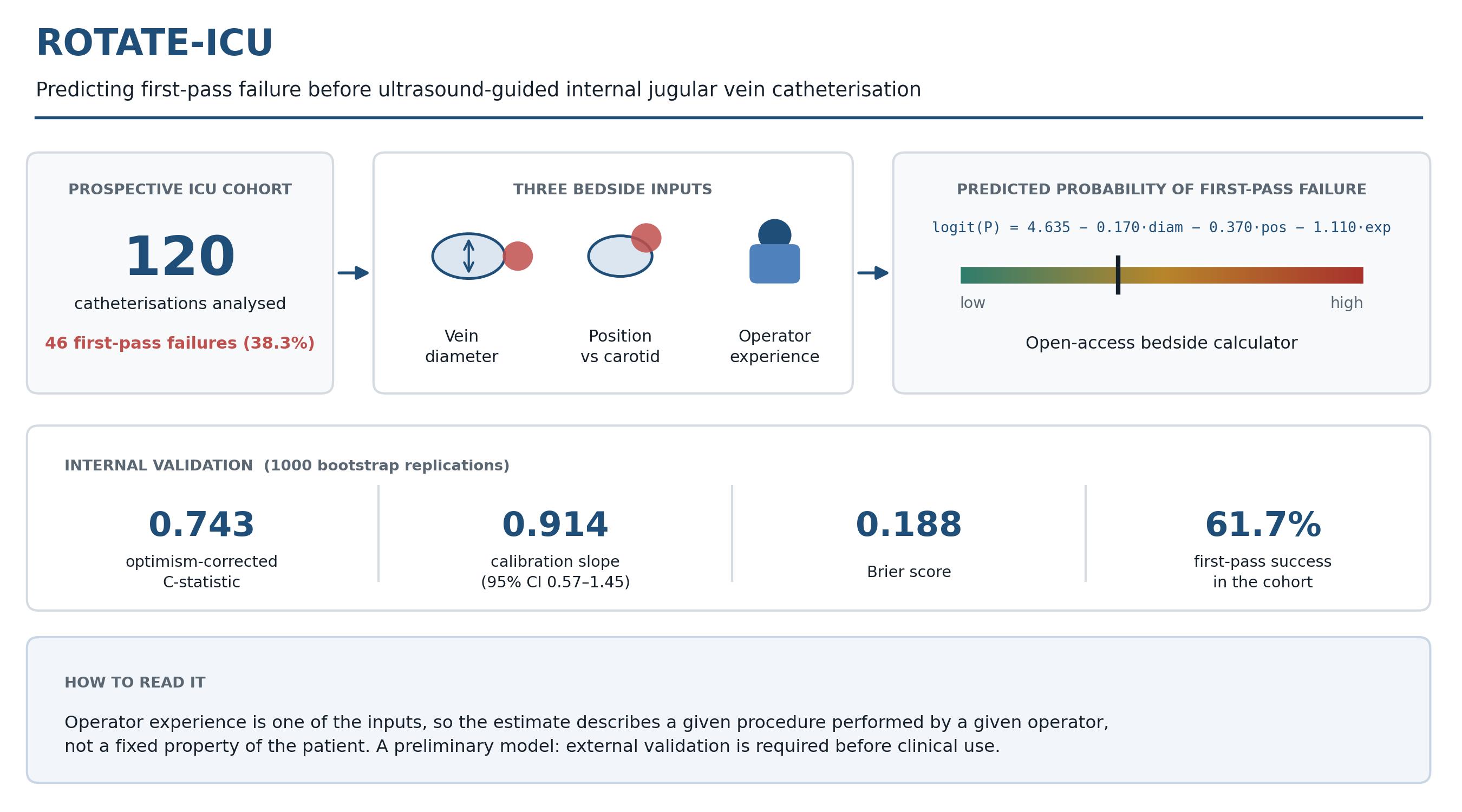
Background: First-pass failure during ultrasound-guided internal jugular vein (IJV) catheterisation is common in critically ill adults and is associated with mechanical complications, yet no model has been developed to estimate its probability before the procedure. We therefore set out to develop and internally validate a preliminary prediction model, ROTATE-ICU. Methods: We conducted a prospective, single-centre, observational cohort study and included 120 of 156 assessed procedures, taking one episode per patient. The pre-specified model combined IJV anterior–posterior diameter, the position of the vein relative to the common carotid artery, and operator experience. The model was fitted with first-pass failure as the outcome and internally validated by bootstrap optimism correction with 1000 replications. Results: First-pass failure occurred in 46 of 120 procedures (38.3%). The odds of failure fell with each millimeter of anterior–posterior diameter (adjusted OR 0.83, 95% CI 0.72–0.95) and with each category of operator experience (adjusted OR 0.29, 95% CI 0.14–0.59), whereas the positional category showed no independent association. The optimism-corrected C-statistic was 0.743, with a calibration slope of 0.914 (95% CI 0.572–1.451) and a Brier score of 0.188. Conclusions: The ROTATE-ICU model estimates the probability of first-pass failure from three variables available at the bedside, of which vein diameter and operator experience carried most of the predictive weight. Because operator experience is itself one of the inputs, the model describes the risk of a given procedure performed by a given operator rather than a fixed property of the patient. The shrinkage-adjusted model and an open-access calculator are available; this is a preliminary model and external validation is now needed.
Keywords:
central venous catheterization
; internal jugular vein
; ultrasonography
; interventional
; clinical prediction model
; first-pass failure
; operator experience
; intensive care units
; internal validation
1. Introduction
Central venous catheterization (CVC) is among the most frequently performed invasive procedures in the intensive care unit and carries well-recognized risks of immediate mechanical complications [1]. Ultrasound guidance improves overall cannulation success and reduces mechanical complications, particularly among less experienced operators [2]. Even so, first-pass failure—the inability to advance the guide wire following the first skin puncture, without needle redirection or an additional skin puncture—remains common, and first-attempt success rates in adult ICU cohorts lie between 60% and 70% [3]. Because the risk of arterial puncture and hematoma rises with the number of needle passes, the first attempt carries disproportionate weight [4]. Identifying patients at elevated risk before the procedure would allow the most experienced available operator to be assigned, or an alternative access site to be considered at an early stage. The intended users of such a model are the intensivists and trainees who perform these procedures, at the point at which the pre-procedural ultrasound scan is already being carried out. A model that contains operator experience as an input describes the risk of a particular procedure performed by a particular operator, not a fixed property of the patient: changing the operator changes one of the inputs and therefore the estimate itself. Whether acting on such an estimate lowers the failure rate is a separate question that requires an interventional design.
The anatomical determinants of cannulation difficulty—IJV lumen diameter and the positional relationship between the vein and the common carotid artery—have been characterized mainly in anaesthetized surgical patients [5] and in healthy volunteers [6], with head position and neck rotation as the principal manipulated variables [7]. Whether these measurements retain their predictive value in critically ill patients, in whom volume status, positive-pressure ventilation and vasopressor support alter venous filling, remains unknown. Operator experience is likewise a determinant of success, and is modifiable through structured training [8], while current recommendations advocate pre-procedural ultrasound assessment of the target vessel without specifying how that assessment should inform the choice of site or operator [9]. Structured risk prediction of this kind has already been established for peripheral intravenous access, where the A-DIVA scale prospectively identifies patients at risk of first-attempt failure [10,11]. To our knowledge, no equivalent model combining pre-procedural ultrasound anatomy with operator experience has been developed or validated for internal jugular cannulation in critically ill patients.
We designed the ROTATE-ICU study to develop and internally validate a pre-specified prediction model for first-pass failure during ultrasound-guided IJV catheterization using pre-procedural ultrasound anatomy and operator experience, and to examine the association between the operator-selected head rotation angle and IJV morphology in a prospective ICU cohort.
2. Materials and Methods
2.1. Study Design and Participants
ROTATE-ICU is a prospective, single-centre, observational cohort study conducted in a 25-bed tertiary intensive care service comprising four separate units at a training and research hospital in Antalya, Türkiye. Patients were recruited from all four units, which share a common medical staff and a single institutional protocol for central venous catheterization. Enrolment ran from 25 June 2026 to 1 September 2026. The study was performed in accordance with the Declaration of Helsinki and approved by the Ethics Committee of Antalya Training and Research Hospital (protocol code 2026-250, 25 June 2026), and written informed consent was obtained from all participants or their legal proxies before enrolment. The study was registered prospectively at ClinicalTrials.gov (NCT07764562); the three pre-specified predictor variables (IJV AP diameter, IJV–CCA positional category, and operator experience), the primary outcome of first-pass success, and the planned statistical approach were declared in the registration record before the first patient was enrolled. This report follows the STROBE recommendations for observational studies [12] and the TRIPOD+AI statement for prediction model development [13]; the completed checklists are provided as Supplementary Material. The study was designed with reference to the PROBAST domains of participant selection, predictor and outcome definition, and analysis [14].
Adults aged 18 years or older who were admitted to the ICU and required ultrasound-guided IJV catheterization for any indication were eligible. Patients were excluded if the procedure was performed during active cardiopulmonary resuscitation, if there was an anatomical abnormality, hematoma, or active soft-tissue infection of the neck, if non-IJV access was planned, if a non-ultrasound-guided technique was used, or if consent was declined. Only the first eligible catheterization episode per patient was included.
Of 156 catheter procedures assessed, 120 were included (Figure 1). Procedures were excluded because insertion was performed as an emergency, so that pre-procedural assessment was not feasible (n = 15), or because the subclavian (n = 14) or femoral (n = 7) route was used. Enrolment continued until the end of the pre-specified data-collection period, at which point the study was closed with 120 patients enrolled; the target of 150 was not reached because the study period expired, and not because of an interim analysis or a safety-related decision to stop. The model was fitted on all 120 procedures with 46 events. Assessed against the criteria of Riley and colleagues, the achieved sample was close to the minimum required for stable coefficient estimation but well short of what would be needed to estimate absolute risk precisely, for which about 364 procedures would have been necessary; the model is therefore reported as preliminary [15,16]. The calculation is set out in the Supplementary Material.
2.2. Pre-Procedural Assessment and Data Collection
Seven pre-specified parameters were recorded immediately before each procedure, with the patient in 15° Trendelenburg position and the head at the rotation angle chosen by the operator. All measurements were performed by trained study coordinators following a standardized protocol based on current recommendations for pre-procedural vascular ultrasound; all images were de-identified and stored in a dedicated study database.
The recorded variables were: (i) IJV anterior–posterior diameter, taken as the mean of three transverse-plane measurements (mm); (ii) skin-to-vessel depth (mm); (iii) the operator-selected head rotation angle (0°, 15°, 30°, 45°, 60°, or 75°), recorded before anatomical assessment; (iv) the IJV–CCA positional category, coded 1–4 on a pre-specified anatomical overlap hierarchy (anteromedial = 1, anterior = 2, anterolateral = 3, lateral = 4), with the small medial subgroup combined with the anteromedial category; (v) body mass index; (vi) operator experience, based on the self-reported lifetime number of ultrasound-guided central venous catheterisations and categorised as low (<20), intermediate (20–50), or high (>50 procedures); and (vii) catheter type.
Of these seven parameters, three—IJV anterior–posterior diameter, the IJV–CCA positional category and operator experience—were pre-specified as candidate predictors in the registration record. The remaining parameters were recorded to characterise the cohort and were not eligible for inclusion in the model.
The ordinal coding of the positional category assumes a monotone decrease in the risk of carotid overlap from code 1 to code 4.
The study coordinator remained at the bedside throughout the procedure and recorded the number of skin punctures, the procedure duration, and any procedure-related complications prospectively as they occurred. Because the same coordinator performed the pre-procedural measurements, outcome assessment could not be blinded to the predictor values; the outcome is, however, an objective event observed at the bedside and leaves little room for interpretation.
2.3. Outcomes
The primary outcome was first-pass success, defined as advancement of the guide wire following the first skin puncture, without needle redirection or an additional skin puncture. For model development this outcome was expressed as its complement, first-pass failure, which is the probability the model reports.
Secondary outcomes were difficult catheterization, a composite of three or more skin punctures, a procedure duration exceeding 10 min from the first skin puncture to intravascular catheter placement, or a change in vascular access site; the total number of skin punctures; procedure duration, measured over the same interval and analyzed as a continuous variable; and procedure-related complications, namely arterial puncture, hematoma larger than 2 cm, catheter malposition requiring correction or repositioning, and pneumothorax detected within 24 h of the procedure.
Two further outcomes were pre-specified in the trial registration: the association of the operator-selected head rotation angle with IJV anterior–posterior diameter and with the IJV–CCA positional category, and the frequency of complete IJV collapse, defined as a vein that could not be identified on the pre-procedural scan.
2.3.1. Open Science and Patient Involvement
No separate study protocol was prepared; the predictors, primary outcome and planned analysis were specified in the ClinicalTrials.gov registration record. The analytical code used for model development and internal validation is available with the web calculator in the study repository. Patients and members of the public were not involved in the design, conduct, reporting or dissemination of the study.
2.4. Statistical Analysis
The model was specified before any analysis of the data and comprised three variables selected on mechanistic grounds: IJV anterior–posterior diameter (continuous), positional category (ordinal, 1–4) and operator experience (ordinal, 1–3). No data-driven variable selection was undertaken at any stage. Data were complete for all 120 procedures, and no imputation was required.
The registered outcome measure was first-pass success. Because the clinical output of the model is the probability of failure, the model was fitted by logistic regression with first-pass failure as the dependent variable; the two parameterizations describe the same binary outcome and differ only in the sign of the coefficients. Odds ratios below unity therefore denote predictors associated with a lower risk of failure.
Coefficients were shrunk to reduce over fitting and the intercept was re-estimated so that the mean predicted risk matched the observed risk [17,18,19]; the shrinkage factor and the method are given in the Supplementary Material. Discrimination was quantified by the C-statistic and corrected for optimism by bootstrap resampling with 1000 replications; the calibration slope and its interval were obtained from 2000 replications with a fixed random seed, so that the reported values are reproducible from the analysis code [17,20]. Calibration was assessed by the calibration slope with a bootstrap 95% confidence interval and displayed graphically, with observed proportions plotted against predicted probabilities using a loess smoother together with grouped estimates by quintile of predicted risk (Figure 3B); the Brier score was reported as a measure of overall performance [21]. Three pre-planned checks were carried out: the assumption of a linear relationship between anterior–posterior diameter and the log odds of the outcome was examined by adding a quadratic term; the model was refitted in the subgroup receiving hemodialysis catheters; and the model was compared with a decision rule based on diameter alone. Because each operator contributed several procedures, the analysis was additionally repeated with allowance for clustering by operator; this analysis was not pre-planned. The decision curve analysis was exploratory, because the clinical action to be taken at a given threshold is not established, and is reported in the Supplementary Material.
Pre-procedural measurements were compared across operator-selected rotation angles, which were grouped as 0°, 15°, 30°, 45° and ≥60° because only one procedure was performed at 75°. The Kruskal–Wallis test was used for anterior–posterior diameter and the chi-square test for positional category; because expected cell counts were sparse, an exact p value was obtained by Monte Carlo simulation with 20,000 samples. The sample-size calculation, the shrinkage and clustering analyses, additional calibration measures, an exploratory decision curve analysis [22] and the variance–covariance matrix of the final model coefficients are provided in the Supplementary Material. All tests were two-tailed with α = 0.05. Analyses were performed in R version 4.3 (R Foundation for Statistical Computing, Vienna, Austria) and SPSS version 29.0 (IBM Corp., Armonk, NY, USA).
3. Results
3.1. Participants
Of 156 procedures assessed, 120 were enrolled and 36 excluded (Figure 1). Pre-procedural ultrasound data were not recorded for the excluded procedures, so a formal comparison with the included cohort was not possible. Baseline characteristics are shown in Table 1 and procedural outcomes in Table 3. The median age was 61.0 years [51.0–68.0], 69 patients (57.5%) were male, the mean body mass index was 26.3 kg/m² (SD 4.6), and the median Glasgow Coma Scale score was 14 [12,13,14,15]. Coagulopathy was present in 52 patients (43.3%), and 80 catheters (66.7%) were placed for hemodialysis. Operator experience was high in 70 procedures (58.3%), intermediate in 42 (35.0%), and low in 8 (6.7%). Rotation angles selected by operators were 0° in 9 procedures, 15° in 24, 30° in 44, 45° in 37, and 60° or more in 6. The procedures were performed by five operators, each linked to an anonymous identifier: one specialist (61 procedures), one senior resident (41), one fellow (9) and two junior residents (6 and 3). Observed failure rates ranged from 26% for the specialist to 83% for the junior resident with six procedures. Two operators crossed an experience threshold during the study, so experience was not fixed within an operator.
3.2. IJV Morphology and Head Rotation
The distribution of IJV anterior–posterior diameter differed little across the rotation angles selected by operators (Kruskal–Wallis H = 1.198, df = 4, p = 0.878; Figure 2A). Because each operator selected a single rotation angle and one measurement was obtained at that angle, these data show that groups defined by different selected angles had similar diameter distributions; the design cannot establish whether head rotation alters the diameter within an individual patient. The distribution of IJV–CCA positional categories, by contrast, differed markedly across rotation-angle groups (χ² = 67.28, df = 12; Monte Carlo p < 0.001 based on 20,000 samples; Figure 2B), the anterolateral category being most prevalent between 30° and 45°. Since 13 of the 20 expected cell counts fell below five, an exact p value was used and no effect-size statistic is reported.
3.3. Prediction Model and Internal Validation
First-pass failure occurred in 46 of 120 procedures (38.3%), giving 15 events per candidate predictor for model development. Full regression results are given in Table 2 and are displayed alongside the univariable estimates in Supplementary Figure S2.
IJV anterior–posterior diameter (adjusted OR per mm 0.825, 95% CI 0.720–0.945; p = 0.005) and operator experience (adjusted OR per category 0.285, 95% CI 0.139–0.586; p = 0.001) were each independently associated with first-pass failure, whereas the positional category was not (adjusted OR 0.658, 95% CI 0.387–1.119; p = 0.122). A larger diameter and greater operator experience therefore correspond to a lower probability of failure.
The heuristic shrinkage factor was 0.885 (LR χ² = 26.03, df = 3), and the shrinkage-adjusted model is logit(P_failure) = 4.635 − 0.170 × AP_mm − 0.370 × IJV_pos − 1.110 × Exp, where AP_mm is the anterior–posterior diameter in millimeters, IJV_pos is coded 1–4 and Exp 1–3. The apparent C-statistic was 0.762 and the optimism-corrected value 0.743 (optimism 0.019). The bootstrap-corrected calibration slope of the unshrunk model was 0.914 (95% CI 0.572–1.451), which motivated the shrinkage applied to the final coefficients; after shrinkage the apparent slope was 1.13 with an intercept of 0.06 on the failure scale. Calibration of the final model is shown in Figure 3B, where the flexible calibration curve follows the diagonal across the range of predicted risk. The width of the confidence interval nevertheless indicates that calibration is uncertain in this derivation cohort and may not hold in a new population, so external validation with prospective recalibration will be necessary. The Brier score was 0.188 (Supplementary Table S1). The assumption of a linear relationship between diameter and the log odds of the outcome was not rejected (p = 0.08). Analyses accounting for clustering by operator did not materially alter the findings; detailed results are given in Supplementary Table S1. The positional category contributed little: a model containing diameter and operator experience alone achieved a C-statistic of 0.741, against 0.762 for the three-variable model. Restricted to the 80 hemodialysis catheters, discrimination was similar (C = 0.741). An exploratory decision curve analysis is presented in the Supplementary Material, together with the full set of calibration measures and the model-checking analyses.
3.4. Difficult Catheterisation and Procedural Complications
The difficult catheterization composite was met in 70 of 120 procedures (58.3%; Table 3). The median number of skin punctures was 1 [1,2] and the median procedure duration 12 min [8,9,10,11,12,13,14,15,16]. Because the median procedure already exceeded the 10-minute criterion, that criterion dominated the composite: 67 procedures met it, against 24 with three or more skin punctures and 15 with a change of access site. Among the 74 procedures with first-pass success, 27 (36.5%) met the composite on duration alone, reflecting the time needed for guide wire threading, dilatation and catheter advancement after the vein has been entered on the first attempt. Arterial puncture occurred in 6 procedures (5.0%), hematoma in 14 (11.7%), catheter malposition in 15 (12.5%), and pneumothorax in 1 (0.8%).

Table 3.
Procedural Outcomes by First-Pass Success (n = 120).
| Outcome | All (n = 120) | FP success (n = 74) |
FP failure (n = 46) |
SMD |
|---|---|---|---|---|
| Procedural process | ||||
| Procedure time (min) | 12.0 [8.0–16.0] | 8.6 [7.0–13.8] | 15.0 [12.2–17.8] | 1.01 ‡ |
| Skin punctures (n) | 1 [1–2] | 1 [1–1] | 3 [2–4] | 2.39 ‡ |
| Secondary outcomes | ||||
| Difficult catheterisation (composite) † | 70 (58.3) | 27 (36.5) | 43 (93.5) | 1.32 ‡ |
| ≥3 skin punctures | 24 (20.0) | 0 (0.0) | 24 (52.2) | |
| Procedure duration >10 min § | 67 (55.8) | 27 (36.5) | 40 (87.0) | |
| Access-site change | 15 (12.5) | 0 (0.0) | 15 (32.6) | |
| Procedural complications | ||||
| Arterial puncture | 6 (5.0) | 1 (1.4) | 5 (10.9) | 0.41 |
| Haematoma (>2 cm) | 14 (11.7) | 0 (0.0) | 14 (30.4) | 0.94 ‡ |
| Catheter malposition | 15 (12.5) | 0 (0.0) | 15 (32.6) | 0.98 ‡ |
| Pneumothorax | 1 (0.8) | 0 (0.0) | 1 (2.2) | 0.21 |
| Any complication | 26 (21.7) | 1 (1.4) | 25 (54.3) | 1.47 ‡ |
Values are median [IQR] or n (%). FP, first-pass; SMD, standardised mean difference. Procedure time was measured from the first skin puncture to intravascular catheter placement; fixation and dressing are not included. † Composite of three or more skin punctures, procedure duration exceeding 10 min, or a change in access site. ‡ SMD > 0.50, indicating a large between-group difference. § Twenty-seven procedures with first-pass success met this criterion, in each case because of the time required for guidewire threading, dilatation and catheter advancement after the vein was entered on the first attempt.
4. Discussion
The central finding is straightforward: a smaller IJV anterior–posterior diameter and lower operator experience were each independently associated with first-pass failure during ultrasound-guided IJV catheterization in critically ill adults, and the model retained moderate discrimination after bootstrap optimism correction. The three-variable ROTATE-ICU model combines these two predictors with the IJV–CCA positional category and is implemented as a web-based calculator that returns the predicted probability of failure. It should be regarded as a preliminary procedure–operator model for external validation, not as a clinically deployable patient-only score.
First-pass success was achieved in 61.7% of procedures, which sits at the lower end of the 60–70% range reported for adult ICU cohorts [3] and confirms that ultrasound guidance alone does not make the first attempt reliable. Both principal predictors are clinically intuitive, and the model behaved consistently in the hemodialysis subgroup, which made up two-thirds of the cohort. The positional category was not independently associated with the outcome and added little to discrimination. It was retained because it was pre-specified, but the two-variable model performs almost identically and is the more defensible candidate to carry into external validation; both are reported in full so that either can be evaluated. The calibration slope of 0.914 carries a confidence interval from 0.572 to 1.451, wide enough to encompass meaningful miscalibration in a new population, so external data are needed before the score can be expected to transport to other ICU settings.
A comparable approach has already proved workable for peripheral intravenous access, where the A-DIVA scale predicts first-attempt failure from a small set of bedside variables and has been externally validated across multiple centres [10,11]. ROTATE-ICU applies the same logic to a more consequential procedure, with the addition that operator experience is treated as a predictor rather than as a source of noise to be adjusted away. That addition changes what the model estimates. Because experience is an input, the estimate belongs to the pairing of a patient with a particular operator, and reassigning the procedure produces a different estimate rather than a lower risk under the same one. The model is therefore best read as a procedure–operator risk estimate, and its most natural use is to display the predicted probability for the operator at the bedside alongside that for a more experienced colleague. Whether acting on that comparison improves outcomes cannot be answered by this design and would require an impact study.
The distribution of IJV–CCA positional categories differed across the rotation angles operators selected, whereas the distribution of vein diameter did not. Because each patient was assessed at a single operator-chosen angle, these are between-patient comparisons and cannot establish that rotating the head alters anatomy within an individual. That the positional relationship varied across angle groups while diameter did not is nonetheless consistent with rotation acting mainly on the geometry between the vessels rather than on venous filling, and it suggests that the two anatomical inputs to the score may behave differently under changes in head position. An optimal rotation angle cannot be derived from this design, and within-patient measurements at several angles would be needed to address that question.
Using the score requires no more than the pre-procedural ultrasound scan that is already recommended before internal jugular cannulation: the operator measures the vein and classifies its position relative to the carotid artery, and enters these together with their own experience category. Both inputs depend on competence in vascular ultrasound, and the reliability reported here was achieved by coordinators who had completed standardized training. No vein was unidentifiable on the pre-procedural scan, so complete IJV collapse, a further registered outcome, did not occur in this cohort.
Two features of the data limit how far the secondary findings can be taken. The difficult catheterization composite was driven almost entirely by its duration criterion, which the median procedure in this cohort exceeded, so it functions as a marker of overall procedure length rather than of cannulation difficulty. Complications were recorded almost exclusively in procedures with first-pass failure, including catheter malposition, which has no direct mechanistic link to the number of needle passes and is unlikely to follow from it. Difficult anatomy plausibly underlies both, and procedures that were difficult from the outset may also have been examined more closely afterwards; the complication counts are therefore best read as descriptive.
Several limitations bear on how these results should be read. The model was derived at a single centre from a cohort of 120 patients with 46 events, and no external validation has been undertaken. Only five operators performed the procedures and one specialist accounted for 61 of the 120, so the effect attributed to experience may reflect individual technique as much as accumulated volume. Two operators crossed an experience threshold during the study, which loosens that confounding, and analyses accounting for operator clustering gave the same estimates; but five clusters are too few for reliable operator-level inference, and the confidence interval for the experience effect should not be taken at face value. Operator experience was self-reported without logbook verification. Hemodialysis catheters accounted for 66.7% of procedures, which limits generalizability to units with a different case mix. The ordinal coding of both categorical predictors assumes an equal-interval gradient in risk. For the positional category this assumption was examined and not rejected, though the comparison was close; for operator experience it was not tested, since only eight procedures fell in the lowest category, and the true effect may differ across category transitions. Procedures performed as emergencies and those using the subclavian or femoral route were excluded by design, so the cohort represents elective internal jugular catheterization rather than central venous access as a whole. Because no pre-procedural data were recorded for the excluded procedures, the direction and magnitude of any resulting selection bias cannot be determined. Enrolment closed at 120 rather than the planned 150 because the data-collection period expired. The sample also falls short of what would be needed to estimate absolute risk precisely, which is a further reason to treat the model as preliminary. Prospective external validation in a multicenter, geographically distinct cohort, with independent verification of operator experience and with clustering at operator level taken into account, is the necessary next step.
5. Conclusions
In critically ill adults undergoing ultrasound-guided internal jugular vein catheterization, the probability of first-pass failure can be estimated before the procedure from three variables available at the bedside: the anterior–posterior diameter of the vein, its position relative to the common carotid artery, and the experience of the operator. Vein diameter and operator experience carried most of the predictive weight, and because experience is one of the inputs the estimate applies to a given procedure performed by a given operator. Internal validation showed moderate discrimination, with an optimism-corrected C-statistic of 0.743, alongside a calibration slope of 0.914 estimated with considerable imprecision (95% CI 0.572–1.451) and a Brier score of 0.188. The model is available as an open-access web calculator (https://doi.org/10.5281/zenodo.22175651). It should be regarded as preliminary: external validation in an independent cohort, with clustering at operator level taken into account, is required before any clinical use.
Supplementary Materials
The following supporting information can be downloaded at the website of this paper posted on Preprints.org, Table S1: full performance and calibration statistics, the sensitivity and model-checking analyses, and the variance–covariance matrix of the final model coefficients. Figure S1: bootstrap distribution of the optimism-corrected C-statistic (1000 replications). Figure S2: forest plot of univariable and adjusted odds ratios. Figure S3: decision curve analysis. Checklist S1: completed STROBE checklist. Checklist S2: completed TRIPOD+AI checklist.
Author Contributions
Conceptualization, Ş.A.; Methodology, Ş.A.; Software, Ş.A.; Validation, Ş.A., M.A. and D.T.Ö.; Formal Analysis, Ş.A.; Investigation, Ş.A., M.A. and D.T.Ö.; Resources, M.A.; Data Curation, D.T.Ö.; Writing—Original Draft Preparation, Ş.A.; Writing—Review and Editing, M.A. and D.T.Ö.; Visualization, Ş.A.; Supervision, M.A.; Project Administration, Ş.A. All authors have read and agreed to the published version of the manuscript.
Funding
This research received no external funding.
Institutional Review Board Statement
The study was conducted in accordance with the Declaration of Helsinki and approved by the Ethics Committee of Antalya Training and Research Hospital (protocol code 2026-250, date of approval: 25 june 2026).
Informed Consent Statement
Informed consent was obtained from all subjects involved in the study or from their legal proxies.
Data Availability Statement
The shrinkage-adjusted model coefficients and the variance–covariance matrix are provided in Supplementary Table S1, so that the model can be applied and evaluated in external cohorts without access to the derivation data. The web calculator implementing the model, together with the analytical code used for model development and internal validation, is openly available at https://doi.org/10.5281/zenodo.22175651. The de-identified individual patient data supporting the findings can be made available by the corresponding author on reasonable request, subject to approval by the institutional ethics committee. The study is registered at ClinicalTrials.gov (NCT07764562).
Conflicts of Interest
The authors declare no conflicts of interest.
References
- Parienti, J.J.; Mongardon, N.; Mégarbane, B.; Mira, J.P.; Kalfon, P.; Gros, A.; Marqué, S.; Thuong, M.; Pottier, V.; Ramakers, M.; et al. Intravascular complications of central venous catheterization by insertion site. N. Engl. J. Med. 2015, 373, 1220–1229. [Google Scholar] [CrossRef]
- Airapetian, N.; Maizel, J.; Langelle, F.; Modeliar, S.S.; Karakitsos, D.; Dupont, H.; Slama, M. Ultrasound-guided central venous cannulation is superior to quick-look ultrasound and landmark methods among inexperienced operators: A prospective randomized study. Intensive Care Med. 2013, 39, 1938–1944. [Google Scholar] [CrossRef]
- Trabelsi, B.; Hajjej, Z.; Drira, D.; Yedes, A.; Labbene, I.; Ferjani, M.; Ben Ali, M. Comparison of ultrasound-guided internal jugular vein and supraclavicular subclavian vein catheterization in critically ill patients: A prospective, randomized clinical trial. Ann. Intensive Care 2022, 12, 91. [Google Scholar] [CrossRef]
- McGee, D.C.; Gould, M.K. Preventing complications of central venous catheterization. N. Engl. J. Med. 2003, 348, 1123–1133. [Google Scholar] [CrossRef]
- Troianos, C.A.; Kuwik, R.J.; Pasqual, J.R.; Lim, A.J.; Odasso, D.P. Internal jugular vein and carotid artery anatomic relation as determined by ultrasonography. Anesthesiology 1996, 85, 43–48. [Google Scholar] [CrossRef]
- Norin, H.; Pikwer, A.; Fellert, F.; Åkeson, J. Internal jugular dimensions and common carotid overlapping assessed in a cross-sectional study by ultrasonography at three neck levels in healthy volunteers. J. Vasc. Access 2017, 18, 69–72. [Google Scholar] [CrossRef]
- Lamperti, M.; Subert, M.; Cortellazzi, P.; Vailati, D.; Borrelli, P.; Montomoli, C.; D'Onofrio, G.; Caldiroli, D. Is a neutral head position safer than 45-degree neck rotation during ultrasound-guided internal jugular vein cannulation? Results of a randomized controlled clinical trial. Anesth. Analg. 2012, 114, 777–784. [Google Scholar] [CrossRef]
- Barsuk, J.H.; McGaghie, W.C.; Cohen, E.R.; Balachandran, J.S.; Wayne, D.B. Use of simulation-based mastery learning to improve the quality of central venous catheter placement in a medical intensive care unit. J. Hosp. Med. 2009, 4, 397–403. [Google Scholar] [CrossRef]
- Saugel, B.; Scheeren, T.W.L.; Teboul, J.L. Ultrasound-guided central venous catheter placement: A structured review and recommendations for clinical practice. Crit. Care 2017, 21, 225. [Google Scholar] [CrossRef]
- van Loon, F.H.J.; Puijn, L.A.P.M.; Houterman, S.; Bouwman, A.R.A. Development of the A-DIVA Scale: A clinical predictive scale to identify difficult intravenous access in adult patients based on clinical observations. Medicine 2016, 95, e3428. [Google Scholar] [CrossRef]
- van Loon, F.H.J.; van Hooff, L.W.E.; de Boer, H.D.; Koopman, S.S.H.A.; Buise, M.P.; Korsten, H.H.M.; Dierick-van Daele, A.T.M.; Bouwman, A.R.A. The modified A-DIVA scale as a predictive tool for prospective identification of adult patients at risk of a difficult intravenous access: A multicenter validation study. J. Clin. Med. 2019, 8, 144. [Google Scholar] [CrossRef]
- von Elm, E.; Altman, D.G.; Egger, M.; Pocock, S.J.; Gøtzsche, P.C.; Vandenbroucke, J.P. The Strengthening the Reporting of Observational Studies in Epidemiology (STROBE) statement: Guidelines for reporting observational studies. Lancet 2007, 370, 1453–1457. [Google Scholar] [CrossRef]
- Collins, G.S.; Moons, K.G.M.; Dhiman, P.; Riley, R.D.; Beam, A.L.; Van Calster, B.; Ghassemi, M.; Liu, X.; Reitsma, J.B.; van Smeden, M.; et al. TRIPOD+AI statement: Updated guidance for reporting clinical prediction models that use regression or machine learning methods. BMJ 2024, 385, e078378. [Google Scholar] [CrossRef]
- Wolff, R.F.; Moons, K.G.M.; Riley, R.D.; Whiting, P.F.; Westwood, M.; Collins, G.S.; Reitsma, J.B.; Kleijnen, J.; Mallett, S. PROBAST: A tool to assess the risk of bias and applicability of prediction model studies. Ann. Intern. Med. 2019, 170, 51–58. [Google Scholar] [CrossRef]
- Peduzzi, P.; Concato, J.; Kemper, E.; Holford, T.R.; Feinstein, A.R. A simulation study of the number of events per variable in logistic regression analysis. J. Clin. Epidemiol. 1996, 49, 1373–1379. [Google Scholar] [CrossRef]
- Riley, R.D.; Ensor, J.; Snell, K.I.E.; Harrell, F.E., Jr.; Martin, G.P.; Reitsma, J.B.; Moons, K.G.M.; Collins, G.S.; van Smeden, M. Calculating the sample size required for developing a clinical prediction model. BMJ 2020, 368, m441. [Google Scholar] [CrossRef]
- Harrell, F.E., Jr. Regression Modeling Strategies: With Applications to Linear Models, Logistic and Ordinal Regression, and Survival Analysis, 2nd ed.; Springer: Cham, Switzerland, 2015. [Google Scholar]
- van Houwelingen, J.C.; le Cessie, S. Predictive value of statistical models. Stat. Med. 1990, 9, 1303–1325. [Google Scholar] [CrossRef]
- Steyerberg, E.W. Clinical Prediction Models: A Practical Approach to Development, Validation, and Updating, 2nd ed.; Springer: Cham, Switzerland, 2019. [Google Scholar]
- Harrell, F.E., Jr.; Lee, K.L.; Mark, D.B. Multivariable prognostic models: Issues in developing models, evaluating assumptions and adequacy, and measuring and reducing errors. Stat. Med. 1996, 15, 361–387. [Google Scholar] [CrossRef]
- Steyerberg, E.W.; Vickers, A.J.; Cook, N.R.; Gerds, T.; Gonen, M.; Obuchowski, N.; Pencina, M.J.; Kattan, M.W. Assessing the performance of prediction models: A framework for some traditional and novel measures. Epidemiology 2010, 21, 128–138. [Google Scholar] [CrossRef]
- Vickers, A.J.; Elkin, E.B. Decision curve analysis: A novel method for evaluating prediction models. Med. Decis. Mak. 2006, 26, 565–574. [Google Scholar] [CrossRef]
Figure 1.
Patient flow. Of 156 catheter procedures assessed, 120 were enrolled and 36 excluded (emergency insertion, n = 15; subclavian access, n = 14; femoral access, n = 7). All 120 procedures had complete data and contributed to model development, with 46 events.
Figure 1.
Patient flow. Of 156 catheter procedures assessed, 120 were enrolled and 36 excluded (emergency insertion, n = 15; subclavian access, n = 14; femoral access, n = 7). All 120 procedures had complete data and contributed to model development, with 46 events.
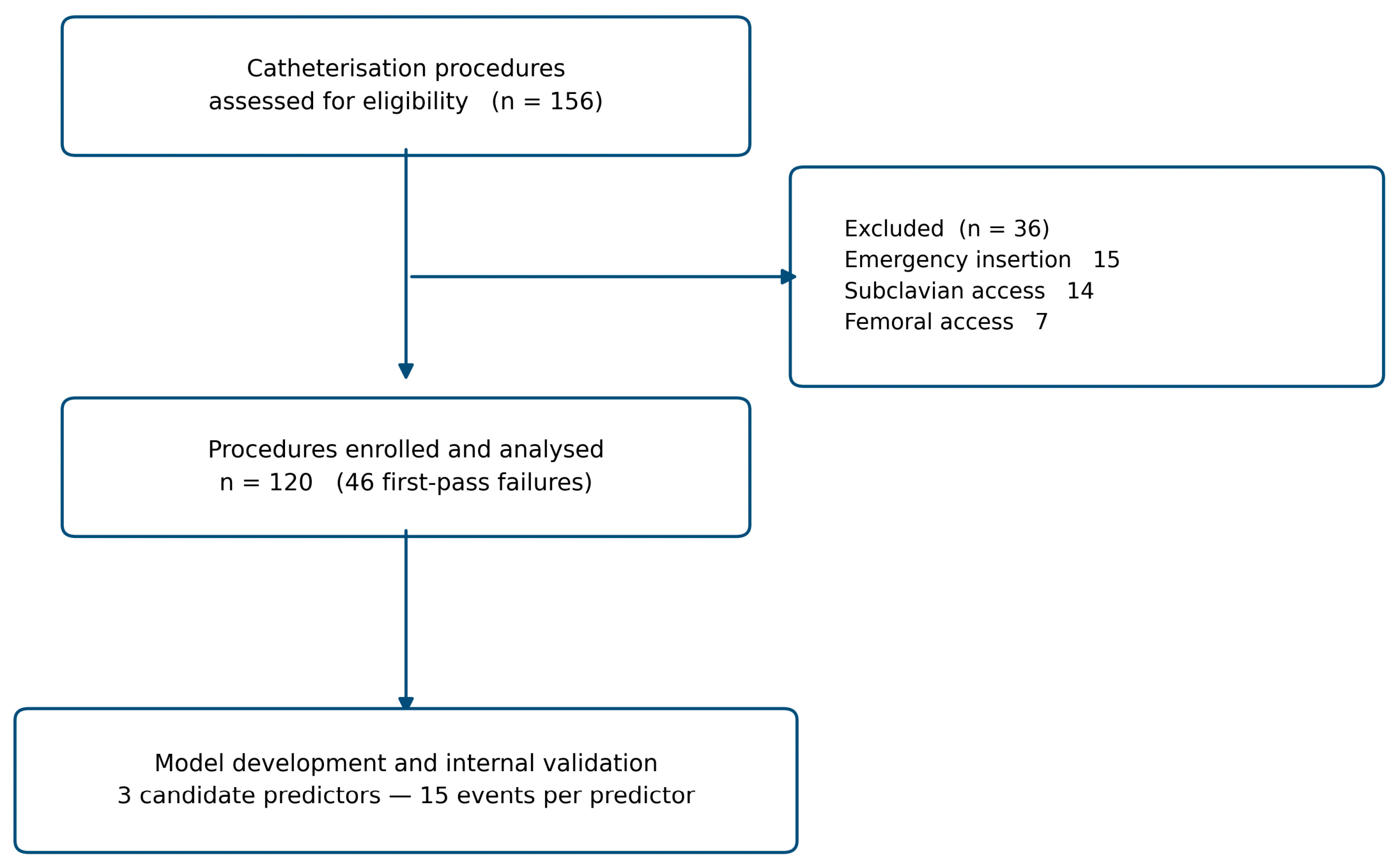
Figure 2.
Operator-selected head rotation angle and pre-procedural IJV morphology. (A) IJV anterior–posterior diameter by rotation-angle group; the distributions did not differ across groups (Kruskal–Wallis H = 1.198, df = 4, p = 0.878). Because each patient was assessed at a single operator-chosen angle, these are between-patient comparisons and cannot establish a within-patient effect of head rotation on vein diameter. (B) Distribution of IJV–CCA positional categories by rotation-angle group; the distributions differed across groups (χ² = 67.28, df = 12; Monte Carlo p < 0.001 based on 20,000 samples). Since 13 of the 20 expected cell counts fell below five, an exact p value was used and no effect-size statistic is reported. One procedure performed at 75° is included in the ≥60° group.
Figure 2.
Operator-selected head rotation angle and pre-procedural IJV morphology. (A) IJV anterior–posterior diameter by rotation-angle group; the distributions did not differ across groups (Kruskal–Wallis H = 1.198, df = 4, p = 0.878). Because each patient was assessed at a single operator-chosen angle, these are between-patient comparisons and cannot establish a within-patient effect of head rotation on vein diameter. (B) Distribution of IJV–CCA positional categories by rotation-angle group; the distributions differed across groups (χ² = 67.28, df = 12; Monte Carlo p < 0.001 based on 20,000 samples). Since 13 of the 20 expected cell counts fell below five, an exact p value was used and no effect-size statistic is reported. One procedure performed at 75° is included in the ≥60° group.
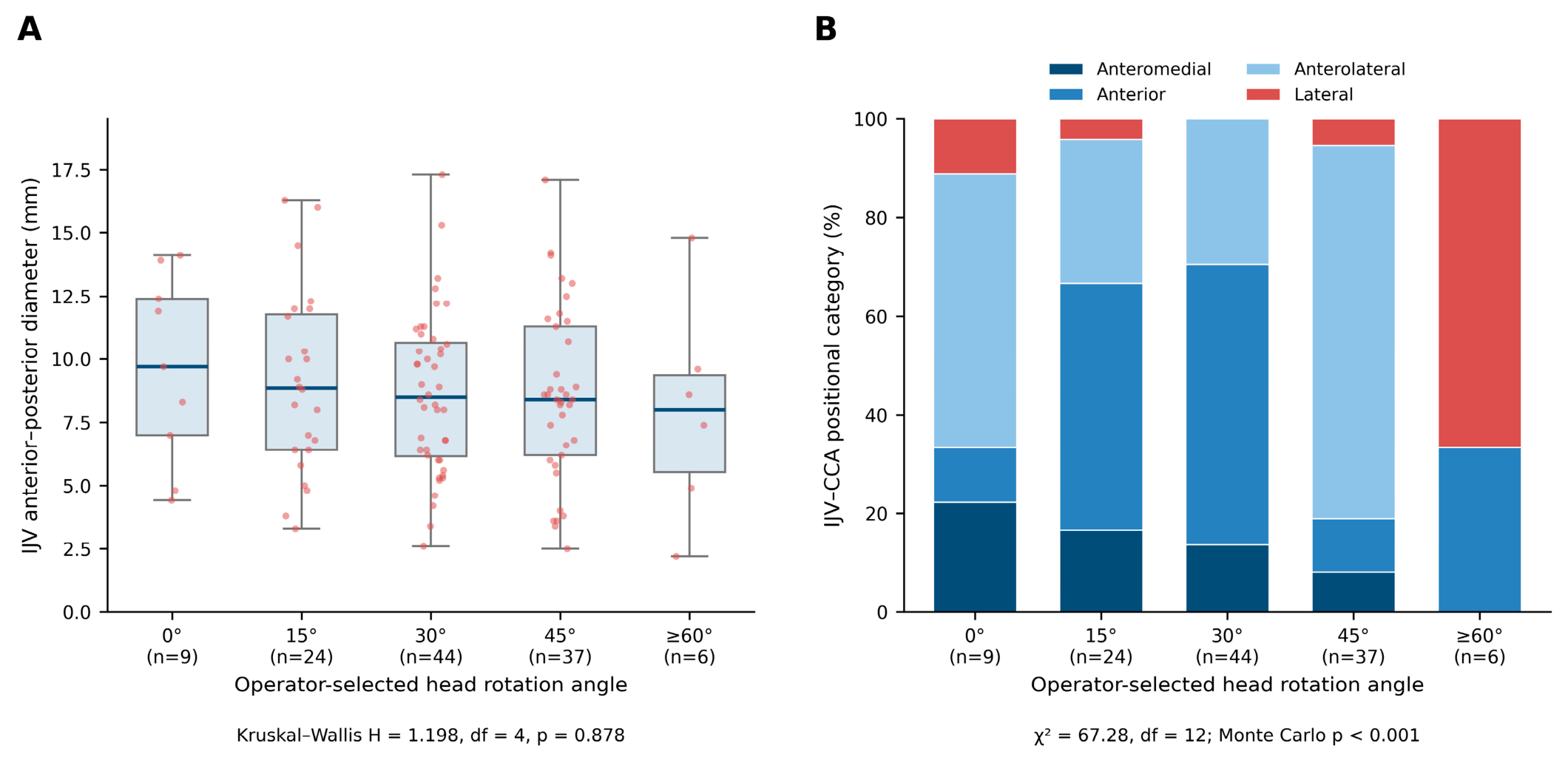
Figure 3.
Discrimination and calibration of the ROTATE-ICU model (n = 120). (A) Receiver operating characteristic curves for the pre-specified model and for each of its variables considered alone; C-statistics were 0.762 for the full model, 0.683 for operator experience, 0.652 for AP diameter and 0.589 for the positional category. The optimism-corrected value for the full model was 0.743. Discrimination is identical whether the outcome is expressed as first-pass failure or first-pass success. (B) Calibration of the final shrinkage-adjusted model on the failure scale (n = 120). The dashed line indicates ideal calibration, the solid curve is a loess smoother, and circles are observed proportions within quintiles of predicted risk with 95% confidence intervals; the histogram shows predicted probabilities in patients with first-pass failure (above the axis) and first-pass success (below). The bootstrap-corrected calibration slope of the unshrunk model was 0.914 (95% CI 0.572–1.451), which motivated the shrinkage applied to the final coefficients; after shrinkage the apparent slope was 1.13 with an intercept of 0.06. Hosmer–Lemeshow χ² = 7.34, df = 8, p = 0.501.
Figure 3.
Discrimination and calibration of the ROTATE-ICU model (n = 120). (A) Receiver operating characteristic curves for the pre-specified model and for each of its variables considered alone; C-statistics were 0.762 for the full model, 0.683 for operator experience, 0.652 for AP diameter and 0.589 for the positional category. The optimism-corrected value for the full model was 0.743. Discrimination is identical whether the outcome is expressed as first-pass failure or first-pass success. (B) Calibration of the final shrinkage-adjusted model on the failure scale (n = 120). The dashed line indicates ideal calibration, the solid curve is a loess smoother, and circles are observed proportions within quintiles of predicted risk with 95% confidence intervals; the histogram shows predicted probabilities in patients with first-pass failure (above the axis) and first-pass success (below). The bootstrap-corrected calibration slope of the unshrunk model was 0.914 (95% CI 0.572–1.451), which motivated the shrinkage applied to the final coefficients; after shrinkage the apparent slope was 1.13 with an intercept of 0.06. Hosmer–Lemeshow χ² = 7.34, df = 8, p = 0.501.
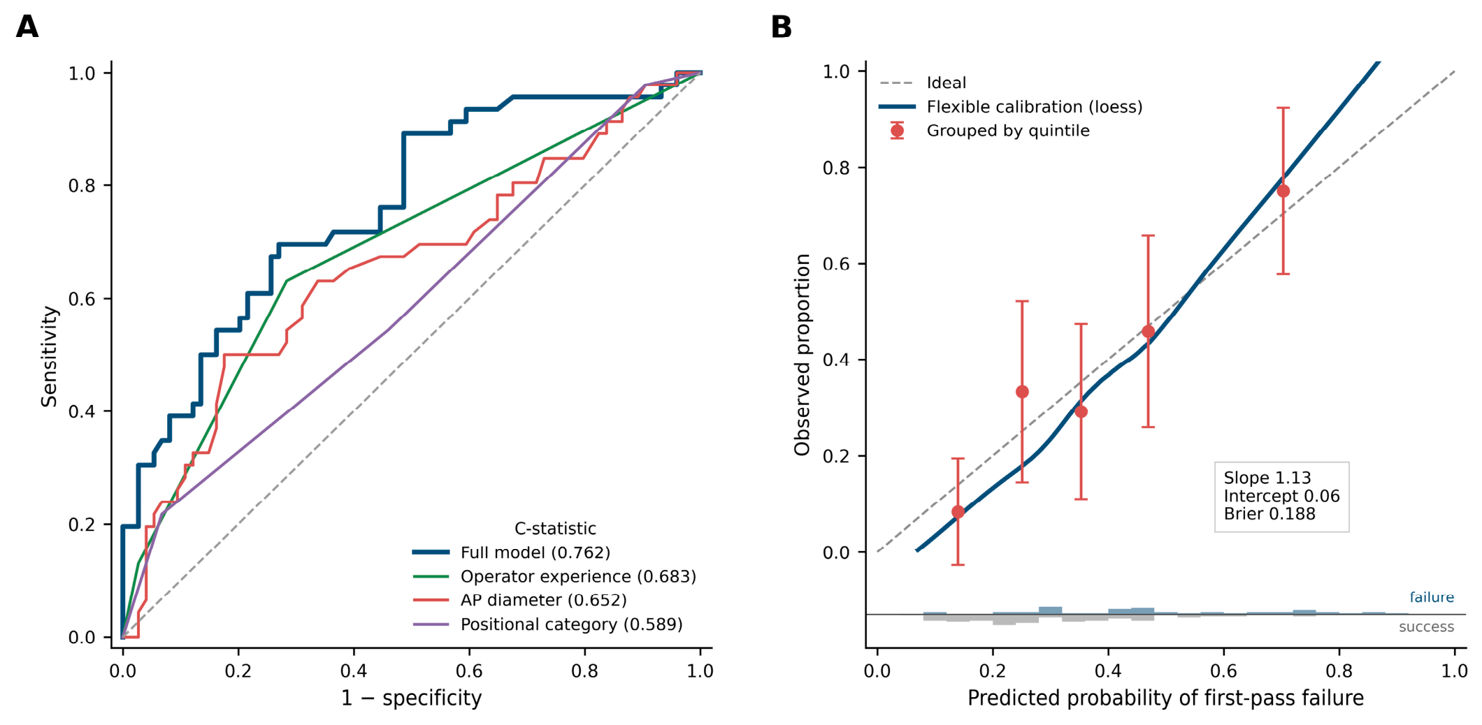
Table 1.
Baseline Characteristics (n = 120).
| Variable | All (n = 120) | FP success (n = 74) |
FP failure (n = 46) |
SMD |
|---|---|---|---|---|
| Demographics | ||||
| Age (years) | 61.0 [51.0–68.0] | 61.0 [52.0–67.0] | 62.5 [49.5–74.2] | 0.08 |
| Male sex, n (%) | 69 (57.5) | 43 (58.1) | 26 (56.5) | 0.03 |
| BMI (kg/m²) | 26.3 ± 4.6 | 26.4 ± 4.1 | 26.2 ± 5.3 | 0.04 |
| Glasgow Coma Scale | 14 [12–15] | 14 [13–15] | 14 [7–15] | 0.49 |
| Coagulopathy, n (%) | 52 (43.3) | 32 (43.2) | 20 (43.5) | 0.00 |
| Procedure | ||||
| Haemodialysis catheter, n (%) | 80 (66.7) | 53 (71.6) | 27 (58.7) | 0.27 |
| Right IJV access, n (%) | 72 (60.0) | 49 (66.2) | 23 (50.0) | 0.33 |
| Model variables | ||||
| IJV AP diameter (mm) ★ | 8.7 ± 3.4 | 9.4 ± 3.3 | 7.7 ± 3.3 | 0.51 ‡ |
| Skin-to-vessel depth (mm) | 10.6 [6.9–13.0] | 10.6 [7.8–12.8] | 10.8 [6.8–13.4] | 0.07 |
| Head rotation angle, n (%) § | ||||
| 0° | 9 (7.5) | 6 (8.1) | 3 (6.5) | |
| 15° | 24 (20.0) | 14 (18.9) | 10 (21.7) | |
| 30° | 44 (36.7) | 25 (33.8) | 19 (41.3) | |
| 45° | 37 (30.8) | 23 (31.1) | 14 (30.4) | |
| ≥60° | 6 (5.0) | 6 (8.1) | 0 (0.0) | |
| IJV positional category, n (%)★ | ||||
| Anteromedial (code 1) | 15 (12.5) | 5 (6.8) | 10 (21.7) | |
| Anterior (code 2) | 44 (36.7) | 29 (39.2) | 15 (32.6) | |
| Anterolateral (code 3) | 53 (44.2) | 33 (44.6) | 20 (43.5) | |
| Lateral (code 4) | 8 (6.7) | 7 (9.5) | 1 (2.2) | |
| Operator experience, n (%)★ | ||||
| Low (<20 procedures) | 8 (6.7) | 2 (2.7) | 6 (13.0) | |
| Intermediate (20–50) | 42 (35.0) | 19 (25.7) | 23 (50.0) | |
| High (>50) | 70 (58.3) | 53 (71.6) | 17 (37.0) |
Values are median [IQR], mean ± SD, or n (%). FP, first-pass; SMD, standardised mean difference; AP, anterior–posterior; IJV, internal jugular vein. ★ Variable included in the pre-specified model. ‡ SMD > 0.50, indicating a large between-group difference. § One procedure was performed at 75° and is included in the ≥60° category. Data were complete for all variables, and all 120 procedures contributed to the model.
Table 2.
ROTATE-ICU Model — Logistic Regression Results (outcome: first-pass failure).
| Variable | β_orig | β_adj | SE | Univariable OR (95% CI) | p | Adjusted OR (95% CI) | p |
|---|---|---|---|---|---|---|---|
| IJV AP diameter (per mm) ★ | −0.193 | −0.170 | 0.069 | 0.852 (0.755–0.961) | 0.009 | 0.825 (0.720–0.945) | 0.005 |
| IJV positional category (ordinal) ★ | −0.418 | −0.370 | 0.271 | 0.608 (0.376–0.984) | 0.043 | 0.658 (0.387–1.119) | 0.122 |
| Operator experience (ordinal) ★ | −1.254 | −1.110 | 0.368 | 0.292 (0.151–0.565) | <0.001 | 0.285 (0.139–0.586) | 0.001 |
| Skin-to-vessel depth | — | — | — | 0.980 (0.881–1.091) | 0.717 | — | — |
| Head rotation angle | — | — | — | 0.842 (0.586–1.210) | 0.354 | — | — |
| BMI | — | — | — | 0.991 (0.915–1.075) | 0.834 | — | — |
| Catheter type (haemodialysis vs 7 Fr) | — | — | — | 0.563 (0.260–1.222) | 0.146 | — | — |
Odds ratios are for first-pass failure, so that a value below 1.0 denotes a lower risk of failure. ★ Variable in the pre-specified model; variables were selected on mechanistic grounds before analysis and not on the basis of p values. All estimates were calculated on the full cohort (n = 120, 46 events, 15 events per candidate predictor); data were complete. β_adj = β_orig × S, where S = 0.885 is the heuristic shrinkage factor. Intercept: β_orig = 5.294; β_adj = 4.635, re-estimated by offset logistic regression so that calibration-in-the-large was preserved. Apparent C-statistic 0.762; optimism-corrected 0.743 (optimism 0.019). Bootstrap-corrected calibration slope of the unshrunk model 0.914 (95% CI 0.572–1.451); Brier score 0.188. Sensitivity and model-checking analyses — the linearity of AP diameter, the two-variable model, the haemodialysis subgroup, and the coding of the positional category — are reported in Supplementary Table S1.
Disclaimer/Publisher’s Note: The statements, opinions and data contained in all publications are solely those of the individual author(s) and contributor(s) and not of MDPI and/or the editor(s). MDPI and/or the editor(s) disclaim responsibility for any injury to people or property resulting from any ideas, methods, instructions or products referred to in the content. |
© 2026 by the authors. Licensee MDPI, Basel, Switzerland. This article is an open access article distributed under the terms and conditions of the Creative Commons Attribution (CC BY) license.
Copyright: This open access article is published under a Creative Commons CC BY 4.0 license, which permit the free download, distribution, and reuse, provided that the author and preprint are cited in any reuse.