

Submitted:
22 October 2026
Posted:
26 October 2026
You are already at the latest version
Abstract
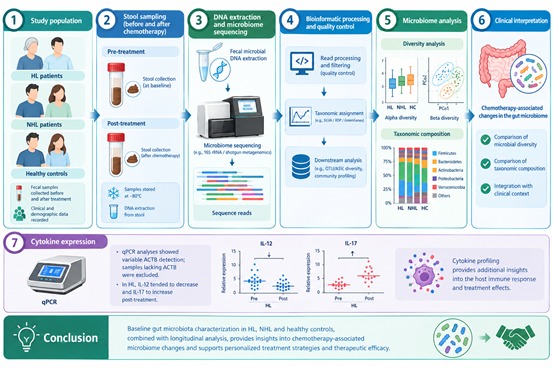
Objective: This study aimed to investigate changes in gut microbiota composition during chemotherapy in patients with Hodgkin lymphoma (HL), non-Hodgkin lymphoma (NHL), and healthy controls, and to evaluate their potential impact on treatment outcomes. Additionally, changes in IL-12 and IL-17 levels during chemotherapy were assessed. Materials and Methods: Fifteen samples from patients with HL were collected after 2–3 cycles of therapy, whereas 15 samples from patients with NHL were collected after 3–4 cycles of therapy. Samples were also collected from healthy controls (n = 33). Microbial alpha diversity and microbiota composition were assessed using 16S rRNA gene sequencing and an operational taxonomic unit (OTU) table generated through R/RStudio. The expression levels of IL-12 and IL-17 were quantified by real-time quantitative PCR (RT-qPCR). Results: In HL patients, genera such as Agathobacter, Romboutsia, and Veillonella were predominantly identified in pre-treatment samples, whereas Dorea, Lactobacillus, and Weissella were enriched post-treatment. Actinobacteriota and Verrucomicrobiota were slightly more abundant before treatment. In NHL patients, Alistipes, Phascolarctobacterium, and Roseburia were characteristic of pre-treatment samples, while Lachnoclostridium, Parabacteroides, and UBA1819 were enriched post-treatment. Actinobacteriota levels increased modestly after chemotherapy. KEGG pathway analysis revealed distinct alterations in HL and NHL, and IL-12 levels differed significantly between NHL patients and healthy controls post-chemotherapy. Conclusions: Baseline gut microbiota profiling may help develop personalized strategies to mitigate chemotherapy-induced dysbiosis and enhance treatment efficacy. Microbiome modulation represents a promising adjunctive approach in lymphoma management.
Keywords:
Hodgkin lymphoma
; non-Hodgkin lymphoma
; healthy controls
; gut microbiota
; dysbiosis
1. Introduction
Hodgkin lymphoma (HL) and non-Hodgkin lymphoma (NHL) are malignant disorders of the lymphatic system with diverse clinical outcomes and treatment strategies [1]. Although chemotherapy, immunotherapy, and targeted agents have significantly improved survival, interindividual differences in treatment response remain substantial [2]. In recent years, the gut microbiota has emerged as a critical modulator of immune homeostasis and cancer therapy outcomes. Increasing evidence links microbial diversity and composition to therapeutic efficacy and toxicity across multiple lymphoma subtypes [3,4,5,6,7,8]. The intestinal microbiota exerts immunomodulatory effects, partly contributing to anti-tumor responses, and may influence the activity of chemotherapeutic agents and immune checkpoint inhibitors [9,10,11].
The present study aimed to investigate gut microbial diversity in patients with HL and NHL using next-generation sequencing of fecal microbial DNA. Microbial profiles before and after chemotherapy were compared with those of healthy controls. In addition, expression of the ACTB gene, commonly used as a housekeeping reference, along with the immune-related cytokines IL-12 and IL-17, was analyzed to explore their potential association with treatment-related microbiota changes.
2. Materials and Methods
2.1. Patients
A total of 30 newly diagnosed lymphoma patients 15 with HL and 15 with NHL were enrolled from the Department of Hematology, Istanbul Faculty of Medicine. First 15 with HL and 15 with non-NHL patients included were those who had been diagnosed, followed up by our outpatient clinic, and had regular follow-up visits. In all cases, the diagnosis was confirmed in accordance with the WHO classification. The inclusion criteria for patients were: no prior treatment, age ≥18 years, and non-pregnant status. Thirty-three healthy volunteers were also included as controls. Controls were required to have no history of malignancy or chronic gastrointestinal inflammatory disease, no antibiotic use within the previous four weeks, no active disease within the previous two weeks, age ≥18 years, and non-pregnant status. Participants with recent antibiotic use (≤30 days before enrollment) were excluded. Two fecal samples were collected from each subject. Samples from patients with HL were collected after 2–3 cycles of therapy, whereas samples from patients with NHL were collected after 3–4 cycles of therapy. Samples were also collected from healthy controls (n = 33). Stool samples were stored at –80 °C until analysis. RNA isolation was performed immediately due to the labile nature of RNA.
2.2. Nucleic Acid Isolation from Stool Samples
DNA was extracted with the ZymoBIOMICS DNA Kit (USA) using optimized combinations of mechanical, chemical, and enzymatic lysis. Silica columns were used for DNA-protein separation, RNase removed RNA, and DNA was eluted in nuclease-free water. Concentration and purity were assessed spectrophotometrically, and only samples with OD260/280 = 1.8–2.0, OD260/230 = 2.0–2.2, and DNA ≥10 ng/µL were included.
2.3. Microbiome Sequencing
Microbiome sequencing was performed on the Illumina MiSeq platform using the Nextera XT DNA Library and Index Kits, following the manufacturer’s protocol. DNA was fragmented, adapters ligated, and purified. A pooling strategy was used instead of panel-based systems, with Illumina MiSeq v3 reagents and 5% PhiX control for quality monitoring
2.4. Bioinformatics and Statistical Analysis
Sequencing produced fastq and bam files, which were checked for amplicon quality and read depth. Initial analyses used BBTools, MagicBlast, and SAMtools. OTUs were assigned via BLASTn with the NCBI 16S rRNA database, and OTU tables were analyzed in R (phyloseq, microbiome, vegan, ggplot2). Alpha diversity indices and statistical analyses were performed in RStudio using non-parametric tests (Wilcoxon or Mann–Whitney) due to non-normal data distribution, along with Bonferroni correction.
2.5. Real-Time Polymerase Chain Reaction (rtPCR)
Total RNA was isolated from blood using the QIAGEN RNeasy Kit with DNase digestion. RNA quality was checked (A260/280 ≈ 1.8–2.0), and cDNA was synthesized with QuantiTect, diluted 1:5–1:10 for SYBR Green qPCR. Thermal cycling included 95 °C activation, 40 cycles of denaturation, annealing (55–60 °C), and extension, followed by melt curve analysis. Relative IL-12 and IL-17 expression was calculated using ΔΔCt normalized to ACTB and compared to healthy controls.
3. Results
3.1. Patient Characteristics
Baseline demographic and clinical features of patients are summarized in Table 1 and Table 2. The mean age was 37.5 ± 11.7 years in HL and 49.5 ± 14.7 years in NHL. The male-to-female ratio was approximately 1.4:1. Subtype distribution and interim PET-CT responses are detailed in Table 1 and Table 2.
3.2. Microbiota Composition
In this study, across all samples, Bacillota (Firmicutes) and Bacteroidota were the dominant phylum, followed by Proteobacteria, Verrucomicrobiota, and Actinobacteria (Figure 1). At the genus level, a total of 899 genera were detected. The most abundant taxa included Prevotella, Escherichia, Bacteroides, and Faecalibacterium, Pseudomonas, Lactobacillus, Klebsiella, Alloprevotella, Collinsella, and Enterobacter.
3.3. Group-Specific Changes in HL and NHL Groups
In HL, Bacillota predominated pre and after-therapy, with modest shifts in Verrucomicrobiota and Actinobacteria but no consistent treatment-related pattern. At the genus level, Romboutsia and an undefined Lachnospiraceae genus decreased after treatment, whereas Lactobacillus and Weissella increased (Figure 2a–c). Compared with controls, HL patients after therapy had lower Akkermansia and Barnesiella but higher Lactobacillus abundance. In NHL, Bacillota and Bacteroidota also dominated. Proteobacteria levels were generally higher compared to HL and controls. Genera such as Prevotella increased after treatment, while Phascolarctobacterium and Roseburia decreased (Figure 2d,e).
3.4. Alpha and Beta Diversity
Alpha diversity was significantly reduced in HL post-treatment compared to controls across all indices, and also declined from pre- to post-treatment using Shannon and Inverse Simpson metrics (Figure 3a,b). No significant diversity changes were observed in the NHL group (Figure 3c,d).
CoA plot based on Bray–Curtis distance, comparing gut microbiota composition between control and post-treatment HL samples (Figure 3e,f). To assess beta diversity, there is no apparent clustering in the control and pre-treatment (Figure 3g), pre- and post-treatment samples in the NHL groups (Figure 3h).
3.5. Functional Predictions
In HL, two Level 2 pathways (Transport and Catabolism and Substance Dependence) were found to differ significantly between the control and post-chemotherapy groups (Supplementary Figure S1a). Additionally, 20 Level 3 pathways were identified as significantly different between these two groups. By contrast, only four Level 3 KEGG pathways showed significant differences between the pre- and post-chemotherapy groups in HL patients. These pathways were D-Arginine and D-Ornithine Metabolism, Glycosaminoglycan Degradation, Glycosphingolipid Biosynthesis, and Sesquiterpenoid and Triterpenoid Biosynthesis (Supplementary Figure S1b). In NHL, one Level 1 pathway (Environmental Information Processing), two Level 2 pathways (Membrane Transport, Translation) (Supplementary Figure S1c,d), and 20 Level 3 pathways differed significantly between control and post-treatment groups (Supplementary Figure S1e).
3.6. Cytokine Expression
qPCR analyses showed variable ACTB detection; samples lacking ACTB were excluded. In HL, IL-12 tended to decrease and IL-17 to increase post-treatment, though not significantly (Supplementary Figure S2a). In NHL, IL-12 expression was significantly lower in post-treatment compared to controls (p = 0.011) (Supplementary Figure S2b).
4. Discussion
Research on gut microbiota in oncology has expanded rapidly since the early 2000s, with growing evidence linking microbial composition to treatment efficacy, toxicity, and clinical outcomes [5,6,7,12,13]. Although microbiota chemotherapy interactions have been widely studied in solid tumors, lymphoma specific data, particularly comparing HL and NHL, are scarce. Therefore, this study provides disease-specific preliminary insights rather than proposing a pioneering concept.
In our study, chemotherapy was associated with significant alterations in gut microbiota, particularly in HL patients, where alpha diversity markedly decreased after treatment. These findings support the idea that chemotherapy, along with the frequent use of antibiotics and antifungals, disrupts microbial diversity, reducing beneficial taxa and promoting potentially pro-inflammatory or pathogenic genera. [14,15,16,17].
At the phylum level, HL patients showed dominance by Bacillota followed by Bacteroidota, with post-treatment samples exhibiting decreased Verrucomicrobiota compared to controls. Genus-level analyses revealed loss of butyrate-producing taxa such as Romboutsia and Lachnospiraceae, alongside increases in Lactobacillus and Weissella. In NHL patients, compositional shifts were observed most notably an increase in Prevotella and reduction of SCFA-producers like Roseburia and Phascolarctobacterium although overall diversity remained stable. These observations align with previous reports of chemotherapy-induced dysbiosis and opportunistic pathogen enrichment. Beta diversity assessment using Bray–Curtis distances and PCoA plots showed no clear clustering between groups, suggesting that chemotherapy affects specific taxa and microbial diversity rather than causing complete restructuring of the community.
In this study, analysis of unique genera across pre- and, post-treatment, and control groups in both HL and NHL patients revealed notable shifts in gut microbiota composition consistent with previously reported findings. Liang et al. reported that Phascolarctobacterium was associated with increased lymphoma risk, whereas the phylum Lentisphaerae, the family Desulfovibrionaceae, and the genus Haemophilus (Proteobacteria), as well as the family and genus Methanobrevibacter (Euryarchaeota), were protective [18]. In NHL patients, post-chemotherapy fecal samples showed decreases in Bacillota (Firmicutes) and Actinobacteria, increases in Proteobacteria, and reductions in butyrate-producing bacteria (Roseburia, Coprococcus, Bifidobacterium, Ruminococcus, Faecalibacterium), along with an increase in Enterobacteriaceae including Citrobacter, potentially impairing colonic homeostasis [19] Studies in DLBCL patients reported elevated Proteobacteria and Enterobacteriaceae (notably Escherichia coli) in untreated patients compared with healthy controls, whereas higher levels of Lactobacillus fermentum and other Lactobacillus species correlated with complete remission [20]. Similarly, DLBCL patients exhibited reduced microbial diversity and increased Enterobacteriaceae, including E. coli and Klebsiella pneumoniae, compared with healthy controls [21]. In our study, patient 1 in the NHL lgroup received multiple antibiotic treatments but demonstrated only a partial response to therapy. The patient subsequently died and hepatotoxicity developed prior to death. Post-treatment microbiota analyses revealed a marked increase in the genus Lachnoclostridium, An increase in this bacterial genus has been suggested to contribute to microbial dysbiosis and may interact with immune cells, potentially increasing susceptibility to tissue damage, particularly in the ileum. In the same patient, a reduction or loss of commensal bacterial communities such as Parabacteroides was observed following treatment, whereas an increase in species belonging to the Escherichia-Shigella group was detected. Notably, these bacterial taxa were present at considerably lower levels at the time of the initial diagnosis. Furthermore, the low gut microbiota diversity observed at diagnosis may have negatively influenced the clinical course of the disease and may be associated with the poor prognosis observed in this patient. Also in the NHL group, analysis of the four patients who ultimately died revealed that all had reduced gut microbiota diversity at the time of initial diagnosis. In addition to the previously discussed patient 1, patients 2, 8, and 10 were also evaluated. The gut microbiota of patient 2 was dominated by the genus Bacteroides prior to treatment. Patient 8 exhibited high levels of bacteria belonging to the Escherichia–Shigella group before treatment. In contrast, patient 10 showed a relatively high abundance of the commensal genus Akkermansia at the time of diagnosis; however, a marked decrease in this genus was observed following treatment. However, these observations are based on a very small number of patients and should be interpreted cautiously. These findings are descriptive and hypothesis-generating rather than conclusive.
Within the Hodgkin lymphoma (HL) patient group, patient 6 exhibited a relatively high abundance of the commensal genus Faecalibacterium at the initiation of treatment. Notably, this patient achieved a complete response to therapy and demonstrated a favorable prognosis. Overall, patients in this disease group showed greater microbiota diversity at the time of diagnosis, with relatively higher abundances of commensal genera such as Veillonella and Prevotella. This observation may be associated with the generally favorable treatment responses observed in this patient group.
Functional predictions indicated that chemotherapy affected microbial metabolic pathways, including amino acid metabolism (asparagine, arginine, tryptophan), membrane transport, and xenobiotic degradation [22,23,24,25]. In HL, changes included triterpenoid biosynthesis pathways, which are associated with membrane adaptation and oxidative stress responses. In NHL, higher-level pathway shifts particularly Environmental Information Processing and Membrane Transport suggest broader microbial adaptive restructuring, potentially reflecting altered host–microbe signaling and nutrient exchange capacity under chemotherapy-induced stress. Microbiota-derived metabolites can influence immune responses within the tumor microenvironment by modulating pathways such as mTOR and amino acid sensing, and by regulating the IL-17/IL-12 axis [26,27,28,29]. In this study, following treatment, HL patients tended to exhibit decreased IL-12 and increased IL-17 levels, whereas in post-treatment NHL patients had significantly lower IL-12 compared to controls. These results suggest disease specific microbiota–immune interactions that may impact treatment outcomes and infection risk. However, given the absence of statistical significance and limited sample size, these findings should be interpreted cautiously. The lack of a statistically significant IL-17 shift in NHL indicates that chemotherapy-associated immune remodeling may not uniformly affect Th17 pathways. These observations support the concept of disease-specific immune remodeling.
Overall, our findings demonstrate that chemotherapy induces distinct, disease specific changes in gut microbiota composition, predicted function, and immune signaling in lymphoma patients. Future studies integrating shotgun metagenomics, metabolomics, short-chain fatty acid measurements, and longitudinal immune profiling are needed to validate and extend these preliminary observations.
Limitations
This study has several limitations. First, the sample size was relatively small (n = 15 per group), limiting statistical power and preventing subgroup analyses based on lymphoma subtype, chemotherapy regimen, disease stage, or antibiotic exposure. Microbiota composition is influenced by diet, genetics, and prior antibiotic use, which could not be fully controlled. Second, only two sampling time points were included, without long-term follow-up. Consequently, the direct relationship between microbiota alterations and clinical outcomes, such as treatment response, infections, or treatment-related toxicities, could not be assessed. Third, 16S rRNA sequencing provides limited species- or strain-level resolution and does not directly measure microbial function. KEGG pathway analyses were based on inferred metagenomic predictions rather than experimentally validated metabolic activity; therefore, functional findings should be considered hypothesis-generating.
Furthermore, the causal relationship between microbiota alterations and IL-12/IL-17 modulation can not be established. Chemotherapy may independently affect immune signaling and microbial ecology, while immune suppression may secondarily alter microbiota composition. Finally, the absence of matched dietary or household controls and the exclusion of some samples due to ACTB normalization variability may have reduced statistical power, particularly in the HL subgroup. Despite these limitations, this study provides preliminary disease-specific insights into microbiota–immune–treatment interactions in lymphoma patients. Therefore, the findings should be interpreted cautiously and considered exploratory rather than definitive.
5. Conclusions
In conclusion, our study demonstrates that chemotherapy induces distinct, disease-specific alterations in gut microbiota composition, diversity, predicted metabolic pathways, and immune cytokine profiles in patients with HL and NHL Although the sample size is limited and the findings are preliminary, integrating microbiota composition, functional predictions, and cytokine responses provides a more comprehensive view of the microbiota-immune-treatment relationship in lymphoma. These results highlight the potential of gut microbiota as a biomarker for treatment response and prognosis and suggest that microbiota-targeted interventions could be explored as supportive strategies in lymphoma therapy. Future studies with larger cohorts, extended follow-up, and multi-omics analyses are needed to validate and expand these findings.
Supplementary Materials
The following supporting information can be downloaded at the website of this paper posted on Preprints.org.
Author Contributions
Conceptualization, O.O., SKB., Z.A., F.O.S., T.O.T., E.E.G., Methodology, O.O., S.K.B, T.O.T., Z.A., F.O.S., E.E.G., U.S., Formal Analysis. O.O., S.K.B., T.O.T., Z.A., F.O.S., E.E.G., U.S, PO.; Investigation, O.O., S.K.B., T.O.T., Z.A., F.O.S., E.E.G., U.S.; Recruited patients and collected clinical data at Haemotology Department, S.K.B. and T.O.T., Writing—Original Draft Preparation Z.A, E.E.G.; Writing—Review and Editing, O.O., S.K.B., T.O.T., Z.A., F.O.S., E.E.G., U.S, PO.; Visualization O.O., U.S.; Supervision O.O., Project Administration O.O; Funding Acquisition O.O.,, All authors have read and agreed to the published version of the manuscript.
Funding
This study was supported by Istanbul University Scientific Research Projects Unit (BAP) [Grant No. 36421].
Institutional Review Board Statement
The study was conducted in accordance with the Declaration of Helsinki, and approved by Ethics Committee for Clinical Research, Istanbul Faculty of Medicine (protocol No:2019/1548) for studies involving humans.
Informed Consent Statement
Informed consent was obtained from all subjects involved in the study.
Data Availability Statement
The data are not publicly available due to patient confidentiality, but are available from the corresponding author upon reasonable request.
Conflicts of Interest
The authors declare that they have no conflict of interest.
References
- Xiao, C., et al., [Clinical and pathological reassessment of 493 cases of non-Hodgkin’s lymphomas according to current WHO classification of lymphoid neoplasms]. Zhonghua Bing Li Xue Za Zhi, 2005. 34(1): p. 22–7. [PubMed]
- Cozen, W., et al., Fecal microbiota diversity in survivors of adolescent/young adult Hodgkin lymphoma: a study of twins. Br J Cancer, 2013. 108(5): p. 1163–1167. [CrossRef] [PubMed]
- Santoni, M., et al., Re: Gut Microbiome Influences Efficacy of PD-1-based Immunotherapy Against Epithelial Tumors. Eur Urol, 2018. 74(4): p. 521–522. [CrossRef] [PubMed]
- Peled, J.U., et al., Intestinal Microbiota and Relapse After Hematopoietic-Cell Transplantation. J Clin Oncol, 2017. 35(15): p. 1650–1659. [CrossRef] [PubMed]
- Jian, X., et al., Alterations of gut microbiome accelerate multiple myeloma progression by increasing the relative abundances of nitrogen-recycling bacteria. Microbiome, 2020. 8(1): p. 74. [CrossRef] [PubMed]
- Yi, M., et al., Gut microbiome modulates efficacy of immune checkpoint inhibitors. J Hematol Oncol, 2018. 11(1): p. 47. [CrossRef] [PubMed]
- Chrysostomou, D., et al., Gut Microbiota Modulation of Efficacy and Toxicity of Cancer Chemotherapy and Immunotherapy. Gastroenterology, 2023. 164(2): p. 198–213. [CrossRef] [PubMed]
- Ting, N.L., H.C. Lau, and J. Yu, Cancer pharmacomicrobiomics: targeting microbiota to optimise cancer therapy outcomes. Gut, 2022. 71(7): p. 1412–1425. [CrossRef] [PubMed]
- Lérias, J.R., et al., Microbes as Master Immunomodulators: Immunopathology, Cancer and Personalized Immunotherapies. Front Cell Dev Biol, 2019. 7: p. 362. [CrossRef] [PubMed]
- Yang, Q., et al., A Review of Gut Microbiota-Derived Metabolites in Tumor Progression and Cancer Therapy. Adv Sci (Weinh), 2023. 10(15): p. e2207366. [CrossRef] [PubMed]
- Alexander, J.L., et al., Gut microbiota modulation of chemotherapy efficacy and toxicity. Nat Rev Gastroenterol Hepatol, 2017. 14(6): p. 356–365. [CrossRef] [PubMed]
- Routy, B., et al., Gut microbiome influences efficacy of PD-1-based immunotherapy against epithelial tumors. Science, 2018. 359(6371): p. 91–97. [CrossRef] [PubMed]
- Russo, F., et al., The effects of fluorouracil, epirubicin, and cyclophosphamide (FEC60) on the intestinal barrier function and gut peptides in breast cancer patients: an observational study. BMC Cancer, 2013. 13: p. 56. [CrossRef] [PubMed]
- Li, S., S. Zhu, and J. Yu, The role of gut microbiota and metabolites in cancer chemotherapy. J Adv Res, 2024. 64: p. 223–235. [CrossRef] [PubMed]
- Fernandes, M.R., et al., Targeting the gut microbiota for cancer therapy. Nat Rev Cancer, 2022. 22(12): p. 703–722. [CrossRef] [PubMed]
- Ramirez, J., et al., Antibiotics as Major Disruptors of Gut Microbiota. Front Cell Infect Microbiol, 2020. 10: p. 572912. [CrossRef] [PubMed]
- Patangia, D.V., et al., Impact of antibiotics on the human microbiome and consequences for host health. Microbiologyopen, 2022. 11(1): p. e1260. [CrossRef] [PubMed]
- Liang, J., et al., Causal relationships between gut microbiota and lymphoma: a bidirectional Mendelian randomization study. Front Cell Infect Microbiol, 2024. 14: p. 1374775. [CrossRef] [PubMed]
- Li, B., et al., The causal relationship between gut microbiota and lymphoma: a two-sample Mendelian randomization study. Front Immunol, 2024. 15: p. 1397485. [CrossRef] [PubMed]
- Xu, Z.F., et al., The Gut Microbiome Correlated to Chemotherapy Efficacy in Diffuse Large B-Cell Lymphoma Patients. Hematol Rep, 2024. 16(1): p. 63–75. [CrossRef] [PubMed]
- Yoon, S.E., et al., The influence of microbial dysbiosis on immunochemotherapy-related efficacy and safety in diffuse large B-cell lymphoma. Blood, 2023. 141(18): p. 2224–2238. [CrossRef] [PubMed]
- Yang, Q., et al., The Interaction between Gut Microbiota and Host Amino Acids Metabolism in Multiple Myeloma. Cancers (Basel), 2023. 15(7): p.1942. [CrossRef] [PubMed]
- Hou, S. and Xue, K. Gut microbiota and metabolomic profiling in Relapsed/Refractory lymphoma. Blood. 2025. 146: p.7087. [CrossRef]
- Xu, Y. Alteration of gut microbiome and correlated amino acid metabolism are associated with acute myelocytic leukemia carcinogenesis. Cancer Med. 2023. 12(15): p.16431-16443. [CrossRef] [PubMed]
- Szefel, J., A. Danielak, and W.J. Kruszewski, Metabolic pathways of L-arginine and therapeutic consequences in tumors. Adv Med Sci, 2019. 64(1): p. 104–110. [CrossRef] [PubMed]
- Flannigan., K., L., et al., IL-17A-mediated neutrophil recruitment limits expansion of segmented filamentous bacteria. Mucosal Immunol. 2017. 10(3): p.673-684. [CrossRef] [PubMed]
- Weaver., C.T., et al., Th17: an effector CD4 T cell lineage with regulatory T cell ties. Immunity. 2006. 24(6): p. 677-688. [CrossRef] [PubMed]
- Chao-Yue., S., et al., T helper 17 (Th17) cell responses to the gut microbiota in human diseases. Biomed Pharmacother. 2023. 161: p.114483. [CrossRef] [PubMed]
- Tanabe., T. The effect of probiotics and gut microbiota on Th17 cells. Int Rev Immunol. 2013. 32(5-6): p.511-25. [CrossRef] [PubMed]
Figure 1.
Relative abundance of fecal microbiota at the phylum level across all samples.
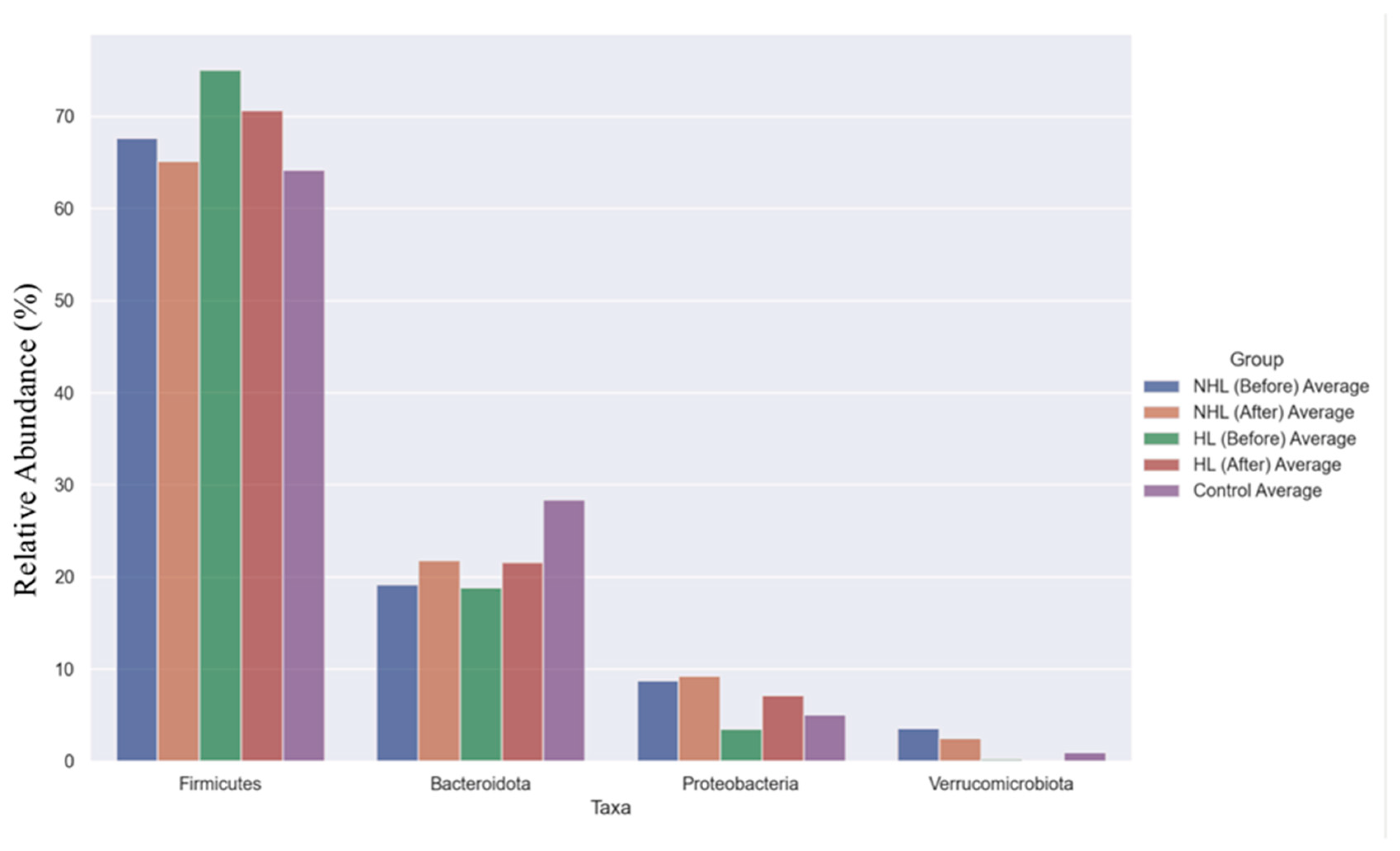
Figure 2.
Genus-level composition of the bacterial microbiota in pre- and post-treatment stool samples from patients with HL and NHL compared with the control group (2a–b: HL; 2c-d: NHL and 2e: control group).
Figure 2.
Genus-level composition of the bacterial microbiota in pre- and post-treatment stool samples from patients with HL and NHL compared with the control group (2a–b: HL; 2c-d: NHL and 2e: control group).
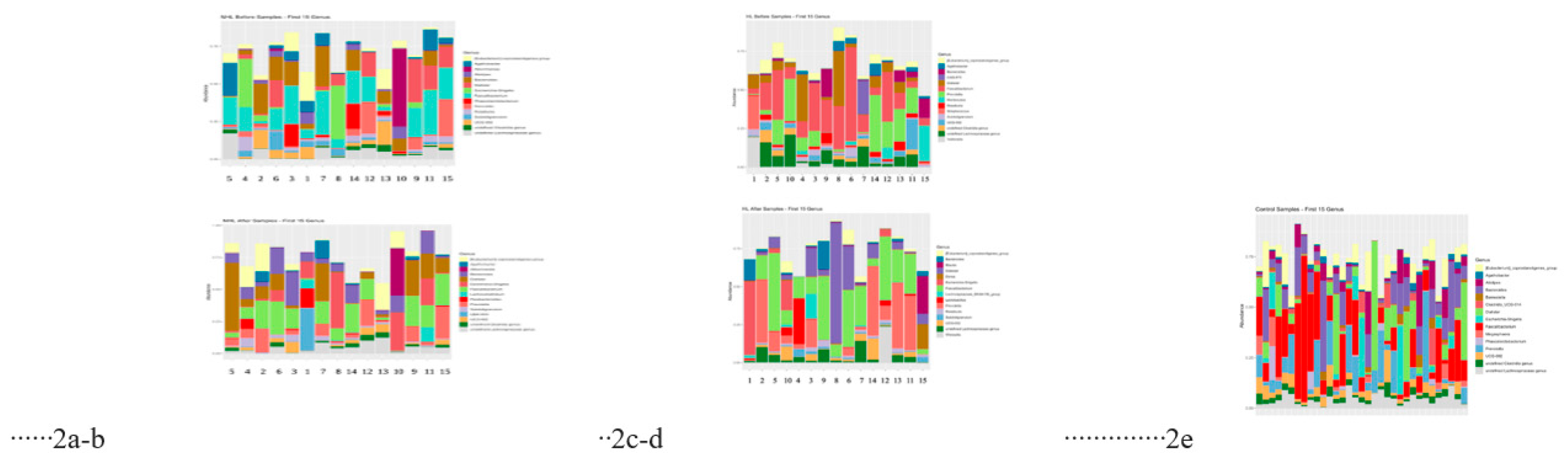
Figure 3.
Alpha diversity analysis of gut microbiota within the HL and NHL groups. Comparison of alpha diversity between the control and post-treatment groups (a) and between the pre- and post-treatment groups (b) within the HL group. Comparison of alpha diversity between the control and post-treatment groups (c) and between the pre- and post-treatment groups (d) within the NHL group. Principal coordinate analysis (PCoA) based on Bray–Curtis distance showing variation in gut microbiota composition in HL and NHL groups. Comparison between control and post-treatment and between pre- and post-treatment within the HL groups (e,f). Comparison between control and post-treatment and between pre- and post-treatment within the NHL groups (g,h).
Figure 3.
Alpha diversity analysis of gut microbiota within the HL and NHL groups. Comparison of alpha diversity between the control and post-treatment groups (a) and between the pre- and post-treatment groups (b) within the HL group. Comparison of alpha diversity between the control and post-treatment groups (c) and between the pre- and post-treatment groups (d) within the NHL group. Principal coordinate analysis (PCoA) based on Bray–Curtis distance showing variation in gut microbiota composition in HL and NHL groups. Comparison between control and post-treatment and between pre- and post-treatment within the HL groups (e,f). Comparison between control and post-treatment and between pre- and post-treatment within the NHL groups (g,h).
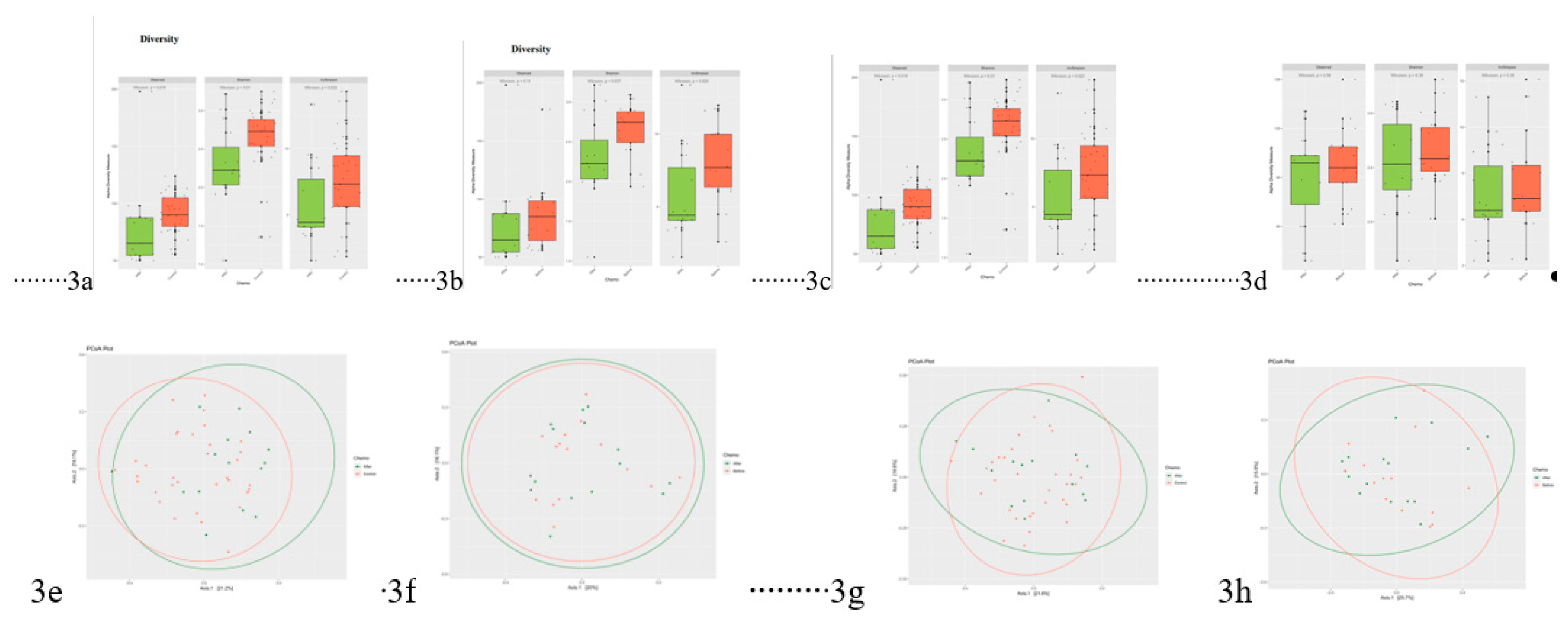

Table 1.
Summary of demographic and clinical characteristics of patients with HL (n=15).
| Patient No | Age/Sex | Hodgkin Lymphoma Subtype | Stage | International Prognostic Score (IPS) | Induction Regimen/ Cycle(s) | Antibiotics | Total Induction Cycle(s) |
Induction Treatment Response |
Relapsed/ Refractory |
Follow Up (Month/s) / Therapy Releated Toxicity / Last Status |
| 1 | 49/M | Mixed cellularity | IIIB | 5 | 2 x (A-AVD) | Metronidazole, ciprofloxacine | 6 | CR | No | 58,7 / Hepatotoxicity / Alive |
| 2 | 55/F | Nodular sclerosing | IIB | N/A | 2 x ABVD | Ciprofloxacine, clarithromycin | 6 | CR | No | 54,8 / No toxicity / Alive |
| 3 | 39/F | Nodular sclerosing | IVB | 2 | 2 x ABVD, 1 x (A-AVD) |
- | 6 | CR | No | 57,5 / No toxicity / Alive |
| 4 | 26/M | Nodular sclerosing | IVB | 3 | 3 x ABVD | Valaciclovir | 6 | CR | No | 38,9 / No toxicity / Alive |
| 5 | 42/M | Classical HL | IVB | 4 | 2 x ABVD | Valaciclovir | 6 | CR | No | 23,9 / No toxicity / Alive |
| 6 | 26/M | Lymphocyte- predominant | IIA | N/A | 2 x ABVD | - | 6 | CR | No | 48,9 / No toxicity / Alive |
| 7 | 33/M | Nodular sclerosing | IIB | N/A | 2 x ABVD | Clindamycin | 4 | CR | No | 53,5 / No toxicity / Alive |
| 8 | 32/M | Nodular sclerosing | IIA | N/A | 2 x ABVD | - | 2 | PD | Primary Refractory Disease | 34,3 / Neuropathy / Alive |
| 9 | 25/F | Classical HL | IVB | 2 | 2 x (A-AVD) | Amoxicillin clavulanate | 6 | CR | No | 49,6 / No toxicity / Alive |
| 10 | 22/M | Classical HL | IVB | 2 | 2 x ABVD | Amoxicillin clavulanate | 6 | CR | No | 48,4 / No toxicity / Alive |
| 11 | 38/F | Classical HL | IIIB | 3 | 2 x ABVD | - | 6 | CR | No | 27,5 / No toxicity / Alive |
| 12 | 47/F | Nodular sclerosing | IVB | 3 | 2 x ABVD | - | 6 | CR | No | 34,5 / No toxicity / Alive |
| 13 | 31/F | Nodular sclerosing | IVB | 3 | 2 x ABVD | Amoxicillin clavulanate, Kotrimoxazol | 2 | PD | Primary Refractory Disease | 1 / Neuropathy / Alive |
| 14 | 62/M | Classical HL | IIIB | 4 | 2 x ABVD | - | 6 | CR | No | 38,5 / Cardiotoxicity / Alive |
| 15 | 35/M | Classical HL | IIA | N/A | 2 x ABVD | Amoxicillin clavulanate | 6 | CR | No | 40,6 / No toxicity / Alive |
# The International Working Group response criteria for Hodgkin lymphoma. A-AVD: Brentuximab vedotin, Doxorubicin, Vinblastine and Dacarbazine, ABVD: Doxorubicin, Bleomycin, Vinblastine and Dacarbazine. SD: Stable disease, CR: Complete response, PR: Partial response, PD: Progressive disease.
Table 2.
Summary of demographic and clinical characteristics of patients with NHL group (n=15).
| Patient No | Age / Sex | Diagnosis | Stage | NCCN-IPI Score | Regimen / Cycle(s) | Antibiotics |
Response on Interim PET-CT# |
Relapsed/Refractory |
Follow Up (Month/s) / Therapy Releated Toxicity / Last Status |
| 1 | 64/F | Indolent B Cell Lymphoma | IIIB | N/A | COP x 3 | Acyclovir, TMP-SMX, Colistin, Piperacillin-Tazobactam, Vancomycin, Teicoplanin, Meropenem, Amphotericin B, Fluconazole, Posaconazole | PR | No |
3,4 / Hepatotoxicity/Died |
| 2 | 54/M | DLBCL-GBC | II | 2 | Dose adjusted R-EPOCH x 4 | Acyclovir, Fluconazole, TMP-SMX | PR | Primary Refractory Disease | 5,1 / Cardiotoxicity/Died |
| 3 | 68/F | DLBCL-ABC | IIIB | 3 | R-CHOP x 4 | Acyclovir, Fluconazole, TMP-SMX | CR | No | 34,8 / No toxicity / Alive |
| 4 | 38/M | DLBCL-ABC | IVA | 3 | R-CHOP x 4 | - | CR | No | 54,8 / No toxicity / Alive |
| 5 | 35/M | DLBCL-ABC | II | 0 | R-CHOP x 4 | - | CR | No | 51,2 / No toxicity / Alive |
| 6 | 66/F | HGBCL | IIIB | 5 | R-CHOP x 4† | Amoxicillin-Clavulanate | CR | No | 58,5 / No toxicity / Alive |
| 7 | 31/M | HGBCL | II | 0 | R-CHOP x 3 | - | CR | No | 57,8 / No toxicity / Alive |
| 8 | 67/F | HGBCL | IVB | 4 | R-CHOP x 4 | - | CR | Relapsed after 10 months | 12 / Hepato-, Cardiotoxicity/Died |
| 9 | 24/M | HGBCL | IIB | 0 | R-CHOP x 3 | - | CR | No | 20,9 / No toxicity / Alive |
| 10 | 52/M | ALK(-) ALCL | IVB | N/A | CHOEP x 4 | Ciprofloxacin, Amoxicillin-Clavulanate | SD | Primary Refractory Disease | 37,3 / No toxicity / Died |
| 11 | 47/F | HGBCL | IVB | 4 | R-CHOP x 4 | Amoxicillin-Clavulanate | CR | Primary Refractory Disease | 39,9 / No toxicity / Alive |
| 12 | 35/M | HGBCL | IB | 0 | R-CHOP x 3 | - | CR | No | 50,9 / No toxicity / Alive |
| 13 | 45/M | HGBCL | II | 1 | R-CHOP x 3 | - | PR | No | 45,1 / No toxicity / Alive |
| 14 | 67/F | DLBCL-ABC | IIA | 2 | R-CHOP x 4† | - | CR | No | 51,4 / No toxicity / Alive |
| 15 | 50/F | HGBCL | IVB | 4 | R-CHOP x 3 | - | PR | No | 48 / Hepato-, Cardiotoxicity / Alive |
† Prophylactic use of intrathecal methotrexate for double-expressor high-grade B-cell lymphoma. # International Working Group consensus response evaluation criteria in lymphoma (RECIL 2017). DLBCL – GBC: Diffuse large B-cell lymphoma, germinal center B-cell type, DLBCL – ABC: Diffuse large B-cell lymphoma, activated B-cell type, HGBCL: High-grade B-cell lymphoma, ALCL: Anaplastic large cell lymphoma, COP: Cyclophosphamide, Vincristine, and Prednisone, R-EPOCH: Rituximab, Etoposide, Prednisone, Vincristine, Cyclophosphamide, and Doxorubicin, R-CHOP: Rituximab, Cyclophosphamide, Doxorubicin, Vincristine, and Prednisone, CHOEP: Cyclophosphamide, Doxorubicin, Vincristine, Etoposide, and Prednisone, Vinblastine and Dacarbazine. PR: Partial response, CR: Complete response, SD: Stable disease, PD: Progressive disease.
Disclaimer/Publisher’s Note: The statements, opinions and data contained in all publications are solely those of the individual author(s) and contributor(s) and not of MDPI and/or the editor(s). MDPI and/or the editor(s) disclaim responsibility for any injury to people or property resulting from any ideas, methods, instructions or products referred to in the content. |
© 2026 by the authors. Licensee MDPI, Basel, Switzerland. This article is an open access article distributed under the terms and conditions of the Creative Commons Attribution (CC BY) license (http://creativecommons.org/licenses/by/4.0/).
Copyright: This open access article is published under a Creative Commons CC BY 4.0 license, which permit the free download, distribution, and reuse, provided that the author and preprint are cited in any reuse.