

Submitted:
22 October 2026
Posted:
23 October 2026
You are already at the latest version
Abstract
Hydrogels, three-dimensional hydrophilic polymer networks that can imbibe large volumes of water or biological fluids without dissolving, have become one of the most versatile platforms in pharmaceutical and biomaterial science. Their high water content, soft tissue-like mechanics, biocompatibility and tunable network architecture allow them to be engineered as topical, ophthalmic, mucosal, oral and injectable dosage forms and as scaffolds for regenerative medicine. This illustrated review provides a critical, practice-oriented account of the field, with particular emphasis on how hydrogel dosage forms are prepared. Physical cross-linking methods (ionic gelation, polyelectrolyte complexation, thermoresponsive gelation, freeze–thaw cycling and supramolecular assembly) and chemical cross-linking methods (free-radical and photo-initiated polymerization, small-molecule cross-linkers, click and dynamic covalent chemistries, and enzyme-mediated and radiation cross-linking) are examined in terms of their mechanisms, typical materials and conditions, critical process parameters, advantages and limitations. The preparation of conventional pharmaceutical gels based on carbomers, cellulose ethers, poloxamers and polysaccharides is described step by step, together with drug-loading strategies, processing into beads, micro- and nanogels, films, sponges, microneedles and printed constructs, and a troubleshooting guide for the most frequent formulation failures. Advanced architectures that overcome the intrinsic weakness of conventional networks, including hybrid, interpenetrating, nanocomposite, double-network and Tetra-PEG hydrogels, are then discussed, followed by the characterization and quality-control methods needed to establish critical quality attributes such as gel fraction, swelling and mesh size, rheology, morphology, release kinetics, stability, microbiological quality and biocompatibility. The review subsequently analyzes drug release mechanisms, route-specific delivery strategies, stimuli-responsive and hierarchical systems, the selection of drug candidates with reference to physicochemical properties and the Biopharmaceutics Classification System, and applications in tissue engineering and wound care. Finally, clinically approved products and the main translational barriers, namely mechanical fragility, burst release, residual reagents, sterilization, batch-to-batch variability and regulatory classification, are critically evaluated to identify realistic routes towards next-generation, clinically useful hydrogel dosage forms.
Keywords:
hydrogels
; preparation methods
; cross-linking
; ionic gelation
; thermoresponsive gels
; photopolymerization
; click chemistry
; topical gels
; drug delivery
; quality control
1. Introduction
In pharmaceutical terminology, hydrogels are defined as three-dimensional networks of hydrophilic polymers that can absorb and retain large quantities of water or biological fluids without dissolving [1]. Their capacity to imbibe water arises from hydrophilic functional groups, such as hydroxyl (–OH), carboxyl (–COOH), amide (–CONH2) and sulfonic acid (–SO3H) groups, which interact with water through hydrogen bonding and ion–dipole interactions, whereas cross-links between the polymer chains prevent dissolution and preserve the integrity of the swollen network [1,2]. The usefulness of hydrogels in pharmacy and medicine stems largely from their resemblance to the native extracellular matrix (ECM): they can contain more than 90% water, are soft and elastic, and possess an interconnected porosity that permits the diffusion of nutrients, metabolites and drugs [3,4]. These features generally translate into good biocompatibility, minimal mechanical irritation of surrounding tissues and comparatively mild tissue responses, which explains their use in contact with skin, mucosae, the eye and internal tissues [3].
Most histories of medical hydrogels begin in Prague in 1960, when Wichterle and Lím reported cross-linked poly(2-hydroxyethyl methacrylate) (pHEMA) as a material intended for permanent contact with living tissue; within a few years the same polymer had become the first soft contact lens [5]. The literature has grown enormously since then. Roughly 1,000 papers on hydrogels had appeared by 1982, compared with more than 100,000 by 2020 [6], and over those decades the emphasis moved from passive, water-swollen matrices towards gels that respond to their environment and give cells instructive signals [2,7]. What has not changed is the position a hydrogel occupies in a drug product. It is seldom a neutral container; it is the interface at which the synthetic delivery system and the body actually meet, and that holds for nearly every application discussed here [7].
Several properties explain the sustained pharmaceutical interest in these materials. Many hydrogels are well tolerated and, depending on the network chemistry, biodegradable [8]. Gels based on chitosan, alginate or hyaluronic acid are generally of low toxicity and are broken down, by enzymes or by hydrolysis, into fragments that can be metabolized or excreted, although the degradation products of each formulation still require their own safety assessment [9]. Their properties are also unusually adjustable: the choice of polymer, its molecular weight and concentration, and the type and density of cross-links together set the swelling, strength, mesh size and degradation rate, which can therefore be matched to a given route and indication [7,10]. A further advantage, and for biologics probably the decisive one, is that the network protects peptides, proteins and nucleic acids from enzymatic attack and releases them over hours to months, making fewer doses, better adherence and lower systemic exposure achievable [4,10]. More recently, ‘smart’ gels that respond to pH, temperature, ionic strength, enzymes or light have brought site-specific and on-demand release within practical reach [11].
The limitations are equally clear. Mechanical weakness is mentioned most often. A swollen network contains little polymer by volume and its cross-links are rarely evenly distributed, so many hydrogels, especially those made from natural polymers, are soft, brittle or prone to fracture and creep [11]; this alone excludes them from load-bearing sites such as articular cartilage. Chemically cross-linked materials raise a different concern, namely cytotoxic residues of monomer, cross-linker, initiator or catalyst, which have to be removed by validated purification and controlled analytically before clinical use [10]. The aqueous matrix is also a poor host for lipophilic drugs, which limits both loading and uniformity, while the mesh of most swollen gels (typically 5–100 nm) is far larger than a small-molecule drug, which is why rapid diffusion and an initial burst are so common [4,12]. Large-scale manufacture, terminal sterilization and long-term storage of water-rich materials that tolerate neither heat nor radiation well remain serious obstacles to clinical translation [13].
Hydrogel design is therefore seldom a matter of optimizing a single variable. In practice it means finding a workable balance between biocompatibility, mechanical performance and therapeutic function, a balance we refer to in this review as the ‘design trilemma’. Raising the cross-link density illustrates the point well: the gel becomes stiffer and releases drug more slowly, but it also swells less, its mesh tightens, it may become brittle, and cells and macromolecular drugs move through it with greater difficulty [7,14]. Natural polymers pull in the opposite direction, offering better bioactivity and cytocompatibility at the expense of strength and batch reproducibility. Much of the current interest in hybrid and composite gels that combine natural and synthetic components stems from this tension. Table 1 lists the main trade-offs, their underlying causes and the strategies currently used to manage them [1].
Many reviews classify hydrogels and survey their applications. Far fewer explain, in practical terms, how a given chemistry is turned into a reproducible and properly characterized dosage form, and the relevant information is scattered across the polymer, pharmaceutical and biomedical literature. This review was written to fill part of that gap, with formulation scientists as its main readers. After a short account of cross-linking principles (Section 2) and applications (Section 3), Section 4 deals in detail with physical and chemical preparation methods, the manufacture of conventional pharmaceutical gels, drug loading, processing into final dosage forms and the manufacturing problems encountered most often. Section 5 covers advanced network architectures and Section 6 the characterization and quality-control tests used to define critical quality attributes. Section 7 to 10 then discuss drug release, route-specific delivery, the selection of drug candidates and regenerative applications, and Section 11 considers approved products, barriers to translation and future directions.
2. Cross-Linking Principles and Choice of Preparation Route
Every hydrogel owes its existence to cross-links. Junctions formed between linear or branched macromolecules convert a free-flowing polymer solution into an insoluble viscoelastic network that swells in water instead of dissolving, and the chemistry, density and lifetime of these junctions determine both the mechanical behavior of the gel and how quickly drugs diffuse through it [1,34,35]. Two broad types of junctions are used. Physical cross-links (ionic bridges, hydrogen bonds, hydrophobic domains, crystallites and chain entanglements) are reversible; because no reactive reagents are involved, purification is straightforward and sensitive payloads are spared, but the resulting gels tend to be weaker and may creep, erode or dissolve when diluted [35,36,37,38,39]. Chemical cross-links, by contrast, are covalent and permanent. They are introduced by chain-growth (free-radical) polymerization, by step-growth reactions such as Michael addition and click chemistry, or with small-molecule cross-linkers, enzymes or ionizing radiation, and they give stronger, more durable networks at the price of residual reagents that have to be removed [30,40,41,42,43]. Either type can be processed into bulk gels and films, micro- and nanogels, or liquids that gel in situ after administration [27,44,45]. Section 4 examines the individual methods, their critical process parameters and their limitations; the emphasis here is on how a formulator might choose between them.
That choice should be driven by the intended product rather than by the chemistry alone, and five questions help to make it. How sensitive is the payload (protein, nucleic acid, living cells or small molecule) to heat, organic solvents, radicals and extremes of pH? Must the gel form in situ, or can it be performed and then administered? What mechanical strength and residence time are required at the site of application? What level of residual reagents is acceptable, and can the process remove and quantify them? And how will the product be sterilized or preserved, scaled up and stored? Figure 1 arranges these questions as a decision tree. In most cases the answers point towards physical or enzymatic gelation for cell- and protein-loaded systems, towards simple physically gelled polymers for topical, ophthalmic and mucosal products, and towards covalent or radiation cross-linking for devices and implants that must withstand prolonged mechanical or osmotic stress [7,25].
3. Overview of Hydrogel Applications
Hydrogels are now established biomaterials in pharmaceutical science and in regenerative medicine, mainly because a single material can combine a hydrophilic network, adjustable viscoelasticity and biological versatility [1,44]. In pharmacotherapy their principal role is in drug delivery systems (DDS) [44], in which the gel acts as a transport matrix that modifies the pharmacokinetics, bioavailability and local distribution of the drug for a given route [44,46]. Injected parenterally, in situ-gelling matrices form local depots that release drug over extended periods and smooth out the peaks and troughs typical of conventional dosing [27,28]. Applied to the skin as patches, films or microneedle arrays, they hydrate the stratum corneum and, since hepatic first-pass metabolism is avoided, support efficient cutaneous and transdermal delivery [47,48]. After oral administration, pH-responsive gels can protect acid-labile peptides and proteins in the stomach before swelling and releasing them in the intestine [49,50]. In ophthalmology, mucoadhesive inserts and in situ-gelling drops prolong precorneal residence and counteract the rapid tear drainage that limits conventional eye drops [13,37].
Hydrogels are equally important in tissue engineering and regenerative medicine (TERM) [1,51]. Their high equilibrium water content and their structural resemblance to the native extracellular matrix (ECM) allow them to give cells mechanical and biochemical cues similar to those of real tissue [1,52]. Oxygen, nutrients and metabolic waste diffuse freely through the hydrated mesh, while the network itself supports cell attachment, proliferation and lineage-specific differentiation [52,53]. Hydrogel scaffolds are therefore being studied for some of the most difficult repair problems in medicine, including osteochondral defects, dermal wounds, avascular cartilage and infarcted myocardium [17,51,54,55].
Wound care is probably where hydrogels have had their most visible clinical impact. By keeping the wound bed moist, hydrogel dressings support autolytic debridement, re-epithelialization and the formation of granulation tissue [15,56,57]. Their porous structure can absorb excess exudate without drying the underlying tissue, and the gel layer acts as a physical barrier to bacteria from outside [56,57]. Newer dressings go further. Rather than simply covering the wound, they are designed to act on it [15,44,56], serving at once as a scaffold for new tissue, a carrier for living cells, and a reservoir from which antiseptics, anti-inflammatory agents or angiogenic growth factors are released either steadily or in response to a trigger [44,53,56]. A bioactive dressing of this kind is, in effect, both a responsive drug depot and a template for skin regeneration [15,56,57]. It is this convergence of delivery and regeneration, together with the pace of recent technical progress, that prompted the present review of hydrogel design, synthesis, characterization and translation across pharmaceutical and biomedical applications [1,10,44].
4. Hydrogel Preparation Methods: Principles, Critical Process Parameters and Practical Considerations
The conversion of a polymer solution into a three-dimensional hydrogel is the step that fixes most of the final physicochemical and therapeutic properties of the product. Network structure, and therefore swelling, strength, degradation and release, depends not only on the cross-linking mechanism but also on raw-material attributes (polymer type, grade, molecular weight and purity) and on process parameters such as concentration, temperature, pH, mixing, curing time and the order in which components are combined [25,36,58]. In this section, preparation methods are grouped according to their cross-linking mechanism. For each method, the underlying principle, typical materials and conditions, critical process parameters (CPPs), advantages and limitations are discussed. The preparation of conventional pharmaceutical gels, drug-loading and processing strategies, and a troubleshooting guide are then presented (Section 4.3, Section 4.4, Section 4.5 and Section 4.6).
4.1. Physically Cross-linked Hydrogels: Reversible and Stimuli-Responsive Networks
Physical hydrogels are formed by reversible, non-covalent interactions, including ionic interactions, hydrogen bonding, hydrophobic association, crystallite formation and chain entanglement. Because they generally do not require potentially cytotoxic cross-linkers or initiators, purification is simpler and cytocompatibility is usually superior. The transient nature of these junctions also confers responsiveness, allowing sol–gel transitions in response to temperature, pH or ions, which is the basis of most in situ-gelling pharmaceutical products [25,37]. The price of this reversibility is lower and less predictable mechanical stability, sensitivity to dilution and ion exchange, and a tendency towards syneresis or erosion during storage and use.
4.1.1. Ionic (Ionotropic) Gelation
Ionic gelation relies on electrostatic cross-linking between a charged polymer and multivalent counter-ions of opposite charge. The most widely used system is sodium alginate, a linear copolymer of β-D-mannuronic (M) and α-L-guluronic (G) acid residues, cross-linked by divalent cations (Figure 2). Ca2+ ions bind cooperatively between G-blocks of adjacent chains to form ‘egg-box’ junction zones, so the G-content and G-block length of the alginate largely determine gel strength: high-G alginates give stiff, porous but more brittle gels, whereas high-M alginates give softer, more elastic gels [32,49]. The affinity of alginate for divalent cations decreases approximately in the order Pb2+ > Cu2+ > Cd2+ > Ba2+ > Sr2+ > Ca2+ > Co2+, Ni2+, Zn2+ > Mn2+, whereas Mg2+ forms little or no gel [32]. Ba2+ and Sr2+ therefore produce stronger and more stable gels than Ca2+, but the toxicity of Ba2+ and Cu2+ limits their pharmaceutical use, and calcium chloride remains the cross-linker of choice. The same principle applies to other polyanions, such as low-methoxyl pectin and gellan gum (Ca2+, Na+, K+) and κ-carrageenan (K+), and to the polycation chitosan, which is cross-linked by multivalent anions such as sodium tripolyphosphate (TPP) to produce nano- and microparticles [38,59].
Two procedural modes are used (Figure 3). In external (diffusion) gelation, the alginate solution, typically 1–3% w/v, is extruded dropwise through a needle or nozzle into a stirred bath of CaCl2, typically 0.05–0.5 M (approximately 0.5–5% w/v), or a cast film or gel is immersed in the bath; Ca2+ diffuses inwards and cross-links the droplet from the surface towards the core within seconds to minutes. This method is simple and underlies most alginate beads and microcapsules, but the rapid, diffusion-limited reaction produces a polymer concentration gradient, with a dense surface and a weaker, less cross-linked core [32,60]. In internal (in situ) gelation, an insoluble or sparingly soluble calcium source, such as CaCO3 or CaSO4, is dispersed in the alginate solution and Ca2+ is released slowly and uniformly, for example by adding D-glucono-δ-lactone (GDL), whose gradual hydrolysis to gluconic acid lowers the pH and solubilizes the carbonate [49,61]. Slower gelation gives more homogeneous, mechanically stronger and more reproducible gels that can be cast into molds of defined shape [60].
The quality of ionically gelled products is governed by a small number of critical parameters. Polymer concentration, molecular weight and M/G composition determine viscosity, droplet formation and network density. Cation type and concentration, curing (residence) time in the bath, temperature and stirring determine the extent and homogeneity of cross-linking. For bead production, needle internal diameter, extrusion rate, dropping height and, where used, coaxial air flow, vibration frequency or electrostatic potential determine particle size and sphericity; electrostatic or vibrating-nozzle generators, or emulsification methods, are needed to obtain microbeads smaller than a few hundred micrometers [32]. After curing, beads are washed to remove excess cations and may be coated with a polycation, such as chitosan or poly-L-lysine, to form a polyelectrolyte membrane that reduces porosity and drug leakage.
Much of the appeal of ionic gelation lies in its simplicity: it is quick, inexpensive, entirely aqueous and mild enough for cells, proteins and probiotics. Three of its weaknesses are, in our view, often underestimated. The first is drug loss. Small water-soluble drugs diffuse into the gelling bath and the wash water, and encapsulation efficiency can be poor unless the curing time is kept short, the drug is complexed beforehand or the bath is pre-saturated with drug. The second is stability. In physiological fluids Na+ exchanges with the bound Ca2+, and phosphate, citrate and other chelators remove the cross-linking ions, so calcium-alginate gels swell, weaken and finally dissolve in a poorly controlled way. The third concerns elimination: because humans have no enzyme that degrades alginate, the gel can disappear only through ion exchange, and high-molecular-weight chains may not be cleared efficiently by the kidneys [32]. A clinically useful variant of the method is ion-activated in situ gelation. Low-acyl gellan gum instilled as an eye drop gels on contact with the cations of tear fluid and so remains longer on the ocular surface, the principle behind a marketed once-daily timolol product [62,63].
4.1.2. Polyelectrolyte Complexation
Mixing two oppositely charged polyelectrolytes produces a polyelectrolyte complex (PEC). Numerous electrostatic contacts form between the chains, and the release of small counter-ions makes the process entropically favorable, so no covalent cross-linker is required. In pharmaceutical work chitosan is the usual polycation, paired with alginate, pectin, carrageenan, xanthan gum, hyaluronic acid or carboxymethylcellulose, or with synthetic polyanions such as poly(acrylic acid) [39,58]. The kind of complex that forms depends on the charge density and molecular weight of each polymer, on the charge ratio at which they are mixed, on ionic strength, and on pH, which sets the degree of ionization of both partners (chitosan has a pKa of about 6.3–6.5). The order and rate of mixing matter more than is often appreciated. Concentrated solutions mixed quickly tend to precipitate as heterogeneous aggregates rather than forming a continuous gel, which is why PEC hydrogels are usually made from dilute solutions, built up layer by layer on a template, or applied as coatings on preformed ionic gels. PECs are mucoadhesive and pH-responsive, and they have been explored widely for oral, nasal and wound applications. Their swelling and integrity nonetheless change markedly with the pH and salt content of the surrounding medium, and in vitro results need to be read with this in mind.
4.1.3. Thermoresponsive (Temperature-Induced) Gelation
Certain polymers undergo a reversible sol–gel transition in response to temperature. Polymers with lower critical solution temperature (LCST)-type behavior are soluble when cold and gel or phase-separate on warming; this group includes poloxamers, methylcellulose and hydroxypropyl methylcellulose, poly(N-isopropylacrylamide) (PNIPAAm, LCST about 32 °C) and biodegradable polyester–PEG block copolymers such as PLGA–PEG–PLGA [64,65]. This behavior is ideal for injectable or instillable in situ-forming systems that are administered as free-flowing liquids at room temperature and gel at body temperature (Figure 4). Polymers with upper critical solution temperature (UCST)-type behavior, such as gelatin, agarose and gellan gum, behave in the opposite way: they dissolve when hot and gel on cooling through the formation of helical junction zones, which makes them suitable for preformed gels, soft capsules and printed constructs but not for in situ gelation at 37 °C [36,64]. Poloxamer 407 (Pluronic® F127), a PEO–PPO–PEO triblock copolymer containing approximately 70% w/w PEO, is the most widely used thermogelling excipient because of its regulatory acceptance and its ability to gel near body temperature at appropriate concentrations [66,67].
Poloxamer gels are usually made by the ‘cold method’. The polymer is added slowly, with gentle stirring, to purified water or buffer kept at about 4–5 °C, and the mixture is stored in a refrigerator, typically overnight, until a clear solution forms; heat-labile drugs and other excipients are then dissolved or dispersed in this cold solution [67]. At refrigerator temperature both the PEO and PPO blocks are hydrated and the copolymer exists as unimers. On warming, the PPO blocks lose their water of hydration and the unimers associate into micelles, each with a hydrophobic PPO core and a hydrated PEO corona. Provided that the concentration is sufficient, further warming increases the micellar volume fraction until the micelles pack into an ordered, usually cubic, lattice. Viscosity then rises steeply and the liquid sets to a semi-solid gel; the corresponding temperature is the sol–gel transition temperature, Tsol–gel [64,67].
Polymer concentration is the variable with the greatest influence. Poloxamer 407 seldom gels below about 15–16% w/w, and Tsol–gel decreases as the concentration increases, which is why formulations intended to be liquid at room temperature yet gel at 30–37 °C generally contain about 16–25% w/w [66,67]. Additives shift the transition as well: poloxamer 188, which is more hydrophilic, and ethanol raise Tsol–gel, while many salts and some drugs lower it. For this reason, the transition should always be measured on the finished formulation and not inferred from data on the polymer alone. Once administered, poloxamer gels are mechanically weak and dissolve quickly in excess fluid, usually within hours and rarely persisting more than a few days in vivo, so the period over which they can sustain release is limited. Poloxamer 407 is also non-biodegradable, and high parenteral doses induce hyperlipidemia in rodents, which deserves attention in safety assessment [67]. Blending with carbomers, hydroxypropyl methylcellulose, chitosan, xanthan gum or carrageenan improves gel strength, residence time and mucoadhesion, but each of these additives also changes Tsol–gel, so the blend has to be optimized as a whole [66].
4.1.4. Freeze–Thaw Cycling (Cryogelation of Poly(vinyl alcohol))
Freeze–thaw cycling cross-links polymers that are able to crystallize, and in practice it is used almost exclusively for poly(vinyl alcohol) (PVA). The resulting gels are elastic, resilient and opaque, and they contain no chemical cross-linker, initiator or catalyst. These properties explain why freeze–thaw PVA has been studied for wound dressings, ocular devices, cartilage substitutes and drug-eluting matrices [68,69]. On its own, however, PVA is bioinert and degrades only with difficulty. It is therefore often blended with sodium alginate, chitosan, gelatin or hyaluronic acid to add bioactivity, mucoadhesion or degradability, usually at some cost in crystallinity and strength [68,69].
PVA is first dissolved in water, typically at 5–20% w/v, by stirring at about 80–95 °C until the solution is clear. Fully hydrolyzed grades (degree of hydrolysis ≥ 98%) of medium to high molecular weight work best, because residual acetate groups interfere with crystallization. The solution is degassed, cast into molds and taken through repeated freeze–thaw cycles, for example freezing at −20 °C for 8–24 h followed by thawing at room temperature for 2–8 h [68,70]. As ice forms, the polymer is excluded into shrinking pockets of concentrated, unfrozen solution, where neighboring chains are pressed together and form hydrogen-bonded crystallites. The ice melts on thawing and leaves macropores behind, but the crystallites survive and hold the network together as physical junctions (Figure 5) [68,70].
Several processing variables can be adjusted to tune the gel [69,70]. Increasing the number of freeze–thaw cycles raises crystallinity, modulus and resistance to dissolution and lowers swelling, although beyond five or six cycles the gains are usually small. Freezing and thawing conditions matter as much as the number of cycles. Slow freezing at a moderate temperature, for example −20 °C, gives fewer but larger ice crystals and therefore larger pores; rapid freezing at −80 °C or in liquid nitrogen gives many small crystals and a finer pore structure. Slow thawing allows more time for crystallite growth and generally produces a stronger gel [68]. Higher PVA concentrations or molecular weights increase the number of chain contacts per unit volume, giving stiffer gels with lower water content, and additives such as dimethyl sulfoxide, glycerol, salts or a second polymer modify crystallization in their own right. The method is not without limitations: it is slow, it works only with crystallizable polymers, the gels are opaque, heat-sensitive drugs can be added only after the hot PVA solution has cooled, and ice formation may damage biologics.
4.1.5. Hydrogen Bonding, Hydrophobic Association and Supramolecular Assembly
Other non-covalent mechanisms are also used, alone or in combination with those described above. Proton-donating polymers such as poly(acrylic acid) or poly(methacrylic acid) form hydrogen-bonded complexes with proton acceptors such as PEG or poly(vinylpyrrolidone) as long as their carboxyl groups remain un-ionized; ionization breaks the complexes apart, a pH dependence that has been exploited for oral protein delivery [2,71]. Hydrophobic association is responsible for the gelation of hydrophobically modified polysaccharides, and self-assembling peptides and peptide amphiphiles form nanofibrous networks in physiological salt solutions. Additional reversible junctions can be created by host–guest inclusion, for example between cyclodextrins and PEG chains or adamantane groups, and by stereocomplex formation between poly(L-lactide) and poly(D-lactide) blocks [25]. Supramolecular gels of this kind are generally shear-thinning and self-healing and are well suited to injection, but limited gel strength, uncertain long-term stability and high cost still restrict their use in conventional dosage forms. The physical methods are compared in Table 2.
4.2. Chemically Cross-Linked Hydrogels: Permanent and Stable Networks
Covalent cross-links give a permanent network whose mechanical properties are defined and reproducible, and which resists dissolution under physiological conditions far better than a physical gel. Covalent chemistry also gives the formulator precise control over cross-link density and allows cleavable linkages to be built in, so that the degradation rate can be designed rather than simply accepted. These benefits depend on reactive reagents, however. Residual monomers, cross-linkers, initiators and catalysts have to be removed after synthesis and their levels controlled, and the reaction must be shown to be compatible with the payload [10,25].
4.2.1. Free-Radical (Chain-Growth) Polymerization and Photopolymerization
Free-radical polymerization is the most widely used route to synthetic hydrogels such as pHEMA, polyacrylamide, poly(acrylic acid) and PNIPAAm. A typical formulation contains (i) one or more water-soluble vinyl monomers or macromers, for example 2-hydroxyethyl methacrylate (HEMA), acrylamide, acrylic acid, NIPAAm, PEG diacrylate (PEGDA) or methacrylated natural polymers such as gelatin methacryloyl (GelMA); (ii) a multifunctional cross-linker bearing two or more vinyl groups, such as N,N′-methylenebis(acrylamide) (MBAA) or ethylene glycol dimethacrylate (EGDMA), commonly at 0.1–5 mol% relative to monomer; and (iii) an initiator that produces radicals thermally (e.g., potassium or ammonium persulfate at 50–70 °C), by a redox reaction at room temperature (e.g., ammonium persulfate with N,N,N′,N′-tetramethylethylenediamine, APS/TEMED) or photochemically [42,73]. The reaction proceeds through initiation, in which primary radicals add to monomer; propagation, in which monomer units are added successively to the growing chain; and termination by radical combination or disproportionation. Because each cross-linker molecule contains more than one polymerizable group, it becomes incorporated into two or more growing chains, generating the three-dimensional network (Figure 6) [42].
Photopolymerization is particularly valuable for biomedical systems because it proceeds rapidly at physiological temperature and pH and offers spatial and temporal control, enabling in situ curing, micropatterning and light-based 3D printing [74]. Cytocompatible photoinitiators include Irgacure 2959, which requires ultraviolet light at about 365 nm and has limited water solubility, and lithium phenyl-2,4,6-trimethylbenzoylphosphinate (LAP), which is more water-soluble, absorbs up to about 405 nm and permits visible-light curing with lower cytotoxicity, typically at 0.05–0.5% w/v [75]. GelMA, prepared by reacting gelatin with methacrylic anhydride, combines the cell-adhesive RGD sequences and enzymatic degradability of gelatin with the tunable stiffness of a photo-cross-linked network and has become a reference material for tissue engineering and bioprinting [72]. Process variants include solution polymerization to form bulk gels, films and contact lenses; inverse (water-in-oil) suspension or emulsion polymerization to form micro- and nanogels; and graft copolymerization of acrylic monomers onto polysaccharides to form superabsorbent and pH-responsive networks [58].
Free-radical gelation is sensitive to several process variables. Dissolved oxygen scavenges radicals and inhibits polymerization, slowing gelation and leaving tacky, under-cured surfaces, which is why solutions are purged with nitrogen or argon or polymerized in closed molds. Commercial monomers contain inhibitors such as hydroquinone monomethyl ether that sometimes have to be removed before use. More cross-linker gives a higher cross-link density and modulus but lower swelling and a smaller mesh size [42]. A higher initiator concentration or light intensity accelerates gelation, but it also shortens the chains, introduces more network defects and generates more heat; newer initiating systems capable of rapid gelation under harsh conditions are being developed partly to widen this processing window [76]. Since the reaction is exothermic, the temperature of a bulk sample may rise enough to damage proteins or cells and to create voids. From a pharmaceutical standpoint, however, the main concern is what remains in the gel afterwards, namely unreacted monomer (acrylamide, for instance, is neurotoxic), soluble oligomers and fragments of cross-linker and initiator. Gels are therefore washed or dialyzed against water or buffer, often for several days with frequent medium changes, before residual levels are determined by HPLC or GC and the gel fraction is measured [73]. A drug present during polymerization is exposed to radicals and light and may be degraded or covalently bound to the network; post-loading avoids this but usually gives lower loading (Section 4.4). Controlled radical techniques such as atom-transfer radical polymerization (ATRP) and reversible addition–fragmentation chain-transfer (RAFT) polymerization produce more uniform networks, although they add reagents that must also be removed [73].
4.2.2. Cross-linking of Preformed Polymers with Small-Molecule Cross-linkers
Many pharmaceutical hydrogels are produced not from monomers but by covalently cross-linking preformed polymers that bear hydroxyl, amine or carboxyl groups. Glutaraldehyde reacts with the amine groups of chitosan, gelatin and collagen (and, under acid catalysis, with the hydroxyl groups of PVA) to form imine and acetal linkages; it is efficient and inexpensive but cytotoxic, so unreacted aldehyde must be quenched (e.g., with glycine) and removed. Genipin, a natural iridoid obtained from Gardenia fruit, cross-links amine-containing polymers spontaneously under mild conditions and is substantially less cytotoxic than glutaraldehyde, although the reaction is slow (hours to days) and produces a characteristic blue color [26]. Water-soluble carbodiimides such as 1-ethyl-3-(3-dimethylaminopropyl)carbodiimide (EDC), usually combined with N-hydroxysuccinimide (NHS), activate carboxyl groups to form amide bonds with amines; because no part of the reagent is incorporated into the network (‘zero-length’ cross-linking) and the urea by-product is water-soluble, EDC/NHS is widely used for collagen, gelatin and hyaluronic acid [33].
Polysaccharides can also be cross-linked through their hydroxyl groups with diepoxides or polycarboxylic acids. 1,4-Butanediol diglycidyl ether (BDDE), which reacts under alkaline conditions, is the cross-linker used in most hyaluronic acid dermal fillers, for which the residual BDDE content is strictly controlled [33]; epichlorohydrin is used to cross-link dextran and starch; and citric acid or other polycarboxylic acids form ester cross-links with cellulose derivatives on heating, a low-toxicity route to superabsorbent materials [25,58]. The degree of cross-linking in these systems depends on the cross-linker-to-polymer ratio, pH, temperature, reaction time and polymer concentration, and it should be confirmed by gel fraction, swelling and spectroscopic measurements rather than assumed from the feed ratio.
4.2.3. ‘Click’ and Dynamic Covalent Chemistries
‘Click’ chemistry, as defined by Kolb, Finn and Sharpless, comprises reactions that are modular, high-yielding, highly selective, tolerant of water and oxygen, and free of harmful by-products [77]. These features make click reactions well suited to hydrogel formation in the presence of drugs, proteins and cells, and to injectable systems that cross-link in situ [43,78].
In these systems, two polymer precursors, each functionalized with one of a pair of complementary reactive groups, are mixed (Figure 7). The most important reactions for hydrogel formation are (1) thiol–Michael addition between thiolated polymers (e.g., multi-arm PEG-thiol) and electron-deficient alkenes such as maleimides, vinyl sulfones or acrylates; (2) radical-mediated thiol–ene photo-click reactions, typically with norbornene-functionalized polymers; (3) azide–alkyne cycloaddition, either copper(I)-catalyzed (CuAAC) or strain-promoted (SPAAC) with cyclooctynes; (4) the inverse-electron-demand Diels–Alder reaction between tetrazines and strained alkenes such as norbornene or trans-cyclooctene; and (5) the Diels–Alder reaction between furan and maleimide groups [43,78,79,80]. Closely related dynamic covalent reactions, which are often grouped with click chemistry, include Schiff-base (imine) formation between aldehyde-bearing polymers (e.g., periodate-oxidized alginate, dextran or hyaluronic acid) and amine-bearing polymers (e.g., chitosan or gelatin), hydrazone and oxime formation, and boronate ester formation between phenylboronic acids and diols [10,41].
The preparation itself is simple. Aqueous solutions of the two precursors are mixed, often in a dual-barrel syringe fitted with a static mixer, and the network forms spontaneously within seconds to minutes at room or body temperature and physiological pH [43]. Gelation rate and final network structure depend on precursor concentration, molecular weight and functionality (the number of reactive groups per molecule), on the stoichiometric balance of reactive groups, and on temperature and buffer composition. For thiol–Michael reactions pH is particularly important, since the reaction proceeds through the thiolate anion and therefore accelerates as the pH approaches and exceeds the pKa of the thiol [79]. In practice the gelation time must fall within a fairly narrow window, long enough for mixing and injection without needle blockage but short enough for the precursors to react before they diffuse away from the site. Maleimide–thiol coupling, for example, can be too fast for homogeneous mixing, whereas vinyl sulfones and acrylates react more slowly.
The choice of chemistry also has implications for safety. CuAAC is fast and efficient, but copper is cytotoxic and must be removed, which effectively excludes the reaction from in situ use. SPAAC and tetrazine ligations require no metal catalyst and are bio-orthogonal (they react selectively in the presence of the functional groups found in biological systems), but the strained reagents involved are expensive, synthetically demanding and comparatively hydrophobic [79,80]. Schiff-base and hydrazone networks are reversible. Because their bonds exchange continuously, the gels self-heal, shear-thin and can be injected after they have formed, and their faster hydrolysis under acidic conditions can be used to trigger release in tumors or inflamed tissue. The same reversibility reduces storage stability, however, and unreacted aldehyde groups may react with amine-containing drugs and proteins.
Click and related chemistries thus give well-defined, homogeneous networks under mild conditions and have been particularly successful for injectable gels and for cell encapsulation in tissue engineering and regenerative medicine [78]. The obstacles to their pharmaceutical use are mostly practical. Functionalized precursors need multistep synthesis and complete characterization (degree of substitution, residual reagents and stability), they are more expensive than conventional polymers, and every new functionalized polymer has to be qualified as a new excipient or device component before it can enter a product.
4.2.4. Enzyme-Mediated Cross-linking
Enzymes catalyze cross-linking under physiological conditions with high substrate specificity [40]. In the most widely used system, horseradish peroxidase (HRP) in the presence of hydrogen peroxide couples phenol groups introduced into polymers such as hyaluronic acid, gelatin, dextran or alginate (as tyramine or hydroxyphenylpropionic acid conjugates); the gelation time, which can be tuned from seconds to minutes, depends mainly on HRP concentration, whereas cross-link density and stiffness depend mainly on H2O2 concentration, so the two can be controlled independently [81]. Microbial transglutaminase forms ε-(γ-glutamyl)lysine isopeptide bonds in gelatin, collagen, casein and other proteins, and tyrosinase oxidizes tyrosine or catechol residues. Enzymatic methods are well suited to injectable and cell-laden gels, but residual H2O2 can be cytotoxic, enzymes may be immunogenic or difficult to remove, and enzyme activity varies between batches, which complicates reproducibility and specification.
4.2.5. Radiation Cross-linking
Gamma rays and electron beams cross-link water-soluble polymers such as PVP, PVA, PEG and carboxymethylcellulose in aqueous solution without any chemical additive. Radiolysis of water generates hydroxyl radicals, which abstract hydrogen atoms from the polymer chains, and the resulting macroradicals recombine to form covalent C–C cross-links [30,34]. Absorbed doses of the order of 10–50 kGy are typically used; network properties depend on polymer concentration, dose, dose rate and the presence of oxygen, and chain scission competes with cross-linking for some polymers, notably many polysaccharides. The method requires no initiators or cross-linkers and can combine cross-linking with sterilization, which is the basis of commercial PVP- and PVA-based hydrogel wound dressings [30]. Its disadvantages are the need for specialized irradiation facilities and the risk of degrading co-irradiated drugs or proteins, which are therefore usually loaded after irradiation under aseptic conditions. The chemical methods are compared in Table 3.
4.3. Preparation of Conventional Pharmaceutical Hydrogel Dosage Forms
Most hydrogel medicines on the market for cutaneous, ophthalmic, nasal, oromucosal, vaginal and rectal use are not covalently cross-linked networks at all. They are aqueous systems thickened or gelled by a handful of well-characterized gelling agents. The European Pharmacopoeia reflects this, describing hydrophilic gels as preparations whose bases usually consist of water, glycerol or propylene glycol gelled with agents such as poloxamers, starch, cellulose derivatives, carbomers and magnesium aluminum silicates [82]. These are the gels that pharmacies, hospitals and manufacturers actually make in quantity, and their formulation arguably deserves as much care as that of the advanced systems that dominate the research literature. Table 4 summarizes the properties and processing requirements of the gelling agents used most often.
A general manufacturing sequence, summarized in Figure 8, can be described as follows [83,84]. (1) The gelling agent and grade are selected according to the route, the required viscosity and clarity, and compatibility with the drug and preservative. (2) The polymer is dispersed in the aqueous vehicle without forming agglomerates: powders are sifted slowly into the vortex of a moderately stirred liquid or pre-wetted with a non-solvent such as glycerol or propylene glycol, whereas cellulose ethers such as HPMC and methylcellulose are dispersed in hot water (about 80–90 °C) and then hydrated by adding the remaining water cold (the ‘hot/cold’ technique); poloxamers are dissolved by the cold method described in Section 4.1.3. (3) The polymer is allowed to hydrate fully, which may take from 30 min to overnight depending on the polymer. (4) Water-soluble excipients and the drug are incorporated: humectants (e.g., glycerol or propylene glycol, commonly 5–15% w/w), preservatives, chelating agents (e.g., disodium edetate) and antioxidants are dissolved in part of the aqueous phase, poorly water-soluble drugs are pre-dissolved in a co-solvent or levigated before dispersion, and the order of addition is chosen to avoid drug–polymer and electrolyte–polymer interactions. (5) Gelation is induced where required, for example by neutralizing a carbomer dispersion to pH 5.5–7.0 with sodium hydroxide, triethanolamine or trometamol under slow, low-shear mixing. (6) Entrapped air is removed by standing, gentle centrifugation or mixing under vacuum. (7) pH, viscosity, appearance and assay are checked against specifications, and the batch is made up to final weight. (8) The gel is filled into suitable containers, such as laminated or aluminum tubes or airless dispensers for multidose products, or sterile single-dose units for ophthalmic and parenteral products.
A number of formulation details have a strong influence on quality. Carbomer dispersions are acidic (pH about 2.5–3.5 at 0.5–1% w/v) and become viscous only once the carboxyl groups are ionized; viscosity is highest at about pH 6–7 and falls sharply in the presence of electrolytes, cationic drugs or polymers, or after excessive shear following neutralization [83,84]. Ionic gelling agents are therefore poorly suited to formulations containing high concentrations of drug salts, whereas non-ionic cellulose ethers and poloxamers tolerate electrolytes better. Preservatives must also be compatible with the gelling agent. Cationic benzalkonium chloride can interact with anionic carbomers and alginates, and parabens are partly bound by non-ionic polymers and solubilized in poloxamer micelles, lowering their free, active concentration, so preservative efficacy has to be demonstrated on the final formulation [31,85]. Where gelatin or other animal-derived materials are used, BSE/TSE risk and, in many markets, halal certification affect the choice of supplier and source. Finally, water-rich gels lose water readily, so container permeability and headspace should be included in stability studies.
4.4. Drug-Loading Strategies
A drug can be introduced into a hydrogel in two basic ways, and the choice has consequences for loading, stability and release [4,7]. In in situ (pre-gelation) loading, the drug is dissolved or dispersed in the precursor solution and becomes entrapped as the network forms. Loading is usually high and reproducible, and this is the standard approach for physically gelled and click-cross-linked systems, but the drug is exposed to whatever gelation involves (radicals, light, heat, pH changes or reactive cross-linkers) and may be degraded or chemically modified; some is also lost if a chemically cross-linked gel is purified afterwards. In post-loading, a gel that has already been formed and purified, often dried to a xerogel, is soaked in a concentrated drug solution so that the drug enters by diffusion and partitioning. Reaction-related damage is avoided, but the amount loaded is limited by the partition coefficient of the drug between gel and solution and, for macromolecules, by the mesh size. Post-loaded drug also tends to accumulate near the surface, particularly during drying, which contributes to burst release.
Loading values can be compared only if they are defined consistently. Drug loading is normally expressed as DL (%) = (mass of drug in the gel / total mass of the drug-loaded gel) × 100 and encapsulation (entrapment) efficiency as EE (%) = (mass of drug in the gel / mass of drug initially added) × 100, with a clear statement of whether wet or dry mass was used. Release can be extended beyond what simple entrapment provides by introducing some affinity between drug and network: charged drugs can be bound electrostatically, lipophilic drugs held in hydrophobic domains, and many molecules complexed with cyclodextrins or other host–guest partners. Growth factors can be retained by heparin-based binding, and drugs can be tethered covalently through cleavable linkers. Another option is to load the drug into nanocarriers that are then embedded in the gel (Section 7.4) [7,22,53].
4.5. Processing Hydrogels into Final Dosage Forms
The same network chemistry can lead to very different products. Bulk gels, sheets and films are cast or molded and, in some cases, dried under controlled conditions to xerogel films that rehydrate at the site of application, with plasticizers such as glycerol added to prevent brittleness. Beads and microgels can be made by extrusion–dripping, with or without coaxial air flow or vibrating-nozzle or electrostatic assistance; by emulsifying an aqueous precursor phase in oil containing a lipophilic surfactant (e.g., sorbitan oleate) and then inducing gelation; or by microfluidic droplet generation, which yields very uniform particles but only at small scale [32,44]. Nanogels are obtained by inverse miniemulsion polymerization, by ionic gelation at low polymer concentration (e.g., chitosan–TPP) or by self-assembly of amphiphilic polymers followed by cross-linking [22,59]. Freeze-drying converts hydrogels into porous sponges and wafers. Their pore structure is templated by ice crystals and so depends on the freezing rate, and lyoprotectants together with controlled freezing are needed if proteins are to be preserved and structural collapse avoided.
Hydrogel-forming microneedle arrays are produced by micromolding cross-linkable polymer blends. After insertion through the stratum corneum the needles absorb interstitial fluid and swell into continuous conduits, through which drug from an attached reservoir diffuses into the skin; the array is then removed intact, leaving no polymer behind in the tissue [86]. Extrusion-based 3D printing and bioprinting impose different requirements. The ink has to thin under shear as it passes through the nozzle and recover its structure quickly once deposited, and a secondary ionic, photochemical or thermal cross-linking step is usually needed. Printing pressure, nozzle diameter, print speed and ink rheology jointly govern filament fidelity and cell survival, and numerical simulation can help to optimize them [87]. Whatever the format, moving from laboratory molds to commercial batch sizes changes heat and mass transfer, mixing and curing times, so critical process parameters should be re-established during scale-up rather than simply transferred.
4.6. Common Formulation Problems and Practical Remedies
Many of the failures met in hydrogel development recur from project to project and can be traced to a limited number of root causes. Table 5 summarizes the problems most often encountered during laboratory preparation and scale-up, their probable origins and the measures that usually correct or prevent them.
5. Strategies for Engineering Superior Hydrogels
Choosing a preparation method is only the first step. The intrinsic weaknesses of conventional networks can be offset by several design strategies, each aimed at a better balance between mechanical strength, mass transport and biological performance [16].
The simplest approach is to adjust the cross-link density, for example through the cross-linker concentration or the number of freeze–thaw cycles. This approach is limited, however, because stiffness, swelling and mesh size are coupled: a more highly cross-linked network is stiffer but swells less, is often more brittle and restricts the diffusion of nutrients and macromolecular drugs. Combining materials is generally more effective. Hybrid hydrogels that combine natural polymers (e.g., gelatin, hyaluronic acid or chitosan) with synthetic polymers (e.g., PEG or PVA) aim to unite the bioactivity of the former with the mechanical control and reproducibility of the latter [16]. Interpenetrating polymer networks (IPNs), in which two networks are formed in the presence of each other without covalent links between them, and semi-IPNs, in which a linear polymer is entrapped within a network, allow swelling, strength and responsiveness to be combined. Nanocomposite hydrogels incorporate inorganic or carbon-based nanoparticles, such as nanoclays (e.g., laponite), silica, hydroxyapatite or graphene oxide, which act as multifunctional cross-linking points and can add toughness, osteoconductivity, electrical conductivity or photothermal activity; their translation, however, requires additional evidence on nanoparticle biodistribution, persistence and long-term safety [16].
To overcome the trade-off between strength and water content more fundamentally, new network architectures have been introduced. Double-network (DN) hydrogels, first described by Gong and colleagues, consist of a densely cross-linked, stiff and brittle first network interpenetrated by a loosely cross-linked, soft and ductile second network. On deformation, the sacrificial first network fractures and dissipates energy while the second network maintains integrity, giving a fracture toughness far greater than that of either network alone [17,19]. Tetra-PEG hydrogels, formed by end-linking two complementary four-arm PEG macromers, have an almost ideal, homogeneous network structure with few defects, which confers high strength and predictable mechanics and makes them valuable model networks [18,20]. Dynamic and self-healing networks based on reversible covalent or supramolecular bonds (Section 4) add shear-thinning and recovery, which are particularly valuable for injectable products.
An important caveat is that many of the toughest systems reported, including the original DN gels, are based on non-degradable synthetic networks prepared from toxic monomers, and their multistep synthesis, purification and characterization are considerably more demanding than those of conventional gels. For pharmaceutical development, the relevant question is therefore not which architecture achieves the highest mechanical performance, but which provides sufficient performance for the intended site and duration of use with the simplest, most controllable and most readily purified process.
6. Characterization and Quality Control of Hydrogel Dosage Forms
A hydrogel formulation can be considered reproducible only when its critical quality attributes (CQAs) have been defined and measured with appropriate, validated methods. Because the properties of a gel depend on its history (preparation, drying, rehydration and storage), test conditions, including medium composition, pH, ionic strength, temperature and equilibration time, must be specified and should reflect the intended use [10,13,58]. The main CQAs and methods are summarized in Table 6, and the principal considerations are discussed below.
6.1. Network Formation, Composition and Residual Reagents
Covalent network formation is usually confirmed by Fourier-transform infrared (FTIR) spectroscopy, in which the vinyl C=C band of acrylates near 1635 cm−1 disappears as polymerization proceeds and an imine C=N band near 1640 cm−1 appears when Schiff-base cross-links form. Nuclear magnetic resonance spectroscopy gives complementary evidence and is also the standard method for determining the degree of substitution of functionalized precursors such as GelMA [72]. Cross-linking efficiency is assessed directly from the gel fraction: a dried gel of known mass (Wi) is extracted to remove the soluble fraction, the insoluble network is dried and reweighed (Wd), and the gel fraction (%) is calculated as (Wd/Wi) × 100. For chemically cross-linked products, residual monomers, cross-linkers, initiators and catalysts should be quantified with validated chromatographic methods, while for natural polymers molecular weight, composition, endotoxin and bioburden need to be specified as critical material attributes.
6.2. Swelling, Water Content and Network Parameters
Swelling is usually determined gravimetrically. A dried gel of mass Wd is immersed in an appropriate medium and, at intervals, removed, blotted free of surface liquid and weighed (Ws). The swelling ratio is calculated as SR (%) = [(Ws − Wd)/Wd] × 100 and the equilibrium water content as EWC (%) = [(Ws − Wd)/Ws] × 100. One frequent pitfall should be stressed: polyelectrolyte gels swell very differently according to pH and ionic strength, so values measured in deionized water say little about behavior in body fluids and need to be complemented by measurements in buffers or simulated biological fluids. Where the polymer–solvent interaction parameter (χ) is known, equilibrium swelling data can be used to estimate the average molecular weight between cross-links (Mc) by the Flory–Rehner theory or, for networks cross-linked in solution, by the Peppas–Merrill modification. The mesh size is then obtained as ξ = v2,s−1/3⟨r02⟩1/2, where v2,s is the polymer volume fraction in the swollen state and ⟨r02⟩1/2 the root-mean-square end-to-end distance of the chains between cross-links [88,89]. These models assume an ideal network, so the values they give are more useful for comparing formulations than as absolute quantities, and where possible they should be checked against cross-link densities derived from rheology.
6.3. Rheological, Mechanical and Application-Related Properties
Oscillatory rheology is the most informative method for characterizing gelation and gel structure. An amplitude (strain) sweep defines the linear viscoelastic region; a frequency sweep within this region shows whether the storage modulus (G′) exceeds the loss modulus (G″) and is largely frequency-independent, which is characteristic of a true gel; a time sweep at constant temperature follows gelation kinetics; and a temperature ramp, typically at 1–2 °C/min, determines Tsol–gel of thermoresponsive systems. The crossover of G′ and G″ is often used as a practical gel point, although for chemically cross-linking systems the frequency-independent loss tangent criterion of Winter and Chambon is more rigorous [90]. Simple tube-inversion tests are useful for screening but are not quantitative. For semi-solid products, steady-shear viscosity and shear-thinning, thixotropic recovery, spreadability, texture profile parameters (hardness, cohesiveness and adhesiveness) and mucoadhesive detachment force determine performance and patient acceptability [84]. Compressive or tensile modulus, fracture strain and toughness are required for scaffolds and devices, and the injection force through the intended needle should be measured for injectable products.
6.4. Morphology and Solid-State Properties
Scanning electron microscopy (SEM) of freeze-dried gels is widely used to visualize porosity, but the pores observed are largely created by ice crystals during freezing and may differ substantially from the structure of the hydrated gel; cryo-SEM, environmental SEM or confocal microscopy of hydrated samples give more representative images, and porosity can also be estimated by solvent displacement. Differential scanning calorimetry distinguishes freezing (free) from non-freezing (bound) water and detects drug crystallinity, complemented by X-ray diffraction, whereas thermogravimetric analysis provides water content and thermal stability.
6.5. Drug Content, In Vitro Release and Kinetic Analysis
Drug content uniformity and encapsulation efficiency (Section 4.4) should be determined with validated assays after complete extraction or dissolution of the gel. In vitro release testing must maintain sink conditions and use a method appropriate to the dosage form: dialysis-membrane or sample-and-separate methods for particles and injectable depots; membrane-less methods, in which the gel is overlaid with medium, for in situ-forming systems; and vertical diffusion (Franz) cells with synthetic membranes for semi-solid topical products, as described in USP general chapter <1724>, with the membrane surface maintained at about 32 °C [91]. For mucosal and skin permeation studies, excised tissue or validated synthetic membranes may be used.
Release data are commonly fitted to the mathematical models listed in Table 7 [92,93,94,95,96]. The Korsmeyer–Peppas power law is the most widely used of these, mainly because its release exponent (n) gives an indication of the dominant release mechanism; it is, however, valid only for the first 60% of release, and the critical values of n depend on the geometry of the dosage form. Model fitting should be interpreted with care. A high coefficient of determination does not by itself establish a mechanism, and models with different numbers of parameters ought to be compared using the adjusted R2 or the Akaike information criterion rather than the raw R2. Mechanistic conclusions are considerably more convincing when supported by independent measurements of swelling, erosion and drug–polymer interaction, and when the aim is only to compare formulations, model-independent measures such as the similarity factor (f2) are often more informative.
6.6. Stability, Microbiological Quality and Biocompatibility
Stability studies should follow ICH Q1A(R2), using long-term conditions appropriate to the intended climatic zone, for example 25 °C/60% RH, 30 °C/65% RH or 30 °C/75% RH (Zone IVb), with accelerated testing at 40 °C/75% RH, and should monitor appearance, pH, viscosity or rheology, assay, degradation products, syneresis, water loss and microbiological quality [97]. Thermoresponsive and physically cross-linked products should additionally undergo temperature-cycling studies. Non-sterile multidose products require microbial enumeration and preservative efficacy testing, while ophthalmic, parenteral and implantable products must be sterile and, where relevant, comply with bacterial endotoxin limits [31,85]. Biocompatibility should be evaluated according to the ISO 10993 series, beginning with in vitro cytotoxicity testing of extracts (ISO 10993-5) and, depending on the nature and duration of tissue contact, followed by sensitization, irritation, systemic toxicity and implantation studies [98].
7. Hydrogels as Platforms for Drug Delivery
Owing to their high water content, porosity and biocompatibility, hydrogels are attractive matrices for advanced drug delivery systems (DDS). The aim of a hydrogel DDS is to improve on conventional dosage forms by controlling the rate, duration and location of drug release, thereby increasing therapeutic efficacy, reducing dosing frequency, improving adherence and limiting systemic adverse effects through local administration. Hydrogels can be adapted to almost every route of administration, each requiring a release strategy designed to overcome specific physiological barriers [46].
7.1. Fundamental Mechanisms of Drug Release from Hydrogels
Drug release from a hydrogel results from the interplay of physical and chemical processes, and rational design requires an understanding of each. The three principal mechanisms are diffusion, swelling and chemical degradation or erosion of the network, which frequently act together; their relative contributions depend on the ratio of drug size to mesh size, the rate of water uptake and the degradability of the network [53,96].
7.1.1. Diffusion-Controlled Release
Diffusion is the net movement of drug molecules, driven by random thermal motion, from a region of high concentration in the gel to a region of lower concentration in the surrounding biological fluid (Figure 9) [53]. In a swollen gel, physically entrapped drug dissolves in the water-filled mesh and diffuses through it into the external medium at a rate described by Fick’s laws, which depends on the concentration gradient, the diffusivity of the drug and the ratio of the hydrodynamic radius of the drug to the mesh size of the network. When the drug is much smaller than the mesh, as is typical of small molecules, its diffusivity in the gel approaches that in water and release is rapid, usually complete within hours to a few days; as the drug size approaches the mesh size, diffusion becomes increasingly hindered, and when the drug is larger than the mesh, release requires network swelling or degradation [4,7]. Diffusion contributes to release from all hydrogels and is usually the dominant mechanism in non-degradable networks. It is predictable and can be described by well-established models (Table 7), but for small molecules it rarely provides prolonged release unless combined with drug–polymer interactions or nanocarriers [4,7].
7.1.2. Swelling-Controlled Release
Swelling-controlled release is most relevant to dosage forms administered in a dry or partially hydrated (glassy) state, such as xerogel tablets, films, inserts and microparticles. On contact with biological fluid, water penetrates the glassy polymer and a moving swelling front separates an outer, swollen, rubbery layer from an inner, dry, glassy core (Figure 10). In the swollen region, chain relaxation increases the mesh size and the drug can dissolve and diffuse out, whereas it remains immobilized in the glassy core; release is therefore controlled by the rate of advance of the swelling front [95]. The kinetics are classified as Fickian (Case I) when the rate of water diffusion is much slower than the rate of polymer relaxation, Case II when diffusion is much faster than relaxation so that relaxation controls release, and anomalous (non-Fickian) when the two rates are comparable [93,95]. Because the swelling rate can be tuned through polymer composition, cross-link density and the responsiveness of the network to pH or temperature, swelling control offers a means of programming release profiles that cannot be obtained by simple diffusion [71].
7.1.3. Chemically Controlled Release (Degradation and Erosion)
In chemically controlled systems, drug release results from cleavage of the polymer backbone or of the cross-links, which progressively loosens and ultimately dissolves the network (Figure 11) [7,96]. Cleavage may occur by hydrolysis of ester, anhydride, imine or hydrazone bonds, or by enzymes such as matrix metalloproteinases, hyaluronidase, collagenase or lysozyme, which can be targeted by incorporating enzyme-sensitive peptide or polysaccharide sequences; release may also be controlled by cleavage of covalent drug–polymer linkages. Erosion can proceed at the surface, where degradation confined to the outer layer may approach zero-order release, or throughout the bulk of the matrix. Because water penetrates hydrogels much faster than most bonds hydrolyze, hydrogels almost always undergo bulk degradation; true surface erosion is characteristic of hydrophobic polymers such as polyanhydrides and is rarely achieved in hydrogels unless degradation is very rapid or enzymatic attack is restricted to the surface [7,96].
Degradation-controlled release is particularly important for implantable and injectable depots, because the vehicle disappears after it has fulfilled its function and does not require surgical removal. In bulk-degrading networks, the cross-link density decreases progressively and the mesh size increases, so release often accelerates in the later stages until the gel dissolves completely (reverse gelation). The release rate therefore reflects the degradation kinetics, which must be characterized under conditions relevant to the target tissue, and the safety of the degradation products, including oligomers, cross-linker fragments and acidic by-products, must be established [7,96].
7.2. Hydrogel Applications Across Major Administration Routes
The route of administration determines the physiological barriers that a delivery system must overcome, and hydrogels can be formulated accordingly as oral, cutaneous and transdermal, parenteral, ophthalmic and other mucosal dosage forms.
7.2.1. Oral Drug Delivery
The oral route is preferred for its convenience and patient acceptability, but it exposes labile drugs to a hostile environment: strongly acidic gastric fluid (approximately pH 1–3 in the fasted state), digestive enzymes, a protective mucus layer and the epithelial barrier. Anionic pH-sensitive hydrogels based on poly(acrylic acid), poly(methacrylic acid) or alginate stay collapsed at gastric pH, where their carboxyl groups are protonated, and so protect the payload; in the small intestine (pH about 6–7.5) the carboxyl groups ionize, electrostatic repulsion and osmotic swelling enlarge the mesh, and the drug is released (Figure 12) [50,71]. Cationic gels such as chitosan behave in the opposite way and swell in the stomach, which can be useful for gastric delivery. Mucoadhesive polymers lengthen residence at the absorption site, and complexation hydrogels such as poly(methacrylic acid-g-ethylene glycol) can additionally bind calcium, which inhibits calcium-dependent proteases and transiently opens tight junctions [2]. These advances should not be overstated, however. After several decades of work, the oral bioavailability of proteins delivered in hydrogel carriers is still generally low and variable, largely because protection from gastric acid does nothing to overcome the epithelial barrier [7].
7.2.2. Cutaneous and Transdermal Drug Delivery
Delivery through the skin is non-invasive, avoids hepatic first-pass metabolism and gastrointestinal degradation, provides sustained input and allows therapy to be stopped simply by removing the dosage form [48]. Its principal barrier is the stratum corneum, which restricts passive permeation largely to potent, moderately lipophilic molecules of low molecular weight (generally below about 500 Da) [99]. Hydrogel patches and gels are occlusive and release water to the skin surface, which hydrates the stratum corneum, increases the mobility of its intercellular lipids and enhances drug partitioning and diffusion (Figure 13A); they are also cooling, non-greasy and well tolerated, and can incorporate chemical penetration enhancers or nanocarriers. For larger molecules, hydrogel-forming microneedle arrays offer a minimally invasive alternative: the needles penetrate the stratum corneum painlessly, imbibe interstitial fluid and swell to form continuous hydrogel conduits through which drug from an attached reservoir diffuses into the viable epidermis and dermis (Figure 13B) [47,86]. For topical (local) therapy the aim is retention in the skin rather than systemic absorption, so the gel should be optimized for spreadability, residence and skin deposition rather than transdermal flux.
7.2.3. Parenteral (Injectable) Drug Delivery
Injectable hydrogels enable local and sustained drug delivery, reducing systemic exposure and the frequency of injections. The most advanced systems are in situ-forming hydrogels, which are injected as low-viscosity solutions through conventional needles and undergo a sol–gel transition in response to body temperature, pH or ions, or cross-link covalently after the precursors are mixed (Figure 14) [28,29]. The resulting soft depot conforms to the shape of the tissue space and releases drug by diffusion and/or matrix erosion over days, weeks or, for some systems, months. Alternatively, preformed shear-thinning or self-healing gels can be injected after gelation, and injectable microgels combine injectability with defined particle-level properties. Key design requirements are an appropriate gelation time (fast enough to prevent the formulation from dispersing away from the injection site, yet slow enough to allow injection), acceptable injection force, sterility, local tolerability, and degradation and clearance of the vehicle. Clinical experience also shows that controlled local release alone does not guarantee efficacy: a paclitaxel-loaded PLGA–PEG–PLGA thermogel (OncoGel) was discontinued after a Phase IIb study in esophageal cancer showed no effect on tumor response, underlining the importance of drug distribution within the target tissue and of clinically meaningful endpoints [100].
7.2.4. Ophthalmic and Other Mucosal Routes
Conventional eye drops are rapidly removed by tear turnover and nasolacrimal drainage, so that typically less than 5% of the instilled dose reaches intraocular tissues. In situ-gelling ophthalmic systems are instilled as drops and gel on the ocular surface in response to temperature (poloxamers), pH (e.g., cellulose acetate phthalate latex) or the cations of tear fluid (gellan gum, alginate), prolonging precorneal residence; the gellan-based timolol formulation, which permits once-daily dosing, is the classic example [62,63]. Carbomer, hyaluronate and other mucoadhesive gels are likewise used for dry eye and as vehicles for ocular drugs, although transient blurred vision and the requirements for sterility, isotonicity and physiological pH constrain formulation. Analogous mucoadhesive and in situ-gelling hydrogels prolong residence in the nasal cavity, oral cavity, vagina and rectum, where they are used for local therapy and, in the case of the nasal route, for systemic and nose-to-brain delivery [13,62].
7.3. ‘Smart’ Stimuli-Responsive Systems
A major direction in hydrogel DDS is ‘on-demand’ delivery by ‘smart’ hydrogels whose properties change in response to biological or external stimuli (Figure 15). Internally responsive systems adapt to their local physiological microenvironment. pH-responsive gels exploit pH gradients along the gastrointestinal tract or the acidic microenvironment of tumors and inflamed tissues; glucose-responsive gels incorporating glucose oxidase or phenylboronic acid groups have been developed for self-regulated insulin delivery; and enzyme-responsive gels contain cross-links that are cleaved by enzymes overexpressed at the disease site, such as matrix metalloproteinases in tumors, so that release is concentrated where it is needed [11]. Externally responsive systems allow release to be controlled by the clinician or patient [11]. Light-responsive hydrogels contain photocleavable linkers, photoisomerizable groups or photothermal agents that alter the network structure on irradiation, usually with near-infrared light, which penetrates tissue more deeply than ultraviolet or visible light. Ultrasound-responsive hydrogels release drug through thermal effects, in which absorbed acoustic energy heats the gel and induces a phase transition, and through non-thermal mechanical effects, in which acoustic cavitation and microstreaming disrupt the matrix [11,101]. Magnetic and electrical stimuli have also been investigated. For all these systems, however, the response must occur within the narrow range of stimuli that is physiologically achievable and safe, response kinetics must be sufficiently fast and reproducible, and the additional components (sensing groups, photothermal agents or nanoparticles) increase the complexity of manufacture, characterization and regulatory approval; consequently, very few externally triggered systems have yet reached clinical use.
7.4. Hierarchical Delivery: Hydrogel–Nanocarrier Systems
A further level of control is achieved with multiscale, hierarchical systems in which nanocarriers, such as nanogels, liposomes, niosomes, micelles or polymeric nanoparticles, are dispersed in a macroscopic hydrogel (Figure 16). The gel acts as an injectable or implantable depot that localizes the nanocarriers and controls their release, while the nanocarriers solubilize hydrophobic drugs, protect labile payloads, modulate release and, once released, can be internalized by target cells through endocytosis [22]. For intracellular targets, the payload must escape from the endosome before it is degraded in lysosomes. One proposed mechanism is the ‘proton sponge’ effect, in which polymers bearing protonatable amines, such as polyethylenimine, buffer the acidifying endosome, causing an influx of protons, chloride ions and water, osmotic swelling and rupture of the endosomal membrane, although its contribution relative to direct membrane destabilization remains debated. Hierarchical systems thus combine localization with sustained and intracellular delivery and are being explored for gene therapy, cancer immunotherapy and regenerative medicine [45]. Their additional complexity, however, requires characterization of the nanocarrier (size, charge, loading and stability), of the gel and of their interactions, all of which affect release and shelf life.
8. Selecting the Therapeutic Cargo: Drug Candidates for Hydrogel Delivery
The performance of a hydrogel DDS depends on the compatibility between the network and its payload. Although the hydrogel provides the platform for controlled delivery, loading, stability and release are governed to a large extent by the physicochemical properties of the drug, which must therefore be considered together with network design [4,7]. This section examines the drug properties that determine suitability for hydrogel delivery, considers them within the framework of the Biopharmaceutics Classification System (BCS) and discusses the therapeutic classes most likely to benefit [4,102].
8.1. Physicochemical Properties Determining Drug Suitability
Aqueous solubility and lipophilicity. Hydrophilic drugs are the natural partners of hydrogels. They dissolve readily in the precursor solution and end up evenly distributed through the matrix [4,23]. Hydrophobic drugs are a different matter, because their limited solubility in the gel leads to low loading, uneven dispersion, crystallization and release that is hard to predict. There are several ways around this. Poloxamer and other amphiphilic gels contain micelles whose hydrophobic cores can solubilize lipophilic drugs, cyclodextrins can form inclusion complexes, and co-solvents can be used within toxicological limits. The drug can also be dispersed in the gel as nanocrystals, nanoemulsion droplets or drug-loaded nanoparticles [23,45].
Molecular weight and size. The release mechanism depends largely on the size of the drug relative to the mesh of the network. Small molecules, typically below about 500 Da, are much smaller than the mesh of most swollen hydrogels and are released mainly by diffusion, at a rate governed by cross-link density and by any interaction with the polymer. Proteins, peptides and nucleic acids are a different case. Because their dimensions can approach or exceed the mesh size, they are held sterically within the network and are released only as it swells, degrades or erodes. This size-dependent entrapment is one of the main reasons for using hydrogels to deliver biologics, since release can then be programmed through the degradation of the network [7,46].
Ionic charge. Electrostatic interactions provide another means of controlling release. Gels bearing ionizable groups, for instance the carboxyl groups of alginate, hyaluronic acid and carbomers or the amine groups of chitosan, can bind drugs of opposite charge; a cationic drug in an anionic network is the usual example. Since such binding is reversible and depends on pH and ionic strength, it slows diffusion and prolongs release compared with an uncharged drug of similar size [4,7]. The same interaction can also cause problems. Strong binding may reduce the fraction of free drug available for absorption, and some drug–polymer combinations are simply incompatible, as when a cationic drug causes a carbomer gel to lose its viscosity.
8.2. The Biopharmaceutics Classification System (BCS) as a Framework for Hydrogel Formulation
The BCS classifies drugs according to the two parameters that govern oral absorption, aqueous solubility and intestinal permeability [102]. It was developed for oral immediate-release products, and for cutaneous, ophthalmic and parenteral routes other descriptors, such as molecular weight, log P and the ‘500 Dalton rule’ for skin permeation, are more relevant [99]. Even so, the BCS provides a convenient framework for identifying the main biopharmaceutical limitation that a hydrogel formulation has to overcome (Figure 17).
1. BCS Class I (high solubility, high permeability): These drugs are well absorbed and present few biopharmaceutical challenges. Hydrogels are used not to improve bioavailability but to add value through controlled or prolonged release, which can reduce dosing frequency, improve adherence and attenuate peak-related adverse effects.
2. BCS Class II (low solubility, high permeability): Absorption of these drugs is limited by their dissolution rate. Although this may appear counterintuitive for an aqueous system, hydrogels can improve their bioavailability when used as carriers for nanocrystals, amorphous solid dispersions or micellar and cyclodextrin-solubilized forms, which increase the dissolution rate and apparent solubility after release; amphiphilic gels and incorporated solubilizers can also create microenvironments that help maintain supersaturation.
3. BCS Class III (high solubility, low permeability): These hydrophilic drugs are compatible with hydrogel matrices but are poorly absorbed across the intestinal epithelium. Here the main contribution of the hydrogel is mucoadhesion, which prolongs residence at the absorption site and maintains a high local concentration gradient; some mucoadhesive polymers, such as chitosan and thiolated polymers, can also transiently open tight junctions or inhibit efflux and enzymatic degradation.
4. BCS Class IV (low solubility, low permeability): These drugs are the hardest of all to formulate for oral use. A hydrogel alone cannot solve both problems, so it is normally used within a hierarchical system, acting as a macroscopic carrier for lipid nanocarriers or polymeric nanoparticles that deal with solubility and permeability while the gel keeps them at the site and sustains their release [22,45].
8.3. Key Therapeutic Classes Delivered via Hydrogels
Some therapeutic classes benefit more than others from the particular properties of hydrogels, notably labile biologics and drugs that act best when delivered locally over long periods [4,7]. Figure 18 summarizes how drugs of different size, charge and solubility are accommodated within the network.
1. Small-molecule drugs: Numerous conventional small-molecule drugs have been formulated as hydrogels, among them non-steroidal anti-inflammatory drugs such as ibuprofen and diclofenac for topical pain relief, antimicrobials for wound and periodontal infections, local anesthetics, and potent anticancer agents including cisplatin, carboplatin and doxorubicin. The usual objective is local delivery (intratumoral, intra-articular or periodontal), so that high concentrations are reached at the target while systemic toxicity is limited and the duration of action prolonged [28,29]. Because small molecules diffuse rapidly out of most gels, however, prolonged release generally requires drug–polymer interactions, prodrugs or nanocarriers.
2. Biologics (proteins, peptides and nucleic acids): This is arguably the most transformative area of hydrogel application. The activity of biologics depends on their three-dimensional structure, which is readily damaged by enzymes, extremes of pH, interfaces and the organic solvents used in many other sustained-release technologies, such as polyester microspheres [12]. The aqueous environment of hydrogels approximates physiological conditions and helps to preserve native structure and bioactivity, and the network provides a physical barrier against proteolytic enzymes. Hydrogels have therefore been used to deliver insulin, growth factors, cytokines, antibodies and genetic material such as plasmid DNA, siRNA and mRNA [7,53]. Important challenges remain, including protein aggregation or adsorption to the polymer, chemical modification during in situ cross-linking, incomplete release, and the maintenance of stability during storage and during release periods that may last several weeks.
9. Hydrogels as Enabling Platforms for Tissue Engineering and Regenerative Medicine
Hydrogels are as central to tissue engineering and regenerative medicine (TERM) as they are to drug delivery. TERM sets out to repair, replace or regenerate damaged tissues and organs by bringing together three components: therapeutic cells, signaling molecules such as growth factors that instruct those cells, and a supporting scaffold that is usually designed to degrade. A hydrogel can combine all three in one system, and its similarity to the native extracellular matrix, in both composition and structure, makes it particularly well suited to the task [51].
9.1. The Hydrogel as a Biomimetic Scaffold
In TERM, the scaffold is a three-dimensional template that supplies the physical and biological cues cells need in order to attach, proliferate and differentiate into functional tissue (Figure 19). The high water content of hydrogels provides a physiological environment, and their porosity allows the diffusion of oxygen, nutrients and metabolic waste on which cell survival depends. Their mechanical properties, stiffness in particular, can be adjusted to approximate those of the target tissue, from very soft gels for neural tissue to stiffer matrices for cartilage and bone precursors, and substrate stiffness is now recognized as a strong cue directing stem cell differentiation. Bioinert synthetic polymers such as PEG can be functionalized with cell-adhesive ligands, for example the arginine–glycine–aspartic acid (RGD) motif that binds integrin receptors, and with protease-cleavable sequences that allow cells to remodel the matrix [52,103]. In a three-dimensional hydrogel environment, proliferation, migration and differentiation proceed in a more physiologically relevant way than in conventional two-dimensional culture on rigid plastic, and this biomimicry is essential for engineering complex tissues such as skin, cartilage and bone [52].
9.2. Injectable Hydrogels for Minimally Invasive Cell Therapy
Cell therapy involves the administration of living cells to treat disease or repair damaged tissue. A major practical limitation is the poor survival and retention of cells after administration, because most cells injected as suspensions are rapidly lost from the target site or die as a result of mechanical stress during injection, loss of anchorage and the hostile microenvironment of injured tissue. In situ-forming hydrogels have been developed to address this problem [27]. Therapeutic cells, such as mesenchymal stem cells, are gently mixed with a liquid polymer precursor, drawn into a syringe and injected directly into the target site, for example a cartilage defect or infarcted myocardium (Figure 20). A sol–gel transition triggered by temperature, ions or a cytocompatible chemical reaction then forms a cell-laden hydrogel niche that fills and conforms to the defect. Compared with injection of cell suspensions, this approach can substantially improve cell retention and survival, partly because shear-thinning gels protect cells from extensional forces during injection and the gel provides adhesion sites and a degree of protection from host immune cells; it also avoids open surgery [104]. Gelation conditions, injection shear, oxygen and nutrient supply within the gel, and the rate of degradation relative to tissue formation must nevertheless be optimized for each cell type.
10. Specific Applications of Hydrogels in Regenerative Medicine
The versatility of hydrogel platforms has led to their application in the regeneration of a wide range of tissues.
1. Wound healing: Hydrogels have transformed wound care (Figure 21). As dressings, they maintain a moist wound environment that favors autolytic debridement, cell migration and re-epithelialization, and they do not adhere to the wound bed, so dressing changes are less painful and less traumatic. Amorphous hydrogels donate moisture to dry and necrotic wounds, whereas sheet hydrogels and more absorbent composites can take up moderate amounts of exudate while remaining permeable to gases and acting as a barrier to bacteria. Dressings can also be loaded with antimicrobials to prevent infection, anti-inflammatory agents to modulate the wound microenvironment and growth factors to stimulate cell proliferation and angiogenesis, all of which may accelerate healing [57]. Conventional hydrogels, however, have limited absorptive capacity and are generally unsuitable for heavily exuding wounds.
2. Cartilage and bone regeneration: Articular cartilage and large bone defects repair poorly on their own, and regenerative treatments are therefore in considerable clinical demand. Hydrogels can be implanted or injected at these sites as biodegradable carriers that guide regeneration. Because the sites are mechanically loaded, attention has turned to more robust systems such as double-network, interpenetrating and nanocomposite hydrogels, often reinforced with hydroxyapatite or other bioceramics. These gels can be combined with chondrocytes or osteoprogenitor cells and with growth factors, for example transforming growth factor-β (TGF-β) for cartilage and bone morphogenetic protein-2 (BMP-2) for bone [54].
3. Cardiac and neural repair: Tissue damage after myocardial infarction, stroke or spinal cord injury is largely irreversible, and injectable hydrogels are being investigated as local treatments. In preclinical models, gels injected into infarcted myocardium, alone or carrying cardioprotective drugs, stem cells or growth factors, have been shown to support the ventricular wall, limit adverse remodeling, reduce cell death and promote angiogenesis, with corresponding improvements in cardiac function. In the nervous system, hydrogels can fill lesion cavities, provide a matrix along which regenerating axons can grow, and release neurotrophic factors that promote neuronal survival and regeneration [55].
11. Translational Hurdles and Future Directions
The preceding sections have described the remarkable versatility of hydrogels as pharmaceutical platforms. Nevertheless, the translation of laboratory-scale hydrogel systems into approved, reproducibly manufactured and clinically adopted products remains limited by a well-defined set of hurdles that must be addressed before the promise of ‘third-generation’ intelligent systems can be realized in practice. The products that have succeeded (Table 8) are instructive: most are based on simple, well-characterized chemistries, and their value lies in solving a clearly defined clinical problem rather than in material sophistication [62].
11.1. Translational and Clinical Challenges
The mechanical weakness of many hydrogel networks is still the most persistent technical obstacle. Poor strength and stability, uncontrolled burst release and low loading of hydrophobic drugs (Table 1) continue to limit the formulations that can realistically reach the clinic [4,62]. Double-network and nanocomposite hydrogels (Section 5) have largely closed the mechanical gap in the laboratory, but their multistep synthesis and reliance on specialized monomers or nanoparticles raise new questions of manufacturability, safety and cost.
Manufacture and sterilization pose a second, equally serious problem. Many hydrogels cannot be terminally sterilized: autoclaving may degrade or denature natural polymers such as gelatin and hyaluronic acid, and gamma or electron-beam irradiation often causes chain scission or additional cross-linking that alters swelling and release unpredictably [30]. Injectable and in situ-forming systems are therefore usually manufactured aseptically, at greater cost and regulatory burden, and sterile filtration is feasible only for low-viscosity precursor solutions. Batch-to-batch reproducibility is a further concern, particularly for natural polymers whose molecular weight, composition and impurity profile vary with source and processing, and GMP-grade, low-endotoxin polymers are expensive. Residual monomers, cross-linkers and initiator fragments must also be removed and quantified before batch release, adding validated purification and analytical steps to each production cycle [25,58]. Scale-up brings its own difficulties, because heat transfer, mixing and gelation kinetics in commercial equipment can differ considerably from those in small laboratory molds.
Regulatory classification adds a further layer of complexity. Many hydrogel products are combination products in which a device-like matrix and a pharmacologically active substance are evaluated together, and whether a product is regulated as a medicine, a device or a combination depends on its primary mode of action and on the jurisdiction. Harmonized guidance is still evolving in several areas of direct relevance to hydrogels: demonstrating batch-to-batch consistency of a swollen and possibly stimuli-responsive network, designing release tests for degrading matrices that are both discriminating and clinically relevant, qualifying new functionalized polymers as excipients, and establishing the safety of degradation products. The ‘design trilemma’ introduced in Section 1, between biocompatibility, mechanical performance and therapeutic function, is therefore as much a development, quality and regulatory problem as a materials-science one. We believe the most effective way to reduce this risk is to apply quality-by-design principles from the start of development, linking critical material attributes and process parameters to CQAs (Section 4 and Section 6).
11.2. Future Outlook
The field is moving beyond ‘first-generation’ passive matrices and ‘second-generation’ single-stimulus systems towards what may be called ‘third-generation’ intelligent hydrogels. Three directions stand out. The first is multi-stimuli-responsive materials that combine several physiological triggers, such as pH, temperature, glucose, enzymes and reactive oxygen species. The second is hybrid gels that unite the bioactivity of natural polymers with the reproducibility of synthetic ones, and the third is additive manufacturing [16,87]. 3D and 4D printing of hydrogel inks, in which printed constructs change shape or properties over time in response to physiological cues, could make patient-specific dosage forms and self-reshaping scaffolds a practical possibility [87,107]. Machine learning is also beginning to speed up formulation work by relating composition and process variables to properties, provided that the datasets behind the models are large enough and of good quality [14]. Meanwhile, minimally invasive, in situ-forming depots will probably continue to blur the line between conventional dosage forms and interventional therapy. None of the remaining obstacles, whether mechanical robustness, sterilization, scalable GMP manufacture or regulatory validation, is trivial. Each is tractable, however, and the systems reviewed here show that progress on all of them is already under way.
12. Conclusions
Hydrogels have developed from simple water-swollen networks into versatile, programmable platforms for pharmacy and medicine, yet their behavior is still governed by a small number of principles: a three-dimensional hydrophilic network, a structural resemblance to the extracellular matrix and a persistent trilemma between biocompatibility, mechanical performance and biological function. As this review has shown, choosing between physical methods (ionic gelation, polyelectrolyte complexation, thermoresponsive gelation, freeze–thaw cycling and supramolecular assembly) and chemical methods (free-radical and photopolymerization, small-molecule cross-linkers, click and dynamic covalent chemistry, and enzymatic and radiation cross-linking) is essentially a compromise between a simple, cytocompatible process and a permanent, strong and tunable network. For the practitioner, success depends at least as much on control of raw-material attributes and critical process parameters (polymer grade and concentration, ion and cross-linker ratios, temperature, pH, curing time, oxygen exclusion and purification) as on the chemistry itself, and on defining CQAs with appropriate characterization methods. Hybrid, interpenetrating, nanocomposite, double-network and Tetra-PEG architectures allow this compromise to be engineered, and diffusion, swelling and erosion provide the mechanistic basis for controlling release by the various routes of administration. Clinical experience suggests that the most successful hydrogel products are often the simplest ones, in which a robust, well-characterized and manufacturable system addresses a clearly defined need. The main outstanding problems are mechanical fragility, burst release, residual reagents, sterilization, the variability of natural polymers, the cost of GMP-grade materials and still-evolving regulatory expectations for combination products. Whether multi-stimuli-responsive, hybrid and 3D/4D-printed hydrogels become active components of future therapies, rather than passive carriers, will depend largely on how well these problems are addressed through quality-by-design, simpler and more reproducible chemistries and clinically relevant testing.
Author Contributions
Conceptualization, S.M.M.; methodology, K.R. and S.M.M.; software, K.R. and S.M.M.; validation, K.R. and S.M.M.; formal analysis, K.R. and S.M.M.; investigation, K.R. and S.M.M.; resources, K.R. and S.M.M.; data curation, K.R. and S.M.M.; writing—original draft preparation, K.R. and S.M.M.; writing—review and editing, K.R. and S.M.M.; visualization, K.R. and S.M.M.; supervision, S.M.M.; project administration, S.M.M.; funding acquisition, S.M.M. All authors have read and agreed to the published version of the manuscript.
Funding
This research received no external funding. The APC was funded by the Research Management Centre, Management and Science University.
Institutional Review Board Statement
Not applicable.
Informed Consent Statement
Not applicable.
Data Availability Statement
No new data were created or analyzed in this study. Data sharing is not applicable to this article.
Acknowledgments
During the preparation of this manuscript, the authors used Quillbot (Version 2.3.1.0) for the purposes of text editing (e.g., grammar, structure, spelling, punctuation, and formatting) and used Canva (Version 1.126.0.0) for the purposes of creating all figures. The authors have reviewed and edited the output and take full responsibility for the content of this publication.
Conflicts of Interest
The authors declare no conflicts of interest.
References
- Kasai, R.D.; Radhika, D.; Archana, S.; Shanavaz, H.; Koutavarapu, R.; Lee, D.-Y.; Shim, J. A review on hydrogels classification and recent developments in biomedical applications. Int. J. Polym. Mater. Polym. Biomater. 2023, 72, 1059–1069. [Google Scholar] [CrossRef]
- Peppas, N.A.; Bures, P.; Leobandung, W.; Ichikawa, H. Hydrogels in pharmaceutical formulations. Eur. J. Pharm. Biopharm. 2000, 50, 27–46. [Google Scholar] [CrossRef]
- Mittal, R.K.; Krishna, G.; Mishra, R.; Uddin, R.; Sharma, V. From synthesis to solutions: Hydrogels’ impact on the biomedical landscape. Curr. Pharm. Biotechnol. 2025, 26, 1634–1651. [Google Scholar] [CrossRef]
- Hoare, T.R.; Kohane, D.S. Hydrogels in drug delivery: Progress and challenges. Polymer 2008, 49, 1993–2007. [Google Scholar] [CrossRef]
- Wichterle, O.; Lím, D. Hydrophilic gels for biological use. Nature 1960, 185, 117–118. [Google Scholar] [CrossRef]
- Sengar, R.; Dubey, S. Introduction and History of Hydrogels. In Applications of Hydrogels in Modern Wastewater Treatment; Advances in Wastewater Research; Springer: Singapore, 2026; pp. 1–26. [Google Scholar] [CrossRef]
- Li, J.; Mooney, D.J. Designing hydrogels for controlled drug delivery. Nat. Rev. Mater. 2016, 1, 16071. [Google Scholar] [CrossRef]
- Rahim, Z.A.; Saqib Iqbal, M.; Bakar, N.A. Smart hydrogel for biomedical applications: TRIZ patent literature review. J. Chem. Technol. Biotechnol. 2025, 100, 2522–2531. [Google Scholar] [CrossRef]
- Satchanska, G.; Davidova, S.; Petrov, P.D. Natural and synthetic polymers for biomedical and environmental applications. Polymers 2024, 16, 1159. [Google Scholar] [CrossRef]
- Kumar, A.; Pandey, S.; Kumar, K.; Krishnamoorthi, S.; Tungala, K. Hydrogels: Classification, cross-linking methods, characteristics, and current trends in biomedical applications. Polym. Bull. 2026, 83. [Google Scholar] [CrossRef]
- Zhang, Y.; Wu, B.M. Current advances in stimuli-responsive hydrogels as smart drug delivery carriers. Gels 2023, 9, 838. [Google Scholar] [CrossRef]
- Mawazi, S.M.; Marzo, R.; Aljuaid, M. Formulation of a Positively Charged Testosterone Niosome-Loaded Hydrogel: Optimization, IVIVC Correlation, and Pharmacokinetic Evaluation. Acta Pol. Pharm. Drug Res. 2025, 82. Available online: https://www.ptfarm.pl/wydawnictwa/czasopisma/acta-poloniae-pharmaceutica/110/-/30903 (accessed on 22 October 2026). [CrossRef]
- Hameed, H.; Faheem, S.; Paiva-Santos, A.C.; Sarwar, H.S.; Jamshaid, M. A comprehensive review of hydrogel-based drug delivery systems: Classification, properties, recent trends, and applications. AAPS PharmSciTech 2024, 25, 64. [Google Scholar] [CrossRef]
- Aboti, I.; Dhoble, N.; Padole, N.; Dhapake, P.; Baheti, J. Artificial intelligence & machine learning in hydrogel: Revolutionizing design and optimization – Detailed review. Next Res. 2026, 5, 101335. [Google Scholar] [CrossRef]
- Fan, F.; Saha, S.; Hanjaya-Putra, D. Biomimetic hydrogels to promote wound healing. Front. Bioeng. Biotechnol. 2021, 9, 718377. [Google Scholar] [CrossRef]
- Lin, X.; Zhao, X.; Xu, C.; Wang, L.; Xia, Y. Emerging fabrication strategies of hydrogels and its applications. J. Polym. Sci. 2022, 60, 2525–2542. [Google Scholar]
- Ning, X.; Huang, J.; A, Y.; Yuan, N.; Chen, C.; Lin, D. Research advances in mechanical properties and applications of dual network hydrogels. Int. J. Mol. Sci. 2022, 23, 15757. [Google Scholar] [CrossRef]
- Orozco-Noriega, V.A.; Cortez-Lemus, N.A. Synthetic strategies for the preparation of hydrogels using star-shaped polymers. Polym. Bull. 2025, 82, 8955–8990. [Google Scholar] [CrossRef]
- Gong, J.P.; Katsuyama, Y.; Kurokawa, T.; Osada, Y. Double-network hydrogels with extremely high mechanical strength. Adv. Mater. 2003, 15, 1155–1158. [Google Scholar] [CrossRef]
- Sakai, T.; et al. Design and fabrication of a high-strength hydrogel with ideally homogeneous network structure from tetrahedron-like macromonomers. Macromolecules 2008, 41, 5379–5384. [Google Scholar] [CrossRef]
- Lu, H.; Cai, Z.; Hu, P. Recent advances in polymeric delivery vehicles for controlled and sustained drug release. Pharmaceutics 2024, 16, 1184. [Google Scholar] [CrossRef]
- Liu, B.; Chen, K. Advances in hydrogel-based drug delivery systems. Gels 2024, 10, 262. [Google Scholar] [CrossRef]
- McKenzie, M.; Betts, D.; Suh, A.; Bui, K.; Kim, L.D.; Cho, H. Hydrogel-based drug delivery systems for poorly water-soluble drugs. Molecules 2015, 20, 20397–20408. [Google Scholar] [CrossRef]
- Xiao, T.; et al. Tailoring physicochemical properties of photothermal hydrogels toward intrinsically regenerative therapies. Adv. Funct. Mater. 2025, 35, 2414419. [Google Scholar] [CrossRef]
- Hennink, W.E.; van Nostrum, C.F. Novel crosslinking methods to design hydrogels. Adv. Drug Deliv. Rev. 2002, 54, 13–36. [Google Scholar] [CrossRef]
- Butler, M.F.; Ng, Y.-F.; Pudney, P.D.A. Mechanism and kinetics of the crosslinking reaction between biopolymers containing primary amine groups and genipin. J. Polym. Sci. A Polym. Chem. 2003, 41, 3941–3953. [Google Scholar] [CrossRef]
- Parvin, N.; Joo, S.W.; Mandal, T.K. Injectable biopolymer-based hydrogels: A next-generation platform for minimally invasive therapeutics. Gels 2025, 11, 383. [Google Scholar] [CrossRef]
- Lee, Y.; Kim, M.; Kim, N.; Byun, S.; Seo, S.; Han, J.Y. Injectable hydrogel systems for targeted drug delivery: From site-specific application to design strategy. Appl. Sci. 2025, 15, 11599. [Google Scholar] [CrossRef]
- Yu, L.; Ding, J. Injectable hydrogels as unique biomedical materials. Chem. Soc. Rev. 2008, 37, 1473–1481. [Google Scholar] [CrossRef]
- Rosiak, J.M.; Yoshii, F. Hydrogels and their medical applications. Nucl. Instrum. Methods Phys. Res. B 1999, 151, 56–64. [Google Scholar] [CrossRef]
- United States Pharmacopeia. General Chapter <51> Antimicrobial Effectiveness Testing. In United States Pharmacopeia and National Formulary (USP–NF); United States Pharmacopeial Convention: Rockville, MD, USA.
- Lee, K.Y.; Mooney, D.J. Alginate: Properties and biomedical applications. Prog. Polym. Sci. 2012, 37, 106–126. [Google Scholar] [CrossRef]
- Burdick, J.A.; Prestwich, G.D. Hyaluronic acid hydrogels for biomedical applications. Adv. Mater. 2011, 23, H41–H56. [Google Scholar] [CrossRef]
- Yadav, J.; Chahal, S.; Kumar, P.; Kumar, C. Thermo-responsive smart hydrogels: Molecular engineering, dynamic cross-linking strategies, and therapeutics applications. Gels 2025, 12, 12. [Google Scholar] [CrossRef]
- Formanek, M.; Rovigatti, L.; Zaccarelli, E.; Sciortino, F.; Moreno, A.J. Gel formation in reversibly cross-linking polymers. Macromolecules 2021, 54, 6613–6627. [Google Scholar] [CrossRef]
- Pyzhov, V.; Bakhrushina, E.; Grebennikova, V. Polymer matrices for reversible thermogelling hydrogels: Principles, fabrication, and drug delivery prospects. Polymers 2026, 18, 681. [Google Scholar] [CrossRef]
- Caló, E.; Khutoryanskiy, V.V. Biomedical applications of hydrogels: A review of patents and commercial products. Eur. Polym. J. 2015, 65, 252–267. [Google Scholar] [CrossRef]
- Gadziński, P.; Froelich, A.; Jadach, B.; Wojtyłko, M. Ionotropic gelation and chemical crosslinking as methods for fabrication of modified-release gellan gum-based drug delivery systems. Pharmaceutics 2023, 15, 108. [Google Scholar] [CrossRef]
- Yang, J.; Chen, Y.; Zhao, L.; Zhang, J.; Luo, H. Constructions and properties of physically cross-linked hydrogels based on natural polymers. Polym. Rev. 2023, 63, 574–612. [Google Scholar] [CrossRef]
- Li, Z.; Lu, F.; Liu, Y. A review of the mechanism, properties, and applications of hydrogels prepared by enzymatic cross-linking. J. Agric. Food Chem. 2023, 71, 10238–10249. [Google Scholar] [CrossRef]
- Lin, J.; Ren, H.; Shen, X.; Liu, Z. In situ click chemistry forming pH/ROS-responsive and antifouling hydrogel to prevent intrauterine adhesion and restore fertility. Adv. Healthc. Mater. 2026, 15, e71444. [Google Scholar] [CrossRef]
- Li, S. The future of free radical polymerizations. Chem. Mater. 2024, 36, 1779–1780. [Google Scholar] [CrossRef]
- Li, Q.; Hu, Z.; Ji, X. Hydrogel-based macroscopic click chemistry. Angew. Chem. Int. Ed. 2023, 62, e202315086. [Google Scholar] [CrossRef]
- Idumah, C.I.; Nwuzor, I.C.; Odera, S.R.; Timothy, U.J.; Ngenegbo, U.; Tanjung, F.A. Recent advances in polymeric hydrogel nanoarchitectures for drug delivery applications. Int. J. Polym. Mater. Polym. Biomater. 2024, 73, 1–32. [Google Scholar] [CrossRef]
- Zhang, C.W.; Zhou, Q.; Peng, H. Hierarchical engineering for biopolymer-based hydrogels with tailored property and functionality. Adv. Mater. 2025, 37, 2414897. [Google Scholar] [CrossRef]
- Thang, N.H.; Chien, T.B.; Cuong, D.X. Polymer-based hydrogels applied in drug delivery: An overview. Gels 2023, 9, 523. [Google Scholar] [CrossRef]
- Alex, M.; Alsawaftah, N.; Husseini, G.A. State-of-the-art and prospective hydrogel-based transdermal drug delivery systems. Appl. Sci. 2024, 14, 2926. [Google Scholar] [CrossRef]
- Ramadon, D.; McCrudden, M.T.C.; Courtenay, A.J.; Donnelly, R.F. Enhancement strategies for transdermal drug delivery systems: Current trends and applications. Drug Deliv. Transl. Res. 2022, 12, 758–791. [Google Scholar] [CrossRef]
- Li, Y.H.; Wang, Y.S.; Zhao, J.S.; Li, Z.Y.; Chen, H.H. A pH-sensitive curcumin loaded microemulsion-filled alginate and porous starch composite gels: Characterization, in vitro release kinetics and biological activity. Int. J. Biol. Macromol. 2021, 182, 1863–1873. [Google Scholar] [CrossRef]
- Raeisi, A.; Farjadian, F. Advances in hydrogel-based drug delivery systems. Front. Chem. 2024, 12, 1336717. [Google Scholar] [CrossRef]
- Revete, A.; Aparicio, A.; Cisterna, B.A.; Revete, J.; Luis, L.; Ibarra, E.; Segura González, E.A.; Molino, J.; Reginensi, D. Advancements in the use of hydrogels for regenerative medicine: Properties and biomedical applications. Int. J. Biomater. 2022, 2022, 3606765. [Google Scholar] [CrossRef]
- Radulescu, D.M.; Neacsu, I.A.; Grumezescu, A.M.; Andronescu, E. New insights of scaffolds based on hydrogels in tissue engineering. Polymers 2022, 14, 799. [Google Scholar] [CrossRef]
- Shan, B.H.; Wu, F.G. Hydrogel-based growth factor delivery platforms: Strategies and recent advances. Adv. Mater. 2024, 36, 2210707. [Google Scholar] [CrossRef]
- De Leon-Oliva, D.; Boaru, D.L.; Perez-Exposito, R.E.; Fraile-Martinez, O.; García-Montero, C.; Diaz, R.; Bujan, J.; García-Honduvilla, N.; Lopez-Gonzalez, L.; Álvarez-Mon, M.; Saz, J.V.; de la Torre, B.; Ortega, M.A. Advanced hydrogel-based strategies for enhanced bone and cartilage regeneration: A comprehensive review. Gels 2023, 9, 885. [Google Scholar] [CrossRef]
- Gao, Y.; Zhang, T.L.; Zhang, H.J.; Gao, J.; Yang, P.F. A promising application of injectable hydrogels in nerve repair and regeneration for ischemic stroke. Int. J. Nanomed. 2024, 19, 327–345. [Google Scholar] [CrossRef]
- Brumberg, V.; Astrelina, T.; Malivanova, T.; Samoilov, A. Modern wound dressings: Hydrogel dressings. Biomedicines 2021, 9, 1235. [Google Scholar] [CrossRef]
- Olteanu, G.; Neacșu, S.M.; Joița, F.A.; Musuc, A.M.; Lupu, E.C.; Ioniță-Mîndrican, C.-B.; Lupuliasa, D.; Mititelu, M. Advancements in regenerative hydrogels in skin wound treatment: A comprehensive review. Int. J. Mol. Sci. 2024, 25, 3849. [Google Scholar] [CrossRef]
- Ahmed, E.M. Hydrogel: Preparation, characterization, and applications: A review. J. Adv. Res. 2015, 6, 105–121. [Google Scholar] [CrossRef]
- Calvo, P.; Remuñán-López, C.; Vila-Jato, J.L.; Alonso, M.J. Novel hydrophilic chitosan–polyethylene oxide nanoparticles as protein carriers. J. Appl. Polym. Sci. 1997, 63, 125–132. [Google Scholar] [CrossRef]
- Kuo, C.K.; Ma, P.X. Ionically crosslinked alginate hydrogels as scaffolds for tissue engineering: Part 1. Structure, gelation rate and mechanical properties. Biomaterials 2001, 22, 511–521. [Google Scholar] [CrossRef]
- Dash, S.; Gutti, P.; Behera, B.; Mondal, D. Anionic species from multivalent metal salts are differentially retained during aqueous ionic gelation of sodium alginate and could fine-tune the hydrogel properties. Int. J. Biol. Macromol. 2024, 268, 131571. [Google Scholar] [CrossRef]
- Mandal, A.; Clegg, J.R.; Anselmo, A.C.; Mitragotri, S. Hydrogels in the clinic. Bioeng. Transl. Med. 2020, 5, e10158. [Google Scholar] [CrossRef]
- Rozier, A.; Mazuel, C.; Grove, J.; Plazonnet, B. Gelrite: A novel, ion-activated, in-situ gelling polymer for ophthalmic vehicles. Effect on bioavailability of timolol. Int. J. Pharm. 1989, 57, 163–168. [Google Scholar] [CrossRef]
- Cook, M.T.; Haddow, P.; Kirton, S.B.; McAuley, W.J. Polymers exhibiting lower critical solution temperatures as a route to thermoreversible gelators for healthcare. Adv. Funct. Mater. 2021, 31, 2008123. [Google Scholar] [CrossRef]
- Jeong, B.; Bae, Y.H.; Lee, D.S.; Kim, S.W. Biodegradable block copolymers as injectable drug-delivery systems. Nature 1997, 388, 860–862. [Google Scholar] [CrossRef]
- Chen, Y.; Liu, Y.; Wei, L.; An, S. An overview on thermosensitive oral gel based on poloxamer 407. Materials 2021, 14, 4522. [Google Scholar] [CrossRef]
- Dumortier, G.; Grossiord, J.L.; Agnely, F.; Chaumeil, J.C. A review of poloxamer 407 pharmaceutical and pharmacological characteristics. Pharm. Res. 2006, 23, 2709–2728. [Google Scholar] [CrossRef]
- Hassan, C.M.; Peppas, N.A. Structure and applications of poly(vinyl alcohol) hydrogels produced by conventional crosslinking or by freezing/thawing methods. Adv. Polym. Sci. 2000, 153, 37–65. [Google Scholar]
- Adelnia, H.; Ensandoost, R.; Moonshi, S.S.; Gavgani, J.N.; Vasafi, E.I.; Ta, H.T. Freeze/thawed polyvinyl alcohol hydrogels: Present, past and future. Eur. Polym. J. 2022, 164, 110974. [Google Scholar] [CrossRef]
- Stauffer, S.R.; Peppas, N.A. Poly(vinyl alcohol) hydrogels prepared by freezing–thawing cyclic processing. Polymer 1992, 33, 3932–3936. [Google Scholar] [CrossRef]
- Gupta, P.; Vermani, K.; Garg, S. Hydrogels: From controlled release to pH-responsive drug delivery. Drug Discov. Today 2002, 7, 569–579. [Google Scholar] [CrossRef]
- Yue, K.; Trujillo-de Santiago, G.; Alvarez, M.M.; Tamayol, A.; Annabi, N.; Khademhosseini, A. Synthesis, properties, and biomedical applications of gelatin methacryloyl (GelMA) hydrogels. Biomaterials 2015, 73, 254–271. [Google Scholar] [CrossRef]
- Jaseem, S.A.; Davis, R.M.; Kalantar-Zadeh, K. Liquid metals as initiators of free-radical polymerization of hydrogels: A perspective. Adv. Funct. Mater. 2025, 35, 2514024. [Google Scholar] [CrossRef]
- Nguyen, K.T.; West, J.L. Photopolymerizable hydrogels for tissue engineering applications. Biomaterials 2002, 23, 4307–4314. [Google Scholar] [CrossRef]
- Fairbanks, B.D.; Schwartz, M.P.; Bowman, C.N.; Anseth, K.S. Photoinitiated polymerization of PEG-diacrylate with lithium phenyl-2,4,6-trimethylbenzoylphosphinate: Polymerization rate and cytocompatibility. Biomaterials 2009, 30, 6702–6707. [Google Scholar] [CrossRef]
- Wang, Y.; Zhang, T.; Liu, X.; Huang, J. Fast gelation and mechanical reinforcement of tetrahydroxydiboron-induced free radical polymerized hydrogels under harsh conditions. Macromolecules 2024, 57, 9965–9976. [Google Scholar] [CrossRef]
- Kolb, H.C.; Finn, M.G.; Sharpless, K.B. Click chemistry: Diverse chemical function from a few good reactions. Angew. Chem. Int. Ed. 2001, 40, 2004–2021. [Google Scholar] [CrossRef]
- Li, X.; Xiong, Y. Application of ‘click’ chemistry in biomedical hydrogels. ACS Omega 2022, 7, 36918–36928. [Google Scholar] [CrossRef]
- de Miguel-Jiménez, A.; Ebeling, B.; Paez, J.I.; Fink-Straube, C.; Pearson, S.; del Campo, A. Gelation kinetics and mechanical properties of thiol-tetrazole methylsulfone hydrogels designed for cell encapsulation. Macromol. Biosci. 2023, 23, 2200419. [Google Scholar] [CrossRef]
- Bertozzi, C.R. Bioorthogonal chemistry: Chemical transformations in living systems. Acc. Chem. Res. 2011, 44, 651–653. [Google Scholar]
- Kurisawa, M.; Chung, J.E.; Yang, Y.Y.; Gao, S.J.; Uyama, H. Injectable biodegradable hydrogels composed of hyaluronic acid–tyramine conjugates for drug delivery and tissue engineering. Chem. Commun. 2005, 4312–4314. [Google Scholar] [CrossRef]
- European Pharmacopoeia. Semi-Solid Preparations for Cutaneous Application (Monograph 0132). In European Pharmacopoeia, current ed.; European Directorate for the Quality of Medicines & HealthCare (EDQM), Council of Europe: Strasbourg, France.
- Sheskey, P.J.; Cook, W.G.; Cable, C.G. (Eds.) Handbook of Pharmaceutical Excipients, 8th ed.; Pharmaceutical Press: London, UK, 2017. [Google Scholar]
- Islam, M.T.; Rodríguez-Hornedo, N.; Ciotti, S.; Ackermann, C. Rheological characterization of topical carbomer gels neutralized to different pH. Pharm. Res. 2004, 21, 1192–1199. [Google Scholar] [CrossRef]
- Pharmacopoeia, European. Chapter 5.1.3, Efficacy of Antimicrobial Preservation. In European Pharmacopoeia, current ed.; European Directorate for the Quality of Medicines & HealthCare (EDQM), Council of Europe: Strasbourg, France.
- Donnelly, R.F.; et al. Hydrogel-forming microneedle arrays for enhanced transdermal drug delivery. Adv. Funct. Mater. 2012, 22, 4879–4890. [Google Scholar] [CrossRef]
- Wei, Q.; Li, M.; An, Y.; Zhao, X.; Sun, D. Optimization of hydrogel extrusion printing process parameters based on numerical simulation. Phys. Fluids 2024, 36, 113102. [Google Scholar] [CrossRef]
- Flory, P.J.; Rehner, J. Statistical mechanics of cross-linked polymer networks II. Swelling. J. Chem. Phys. 1943, 11, 521–526. [Google Scholar] [CrossRef]
- Canal, T.; Peppas, N.A. Correlation between mesh size and equilibrium degree of swelling of polymeric networks. J. Biomed. Mater. Res. 1989, 23, 1183–1193. [Google Scholar] [CrossRef]
- Winter, H.H.; Chambon, F. Analysis of linear viscoelasticity of a crosslinking polymer at the gel point. J. Rheol. 1986, 30, 367–382. [Google Scholar] [CrossRef]
- United States Pharmacopeia. General Chapter <1724> Semisolid Drug Products: Performance Tests. In United States Pharmacopeia and National Formulary (USP–NF); United States Pharmacopeial Convention: Rockville, MD, USA.
- Korsmeyer, R.W.; Gurny, R.; Doelker, E.; Buri, P.; Peppas, N.A. Mechanisms of solute release from porous hydrophilic polymers. Int. J. Pharm. 1983, 15, 25–35. [Google Scholar] [CrossRef]
- Ritger, P.L.; Peppas, N.A. A simple equation for description of solute release I. Fickian and non-Fickian release from non-swellable devices in the form of slabs, spheres, cylinders or discs. J. Control. Release 1987, 5, 23–36. [Google Scholar] [CrossRef]
- Peppas, N.A.; Sahlin, J.J. A simple equation for the description of solute release. III. Coupling of diffusion and relaxation. Int. J. Pharm. 1989, 57, 169–172. [Google Scholar] [CrossRef]
- Siepmann, J.; Peppas, N.A. Modeling of drug release from delivery systems based on hydroxypropyl methylcellulose (HPMC). Adv. Drug Deliv. Rev. 2001, 48, 139–157. [Google Scholar] [CrossRef]
- Lin, C.-C.; Metters, A.T. Hydrogels in controlled release formulations: Network design and mathematical modeling. Adv. Drug Deliv. Rev. 2006, 58, 1379–1408. [Google Scholar] [CrossRef]
- International Council for Harmonisation (ICH). ICH Q1A(R2): Stability Testing of New Drug Substances and Products; ICH: Geneva, Switzerland, 2003. [Google Scholar]
- ISO 10993-5:2009; Biological Evaluation of Medical Devices, Part 5: Tests for In Vitro Cytotoxicity. International Organization for Standardization: Geneva, Switzerland, 2009.
- Bos, J.D.; Meinardi, M.M.H.M. The 500 Dalton rule for the skin penetration of chemical compounds and drugs. Exp. Dermatol. 2000, 9, 165–169. [Google Scholar] [CrossRef]
- BTG plc. Close Period Update (5 April 2011): OncoGel Phase IIb Results. Available online: https://news.bostonscientific.com/2011-04-05-btg-plc-close-period-update (accessed on 22 October 2026).
- Huebsch, N.; Kearney, C.J.; Zhao, X.; Kim, J.; Cezar, C.A.; Suo, Z.; Mooney, D.J. Ultrasound-triggered disruption and self-healing of reversibly cross-linked hydrogels for drug delivery and enhanced chemotherapy. Proc. Natl. Acad. Sci. U.S.A. 2014, 111, 9762–9767. [Google Scholar] [CrossRef]
- Amidon, G.L.; Lennernäs, H.; Shah, V.P.; Crison, J.R. A theoretical basis for a biopharmaceutic drug classification: The correlation of in vitro drug product dissolution and in vivo bioavailability. Pharm. Res. 1995, 12, 413–420. [Google Scholar] [CrossRef]
- Annabi, N.; et al. 25th anniversary article: Rational design and applications of hydrogels in regenerative medicine. Adv. Mater. 2014, 26, 85–124. [Google Scholar] [CrossRef]
- Liao, J.; Huang, H.; Shen, X. Propelling minimally invasive tissue regeneration with next-era injectable pre-formed scaffolds. Adv. Mater. 2024, 36, 2400700. [Google Scholar] [CrossRef]
- U.S. Food and Drug Administration. FDA Approves Mitomycin for Low-Grade Upper Tract Urothelial Cancer. Available online: https://www.fda.gov/drugs/resources-information-approved-drugs/fda-approves-mitomycin-low-grade-upper-tract-urothelial-cancer (accessed on 22 October 2026).
- Mariados, N.; et al. Hydrogel spacer prospective multicenter randomized controlled pivotal trial: Dosimetric and clinical effects of perirectal spacer application in men undergoing prostate image guided intensity modulated radiation therapy. Int. J. Radiat. Oncol. Biol. Phys. 2015, 92, 971–977. [Google Scholar] [CrossRef]
- Gladman, A.S.; Matsumoto, E.A.; Nuzzo, R.G.; Mahadevan, L.; Lewis, J.A. Biomimetic 4D printing. Nat. Mater. 2016, 15, 413–418. [Google Scholar] [CrossRef]
Figure 1.
Decision tree for selecting a hydrogel preparation method. Four questions, concerning the nature of the payload, the need for in situ gelation, the type of dosage form and the mechanical durability required, lead to five groups of methods, each illustrated with representative examples. Whatever route is chosen, removal of residual reagents, compatibility with sterilization or preservation and scalability should be confirmed before development proceeds.
Figure 1.
Decision tree for selecting a hydrogel preparation method. Four questions, concerning the nature of the payload, the need for in situ gelation, the type of dosage form and the mechanical durability required, lead to five groups of methods, each illustrated with representative examples. Whatever route is chosen, removal of residual reagents, compatibility with sterilization or preservation and scalability should be confirmed before development proceeds.
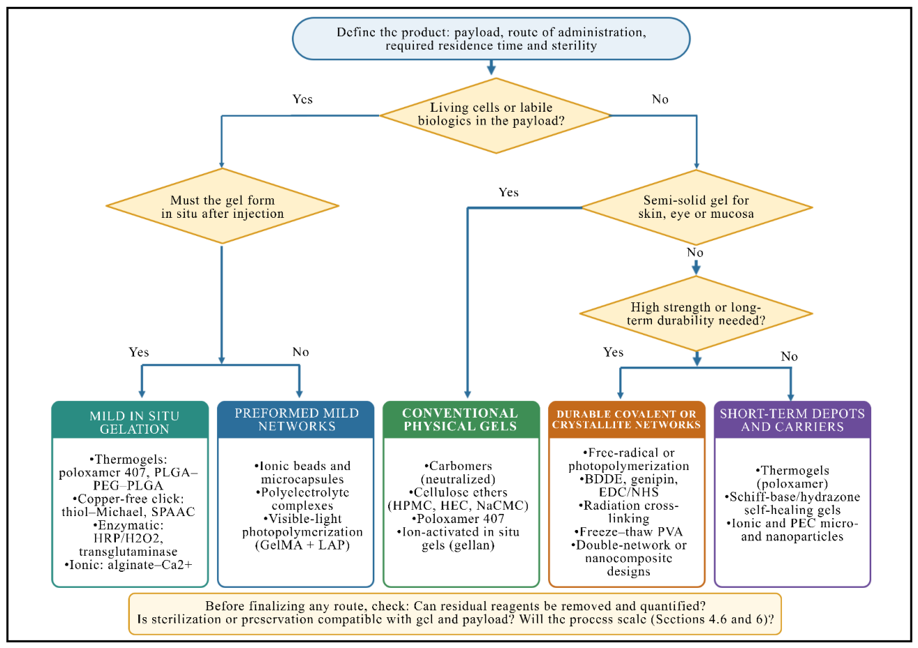
Figure 2.
Schematic representation of ionic gelation. Anionic polymer chains (e.g., sodium alginate) are cross-linked by multivalent cations (e.g., Ca2+), which bind cooperatively between guluronate blocks of adjacent chains to form ‘egg-box’ junction zones and a stable hydrogel network. In external gelation the cations diffuse inwards from a gelling bath, whereas in internal gelation they are released uniformly from a dispersed calcium source.
Figure 2.
Schematic representation of ionic gelation. Anionic polymer chains (e.g., sodium alginate) are cross-linked by multivalent cations (e.g., Ca2+), which bind cooperatively between guluronate blocks of adjacent chains to form ‘egg-box’ junction zones and a stable hydrogel network. In external gelation the cations diffuse inwards from a gelling bath, whereas in internal gelation they are released uniformly from a dispersed calcium source.
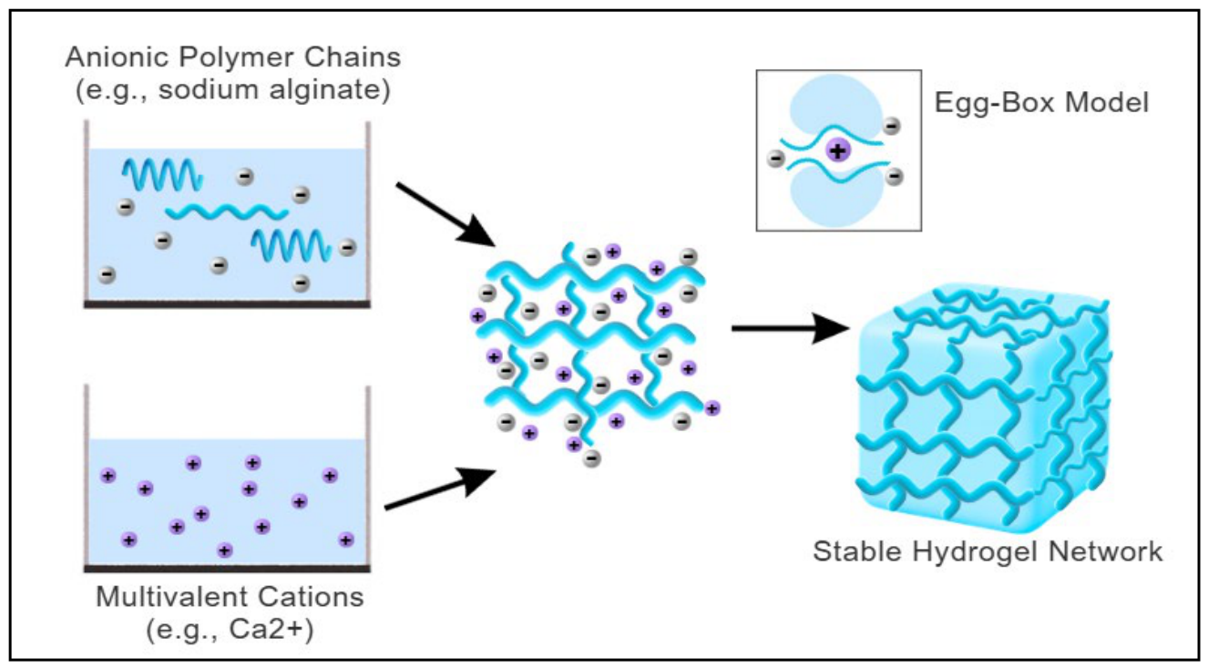
Figure 3.
External versus internal ionic gelation of alginate. (A) In external (diffusion) gelation, alginate droplets fall into a CaCl2 bath and Ca2+ diffuses inwards, producing beads with a densely cross-linked shell and a weaker core. (B) In internal gelation, insoluble CaCO3 dispersed in the alginate solution releases Ca2+ uniformly as D-glucono-δ-lactone (GDL) hydrolyzes and lowers the pH, giving homogeneous, mechanically stronger gels that can be cast into molds.
Figure 3.
External versus internal ionic gelation of alginate. (A) In external (diffusion) gelation, alginate droplets fall into a CaCl2 bath and Ca2+ diffuses inwards, producing beads with a densely cross-linked shell and a weaker core. (B) In internal gelation, insoluble CaCO3 dispersed in the alginate solution releases Ca2+ uniformly as D-glucono-δ-lactone (GDL) hydrolyzes and lowers the pH, giving homogeneous, mechanically stronger gels that can be cast into molds.
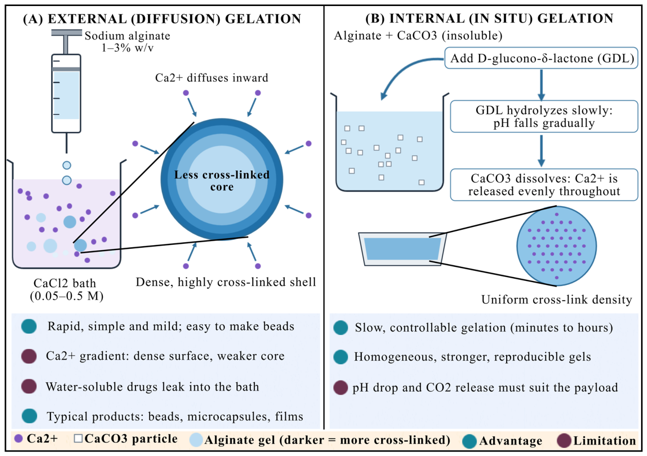
Figure 4.
Illustration of thermoresponsive gelation of poloxamer 407. At low temperature the PEO–PPO–PEO triblock copolymer chains are fully hydrated and the formulation is a free-flowing solution; on warming towards body temperature, dehydration of the PPO blocks drives micellization, and close packing of the micelles above Tsol–gel produces a semi-solid gel.
Figure 4.
Illustration of thermoresponsive gelation of poloxamer 407. At low temperature the PEO–PPO–PEO triblock copolymer chains are fully hydrated and the formulation is a free-flowing solution; on warming towards body temperature, dehydration of the PPO blocks drives micellization, and close packing of the micelles above Tsol–gel produces a semi-solid gel.
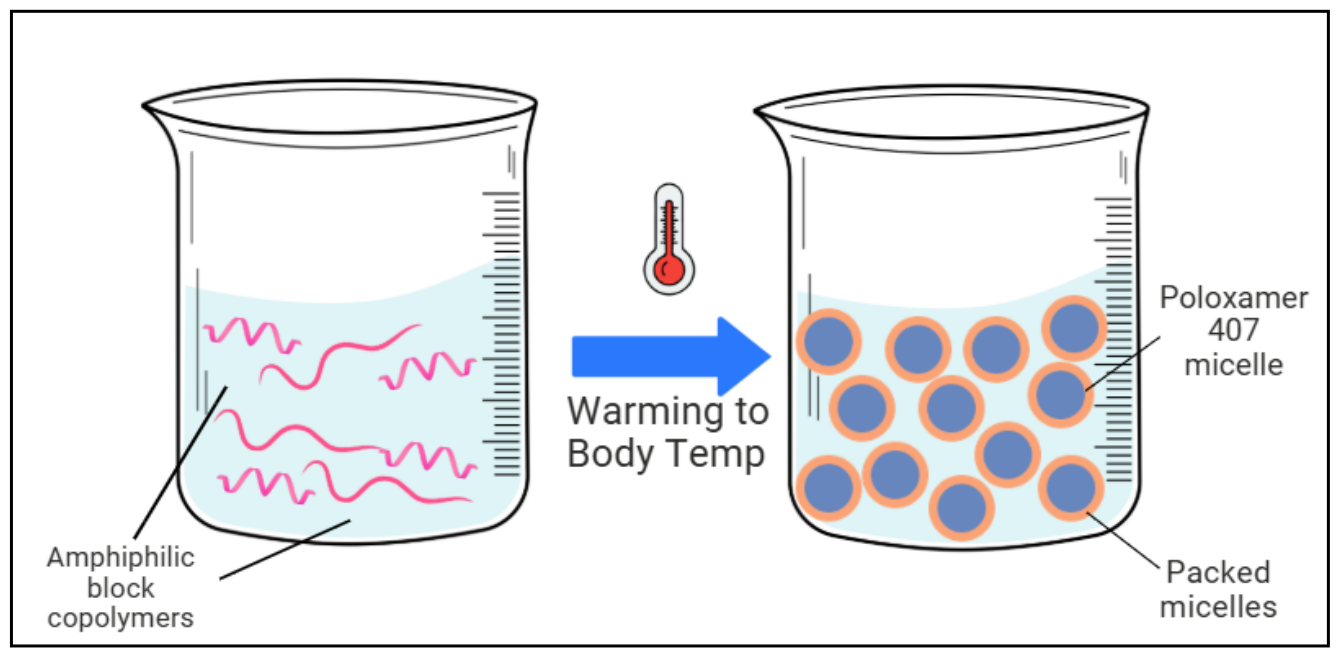
Figure 5.
Freeze–thaw preparation of PVA hydrogels. Freezing an aqueous PVA solution (e.g., at −20 °C) concentrates the polymer chains in the unfrozen regions between ice crystals, where hydrogen-bonded crystallites form; after thawing (e.g., at 25 °C) these crystallites persist as physical cross-links. Each additional cycle increases crystallinity and gel strength.
Figure 5.
Freeze–thaw preparation of PVA hydrogels. Freezing an aqueous PVA solution (e.g., at −20 °C) concentrates the polymer chains in the unfrozen regions between ice crystals, where hydrogen-bonded crystallites form; after thawing (e.g., at 25 °C) these crystallites persist as physical cross-links. Each additional cycle increases crystallinity and gel strength.
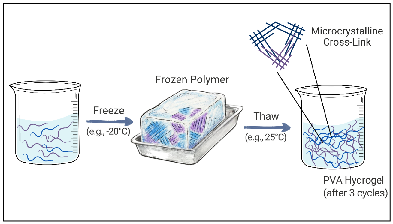
Figure 6.
Free-radical polymerization. An initiator (I) generates primary radicals (R•) thermally, by a redox reaction or by light; the radicals add to vinyl monomers (M) and propagate a growing chain, and a multifunctional cross-linker incorporated into two or more chains creates a permanent covalent network. Oxygen inhibition, the cross-linker ratio and the removal of residual monomer are critical considerations.
Figure 6.
Free-radical polymerization. An initiator (I) generates primary radicals (R•) thermally, by a redox reaction or by light; the radicals add to vinyl monomers (M) and propagate a growing chain, and a multifunctional cross-linker incorporated into two or more chains creates a permanent covalent network. Oxygen inhibition, the cross-linker ratio and the removal of residual monomer are critical considerations.
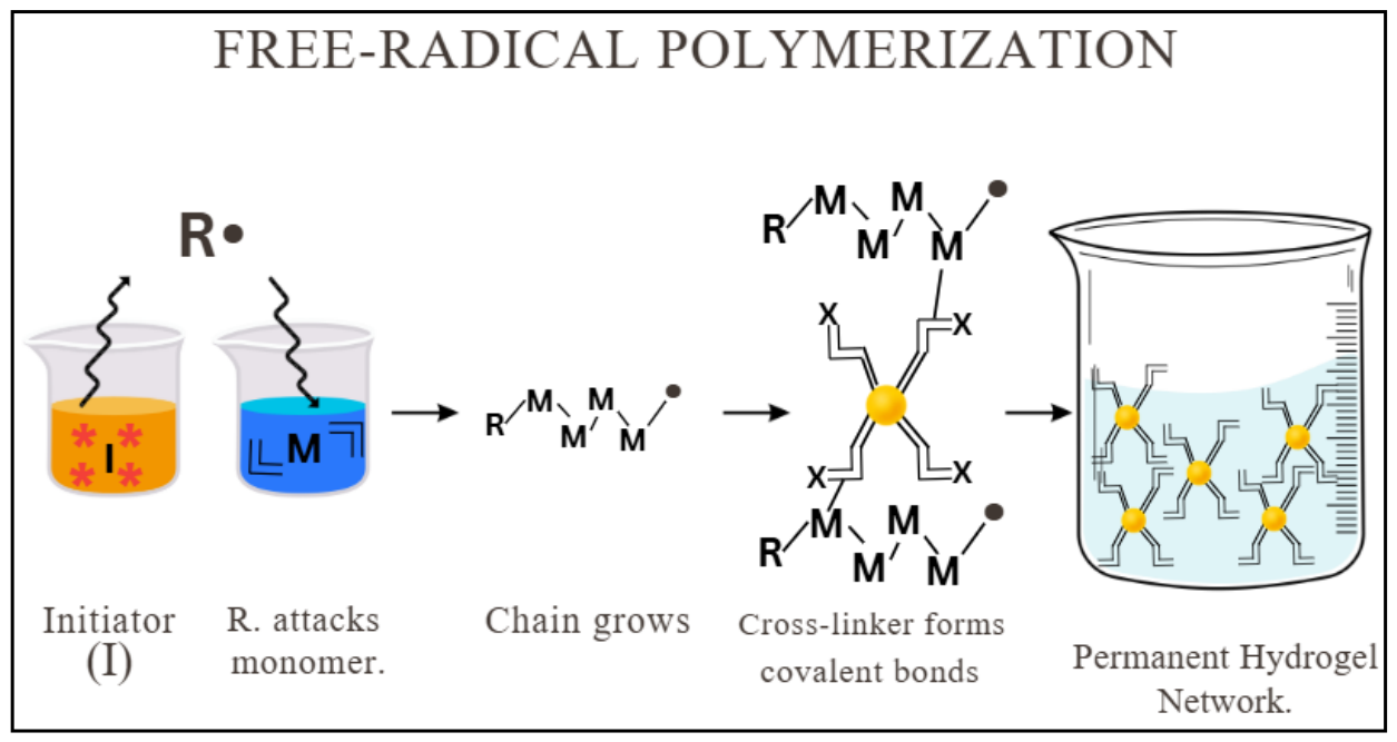
Figure 7.
Concept of ‘click’ chemistry for hydrogel formation. Two polymer precursors bearing complementary reactive groups (here, azide and alkyne) are mixed and react rapidly and selectively under mild aqueous conditions, forming covalent (triazole) cross-links and a homogeneous network. Copper-free variants (e.g., strain-promoted azide–alkyne cycloaddition) are required when gelation takes place in the presence of cells or in vivo.
Figure 7.
Concept of ‘click’ chemistry for hydrogel formation. Two polymer precursors bearing complementary reactive groups (here, azide and alkyne) are mixed and react rapidly and selectively under mild aqueous conditions, forming covalent (triazole) cross-links and a homogeneous network. Copper-free variants (e.g., strain-promoted azide–alkyne cycloaddition) are required when gelation takes place in the presence of cells or in vivo.
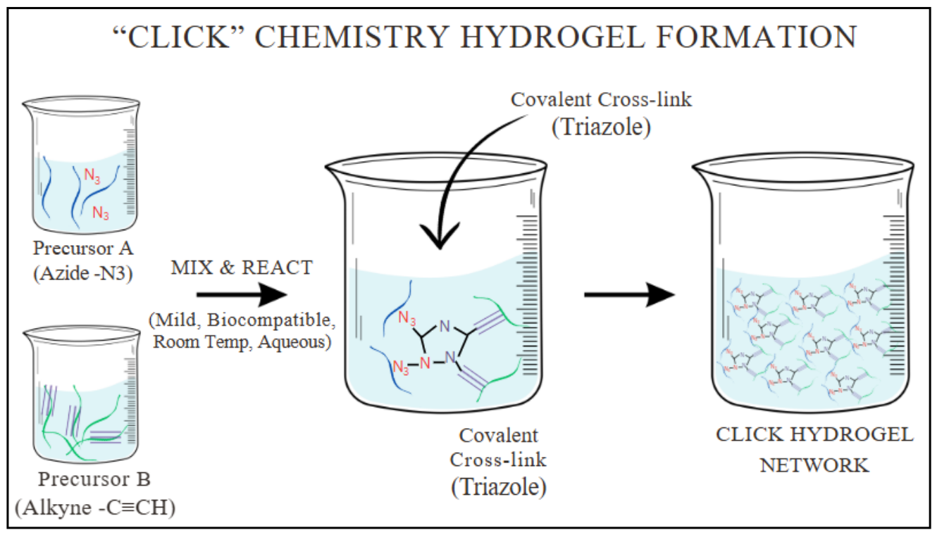
Figure 8.
Preparation of a carbomer hydrogel. Eight process steps run from polymer selection to filling, with critical control points (highlighted boxes) at dispersion, excipient addition, neutralization and final quality testing. EDTA, disodium edetate; TEA, triethanolamine.
Figure 8.
Preparation of a carbomer hydrogel. Eight process steps run from polymer selection to filling, with critical control points (highlighted boxes) at dispersion, excipient addition, neutralization and final quality testing. EDTA, disodium edetate; TEA, triethanolamine.
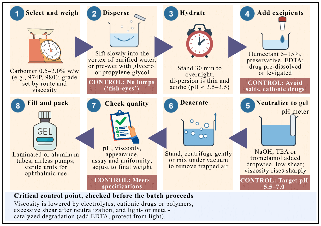
Figure 9.
Diffusion-controlled release. The cross-section shows a swollen hydrogel in which physically entrapped drug molecules move through the water-filled mesh, from the concentrated interior of the gel towards the dilute external medium. How quickly they leave depends on the concentration gradient and on the size of the drug relative to the mesh.
Figure 9.
Diffusion-controlled release. The cross-section shows a swollen hydrogel in which physically entrapped drug molecules move through the water-filled mesh, from the concentrated interior of the gel towards the dilute external medium. How quickly they leave depends on the concentration gradient and on the size of the drug relative to the mesh.
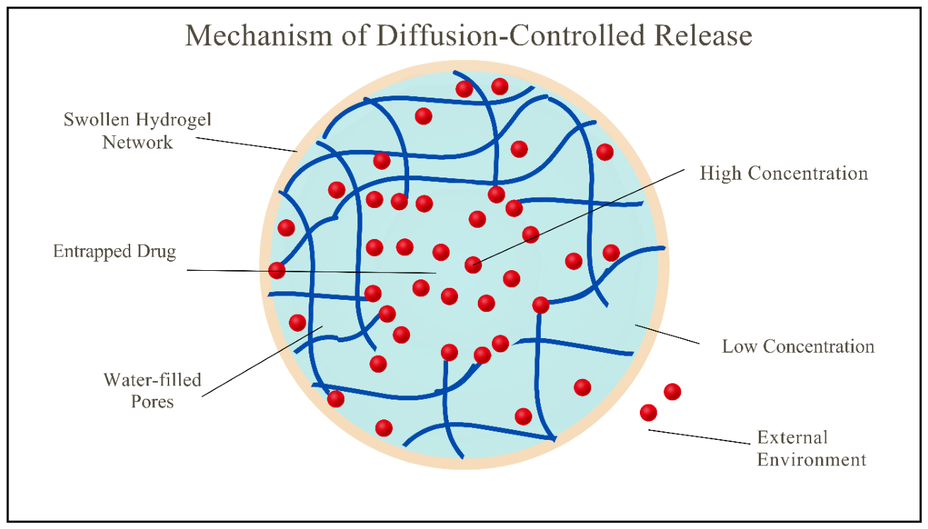
Figure 10.
Mechanism of swelling-controlled release. (A) Dry, glassy hydrogel in which drug molecules are immobilized. (B) The same hydrogel after exposure to aqueous fluid: a swelling front separates a swollen, rubbery outer layer, from which drug is released, from a dry, glassy inner core in which drug remains entrapped.
Figure 10.
Mechanism of swelling-controlled release. (A) Dry, glassy hydrogel in which drug molecules are immobilized. (B) The same hydrogel after exposure to aqueous fluid: a swelling front separates a swollen, rubbery outer layer, from which drug is released, from a dry, glassy inner core in which drug remains entrapped.
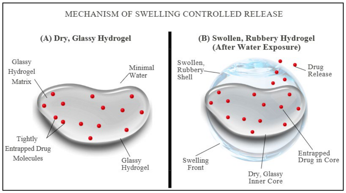
Figure 11.
Mechanism of chemically controlled release. Water molecules (hydrolysis) or enzymes cleave the polymer chains and cross-links of a degradable hydrogel matrix; as the network loosens and erodes, the embedded drug molecules are released into the surrounding medium.
Figure 11.
Mechanism of chemically controlled release. Water molecules (hydrolysis) or enzymes cleave the polymer chains and cross-links of a degradable hydrogel matrix; as the network loosens and erodes, the embedded drug molecules are released into the surrounding medium.
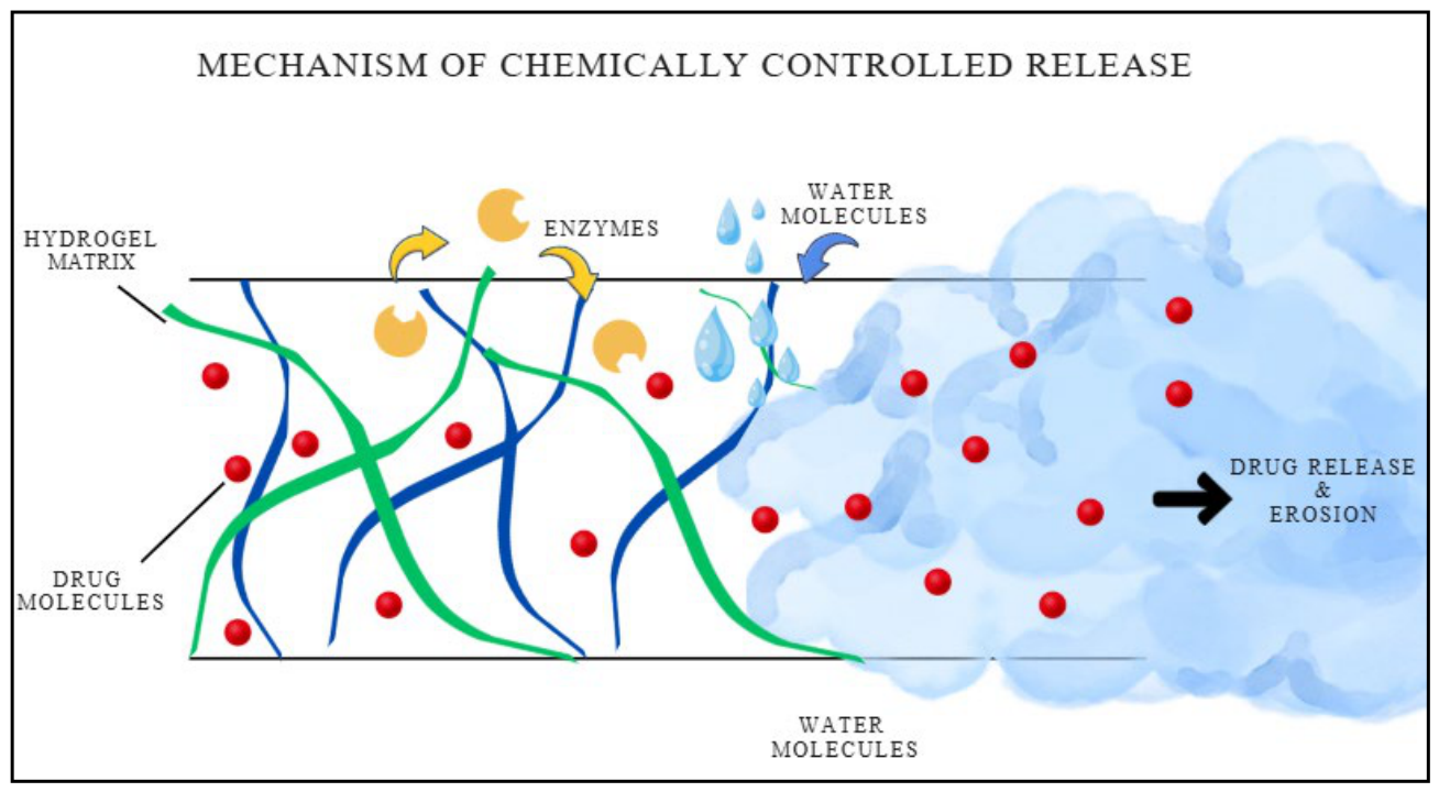
Figure 12.
Oral delivery with a pH-responsive anionic hydrogel. At gastric pH (≈ 1–3) the carboxyl groups are protonated, the network stays collapsed and the drug is protected. In the small intestine (pH ≈ 6–7.5) the same groups ionize; the gel swells and the drug is released for absorption.
Figure 12.
Oral delivery with a pH-responsive anionic hydrogel. At gastric pH (≈ 1–3) the carboxyl groups are protonated, the network stays collapsed and the drug is protected. In the small intestine (pH ≈ 6–7.5) the same groups ionize; the gel swells and the drug is released for absorption.
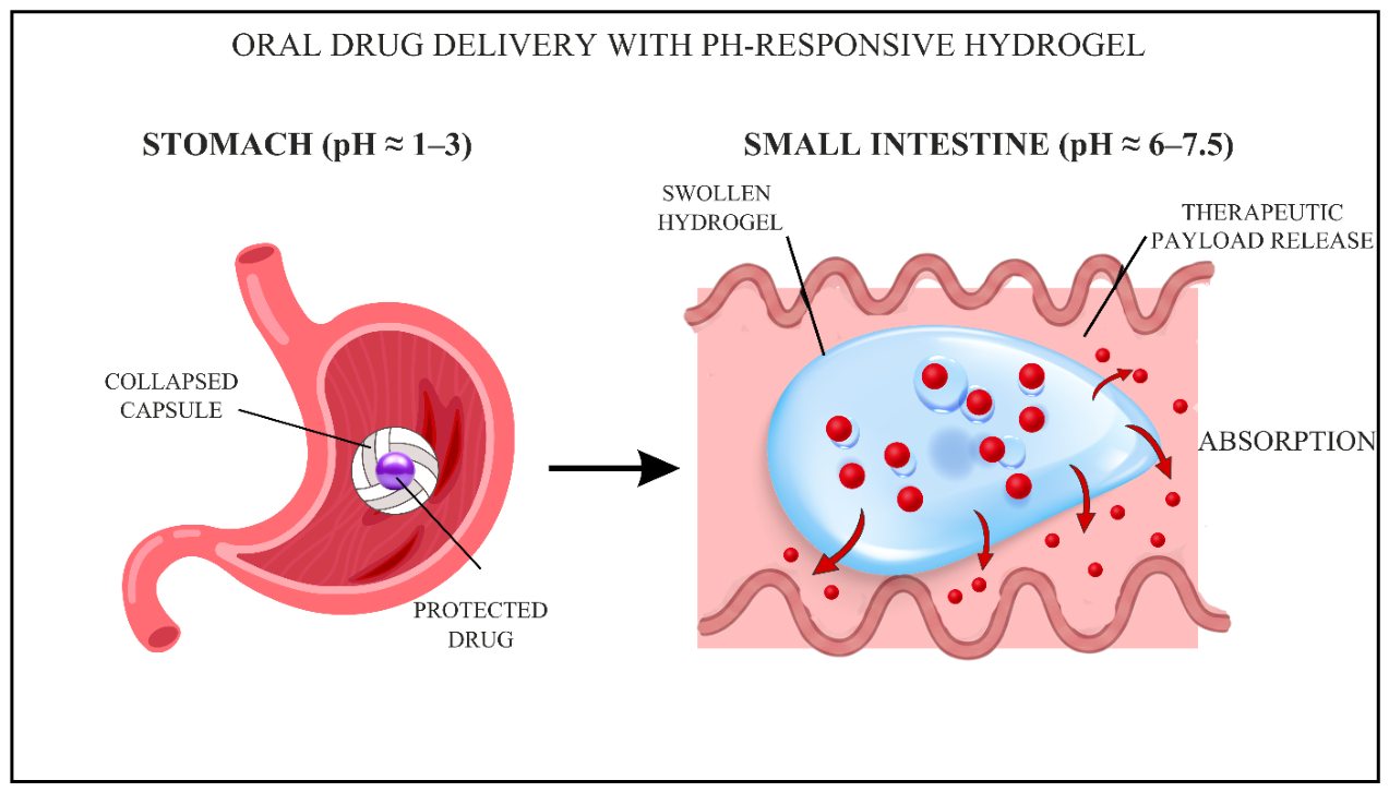
Figure 13.
Cutaneous and transdermal delivery mechanisms. (A) A hydrogel patch on the skin surface hydrates the stratum corneum (depicted as a ‘brick-and-mortar’ structure), increasing the permeability of the intercellular lipids and allowing drug molecules to diffuse into the skin. (B) A hydrogel-forming microneedle array penetrates the stratum corneum and swells to create continuous hydrogel conduits through which drug molecules are delivered into the viable skin layers.
Figure 13.
Cutaneous and transdermal delivery mechanisms. (A) A hydrogel patch on the skin surface hydrates the stratum corneum (depicted as a ‘brick-and-mortar’ structure), increasing the permeability of the intercellular lipids and allowing drug molecules to diffuse into the skin. (B) A hydrogel-forming microneedle array penetrates the stratum corneum and swells to create continuous hydrogel conduits through which drug molecules are delivered into the viable skin layers.
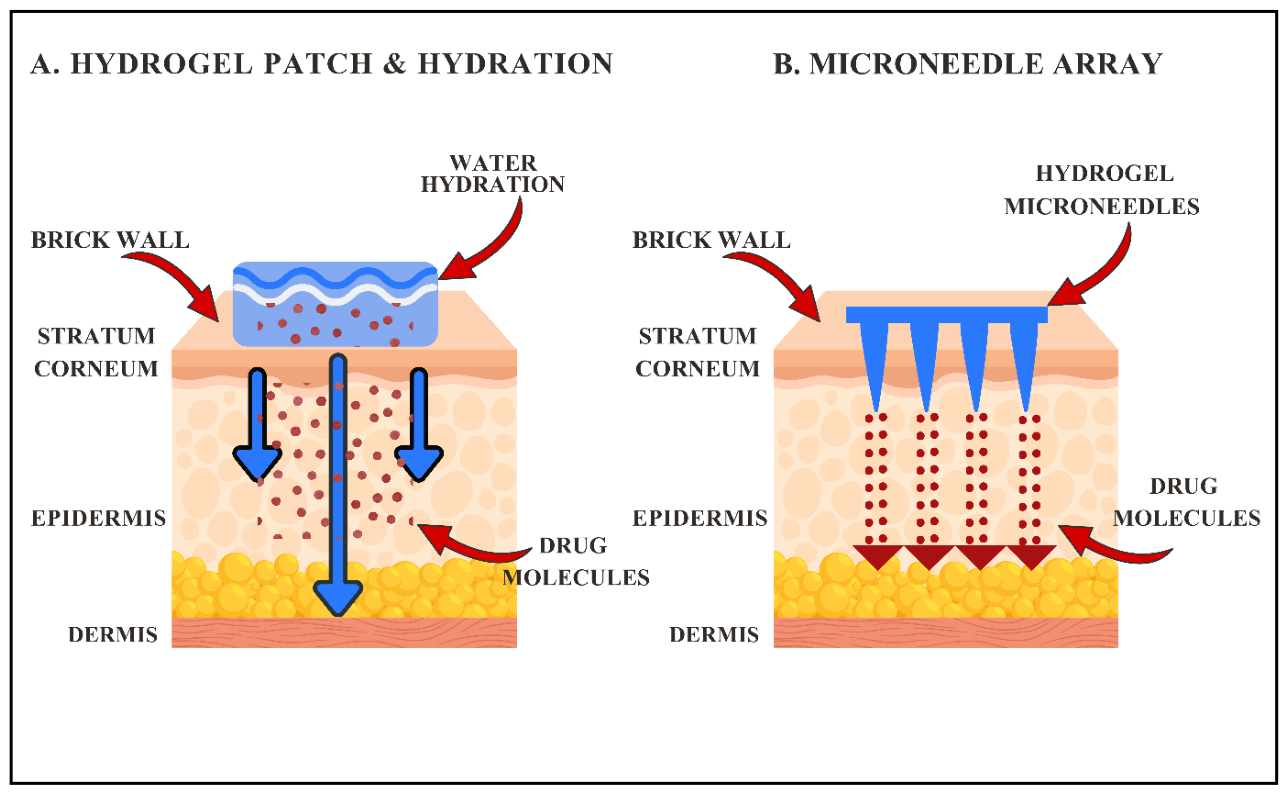
Figure 14.
Injectable in situ-forming hydrogel system. Step 1: a liquid polymer–drug solution is injected into the target tissue. Step 2: the solution undergoes a sol–gel transition in response to body temperature (37 °C). Step 3: the resulting gel depot conforms to the tissue space and provides sustained drug release over an extended period.
Figure 14.
Injectable in situ-forming hydrogel system. Step 1: a liquid polymer–drug solution is injected into the target tissue. Step 2: the solution undergoes a sol–gel transition in response to body temperature (37 °C). Step 3: the resulting gel depot conforms to the tissue space and provides sustained drug release over an extended period.
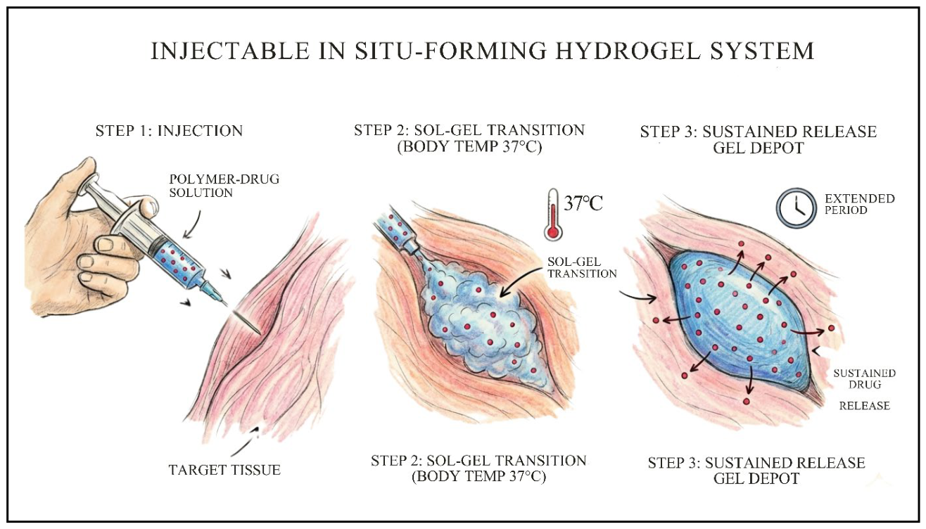
Figure 15.
Mechanisms of ‘smart’ stimuli-responsive hydrogels. (A) Enzymes cleave the cross-links of an enzyme-responsive hydrogel. (B) Light breaks photosensitive linkers in a light-responsive hydrogel. (C) Ultrasound induces acoustic cavitation and matrix disruption in an ultrasound-responsive hydrogel. In each case the stimulus triggers release of the encapsulated drug.
Figure 15.
Mechanisms of ‘smart’ stimuli-responsive hydrogels. (A) Enzymes cleave the cross-links of an enzyme-responsive hydrogel. (B) Light breaks photosensitive linkers in a light-responsive hydrogel. (C) Ultrasound induces acoustic cavitation and matrix disruption in an ultrasound-responsive hydrogel. In each case the stimulus triggers release of the encapsulated drug.
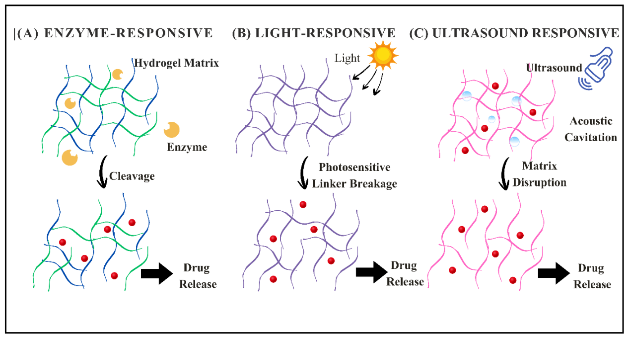
Figure 16.
Hierarchical delivery via a hydrogel–nanocarrier system. A macroscopic hydrogel implanted in tissue releases drug-loaded nanocarriers, which are internalized by target cells by endocytosis and entrapped in endosomes. The inset illustrates the ‘proton sponge’ effect: buffering of the acidic endosome causes an influx of H+, Cl− and water, leading to osmotic swelling, rupture of the endosomal membrane and release of the drug into the cytoplasm.
Figure 16.
Hierarchical delivery via a hydrogel–nanocarrier system. A macroscopic hydrogel implanted in tissue releases drug-loaded nanocarriers, which are internalized by target cells by endocytosis and entrapped in endosomes. The inset illustrates the ‘proton sponge’ effect: buffering of the acidic endosome causes an influx of H+, Cl− and water, leading to osmotic swelling, rupture of the endosomal membrane and release of the drug into the cytoplasm.
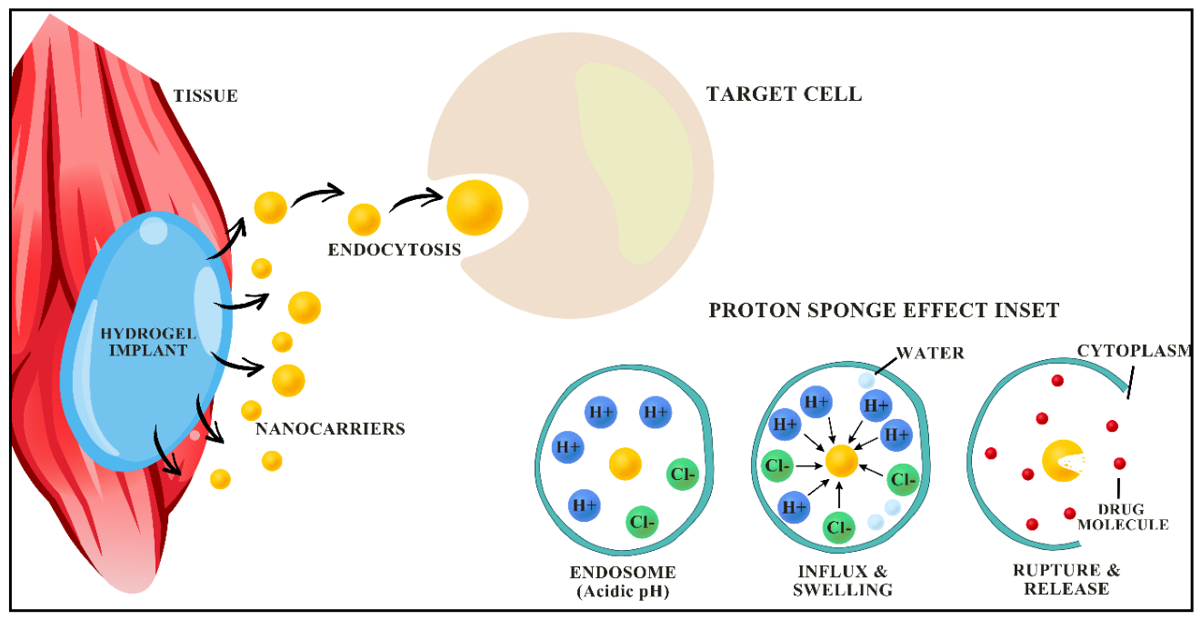
Figure 17.
The Biopharmaceutics Classification System (BCS) and corresponding hydrogel formulation strategies, with solubility on the vertical axis and permeability on the horizontal axis. For Class I drugs a simple hydrogel provides sustained release; for Class II, a hydrogel carrying drug nanocrystals improves dissolution; for Class III, a mucoadhesive hydrogel prolongs residence at the absorption site; and for Class IV, a hydrogel releasing lipid nanoparticles addresses both limitations.
Figure 17.
The Biopharmaceutics Classification System (BCS) and corresponding hydrogel formulation strategies, with solubility on the vertical axis and permeability on the horizontal axis. For Class I drugs a simple hydrogel provides sustained release; for Class II, a hydrogel carrying drug nanocrystals improves dissolution; for Class III, a mucoadhesive hydrogel prolongs residence at the absorption site; and for Class IV, a hydrogel releasing lipid nanoparticles addresses both limitations.
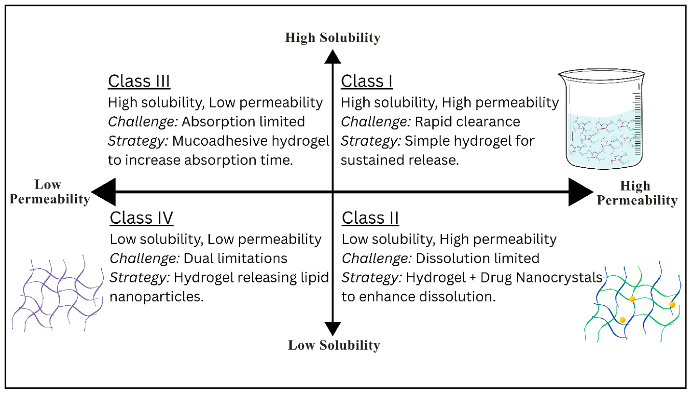
Figure 18.
Drug–hydrogel interactions determined by physicochemical properties. (1) Small hydrophilic drug molecules diffuse freely through the aqueous pores. (2) Large protein or peptide molecules are physically entrapped by the polymer network. (3) Positively charged drug molecules interact electrostatically with negatively charged polymer chains. (4) Hydrophobic drug molecules are solubilized within the cores of micelles formed by amphiphilic polymers.
Figure 18.
Drug–hydrogel interactions determined by physicochemical properties. (1) Small hydrophilic drug molecules diffuse freely through the aqueous pores. (2) Large protein or peptide molecules are physically entrapped by the polymer network. (3) Positively charged drug molecules interact electrostatically with negatively charged polymer chains. (4) Hydrophobic drug molecules are solubilized within the cores of micelles formed by amphiphilic polymers.
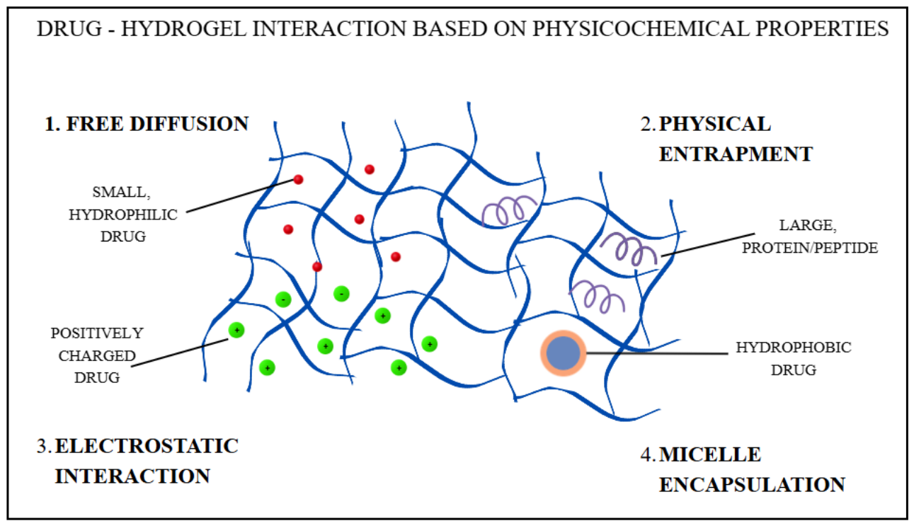
Figure 19.
The hydrogel as an integrated platform for tissue engineering. Therapeutic cells such as stem cells are encapsulated within the porous network, which serves as the scaffold, while growth factors and other signaling molecules released from the matrix direct their proliferation and differentiation until new, functional tissue forms.
Figure 19.
The hydrogel as an integrated platform for tissue engineering. Therapeutic cells such as stem cells are encapsulated within the porous network, which serves as the scaffold, while growth factors and other signaling molecules released from the matrix direct their proliferation and differentiation until new, functional tissue forms.
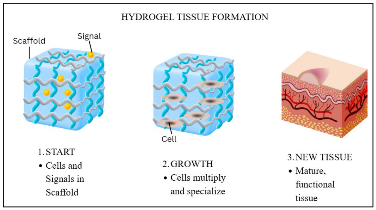
Figure 20.
Injectable hydrogel for localized cell delivery. Step 1: a syringe contains a liquid mixture of polymer precursor and therapeutic stem cells. Step 2: the mixture is injected into a tissue defect (e.g., a cartilage lesion). Step 3: triggered by body temperature, the liquid undergoes a sol–gel transition to form a solid, cell-laden hydrogel scaffold that fills the defect and supports tissue regeneration.
Figure 20.
Injectable hydrogel for localized cell delivery. Step 1: a syringe contains a liquid mixture of polymer precursor and therapeutic stem cells. Step 2: the mixture is injected into a tissue defect (e.g., a cartilage lesion). Step 3: triggered by body temperature, the liquid undergoes a sol–gel transition to form a solid, cell-laden hydrogel scaffold that fills the defect and supports tissue regeneration.
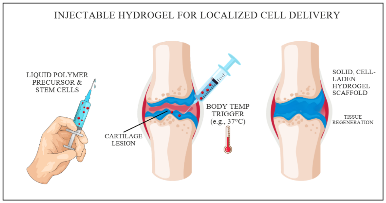
Figure 21.
Comparison of a traditional gauze dressing and a hydrogel wound dressing. Left: a gauze dressing allows moisture loss and bacterial entry and may adhere to the wound bed. Right: a hydrogel dressing maintains hydration, absorbs exudate, acts as a barrier to bacteria and can release therapeutic molecules (e.g., growth factors and antibiotics) into the wound bed to promote healing.
Figure 21.
Comparison of a traditional gauze dressing and a hydrogel wound dressing. Left: a gauze dressing allows moisture loss and bacterial entry and may adhere to the wound bed. Right: a hydrogel dressing maintains hydration, absorbs exudate, acts as a barrier to bacteria and can release therapeutic molecules (e.g., growth factors and antibiotics) into the wound bed to promote healing.
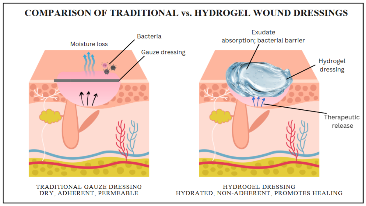

Table 1.
Principal advantages of hydrogels as pharmaceutical dosage forms, the limitations associated with each, their underlying causes and practical mitigation strategies.
Table 1.
Principal advantages of hydrogels as pharmaceutical dosage forms, the limitations associated with each, their underlying causes and practical mitigation strategies.
| Advantage | Associated limitation or challenge | Underlying cause | Practical mitigation strategies | Ref. |
| High biocompatibility and tissue-like softness | Low mechanical strength, brittleness, creep and poor handling | Low polymer volume fraction in the swollen state; heterogeneous cross-link distribution; no energy-dissipating mechanism | Optimize polymer concentration and cross-link density; use interpenetrating, double-network, nanocomposite or Tetra-PEG architectures; reinforce with fibers or meshes; avoid load-bearing applications where possible | [11,15,16,17,18,19,20] |
| Controlled and sustained release | Initial burst release; short release duration for small hydrophilic drugs | Mesh size (typically 5–100 nm) much larger than small molecules; drug accumulated at the surface during loading or drying | Exploit drug–polymer affinity (ionic, hydrophobic, host–guest or covalent tethering); embed drug-loaded nanocarriers; increase cross-link density; apply rate-controlling coatings; wash off surface-associated drug | [4,7,21,22] |
| Aqueous microenvironment that preserves labile biologics | Low loading and non-uniform dispersion of hydrophobic drugs | Thermodynamic incompatibility between lipophilic drugs and the water-swollen matrix | Cyclodextrin inclusion complexes; amphiphilic or micellar gels (e.g., poloxamers); nanocrystals, nanoemulsions, niosomes or liposomes dispersed in the gel (emulgels and nanocarrier-in-gel systems) | [12,23] |
| Highly tunable physicochemical properties | Toxicity of residual synthesis reagents | Incomplete monomer conversion; small-molecule cross-linkers (e.g., glutaraldehyde), initiators, photoinitiators or metal catalysts retained in the network | Prefer physical, enzymatic or bio-orthogonal chemistries; use less toxic cross-linkers (e.g., genipin); validated purification (dialysis, repeated washing); quantify residuals by HPLC or GC; determine gel fraction | [24,25,26] |
| Amenable to minimally invasive delivery (injectable and in situ-forming gels) | Premature gelation, needle clogging, dilution or rapid erosion at the injection site | Gelation kinetics or sol–gel temperature not matched to administration; high viscosity; low gel strength | Shear-thinning and self-healing networks; tune Tsol–gel or gelation time; dual-barrel syringes with static mixers; rheological and injection-force testing | [27,28,29] |
| High water content (comfort, moist wound environment) | Microbial susceptibility; difficult terminal sterilization; water loss during storage | High water activity supports microbial growth; heat and ionizing radiation alter polymer chains and cross-link density | Compatible preservative systems verified by efficacy testing; aseptic processing; sterile filtration of low-viscosity precursors; validated radiation or heat cycles for tolerant systems; barrier packaging | [13,30,31] |
| Accessible from abundant natural and synthetic raw materials | Batch-to-batch variability; cost of GMP-grade, low-endotoxin polymers | Source- and process-dependent molecular weight, composition (e.g., M/G ratio, degree of deacetylation) and impurity profile | Specify critical material attributes (molecular weight, viscosity, composition, endotoxin, bioburden); qualify suppliers; apply quality-by-design (QbD) to define acceptable ranges | [32,33] |
Table 2.
Comparison of physical (non-covalent) preparation methods: representative materials and conditions, the process parameters with the greatest effect on quality, principal strengths and weaknesses, and typical dosage forms.
Table 2.
Comparison of physical (non-covalent) preparation methods: representative materials and conditions, the process parameters with the greatest effect on quality, principal strengths and weaknesses, and typical dosage forms.
| Method and mechanism | Representative materials and typical conditions | Critical process parameters | Advantages | Limitations | Typical dosage forms and examples [Ref.] |
| Ionic (ionotropic) gelation: multivalent counter-ions bridge charged chains (‘egg-box’ junctions for alginate) | Sodium alginate 1–3% w/v with CaCl2 0.05–0.5 M (external) or CaCO3/GDL (internal); low-methoxyl pectin, gellan gum (Ca2+, K+); chitosan 0.1–3% w/v with TPP | M/G ratio and molecular weight; polymer and ion concentration; curing time; pH; needle size, flow rate and drop height; gelation mode | Aqueous, rapid and mild; low cost; suitable for cells, proteins and probiotics; simple extrusion scale-up | Heterogeneous gels with external gelation; drug leakage into the curing bath; instability by ion exchange and chelation; alginate not degraded in humans | Beads, microcapsules, nanoparticles, wound dressings, ion-activated ophthalmic in situ gels [32,38,59,60,61,63] |
| Polyelectrolyte complexation: electrostatic association of oppositely charged polymers with counter-ion release | Chitosan with alginate, pectin, carrageenan, xanthan, hyaluronic acid or carboxymethylcellulose; dilute solutions (≈0.1–1% w/v); pH about 4–6 | Charge ratio; pH and ionic strength; molecular weight; order and rate of mixing | No cross-linker; mucoadhesive; pH-responsive; tunable by charge balance | Precipitation instead of gelation if mixing is uncontrolled; sensitive to salt and pH of release media; variability of natural polymers | Oral and nasal particles, films, wound dressings, coatings on ionic gels [39,58] |
| Thermoresponsive (LCST-type) gelation: dehydration, micellar packing or hydrophobic association on warming | Poloxamer 407 16–25% w/w (cold method, 4–5 °C); methylcellulose and HPMC; PNIPAAm; PLGA–PEG–PLGA copolymers | Polymer concentration and grade; co-solutes (poloxamer 188, salts, ethanol, drug); pH; preparation temperature | Liquid at administration, gel at body temperature; no cross-linker; simple preparation | Low gel strength; rapid dissolution on dilution (poloxamer); Tsol–gel shifted by additives; premature gelation in warm climates or syringes | In situ ophthalmic, nasal, vaginal, rectal, intratumoral and periodontal gels; topical gels [36,64,65,66,67] |
| Cooling-induced (UCST-type) gelation: helix formation and aggregation of chains on cooling | Gelatin 2–10% w/v; agarose 0.5–2% w/v; gellan gum; carrageenan; dissolved hot (about 50–90 °C) and cooled | Concentration; Bloom strength or grade; cooling rate; ionic environment | Simple and reagent-free; good printability (gelatin); widely available materials | Gelatin gels melt near 30–35 °C and are unstable at 37 °C unless cross-linked; animal origin of gelatin (BSE/TSE and halal considerations) | Soft capsules, lozenges, cast gels, scaffolds and bioinks, often cross-linked afterwards [36,72] |
| Freeze–thaw cycling: ice-induced phase separation and hydrogen-bonded crystallite formation | PVA 5–20% w/v (degree of hydrolysis ≥ 98%) dissolved at 80–95 °C; 3–6 cycles of freezing (−20 °C, 8–24 h) and thawing (room temperature, 2–8 h); blends with alginate, chitosan or gelatin | Number of cycles; freezing temperature and time; thawing rate; PVA concentration, molecular weight and degree of hydrolysis | Tough, elastic gels without chemicals; stability improves with cycle number | Time-consuming; limited to crystallizable polymers; opaque; PVA not readily biodegradable; high dissolution temperature | Wound dressings, ocular and cartilage devices, drug-eluting implants [68,69,70] |
| Hydrogen bonding, hydrophobic and supramolecular assembly | PAA or PMAA with PEG or PVP; self-assembling peptides; cyclodextrin host–guest systems; PLLA/PDLA stereocomplexes | pH (degree of ionization); polymer ratio; ionic strength; temperature | Shear-thinning and self-healing; injectable; pH-responsive | Often weak and short-lived in vivo; high cost of peptides and specialized polymers | Oral pH-responsive carriers, injectable depots, cell carriers [2,25,71] |
Table 3.
Comparison of chemical (covalent) hydrogel preparation methods, with representative materials, typical conditions, critical process parameters, advantages, limitations and typical pharmaceutical formats.
Table 3.
Comparison of chemical (covalent) hydrogel preparation methods, with representative materials, typical conditions, critical process parameters, advantages, limitations and typical pharmaceutical formats.
| Method and mechanism | Representative materials and typical conditions | Critical process parameters | Advantages | Limitations | Typical dosage forms and examples [Ref.] |
| Free-radical polymerization (thermal or redox initiation): chain growth of vinyl monomers with a multifunctional cross-linker | HEMA, acrylamide, acrylic acid, NIPAAm; MBAA or EGDMA 0.1–5 mol%; KPS or APS at 50–70 °C, or APS/TEMED at room temperature; N2 purging | Monomer and cross-linker concentration; initiator level; temperature and exotherm; oxygen exclusion; reaction time; purification | Strong, reproducible networks; wide choice of monomers; precise tuning of cross-link density; scalable | Toxic residual monomer, oligomers and initiator fragments; exotherm; payload degradation during in situ loading; most networks non-degradable | Contact lenses, superabsorbents, pH- and thermoresponsive matrices, implants [42,58,73] |
| Photopolymerization: light-initiated radical polymerization of (meth)acrylated or norbornene-functionalized macromers | PEGDA, GelMA (about 5–15% w/v), methacrylated hyaluronic acid; Irgacure 2959 (365 nm) or LAP 0.05–0.5% w/v (365–405 nm) | Degree of substitution of the macromer; photoinitiator type and concentration; wavelength, intensity and exposure time; sample thickness | Rapid curing at 37 °C; spatial and temporal control; enables in situ curing and 3D printing | Limited light penetration in thick or opaque samples; photoinitiator and radical cytotoxicity; requires light-delivery equipment | Cell-laden scaffolds, bioinks, in situ sealants, microgels [72,74,75] |
| Small-molecule cross-linkers: reaction of preformed polymers with bi- or multifunctional reagents | Glutaraldehyde (chitosan, gelatin, PVA); genipin (chitosan, gelatin); EDC/NHS (collagen, gelatin, hyaluronic acid); BDDE (hyaluronic acid); epichlorohydrin (dextran, starch); citric acid (cellulose ethers) | Cross-linker-to-polymer ratio; pH; temperature and time; quenching and washing | Uses established, inexpensive polymers; straightforward; wide range of stiffness | Cytotoxic residues (glutaraldehyde, epoxides); slow reaction (genipin); risk of drug modification during in situ loading | Dermal fillers, beads and microspheres, sponges, films, superabsorbents [25,26,33] |
| Click chemistry: selective coupling of complementary groups (thiol–Michael, thiol–ene, CuAAC, SPAAC, tetrazine ligation, Diels–Alder) | Multi-arm PEG-thiol with PEG-maleimide, -vinyl sulfone or -acrylate; PEG- or HA-norbornene; azide- and cyclooctyne-functionalized polymers | Precursor concentration and functionality; stoichiometry; pH (thiol–Michael); mixing efficiency; light dose (thiol–ene) | Fast, selective and near-quantitative; mild aqueous conditions; homogeneous networks; suitable for in situ use (copper-free variants) | Synthesis and characterization of functionalized precursors; cost; Cu toxicity in CuAAC; very fast reactions may prevent homogeneous mixing | Injectable depots, cell encapsulation, tissue engineering scaffolds [43,77,78,79,80] |
| Dynamic covalent chemistry: reversible imine, hydrazone, oxime or boronate ester bonds | Oxidized alginate, dextran or hyaluronic acid with chitosan, gelatin or hydrazide-modified polymers; phenylboronic acid polymers with diols | Degree of oxidation or substitution; aldehyde-to-amine ratio; pH; polymer concentration | Self-healing, shear-thinning and injectable after gelation; pH-responsive degradation; no catalyst | Limited long-term stability; residual aldehydes may react with drugs and tissues; mechanical weakness | Injectable and sprayable gels, wound dressings, pH-triggered release systems [10,41] |
| Enzyme-mediated cross-linking: enzymatic coupling of phenol, amine or glutamine residues | HRP/H2O2 with tyramine-conjugated hyaluronic acid, gelatin or dextran; microbial transglutaminase with gelatin or collagen; tyrosinase | Enzyme concentration (gelation time); H2O2 concentration (cross-link density); degree of substitution; temperature | Physiological conditions; independently tunable gelation rate and stiffness; good cytocompatibility | H2O2 cytotoxicity; enzyme cost, variability and potential immunogenicity | Injectable and cell-laden gels, tissue adhesives [40,81] |
| Radiation cross-linking: radical generation by gamma or electron-beam irradiation of aqueous polymer solutions | PVP, PVA, PEG, carboxymethylcellulose; typically 10–50 kGy | Polymer concentration; dose and dose rate; oxygen; additives | No initiators or cross-linkers; simultaneous sterilization; industrially established | Requires an irradiation facility; chain scission of sensitive polymers; incompatible with co-irradiated labile drugs | Wound dressings, sheet gels, sterile implants [30,34] |
Table 4.
Common gelling agents used in conventional pharmaceutical hydrogel dosage forms, with typical concentrations, processing requirements, limitations and applications [32,33,66,67,82,83,84].
| Gelling agent (ionic character) | Gelation mechanism | Typical conc. (% w/w) | Processing notes and pH range | Incompatibilities and limitations | Typical dosage forms |
| Carbomers, e.g., Carbopol® 934P, 940, 971P, 974P, 980 (anionic) | Swelling of cross-linked poly(acrylic acid) microgels on neutralization (pKa ≈ 6) | 0.5–2.0 | Disperse without lumps, hydrate, then neutralize to pH 5.5–7.0 (NaOH, triethanolamine, trometamol); avoid excessive shear after neutralization | Electrolytes, cationic drugs and polymers, strong acids, phenol, resorcinol; light- and metal-catalyzed oxidative degradation (add EDTA) | Topical, ophthalmic, vaginal, rectal and mucoadhesive gels; emulgels |
| Hydroxypropyl methylcellulose (non-ionic) | Hydration and chain entanglement; thermal gelation at elevated temperature | ≈2–5 (grade-dependent) | Hot/cold dispersion technique; stable at about pH 3–11; clear gels | Salting-out by high electrolyte concentrations; partial binding of some preservatives | Topical and ophthalmic gels, mucoadhesive systems, in situ gel blends |
| Hydroxyethylcellulose (non-ionic) | Hydration and chain entanglement | ≈1–3 | Disperse in water and allow to hydrate; wide pH tolerance; clear gels | Low intrinsic mucoadhesion; solutions susceptible to microbial growth | Lubricant and vaginal gels, catheter and ultrasound gels |
| Sodium carboxymethylcellulose (anionic) | Hydration and chain entanglement | 3–6 | Disperse with vigorous stirring or pre-wet with glycerol; most stable at about pH 7–9 | Strong acids, polyvalent metal salts (e.g., Al3+, Fe3+), cationic drugs | Topical and wound gels, oromucosal and dental gels |
| Methylcellulose (non-ionic) | Hydrophobic association on heating (gelation at about 50–60 °C, lowered by salts) | ≈1–5 | Hot/cold dispersion technique | Gels above body temperature unless modified with salts; salting-out | Topical gels; ophthalmic and nasal thermogelling blends |
| Poloxamer 407 (non-ionic) | Micellization and micellar packing on warming (LCST-type) | 16–30 (in situ gels ≈16–25) | Cold method (4–5 °C), refrigerate until clear; measure Tsol–gel on the final formulation | Weak gel and rapid dissolution in excess fluid; Tsol–gel shifted by drugs, salts and co-solvents; binds parabens | In situ ophthalmic, nasal, vaginal, rectal, periodontal and intratumoral gels; topical gels |
| Sodium alginate (anionic) | Ionotropic gelation with Ca2+; viscous solutions without ions | 0.5–3 | Disperse and hydrate; requires pH above about 4 (alginic acid precipitates at low pH) | Calcium chelators (phosphate, citrate, EDTA) dissolve Ca2+ gels; cationic drugs | Beads, wound dressings, raft-forming oral liquids, cell encapsulation |
| Low-acyl gellan gum (anionic) | Ion-activated gelation (Na+, K+, Ca2+); gelation on cooling | 0.1–1.0 | Dissolve hot in deionized water; gels in tear fluid or other cation-containing fluids | Premature gelation by electrolytes in the formulation | Ophthalmic and nasal in situ gels; oral liquids |
| Xanthan gum (anionic) | Ordered helical association; weak, strongly shear-thinning gels (synergistic with locust bean gum) | 0.5–2 | Disperse under high shear or pre-wet; stable over wide pH and temperature ranges | Some cationic preservatives and surfactants; limited clarity | Topical gels, suspensions, mucoadhesive blends with poloxamers |
| Chitosan (cationic) | Entanglement in dilute acid; ionic gelation with TPP; polyelectrolyte complexation | 1–3 (in 1% v/v acetic or lactic acid) | Soluble only below about pH 6.3–6.5; properties depend on degree of deacetylation and molecular weight | Anionic drugs, polymers and surfactants; poor solubility at neutral pH; batch variability | Mucoadhesive, wound and antimicrobial gels; nano- and microparticles |
| Gelatin (amphoteric) | Triple-helix formation on cooling (UCST-type) | 2–10 | Dissolve at 40–60 °C; gels on cooling below about 30 °C; choose Type A or Type B according to isoelectric point | Melts at body temperature unless cross-linked; animal origin (BSE/TSE and halal considerations) | Soft capsules, lozenges, scaffolds, bioinks (often as GelMA) |
| Sodium hyaluronate (anionic) | Viscoelastic entanglement; covalent cross-linking (e.g., BDDE) for durable gels | 0.1–1 (solutions); ≈2 for cross-linked fillers | Hydrate slowly without high shear; aseptic processing usual | Degraded by hyaluronidase and free radicals; high cost; endotoxin control required | Eye drops and ophthalmic viscoelastics, intra-articular injections, dermal fillers |
Table 5.
Troubleshooting guide for hydrogel preparation: common problems, probable causes and corrective or preventive actions.
Table 5.
Troubleshooting guide for hydrogel preparation: common problems, probable causes and corrective or preventive actions.
| Problem observed | Likely root cause(s) | Corrective and preventive actions |
| Lumps (‘fish-eyes’) or undissolved polymer during dispersion | Powder added too quickly, so a hydrated gel layer forms around a dry core and prevents wetting | Sift the polymer slowly into the vortex; pre-wet with glycerol or propylene glycol; use the hot/cold technique for cellulose ethers and the cold method for poloxamers; allow complete hydration (up to overnight) before adding other components |
| Entrapped air bubbles, foaming or turbidity | High-speed mixing after viscosity build-up; surfactants in the formulation; rapid addition of neutralizer | Use low-shear mixing in later stages; add neutralizer slowly; deaerate by standing, gentle centrifugation or mixing under vacuum |
| Carbomer gel with low or variable viscosity | Incomplete neutralization or pH outside about 5.5–7.5; electrolytes or cationic ingredients; excessive shear after neutralization; light- or metal-catalyzed degradation | Control pH with a calibrated method; minimize or relocate electrolytes; use non-ionic gelling agents for salt-rich formulations; add EDTA and use light-protective packaging |
| Weak, heterogeneous or misshapen ionotropic beads | Excessively rapid external gelation; low-G alginate; insufficient curing; unsuitable viscosity or dropping height | Use internal gelation (CaCO3/GDL) or a lower bath concentration; select high-G alginate; optimize curing time, viscosity, needle size and dropping distance |
| Low drug encapsulation efficiency in beads or microparticles | Diffusion of water-soluble drug into the curing bath and wash media; charge repulsion between drug and polymer | Minimize curing and washing times; pre-saturate the bath with drug or use drug complexes; adjust pH to favor drug–polymer interaction; apply a polyelectrolyte (e.g., chitosan) coating; consider emulsion-based methods |
| Poloxamer formulation gels at room temperature or fails to gel at 30–37 °C | Tsol–gel shifted by polymer concentration, drug, salts or co-solvents; concentration error caused by water loss during cold preparation | Adjust poloxamer 407 concentration; blend with poloxamer 188 to raise Tsol–gel; measure Tsol–gel on the complete formulation; compensate for water loss; store refrigerated |
| Slow or incomplete free-radical polymerization; tacky surfaces | Oxygen inhibition; residual inhibitor in the monomer; insufficient initiator or light dose; light attenuation in thick samples | Purge with nitrogen or polymerize in closed molds; remove inhibitor; optimize initiator concentration, light intensity and exposure time; limit sample thickness |
| Cytotoxicity or irritation of chemically cross-linked gels | Residual monomer, cross-linker (e.g., glutaraldehyde), initiator or photoinitiator fragments, copper catalyst, or extreme pH | Extend purification (dialysis or repeated washing with frequent medium changes); quench aldehydes; select less toxic chemistries (e.g., genipin, copper-free click); quantify residuals and test cytotoxicity (ISO 10993-5) |
| Excessive burst release | Surface-associated drug; mesh size much larger than the drug; drug migration during drying; rapid initial swelling | Wash off surface drug; increase polymer concentration or cross-link density; introduce drug–polymer affinity; incorporate nanocarriers; apply rate-controlling layers |
| Syneresis (expulsion of liquid) during storage | Progressive network contraction in ionically, hydrophobically or helix-associated gels; excess cross-linking ions; temperature cycling | Optimize the polymer-to-ion ratio; add humectants or a secondary polymer; control storage temperature; include temperature-cycling studies in stability protocols |
| Microbial growth or failure of preservative efficacy testing | High water activity; preservative bound by the polymer or solubilized in micelles; incompatibility with charged gelling agents | Select compatible preservatives and verify by efficacy testing (USP <51>, Ph. Eur. 5.1.3); use protective packaging (tubes, airless pumps) or sterile single-dose units |
| Needle clogging or high injection force | Gelation before or during injection; high viscosity; aggregated particles | Design shear-thinning or slower-gelling systems; use dual-barrel syringes with static mixers; select an appropriate needle gauge; measure injection (glide) force on a texture analyzer |
Table 6.
Critical quality attributes (CQAs) of hydrogel dosage forms, recommended characterization methods, typical outputs and practical pitfalls.
Table 6.
Critical quality attributes (CQAs) of hydrogel dosage forms, recommended characterization methods, typical outputs and practical pitfalls.
| Critical quality attribute | Recommended method(s) | Typical output or conditions | Interpretation and pitfalls |
| Network formation and chemical structure | FTIR; 1H NMR; Raman spectroscopy | Disappearance of reactive groups; degree of substitution of functionalized polymers | FTIR is qualitative unless calibrated; insoluble networks require solid-state or HR-MAS NMR |
| Gel fraction and residual reagents | Sol–gel extraction; HPLC; GC | Gel fraction (%); residual monomer, cross-linker and initiator (ppm) | Extraction solvent and time must be sufficient; a low gel fraction indicates incomplete cross-linking |
| Swelling and equilibrium water content | Gravimetry in water, buffers and simulated fluids at 37 °C | SR (%), EWC (%), swelling kinetics | Strongly dependent on pH and ionic strength; blotting technique affects reproducibility |
| Cross-link density and mesh size | Flory–Rehner/Peppas–Merrill analysis; rubber elasticity theory from the modulus | Mc (g/mol); ξ (nm) | Model assumptions (ideal network, known χ) limit absolute accuracy; use comparatively |
| Gelation behavior | Oscillatory rheology (time and temperature sweeps); tube inversion | Gelation time; Tsol–gel; G′/G″ crossover | Tube inversion is only qualitative; heating rate and geometry influence Tsol–gel |
| Viscoelasticity and flow | Amplitude and frequency sweeps; steady shear; thixotropy (recovery) tests | G′, G″, tan δ; viscosity versus shear rate; recovery (%) | Measure within the linear viscoelastic region; prevent evaporation and wall slip |
| Application-related properties | Texture analysis; spreadability; mucoadhesion; injection force | Hardness, adhesiveness, spreadability; detachment force; glide force (N) | Probe, speed, needle gauge and substrate must be standardized |
| Mechanical strength | Compression, tensile and fracture tests | Young’s modulus; fracture stress and strain; toughness | Control strain rate and hydration state; report swollen dimensions |
| Morphology and porosity | SEM, cryo-SEM, ESEM, confocal microscopy; solvent displacement | Pore size distribution; interconnectivity; porosity (%) | Freeze-drying creates artifactual pores; prefer imaging of hydrated samples |
| Thermal and solid-state properties | DSC; TGA; XRD | Free and bound water; drug crystallinity; thermal stability | Sample preparation and drying alter the state of water |
| pH and appearance | Potentiometry; visual and microscopic inspection | pH; clarity, color, homogeneity, phase separation | Use electrodes suitable for viscous samples or a standardized dilution |
| Drug content and encapsulation efficiency | Validated HPLC or UV assay after extraction or dissolution of the gel | Assay (%); content uniformity; EE (%); DL (%) | Specify wet or dry basis; confirm complete extraction |
| In vitro release | Vertical diffusion (Franz) cells (USP <1724>); dialysis; membrane-less methods | Cumulative release versus time; kinetic parameters; f2 | Maintain sink conditions; the membrane or medium may become rate-limiting |
| Degradation and erosion | Mass loss in buffer or enzyme solution; rheology over time; GPC of degradation products | Mass remaining (%); degradation half-life | In vitro enzyme concentrations rarely match in vivo conditions |
| Stability | ICH Q1A(R2) studies; temperature cycling | Changes in assay, pH, rheology, appearance and syneresis over time | Water loss through the packaging can mimic or mask instability |
| Microbiological quality and sterility | Microbial enumeration; preservative efficacy (USP <51>, Ph. Eur. 5.1.3); sterility; bacterial endotoxins | CFU/g; log reduction; sterility; EU/mL | Preservatives may be inactivated by polymers; viscous gels need validated sample preparation |
| Biocompatibility | ISO 10993-5 cytotoxicity of extracts; irritation and sensitization; implantation | Cell viability (%); irritation scores | Test extracts and degradation products, not only the intact gel |
Table 7.
Mathematical models commonly used to analyze drug release from hydrogels and the interpretation of their parameters [92,93,94,95,96].
| Model | Equation | Parameters | Interpretation and applicability |
| Zero order | Mt/M∞ = k0t | k0: zero-order rate constant | Constant release rate independent of the amount remaining; approached by reservoir devices, surface-eroding systems and some swelling-controlled systems |
| First order | ln(1 − Mt/M∞) = −k1t | k1: first-order rate constant | Release rate proportional to the amount of drug remaining; typical of porous matrices containing water-soluble drugs |
| Higuchi | Mt/A = kHt1/2 | kH: Higuchi constant; A: release area | Fickian diffusion from a planar, non-swelling matrix under sink conditions; strictly valid when drug loading exceeds solubility and swelling is limited |
| Korsmeyer–Peppas (power law) | Mt/M∞ = ktn | k: kinetic constant; n: release exponent | Valid for Mt/M∞ ≤ 0.6. Thin film: n = 0.5 Fickian, 0.5 < n < 1.0 anomalous, n = 1.0 Case II; cylinder: 0.45, 0.45–0.89, 0.89; sphere: 0.43, 0.43–0.85, 0.85; values above the upper limit indicate super Case II transport |
| Peppas–Sahlin | Mt/M∞ = k1tm + k2t2m | k1: diffusional constant; k2: relaxational constant; m: Fickian exponent for the geometry | Quantifies the relative contributions of Fickian diffusion and polymer relaxation |
| Hixson–Crowell | W01/3 − Wt1/3 = kHCt | W0, Wt: initial and remaining amount; kHC: constant | Release governed by dissolution or erosion with a changing surface area; suitable for eroding gels and particles |
Table 8.
Representative clinically approved or marketed hydrogel-based products, together with an investigational system that failed in clinical development, and the lessons they provide for formulators.
Table 8.
Representative clinically approved or marketed hydrogel-based products, together with an investigational system that failed in clinical development, and the lessons they provide for formulators.
| Product (type) | Hydrogel system and gelation mechanism | Route and indication | Significance for formulators [Ref.] |
| Soft contact lenses (pHEMA and silicone hydrogels) | Free-radical polymerized, covalently cross-linked networks | Ophthalmic medical devices for vision correction | First large-scale biomedical hydrogel; shows the importance of residual monomer control, sterilization and oxygen permeability [5,62] |
| Timolol maleate gel-forming ophthalmic solution (gellan gum) | Ion-activated in situ gelation of low-acyl gellan in tear fluid | Ophthalmic; glaucoma and ocular hypertension | Simple physical gelation prolongs precorneal residence and permits once-daily dosing [62,63] |
| Histrelin acetate subcutaneous implant (Supprelin® LA) | Non-degradable hydrogel reservoir of cross-linked hydrophilic methacrylate polymers | Subcutaneous implant; central precocious puberty | Hydrogel membrane controls release of a peptide for about 12 months; requires surgical insertion and removal [62] |
| Mitomycin for pyelocalyceal solution (Jelmyto®) | Poloxamer-based reverse thermal gel: liquid when chilled, gel at body temperature | Instillation into the upper urinary tract; low-grade upper tract urothelial cancer | Thermoresponsive gelation prolongs the dwell time of a cytotoxic drug in a cavity normally flushed by urine; approved by the FDA in 2020 [105] |
| Perirectal hydrogel spacer (SpaceOAR®) | In situ-forming PEG hydrogel produced by covalent reaction of a PEG precursor with a trilysine solution | Transperineal injection; protection of the rectum during prostate radiotherapy | Absorbable implant that maintains space for about 3 months and is absorbed in about 6 months; illustrates clinical in situ covalent cross-linking [106] |
| Hyaluronic acid dermal fillers | BDDE-cross-linked hyaluronic acid gels | Intradermal or subcutaneous injection; soft-tissue augmentation | Cross-link density and particle size control residence and injectability; residual cross-linker is a critical specification [33,62] |
| Amorphous and sheet hydrogel wound dressings | Physically gelled cellulose derivatives (e.g., carboxymethylcellulose with propylene glycol) or radiation-cross-linked PVP/PVA sheets | Topical; dry, sloughy or necrotic wounds and burns | Low-cost, high-volume products whose primary function is moisture management rather than drug delivery [30,62] |
| Carbomer- and cellulose-based topical, ophthalmic and vaginal gels | Physically gelled polymer dispersions | Cutaneous, ophthalmic and mucosal; numerous local therapies | The largest group of hydrogel medicines; success depends on robust excipient grades, preservation, rheology and stability rather than on novel chemistry [82,83] |
| Paclitaxel thermogel (OncoGel; investigational) | PLGA–PEG–PLGA thermoresponsive triblock copolymer | Intratumoral; esophageal cancer | Discontinued after a Phase IIb study showed no effect on tumor response: sustained local release must translate into adequate drug distribution and clinical benefit [100] |
Disclaimer/Publisher’s Note: The statements, opinions and data contained in all publications are solely those of the individual author(s) and contributor(s) and not of MDPI and/or the editor(s). MDPI and/or the editor(s) disclaim responsibility for any injury to people or property resulting from any ideas, methods, instructions or products referred to in the content. |
© 2026 by the authors. Licensee MDPI, Basel, Switzerland. This article is an open access article distributed under the terms and conditions of the Creative Commons Attribution (CC BY) license.
Copyright: This open access article is published under a Creative Commons CC BY 4.0 license, which permit the free download, distribution, and reuse, provided that the author and preprint are cited in any reuse.