

Submitted:
22 October 2026
Posted:
23 October 2026
You are already at the latest version
Abstract
Background: Telerehabilitation can improve access to rehabilitation services for older adults, but limited clinical supervision may increase the risk of undetected compensatory movements. This study evaluated whether inertial measurement unit (IMU) based machine learning model could automatically detect compensations during rehabilitation exercises. Methods: Five exercise-specific classifiers were developed using a controlled dataset of compensatory movements performed by fifteen healthy kinesiologists. For each exercise, 450 correct and 450 compensatory repetitions were collected while participants wore both the prototype and commercial IMUs. Biomechanical and statistical features were extracted, and LASSO regression was applied for feature selection. Five different classifiers were optimized through grid search and evaluated using subject-wise leave-one-out cross-validation. The selected models were subsequently validated in eleven community-dwelling older adults aged 69.7 ± 7.1 years. Results: In controlled conditions, the prototype achieved performance comparable to the commercial IMU (accuracy and precision ≥ 95%). In older adults, accuracy ranged from 67% to 96%, whereas precision ranged from 32% to 100%, indicating exercise-dependent differences. Conclusions: A low-cost IMU combined with lightweight machine learning can detect compensatory movements with performance comparable to a research-grade sensor under controlled conditions. Larger clinical samples, different exercises and multi-level compensation labels are required to improve generalizability and support real-time telerehabilitation feedback.
Keywords:
telerehabilitation
; older adults
; movement patterns
; wearable sensors
1. Introduction
Telerehabilitation, defined as remote rehabilitation services via telecommunications technology, represents a transformative advancement in healthcare delivery, particularly for populations with limited mobility and access to healthcare facilities [1]. By offering assessment, monitoring, and therapeutic interventions remotely, telerehabilitation addresses the challenge of providing consistent care to individuals who may struggle with transportation or physical limitations, such as elderly or frail populations. The COVID-19 pandemic accelerated the adoption of telerehabilitation, forcing healthcare systems to find alternative ways to maintain care continuity while adhering to social distancing guidelines [2].
Given the rising demand for rehabilitation services, driven by population aging and the increasing burden of chronic conditions, telerehabilitation has emerged as a viable solution to overcome longstanding barriers to essential healthcare services [3].
According to the World Health Organization (WHO), approximately 2.4 billion people worldwide are in need of rehabilitation due to injuries, chronic diseases, or other health conditions [4]. This number is expected to increase, particularly as populations age and the prevalence of age-related conditions and non-communicable diseases continues to rise. Elderly individuals represent a significant proportion of those requiring rehabilitation services, as they are more prone to physical and cognitive impairments that reduce their functional independence [1]. The global population aged 60 years and older is expected to double by 2050, from 1 billion in 2020 to 2.1 billion, underscoring the critical need for scalable rehabilitation solutions [5].
A systematic review with meta-analysis [6] showed that telerehabilitation was found more effective in improving physical performance, particularly balance, muscle strength, and range of motion, compared to conventional treatments. Telerehabilitation also facilitates the transition from hospital-based care to home-based recovery, ensuring continuity of care across different stages of the rehabilitation process [7]. While telerehabilitation has demonstrated substantial promise, its implementation presents significant challenges. From a technological standpoint, many regions, particularly rural areas, lack the infrastructure necessary to support remote monitoring [8,9]. In addition, the cost of the technology can be prohibitive in some healthcare settings [3]. Even when technology is available, elderly people often face challenges related to digital literacy, which can limit the effectiveness of remote interventions [1]. Training programs for both patients and healthcare providers are essential to address these barriers and ensure that telerehabilitation is accessible and effective [2].
In addition to these issues, one of the most significant challenges of telerehabilitation is ensuring that exercises are performed correctly. Compensatory movements, which are maladaptive patterns that develop when patients perform exercises incorrectly, can delay or even impair recovery [10]. Video-based teleconferencing platforms can enable real-time interaction between therapists and patients, ensuring that exercises are performed correctly and preventing the development of harmful compensatory movements [11]. However, this approach does not fully resolve the problem, since the identification of compensations continues to depend on the therapist’s direct observation and are not available during unsupervised sessions.
Technologies such as wearable sensors and motion-capture devices which are integrated into some telerehabilitation platforms to monitor patients’ movements in real time, can enable therapists to detect and correct compensatory movements before they become problematic [12]. For example, sensors that measure joint angles and muscle activity can provide detailed information about a patient’s performance, allowing to adjust exercise regimens accordingly [10].
From a clinical perspective, technological approaches for compensatory movement detection have mainly been developed for specific rehabilitation tasks and clinical populations. In upper-limb rehabilitation after stroke, motion-tracking systems have been applied to identify compensatory strategies during reaching tasks [11,13,14]. For example, [11] used a commercial Kinect device to measure trunk compensatory movements during repetitive upper-limb reaching practice. Beyond trunk compensation, compensatory strategies may also involve the affected limb itself, such as increased upper-arm elevation to compensate for reduced lower-arm motion [15], highlighting the variety of movement patterns that may need to be identified during rehabilitation. Similar approaches have also been explored in other clinical contexts. For instance, [16] developed a classification model based on accelerometer, gyroscope, and magnetometer data to detect typical compensatory movements during clinically prescribed exercises for low back pain. Although these studies demonstrate the feasibility of technology-assisted compensation detection, most available approaches remain tailored to specific tasks, body regions, or clinical conditions. Accordingly, [17] reported that end-users, rehabilitation professionals, and other stakeholders still perceive a need for reliable and applicable methodologies to measure compensatory movements and support effective home-based telerehabilitation.
These limitations highlight the need for accessible and adaptable sensing solutions that can be applied wide-ranging.
To address these, the present study aimed to develop a low-cost IMU-based prototype integrated with a machine-learning pipeline for the automatic detection of compensatory movements across different motor rehabilitation exercises. To this aim, the performance of machine-learning models developed using data acquired from the low-cost IMU prototype were compared with that of equivalent models based on data from a commercial reference sensor. The secondary objective was to validate the selected models with data from older adults performing the same rehabilitation exercises. We hypothesized that models trained on simulated data would generalize sufficiently to detect analogous compensations in elders performing the same exercises. Together, these analyses were designed to assess the feasibility of using low-cost wearable sensors for clinically meaningful detection of compensatory movements during rehabilitation exercises.
2. Materials and Methods
2.1. Study Design and Ethics
We conducted a two-phase, cross-sectional observational study that combined model development using erroneous compensatory movements voluntarily performed by kinesiologists and rehabilitation therapists (Phase I) and external validation of the trained classifiers in community-dwelling older adults (Phase II). The study was approved by the Research Committee of the University of Rome “Foro Italico” (CAR code 158/2023) and conducted in accordance with the Declaration of Helsinki, as revised in 2024. Written informed consent was obtained from all participants before any study procedures were initiated.
2.2. Participants
2.2.1. Phase I
Fourteen volunteer kinesiologists and one occupational therapist (7 men, 8 women; age: 26.3 ± 3.8 years; mass: 65.3 ± 13.9 kg; height: 1.70 ± 0.1 m) were recruited. The inclusion criteria required participants to hold a bachelor’s degree in kinesiology, occupational therapy, or rehabilitation, to have no musculoskeletal or neurological disorders in the twelve months preceding the study, and to demonstrate the ability to understand and follow the instructions provided during the testing sessions.
2.2.1. Phase II
Eleven community-dwelling older adults (6 men, 5 women; age: 69.7 ± 7.1 years; mass; 74.3 ± 14.5 kg; height:1.68 ± 0.93 cm) were contacted via senior-centre mailing lists. Inclusion criteria were age ≥ 65 years, independent ambulation (with or without aid) and medical clearance for physical activity. Exclusion criteria included active pain limiting exercise, uncorrected visual impairment, vestibular deficits, and cardiac contraindications.
2.3. Exercises Protocol
The proposed exercise protocol included five different exercises selected from the REHACT protocol [18], which typically entail specific compensatory movements [19]. In particular, the included exercises were:
- Sitting shoulder flexion without back support (SF), in which one typical compensation is a flexion on the lumbar spine during the arms lift;
- Seated knee extension without back support (KE), in which the compensation selected was an excessive extension of the column to lift the leg;
- Half squat (SQ): the exercise consisted of performing a partial squat while using external support. The compensatory pattern was characterized by reduced hip flexion associated with excessive forward displacement of the knees, reflecting an altered lower-limb movement strategy.
- Bird Dog with legs only (BD1): the exercise consisted of lifting one upper limb while maintaining a stable trunk position. The compensatory movement was characterized by a loss of trunk stability, resulting in excessive lumbar or trunk rotation during arm elevation.
- Bird Dog with arms only (BD2): the exercise consisted of extending one lower limb while maintaining trunk and pelvic stability. The compensatory movement was characterized by a loss of lumbopelvic control, with excessive lumbar extension and/or pelvic rotation during leg extension.
Figure 1 gives a representation of the correct and compensatory movements.
In Phase 1, each participant performed three sets of 10 repetitions for each exercise and for each condition (correct and compensated), resulting in 900 repetitions per exercise (450 correct and 450 compensated) and 4500 repetitions across the five exercises. In Phase 2, the elders performed 3 sets of 10 repetitions for each exercise.
2.3. Instruments
Custom made single-board 9-axis IMU prototypes (LSM9DS1, STMicroelectronics, Geneva, CH) were developed and validated previously [18]. The sensors dimensions are 50 × 35 × 20 mm; with a weight of 42 g and present a full-scale range of ±4 g for the accelerometer, ±2000 °/s for the gyroscope, and ±6 Gauss for the magnetometer, sampled at 30 Hz. For benchmarking, we simultaneously recorded the movements with a commercial IMU (OPAL, APDM Inc., Portland, USA; 128 Hz; ±200 g; ±2000 °/s; ±8 Gauss). Sensor placement varied according to the exercise: for BD2, KE, and SQ, two sensors were positioned over the malleoli to monitor lower-limb movements, whereas for SF and BD1, the sensors were placed on the wrists to track upper-limb movements. For all five exercises, an additional sensor was positioned on the lumbar region and used as the primary sensor for detecting the selected compensatory movements. Reference IMUs were placed directly over the prototype sensors and secured with additional elastic bands.
2.4. Classifiers Implementation
Figure 2 explains the methodology pipeline used for developing the classifiers.
2.4.1. Signals Pre-Processing
Data pre-processing first involved low-pass filtering of the IMU signals using a 4th-order Butterworth filter. The optimal cut-off frequency was determined separately for each signal according to the residual analysis described by [20], resulting in a mean cut-off frequency of 10 Hz across the analysed signals.
The filtered signals were subsequently segmented into individual movement cycles using the automatic procedure previously described by [18]. Briefly, repetitions were identified from acceleration peaks using a threshold set at 30% of the maximum peak acceleration. Peak detection was performed along the exercise-specific axis showing the greatest movement amplitude (y-axis for SQ, SF, BD1, and BD2; x-axis for KE) using the MATLAB” findpeaks“function. The minima immediately preceding and following each detected peak were used to define the beginning and end of each repetition. The same segmentation procedure was applied to all exercises without modifications.
Finally, each repetition was labelled as either “compensated” or “correct” for subsequent classifier training.
2.4.2. Features Extraction
Biomechanical features were grouped into three domains: quantity, intensity, and quality of movement [18].
Quantity included basic rehabilitation-related metrics describing exercise execution, in particular number of repetitions and its duration of each repetition.
Intensity was described using acceleration- and angular velocity-based parameters. Specifically, acceleration peaks along the three axes and angular velocity peaks around the three axes were extracted as the maximum values within each segmented repetition. In addition, the range of angular velocity (RAV) was calculated as the difference between the maximum and minimum values of the angular velocity norm [21,22]. Movement intensity (MI) was computed from the Euclidean norm of the triaxial acceleration signal and used to quantify the overall intensity of movement during the exercise [21,22].
Quality of movement included parameters related to smoothness, stability, and range of motion. Movement smoothness was assessed using the log dimensionless jerk (LDLJ), extracted from filtered acceleration data for each repetition [23,24]. Movement stability was evaluated through dynamic time warping (DTW), which compared acceleration signals between consecutive repetitions within the same set. Lower DTW distances indicated greater movement stability [21]. Finally, the peak range of motion was used to quantify the maximum joint excursion during each repetition [18].
In addition, statistical features were extracted from the nine accelerometer, gyroscope and magnetometer axes to characterize the overall distribution and variability of the signals within each segmented movement cycle. Specifically, the mean and median values were computed to characterize the central tendency of the signal, while standard deviation, variance, range, and interquartile range were used to quantify signal dispersion and variability. In addition, skewness was calculated to assess the asymmetry of the signal distribution, whereas kurtosis was used to describe the tailedness of the distribution. Finally, signal energy was extracted as a measure of the overall magnitude of the signal over the analyzed movement cycle.
2.4.3. Features Reduction
Feature selection was performed on an independent dataset comprising 7 kinesiologists, separate from the dataset used for classifier development and validation (8 kinesiologists). LASSO regression [25,26] was implemented in MATLAB using the lasso function. Predictors were standardized using z-score normalization, and the regularization parameter (λ) was optimized using 10-fold cross-validation. The value corresponding to the minimum cross-validated mean squared error (MSE) was selected, and features with non-zero coefficients were retained. The same selected features were then used for subsequent classifier development and validation.
2.4.4. Models’ Development
Five different types of classifiers were trained and tested using Spyder (v.5.4.3):
- Random Forest Classifier (RF),
- Decision Tree Classifier (DT),
- Gradient Boosting Classifier (GB),
- Support Vector Machine (SVM)
- K-Nearest Neighbors Network (KNN).
Leave-one-out cross-validation (LOOCV) was used to estimate model performance on unseen participants while maintaining subject independence [27]. The remaining post-LASSO dataset (8 kinesiologists) was partioned, where the repetitions performed by 7 subjects were used as the training set, while the repetitions performed by the remaining subject were used as the test set. This process was repeated iteratively for each subject until all subjects’repetitions had been used once as the test set. For each model, a hyperparameter tuning was performed using the grid search method [28,29]. Hyperparameter optimization was performed within each subject-wise LOOCV iteration using grid search on the training data only. The optimal hyperparameter combination was identified independently for each fold, and the combination most frequently selected across folds was reported as the representative model configuration.
Classifier performance was assessed using accuracy and precision [30]: accuracy quantified the overall classification performance; precision evaluated the reliability of positive predictions. For each exercise, the model selected for subsequent validation was the classifier showing the greatest agreement in accuracy and precision between the prototype and the commercial reference IMU. When more than one classifier showed comparable differences, the model with the highest overall classification performance was selected.
In Phase II, each older adult performed all the exercises included in the study (Figure 1) while wearing only prototypes. Each repetition was initially annotated during data collection by the attending kinesiologist and subsequently reviewed independently by a second operator using video recordings. In case of disagreement between the two raters, a third independent evaluator reviewed the video, and the final label was assigned by consensus. For the validation set, sensitivity and specificity [30] were also calculated corresponding to the proportion of correctly identified compensatory repetitions and the proportion of correctly identified non-compensatory repetitions, respectively.
3. Results
3.1. Phase I
For each exercise, a total of 900 repetitions (450 correct and 450 compensated) were used for model development. Table 1 presents for each exercise the selected model in identifying motor compensations using data from the group of tested kinesiologists. The other models developed are provided in Supplementary Materials.
3.2. Phase II
The compensatory movements in Phase II belonged to the same predefined compensation categories simulated by the kinesiologists during model development. However, their expression in older adults was less standardized, with variability in movement strategy and compensation severity within the same target category. Some participants exhibited more than one of the selected compensations. The number of older adults exhibiting each predefined compensatory pattern at least once was as follows:
- SQ: 6 subjects had a deficit in hip flexion while performing the squat.
- KE:7 subjects showed excessive trunk extension following knee extension.
- SF: 4 subjects compensated with incorrect back movement while lifting their arms.
- BD1: 6 subjects showed insufficient control of the core muscles, performing the Bird Dog with the use of the legs.
- BD2: 3 subjects showed insufficient control of the core muscles during arm lifting.
Table 2 presents the results of the validation performed on the older-adult dataset. The number of participants reported above indicates the subjects who exhibited the predefined compensatory pattern at least once during the exercise; however, not all repetitions performed by these participants were necessarily compensated. Accordingly, the numerator reported in Table 2 represents the actual number of individual repetitions classified as compensated out of the 330 repetitions performed for each exercise. Repetitions were initially annotated by the operator who collected the data and were subsequently independently reviewed through video analysis by two additional operators. Only repetitions showing the same predefined compensatory patterns used for model development were classified as compensated.
4. Discussion
This study showed that a single low-cost IMU combined with lightweight machine-learning classifiers achieved high performance in detecting predefined compensatory movements under controlled simulated conditions, whereas validation in older adults resulted in exercise-dependent performance. These findings indicate that classification performance is influenced not only by sensing technology, but also by the complexity and variability of the compensatory strategies represented in the data.
4.1. Models and Classifiers
The models trained on data from the prototype sensor and from the commercial sensor achieved similar performance, with high accuracy and precision values, often exceeding 95% (Table 1). Specifically, the models that showed the smallest differences in identifying compensations were the RF for exercises KE, SF, BD1, and BD2 and the SVM for SQ (Table 1). For SQ, the selected SVM achieved an accuracy and precision of 99% using the prototype sensor, compared with 92% accuracy and 94% precision using the commercial sensor. The lower performance of the commercial sensor may partly reflect its placement over the prototype sensor and, therefore, its slightly greater distance from the body segment. However, the performance observed for most Phase I models should be interpreted cautiously. Although LASSO feature selection and subject-wise LOOCV were used, the Phase I dataset was collected under highly controlled conditions, with trained professionals deliberately reproducing predefined compensatory patterns. This standardization likely increased the separability between correct and compensated repetitions and may partly explain the very high classification scores. The lower and more heterogeneous performance observed in Phase II further supports the need to validate these models on naturally occurring compensatory movements.
These findings are similar with previous studies supporting the feasibility of IMU-based machine-learning approaches for monitoring rehabilitation exercises. For instance, [16] investigated compensatory movement detection in 30 healthy adults performing three low-back pain exercises in both clinically prescribed and simulated compensatory conditions, with three sets of six repetitions per condition. Using forward feature selection and binary decision trees evaluated through 5-fold cross-validation, high classification accuracy was obtained for Bird-Dog and Prone-Rocking, whereas lower performance was observed for Rowing, suggesting that compensation detection may depend on the specific motor task. Similarly, [31] demonstrated high classification performance in 19 healthy adults performing McKenzie low-back rehabilitation exercises, achieving an F1-score of approximately 0.94 using participant-grouped cross-validation. Although focused on exercise recognition rather than compensatory movement detection, their findings further support the feasibility of wearable IMU-based machine-learning approaches for rehabilitation monitoring. Importantly, both studies primarily developed and evaluated their models in healthy adults, leaving their generalizability to older or clinical populations largely unexplored.
Overall, these findings demonstrate that the proposed low-cost IMU can provide classification performance comparable to a commercial one under controlled laboratory conditions. This result represents an important endpoint for future wearable systems for home-based rehabilitation.
4.2. Real-Word Applications
Validation among the elderly led to heterogeneous results (Table 2). Regarding exercises (Table 2), KE maintained excellent classification performance, achieving an accuracy of 96% and a precision of 100%, indicating that the model generalized well from simulated compensatory movements to executions performed by older adults. Similarly, the SVM used to identify the compensation associated with reduced hip flexion during SQ achieved an accuracy of 94% and a precision of 100%, showing high effectiveness in recognizing this compensatory strategy. These findings suggest that, for these exercises, the low-cost prototype sensor combined with machine-learning models can reliably detect specific compensatory patterns during rehabilitation.
Conversely, SF and BD1 showed lower classification performance, achieving accuracies of 79% and 69% and precisions of 86% and 83%, respectively. BD2 showed the poorest overall performance, with an accuracy of 67% and a precision of 32%. Despite a sensitivity of 84%, its lower specificity (63%) indicates that the classifier generated a relatively high number of false-positive detections.
The heterogeneous performance observed across exercises highlights that compensatory movement detection performance could be exercise dependent. Exercises such as SQ and KE involve larger and more stereotyped movement patterns and compensations, which are easier to identify from inertial signals. Conversely, compensations occurring during SF, BD1, and especially BD2 appear to be more subtle and variable among participants, making their automatic identification considerably more challenging.
Although grid search was applied to optimize model hyperparameters, the lower performance observed for some exercises cannot be attributed also to study procedures. The target compensation categories observed in older adults were the same as those used during model development; however, their severity could be more heterogeneous than the standardized patterns voluntarily simulated by the kinesiologists. Consequently, atypical expressions of the target compensations may have been more difficult to classify because these variants were not fully represented in the training dataset. This mismatch between the variability represented during training and that observed during validation may explain the lower performance of SF, BD1, and BD2.
4.3. Limitations
This study presents several limitations that should be considered when interpreting the results.
First, the machine-learning models were developed using standardized compensatory movements voluntarily performed by healthy young professionals. Although these movements represented the same target compensation categories subsequently evaluated in older adults, the training dataset did not capture the full variability in severity observed in the target population. Therefore, the validation results should be interpreted as preliminary.
Second, the validation phase involved a relatively small sample of older adults, limiting the generalizability of the findings. Larger datasets are required to improve model robustness and enable external validation.
Third, only binary classification (correct versus compensated movement) was investigated. However, compensatory movements often occur with different degrees of severity rather than as discrete events. Consequently, binary classification may oversimplify motor behavior and reduce the capability of the models to identify mild or atypical compensations.
Finally, the developed classifiers were exercise-specific and were evaluated only for the five rehabilitation exercises included in this study. Consequently, their applicability for other rehabilitation tasks remains unknown and should be investigated in future research.
4.4. Clinical and Future Implications
The use of inexpensive and widely available IMU hardware may support the development of accessible tools for home-based rehabilitation, where telerehabilitation can reduce travel requirements and facilitate remote monitoring [7,12]. However, implementation remains influenced by factors such as technological infrastructure, connectivity, and digital literacy [4,5,8].
Future work should prioritize larger and more representative training datasets that include other types of compensatory movements and different levels of compensation severity. Such datasets could enable the development of multi-class or regression-based models capable of characterizing compensation beyond binary detection. Subsequently, lightweight on-device implementations, such as TinyML, could be explored to support real-time compensation detection and feedback during home-based rehabilitation.
5. Conclusions
In conclusion, this study demonstrates that a single low-cost IMU, when combined with lightweight machine learning algorithms, achieved classification performance comparable to that of a commercial sensor under simulated conditions. The effectiveness of the proposed framework derives not only from the sensing hardware, but from the integration of low-cost wearable technology with an exercise-specific analytical pipeline.
Validation in older adults showed that this approach can also be transferred to more realistic conditions, although models performance varied across exercises. Classifiers developed for squat and knee extension exercises maintained excellent results, with respect to the others exercises.
Overall, the proposed framework highlights the potential of IMU-based machine learning to support the automatic detection of predefined compensatory movements during telerehabilitation. Such systems could assist clinicians by identifying selected compensatory patterns during exercise execution and providing objective information to support remote supervision.
Supplementary Materials
The following supporting information can be downloaded at the website of this paper posted on Preprints.org.
Author Contributions
conceptualization, F.C. and V.C.; methodology, F.C., M.T.,V.C.; software, F.C.; validation, F.C., E.D., and L.L.; formal analysis, F.C., M.T.; investigation, F.C., E.D., L.L., M.T. ; data curation, F.C.; writing—original draft preparation, F.C., M.T., V.C.; writing—review and editing, F.C. and V.C.; visualization, F.C. and V.C.; supervision, V.C.; project administration, E.D., L.L., and V.C.; funding acquisition, V.C. All authors have read and agreed to the published version of the manuscript.
Funding
This research project “teleREHabilitation for respiratory and motor reACTivation exercises” (REHACT) was funded by the Italian National Institute of Social Security (INPS), Call: Industry 4.0, 2020-21.
Institutional Review Board Statement
The study was conducted in accordance with the Declaration of Helsinki and approved by the Institutional Review Board of University of Rome Foro Italico (protocol code CAR158/2023 Rev, date of approval 04. 09. 2024,).
Informed Consent Statement
Informed consent was obtained from all subjects involved in the study.
Data Availability Statement
The raw data supporting the conclusions of this article will be made available by the authors on request.
Conflicts of Interest
The authors declare the following conflicts of interest: Emanuele D’Angelantonio and Leandro Lucangeli are employed in Technoscience producing the prototype assessed in the study. The company funded their PhD research, but was not involved in the study design, data collection, analysis, or manuscript preparation. All these aspects were managed by Valentina Camomilla and Federico Caramia, who do not have any conflicts of interest associated with this publication.:
Abbreviations
The following abbreviations are used in this manuscript:
| IMU | Inertial Measurement Unit |
| WHO | World Health Organization |
| REHACT | teleREHabilitation for respiratory and motor reACTivation exercises |
| SF | Shoulder Flexion |
| KE | Knee Extension |
| SQ | Half Squat |
| BD1 | Bird Dog with legs only |
| BD2 | Bird Dog with arms only |
| RAV | Range of Angular Velocity |
| MI | Movement Intensity |
| LDLJ | Log Dimensionless Jerk |
| DTW | Dynamic Time Warping |
| LASSO | Least Absolute Shrinkage and Selection Operator |
| CV | Cross-Validation |
| MSE | Mean Squared Error |
| LOOCV | Leave-One-Out Cross-Validation |
| RF | Random Forest |
| DT | Decision Tree |
| GB | Gradient Boosting |
| SVM | Support Vector Machine |
| KNN | K-Nearest Neighbors |
| TinyML | Tiny Machine Learning |
| INPS | Italian National Institute of Social Security |
| IMU | Inertial Measurement Unit |
| WHO | World Health Organization |
| REHACT | teleREHabilitation for respiratory and motor reACTivation exercises |
| SF | Shoulder Flexion |
References
- Oh-Park, M.; Lew, H.L.; Raghavan, P. Telerehabilitation for Geriatrics. Phys. Med. Rehabil. Clin. N. Am. 2021, 32, 291–305. [CrossRef] [PubMed]
- Lee, A.C.; Deutsch, J.E.; Holdsworth, L.; Kaplan, S.L.; Kosakowski, H.; Latz, R.; McNeary, L.L.; O’Neil, J.; Ronzio, O.; Sanders, K.; et al. Telerehabilitation in Physical Therapist Practice: A Clinical Practice Guideline From the American Physical Therapy Association. Phys. Ther. 2024, 104, pzae045. [CrossRef] [PubMed]
- Reeder, B.; Chung, J.; Stevens-Lapsley, J. Current Telerehabilitation Research With Older Adults at Home: An Integrative Review. J. Gerontol. Nurs. 2016, 42, 15–20. [CrossRef] [PubMed]
- World Health Organization Regional Office for Europe. Support Tool to Strengthen Telemedicine: Resource for Assessment, Strategy Development, and Strengthening of Telemedicine Services: Web Annex: Assessment Item Sheets; WHO Regional Office for Europe: Copenhagen, Denmark, 2024.
- Saigí-Rubió, F.; Borges do Nascimento, I.J.; Robles, N.; Ivanovska, K.; Katz, C.; Azzopardi-Muscat, N.; Novillo Ortiz, D. The Current Status of Telemedicine Technology Use Across the World Health Organization European Region: An Overview of Systematic Reviews. J. Med. Internet Res. 2022, 24, e40877. [CrossRef] [PubMed]
- Jirasakulsuk, N.; Saengpromma, P.; Khruakhorn, S. Real-Time Telerehabilitation in Older Adults With Musculoskeletal Conditions: Systematic Review and Meta-analysis. JMIR Rehabil. Assist. Technol. 2022, 9, e36028. [CrossRef] [PubMed]
- Maresca, G.; Maggio, M.G.; De Luca, R.; Manuli, A.; Tonin, P.; Pignolo, L.; Calabrò, R.S. Tele-Neuro-Rehabilitation in Italy: State of the Art and Future Perspectives. Front. Neurol. 2020, 11, 563375. [CrossRef] [PubMed]
- Leochico, C.F.D.; Espiritu, A.I.; Ignacio, S.D.; Mojica, J.A.P. Challenges to the Emergence of Telerehabilitation in a Developing Country: A Systematic Review. Front. Neurol. 2020, 11, 1007. [CrossRef] [PubMed]
- Nuara, A.; Fabbri-Destro, M.; Scalona, E.; Lenzi, S.E.; Rizzolatti, G.; Avanzini, P. Telerehabilitation in Response to Constrained Physical Distance: An Opportunity to Rethink Neurorehabilitative Routines. J. Neurol. 2022, 269, 627–638. [CrossRef] [PubMed]
- Esparza Y, W.; Aladro-Gonzalvo, A.; Pérez-Medina, J.-L.; Jimenes, K.B.; Baldeon, J. Detecting Human Factors that Induce Errors in Movement Patterns for the Development of a Web-Based Telerehabilitation Platform. In Advances in Human Factors and Systems Interaction; Nunes, I.L., Ed.; Advances in Intelligent Systems and Computing, Volume 959; Springer: Cham, Switzerland, 2020; pp. 165–176. [CrossRef]
- Foreman, M.H.; Engsberg, J.R. A Virtual Reality Tool for Measuring and Shaping Trunk Compensation for Persons with Stroke: Design and Initial Feasibility Testing. J. Rehabil. Assist. Technol. Eng. 2019, 6, 2055668318823673. [CrossRef] [PubMed]
- Peretti, A.; Amenta, F.; Tayebati, S.K.; Nittari, G.; Mahdi, S.S. Telerehabilitation: Review of the State-of-the-Art and Areas of Application. JMIR Rehabil. Assist. Technol. 2017, 4, e7. [CrossRef] [PubMed]
- Reither, L.R.; Foreman, M.H.; Migotsky, N.; Haddix, C.; Engsberg, J.R. Upper Extremity Movement Reliability and Validity of the Kinect Version 2. Disabil. Rehabil. Assist. Technol. 2018, 13, 54–59. [CrossRef] [PubMed]
- Valdés, B.A.; Glegg, S.M.N.; Van der Loos, H.F.M. Trunk Compensation During Bimanual Reaching at Different Heights by Healthy and Hemiparetic Adults. J. Mot. Behav. 2017, 49, 580–592. [CrossRef] [PubMed]
- van Kordelaar, J.; van Wegen, E.E.H.; Kwakkel, G. Unraveling the Interaction between Pathological Upper Limb Synergies and Compensatory Trunk Movements during Reach-to-Grasp after Stroke: A Cross-Sectional Study. Exp. Brain Res. 2012, 221, 251–262. [CrossRef] [PubMed]
- Sellmann, A.; Wagner, D.; Holtz, L.; Eschweiler, J.; Diers, C.; Williams, S.; Disselhorst-Klug, C. Detection of Typical Compensatory Movements during Autonomously Performed Exercises Preventing Low Back Pain (LBP). Sensors 2022, 22, 111. [CrossRef] [PubMed]
- Langerak, A.J.; Regterschot, G.R.H.; Selles, R.W.; Meskers, C.G.M.; Evers, M.; Ribbers, G.M.; van Beijnum, B.J.F.; Bussmann, J.B.J. Requirements for Home-Based Upper Extremity Rehabilitation Using Wearable Motion Sensors for Stroke Patients: A User-Centred Approach. Disabil. Rehabil. Assist. Technol. 2024, 19, 1392–1404. [CrossRef] [PubMed]
- Caramia, F.; D’Angelantonio, E.; Lucangeli, L.; Camomilla, V. Validation of Low-Cost IMUs for Telerehabilitation Exercises. Sensors 2025, 25, 3129. [CrossRef] [PubMed]
- MacAuley, D., Ed. Oxford Handbook of Sport and Exercise Medicine, 2nd ed.; Oxford University Press: Oxford, UK, 2012. [CrossRef]
- Winter, D.A. Biomechanics and Motor Control of Human Movement, 2nd ed.; John Wiley & Sons: New York, NY, USA, 1990.
- Komaris, D.-S.; Tarfali, G.; O’Flynn, B.; Tedesco, S. Unsupervised IMU-Based Evaluation of At-Home Exercise Programmes: A Feasibility Study. BMC Sports Sci. Med. Rehabil. 2022, 14, 28. [CrossRef] [PubMed]
- Strohrmann, C.; Labruyère, R.; Gerber, C.N.; van Hedel, H.J.; Arnrich, B.; Tröster, G. Monitoring Motor Capacity Changes of Children during Rehabilitation Using Body-Worn Sensors. J. Neuroeng. Rehabil. 2013, 10, 83. [CrossRef] [PubMed]
- Hogan, N.; Sternad, D. Sensitivity of Smoothness Measures to Movement Duration, Amplitude, and Arrests. J. Mot. Behav. 2009, 41, 529–534. [CrossRef] [PubMed]
- Melendez-Calderon, A.; Shirota, C.; Balasubramanian, S. Estimating Movement Smoothness From Inertial Measurement Units. Front. Bioeng. Biotechnol. 2021, 8, 558771. [CrossRef] [PubMed]
- Theng, D.; Bhoyar, K.K. Feature Selection Techniques for Machine Learning: A Survey of More Than Two Decades of Research. Knowl. Inf. Syst. 2024, 66, 1575–1637. [CrossRef]
- Tibshirani, R. Regression Shrinkage and Selection via the Lasso. J. R. Stat. Soc. Ser. B Methodol. 1996, 58, 267–288. [CrossRef]
- Hastie, T.; Tibshirani, R.; Friedman, J. Overview of Supervised Learning. In The Elements of Statistical Learning: Data Mining, Inference, and Prediction, 2nd ed.; Springer: New York, NY, USA, 2009; pp. 9–41. [CrossRef]
- Pedregosa, F.; Varoquaux, G.; Gramfort, A.; Michel, V.; Thirion, B.; Grisel, O.; Blondel, M.; Prettenhofer, P.; Weiss, R.; Dubourg, V.; et al. Scikit-Learn: Machine Learning in Python. J. Mach. Learn. Res. 2011, 12, 2825–2830.
- Sidel, J.L.; Bleibaum, R.N.; Tao, K.W.C. Quantitative Descriptive Analysis. In Descriptive Analysis in Sensory Evaluation; Kemp, S.E.; Hort, J.; Hollowood, T., Eds.; Wiley-Blackwell: Chichester, UK, 2018; pp. 287–318. [CrossRef]
- Powers, D.M.W. Evaluation: From Precision, Recall and F-Measure to ROC, Informedness, Markedness and Correlation. J. Mach. Learn. Technol. 2011, 2, 37–63.
- Alfakir, A.; Arrowsmith, C.; Burns, D.; Razmjou, H.; Hardisty, M.; Whyne, C. Detection of Low Back Physiotherapy Exercises With Inertial Sensors and Machine Learning: Algorithm Development and Validation. JMIR Rehabil. Assist. Technol. 2022, 9, e38689. [CrossRef] [PubMed]
Figure 1.
Exercises and their compensations.
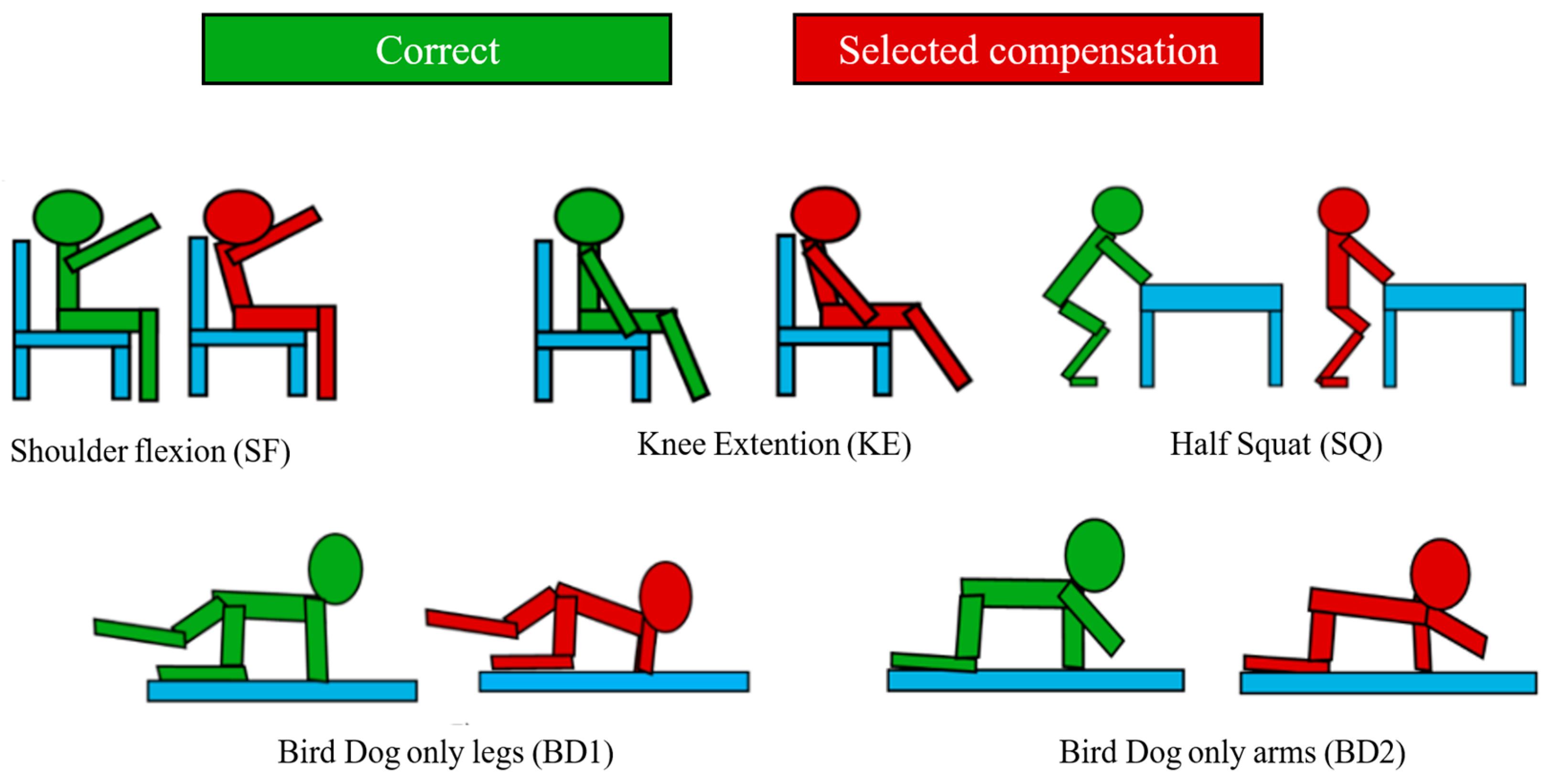
Figure 2.
Schematic overview of the machine-learning pipeline used for compensatory movement detection.
Figure 2.
Schematic overview of the machine-learning pipeline used for compensatory movement detection.
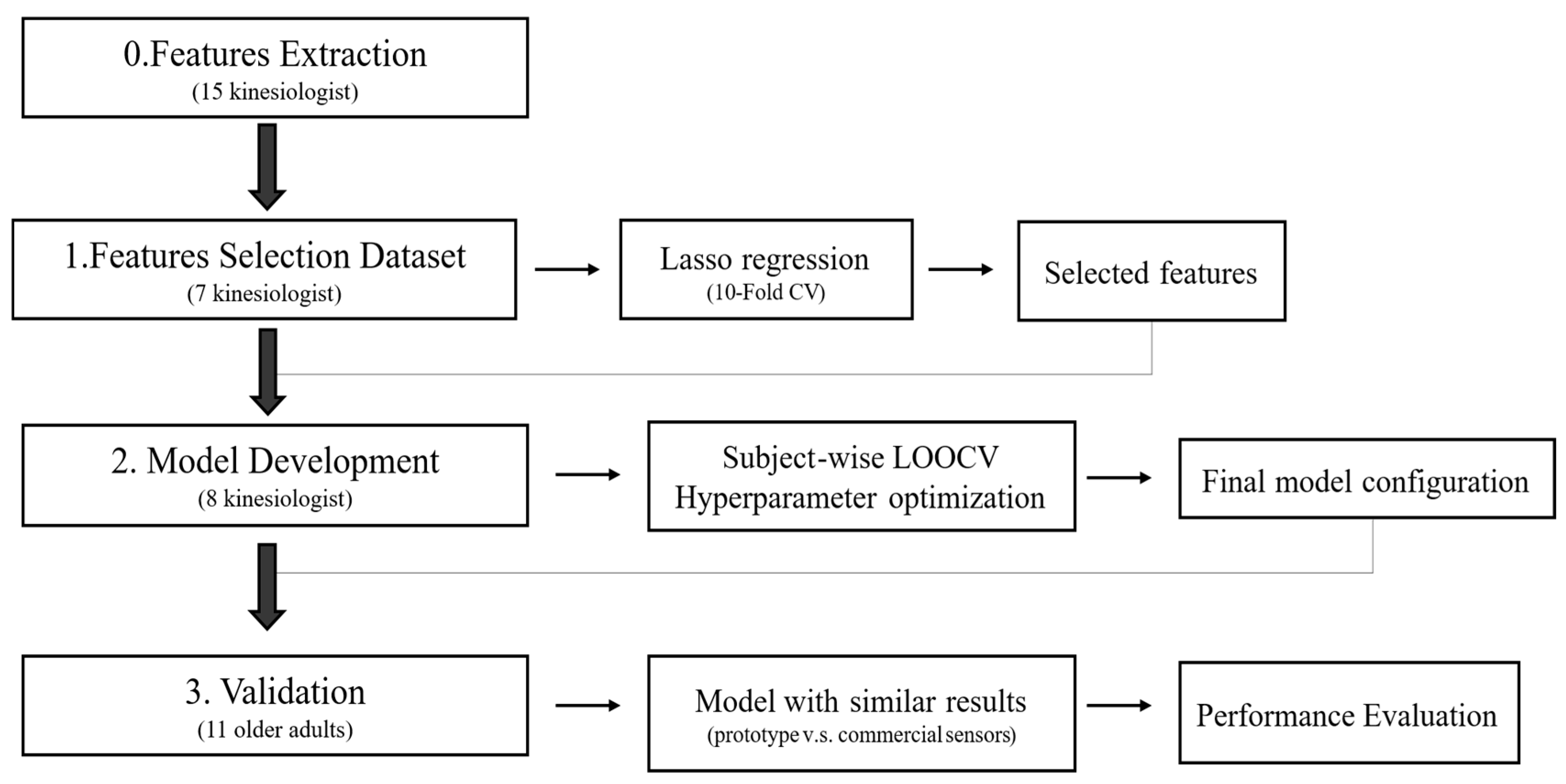

Table 1.
Selected model classifiers for identification of compensatory movements. KE3: knee extension without back support. SQ: Half squat in support on a table. SF3: shoulder flexion without back support. BD1: Bird Dog exercises using only legs. BD2: Bird Dog exercises using only arms. RF: random forest classifier. SVM: Support vector machine classifier. Pr: protytipical sensor. Com: commercial sensor.
Table 1.
Selected model classifiers for identification of compensatory movements. KE3: knee extension without back support. SQ: Half squat in support on a table. SF3: shoulder flexion without back support. BD1: Bird Dog exercises using only legs. BD2: Bird Dog exercises using only arms. RF: random forest classifier. SVM: Support vector machine classifier. Pr: protytipical sensor. Com: commercial sensor.
| Exercises | Features Selected | Model | Selected Hyperparameter | Accuracy(%) | Precision (%) | ||
| Pr. | Com. | Pr. | Com. | ||||
| KE | 67 | RF | Criterior= entropy N.estimators = 50 Max depth= 20 Min samples split= 2 Min samples leaf= 2 |
99 | 99 | 99 | 99 |
| SQ | 84 | SVM | Kernel= linear Degree= 3 Shrinking= True C=1 Class weight=balanced |
99 | 92 | 99 | 94 |
| SF | 73 | RF | Criterior= gini N.estimators = 50 Max depth= 5 Min samples split= 4 Min samples leaf= 2 |
99 | 100 | 99 | 100 |
| BD1 | 69 | RF | Criterior= gini N.estimators = 50 Max depth= 10 Min samples split= 4 Min samples leaf= 2 |
99 | 99 | 99 | 99 |
| BD2 | 70 | RF | Criterior= entropy N.estimators = 50 Max depth= 5 Min samples split= 6 Min samples leaf= 2 |
99 | 100 | 99 | 100 |
Table 2.
Validation test on elderly dataset.KE: knee extension without back support. SQ: Half squat in support on a table. SF: shoulder flexion without back support. BD1: Bird Dog exercises using only legs. BD2: Bird Dog exercises using only arms. Comp: Number of compensated repetitions. T: Total of repetitions.
Table 2.
Validation test on elderly dataset.KE: knee extension without back support. SQ: Half squat in support on a table. SF: shoulder flexion without back support. BD1: Bird Dog exercises using only legs. BD2: Bird Dog exercises using only arms. Comp: Number of compensated repetitions. T: Total of repetitions.
| Exercises | Comp. (n) /T(n) | Accuracy (%) | Precision (%) | Sensitivity (%) | Specificity (%) |
| KE | 180/330 | 94 | 100 | 89 | 100 |
| SQ | 198/330 | 96 | 100 | 93 | 100 |
| SF | 80/330 | 79 | 86 | 15 | 99 |
| BD1 | 124/330 | 69 | 83 | 23 | 97 |
| BD2 | 56/330 | 67 | 32 | 84 | 63 |
Disclaimer/Publisher’s Note: The statements, opinions and data contained in all publications are solely those of the individual author(s) and contributor(s) and not of MDPI and/or the editor(s). MDPI and/or the editor(s) disclaim responsibility for any injury to people or property resulting from any ideas, methods, instructions or products referred to in the content. |
© 2026 by the authors. Licensee MDPI, Basel, Switzerland. This article is an open access article distributed under the terms and conditions of the Creative Commons Attribution (CC BY) license (http://creativecommons.org/licenses/by/4.0/).
Copyright: This open access article is published under a Creative Commons CC BY 4.0 license, which permit the free download, distribution, and reuse, provided that the author and preprint are cited in any reuse.