
Submitted:
20 October 2026
Posted:
22 October 2026
You are already at the latest version
Abstract
Insect chemosensory receptors are essential for detecting environmental chemical cues that influence insect survival and behavior. Advances in genome and transcriptome sequencing have expanded the identification of odorant receptors (ORs), gustatory receptors (GRs), and ionotropic receptors (IRs), while also providing new opportunities to investigate their expression, evolution, and function. Nevertheless, the functions of many identified receptors remain unknown. This review summarizes recent methods and findings in insect chemosensory receptor research, covering receptor identification and annotation using genomic and transcriptomic data, cell-specific expression analysis using single-cell and single-nucleus sequencing, and evolutionary analysis based on comparative genomics and phylogenetics. We also discuss structural studies of OR–Orco complexes and GRs, computational approaches to receptor–ligand prediction, including structure modeling, molecular docking, and machine learning, and experimental methods for functional characterization. By examining how these approaches complement one another, we highlight current progress and outstanding challenges in linking receptor sequences to ligand recognition and biological function.
Keywords:
olfactory receptors
; gustatory receptors
; ionotropic receptors
; comparative genome
; muti-omics
; receptor structure
; functional validation
1. Introduction
Insects occupy diverse ecological niches and have varied effects on ecosystems and human activities [1]. Some species damage crops or transmit pathogens, while others contribute to pollination and natural pest control [2,3]. Chemical cues are central to many of these interactions, allowing insects to locate food and hosts, recognize mates, select oviposition sites, and avoid predators [4]. Insect chemosensation is mediated by several receptor families, particularly odorant receptors (ORs), gustatory receptors (GRs), and ionotropic receptors (IRs). These receptors localize to the membranes of sensory neurons within multiple sensillar classes on the antennae, maxillary palps, proboscis, legs, and ovipositors [3,4,5]. At the molecular level, ORs and GRs belong to an insect-associated family of seven-transmembrane ion channels, whereas IRs are evolutionarily related to ionotropic glutamate receptors and contribute to the detection of diverse chemical and environmental cues, including acids, amines, humidity, and temperature [6,7,8]. Odorant-binding proteins (OBPs) and chemosensory proteins (CSPs) also participate in insect chemosensation by interacting with chemical cues in the sensillum environment [6,7,8]. The expansion and diversification of chemosensory receptor families have provided a molecular basis for differences in chemical perception.
Genome sequencing has revealed large repertoires of insect chemosensory receptors, but identifying these genes is only the first step toward understanding their functions. Even in Drosophila melanogaster, the OR repertoire has required continued annotation and characterization over more than two decades [9,10,11]. The case of Or46aB, whose antennal neurons were not localized until 2025 despite the earlier identification of the gene, illustrates the gap between gene discovery and cellular characterization [12]. This gap is more pronounced in less-studied insects, where many predicted ORs, GRs, and IRs have no known ligands. Testing large numbers of candidates individually using heterologous expression and electrophysiology is time-consuming, making it difficult to keep pace with genome-wide discovery. Genome and transcriptome analyses can help identify candidate receptors and characterize their expression, while computational approaches provide additional criteria for selecting candidates for experimental testing [13,14].
Comparisons across insect species present a related problem. Chemosensory receptor families have undergone lineage-specific expansion and gene loss, accompanied by substantial sequence divergence [15]. Consequently, sequence similarity alone may not reliably distinguish receptor orthologs from lineage-specific duplicates or indicate whether receptors recognize similar ligands. Comparative genomics and phylogenetic analysis help resolve these relationships and identify receptor lineages for further investigation, although functional assignments ultimately require experimental evidence [16,17,18].
Structural biology provides a complementary approach to investigating receptor function. Cryo-electron microscopy (cryo-EM) structures of OR–Orco complexes and insect gustatory receptors have provided experimentally determined structural information for receptor families that were previously poorly characterized at the structural level. These structures offer templates for comparative modelling and provide a framework for molecular docking and machine-learning approaches to receptor–ligand prediction [19,20,21]. At the same time, recent studies indicate that prediction performance depends strongly on receptor family, structural templates, and modelling strategy. For some insect chemosensory receptors, template-based models have shown advantages over predictions generated by general-purpose structure-prediction models such as AlphaFold3 in ligand-binding studies [22]. Thus, structural prediction is becoming a useful complement to, rather than a replacement for, experimental characterization.
Functional studies have also revealed that receptor activity cannot always be interpreted as a simple one-receptor–one-neuron relationship. Alternative splicing, miRNA-mediated post-transcriptional regulation, receptor coexpression, receptor co-option, and compensatory responses to receptor loss can influence chemosensory phenotypes [23,24,25,26]. Non-neuronal support cells also contribute to receptor-associated processes. In addition, these receptors have been linked to functions extending beyond odor and taste perception, including immunity, insecticide resistance, gut–microbiota interactions, reproduction, and lifespan regulation [27,28,29,30]. Together, these findings suggest the importance of considering receptor expression, regulation, cellular context, and physiological function. Integrating genomic and transcriptomic data with structural information and other omics datasets may therefore help explaining the functions and phenotypes of chemosensory receptors.
In this review, we examine bioinformatic and multi-omics approaches to the identification, evolutionary analysis, and functional characterization of insect ORs, GRs, and IRs. We discuss genome- and transcriptome-based discovery, single-cell and single-nucleus transcriptomics, comparative and phylogenetic analyses, structural studies, and computational methods for predicting receptor–ligand interactions. Furthermore, consider experimental strategies for validating these predictions and establishing their physiological and behavioral relevance. Finally, we highlight current methodological limitations and future directions for elucidating the molecular basis of insect chemical perception (Figure 1). Given recent advances in insect chemosensory receptor research, this review aims to provide a comprehensive overview of current approaches to chemosensory receptor identification, evolutionary analysis, and functional validation, while highlighting key challenges and future directions for elucidating the molecular basis of insect chemical perception.
2. Omics-Based Identification of Insect Chemosensory Receptors
Early efforts to identify insect ORs, GRs, and IRs were hampered by limited knowledge of these families, low sequence conservation and the large number of receptors lacking experimentally identified ligands [15,31]. The presence of a candidate sequence in a genome does not, by itself, establish that the gene is complete, expressed, or associated with a particular chemosensory cell. Genome analysis provides the basis for defining the potential receptor repertoire, whereas transcriptomic data indicate which receptors are expressed across tissues, sexes, and developmental stages. Single-cell and single-nucleus approaches add a cellular dimension by assiging receptor expression to specific neuronal and non-neuronal populations and by revealing coexpression patterns that are difficult to resolve in bulk RNA-seq. These datasets therefore address different aspects of receptor identification: the genome defines what may be encoded, transcriptomes indicate what is expressed, and single-cell datasets show where expression occurs.
2.1. Genome, Transcriptome, and Single-Cell-Based Identification
Genome-based identification of chemosensory receptor genes generally combines sequence similarity searches, profile-based searches, gene prediction, and transcript evidence. Protein homology searches using BLASTp against predicted proteomes, or tBLASTn against genome or transcript sequences, remain a common starting point when curated receptor sequences from related species are available [32,33]. For ORs, dedicated annotation tools can improve recovery because these genes are highly divergent and are often missed or incorrectly modelled by general gene-annotation pipelines. For example, the insectOR web server uses protein-to-genome alignments to identify OR loci, resolve tandem arrays, and refine gene boundaries, and showed higher sensitivity than MAKER and the NCBI eukaryotic annotation pipeline in two test genomes [34]. Profile-based searches provide a complementary strategy when sequence similarity is weak. Hidden Markov models implemented in HMMER can detect conserved family signatures and have been used to identify domains associated with ORs, IRs, and other chemosensory proteins [32,35,36]. Ab initio gene prediction can further provide candidate gene models when closely related reference annotations are unavailable, using programs such as AUGUSTUS, SNAP, GeneWise, Exonerate, BRAKER2, and MAKER [34,37,38,39,40,41,42,43]. RNA-seq evidence can then be incorporated to refine exon boundaries and support transcriptional evidence, either through transcriptome assembly and genome alignment or through evidence-based annotation pipelines such as MAKER and BRAKER [32,42,43,44]. Candidate sets from any of these routes are then filtered and described with a shared downstream toolkit: SignalP for signal peptides, TMHMM or DeepTMHMM for transmembrane topology, and InterProScan for conserved domains [32,33,44,45,46,47,48].
No single strategy is sufficient for all receptor families or genome assemblies. Homology-based searches are effective when suitable reference sequences are available but can miss highly divergent genes; HMM-based searches are less dependent on pairwise sequence similarity but still depend on the quality and specificity of the underlying profiles; and ab initio prediction can generate incomplete or spurious models in poorly annotated genomes. In practice, published receptor repertoires often combine several approaches followed by manual inspection and phylogenetic or structural validation.
Genome-level annotation also provides the scale required to examine the evolution of receptor repertoires. Comparative analyses can reveal lineage-specific expansions and losses, tandem duplications, chromosomal organization, and associations with rapidly evolving genomic regions such as transposable elements (TEs). For example, broad phylogenomic analyses of insect and non-insect hexapods have revealed previously unrecognized gustatory receptor lineages and provided new evidence for the evolutionary origin of insect ORs [49]. Re-annotation of multiple aphid genomes further showed extensive variation in OR and GR repertoires and associations between receptor evolution, transposable elements, and host range [50]. High-quality chromosome- or telomere-to-telomere assemblies can provide additional resolution of tandem receptor arrays and gene duplications, as illustrated by the OR88a cluster in Bactrocera dorsalis [51]. In some species, genome-wide analyses reveal extensive receptor loss rather than gene family expansion. For example, the ambrosia beetle Trypodendron lineatum has a reduced repertoire of ORs, GRs, and IRs, consistent with changes in chemosensory gene content across insect lineages [52]. Reliable comparisons, however, depend on the quality of genome assemblies and gene annotation, since missing or fragmented genes can be mistaken for genuine gene loss.
Transcriptomic data complement genome-based annotation by revealing receptor expression across tissues and biological conditions. Bulk RNA-seq can identify differences associated with sex, developmental stage, or physiological state, while long-read sequencing helps resolve full-length transcripts and alternative isoforms. In the soybean pod borer Leguminivora glycinivorella, multi-tissue RNA-seq detected male-biased expression of pheromone receptors and other chemosensory genes in the antennae [53]. Receptor expression is not restricted to antennal tissues: ORs, GRs, and IRs have also been detected in the mouthparts of Spodoptera frugiperda [54], broadening the anatomical scope of transcriptome-based receptor surveys. Developmental transcriptomes of Monochamus alternatus identified persistent IR expression across developmental stages and antennal enrichment of IR8a and IR25a [55]. Long-read sequencing in Microplitis pallidipes further demonstrated the value of full-length transcript information for resolving chemosensory gene isoforms [56].
Bulk transcriptomics identifies which receptor genes are expressed, whereas single-cell and single-nucleus RNA sequencing (scRNA-seq and snRNA-seq) assigns these transcripts to specific cell types, including rare chemosensory neurons. These approaches therefore extend receptor identification from gene lists to the cellular organization, receptor combinations, and regulatory programs of insect sensory systems.
Single-cell analyses have challenged the assumption that each olfactory sensory neuron expresses only one receptor. In female Aedes aegypti antennae, analysis of approximately 46,000 cells showed that around half of the olfactory sensory neuron subtypes expressed more than one receptor. Coexpression occurred predominantly within the same receptor family, whereas OR–IR combinations were relatively uncommon [57]. A single-nucleus multiomic study of Apis mellifera identified three patterns of OR transcription: expression of a single receptor, polycistronic transcription from one active promoter, and coexpression associated with multiple promoters. Multiple OR transcripts were detected in approximately half of the OSNs [58]. Similar observations were made in the ant Harpegnathos saltator, where multiple transcripts from tandem OR arrays could be detected in individual nuclei. Subsequent analyses suggested, however, that only the most upstream OR transcript was generally exported to the cytoplasm and translated [59,60]. These findings illustrate an important distinction between transcriptional detection and functional receptor expression: the presence of multiple receptor transcripts in a nucleus does not necessarily indicate that multiple receptors are translated and active.
Single-cell datasets can also reveal species- and sex-specific differences in sensory-cell composition. Comparative snRNA-seq of antennae from Bactrocera dorsalis and Drosophila melanogaster found greater conservation among support cells than among sensory neurons, whose receptor profiles were more divergent between species [61]. An adult Aedes aegypti atlas spanning 19 tissues and more than 367,000 nuclei further expanded the catalog of receptor-expressing cells across sensory organs and identified sex-biased expression in the antenna and brain [62].
Developmental single-cell studies provide an additional layer of information. An antennal sensory-cell atlas in D. melanogaster showed that receptor expression in mature neurons is associated with selective transcription, combinatorial expression of receptor subunits, and programmed cell death [63]. In H. saltator, loss of orco led to extensive olfactory sensory neuron (OSN) loss during pupal development. Some surviving neurons began expressing IR-complex components, suggesting that OR signaling may influence both neuronal survival and sensory-cell identity [64]. This finding also shows why receptor expression needs to be examined in its cellular and developmental context.
2.2. Phylogenetic, Comparative, and Multi-Omics Analysis
Phylogenetic analysis helps resolve relationships among insect chemosensory receptors, whose sequences have diverged through gene duplication, gene loss, and lineage-specific evolution. Similarity at the sequence level does not always provide sufficient evidence for assigning rapidly evolving receptors to established groups. Phylogenetic reconstruction can distinguish orthologous and paralogous genes and identify divergent lineages that might otherwise remain unclassified. Mitchell et al. [49], for example, combined broad taxonomic sampling, homology searches, and phylogenetic analysis to identify previously unrecognized GR lineages. They also found that a GR lineage from non-insect hexapods was the sister group of insect ORs, supporting the hypothesis that insect ORs originated from within the sugar GR lineage. These relationships can guide the selection of receptors for functional study, although related receptors may differ in ligand specificity and expression following gene duplication.
Comparisons of receptor repertoires across species reveal different evolutionary outcomes. In paper wasps of the genus Polistes, OR family expansion has occurred largely through tandem duplication, with evidence of relaxed selective constraint in some lineages [16]. Analysis of more than 55,000 ORs from 163 ant genomes identified enrichment of positive selection in 9-exon ORs along the ancestral formicoid branch, implicating this receptor group in sensory adaptation during ant diversification [65]. By contrast, three independently evolved lineages of socially parasitic, slave-making ants have undergone convergent losses of ORs and GRs [17]. Individual genes can follow different trajectories as well. Triplication of Or67b in Scaptomyza flava was followed by neofunctionalization and subfunctionalization and has been linked to adaptation to mustard plants [66], whereas the HarmOR42 lineage shows strong purifying selection, consistent with continued functional constraint [67]. Receptor family evolution therefore involves not only expansion and loss, but also differences in the extent to which duplicated genes diverge or retain ancestral functions.
Combining genomic and transcriptomic data can help examine whether evolutionary changes in receptor repertoires are accompanied by differences in expression and function. Lu et al. [68] combined genomic and transcriptomic data to characterize the OR repertoire of L. boulardi and identified two female-biased receptors, LbouOR167 and LbouOR136. Functional assays showed that both receptors detect yeast-derived ethyl esters, and identified Leu159 as an important residue involved in ligand recognition. In B. dorsalis, Liu et al. [51] combined a single-individual telomere-to-telomere (T2T) genome assembly with multi-tissue RNA-seq and comparative genomic analyses. This analysis identified a three-copy OR88a cluster generated by tandem duplication, and behavioral experiments linked the receptor cluster to the response to methyl eugenol. Together, these studies connect receptor evolution and genomic architecture with expression and function, from residue-level ligand recognition to copy-number variation and behavioral response.
3. Computational Prediction of Receptor Function
Computational approaches have become useful for investigating insect chemosensory receptors, particularly given the large number of potential receptor–ligand combinations that need to be examined [13,14]. Sequence analysis and structural modeling can provide clues about receptor function, while ligand prediction and residue-level analyses help identify candidates for further testing [69,70]. The available methods differ in their inputs and applications, ranging from receptor identification to structural and ligand-binding analysis. Table 1 lists representative tools and their uses in chemosensory receptor research.
3.1. Sequence- and Structure-Based Prediction
3.1.1. Sequence Features, Key Residues, and Membrane Topology
Sequence analysis provides an initial means of identifying residues that may contribute to receptor structure or function. Conserved motifs and residues can identify structural cores, subunit interfaces, and regions that may contribute to ligand binding or channel gating. Sequence divergence can also be informative, particularly when closely related receptors differ in ligand specificity or physiological function. Comparing conserved and variable sites across receptor homologs can therefore provide candidate residues for subsequent structural and functional analysis.
Membrane topology is particularly important for structural analysis. Insect ORs and GRs generally contain seven transmembrane helices, with a membrane orientation opposite to that of classical G-protein-coupled receptors. IRs are evolutionarily related to ionotropic glutamate receptors and contain an extracellular ligand-binding domain and an ion-conducting pore. These differences affect both structural modelling and the interpretation of predicted functional sites [90]. Bibi et al. [91] performed a computational analysis of all 72 GRs from Aedes aegypti, combining phylogenetic relationships, physicochemical properties, conserved motifs, and three-dimensional models. The analysis identified the 7tm_7 superfamily domain and several candidate functional residues. Such analyses provide targets for mutagenesis and ligand screening, but sequence conservation or a predicted structural feature alone does not establish ligand specificity.
3.1.2. Structural Modeling and Model Assessment
Three-dimensional receptor models provide structural information that cannot be obtained from sequence comparison alone and can be used to examine transmembrane architecture, putative binding pockets, and receptor–ligand interactions. Current approaches include template-based modelling and deep-learning methods such as AlphaFold and ColabFold [71,92]. When experimentally determined structures of related receptors are available, template-based modelling can preserve family-specific structural features and helix arrangements. Deep-learning methods extend structure prediction to receptors for which suitable experimental templates are unavailable.
Jabeen et al. [21] used the experimentally determined MhOR5 structure as a reference to compare template-based models with AlphaFold3 predictions for fruit fly OR–odorant complexes. Template models generated using transmembrane and hydrophobicity-guided alignments performed better in distinguishing binding from non-binding complexes in their benchmark. Chidambara Thanu et al. [22] compared models for six ORs from three insect orders using available mutagenesis data and also found advantages for template-based models in several ligand-prediction tasks. These studies indicate that experimentally characterized receptor structures remain useful for modelling related receptors and evaluating ligand-binding hypotheses. Model performance can vary with the quality of the template, sequence alignment, receptor conformation, and the region being modelled.
Structural prediction has also been scaled to entire receptor repertoires. Kalepu et al. [93] developed a workflow combining functional annotation with ColabFold modelling and applied related methods to 110 ORs from the red palm weevil, Rhynchophorus ferrugineus. Molecular-dynamics simulations of RferOR18148 and RferOrco were used to examine the predicted structures in a membrane environment and identified a central cavity as a potential ligand-binding site [94]. Such analyses provide structural hypotheses that can be tested experimentally.
3.1.3. Inferring Functional Sites and Receptor Specificity
Sequence comparison and structural modelling can be used together to identify residues that may contribute to ligand recognition and receptor specificity. Candidate residues can be selected from conserved or divergent sites, examined in structural models, and further evaluated by molecular docking, molecular dynamics, or site-directed mutagenesis. Functional assays can test whether changes at these positions affect receptor activity. One example is the Drosophila Or22a system, in which variation at the Or22a/Or22b locus has been linked to interspecific differences in behavioral responses to methyl and ethyl hexanoate [95]. In D. melanogaster, Or22a is expressed in ab3A basiconic sensilla and is particularly sensitive to esters such as methyl hexanoate [96]. In the noni-fruit specialist D. sechellia, Auer et al. [97] combined calcium imaging, receptor mutagenesis, and cross-species allele transfer to examine how Or22a tuning contributes to attraction to noni. Chimeric receptors localized the difference in ester sensitivity to to the N-terminal 100 amino acids of DsecOr22a. Further mutagenesis narrowed the effect to residues 45, 67, and 93, which are I45, I67, and M93 in DmelOr22a and V45, M67, and I93 in DsecOr22a. Substitution of M93 with I in DmelOr22a largely reproduced the stronger response to methyl esters, whereas the reciprocal I93M substitution broadened the response profile. A triple mutant further altered receptor responses and behavioral attraction in vivo [97].
This example shows how comparative sequence analysis can be followed by targeted experiments to identify individual residues contributing to receptor specificity and ultimately link sequence variation to behavioral phenotypes.
3.2. Ligand Prediction, Docking, and Machine Learning
3.2.1. Molecular Docking and Molecular Dynamics
Molecular docking predicts plausible ligand poses within a receptor-binding pocket and ranks candidate compounds using approximate scoring functions. Molecular-dynamics simulations can further examine the stability of predicted complexes, receptor–ligand contacts, and conformational changes under defined membrane and solvent conditions. These approaches provide structural hypotheses for receptor–ligand recognition, but docking scores should not be interpreted as direct measurements of binding affinity or receptor activation. The accuracy of these analyses depends on the receptor model, binding-site definition, treatment of receptor and ligand flexibility, and other modelling parameters. Docking scores are therefore most useful for prioritizing candidates, which can then be evaluated using receptor-based functional assays and behavioral experiments to identify biologically relevant receptor–ligand interactions. In the potato tuber moth, Chen et al. [98] combined molecular docking with heterologous expression and CRISPR-mediated gene disruption to identify ORs involved in responses to trans-nerolidol, benzyl tiglate, and 3-carene.
Docking combined with mutagenesis can also be used to examine candidate ligand-binding residues. Ma et al. [99] showed that OachOR16 from the camphor-tree specialist Orthaga achatina responds to camphor and identified Ser209 as a residue involved in ligand recognition. Lu et al. [100] used multiple docking algorithms to screen 42 ORs from Callosobruchus maculatus and prioritized CmacOR3 as a candidate receptor for the pheromone components Z32A and Z33A. RNAi supported its in vivo relevance, although receptor-level assays are still needed to establish the direct receptor–ligand interaction.
Docking can also provide a structural basis for comparing homologous receptors. Hou et al. [101] reported that several BmorOR56 orthologs respond to cis-jasmone and are subject to strong purifying selection. Their structural models showed similar pocket features, consistent with conservation of receptor function. Structural similarity and evolutionary conservation, however, provide supporting evidence rather than direct evidence of ligand specificity. Ligand access may also involve transient conformational changes that are not captured by a static receptor model [102].
3.2.2. Virtual Screening and Reverse Chemical Ecology
Virtual screening extends docking or ligand-similarity analysis to large chemical libraries and can reduce the number of compounds selected for receptor assays. Reverse chemical ecology uses the receptor as the starting point for chemical discovery: candidate compounds are first predicted or selected computationally, followed by receptor assays and behavioral testing. This contrasts with conventional chemical ecology, in which biologically relevant volatiles are first collected from plants, hosts, or conspecifics and then tested for activity.
Comte et al. [14] applied structure-based virtual screening to a non-pheromone OR from Spodoptera littoralis. Docking-based prioritization followed by receptor assays and behavioral experiments identified previously unrecognized behaviorally active volatiles. The study also identified physicochemical features of the receptor pocket associated with different response profiles, providing a basis for further candidate selection.
Orco has also been used as a target for receptor-guided chemical screening. Lee et al. [103] identified 2,4-di-tert-butylphenol as an Orco agonist and used electrophysiological and mutational analyses to implicate W146 in its activity. Because Orco is shared by most insect OR complexes, compounds acting on Orco have the potential to influence multiple olfactory pathways. Their activity therefore needs to be evaluated at both the receptor and organismal levels, particularly when such compounds are considered for pest management.
3.2.3. Machine and Deep Learning
Machine-learning approaches provide a ligand-based alternative to structure-dependent prediction. Models can be trained on molecular descriptors, chemical fingerprints, or experimentally measured receptor–ligand responses and then used to rank untested compounds. Quantitative structure–activity relationship models are one common implementation and can prioritize potential agonists or antagonists from large chemical libraries [13]. Because these models do not necessarily require a high-resolution receptor structure, they are useful for receptors for which structural information remains limited.
Caballero-Vidal et al. [104] applied machine-learning-based virtual screening to the lepidopteran receptor SlitOR25. Approximately three million compounds were screened in silico, after which 32 commercially available candidates were tested experimentally and 11 new agonists were identified. Among the models evaluated, the support-vector machine achieved the best performance in this dataset. In a subsequent study, computational predictions for SlitOR24 and SlitOR25 were tested using electrophysiological and behavioral assays, leading to the identification of additional active compounds, some of which attracted larvae [105]. Together, these studies illustrate the use of machine learning to prioritize compounds for experimental testing when the initial candidate pool is too large for direct screening.
Machine learning has also been used to Orco modulators. Kepchia et al. [106] tested 83 compounds and used the resulting activity data to rank 1,280 odorants. Their analysis identified 2-tert-butyl-6-methylphenol as a potential Orco antagonist with behavioral activity. Its effects were further examined using electrophysiological recordings and larval behavioral assays in Drosophila melanogaster. Resources such as iORandLigandDB, which brings together receptor structures, ligand information, and docking results, can also support model development and the selection of candidate compounds [78].
The reliability of these predictions depends in part on the quality and coverage of the training data. Available insect receptor datasets are still limited in size, with some receptor families and insect taxa represented much more extensively than others. Inactive compounds are also often underrepresented, making it difficult to distinguish true receptor agonists from compounds that simply lack experimental evidence of activity.
4. Experimental Validation of Chemosensory Receptors
Computational analyses can prioritize candidate receptors, ligands, and interaction residues, but receptor function ultimately needs to be tested experimentally. Different assays address different levels of function. Receptor-based assays determine whether a candidate receptor responds to a compound, whereas neuronal assays place the receptor in a sensory-cell context. Genetic manipulation can test its contribution in vivo, and behavioral assays can determine whether receptor activity is associated with a measurable change in animal behavior. In practice, receptor assignment is strongest when evidence from several levels converges on the same receptor–ligand relationship.
4.1. Receptor- and Neuron-Level Functional Assays
4.1.1. Heterologous Expression in Oocytes and Cultured Cells
Heterologous expression provides a controlled system for testing whether a candidate receptor responds to a putative ligand. Xenopus oocytes, mammalian cells, and insect cell lines have all been used to characterize insect ORs, GRs, and IRs. When receptor function requires accessory proteins, the candidate receptor can be expressed together with the appropriate co-receptors, such as Orco for many insect ORs or the relevant IR co-receptors for specific IR complexes. Ligand-evoked activity can then be measured by two-electrode voltage clamp (TEVC), whole-cell patch clamp, or Ca²⁺ imaging [107].
Heterologous systems allow ligand selectivity, dose–response relationships, response kinetics, and the effects of receptor mutations to be compared under controlled conditions. Oocytes often generate robust current responses and are particularly useful for examining receptor–co-receptor combinations, whereas cultured cells provide greater flexibility for dose–response measurements and comparisons among receptor variants [108]. Early heterologous studies established that insect ORs assemble with the conserved co-receptor Orco into heteromeric, ligand-gated, non-selective cation channels [109,110]. Oocyte-based TEVC assays were subsequently used to screen the OR repertoire of Anopheles gambiae [111].
Heterologous expression is also useful for testing hypotheses generated from sequence analysis, structural modelling, or molecular docking. Mutant and orthologous receptors can be compared under the same experimental conditions, allowing changes in ligand sensitivity or response profiles to be related to specific sequence differences. The interpretation of a negative result, however, requires attention to receptor expression and membrane localization, co-receptor availability, and the compatibility of the expression system with receptor assembly and trafficking. Appropriate controls, including mock-transfected cells, co-receptor-only controls, unrelated receptors, and structurally related compounds, are therefore important when assigning ligand specificity. Receptor abundance and surface localization are particularly relevant when comparing mutants or orthologues [112].
4.1.2. Reconstituted and Native Neuronal Assays
In vivo reconstituted neuronal systems test candidate receptors within a defined sensory-neuron and sensillum context. The Drosophila empty-neuron system is one of the most widely used examples [113,114]. A candidate receptor is expressed in an olfactory sensory neuron in which the endogenous receptor has been removed or silenced, and ligand-evoked activity is measured by single-sensillum recording or neuronal imaging. The system retains the neuronal signaling machinery and much of the sensillum environment while allowing a defined receptor to be tested in vivo.
Several Drosophila reconstruction systems have been used for this purpose. The ab3A empty-neuron system, for example, allows ORs to be expressed in neurons lacking the endogenous Or22a and Or22b receptors in the Δhalo background. Candidate ORs can also be re-expressed together with Orco in orco mutant backgrounds, while the ab1C neuronal system has been used as an in vivo decoder for GR function [115,116]. These platforms have facilitated cross-species characterization of chemosensory receptors. AgOr1 from Anopheles gambiae, the silkworm pheromone receptor BmorOR1, ant 9-exon ORs, and OR repertoires from moths have all been expressed in Drosophila neurons for functional characterization and ligand identification [117,118,119,120].
Neuronal reconstruction is particularly useful when receptor activity depends on the cellular environment. At the same time, an empty-neuron system does not reproduce every component of the native sensory organ. Pheromone-receptor responses, for example, may depend on species-specific odorant-binding proteins, degrading enzymes, sensillum lymph, and other accessory factors [112,118]. For IRs, there is still no broadly used reconstruction system equivalent to the Drosophila ab3A empty neuron. IR functionhas therefore commonly been examined through combining co-receptor expression, co-receptor mutation or knockdown, heterologous reconstitution, and recordings from native neurons [121,122,123,124].
Native neuronal assays provide a more physiological context than heterologous systems or reconstructed neurons. Single-sensillum recording measures stimulus-evoked changes in spike activity from individual sensilla, while Ca²⁺ imaging can monitor responses across populations of sensory neurons or downstream neural circuits. These approaches reveal how chemical stimuli are detected and represented within the sensory system, but a neuronal response alone does not necessarily identify the receptor responsible. Receptor-specific expression data, genetic manipulation, or rescue experiments can therefore provide additional evidence for linking a neuronal response to a particular receptor.
4.1.3. Electrophysiology, Ca²⁺ Imaging, and Site-Directed Mutagenesis
Electrophysiological approaches provide quantitative measurements of receptor or neuronal activity. In heterologous systems, TEVC and whole-cell voltage clamp can be used to generate concentration–response curves and compare maximal responses, apparent sensitivity, and response kinetics among receptors or receptor variants. In intact sensilla, single-sensillum recording measures odor-evoked changes in neuronal firing while retaining the local sensory environment. Ca²⁺ imaging provides a complementary approach and allows responses from multiple cells or neurons to be monitored simultaneously, making it useful for screening receptor–ligand combinations and mapping stimulus-responsive populations.
The combination of electrophysiology and Ca²⁺ imaging can also reveal receptor modulation in native sensory systems. Sun et al. [125] integrated peripheral electrophysiological recordings with Ca²⁺ imaging to show that male-produced benzaldehyde suppresses the response of Mythimna separata to (Z)-11-hexadecenal. Their results supported a model in which benzaldehyde acts as a competitive antagonist of the sex-pheromone receptor MsepPR3, with the antagonistic effect involving or occurring near the receptor ligand-binding site. In another study, Andreu et al. [126] combined transcuticular Ca²⁺ imaging with electrophysiological recording in the Drosophila empty-neuron system to identify AmelOR136 and AmelOR109 as receptors for alarm-pheromone components in the honey bee, Apis mellifera. Together, these studies show that electrophysiology offers quantitative measurements of receptor or neuronal responses, whereas Ca²⁺ imaging allows parallel measurements across cells and sensory pathways.
Site-directed mutagenesis provides a direct experimental route for testing residues highlighted by sequence comparison, structural modelling, or molecular docking. Changes in response amplitude or ligand selectivity after mutation can implicate a residue in receptor function. Interpreting such effects requires consideration of receptor expression, membrane trafficking, protein stability, and folding, particularly when a mutation causes a large loss of activity. Reciprocal substitutions, concentration–response analysis, and expression or localization controls can help distinguish effects on ligand recognition from more general effects on receptor integrity.
Dong et al. [127] applied this strategy to the aphid sex-pheromone receptor ApOR22. Among 13 alanine-substitution mutants targeting residues in the predicted ligand-binding pocket, 11 showed markedly reduced responses, whereas M121A and L361A increased receptor sensitivity. Additional substitutions, including M121V, L122M, and Y125N, further clarified the contribution of residues in the S2 region to ligand specificity. All variants were tested using TEVC, and the results were interpreted together with cryo-EM structures and molecular dynamics simulations. Similarly, Frunze et al. [128] showed that a single mutation in the Aedes aegypti receptor AaOr8 eliminated its response to 1-octen-3-ol, with evidence from site-directed mutagenesis, molecular docking, and in vivo Ca²⁺ imaging
Sequence variation can therefore have substantial effects on receptor specificity. Two substitutions were sufficient to alter the ligand response of closely related moth pheromone receptors [129]. In Drosophila, a single amino-acid polymorphism in OR59B altered odor responses and sensitivity to DEET [130]. The Or22a study discussed in Section 3 further linked interspecific differences in odor response to substitutions at I45, I67, and M93 [97]. Such studies provide a route from sequence variation to receptor-level function and, when combined with neuronal and behavioral experiments, to phenotypic consequences.
4.2. Genetic and Behavioral Validation
In vivo genetic experiments go beyond demonstrating that a receptor can respond to a ligand. They test whether the receptor contributes to sensory processing and behavior in the intact animal by reducing, eliminating, or specifically modifying its function in native neurons or tissues. The main approaches are RNA interference (RNAi) and CRISPR-based genome editing. RNAi is useful for transient or partial knockdown, whereas CRISPR can produce targeted knockout, knock-in, or allele replacement.
RNAi can reduce receptor transcript levels and is useful for rapid or tissue-specific functional analysis. Its effectiveness may vary with tissue, developmental stage, and delivery method, and incomplete knockdown or off-target effects may complicate interpretation. For this reason, receptor expression should be quantified after treatment and, where possible, independent RNAi constructs and appropriate controls should be used. Rescue experiments provide additional support when a behavioral or physiological phenotype can be restored by re-expressing the receptor. RNAi has been used to examine the locust aggregation-related receptor OR35 [131], ORs in Apolygus lucorum, and Orco in the whitefly and bean weevil [132,133,134]. These studies also illustrate how knockdown efficiency and tissue specificity can affect the interpretation of RNAi results.
CRISPR-based approaches provide a more direct way to test receptor function in vivo. Gene knockout can determine whether a receptor contributes to a sensory response, whereas knock-in or allele-replacement strategies can examine specific residues, natural variants, or interspecific sequence differences. Editing a predicted ligand-interaction site can therefore connect a molecular model with neuronal and behavioral phenotypes in the native animal. Tissue-specific or inducible approaches are particularly useful when receptor genes also affect development.
In ants, for example, orco mutations impair social behavior and are associated with changes in antennal-lobe development [135,136]. Orco knockout studies in different insect pests have further implicated OR pathways in host choice, oviposition, and other behaviors [137,138,139,140]. Genetic manipulation of individual receptors can also reveal more specific ecological functions. Specific ORs contribute to parasitoid avoidance in the diamondback moth, whereas OR39 in mosquitoes provides an example of functional redundancy among olfactory pathways [141,142].
5. Structural Basis of Chemosensory Receptor Function
Advances in single-particle cryo-electron microscopy (cryo-EM) have provided structural information for several major families of insect chemosensory receptors. Structures of ORs, the conserved co-receptor Orco, and GRs have revealed the overall architecture of these receptors and provided a framework for examining ligand recognition and ion-channel gating. ORs and GRs belong to the insect-specific seven-transmembrane ion channel (7TMIC) superfamily and form tetrameric ion channels with a central pore (Figure 2A, B). Structural studies have begun to reveal how ligand-binding sites are organized and how ligand binding is coupled to conformational changes in the transmembrane helices and pore. These studies also indicate that ORs and GRs can differ substantially in the architecture and chemical environment of their ligand-binding pockets, consistent with their recognition of chemically diverse odorants and tastants.
IRs are evolutionarily related to ionotropic glutamate receptors (iGluRs) rather than the 7TMIC family and generally function as heteromeric complexes containing conserved co-receptors and divergent tuning subunits (Figure 2C). Their complex assembly, extracellular ligand-binding domains, and challenges in receptor expression and purification have made structural characterization more difficult. As a result, the available structures provide a more developed framework for ORs and GRs than for IRs. The following sections examine the structural principles established for OR–Orco complexes and GRs, followed by the current structural challenges associated with IRs.
5.1. OR–Orco
The first structural insight into insect ORs came from the Orco homotetramer from the fig wasp Apocrypta bakeri, resolved by cryo-EM in 2018 at 3.5 Å [6,143]. The structure showed four seven-transmembrane subunits arranged around a central ion-conducting pore. Because it contained Orco alone, it did not establish the subunit composition or activation mechanism of native OR–Orco complexes. The authors proposed that odorant receptors might replace one or more Orco subunits in the native channel. In 2021, del Mármol et al. showed that MhOR5 from the wingless insect Machilis hrabei, which lacks Orco, can form a functional homotetrameric odorant-gated channel [19]. This finding is consistent with the possibility that some ancestral ORs functioned without a dedicated co-receptor, although it does not establish the evolutionary sequence. In the MhOR5 structures, DEET and eugenol occupied a hydrophobic binding pocket. Ligand binding was associated with an outward movement of the pore-lining S7b helix, including movement of Val468 away from the pore. These results provided a structural link between ligand binding and channel opening.
The architecture of native OR–Orco complex was directly examined in 2024. Wang et al. [20] resolved a pea aphid complex with one OR and three Orco subunits. Functional and structural analyses indicated that ligand-dependent rearrangement occurred mainly in the OR subunit, while the Orco subunits remained comparatively stable. The study also detected 2OR:2Orco and 3OR:1Orco assemblies by single-molecule photobleaching. Whether these assemblies occur as stable, functional complexes in sensory neurons remains unknown.
Around the same time, an independent study of mosquito OR complexes, including AaOR10 from Aedes aegypti and AgOR28 from Anopheles gambiae, also found a 1OR:3Orco arrangement [144]. The study did not detect other stable stoichiometries, and functional assays showed that activation of the single OR subunit could gate the channel. A lipid-like density was observed in the OR binding pocket in the apo state and was displaced upon odorant binding. The authors proposed that this lipid might act as a negative allosteric modulator, but its role in OR regulation remains to be established.
Computational work has further explored OR–Orco assembly and gating. Using AlphaFold3-based models and molecular dynamics simulations, Xue et al. [145] found the 1OR:3Orco arrangement to be the most stable of the tested LmOR35–Orco assemblies. Their simulations also proposed a pathway in which ligand-associated changes in transmembrane helices S2–S6 are transmitted to the pore-lining S7b helix. These results are consistent with the cryo-EM structures, but remain computational predictions.
The available structures have established a basic model for OR–Orco signaling, particularly the asymmetric 1OR:3Orco architecture and the coupling between ligand recognition and pore opening. Several questions remain unresolved. It is not yet known whether alternative stoichiometries occur in native sensory neurons, or whether they reflect particular expression and purification conditions. How pocket shape and amino acid composition confer selectivity for different odorants remains unclear. The effects of membrane lipids on receptor function, as well as the mechanisms underlying receptor desensitization and recovery, also warrant further study.
5.2. GRs
Cryo-EM has provided structural insights into insect gustatory receptors. Ma et al. [146] reported apo and ligand-bound structures of the Drosophila sugar receptors GR43a and GR64a in 2024. Both receptors assembled into tetrameric ion channels belonging to the 7TMIC superfamily, with the four S7 helices forming the central pore. Their ligand-binding pockets differed in size and shape. GR43a contained a relatively narrow, polar cavity suited to fructose binding, whereas the more open cavity of GR64a could accommodate larger sugars, including sucrose and maltose. These structural differences offer a possible explanation for the distinct sugar preferences of the two receptors.
Structures of the silkworm fructose receptor BmGr9 show that ligand binding does not always lead to channel activation. Gomes et al. [147] found that L-sorbose occupied a position similar to D-fructose but activated the receptor less effectively. Structural analysis linked this difference to ligand stereochemistry and its interaction with residues in the binding pocket, which may affect coupling between ligand binding and the pore-forming helices. The results distinguish two steps in receptor function: ligand binding and the conformational changes required for channel opening.
Frank et al. [148] independently resolved BmGr9 structures and identified polar and aromatic interactions that contribute to sugar recognition. A comparison of DmGR43a and DmGR64a by Chen et al. [149] likewise associated the broader tuning of DmGR64a with its larger, more open binding cavity. These studies indicate that pocket size and chemical environment contribute to ligand selectivity, whereas the efficiency of coupling to the channel gate determines whether ligand binding produces an effective response.
The structural framework for GRs remains incomplete. Many insect taste responses are thought to involve complexes containing multiple GR subunits, but available structures have mainly examined homomeric receptors [8,149]. The assembly and gating of native heteromeric GR complexes therefore remain poorly understood. Structural information is also limited for receptors that detect bitter compounds, plant secondary metabolites, and other non-sugar ligands.
5.3. IRs: An Unresolved Structural Gap
Ionotropic receptors (IRs) are structurally distinct from ORs and GRs. Phylogenetic studies indicate that IRs are related to ionotropic glutamate receptors (iGluRs) rather than the 7TMIC superfamily [121]. Each IR subunit contains three transmembrane helices, a re-entrant pore loop, and an extracellular bilobed ligand-binding domain (LBD) (Figure 2C), giving IRs an architecture related to vertebrate AMPA, NMDA, kainate, and GluD-type receptors [150]. Many canonical IRs function as heteromeric complexes containing conserved co-receptors, such as IR8a or IR25a in olfactory neurons, IR76b in gustatory and general chemosensory neurons, or IR93a in thermosensory and hygrosensory systems, together with one or more divergent tuning IRs [122,151]. These complexes participate in the detection of diverse chemical and physical stimuli, including carboxylic acids, amines, amino acids, salts, humidity, and temperature differences [152,153].
Despite the increasing functional information available for IRs, no high-resolution structure of an insect IR complex has yet been reported. Their obligate heteromeric organization, conformational flexibility, and difficulties in obtaining stable receptor complexes have complicated structural analysis. The lack of experimentally determined structures limits direct comparison of IRs with ORs and GRs, particularly with respect to ligand recognition, subunit assembly, and channel gating. Vertebrate iGluR structures provide useful reference frameworks for understanding IR architecture and conformational changes [154,155], but they cannot substitute for insect IR structures. The high sequence divergence of tuning IRs, their diverse ligand preferences, and their specific heteromeric organization make it difficult to infer receptor-specific mechanisms from vertebrate templates alone. For several IRs, the location and orientation of ligand-binding sites therefore remain dependent on indirect evidence from homology modelling or docking [156].
6. Challenges and Future Perspectives
Structural studies have substantially changed our understanding of insect chemosensory receptors, but the current structures represent only a small portion of the receptor diversity encoded in insect genomes. The central question is how receptor sequence and structure translate into ligand recognition, physiological responses, and ultimately insect behavior. Although structural and computational approaches have advanced considerably, these relationships have rarely been established within a single experimental framework.
Most available receptor structures have been determined using purified proteins or reconstituted complexes. These preparations have made high-resolution structural analysis possible, but they do not necessarily reproduce the conditions under which receptors operate in sensory neurons. For example, alternative OR–Orco stoichiometries observed in vitro raise the possibility that receptor assembly varies with the experimental environment [20]. The organization of GR complexes is also incompletely characterized, particularly for receptors whose physiological responses are thought to depend on heteromeric assemblies [8,149]. Membrane lipids may add another layer of regulation. Lipid-like densities have been observed in OR structures, suggesting that membrane components can occupy receptor cavities and influence their conformation [144]. Whether these interactions are specific to individual receptors or shared across different chemosensory receptor families remains to be established. Structural analysis in defined lipid environments, combined with biochemical characterization, could help clarify how membrane composition affects receptor assembly and function.
Structural studies have also begun to reveal how ligand binding is coupled to receptor activation, although the sequence of conformational changes remains poorly resolved. Cryo-EM captures receptor conformations under particular experimental conditions, whereas ligand binding, pore opening, ion permeation, and desensitization occur dynamically. Molecular-dynamics simulations can be used to examine possible transitions between experimentally observed conformations and identify residues that may participate in these processes [145]. These predictions need to be tested against functional measurements. For example, mutations at residues implicated in gating can be assessed for their effects on response amplitude, sensitivity, and kinetics using electrophysiological recordings. Combining these measurements with structural analysis and simulations would help distinguish conformational changes that contribute directly to receptor activation from those that reflect ligand binding or structural stabilization.
Computational prediction of receptor–ligand interactions presents a related problem. Docking, structure prediction, and machine learning can help prioritize candidate ligands, but their performance depends on the range and quality of experimentally characterized interactions available for training and evaluation. Current datasets cover insect species, receptor families, and chemical classes unevenly, while inactive compounds are often poorly represented. A model trained on a narrow set of receptors or structurally related compounds may therefore fail when applied to a divergent receptor or an unfamiliar chemical scaffold. Expanding experimental datasets to include both active and inactive compounds, with quantitative measurements obtained under comparable conditions, would provide a stronger basis for evaluating these methods. Predictions could then be tested through heterologous expression, electrophysiological recordings, and Ca²⁺ imaging, with the resulting measurements used to refine subsequent rounds of modeling.
Establishing receptor activity in a heterologous system is only one step toward understanding its biological role. A receptor that responds to a compound in vitro does not necessarily mediate the detection of that compound in the insect, nor does it establish an effect on behavior. Native-neuron recordings, genetic manipulation, and behavioral assays are needed to determine whether the proposed interaction operates in the intact sensory system. Genomic, transcriptomic, and structural data can help guide these experiments by identifying candidate receptors, defining their expression patterns, and suggesting molecular determinants of ligand recognition. This connection is particularly important for rapidly evolving receptor families, in which sequence similarity alone may provide limited information about ligand specificity or biological function.
7. Conclusions
Genomic and transcriptomic studies have greatly expanded the catalog of insect chemosensory receptors, while comparative analyses have helped place these genes in an evolutionary context. Single-cell approaches add information about their expression in sensory neurons, and cryo-EM structures of OR–Orco complexes and GRs have begun to explain how receptor architecture relates to ligand recognition. Together with advances in structure prediction and machine learning, these developments have created new ways to select candidate receptors, ligands, and functional residues for experimental investigation.
The main limitation is that these advances have not yet produced a comparable level of functional understanding across receptor families. For many receptors, ligand specificity, activation mechanisms, and biological roles remain uncertain. Progress will depend on combining computational predictions with experiments that measure receptor activity and establish its contribution to sensory responses and behavior. More complete genome annotations, broader receptor–ligand datasets, and better representation of inactive compounds will also be important for improving prediction accuracy. Structural information on IRs and receptor complexes examined in more native cellular environments would help address questions that remain unresolved by current studies.
An important next step is to use sequence, structural, and functional evidence to inform one another. Comparative genomics can identify receptor variants worth investigating; structural models can suggest residues involved in ligand recognition; and functional assays can establish whether these predictions hold. Neuronal recordings and behavioral experiments can then determine the biological significance of the interactions. Applying this approach across receptor families and insect species would provide a more coherent account of how chemosensory receptors evolve, recognize chemical cues, and contribute to insect behavior. It would also provide a firmer foundation for identifying receptor targets relevant to pest control and vector management.
Author Contributions
Y.L. was responsible for literature collection, data organization, visualization, and writing the original draft of the manuscript. H.Y. contributed additional text, provided critical revisions and scientific discussions, and supervised the preparation of the manuscript. All authors have read and agreed to the published version of the manuscript.
Funding
This work was supported by the Quzhou Municipal Financially Funded Research Project (Grant No. 2024D048).
Institutional Review Board Statement
Not applicable.
Informed Consent Statement
Not applicable.
Data Availability Statement
No new data were created or analyzed in this study. Data sharing is not applicable to this article.
Acknowledgments
The authors sincerely thank the editors and reviewers for their valuable comments and constructive suggestions, which have greatly helped improve the quality of this manuscript.
Conflicts of Interest
The author declares no conflicts of interest.
References
- Stork, N.E. How many species of insects and other terrestrial arthropods are there on Earth? Annu. Rev. Entomol. 2018, 63, 31–45. [Google Scholar] [CrossRef] [PubMed]
- Wheelwright, M.; Whittle, C.R.; Riabinina, O. Olfactory systems across mosquito species. Cell Tissue Res. 2021, 383, 75–90. [Google Scholar] [CrossRef] [PubMed]
- Paoli, M.; Galizia, C.G. Olfactory coding in honeybees. Cell Tissue Res. 2021, 383, 461–476. [Google Scholar] [CrossRef] [PubMed]
- Mbaluto, C.M.; Ayelo, P.M.; Duffy, A.G.; Erdei, A.L.; Tallon, A.K.; Xia, S.; Caballero-Vidal, G.; Spitaler, U.; Szelenyi, M.O.; Duarte, G.A.; Walker, W.B.; Becher, P.G. Insect chemical ecology: chemically mediated interactions and novel applications in agriculture. Arthropod-Plant Interact. 2020, 14, 671–684. [Google Scholar] [CrossRef] [PubMed]
- Lai, S.-L.; Awasaki, T.; Ito, K.; Lee, T. Clonal analysis of Drosophila antennal lobe neurons: Diverse neuronal architectures in the lateral neuroblast lineage. Development 2008, 135, 2883–2893. [Google Scholar] [CrossRef] [PubMed]
- Hua, J.; Li, H.; Huang, Y.; Li, Y.; Li, Z.; Chen, T.; Pan, C.; Qin, R.; Wang, Y. Insect Odorant Receptors: From Structure and Evolution to Mechanism and Application. Insects 2026, 17. [Google Scholar] [CrossRef] [PubMed]
- Li, Z.; Baruah, A.; Bhamidipati, S.; Montagne, N.; Jacquin-Joly, E.; Meslin, C. Molecular bases of insect odorant receptor function: Specificity and evolution. Biol. Rev. 2026, 101, 1036–1049. [Google Scholar] [CrossRef] [PubMed]
- Gomes, J.V.; Reilly, R.A.; Butterwick, J.A. A structural perspective on insect gustatory receptors. Chem. Senses 2025, 50, bjaf053. [Google Scholar] [CrossRef] [PubMed]
- Clyne, P.J.; Warr, C.G.; Freeman, M.R.; Lessing, D.; Kim, J.; Carlson, J.R. A novel family of divergent seven-transmembrane proteins: Candidate odorant receptors in Drosophila. Neuron 1999, 22, 327–338. [Google Scholar] [CrossRef] [PubMed]
- Gao, Q.; Chess, A. Identification of candidate Drosophila olfactory receptors from genomic DNA sequence. Genomics 1999, 60, 31–39. [Google Scholar] [CrossRef] [PubMed]
- Vosshall, L.B.; Amrein, H.; Morozov, P.S.; Rzhetsky, A.; Axel, R. A spatial map of olfactory receptor expression in the Drosophila antenna. Cell 1999, 96, 725–736. [Google Scholar] [CrossRef] [PubMed]
- Benton, R.; Mermet, J.; Jang, A.; Endo, K.; Cruchet, S.; Menuz, K. An integrated anatomical, functional and evolutionary view of the Drosophila olfactory system. EMBO Rep. 2025, 26, 3204–3225. [Google Scholar] [CrossRef] [PubMed]
- Comte, A.; Fiorucci, S.; Jacquin-Joly, E. Combining Machine Learning and Electrophysiology for Insect Odorant Receptor Studies. Methods Mol. Biol. 2025, 2915, 101–116. [Google Scholar] [CrossRef] [PubMed]
- Comte, A.; Lalis, M.; Brajon, L.; Moracci, R.; Montagné, N.; Topin, J.; Jacquin-Joly, E.; Fiorucci, S. Accelerating Ligand Discovery for Insect Odorant Receptors. Int. J. Biol. Sci. 2025, 21, 2101–2117. [Google Scholar] [CrossRef] [PubMed]
- Benton, R. Multigene Family Evolution: Perspectives from Insect Chemoreceptors. Trends Ecol. Evol. 2015, 30, 590–600. [Google Scholar] [CrossRef] [PubMed]
- Legan, A.W.; Jernigan, C.M.; Miller, S.E.; Fuchs, M.F.; Sheehan, M.J. Expansion and Accelerated Evolution of 9-Exon Odorant Receptors in Polistes Paper Wasps. Mol. Biol. Evol. 2021, 38, 3832–3846. [Google Scholar] [CrossRef] [PubMed]
- Jongepier, E.; Séguret, A.; Labutin, A.; Feldmeyer, B.; Gstöttl, C.; Foitzik, S.; Heinze, J.; Bornberg-Bauer, E. Convergent Loss of Chemoreceptors across Independent Origins of Slave-Making in Ants. Mol. Biol. Evol. 2022, 39. [Google Scholar] [CrossRef] [PubMed]
- Peláez, J.N.; Gloss, A.D.; Goldman-Huertas, B.; Kim, B.; Lapoint, R.T.; Pimentel-Solorio, G.; Verster, K.I.; Aguilar, J.M.; Dittrich, Nelson; Singhal, M.; et al. Evolution of chemosensory and detoxification gene families across herbivorous Drosophilidae. G3 2023, 13. [Google Scholar] [CrossRef] [PubMed]
- del Marmol, J.; Yedlin, M.A.; Ruta, V. The structural basis of odorant recognition in insect olfactory receptors. Nature 2021, 597, 126–131. [Google Scholar] [CrossRef] [PubMed]
- Wang, Y.; Qiu, L.; Wang, B.; Guan, Z.; Dong, Z.; Zhang, J.; Cao, S.; Yang, L.; Wang, B.; Gong, Z.; et al. Structural basis for odorant recognition of the insect odorant receptor OR-Orco heterocomplex. Science 2024, 384, 1453–1460. [Google Scholar] [CrossRef] [PubMed]
- Jabeen, A.; Oakeshott, J.G.; Lee, S.F.; Ranganathan, S.; Taylor, P.W. Template-based modeling of insect odorant receptors outperforms AlphaFold3 for ligand binding predictions. Sci. Rep. 2024, 14, 29084. [Google Scholar] [CrossRef] [PubMed]
- Thanu, Chidambara; Jabeen, A.; Zographos, S.E.; Taylor, P.W.; Ranganathan, S. TBM preferred to AlphaFold 3 for functional models of insect odorant receptors. Comput. Struct. Biotechnol. J. 2025, 27, 3908–3918. [Google Scholar] [CrossRef] [PubMed]
- Scalzotto, M.; Ng, R.; Cruchet, S.; Saina, M.; Armida, J.; Su, C.Y.; Benton, R. Pheromone sensing in Drosophila requires support cell-expressed Osiris 8. BMC Biol. 2022, 20, 230. [Google Scholar] [CrossRef] [PubMed]
- Adavi, E.D.; Dos Anjos; Kotb, S.; Metz, H.C.; Tian, D.; Zhao, Z.; Zung, J.L.; Rose, N.H.; McBride, C.S. Olfactory receptor coexpression and co-option in the dengue mosquito. bioRxiv 2024. [Google Scholar] [CrossRef] [PubMed]
- Wang, Q.; Zhang, J.; Liu, C.; Ru, C.; Qian, Q.; Yang, M.; Yan, S.; Liu, W.; Wang, G. Identification of antennal alternative splicing by combining genome and full-length transcriptome analysis in Bactrocera dorsalis. Front. Physiol. 2024, 15, 1384426. [Google Scholar] [CrossRef] [PubMed]
- Jiang, L.; Wang, P.; Li, C.; Shen, D.; Chen, A.; Qian, H.; Zhao, Q. Compensatory effects of other olfactory genes after CRISPR/cas9 editing of BmOR56 in silkworm, Bombyx mori. Comp. Biochem. Physiol. Part D. Genom. Proteom. 2024, 52, 101275. [Google Scholar] [CrossRef] [PubMed]
- Chen, W.; Zhang, S.; Li, Z. Bitter gustatory receptor modulates the immune response of Coptotermes formosanus against Metarhizium anisopliae. Pestic. Biochem. Physiol. 2026, 218, 106940. [Google Scholar] [CrossRef] [PubMed]
- Wang, Q.; Shentu, X.; Yu, X.; Liu, Y. Insect Odorant-Binding Proteins (OBPs) and Chemosensory Proteins (CSPs): Mechanisms and Research Perspectives in Mediating Insecticide Resistance. Biology 2025, 14. [Google Scholar] [CrossRef] [PubMed]
- Al Darwish; Cacao, M.; Hart, T.; Patel, D.; Russo, S.; Salama, S.; Tariq, M.; Ananda, A.T.; Sun, J.S. Escherichia coli Mono-Association Modulates Ionotropic Receptor-Dependent Behaviors in Drosophila melanogaster. Insects 2026, 17. [Google Scholar] [CrossRef] [PubMed]
- Easwaran, S.; Montell, D.J. A genome-wide association study implicates the olfactory system in Drosophila melanogaster diapause-associated lifespan extension and fecundity. eLife 2025, 13. [Google Scholar] [CrossRef] [PubMed]
- Montagné, N.; de Fouchier; Newcomb, R.D.; Jacquin-Joly, E. Advances in the identification and characterization of olfactory receptors in insects. Prog. Mol. Biol. Transl. Sci. 2015, 130, 55–80. [Google Scholar] [CrossRef] [PubMed]
- He, Z.; Yu, Z.; He, X.; Hao, Y.; Qiao, L.; Luo, S.; Zhang, J.; Chen, B. Genome-wide identification and expression profiling of odorant receptor genes in the malaria vector Anopheles sinensis. Parasit. Vectors 2022, 15, 143. [Google Scholar] [CrossRef] [PubMed]
- Chen, R.; Yan, J.; Wickham, J.D.; Gao, Y. Genomic identification and evolutionary analysis of chemosensory receptor gene families in two Phthorimaea pest species: insights into chemical ecology and host adaptation. BMC Genom. 2024, 25, 493. [Google Scholar] [CrossRef] [PubMed]
- Karpe, S.D.; Tiwari, V.; Ramanathan, S. InsectOR-Webserver for sensitive identification of insect olfactory receptor genes from non-model genomes. PLoS ONE 2021, 16, e0245324. [Google Scholar] [CrossRef] [PubMed]
- Sun, J.; Wu, J.; Sun, S.Y.; Chen, Z.D.; Smagghe, G.; Wang, J.J.; Wei, D.D. The expansion and loss of specific olfactory genes in relatives of parasitic lice, the stored-product psocids (Psocodea: Liposcelididae). BMC Genom. 2025. [Google Scholar] [CrossRef] [PubMed]
- Eddy, S.R. Accelerated Profile HMM Searches. PLoS Comput. Biol. 2011, 7, e1002195. [Google Scholar] [CrossRef] [PubMed]
- Navarro-Escalante, L.; Hernandez-Hernandez, E.M.; Nunez, J.; Acevedo, F.E.; Berrio, A.; Constantino, L.M.; Padilla-Hurtado, B.E.; Molina, D.; et al. A coffee berry borer (Hypothenemus hampei) genome assembly reveals a reduced chemosensory receptor gene repertoire and male-specific genome sequences. Sci. Rep. 2021, 11, 4900. [Google Scholar] [CrossRef] [PubMed]
- Stanke, M.; Waack, S. Gene prediction with a hidden Markov model and a new intron submodel. Bioinformatics 2003, 19, ii215–ii225. [Google Scholar] [CrossRef] [PubMed]
- Korf, I. Gene finding in novel genomes. BMC Bioinform. 2004, 5, 59. [Google Scholar] [CrossRef] [PubMed]
- Birney, E.; Clamp, M.; Durbin, R. GeneWise and Genomewise. Genome Res. 2004, 14, 988–995. [Google Scholar] [CrossRef] [PubMed]
- Slater, G.S.C.; Birney, E. Automated generation of heuristics for biological sequence comparison. BMC Bioinform. 2005, 6, 31. [Google Scholar] [CrossRef] [PubMed]
- Brůna, T.; Hoff, K.J.; Lomsadze, A.; Stanke, M.; Borodovsky, M. BRAKER2: automatic eukaryotic genome annotation with GeneMark-EP+ and AUGUSTUS supported by a protein database. NAR Genom. Bioinform. 2021, 3, lqaa108. [Google Scholar] [CrossRef] [PubMed]
- Cantarel, B.L.; Korf, I.; Robb, S.M.C.; Parra, G.; Ross, E.; Moore, B.; Holt, C.; Sánchez Alvarado, A.; Yandell, M. MAKER: an easy-to-use annotation pipeline designed for emerging model organism genomes. Genome Res. 2008, 18, 188–196. [Google Scholar] [CrossRef]
- Hu, W.; Chen, R.; Su, Q.; Gao, Y. Genome-wide identification and comparative characterization of chemosensory gene families in two Phthorimaea pests. Insects 2026, 17, 178. [Google Scholar] [CrossRef]
- Teufel, F.; Almagro Armenteros, J.J.; Johansen, A.R.; Gíslason, M.H.; Pihl, S.I.; Tsirigos, K.D.; Winther, O.; Brunak, S.; von Heijne, G.; Nielsen, H. SignalP 6.0 predicts all five types of signal peptides using protein language models. Nat. Biotechnol. 2022, 40, 1023–1025. [Google Scholar] [CrossRef] [PubMed]
- Krogh, A.; Larsson, B.; von Heijne, G.; Sonnhammer, E.L.L. Predicting transmembrane protein topology with a hidden Markov model: application to complete genomes. J. Mol. Biol. 2001, 305, 567–580. [Google Scholar] [CrossRef] [PubMed]
- Hallgren, J.; Tsirigos, K.D.; Pedersen, M.D.; Almagro Armenteros, J.J.; Marcatili, P.; Nielsen, H.; Krogh, A.; Winther, O. DeepTMHMM predicts alpha and beta transmembrane proteins using deep neural networks. bioRxiv 2022. [Google Scholar] [CrossRef]
- Jones, P.; Binns, D.; Chang, H.-Y.; Fraser, M.; Li, W.; McAnulla, C.; McWilliam, H.; Maslen, J.; Mitchell, A.; Nuka, G.; et al. InterProScan 5: genome-scale protein function classification. Bioinformatics 2014, 30, 1236–1240. [Google Scholar] [CrossRef] [PubMed]
- Mitchell, R.F.; Moran, K.M.; McKenna, D.D. Early hexapod genomes reveal the deep origin of insect gustatory and odorant receptors. Nat. Commun. 2026. [Google Scholar] [CrossRef] [PubMed]
- Olvera-Vazquez, S.G.; Chen, X.; Mesnil, A.; Meslin, C.; Almeida-Silva, F.; Confais, J.; Bourgeois, Y.; Lombardi, G.; Lougmani, C.; Alix, K.; et al. Comprehensive Annotation of Olfactory and Gustatory Receptor Genes and Transposable Elements Revealed Their Evolutionary Dynamics in Aphids. Mol. Biol. Evol. 2025, 42. [Google Scholar] [CrossRef] [PubMed]
- Liu, W.; Lin, Q.; Wang, Q.; Liu, W.; Jia, J.; Zhang, J.; Yang, L.; Lu, Y.; et al. Telomere-to-telomere genome assembly of the dipteran Bactrocera dorsalis from a single individual. Nat. Commun. 2025. [Google Scholar] [CrossRef] [PubMed]
- Biswas, T.; Vogel, H.; Biedermann, P.H.W.; Lehenberger, M.; Yuvaraj, J.K.; Andersson, M.N. Few chemoreceptor genes in the ambrosia beetle Trypodendron lineatum may reflect its specialized ecology. BMC Genom. 2024, 25, 764. [Google Scholar] [CrossRef] [PubMed]
- Shi, J.; Zhou, Y.; Du, Z.; Li, R.; Wang, Q.; Gao, Y.; Shi, S. Transcriptome Analysis and Identification of Chemosensory Genes in Leguminivora glycinivorella. Biology 2026, 15. [Google Scholar] [CrossRef] [PubMed]
- Dong, J.F.; Hu, Z.J.; Dong, B.X.; Tian, C.H. A mouthpart transcriptome for Spodoptera frugiperda adults: identification of candidate chemoreceptors and investigation of expression patterns. Front. Physiol. 2023, 14, 1193085. [Google Scholar] [CrossRef] [PubMed]
- Han, X.; Weng, M.; Shi, W.; Wen, Y.; Long, Y.; Hu, X.; Ji, G.; Zhu, Y.; Wen, X.; Zhang, F.; et al. The Neurotranscriptome of Monochamus alternatus. Int. J. Mol. Sci. 2024, 25. [Google Scholar] [CrossRef] [PubMed]
- Zhang, H.; Wang, J.Y.; Wan, N.F.; Chen, Y.J.; Ji, X.Y.; Jiang, J.X. Identification and expression profile of odorant-binding proteins in the parasitic wasp Microplitis pallidipes using PacBio long-read sequencing. Parasite 2022, 29, 53. [Google Scholar] [CrossRef] [PubMed]
- David, E.; Dos Anjos, V.L.; Kotb, S.M.; Edwards, M.; Metz, H.C.; Tian, D.; Zhao, Z.; Zung, J.L.; et al. Odorant receptor coexpression and multi-expression in the dengue mosquito. Nat. Commun. 2026. [Google Scholar] [CrossRef] [PubMed]
- Zhang, W.; Nie, Y.; Xu, T.; Li, Y.; Xu, Y.; Chen, X.; Shi, P.; Liu, F. Evolutionary process underlying receptor gene expansion and cellular divergence of olfactory sensory neurons in honeybees. Mol. Biol. Evol. 2025. [Google Scholar] [CrossRef] [PubMed]
- Brahma, A.; Frank, D.D.; Pastor, P.D.H.; Piekarski, P.K.; Wang, W.; Luo, J.D.; Carroll, T.S.; Kronauer, D.J.C. Transcriptional and post-transcriptional control of odorant receptor choice in ants. Curr. Biol. 2023. [Google Scholar] [CrossRef] [PubMed]
- Glotzer, G.L.; Pastor, P.D.H.; Kronauer, D.J.C. Transcriptional interference gates monogenic odorant receptor expression in ants. Curr. Biol. 2025. [Google Scholar] [CrossRef] [PubMed]
- Liu, W.; Zhang, S.; Zhang, J.; Tian, Z.; Xue, Y.; Yang, L.; Xu, J.; Wang, G.; et al. Asymmetric evolution of antennal cell types underlies a derived ammonia-sensing logic for ecological adaptation in Bactrocera dorsalis. Adv. Sci. 2026. [Google Scholar] [CrossRef] [PubMed]
- Goldman, O.V.; DeFoe, A.E.; Qi, Y.; Jiao, Y.; Weng, S.C.; Wick, B.; Houri-Zeevi, L.; Lakhiani, P.; et al. A single-nucleus transcriptomic atlas of the adult Aedes aegypti mosquito. Cell 2025. [Google Scholar] [CrossRef] [PubMed]
- Mermet, J.; Cruchet, S.; Borbora, A.S.; Lee, D.; Chai, P.C.; Jang, A.; Menuz, K.; Benton, R. Multilayer regulation underlies the functional precision and evolutionary potential of the olfactory system. Nat. Commun. 2025. [Google Scholar] [CrossRef] [PubMed]
- Sieriebriennikov, B.; Sieber, K.R.; Kolumba, O.; Mlejnek, J.; Jafari, S.; Yan, H. Orco-dependent survival of odorant receptor neurons in ants. Sci. Adv. 2024, 10, eadk9000. [Google Scholar] [CrossRef] [PubMed]
- Pellen, Y.; Vizueta, J.; Beck, E.; Liebig, J.; Schrader, L.; Privman, E. Adaptive evolution of odorant receptors is associated with elaborations of social organization in ants. Mol. Biol. Evol. 2026. [Google Scholar] [CrossRef] [PubMed]
- Matsunaga, T.; Reisenman, C.E.; Goldman-Huertas, B.; Brand, P.; Miao, K.; Suzuki, H.C.; Verster, K.I.; Ramirez, S.R. Evolution of olfactory receptors tuned to mustard oils in herbivorous Drosophilidae. Mol. Biol. Evol. 2022. [Google Scholar] [CrossRef] [PubMed]
- Guo, M.; Du, L.; Chen, Q.; Feng, Y.; Zhang, J.; Zhang, X.; Tian, K.; Cao, S. Odorant receptors for detecting flowering plant cues are functionally conserved across moths and butterflies. Mol. Biol. Evol. 2021. [Google Scholar] [CrossRef] [PubMed]
- Lu, Y.; Pang, L.; Shi, W.; Feng, T.; Dong, Z.; Sheng, Y.; Zhou, S.; Yu, L.; et al. Microbial odorant detection guides Drosophila parasitoids seeking hosts in fermenting fruits. Adv. Sci. 2026, 13, e75253. [Google Scholar] [CrossRef] [PubMed]
- Carraher, C.; Dalziel, J.; Jordan, M.D.; Christie, D.L.; Newcomb, R.D.; Kralicek, A.V. Towards an understanding of the structural basis for insect olfaction by odorant receptors. Insect Biochem. Mol. Biol. 2015, 66, 31–41. [Google Scholar] [CrossRef] [PubMed]
- Wicher, D.; Miazzi, F. Functional properties of insect olfactory receptors: ionotropic receptors and odorant receptors. Cell Tissue Res. 2021, 383, 7–19. [Google Scholar] [CrossRef] [PubMed]
- Jumper, J.; Evans, R.; Pritzel, A.; Green, T.; Figurnov, M.; Ronneberger, O.; Tunyasuvunakool, K.; Bates, R.; Žídek, A.; Potapenko, A.; et al. Highly accurate protein structure prediction with AlphaFold. Nature 2021, 596, 583–589. [Google Scholar] [CrossRef] [PubMed]
- Abramson, J.; Adler, J.; Dunger, J.; Evans, R.; Green, T.; Pritzel, A.; Ronneberger, O.; Willmore, L.; Ballard, A.J.; Bambrick, J.; et al. Accurate structure prediction of biomolecular interactions with AlphaFold 3. Nature 2024, 630, 493–500. [Google Scholar] [CrossRef] [PubMed]
- Baek, M.; DiMaio, F.; Anishchenko, I.; Dauparas, J.; Ovchinnikov, S.; Lee, G.R.; Wang, J.; Cong, Q.; Kinch, L.N.; Schaeffer, R.D.; et al. Accurate prediction of protein structures and interactions using a three-track neural network. Science 2021, 373, 871–876. [Google Scholar] [CrossRef] [PubMed]
- Lin, Z.; Akin, H.; Rao, R.; Hie, B.; Zhu, Z.; Lu, W.; Smetanin, N.; Verkuil, R.; Kabeli, O.; Shmueli, Y.; et al. Evolutionary-scale prediction of atomic-level protein structure with a language model. Science 2023, 379, 1123–1130. [Google Scholar] [CrossRef] [PubMed]
- Yang, J.; Yan, R.; Roy, A.; Xu, D.; Poisson, J.; Zhang, Y. The I-TASSER Suite: protein structure and function prediction. Nat. Methods 2015, 12, 7–8. [Google Scholar] [CrossRef] [PubMed]
- Waterhouse, A.; Bertoni, M.; Bienert, S.; Studer, G.; Tauriello, G.; Gumienny, R.; Heer, F.T.; de Beer, T.A.P.; Rempfer, C.; Bordoli, L.; et al. SWISS-MODEL: homology modelling of protein structures and complexes. Nucleic Acids Res. 2018, 46, W296–W303. [Google Scholar] [CrossRef] [PubMed]
- Webb, B.; Sali, A. Comparative Protein Structure Modeling Using MODELLER. Curr. Protoc. Bioinform. 2016, 54, 5.6.1–5.6.37. [Google Scholar] [CrossRef] [PubMed]
- Jin, S.; Qian, K.; He, L.; Zhang, Z. iORandLigandDB: A Website for Three-Dimensional Structure Prediction of Insect Odorant Receptors and Docking with Odorants. Insects 2023, 14. [Google Scholar] [CrossRef] [PubMed]
- Trott, O.; Olson, A.J. AutoDock Vina: improving the speed and accuracy of docking with a new scoring function, efficient optimization, and multithreading. J. Comput. Chem. 2010, 31, 455–461. [Google Scholar] [CrossRef] [PubMed]
- Morris, G.M.; Huey, R.; Lindstrom, W.; Sanner, M.F.; Belew, R.K.; Goodsell, D.S.; Olson, A.J. AutoDock4 and AutoDockTools4: automated docking with selective receptor flexibility. J. Comput. Chem. 2009, 30, 2785–2791. [Google Scholar] [CrossRef] [PubMed]
- Friesner, R.A.; Banks, J.L.; Murphy, R.B.; Halgren, T.A.; Klicic, J.J.; Mainz, D.T.; Repasky, M.P.; Knoll, E.H.; Shelley, M.; Perry, J.K.; et al. Glide: a new approach for rapid, accurate docking and scoring. 1. Method and assessment of docking accuracy. J. Med. Chem. 2004, 47, 1739–1749. [Google Scholar] [CrossRef] [PubMed]
- McNutt, A.T.; Francoeur, P.; Aggarwal, R.; Masuda, T.; Meli, R.; Ragoza, M.; Sunseri, J.; Koes, D.R. GNINA 1.0: molecular docking with deep learning. J. Cheminform. 2021, 13, 43. [Google Scholar] [CrossRef] [PubMed]
- Corso, G.; Stärk, H.; Jing, B.; Barzilay, R.; Jaakkola, T. DiffDock: diffusion steps, twists, and turns for molecular docking. In Proceedings of the 11th International Conference on Learning Representations (ICLR), Kigali, Rwanda, 1–5 May 2023. [Google Scholar]
- Salentin, S.; Schreiber, S.; Haupt, V.J.; Adasme, M.F.; Schroeder, M. PLIP: fully automated protein–ligand interaction profiler. Nucleic Acids Res. 2015, 43, W443–W447. [Google Scholar] [CrossRef] [PubMed]
- Laskowski, R.A.; Swindells, M.B. LigPlot+: multiple ligand–protein interaction diagrams for drug discovery. J. Chem. Inf. Model. 2011, 51, 2778–2786. [Google Scholar] [CrossRef] [PubMed]
- Van Der Spoel, D.; Lindahl, E.; Hess, B.; Groenhof, G.; Mark, A.E.; Berendsen, H.J.C. GROMACS: fast, flexible, and free. J. Comput. Chem. 2005, 26, 1701–1718. [Google Scholar] [CrossRef] [PubMed]
- Genheden, S.; Ryde, U. The MM/PBSA and MM/GBSA methods to estimate ligand-binding affinities. Expert Opin. Drug Discov. 2015, 10, 449–461. [Google Scholar] [CrossRef] [PubMed]
- Öztürk, H.; Özgür, A.; Olmez, T. DeepDTA: deep drug–target binding affinity prediction. Bioinformatics 2018, 34, i821–i829. [Google Scholar] [CrossRef] [PubMed]
- Koh, H.Y.; Nguyen, A.T.N.; Pan, S.; May, L.T.; Webb, G.I. Physicochemical graph neural network for learning protein–ligand interaction fingerprints from sequence data. Nat. Mach. Intell. 2024, 6, 673–687. [Google Scholar] [CrossRef]
- Tiwari, V.; Karpe, S.D.; Sowdhamini, R. Topology prediction of insect olfactory receptors. Curr. Opin. Struct. Biol. 2019, 55, 194–203. [Google Scholar] [CrossRef] [PubMed]
- Bibi, M.; Hussain, A.; Ali, F.; Ali, A.; Said, F.; Tariq, K.; Yun, B.W. In Silico Characterisation of the Aedes aegypti Gustatory Receptors. Int. J. Mol. Sci. 2023, 24. [Google Scholar] [CrossRef] [PubMed]
- Mirdita, M.; Schütze, K.; Moriwaki, Y.; Heo, L.; Ovchinnikov, S.; Steinegger, M. ColabFold: making protein folding accessible to all. Nat. Methods 2022, 19, 679–682. [Google Scholar] [CrossRef] [PubMed]
- Kalepu, R.; Hamid, A.A.A.; Hassan, M.; Mohd-Assaad, N.; Muhammad, N.A.N. A Step-by-Step Computational Protocol for Functional Annotation and Structural Modelling of Insect Chemosensory Proteins. Bio-Protoc. 2025, 15, e5523. [Google Scholar] [CrossRef] [PubMed]
- Kalepu, R.; Hassan; Hamid, M.; Abdul; Mohd-Assaad, N. Structural characterization of representative odorant receptors in Rhynchophorus ferrugineus through high-throughput modelling and extended molecular dynamics simulations. J. Comput.-Aided Mol. Des. 2026, 40. [Google Scholar] [CrossRef] [PubMed]
- Richgels, P.K.; Rollmann, S.M. Genetic variation in odorant receptors contributes to variation in olfactory behavior in a natural population of Drosophila melanogaster. Chem. Senses 2012, 37, 229–240. [Google Scholar] [CrossRef] [PubMed]
- Shaw, K.H.; Johnson, T.K.; Anderson, A.; de Bruyne, M.; Warr, C.G. Molecular and Functional Evolution at the Odorant Receptor Or22 Locus in Drosophila melanogaster. Mol. Biol. Evol. 2019, 36, 919–929. [Google Scholar] [CrossRef] [PubMed]
- Auer, T.O.; Khallaf, M.A.; Silbering, A.F.; Zappia, G.; Ellis, K.; Alvarez-Ocana, R.; Arguello, J.R.; Hansson, B.S.; Jefferis, G.S.X.E.; Caron, S.J.C.; Knaden, M.; Benton, R. Olfactory receptor and circuit evolution promote host specialization. Nature 2020, 579, 402–408. [Google Scholar] [CrossRef] [PubMed]
- Chen, R.; Hu, W.; Hu, Q.; Yan, J.; Yin, J.; Khamis, F.M.; Gao, Y.; Leal, W.S. Semiochemicals and odorant receptors underlying potato cultivar susceptibility and resistance to potato tuber moth. Proc. Natl. Acad. Sci. USA 2026, 123, e2537754123. [Google Scholar] [CrossRef] [PubMed]
- Ma, Y.; Yang, T.T.; Ni, S.; Wang, J.X.; He, Y.; Si, Y.X.; Zhang, J.; Dong, S.L.; Yan, Q. The Odorant Receptor Recognizing Camphor in a Camphor Tree Specialist Orthaga achatina (Lepidoptera: Pyralidae). J. Agric. Food Chem. 2024, 72, 2689–2696. [Google Scholar] [CrossRef] [PubMed]
- Lu, H.R.; Li, S.; Zhang, L.J.; Shi, K.; Ge, S.Q.; Zou, Z. Ligand-specific tuning of an olfactory receptor to respond to sex pheromones in cowpea weevil, Callosobruchus maculatus. Insect Biochem. Mol. Biol. 2026, 190, 104532. [Google Scholar] [CrossRef] [PubMed]
- Hou, X.Q.; Jia, Z.; Zhang, D.D.; Wang, G. Odorant receptor orthologues from moths display conserved responses to cis-jasmone. Insect Sci. 2024, 31, 1107–1120. [Google Scholar] [CrossRef] [PubMed]
- Renthal, R. Arthropod repellent interactions with olfactory receptors and ionotropic receptors analyzed by molecular modeling. Curr. Res. Insect Sci. 2024, 5, 100082. [Google Scholar] [CrossRef] [PubMed]
- Lee, S.; Eom, S.; Pyeon, M.; Moon, M.; Yun, J.; Lee, J.; Choi, Y.S.; Lee, J.H. Identification of 2,4-Di-tert-butylphenol as a Novel Agonist for Insect Odorant Receptors. Int. J. Mol. Sci. 2023, 25. [Google Scholar] [CrossRef] [PubMed]
- Caballero-Vidal, G.; Bouysset, C.; Grunig, H.; Fiorucci, S.; Montagne, N.; Golebiowski, J.; Jacquin-Joly, E. Machine learning decodes chemical features to identify novel agonists of a moth odorant receptor. Sci. Rep. 2020, 10, 1655. [Google Scholar] [CrossRef] [PubMed]
- Caballero-Vidal, G.; Bouysset, C.; Gevar, J.; Mbouzid, H.; Nara, C.; Delaroche, J.; Golebiowski, J.; Montagne, N.; Fiorucci, S.; Jacquin-Joly, E. Reverse chemical ecology in a moth: machine learning on odorant receptors identifies new behaviorally active agonists. Cell. Mol. Life Sci. 2021, 78, 6593–6603. [Google Scholar] [CrossRef] [PubMed]
- Kepchia, D.; Xu, P.; Terryn, R.; Castro, A.; Schurer, S.C.; Leal, W.S.; Luetje, C.W. Use of machine learning to identify novel, behaviorally active antagonists of the insect odorant receptor co-receptor (Orco) subunit. Sci. Rep. 2019, 9, 4055. [Google Scholar] [CrossRef] [PubMed]
- Wetzel, C.H.; Behrendt, H.-J.; Gisselmann, G.; Störtkuhl, K.F.; Hovemann, B.; Hatt, H. Functional expression and characterization of a Drosophila odorant receptor in a heterologous cell system. Proc. Natl. Acad. Sci. USA 2001, 98, 9377–9380. [Google Scholar] [CrossRef] [PubMed]
- Sato, K.; Pellegrino, M.; Nakagawa, T.; Nakagawa, T.; Vosshall, L.B.; Touhara, K. Insect olfactory receptors are heteromeric ligand-gated ion channels. Nature 2008, 452, 1002–1006. [Google Scholar] [CrossRef] [PubMed]
- Wicher, D.; Schafer, R.; Bauernfeind, R.; Stensmyr, M.C.; Heller, R.; Heinemann, S.H.; Hansson, B.S. Drosophila odorant receptors are both ligand-gated and cyclic-nucleotide-activated cation channels. Nature 2008, 452, 1007–1011. [Google Scholar] [CrossRef] [PubMed]
- Nakagawa, T.; Sakurai, T.; Nishioka, T.; Touhara, K. Insect sex-pheromone signals mediated by specific combinations of olfactory receptors. Science 2005, 307, 1638–1642. [Google Scholar] [CrossRef] [PubMed]
- Wang, G.; Carey, A.F.; Carlson, J.R.; Zwiebel, L.J. Molecular basis of odor coding in the malaria vector mosquito Anopheles gambiae. Proc. Natl. Acad. Sci. USA 2010, 107, 4418–4423. [Google Scholar] [CrossRef] [PubMed]
- Schmidt, H.R.; Benton, R. Molecular mechanisms of olfactory detection in insects: beyond receptors. Open Biol. 2020, 10, 200252. [Google Scholar] [CrossRef] [PubMed]
- Hallem, E.A.; Ho, M.G.; Carlson, J.R. The molecular basis of odor coding in the Drosophila antenna. Cell 2004, 117, 965–979. [Google Scholar] [CrossRef] [PubMed]
- Dobritsa, A.A.; van der Goes van Naters, W.; Warr, C.G.; Steinbrecht, R.A.; Carlson, J.R. Integrating the molecular and cellular basis of odor coding in the Drosophila antenna. Neuron 2003, 37, 827–841. [Google Scholar] [CrossRef] [PubMed]
- Larsson, M.C.; Domingos, A.I.; Jones, W.D.; Chiappe, M.E.; Amrein, H.; Vosshall, L.B. Or83b encodes a broadly expressed odorant receptor essential for Drosophila olfaction. Neuron 2004, 43, 703–714. [Google Scholar] [CrossRef] [PubMed]
- Freeman, E.G.; Wisotsky, Z.; Dahanukar, A. Detection of sweet tastants by a conserved group of insect gustatory receptors. Proc. Natl. Acad. Sci. USA 2014, 111, 1598–1603. [Google Scholar] [CrossRef] [PubMed]
- Hallem, E.A.; Nicole Fox, A.; Zwiebel, L.J.; Carlson, J.R. Olfaction: mosquito receptor for human-sweat odorant. Nature 2004, 427, 212–213. [Google Scholar] [CrossRef] [PubMed]
- Syed, Z.; Ishida, Y.; Taylor, K.; Kimbrell, D.A.; Leal, W.S. Pheromone reception in fruit flies expressing a moth’s odorant receptor. Proc. Natl. Acad. Sci. USA 2006, 103, 16538–16543. [Google Scholar] [CrossRef] [PubMed]
- Pask, G.M.; Slone, J.D.; Millar, J.G.; Das, P.; Moreira, J.A.; Zhou, X.; Bello, J.; Berger, S.L.; Bonasio, R.; Desplan, C.; Reinberg, D.; Liebig, J.; Zwiebel, L.J.; Ray, A. Specialized odorant receptors in social insects that detect cuticular hydrocarbon cues and candidate pheromones. Nat. Commun. 2017, 8, 297. [Google Scholar] [CrossRef] [PubMed]
- de Fouchier, A.; Walker, W.B.; Montagne, N.; Steiner, C.; Binyameen, M.; Schlyter, F.; Chertemps, T.; Maria, A.; Francois, M.C.; Monsempes, C.; Anderson, P.; Hansson, B.S.; Larsson, M.C.; Jacquin-Joly, E. Functional evolution of Lepidoptera olfactory receptors revealed by deorphanization of a moth repertoire. Nat. Commun. 2017, 8, 15709. [Google Scholar] [CrossRef] [PubMed]
- Benton, R.; Vannice, K.S.; Gomez-Diaz, C.; Vosshall, L.B. Variant ionotropic glutamate receptors as chemosensory receptors in Drosophila. Cell 2009, 136, 149–162. [Google Scholar] [CrossRef] [PubMed]
- Abuin, L.; Bargeton, B.; Ulbrich, M.H.; Isacoff, E.Y.; Kellenberger, S.; Benton, R. Functional architecture of olfactory ionotropic glutamate receptors. Neuron 2011, 69, 44–60. [Google Scholar] [CrossRef] [PubMed]
- Ai, M.; Blais, S.; Park, J.Y.; Min, S.; Neubert, T.A.; Suh, G.S. Ionotropic glutamate receptors IR64a and IR8a form a functional odorant receptor complex in vivo in Drosophila. J. Neurosci. 2013, 33, 10741–10749. [Google Scholar] [CrossRef] [PubMed]
- Hussain, A.; Zhang, M.; Ucpunar, H.K.; Svensson, T.; Quillery, E.; Gompel, N.; Ignell, R.; Grunwald Kadow, I.C. Ionotropic Chemosensory Receptors Mediate the Taste and Smell of Polyamines. PLoS Biol. 2016, 14, e1002454. [Google Scholar] [CrossRef] [PubMed]
- Sun, D.; Zhang, Y.; Wang, X.; Wang, Y.; Wang, G.; Liu, Y. Male-emitted benzaldehyde acts as an olfactory antagonist, disrupting moth sex pheromone communication. Proc. Natl. Acad. Sci. USA 2026, 123, e2600174123. [Google Scholar] [CrossRef] [PubMed]
- Andreu, B.; Mariette, J.; Delarue, A.; Larcher, V.; Hueber, A.; Touboul, D.; Montagne, N.; Chertemps, T.; Jacquin-Joly, E.; Carcaud, J.; Sandoz, J.C. Identification of two odorant receptors tuned to alarm pheromone compounds in the honey bee Apis mellifera. Commun. Biol. 2025, 9, 115. [Google Scholar] [CrossRef] [PubMed]
- Dong, Z.; Wang, Y.; Tian, Y.; Guan, Z.; Bai, M.; Zhang, B.; Jia, Z.; Wu, J.; Cao, S.; Gong, Z.; Zhao, X.; Ma, W.; Wang, B.; Wang, G.; Yin, P. Structural basis of sex pheromone detection in aphids. Cell Res. 2026, 36, 582–594. [Google Scholar] [CrossRef] [PubMed]
- Frunze, O.; Lee, D.; Lee, S.; Kwon, H.W. A single mutation in the mosquito (Aedes aegypti) olfactory receptor 8 causes loss of function to 1-octen-3-ol. Insect Biochem. Mol. Biol. 2024, 167, 104069. [Google Scholar] [CrossRef] [PubMed]
- Yang, K.; Huang, L.Q.; Ning, C.; Wang, C.Z. Two single-point mutations shift the ligand selectivity of a pheromone receptor between two closely related moth species. eLife 2017, 6, e29100. [Google Scholar] [CrossRef] [PubMed]
- Pellegrino, M.; Steinbach, N.; Stensmyr, M.C.; Hansson, B.S.; Vosshall, L.B. A natural polymorphism alters odour and DEET sensitivity in an insect odorant receptor. Nature 2011, 478, 511–514. [Google Scholar] [CrossRef] [PubMed]
- Guo, X.; Yu, Q.; Chen, D.; Wei, J.; Yang, P.; Yu, J.; Wang, X.; Kang, L. 4-Vinylanisole is an aggregation pheromone in locusts. Nature 2020, 584, 584–588. [Google Scholar] [CrossRef] [PubMed]
- Zhang, S.; Wang, X.; Wang, G.; Liu, F.; Liu, Y. An odorant receptor of the green mirid bug, Apolygus lucorum, tuned to linalool. Insect Biochem. Mol. Biol. 2022, 144, 103764. [Google Scholar] [CrossRef] [PubMed]
- Li, F.; Tian, J.; Di, Z.; Qu, C.; Fu, Y.; Yang, S.; Luo, C. Orco mediates olfactory behavior and oviposition in the whitefly Bemisia tabaci. Pestic. Biochem. Physiol. 2024, 199, 105773. [Google Scholar] [CrossRef] [PubMed]
- Zhang, Y.; Du, Z.; Zhang, X.; Liu, X.; Zheng, X.; Zong, L.; Li, C.; Zhang, L.; Wu, F.; Ge, S. Population-dependent effects of Orco on olfactory-guided behavior and lifespan in Callosobruchus maculatus. Pest Manag. Sci. 2026. [Google Scholar] [CrossRef] [PubMed]
- Trible, W.; Olivos-Cisneros, L.; McKenzie, S.K.; Saragosti, J.; Chang, N.C.; Matthews, B.J.; Oxley, P.R.; Kronauer, D.J. C. orco Mutagenesis Causes Loss of Antennal Lobe Glomeruli and Impaired Social Behavior in Ants. Cell 2017, 170, 727–735. [Google Scholar] [CrossRef] [PubMed]
- Yan, H.; Opachaloemphan, C.; Mancini, G.; Yang, H.; Gallitto, M.; Mlejnek, J.; Leibholz, A.; Haight, K.; Ghaninia, M.; Huo, L.; Perry, M.; Slone, J.; Zhou, X.; Traficante, M.; Penick, C.A.; Dolezal, K.; Gokhale, K.; Stevens, K.; Fetter-Pruneda, I.; Bonasio, R.; Zwiebel, L.J.; Berger, S.L.; Liebig, J.; Reinberg, D.; Desplan, C. An Engineered orco Mutation Produces Aberrant Social Behavior and Defective Neural Development in Ants. Cell 2017, 170, 736–747. [Google Scholar] [CrossRef] [PubMed]
- Xu, L.; Jiang, H.B.; Tang, K.Y.; Yan, Y.; Schetelig, M.F.; Wang, J.J. CRISPR-mediated mutagenesis of the odorant receptor co-receptor (Orco) gene disrupts olfaction-mediated behaviors in Bactrocera dorsalis. Insect Sci. 2022, 29, 1275–1286. [Google Scholar] [CrossRef] [PubMed]
- Sun, H.; Bu, L.A.; Su, S.C.; Guo, D.; Gao, C.F.; Wu, S.F. Knockout of the odorant receptor co-receptor, orco, impairs feeding, mating and egg-laying behavior in the fall armyworm Spodoptera frugiperda. Insect Biochem. Mol. Biol. 2023, 152, 103889. [Google Scholar] [CrossRef] [PubMed]
- Chen, X.; Yao, S.; Xie, L.; Li, J.; Xiong, L.; Yang, X.; Chen, Y.; Cao, F.; Hou, Q.; You, M.; et al. Disruption of the odorant receptor co-receptor (Orco) reveals its critical role in multiple olfactory behaviors of a cosmopolitan pest. Insect Biochem. Mol. Biol. 2025, 177, 104248. [Google Scholar] [CrossRef] [PubMed]
- Yao, S.; Chen, X.; Chen, Y.; Xie, L.; Chen, L.; Yue, X.; Xiong, L.; Lin, S.; Liu, Y.; Gurr, G.M.; et al. Functional Redundancy of Odorant Receptors in α-Humulene Mediates Ovipositional Behavior of a Cosmopolitan Insect Pest. J. Agric. Food Chem. 2025, 73, 29380–29391. [Google Scholar] [CrossRef] [PubMed]
- Liu, Y.; Zhang, S.; Cao, S.; Jacquin-Joly, E.; Zhou, Q.; Liu, Y.; Wang, G. An odorant receptor mediates the avoidance of Plutella xylostella against parasitoid. BMC Biol. 2024, 22, 61. [Google Scholar] [CrossRef] [PubMed]
- Hinze, A.; Pelletier, J.; Ghaninia, M.; Marois, E.; Hill, S.R.; Ignell, R. Knockout of OR39 reveals redundancy in the olfactory pathway regulating the acquisition of host seeking in Anopheles coluzzii. Proc. R. Soc. B 2023, 290, 20232092. [Google Scholar] [CrossRef] [PubMed]
- Butterwick, J.A.; del Marmol, J.; Kim, K.H.; Kahlson, M.A.; Rogow, J.A.; Walz, T.; Ruta, V. Cryo-EM structure of the insect olfactory receptor Orco. Nature 2018, 560, 447–452. [Google Scholar] [CrossRef] [PubMed]
- Zhao, J.; Chen, A.Q.; Ryu, J.; del Marmol, J. Structural basis of odor sensing by insect heteromeric odorant receptors. Science 2024, 384, 1460–1467. [Google Scholar] [CrossRef] [PubMed]
- Xue, W.; Xiao, L.; et al. Probing the optimal architecture and molecular mechanism of insect odorant receptor heteromeric channels. Commun. Biol. 2025, 8, 1119. [Google Scholar] [CrossRef] [PubMed]
- Ma, D.; Hu, M.; Yang, X.; Liu, Q.; Ye, F.; Cheng, W.; et al. Structural basis for sugar perception by Drosophila gustatory receptors. Science 2024, 383, eadj2609. [Google Scholar] [CrossRef] [PubMed]
- Gomes, J.V.; Singh-Bhagania, S.; Cenci, M.; Cordon, Chacon; Singh, M.; Butterwick, J.A. The molecular basis of sugar detection by an insect taste receptor. Nature 2024, 629, 228–234. [Google Scholar] [CrossRef] [PubMed]
- Frank, H.M.; Walujkar, S.; Walsh, R.M.; Laursen, W.J.; Theobald, D.L.; Garrity, P.A.; Gaudet, R. Structural basis of ligand specificity and channel activation in an insect gustatory receptor. Cell Rep. 2024, 43, 114035. [Google Scholar] [CrossRef] [PubMed]
- Chen, X.X.; et al. Structure basis for sugar specificity of gustatory receptors in insects. Cell Discov. 2024, 10, 83. [Google Scholar] [CrossRef] [PubMed]
- Croset, V.; Rytz, R.; Cummins, S.F.; Budd, A.; Brawand, D.; Kaessmann, H.; Gibson, T.J.; Benton, R. Ancient protostome origin of chemosensory ionotropic glutamate receptors and the evolution of insect taste and olfaction. PLoS Genet. 2010, 6, e1001064. [Google Scholar] [CrossRef] [PubMed]
- Rytz, R.; Croset, V.; Benton, R. Ionotropic receptors (IRs): Chemosensory ionotropic glutamate receptors in Drosophila and beyond. Insect Biochem. Mol. Biol. 2013, 43, 888–897. [Google Scholar] [CrossRef] [PubMed]
- Cooke, M.; Chembars, M.S.; Pitts, R.J. The conserved IR75 subfamily mediates carboxylic acid detection in insects of public health and agricultural importance. J. Insect Sci. 2025, 25. [Google Scholar] [CrossRef] [PubMed]
- Keane, G.T.; Garrity, P.A. Mosquito heat and humidity detection in host seeking and oviposition behavior. Curr. Opin. Neurobiol. 2026, 99, 103225. [Google Scholar] [CrossRef] [PubMed]
- Zhou, C.; Segura-Covarrubias, G.; Tajima, N. Structural insights into kainate receptor desensitization. Nat. Commun. 2025, 16, 10989. [Google Scholar] [CrossRef] [PubMed]
- Khanra, N.K.; Strauss, A.; Moreno Wasielewski, L.; Lenze, S.; Meyerson, J.; Reiner, A.; Levitz, J. Structures of partially occupied hetero-tetramers provide insight into kainate receptor activation and desensitization. Nat. Commun. 2026, 17, 3789. [Google Scholar] [CrossRef] [PubMed]
- Hou, X.Q.; Zhang, D.D.; Jia, Z.; Liu, Y.; Lofstedt, C.; Wang, G. Functional conservation and diversification in the Lepidoptera ionotropic receptor IR75q.2 lineage. Insect Biochem. Mol. Biol. 2026, 193, 104619. [Google Scholar] [CrossRef] [PubMed]
Figure 1.
Major approaches for the study of insect chemosensory receptors. Schematic overview of the bioinformatic, multi-omics, computational, and experimental methods used to identify receptor repertoires, investigate their evolutionary history and expression patterns, predict receptor–ligand interactions and functional residues, and determine their sensory, behavioral, and ecological functions.
Figure 1.
Major approaches for the study of insect chemosensory receptors. Schematic overview of the bioinformatic, multi-omics, computational, and experimental methods used to identify receptor repertoires, investigate their evolutionary history and expression patterns, predict receptor–ligand interactions and functional residues, and determine their sensory, behavioral, and ecological functions.
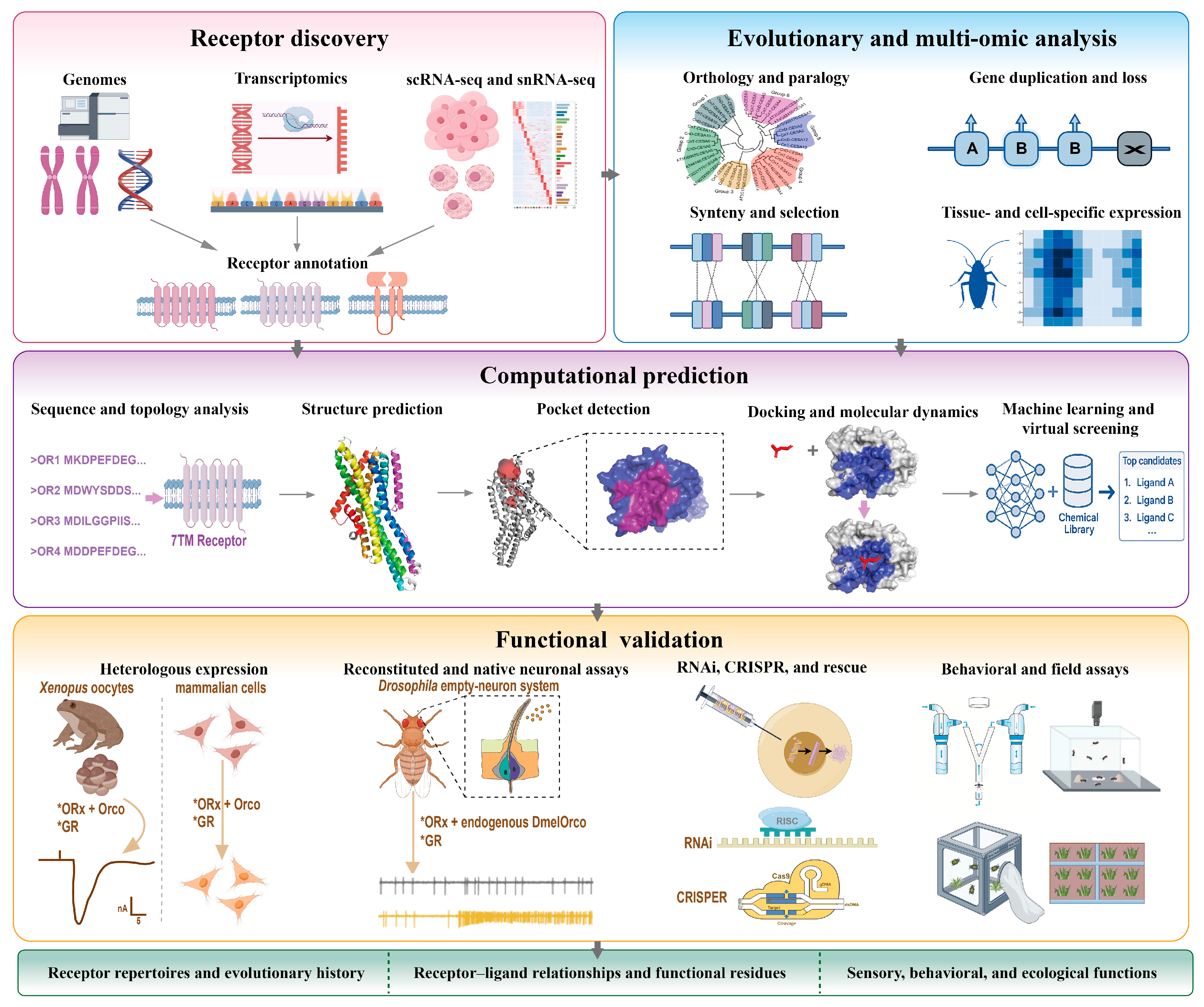
Figure 2.
Architectures of insect chemosensory receptors and representative cryo-EM structures. (A) OR complex comprising a tuning OR and the co-receptor Orco, each with an inverted seven–transmembrane topology (N-in, C-out) that assembles as a ligand-gated cation channel; bottom, representative cryo-EM maps of an Orco homotetramer and a tuning OR–Orco heteromer (e.g., OR5–Orco [20]). (B) GRs share the inverted seven-transmembrane topology and assemble as hetero-oligomeric channels; bottom, cryo-EM maps of GR complexes (e.g., GR9 [147]). (C) Ionotropic receptors (IRs) are iGluR-related heteromers with extracellular S1–S2 ligand-binding domains (and sometimes an amino-terminal domain) that form tetrameric ion channels. Schematics indicate N/C orientation; structures are representative and not to scale.
Figure 2.
Architectures of insect chemosensory receptors and representative cryo-EM structures. (A) OR complex comprising a tuning OR and the co-receptor Orco, each with an inverted seven–transmembrane topology (N-in, C-out) that assembles as a ligand-gated cation channel; bottom, representative cryo-EM maps of an Orco homotetramer and a tuning OR–Orco heteromer (e.g., OR5–Orco [20]). (B) GRs share the inverted seven-transmembrane topology and assemble as hetero-oligomeric channels; bottom, cryo-EM maps of GR complexes (e.g., GR9 [147]). (C) Ionotropic receptors (IRs) are iGluR-related heteromers with extracellular S1–S2 ligand-binding domains (and sometimes an amino-terminal domain) that form tetrameric ion channels. Schematics indicate N/C orientation; structures are representative and not to scale.
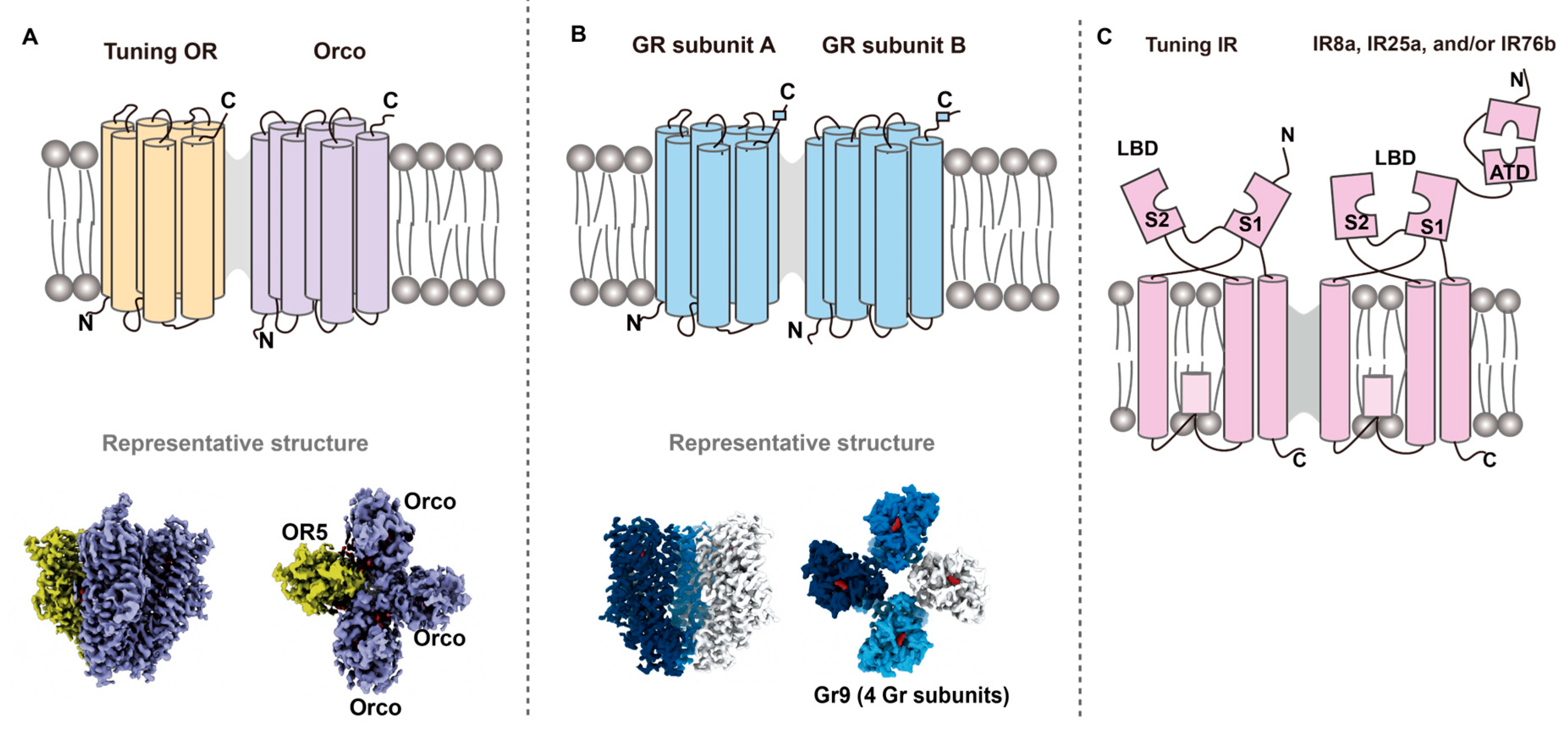

Table 1.
Key software tools and algorithms used for the computational prediction of insect chemosensory receptor function.
Table 1.
Key software tools and algorithms used for the computational prediction of insect chemosensory receptor function.
| Category | Software / Tool | Year | Function | Reference |
| Structure prediction and modelling | AlphaFold2 | 2021 | High-accuracy prediction of protein monomer and complex three-dimensional structures from sequence | [71] |
| AlphaFold3 | 2024 | Prediction of protein–ligand and protein–nucleic acid complexes and of ligand-binding sites | [72] | |
| RoseTTAFold | 2021 | Three-track neural-network prediction of protein structures and interactions | [73] | |
| ESMFold / ESM-2 | 2023 | Protein-language-model-based structure prediction directly from a single sequence, without an MSA | [74] | |
| I-TASSER | 2015 | Template- and threading-based protein structure and function prediction | [75] | |
| SWISS-MODEL | 2018 | Automated homology modelling of protein structures and complexes | [76] | |
| MODELLER | 2016 | Comparative protein structure modelling and optimisation | [77] | |
| Template-based modelling (TBM) for insect ORs | 2024 | Template/homology modelling for ligand-binding prediction in insect ORs | [21,22] | |
| iORandLigandDB | 2023 | Online platform for three-dimensional structure prediction of insect ORs and docking with odorants | [78] | |
| Molecular docking and virtual screening | AutoDock Vina | 2010 | Fast molecular docking based on an empirical scoring function | [79] |
| AutoDock4 / AutoDockTools | 2009 | Molecular docking with optional selective receptor flexibility | [80] | |
| Schrödinger Glide | 2004 | Industrial-grade docking and scoring at multiple precision levels (HTVS/SP/XP) | [81] | |
| GNINA | 2021 | Molecular docking with a deep-learning (CNN) scoring function | [82] | |
| DiffDock | 2023 | Diffusion-model-based blind docking without a predefined binding pocket | [83] | |
| Protein–ligand interaction analysis and visualisation | PLIP | 2015 | Fully automated profiling of protein–ligand interactions (hydrogen bonds, hydrophobic contacts, π–π stacking, salt bridges) and generation of interaction fingerprints | [84] |
| LigPlot+ | 2011 | Two-dimensional ligand–protein interaction diagrams and structural visualisation | [85] | |
| Molecular dynamics and binding free energy | GROMACS | 2005 | Molecular-dynamics simulation: energy minimisation, equilibration and trajectory analysis | [86] |
| MM/PBSA, MM/GBSA | 2015 | Estimation of ligand-binding free energy from MD trajectories | [87] | |
| Machine learning and deep learning | DeepDTA | 2018 | Direct prediction of drug–target binding affinity from sequence, without three-dimensional structures | [88] |
| PSICHIC | 2024 | Graph neural network for learning protein–ligand interaction fingerprints from sequence data | [89] |
Abbreviations: MSA, multiple sequence alignment; CNN, convolutional neural network; MD, molecular dynamics.
Disclaimer/Publisher’s Note: The statements, opinions and data contained in all publications are solely those of the individual author(s) and contributor(s) and not of MDPI and/or the editor(s). MDPI and/or the editor(s) disclaim responsibility for any injury to people or property resulting from any ideas, methods, instructions or products referred to in the content. |
© 2026 by the authors. Licensee MDPI, Basel, Switzerland. This article is an open access article distributed under the terms and conditions of the Creative Commons Attribution (CC BY) license (http://creativecommons.org/licenses/by/4.0/).
Copyright: This open access article is published under a Creative Commons CC BY 4.0 license, which permit the free download, distribution, and reuse, provided that the author and preprint are cited in any reuse.