
Submitted:
20 October 2026
Posted:
21 October 2026
You are already at the latest version
Abstract
Background/Objectives: Primary myelofibrosis (PMF) is a myeloproliferative neoplasm, associated with a somatic mutation in the JAK2, CALR, MPL genes and characterized by progressive bone marrow fibrosis, extramedullary hematopoiesis, splenomegaly, constitutional symptoms, progressive cytopenias, and in a subset of patients, leukemic transformation. The aim of the present study is to review recent studies investigating molecular abnormalities observed in myelofibrosis and showing how these studies have led to the development of new therapeutic strategies for PMF. Methods: An extensive search of the most recent literature was performed, selecting and critically analyzing the most relevant studies. Results: The studies carried out in the last years have provided an extensive molecular characterization of PMF, showing its heterogeneity, characterized by the presence of additional cytogenetic and non-driver gene mutations that contribute to disease evolution. The molecular abnormalities, together with clinical parameters, provided criteria for a risk score stratification of PMF patients. The molecular characterization of PMF has triggered the development of targeted therapeutics represented by JAK inhibitors and antibodies targeting mutant CALR. Conclusions: PMF is a complex and heterogeneous disease that needs to be carefully characterized at molecular and clinical level at diagnosis, to provide a careful risk stratification of individual patients and an adequate personalized treatment.
Keywords:
myeloproliferative neoplasia
; myelofibrosis
; JAK2
; CALR
; MPL
; gene sequencing
; JAK inhibitors
1. Introduction
Myeloproliferative neoplasms (MPN) Philadelphia-negative are clonal hematopoietic disorders comprising polycythemia vera (PV) which is characterized by overproduction of red blood cells, essential thrombocythemia (ET) which is associated with overproduction of platelets and myelofibrosis which id defined by bone marrow fibrosis. PV and ET are chronic-phase myeloproliferative disorders, while MF represents advanced disease that is diagnosed either as first diagnosis or after the diagnosis of ET or PV (secondary MF, SMF) [1].
A fourth type of MPN was proposed by both WHO 2022 and ICC 2022, called pre-PMF which represents a distinct sub entity within the spectrum of MPN and is characterized by unique morphological, clinical and molecular features, distinguishing it from ET and overt PMF [2-3[100. Pre-PMF could represent an early form of PMF and its diagnosis is based on bone marrow morphology, identification of molecular mutations and exclusion of other myeloid neoplasms [2,3].
MPNs share a common origin, as supported by their molecular mechanism, involving oncogenic activation of Janus Kinase 2 (JAK2) though mutations in three different genes: JAK2, calreticulin (CALR) and the thrombopoietin receptor (MPL) [4]. JAK2-V617F mutation is present in >95% of PV patients and in about 50-60% of ET and PMF patients, while CALR (20-30% in ET; 25-35% in PMF) and MPL (3-4% in ET and 5-7% in PMF) are principally associated with ET and PMF [5,6,7]. 10-15% of ET and PMF patients are triple-negative.
JAK2 is the catalytic kinase associated with several cytokine receptors and in MPN is very frequently mutated at the level of 617 aminoacid (JAK2V617F), at the level of the pseudokinase domain JH2 and rarely at the level of exon 12 in the linker between the SH2 and the pseudokinase domain; CALR mutations occurs in exon 9 and cause a frameshift inducing the generation of a mutant protein with a new C-terminal, lacking the KDEL tetrapeptide, exhibiting new functions; MPL mutations frequently occurs at the level of W515 amino acid that plays a key role in preventing receptor dimerization and activation in the absence of cytokine binding [4].
JAK2 V617 determines constitutive activation and cell signaling though the erythropoietin and TPO receptors, with CALR mutant causes MPL binding in the endoplasmic reticulum with consequent dimerization and activation of the receptor; MPL mutant causes constitutive MPL activation [4]. The constitutive activation of EpoR, MPL and other cytokine receptors causes proliferation and increased survival of myeloid progenitors with their consequent accumulation and progressive expansion and development of a leukemic process.
In the present study we analyzed recent progresses made in the understanding of the pathogenesis and molecular heterogeneity of PMF.
2. Aim and Methodology
The. aim of the present study is to review recent studies that have characterized the molecular heterogeneity of PMF and its implication for prognosis and treatment.
This study was based on a literature search of the PubMed database, using the terms primary myelofibrosis, cytogenetic abnormalities in PMF, mutations in PMF, risk stratification of PMF patients. The search period was expanded from 1990 to 2026. The inclusion criteria for the screened literature were limited to studies published in peer-reviewed publications in high-impact journals in hematology.
Furthermore, the Google search engine was used to find the link to major recent international hematology and oncology meetings, which were screened for congress abstracts related to PMF.
3. PMF: General Features
PMF is a MPN characterized by the increased proliferation of abnormal megakaryocytes and granulocytic elements in the bone marrow, associated with medullary fibrosis, osteosclerosis and extramedullary hematopoiesis. At diagnosis, usually PMF patients present a number of features represented by hypercellular bone marrow with medullary fibrosis and osteosclerosis, presence in the peripheral blood of red blood cells with abnormal morphology, erythroid and granulocytic precursors and splenomegaly [8]. PMF is characterized by typical histopathology features. Particularly, the histopathology of PMF is characterized by a biphasic evolution featuring abnormal megakaryocyte proliferation, granulocytic hyperplasia and progressive reticulum and collagen bone marrow fibrosis. In the prefibrotic phase bone marrow is characterized by hypercellularity due to the proliferation of granulocytic cells and by the presence of abnormal megakaryocytic cells increased in number, highly polymorphic, hyperlobate, with “cloud-like” nuclei, forming clusters near to bone trabeculae or blood vessels; at this stage, reticulin and collagen fibrosis are typically absent. In the overt fibrotic stage, a progressive deposition of reticulin and collagen fibers is gradually observed in bone marrow, replacing normal hematopoietic tissue, accompanied by fat spaces and prominent, dilated blood vessels; at this stage, abnormal megakaryocytes forming dense clusters appear as isolated or “naked” nuclei in the fibrotic stroma; in advanced cases, osteosclerosis is observed with formation of bone trabeculae.
Bone marrow fibrosis occurring in PMF if graded into four different stages, from MF-0 (absent) to MF-3 (osteosclerosis with intensive collagen fibrosis) and need to be quantified in bone marrow trephine biopsies by specific histopathological techniques [9]. (Table 1) The fibrosis starts with the deposition of reticulin (collagen III), observed in MF-1 and is followed by accumulation of collagen I, observed in MF-2 and MF-3; finally, osteosclerosis occurs at late stages and is characterized by bone remodeling and formation, associated with intensive fibrosis. The histopathological evaluation of bone marrow fibrosis is diagnostically and prognostically fundamental in PMF. This evaluation is based on reticulin fibrosis (R, assessed using silver impregnation stains), collagen deposition (C, assessed by a Masson’s trichrome stain) and osteosclerosis grading (O) from 0 to 3; the total RCO score provides a prognostically significant measure of the degree of bone marrow fibrosis [10]. Importantly, the RCO score was correlated with overall mortality and discriminated the overall survival of both low- and high-grade patients [10]. Recent studies have confirmed that RCO score provides a reliable assessment of PMF stromal changes, had a significant clinical, genetic and prognostic relevance and its integration into other prognostic models may significantly improve prognostic stratification [11].
Extramedullary hematopoiesis in PMF corresponds to blood cell production happening outside the bone marrow because scarring prevents normal blood cell production inside bone marrow. As bone marrow function deteriorates, hematopoietic stem and progenitor cells migrate to extramedullary sites such as the spleen, liver and less commonly the lungs, where they establish hematopoietic niches [12]. Tis compensatory process reflects mechanisms of adaptability of hematopoiesis and the pathologic modeling of the marrow microenvironment driven by cytokine dysregulation, aberrant adhesion signaling, conditions that favor hematopoietic proliferation outside of the marrow [12]. Extramedullary hematopoiesis in PMF occurs primarily in the spleen; single cell studies in PMF splenic cells showed a pronounced expansion of hematopoietic stem and progenitor cells, associated with aberrant differentiation toward the erythroid and megakaryocytic lineages and modifications of tissue environment with enrichment of inflammatory pathways and emergence of dysfunctional T-cells, conditions influencing hematopoietic cell differentiation and impairing tumor immune surveillance [13].
The increased bone marrow fibrosis, the hallmark of myelofibrosis, is driven by the interaction between clonal MPN cells and bone marrow stromal cells. Particularly, the clonal expansion and differentiation of PMF progenitors produce large, abnormal megakaryocytes that cluster in the bone marrow and release contents from their alpha-granules. Megakaryocytes and associated cells secrete high levels on inflammatory and growth factors, including transforming growth factor-beta (TGF-β), platelet-derived growth factor (PDGF), and basic fibroblast growth factor (bFGF). These and other cytokines mobilize and activate mesenchymal stem cells and fibroblasts, prompting the excessive deposition of reticulin and collagen fibers (scar tissue). Concomitantly, it is observed an upregulation of vascular endothelial growth factor (VEGF), stimulating neoangiogenesis in the marrow microenvironment.
Several studies have shown that myofibroblasts are fibrosis-driving cells in PMF; particularly, Gli1+ (glioma-associated oncogene homolog 1, a transcription factor in the hedgehog signaling pathway) and LepR+ (Leptin Receptor) mesenchymal stromal cells are progenitors of fibrosis fibrosis-causing myofibroblasts in the bone marrow; these stromal cells downregulate the expression of key hematopoietic stem cell-supporting factors and upregute genes associated with fibrosis and osteogenesis, supporting the fibrogenic conversion of myofibroblasts [14,15,16].
According to these studies, HSCs carrying JAK2, CLAR or MPL generate abnormal megakaryocytic cells that secrete high levels of cytokines that in turn activate resident mesenchymal non neoplastic stromal cells within the bone marrow microenvironment, driving their differentiation into myofibroblasts, that release and deposit reticulin and collagen, thus promoting bone marrow fibrosis. Recent studies support the involvement in marrow fibrosis also of another cell type, fibrocytes, collagen-producing cells derived from the monocyte lineage. These cells, abundantly detected in the bone marrow of PMF patients, are of clonal origin and originate from the neoplastic clone in PMF [17]. In JAK2-V617F transgenic mice the majority of collagen- and fibronectin-producing cells in bone marrow and spleen were fibrocytes of neoplastic origin; depletion of monocyte neoplastic cells eliminated collagen- and fibronectin-producing fibrocytes, reduced TGF-β levels, markedly improved fibrosis, without affecting megakaryocytes and progenitor/stem cells [18]. Recently, using multiplex immunofluorescence of bone marrow biopsies from patients with JAK2-V617F-mutated PMF, Uchida and coworkers showed that fibrocytes represent the predominant collagen-producing population in early phases of fibrosis (stages 0-1), while myofibroblasts are only a minoritarian population [18]. At later stages, when bone marrow fibrosis progresses, fibrocytes and myofibroblasts expand , promoting extensive fibrosis [19]. According to these findings, it was proposed a model in which monocyte-derived fibrocytes largely contribute to the initial development of bone marrow fibrosis, while myofibroblasts significantly contribute to the fibrotic process only at later stages [19]. Furthermore, the study of JAK2-V617F transgenic mice led to the identification of the HMG-CoA inhibitor pivastatin as a potent inhibitor of fibrocyte proliferation, with relevant clinical implications [19].
4. Driver Mutations and Clinical Phenotype
PMF patients display a consistent degree of clinical and phenotypic heterogeneity that could be related to their genetic heterogeneity at the level of driver mutations. Thus, Rumi et al. explored a group of 617 PMF patients (64.7% with JAK2, 22.7% with CALR, 4% with MPL mutations and 8.8% triple-negative [20]. Patients with CALR mutation had a lower risk of developing anemia, thrombocytopenia, leukocytosis as well as thrombotic events, compared to the other subtypes; furthermore, CALR-mutant patients developed later splenomegaly compared to the other subtypes [20]. Median overall survival was significantly different in the various subtypes: mOS 17.7 years in CALR, 9.2 years in JAK2, 9.1 years in MPL-mutant and 3.2 in triple-negative patients [20].
Most of CALR-mutant PMF patients harbor only one of two mutations: type 1, the commonest (79%) corresponds to a 52 bp deletion at the level of exon 9, and type 2 (less frequent, 21%) corresponds to a 5-bp insertion at the level of exon 9 [21]. Type 1 mutations show a stronger association with classic myelofibrosis features. Type 2 mutations present with higher platelet counts and a phenotype that more closely mimics essential thrombocythemia [21]. The favorable prognostic impact of CALR mutations in PMF seems to be restricted to type 1, but not to type 2 mutations [21]. However, another study failed to observe a significant difference in the outcomes of type 1 and type 2 CALR mutations [19]. This study showed also that patients with type 1 but not with type 2 mutations showed abnormal cytosolic calcium signals in cultured megakaryocytes [22]. CALR mutation variant allele frequency (VAF) was variable in both type 1 and type 2 CALR mutants; although the VAF had a moderate impact at the phenotypic level, did not affect the outcomes of these patients [23]
Tefferi et al. reported the prognostic impact of driver mutations in 1095 patients with PMF and observed that survival with type 1-like CALR was longer compared to JAK2, tpe-2-like CALR, MPL and triple negative mutational status [24]. Furthermore, in 210 patients with high-risk ASXL1/SRSF2 mutations, survival was significantly longer in the presence vs absence of type 1-like CALR mutations [24].
Although earlier studies identified a low JAK2-V617F allele burden as a risk factor for shortened survival in PMF, as compared to patients whose burden was higher than 50% [25,26], more recent studies have shown that a high allele burden in PMF is typically linked to a more pronounced myeloproliferative presentation, including higher white blood cell counts, higher LDH levels, higher plasma C reactive protein levels, a larger spleen size and disease evolution [27,28]. Furthermore, Barosi et al. reported that in 462 PMF patients, those possessing a homozygous JAK2-V617F genotype (allele burden >50%) exhibit a strong determinant for blast transformation, with a hazard ratio of 2-04 compared to the heterozygous genotype [29].
The relationship between JAK2 mutant VAF and PMF outcomes is complex and affected by many variables. Thus, Wang et al. showed that in JAK2 mutant PMF patients the presence of high-risk mutations (including ASXL1, EZH2, IDH and U2AF1 genes) negatively impact the outcomes in JAK2-V617 VAF mutant patients, this effect being limited to patients with low JAK2-V617F VAF and not observed in patients with high JAK2-V617F VAF [30].
5. Cytogenetic Abnormalities
According to detailed in a Compendium of cytogenetic abnormalities in PMF carried out in a cohort of 826 patients, about 43% of tested patients have an abnormal karyotype [31]. The most frequent changes include: del(20q), occurring in 23% of cases; del(13q), observed in 18% of cases; trisomy 8 (+8) in 11% of cases; trisomy 9 (+9) in 10% of cases; chromosome 1q duplication, in 10% of cases; chromosome 7 changes (-7/7q-) in 7% of cases [31]. Other alterations, including i(17q) in 1.4% of cases, 12p- in 1.1% of cases and inv(3) in 0.6% of cases are much less frequent [31]. Abnormal karyotype frequencies were 43% in JAK2, 42% in CALR, 33% in MPL-mutated and 34% in triple-negative cases [26]. 13q- was associated with mutant CALR and +9 with mutant JAK2 [31].
Kuykendall and coworkers characterized the cytogenetic abnormalities in a population of 312 PMF patients, confirming the findings of the previous study concerning the frequency of the most recurrent chromosomic abnormalities [32]. In addition, the analysis of the outcomes associated with the presence of the various chromosomic abnormalities showed that patients bearing del(20q), del(13q) and trisomy 9 had a median overall survival comparable to patients with normal karyotype (mOS 45 months vs 54 months, respectively), while patients with del(5q), trisomy 8 and chromosome 17 abnormalities had significantly worse OS when compared to patients with normal diploid karyotype (mOS 20 months vs 54 months, respectively) [32].
In 2018, Tefferi et al. performed in 1012 PMF patients’ stepwise analysis of the impact on survival from individual and prognostically ordered cytogenetic abnormalities and developed a three-tiered risk model, with the identification of three risk groups. A favorable risk including normal karyotype, sole del(20q), del(13q), +9, -Y chromosome; an unfavorable risk, including sole +8, del(7q), single/multiple del(5q), monosomal abnormality without a very high risk abnormality; a very high risk including monosomy 7, single/7multiple inv(3) abnormalities, single/multiple i(17q), single/multiple del(12p), single/multiple del(11q), single/multiple trisomies excluding +8 and +9 [33]. The median survival of PMF patients with favorable, unfavorable and very high-risk cytogenetics was 4.4, 2.9 and 1.2 years, respectively [33]. (Table 2)
Chromosome banding along with fluorescence in situ hybridization represents the standard approach for cytogenetic characterization of PMF patients; however, these analyses provide an informative karyotype in only about half of patients. A recent study compared optical genome mapping (OGM) with conventional chromosome analysis (CCA) in patients with primary and secondary myelofibrosis showing that OGM generated interpretable cytogenetic data for every patient, whereas CCA succeeded in fewer more than half cases [31]. Integration of OGM data into DIPSS-plus or MIPSS-plus-version 2 risk models significantly refined allocation of patients [34].
6. Non-Driver Gene Mutations in PMF
Nex generation sequencing (NGS) has given an important contribution to the definition of the numerous molecular alterations observed in PMF patients. Thus, in addition to the driver gene mutations observed at the level of JAK2, CALR and MPL genes showed in about 85% of cases, additional non-driver gene mutations were detected in more than 50% of PMF patients. Additional mutations implied genes involved in histone modification (ASXL1, EZH2), DNA methylation (TET2, DNMT3A, IDH1, IDH2), mRNA splicing (SF3B1, SRSF2, U2AF1, ZRSF2), signaling pathways (CBL, NRAS, KRAS) and transcription control (RUNX1, NFE2, TP53) [35]. The presence and the different combinations of these non-driver mutations influence the clinical course, prognosis and response to therapy [35].
Various studies have provided a characterization of non-driver mutations in PMF patients. In 2013, Vannucchi et al. reported the mutational profile of 879 PMF patients [36]. They identified several recurrent non-driver mutations, including ASXL1 (21.7%), TET2 (/9.7%), SRSF2 (8.5%), DNMT3A (5.7%), EZH2 (5.1%), CBL (4.4%), IDH1-2 (2.6%) [36]. ASXL1, SRSF2 and EZH2 were significantly enriched in IPSS high-risk group with mutational frequencies of 42%, 25% and 12%, respectively [36]. ASXL1, SRSF2 and EZH2 mutations predicted shortened survival, while ASXL1, SRFSF2 and IDH1 or IDH2 mutations predicted leukemic transformation [36]. In 2014, Tefferi et al. reported the frequency of non-driver mutations in 253 PMF patients stratified by the presence or absence of driver mutations JAK2, CALR and MPL [34]. This study showed that the frequency of the most recurrent non-driver gene mutations, including ASXL1, SF3B1, SRSF2, EZH2, U2AF1 and IDH1-2 was comparable in JAK2, MPL and triple-negative PMF; CALR-mutant PMF displayed a significantly lower frequency ofSF3B1, SRSF2 and U2AF1 mutations compared to the other PMF groups, while the frequency of ASXL1, EZH2 and IDH1-2 mutations was comparable in the four groups of PMF [37] (Figure 1).
In 2014, Guglielmelli et al. reported the results of a study carried out in 797 PMF patients aiming to assess the prognostic value of the number of non-driver mutations [38]. The study was limited to the analysis of detrimental non-driver mutations, including ASXL1, SRSF2, EZH2, IDH1-1: 23.6% of patients had one and 7.4% two or more mutations [38]. The presence of two or more mutations predicted the worst survival: median of 12.3 years for no mutation, 7.0 years for one mutation and 2.6 years for two or more mutations [38]. CALR mutations favorably affected survival, independently of the number of mutations [38].
Tefferi et al reported the results of a targeted deep sequence in 182 PMF patients, ASXL1 (36%), TET2 (18%), SRSF2 (18%), U2AF1 (16%), CEBPA (9%) and CBL (5%) were the most frequent non-driver mutations; 35%, 26%, 10%, and 9% of patients harbored 1, 2, 3 or 4 or more non-driver mutations [34]. (Figure 2) SRSF2 and U2AF1 mutations were less frequent in CALR-mutant AML than in JAK2, MPL or triple-negative [39]. The presence of adverse (ASXL1, SRSF2, CBL, KIT, RUNX1, SH2B3 and CEBPA non-driver mutations affected overall survival: mean OS with non-driver mutations not reached, with non-adverse mutations 6.8 years and with adverse mutations 3.6 years [39]. The number of adverse mutations affected OS: mean OS with no adverse mutations 8.5 years, with one or two adverse mutations 4 years and with three or more adverse mutations 0.65 years [39].
Luque Paz and coworkers reported a genomic analysis on 479 patients with PMF and SMF (305 PMF and 174 SMF) by targeted NGS [40]. The most frequently non-driver mutated genes were ASXL1, TET2, SRSF2, U2AF1 and EZH2; SRFS2 and U2AF1 were more frequent in PMF than in SMF, while mutations in NFE2 were more frequent in SMF; SRFSF2 and U2AF1 mutations were more frequently observed in JAK2-mutated than in CALR-mutated patients [40]. The allele burden distribution of non-driver mutants was highly variable, with an allele burden around 40-50% for SRFSF2 and U2AF1, ranging from 0% to 10%% for EZH2 and TP53 and an allele burden with a bimodal distribution, with some values at low allele burden and others centered on 40% to 50% for ASXL1 and TET2 [40]. A hierarchical clustering analysis allowed the identification of four groups with prognostic impact: TP53-mutated patients; patients with ≥1 mutation in EZH2, CBL, U2AF1, SRSF2, IDH1, IDH2, NRAS or KRAS (high-risk group); ASXL1-only mutation, not associated with any high-risk mutation; other patients with other mutations in NFE2, TET2, DNMT3A, SF3B1 [40]. Patients with TP53 mutations or high-risk mutations had a significantly lower OS and a higher incidence of leukemia as compared to patients with isolated ASXL1 or other mutations [40]. Guglielmelli et al. explored a group of 523 MF patients, 330 with PMF (161 pre-PMF and 169 with overt PMF) and 193 with SMF [41]. ASXL1 mutations were observed in 30% of both PMF and SMF patients; compared with pre-PMF, over PMF cohort was enriched in ASXL1 mutations (41% vs 19%) [41]. In PMF patients, ASXL1 mutations were associated with phenotypic characteristics representative of higher-risk disease and clustered with EZH2, SRSF2, U2AF1 and CBL mutations [41]. The subdivision of these patients in the four groups reported by Luque-Paz et al. [40] showed that in PMF: (i) TP53-mutant and high-risk patients had the lowest OS; (ii) ASXL1-mutant-only patients had an intermediate OS between others group and TP53/high-risk mutant groups [41]. In SMF patients, ASXL1-mutant only patients had a MOs comparable to the other mutation groups [38]. Within the high-risk group, among PMF but not SMF patients, the presence of ASXL1 mutations was associated with a reduced OS [41].
Yan and coworkers explored the frequency of ASXL1 mutations at different stages of PMF disease, from pre-PMF to blast accelerated (PMF-BP/AP) through overt phase [39]. Mutations in ASXl1 (31.1% vs 10.5%) and U2AF1 (13.7% vs 1.4%) were more frequent in overt PMF than in pre-PMF; furthermore, mutations in ASXL1 (51.1% vs 31.1%), SRSF2 (26.7% vs 11.2%), RUNX1 (22.2% vs 5.6%), SETBP1 (17.3% vs 7.5%), NRAS (15.6% vs 3.1%) and EZH2 (13.3% vs 5%) were significantly more frequent in PMF-BL/AP compared with overt PMF [42]. Therefore, the frequency of ASXL1 mutations significantly increased during PMF progression from 10.1% in pre-PMF, to 31.1% in overt PMF and 51.1% in PMF-BP/AP [42]. At variance with mutational frequency, ASXL1 VAF did not significantly change during PMF progression [42].
U2AF1 mutations are included in the high-risk mutation group in PMF patients. U2AF1 mutations in PMF patients are significantly associated with anemia [43]. U2AF1 mutations are molecularly heterogeneous and can be classified into two different molecular subtypes Q157 (65% of U2AF1-mutant PMF patients) and S34 (34% of U2AF1-mutant PMF patients) [44]. In PMF patients with U2AF1 overall survival was significantly shorter for patients with U2AF1 Q157 mutations, compared to those with U2AF1 S34 or U2AF1 unmutated cases [44].
As above reported, several studies have shown that TP53-mutant PMF display an OS comparable or even lower that that reported for the group of PMF with high-risk non-driver mutations [40,41]. Tefferi et at explored the outcomes of a cohort of 108 TP53-mutant PMF patients: 61 with chronic phase PMF, 31 with blast phase (BP) PMF and 16 with accelerated phase (AP) PMF; 57% of these patients harbored multihit TP53 mutations (MH-TP53) and 49% monosomal/complex karyotype [45]. MH-TP53 patients were more frequently observed among patients in a blast or accelerated phase, while patients with non-multihit TP53 mutations (HMH-TP53) were more frequent among chronic that BP or AP PMF (76% vs 37%, respectively) [45]. Patients with NHM-TP53 had more frequent TET2, SRSF2 and SF3B1 mutations [45]. The analysis of OS showed that: BP and AP-PMF displayed significantly shorter OS compared to CP-PMF patients; in CP-PMF patients, the presence of MH-TP53 was associated with a significantly shorter OS than that of NMH-TP53; in BP and AP-PMF patients, the presence of TP53 mutations was associated with a shorter OS compared to TP53-WT [45]. MH-TP53 was also associated with inferior survival following allogeneic hematopoietic stem cell transplantation (A-HSCT) [45]. In conclusion, this study showed that the presence of MH-TP53 or NMH-TP53 in BP or AP-PMF and MH-TP53 in CP-PMF was associated with exceptionally poor prognosis [45]. A subsequent study by the same authors confirmed and extended these findings. Particularly, they explored a group of 633 consecutive patients with PMF who performed NGS in chronic phase; TP53 mutation with a VAF >2% was detected in 4.4% of cases with a median VAF of 25.3%: 43% of these patients had MH-TP53 mutations and 57% NMH-TP53 mutations [46]. PMF patients with TP53 mutations were more likely to harbor IDH2 mutations (14% vs 3%), EZH2 mutations (14% vs 4%) and unfavorable karyotype (36% vs 19%) and less likely to harbor CALR-type 1 mutations (4% vs 18%) and SF3B1 (0% vs 6%), compared to TP53-WT PMF [46]. Concerning OS, MH-TP53 but not NMH-TP53 mutations had a significant negative impact on OS; the significant impact of MH-TP53 mutations was confirmed in multivariable analysis that included other molecular risk mutations and unfavorable karyotype [46]. Post- A-HSCT survival was significantly shorter for patients with MH-TP53 mutations vs those with NMH-TP53 mutations vs patients with TP53-WT [46].
A sorter LFS and OS in TP53-mutant PMF patients was reported also by Frioni et al. in a single-center study carried out in a cohort of 250 PMF patients of whom 3.2% had TP53 mutations [47].
Gagelmann and coworkers have explored the impact of TP53 mutations on the outcomes of A-HSCT in PMF patients. To this end, a group of 349 PMF patients was explored: 49 of these patients (13%) had TP53 mutations, of whom 30 had mH-TP53 mutations [48]. Complex karyotype was much more frequent among MH-TP53 compared to NMH-TP53 patients (47% vs 0%) [48]. The TP53 mutational status markedly affected outcomes after allo-HSCT: median survival of TP53-mutant patients was 1.5 years compared to 13.5 years for those with TP53-WT; 6-year OS for NHM-TP53 patients was 56%, for TP53-WT 64% and for MH-TP53 25%; cumulative incidence of relapse was 17% for NHM-TP53, 21% for TP53-WT and 52% for MH-TP53 [48]. Leukemic transformation was significantly more frequent among TP53-mutant PMF compared to TP53-WT PMF (20% vs 2%, respectively) [48].
Bashir et al. have recently reported the analysis of a cohort of 92 triple-negative PMF patients in chronic phase, showing that these patients have a profile of non-driver mutations comparable to that observed in JAK2-mutant patients, except for a higher frequency of DNMT3A mutations (13% vs 5%, respectively) [49]. According to the presence of a favorable or of an unfavorable karyotype, triple-negative as well as JAK2-mutant PMF were subdivided into two subgroups; in triple-negative PMF the presence of an unfavorable karyotype was associated with a marked reduction of OS, while in JAK2-mutant PMF the presence of an unfavorable karyotype only slightly reduced OS [49]. Among triple-negative PMF, the presence of un unfavorable karyotype was associated with a significantly increased frequency of ASXL1 and EZH2 mutations compared to those with a favorable karyotype; among JAK2-mutant PMF, the presence of an unfavorable karyotype was associated with reduced frequency of ASXL1 mutations compared to favorable karyotype [49]. In both triple-negative and JAK2-mutant PMF, the presence of high-risk mutations, such as ASXL1, SRSF2 and U2AF1 was detrimental to both OS and LFS [49].
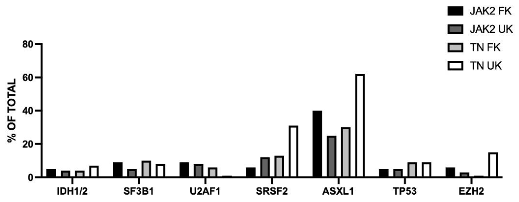
Figure 3.
Non driver gene mutations observed in JAK2-mutated or triple-negative PMF patients. The patients were subdivided into two subgroups according to the presence of a favorable karyotype (FK) or unfavorable karyotype (UK). Original data are reported in Bashir et al. [49].
Figure 3.
Non driver gene mutations observed in JAK2-mutated or triple-negative PMF patients. The patients were subdivided into two subgroups according to the presence of a favorable karyotype (FK) or unfavorable karyotype (UK). Original data are reported in Bashir et al. [49].
Leukemia transformation in PMF, known as blast phase PMF (PMF-BP), is the most feared disease complication, with an incidence estimate around 9-13% [50]. Diagnosis of PMF-BP is based on the presence of ≥20% peripheral blood or bone marrow blasts; the presence of a lower level of blasts (10-19%) corresponds to the accelerated phase of PMF (PMF-AP) [50]. PMF-BP and PMF-AP are associated with a poor prognosis [50]. PMF-BP derives from the expansion of mutated clones driving leukemic transformation and detectable at single cell level in the chronic phase of disease [51].
Targeted NGS studies on MPN patients in blastic phase showed that 37% of these patients displayed ≥3 mutations with the most recurrent mutations involving ASXL1 (57%), TET2 (19%), RUNX1 (17%), TP53 (16%), EZH2 (15%) and SRSF2 (13%); paired chronic-blast phase sample analysis showed more frequent blast phase acquisition of ASXL1, EZH2, LNK, TET2, TP53 and PTPN11 mutations [52].
Abdelmagid and coworkers reported the mutational profile of 103 MPN patients in blast phase and showed that: about 18% had normal karyotype, 19% a complex karyotype, non-monosomal, 36% a monosomal karyotype or monosomy 7 and 28% other chromosomic abnormalities; the most frequent gene mutations were ASXL1 (40%), TP53 (33%), TET2 (18%), FLT3 (18%), SRSF2 (16%), EZH2 (16%), DNMT3A (16%), IDH1 (16%), RUNX1 (11%) and NRAS (9%) [53].
Vallapureddy and coworkers explored in a cohort of 1306 PMF patients the risk factors that predicted leukemic transformation in the first 5 years of disease and over the course of disease [54]. Patients who subsequently developed leukemic transformation (149 patients) were compared to those who remained in chronic phase (1157 patients) [54]. Logistic regression analysis identified ASXL1, IDH1, SRSF2 mutations, very high-risk karyotype, age >70 years, male sex, circulating blasts ≥3%, presence of severe or moderate anemia and constitutional symptoms as predictors of leukemic transformation in the first five years of diagnosis [54]. Based on these findings, it was developed a three-tiered leukemic transformation risk model: high-risk (leukemic transformation incidence 57%), intermediate-risk (leukemic transformation risk 17%) and low-risk (leukemic transformation incidence (8%) [54].
7. Prediction of Survival
The median survival of PMF patients is mainly related to the stage of disease, progressively decreasing following disease evolution from prefibrotic PMF to evert PNF and to PMF-BP. However, the outcome of PMF patients at each disease stage is highly heterogeneous and need to be predicted to define prognosis and to optimize treatment. Thus, many risk models have been developed based on clinical, biologic and genetic parameters attempting to capture heterogeneity of PMF patients and predict their prognosis.
Over the past 20 years, various prognostic systems have been developed for PMF. (Table 3) The International Prognostic Scoring System (IPSS) was introduced in 2009 and applied in clinical practice and clinical trials [55]. IPSS uses five independent clinical and hematologic predictors of inferior survival evaluated at initial diagnosis of patients and each contributing with 1 point for the final score, including age >65 years, hemoglobin <10g/dL, WBC count >25x109/L, circulating blasts ≥1% and constitutional symptoms [55]. Final scores ranging from 0 to ≥4 define four risk groups (low, intermediate-1, intermediate-2, and high-risk) with a corresponding median survival of 11.3, 7.9, 4.0 and 2.3 years [55]. (Table II)
IPSS was modified into the Dynamic IPSS (DIPSS) , which is based on the same prognostic factors, but assigns 2 points and not 1 point to hemoglobin <10 g/dL and can be applied at various times during the clinical course of PMF [56]. (Table II)
DIPSS was further modified into the DIPSS-plus through the inclusion of three additional DIPSS-independent risk factors, including adverse karyotype, platelet counts lower than 100x109/L, and transfusion requirement [57]. This system identifies four risk groups of PMF patients with a median survival ranging from 15.4 for low-risk years to 1.3 years for high-risk patients [57].
Recently, the DIPSS was revised by incorporating new and refined clinical risk variables and excluding those that are subjective (constitutive symptoms) or could exaggerate the need for therapy for older adults (age) [58]. Thus, the risk factors include hemoglobin 8-10 g/dL (women) or 9-11 g/dL (men) 1 point, transfusion dependence or hemoglobin < 8 g/dL 2 points, WBC 11-25x109/L 1 point, WBC >25x109/L 2 points, platelet counts <150x109/L 1 point, monocyte counts ≥1x10⁹/L 1 point, circulating blasts ≥2% 1point [58]. This system identifies four risk groups with a median survival of not reached, 205 months, 95 months and 44 months for the low-, intermediate-1, intermediate-2 and high-risk groups, respectively [58]. The predictive accuracy was comparable between DIPSS and DIPSS-R; furthermore, predictive performance was superior for DIPSS-R vs DIPSS when analysis of patients was stratified by age <65 years and >65 years and the DIPSS-R system assigned a higher proportion of patients to the high-risk category compared to the DIPSS system [58,59]. (Table II)
In 2018, a new prognostic model was proposed, the Mutation-Enhanced International Prognostic Score System (MIPSS70), based on clinical and molecular risk factors [60]. MIPS70 includes the assessment of several risk factors: hemoglobin <10g/dL, platelet count <100x109/L, WBC >25x109/L, constitutional symptoms, bone marrow fibrosis, high molecular risk mutations (ASXL1, EZH2, SRSF2, IDH1-2, and U2AF1Q157), type 1 CALR absent [60]. Patients are classified into three different risk categories: low-risk with a median survival of 27.7 years, intermediate-risk with a median survival of 7.1 years and high-risk with a median survival of 2.3 years [60]. (Table II) Later, a refined version of MIPSS70 was proposed, MIPSS70-plus version 2.0. MIPSS70+ version 2.0 includes five genetic and four clinical risk factors: very high-risk karyotype (4 points), unfavorable karyotype (3 points), presence of 1 (2 points) or ≥2 high risk mutations (3 points), absence of type 1 CALR mutation (2 points), constitutional symptoms (2 points), severe anemia (2 points), moderate anemia (1 point) and circulating blasts ≥2% (1 point) [61]. According to MIPSS+ version 2.0, five risk categories were defined: very low risk (0 points), low risk (1-2 points), intermediate risk (3-4 points), high risk (5-8 points) and very high risk (≥9 points), resulting in a median survival of not reached, 16.4 years, 7.7 years, 4.1 years and 1.8 years, respectively [61].
The Genetically Inspired Prognostic System (GIPSS) was based exclusively on genetic prognostic risk factors, such as mutations and karyotype. The variables include VHR karyotype (2 points), unfavorable karyotype (1 point), absence of type 1 CALR mutation (1 point), ASXL1 mutation (1 point, SRSF2 mutation (1 point) and U2AF1Q517 mutation (1 point). The GIPSS allowed to define four risk categories: low-risk (0 point), intermediate-1 risk (1 point), intermediate-2 risk (2-3 points) and high-risk (≥3 points) with a median survival of 26.4 years, 8.0 years, 4.2 years and 2.0 years, respectively [62].
In addition to the mutations classified as HRM mutations, also CBL, NRAS, KRAS, RUNX1 and TP53 mutations may have a negative prognostic impact in PMF patients. Thus, Loscocco et al. have explored whether the inclusion of these additional mutations among of original MPISS70 may improve the risk stratification of PMF patients [63]. However, the integration of MIPSS70 and MIPSS70 plus models with CBL, NRAS, KRAS, RUNX1 mutations did not improve the performance of these models, largely due to co-mutational profile including ASXL1, EZH2 and SRSF2 [63]. However, TP53 mutations should be including in MIPSS70 score, owing to their independent roel in predicting a dismal outcome [63].
A recent study explored the variables that affect the outcomes of TP53-mutant MPN [64]. This study led to the identification of three variables, allowing a risk classification of TP53-mutated MPN. Thus, the presence of bone marrow fibrosis was an independent predictor of significantly decreased OS and LFS: patients with MF-2 or MF-3 had a significantly shorted OS compared to those with MF-1 [64]. Patients with TP53-mutated multi-hit status had a significantly higher mortality, compared to TP53-mutated single-hit patients [64]. Furthermore, the functional consequences of the specific TP53 mutations, as assessed by the PHANTM score, predicted OS and LFS, independent of TP53 single-hit/multi-hit mutational status [64]. Using these three variables, it was built the FMP model for patients with TP53-mutated MPN [64]. This model contributed to distinguish between TP53-mutated MPN patients at high risk for rapid leukemic transformation and poor survival, from those patients with chronic phase MPN, who despite TP53 mutation did not display a rapid disease progression [64]. The FMP system allowed to stratify TP53-mutated MPN patients in three risk categories: a low-risk with 6.3 years, in intermediate risk with 2.3 years and a high-risk with 0.5 years [64].
Machine learning provided a support for the development of some prognostic scores. Thus, a Spanish group of investigators developed the Artificial Intelligence Prognostic Score System for Myelofibrosis (AIPSS-MF) [65]. This system was based on 8 clinical variables (age at diagnosis, sex, constitutional symptoms, hemoglobin level, leukocyte count, platelet count, blast in peripheral blood and leukoerythroblastosis) and provided a personalized risk score whose predictive accuracy surpassed IPSS [65]. The addition of molecular profiles (EZH2, TP53, IDH1, IDH2, RUNX1, CBL and SRSF2 mutations) to this prognostic score (AIPSSmol-MF) modestly improved the overall prediction capacity in PMF [66].
A model was developed for optimal prognostic stratification of patients with secondary MF post-PV and post-ET; this model called the Myelofibrosis Secondary to PV and ET-Prognostic Model (MYSEC-PM) provided a more accurate evaluation of OS specifically for patients though the integration of various clinical variables [67]. This stratification model was further improved through the integration of molecular variables (MYSEC-kmPM) [68].
8. Treatment
The overall incidence rate of PMF was estimated to be 0.5 per 100,000 person-years; patients with PMF have reduced life expectancy, with a 5-year relative survival of 50.9% in 2009, 57.4% in 2013 and 60.6% in 2016 [69].
Clinical symptoms of PMF include bone marrow fibrosis (causing anemia, thrombocytopenia and bone pain), splenomegaly (causing abdominal pain) and systemic symptoms. The aim of treatment is to delay/inhibit the evolution of disease and to decrease/abrogate hematological abnormalities and constitutional symptoms associated with the disease, to improve and quality of life. Hematopoietic stem cell transplantation is the only curative treatment for PMF.
The introduction of JAK inhibitors has revolutionized the treatment of PMF patients, particularly of those with intermediate-high-risk PMF. Various JAK inhibitors have been approved for the treatment of PMF patients: in 2011, Ruxolitinib, followed by Fedratinib in 2019, Pacritinib in 2022, and Momelotinib in 2023. All these drugs have shown the capacity to improving symptom burden and splenomegaly; Pacritinib and Momelotinib provide also cytopenia-related benefits.
8.1. Ruxolitinib
Ruxolitinib, the first oral selective JAK1/JAK2 inhibitor, has show its efficacy in the context of the pivotal CMFORT trial, with reduction of splenomegaly and disease-related symptoms; particularly, the COMFORT-I trial showed superiority of Ruxolitinib over placebo [70] and the COMFORT-II trial confirmed Ruxolitinib superiority over best available therapy [71]. Long-term follow-up of COMFORT-II trial showed durable clinical effects induced by Ruxolitinib and survival advantages [72]. The results observed in the context of COMFORT trial have been corroborated by large real-world studies and have supported the efficacy and safety of Ruxolitinib in heterogeneous populations of PMF patients [73] and have also shown that Ruxolitinib significantly improved OS compared to standard treatment with hydroxyurea [74]. The COMFORT efficacy trials did not include patients with intermediate-1-risk PMF. The expanded-access JUMP trial included also intermediate-1 patients and showed the efficacy of Ruxolitinib also in this cohort of PMF patients [75,76]. Other studies have supported the efficacy of Ruxolitinib in intermediate-1 patients. Thus, the interim analysis of ROMEI trial on 104 intermediate-1 PMF patients showed that after Ruxolitinib treatment 42.1% and 43.9% of patients demonstrated a symptom response at 24 and 48 weeks, a spleen volume reduction in 38.9% and 41.8% of patients at 24 and 48 weeks; 11.2% and 25.2% of patients reported temporary or permanent discontinuation, respectively, with non-new adverse events reported [77]. Analysis of predictors of response in the context of JUMP study showed scoring factors associated with higher spleen response rates included intermediate-1 vs intermediate-2/high-risk; Ruxolitinib as first-vs second- or later-line therapy and ruxolitinib total daily dose at week 12 [78].
Clinical studies, including rela-world-studies, have provided clear evidence that an optimal efficacy of Ruxolitinib requires the administration of an optimal dosage [79].
Since JAK2 regulates erythropoietin and thrombopoietin signaling, it is evident thart JAK2 inhibitors, such as Ruxolitinib, may worsen or may induce an anemic or a thrombocytopenic condition. Concerning anemia and Ruxolitinib administration, the COMFORT and JUMP studies have shown that both disease-related (present st treatment initiation) and treatment-related (developing on treatment) are associated with worse OS in Ruxolitinib-treated patients [80,81]. To reduce the negative impact of Ruxolitinib on anemia, two approaches have been proposed: to decrease the myelosuppressive effects of Ruxolitinib by reducing its dosing; to maintain the full dosing of Ruxolitinib and associate the administration of anemia-supporting therapies, such as erythropoiesis-stimulating agents. The second option was feasible, as shown by a post-hoc analysis on 101 PMF anemic patients enrolled in the context of the JUMP study [82]. Concerning the effects of Ruxolitinib and platelets, retrospective studies have clearly shown that baseline platelet counts near or below the lower limit of normal were significantly and independently associated with the development of Ruxotilinib moderate of severe thrombocytopenia [83]. A phase II clinical study (NCT 01348490) as well as real-world studies showed that the treatment of thrombocytopenic PMF patients required the use of suboptimal Ruxolitinib and that only patients receiving 10 mg twice daily displayed a clear improvement in spleen size and symptoms [84,85].
Several ongoing studies are exploring the efficacy of Ruxolitinib in combination with other drugs. One study explored the drug combination of Ruxolitinib with Palabresib. Palabresib is an oral, small molecule inhibitor of bromodomain and extra terminal (BET)domain proteins and can act in a complementary manner with Ruxolitinib by targeting inflammatory pathways that are regulated by BET-mediated gene expression [86]. In the phase II clinical trial MANIFEST-2, PMF patients were randomized to receive either Pelabresib-Roxilitinib or Placebo-Rixolitinib [86]. Reduction in spleen volume was observed in 65.9% of patients randomized to receive either Pelabresib-Ruxolitinib versus 35.2% to Placebo-Ruxolitinib; ≥50% reduction in symptoms was observed in 523% vs 46.3% with Pelabresib-Ruxolitinib vs Placebo-Ruxolitinib [86]. Furthermore, bone marrow morphology showed greater improvement with the drug combination [86]. The results observed in MANIFEST-2 study were recently updated to a longer follow-up of 96 weeks, showing that the drug combination Pelabrasib-Ruxolitinib continued to show deep and durable improvements in splenic response and a better rate of symptom control compared to Placebo-Ruxolitinib [87]. Furthermore, fewer progression events and less death were observed with the combination therapy compared with monotherapy [87].
Other studies explored the combination of Ruxolitinib with Selinexor, an oral selective exportin (XPO1) inhibitor, restoring nuclear retention of tumor suppressors such as p53 and inhibitors of inflammatory signaling [88]. In preclinical PMF models Selinexor cooperates with Ruxolitinib in inhibiting the growth of PMF cells, including those resistant to Ruxolitinib [88]. The SENTRY trial explored. The drug combination of Ruxolitinib and Selinexor in JAK inhibitor-naïve PMF patients [89,90]. The phase I of this study involved 24 PMF patients and explored two doses of Selinexor, 40 mg and 60 mg, in combination with Ruxolitinib; 60 mg Selinexor was identified as the optimal dose and, at this dose, a reduction of spleen size and a decrease of symptoms was observed in 79% and 58% of patients, respectively [89]. The phase III of this study explored 353 PMF patients randomly assigned to Ruxolitinib plus Selinexor or Ruxolitinib plus Placebo; at week 24, a spleen reduction volume ≥35% was observed in 49.8% of patients treated with Ruxolitinib plus Selinexor and 28% in the Ruxolitinib plus Placebo group [90]. However, symptom scores in the two groups of patients was similar [90].
Navitoclax is an oral inhibitor of antiapoptotic B-cell lymphoma 2 (BCL-2) family member proteins BCL-2, BCL-XL and BCL-W, preclinical studies have shown a synergistic anti-MF activity of Ruxolitinib and Navitoclax, thus supporting clinical evaluation of this drug combination in PMF patients. In a phase II clinical study, the combination of Navitoclax plus Ruxolitinib was evaluated in PMF patients who had previous Ruxolitinib exposure, showing improvement not only of spleen volume reduction and symptom control but also in reduction of variant allele frequency of driver mutations and improvements in bone marrow fibrosis [91,92]. A recent phase III study TRANSFORM1, extended this analysis also to Ruxolitinib-naïve PMF patients [93]. This last study compared Navitoclax+Ruxolitinib to Placebo+Ruxolitinib in Ruxolitinib-naïve patients [93]. The results of this study showed a significant improvement of spleen volume reduction but not of symptom severity or of anemia level induced by Navitoclax+Ruxolitinib compared to Placebo+Ruxolitinib [93]. VAF reduction, OS and PFS were comparable in the two treatment arms [93].
Although Ruxolitinib now represents the standard therapy for symptomatic myelofibrosis, it is not a curative therapy and the effects on disease biology are limited. In fact, the average reduction in JAK2 allelic burden was 7-22% after 48 weeks of treatment in evaluable patients [70]. Regression of bone marrow fibrosis was observed only in a minority of treated patients. Cases of complete hematological and molecular remissions have been reported but are rare [94]. Furthermore, the of response of PMF patients to Ruxolitinib is highly heterogeneous. A recent study explored the molecular mechanisms underlying Ruxolitinib heterogenous efficacy though a longitudinal single-cell proteogenomic analysis of 12 PMF patients (6 responders and 6 nonresponders to Ruxolitinib) [95]. Responders to Ruxolitinib treatments showed a marked reduction if circulating CD34+ cells and decrease in JAK2V617F allele frequency in granulocytes [95]. Single-cell genomics provided evidence that the mutation acquisition order determines Ruxolitinib sensitivity in that patient in whom the driver mutation either occurred alone or preceded mutations in TET2 or ASXL1 usually responded to therapy, while those in whom epigenetic mutations arose first exhibited limited therapeutic benefit [95]. These results, suggest that Ruxolitinib primarily affects JAK2-only mutated clones, while co-mutated clones evade this treatment and outcompete other neoplastic populations, thus contributing to Ruxolitinib resistance and to disease persistence [95].
8.2. Fedratinib
Fedratinib is a JAK inhibitor synthesized suing structure-based drug design to obtain an inhibitor specific for JAK2, with a much lower affinity for JAK1; Fedratinib inhibits also FLT3 and Ret.
Fedratinib was initially evaluated in PMF patients in the context of a phase-1 dose-evaluation trial, with doses ranging from 30 to 800 mg daily, showing that the maximum tolerated dose (MTD) was 680 mg daily and that after 6 cycles of treatment a spleen reduction ≥50% was observed in 45% of patients treated at the MTD [96]. A phase-2 dose-finding study evaluated the efficacy of various doses of Fedratinib (300, 400 or 500 mg daily): at 24 weeks, the proportion of patients with a ≥35% reduction of spleen volume was 30% (300 mg), 60% (400 mg) and 55% (500); at weeks, the proportion of patients with a ≥50% reduction in total symptoms score was 33% (300 mg), 60% (400mg) and 38% (500 mg) [97]. The JAKARTA and JAKARTA-2 trials provided fundamental data for the approval of Fedratinib for the treatment of PMF patients. In the phase-3 JAKARTA trial, PMF treatment-naïve patients displayed a significant increase in the rate of spleen volume reduction ≥35% compared to placebo [98]. The rate of spleen volume decrease was comparable to that reported in the studies carried out using Ruxolitinib. The JAKARTA-2 study evaluated the efficacy of Fedratinib in patients previously treated with Ruxolitinib, showing significant spleen volume reductions and symptom improvements, thus supporting the efficacy of Fedratinib as second-line therapeutic option [99]. The FREEDOM-2 study provided additional evidence supporting the efficacy of Fedratinib in reducing spleen size and in decreasing disease-related symptoms after Ruxolitinib failure [100]. Real-world studies have confirmed the efficacy of Fedratinib, particularly as second-line therapy after Ruxolitinib failure [101,102].
Ruxolitinib discontinuation syndrome is induced by withdrawal of Ruxolitinib administration and is observed in a minority of patients treated with this drug [103]; this syndrome was not observed after suspension of Fedratinib administration [104].
Interestingly, it was reported the successful administration of Ruxolitinib to patients after Fedratinib failure as first-line or second-line treatments [102].
The safety profile reported for Fedratinib is different from that reported for Ruxolitinib, with frequent gastrointestinal adverse events and rarely, with Wernicke’s encephalopathy [105].
8.3. Pacritinib
Pacritinib is a selective JAK2 inhibitor approved by FDA in 2022 for the treatment of PMF patients with severe thrombocytopenia (platelet counts <50x109/L) Pacritinib inhibits in addition to JAK2, also FLT3 and IRAK1 at doses that inhibit JAK2 and is not myelosuppressive [106].
Two randomized phase III clinical studies (PERSIST-1 and PERSIST-2) evaluated the safety and the efficacy of Pacritinib in a population of PMF patients, including patients with cytopenias. The PERRSIST-1 study including patients with baseline anemia or thrombocytopenia) enrolled 327 PMF patients in first-line of treatment randomized to receive Pacritinib 400 mg one daily or best available therapy; a higher rate of spleen volume reduction ≥35% was observed among the group of patients treated with Pactorinib with respect to those treated with the best available therapy (19% vs 5%, respectively) [107]. The PERTSIST-2 study was carried out as second-line therapy in a group of 311 PMF patients with thrombocytopenia (100x109/L or less platelets) randomized to receive Pacrotinib or best available therapy: in PMF patients with thrombocytopenia, including those with prior anti-JAK therapy, Pacrotinib twice daily at 200 mg or 400 mg was more effective than best available therapy, including Ruxolitinib, for reducing splenomegaly and symptoms [108]. A randomized dose-finding study identified 200 mg dose per day as the optimal Pacrotinib dose showing clinical activity and an acceptable safety profile, with no excess of grade ≥3 hemorrhagic or cardiac adverse events [109].
Interestingly, Pacrotinib inhibits also activin A receptor, type 1 (ACVR1)/activin receptor-like kinase-2 and this activity is exerted with a greater potency compared to other JAK-inhibitors [110]. Inhibition of ACVR1 may improve anemia in patients with PMF; in line with this hypothesis, a retrospective analysis on patients treated with Pacrotinib in the context of PERSIST-2 study showed that among patients who were transfusion-dependent a significantly greater achieved transfusion independency on Pacrotinib compared to those on best available therapy (37% vs 7%) and significantly more had a ≥50% reduction in transfusion burden 849% vs 9%, respectively) [110].
A retrospective analysis carried out on patients enrolled in PERSIST-2 study showed that in PMF patients with platelet count ≤100x109 achieving spleen volume reduction on Pacritinib, but not best available therapy, was associated with significant OS benefit, suggesting that Pacritinib may offer a unique survival advantage in patients with PMF and thrombocytopenia who achieved a significant spleen volume reduction [111].
8.4. Momelotinib
Momelotinib is a JAK1/JAK2 and ACVR1 inhibitor approved by FDA in September 2023 for the treatment of PMF patients with intermediate-2, high-risk disease who also have anemia. This property of Momelotinib is related to its inhibition of ACVR1 with the consequent inhibition of hepcidin expression, thus increasing iron availability to sustain erythropoiesis [112].
Three phase III clinical studies, SIMPLIFY-1, SIMPLIFY-2, and MOMENTUM, have greatly contributed to support the efficacy of Momelotinib in reducing spleen volume, improving PMF-related symptoms, and improving anemia and reducing transfusional need in treatment-naïve and previously-treated patients [113,114,115]. Particularly, the SIMPLIFY-1 phase III randomized study compared in 432 PMF patients with high-risk, intermediate-2 or intermediate-1 symptomatic PMF patients the response to the treatment with Momelotinib (200mg once daily) or with Ruxolitinib, showing after 24 weeks of treatment: a ≥35% reduction of spleen volume size in 26.5 of patients treated with Momelotinib and 29% in those treated with Ruxolitinib; a 50% reduction in symptom score was observed in 28.4% and 42.2% of patients who received Momelotinib and Ruxolitinib, respectively [116]. Transfusion rate, transfusion independence and transfusion dependence were all improved in patients treated with Momelotinib [117]. In the SIMPLIFY-1 trial a part of patients treated with Ruxolitinib switched to Momelitinib after the end of that have completed 24 month of Ruxolitinib treatment; in these patients, after crossover to Momelotinib treatment a rise of hemoglobin levels and an increase in spleen volume reduction was observed (this last event particularly in patients who received Ruxolitinib at 10 mg twice daily [118].
The randomized phase III clinical study SIMPLIFY-2 explored the response to treatment with Momelotinib or best available therapy in 154 PMF patients previously treated with Ruxolitinib and showed that 7% of patients in the Momelotinib group and 6% in the best available treatment group achieved a ≥35 reduction of spleen volume [119]. Transfusion independence rates at week 24 were significantly higher with Momelotinib versus best available therapy [120].
The randomized phase III clinical trial MOMENTUM explored the efficacy of Momelotinib versus Danazol (a synthetic androgen used for treatment of anemia in PMF patients) in a group of 195 PMF patients [121]. Treatment with Momelotinib compared with Danazol resulted in clinically significant improvements in PMF-associated symptoms, anemia measures, and spleen size reduction, with a favorable safety profile [121].
A real-world retrospective study analyzed 221 patients with PMF receiving Momelotinib after Ruxolitinib discontinuation [122]. Before Momelotinib administration, all patients received Ruxolitinib for a median of 31.5 months and discontinued it as a result of resistance, intolerance or both; 97.3% of these patients were cytopenic, 34.8% displayed large splenomegaly and 43.9% were highly symptomatic [122]. Most of these patients rapidly switched to Momelotinib after stopping Ruxolitinib. After 6 months of treatment with Momelotinib, 30% of patients achieved ≥50% spleen length reduction, with higher effects observed in patients who better responded to Ruxolitinib and in those who rapidly switched to Momelotinib after Ruxolitinib stopping [122]. 2-year OS and PFS were 60.9% and 59%; 5% of patients progressed to blast phase [122].
8.5. Gecacitinib
Gecacitinib, also known as Jaktinib, is an inhibitor targeting JAK (JAK1, JAK2, JAK3 and TYK2) and ACVR1. This inhibitory profile was similar to that reported for Momelotinib. Three phase-II clinical trials (ZGJAK002, ZGJAK006 and ZGJ016) and one phase-III clinical trial (ZGJAK016) have been carried out to evaluate the safety and the efficacy of Gecatinib [123,124,125,126]. The interim analysis of the phase-III clinical study supported the superiority of Gecacitinib over hydroxyurea in reducing spleen size in PMF patients [126]. The final results of this phase-III clinical trial showed that, at week 24, a reduction of spleen volume ≥35% was observed in 64.8% of patients treated with Gecatinib and 26.5% of those treated with hydroxyurea; a ≥50% reduction of total symptom score was observed in 62% for Gecacitinib versus 50% for hydroxyurea; in PMF patients with baseline hemoglobin levels <10g/dL, in the 31% of Gecacitinib group a ≥2 g/dL increase was observed, compared to 11% in the hydroxyurea group [127].
A post-hoc analysis carried out in PMF patients treated with Gecacitinib supported its efficacy in thrombocytopenic patients with baseline platelet counts <100x109/L [128]. These patients were subdivided into three groups: Ruxolitinib-naïve; Ruxolitinib intolerant and Ruxolitinib-refractory/relapsed [128]. The evaluation of these patients showed: the spleen size reductions were 75%, 42% and 25%, and reduction of total symptom score were 50%, 33% and 50% in Ruxolitinib-naïve, Ruxolitinib-intolerant and Ruxolitinib-refractory/relapsed thrombocytopenic patients [128].
A recent real-world study supported the efficacy and a favorable safety profile of Gecacitinib in 37 MF patients [129]. This study showed also that Gecacitinib dose lower than 100 mg showed significant efficacy and its use was feasible also in patients with baseline platelet levels <100x109/L [129].
8.6. Comparative Studies of JAK Inhibitors in PMF
No head-to-head randomized clinical trials have been conducted to compare the safety and the efficacy of various JAK inhibitors in PMF patients. Therefore, only an indirect treatment comparison that accounts for differences. Between trial designs and enrolled populations of patients offers a pragmatic solution for the comparison of these treatments. In this context, the matched-adjusted indirect comparison (MAIC) method, facilitates such a comparison by following all the critical steps recommended for this type of analysis.
A MAIC analysis was performed to compare the efficacy of Pacratinib and Momelotinib in the treatment of thrombocytopenic PMF patients, based on the retrospective analysis of the results obtained in phase III clinical trials with these drugs [130]. The parameters involved in this analysis included week-24 percentage of patients achieving ≥50% total symptom score reduction, ≥35% spleen volume reduction and RBC transfusion-independence [130]. All outcomes favored Pacritinib vs Momelotinib; however, none had statistical significance [130].
Duminuco et al. reported the comparative analysis of the efficacy of Fedratinib, Momelotinib and Pacritinib, based on the pooled analysis of phase-III studies for PMF patients, as second-line therapy, post-Ruxolitinib failure [131]. This analysis showed that pooled ≥35% spleen volume reduction at week 24 was 32.7% for Fedratinib, 25.8% for Momelotinib and 21.6% for Pacritinib (200 mg twice daily); pooled ≥50% total symptom score reduction rates were 32.8% for Fedratinib, 25.3% for Momelotinib and 32.4% for Pacritinib [131]. Discontinuation due to adverse events was comparable across agents [131].
Zhang et al. reported the MIAC analysis of Gecacitinib, Fedratinib and Momelotinib in second-line PMF therapy [132]. The results of this comparative analysis showed that: Gecacitinib displayed superior ≥35% spleen volume reduction compared to Fedratinib, Pacritinib and Momelotinib and superior ≥50% total symptom reduction score compared to Pacritinib and Momelotinib [132]. Furthermore, Gecacitinib had also better transfusion independence compared to the three other JAK inhibitors [132].
9. Hematopoietic Stem Cell Transplantation
The number of MF patients undergoing allo-HSCT annually continuously increased during the last years and this was because HSCT is the only curative option for these patients and HSCT has become safer with reduction in non-relapse mortality. HSCT is the standard of care for eligible patients with higher-risk disease.
The first step in HSCT consisted in a thorough selection of candidate patients carried out according to the evaluation of clinical and biological parameters, as recently reviewed in [133,134,135,136]. Usually, patients with intermediate-2 and high DIPSS are considerate candidates for allo-HSCT [133,134,135,136]. The transplant indications and timing, pre-transplant management, criteria of donor selection, and optimization of the conditioning regimen have been recently reviewed [133,134,135,136]. HLA-matched sibling donors represent the preferred donor source.
The optimal conditioning intensity and regimen choice for OSCT in MF remain challenging due to the limited number of prospective trial data. Retrospective analyses deriving from multicenter cohorts showed comparable 5-year OS ranging around 55%-60%, with myeloablative conditioning regimen being associated with higher relapse-free survival but higher non relapse mortality [135].
9.1. Clearance of Driver Mutations in HSCT of PMF
The presence of driver mutations in the large majority of PMF patients offers the opportunity to evaluate whether HSCT was able to induce clearance of driver mutations and had a prognostic relevance.
A recent study by Gagelmann and coworkers evaluated clearance of driver mutations after transplantation in 324 PMF patients (73% with JAK2 mutations, 23% with CALR mutations and 4% with MPL mutations) [137]. At day 30 post-transplantation, mutation clearance was detected in 42% of the patients with JAK2 mutations, 73% of those who had CALR mutations and 54% who had MPL mutations; at day 100, were 63%, 82% and 100%, respectively [134]. Mutation clearance appeared to occur earlier with CALR and MPL mutations than with JAK2 mutations [137]. The cumulative incidence of relapse at 1 year was 6% among patients with mutation clearance at day 30 post-transplantation and 21% among those without mutation clearance at day 30 [137]. Disease-free survival and overall survival at 6 years post-transplantation 61 and 74%, respectively in patients with mutation clearance at day 30 post-transplantation and 41% and 60%, respectively among patients without mutation clearance at day 30 after transplantation [137]. Mutation clearance ta day 30 post-transplantation is a better measure of response compared to evaluation of donor chimerism [137].
The serial evaluation of driver mutations levels is an efficient tool to predict relapse after allo-HSCT [138]. In fact, a retrospective analysis of 34 JAK2-V617F -mutated PMF patients who underwent allo-HSCT showed that a ≥2-fold increase of JAK2 variant allele frequency at day +100 and +180 was highly predictive of eventual relapse [138].
In line with these observations, a panel of experts provided consensus that clearance of driver mutations is central to determining remission of PMF patients after HSCT [139]. Patients in molecular remission can be categorized as: complete remission, remission with poor graft function; remission with incomplete marrow recovery [139].
9.2. HSCT for PMF Patients in Accelerated and Blast Phase
As above discussed, the prognosis of PMF patients in accelerated/blastic phase is poor, with a very limited response to current treatments and with only a short overall survival. Aloo-HSCT is the only valuable therapeutic option for these patients.
Gagelmann and coworkers have explored the outcomes of allo-HSCT in PMF patients [140]. Particularly, these investigators reported the outcome of 349 PMF patients, 35 with accelerated phase, who underwent allo-HSCT after reduced intensity conditioning regimen [140]. After a median 5.9 years of follow-up, 5-year OS was 65% for the accelerated phase group and 64% for the chronic phase group; 5-year RFS was 49% for the accelerated group and 55% for the chronic-phase group; 5-year non relapse mortality was 20% for the accelerated-phase group and 30% for the chronic-phase group; 5-year incidence of relapse was 30% for the accelerated-phase group vs 15% for the chronic-phase group [140]. Thus, these findings showed excellent survival but higher relapse for accelerated-phase PMF [140]. Concerning the accelerated-phase group, some factors affected OS: CALR/MPL-mutated group had higher OS than CALR/MPL-unmutated group (5-year OS 89% vs 61%, respectively); the presence of RAS mutations was associated with markedly lower OS (0% vs 76%, respectively) [140].
Barbullushi et al. reported the outcomes of allo-HSCT in 51 MPN patients in blast phase, mostly with PMF or secondary MF, showing a 5-year OS of 35.9% for patients who underwent a first HSCT and 28.5% for those who underwent a second HSCT [141].
9.3. Transplant Prognostication
Several models have been proposed to predict the survival of PMF patients undergoing HSCT. The MIPSS70 and MIPSS70+ version 2 scoring systems have been previously discussed and can be used also to predict morbidity and mortality after transplantation [60,61]. These systems were primarily based on disease biology, without full consideration of the overall health condition of the patients. This limitation is bypassed by the Myelofibrosis Transplant Scoring System (MTSS) which is based of age, performance status, platelet number, leukocytosis, human leukocyte antigen (HLA)-mismatched unrelated donor, ASXL1 mutation, driver mutation genotype; the patients were stratified into low, intermediate, and very high risk [142]. An improved version of MTSS, iMTSS, integrated the MTSS score with evaluation of cytogenetics and molecular profiling [143].
A simple multivariate scoring system CIBMTR was proposed to evaluate prognosis of PMF patients undergoing allo-HSCT; this system was based on three simple variable such as age >50 years, hemoglobin levels <10 d/dL pre-transplantation and HLA-matching [144]. The advantage of this system is its feasibility, even in centers where genomic is not available [144]
A new scoring system, based on a machine learning prognostic method, was developed by the European Society for Blood and Marrow Transplantation (EBMT) Chronic Malignancies Working Party [145]. This system is based on many parameters, including age, pre-transplantation hemoglobin levels, donor type, comorbidities, performance status, blood blasts, leukocyte count and platelet count [145].
A recent study identified an integrated framework that quantifies biology-driven and patient-driven prognosis for PMF patients undergoing HSCT [146]. This system, called iMTSS, was developed through the analysis of a large cohort of 1550 patients with primary or secondary MF who underwent HSCT [146]. This analysis showed that pos-transplant outcomes resolve two nearly independent risk axes: one genetic-disease axis, based on TP53 allelic state, RAS pathway mutations, ASXL1 and driver mutations, blasts and blood counts, predicted 2-year relapse incidence; a second host and structural axis, based on portal vein thrombosis, donor type, patients’ performance status, and age predicted 2-year non-relapse mortality [146]. The two scores shared only 3.4% of their variance, thus indicating that patients’ disease genetics carried almost no information about non-relapse mortality [146]. Recombined, this framework discriminated OS better than every established prognostic system [146].
10. Therapeutic Targeting of CALR-Mutant MF
WT CALR protein is a 46 kDa calcium-binding lectin chaperone protein localized in the endoplasmic reticulum (ER); this protein is composed of three different domains, with N-terminal and central proline-rich domain involved in chaperone function and the C-domain involved in Ca2+ binding and ER localization through the presence of the KDEL ER retention signal [147]. Mutations in CALR occurring in MPN modify the chaperone function and transform CLRL protein into an aberrant constitutive activator of JAK/STAT signaling pathway [147]. All CALR pathogenic mutations occur at the level of exon 9 and cause a +1 base-pair frameshift, inducing the generation of a shared 36-amino acid C-terminal neoantigen [147]. As above discussed, the majority of CALR mutations fall into two categories, type 1 CALRdel52 and type 2 CALRins5. All CALR mutants share the development of a positively charged C-terminal neoantigen and the loss of the KDEL ER retention signal, with consequent mobilization of the mutant-CALR protein out the ER and localization at the level of the cell membrane and translocation of mutant-CALR/MPL complexes to the cell membrane [147]. The C-terminal neoantigen motif shared between different CALR mutations is presented extracellularly on major histocompatibility complex (MHC) class I molecules and in complex with MPL, thus offering the opportuning for its targeting through vaccination or specific antibodies [147]. Mutant CALR peptide vaccines induce significant T cell responses but have failed to result in hematologic or molecular responses [147]. Mutant-CALR-specific monoclonal antibodies, such as Fc-silent antagonists, are able to block cell signaling originated by mutant CALR-MPL signaling, suppress HSC and HPC proliferation and megakaryopoiesis, with achievement of rapid durable hematologic remissions with low toxicity in early-phase trials [147]. Particularly, the clinical data presented at EHA 2026 using the monoclonal antibody INCA033989 have shown a potential to modify CALR-mutated PMF biology, with high rates of hematologic and molecular responses, VAF reductions that associate closely with response depth [148,149,150].
A recent experimental study by Thompson-Peach et al. reported an efficient targeting of mutant CALR based on the combination of two anti-CALR antibodies targeting simultaneously targeting distinct portions of the CALR-mutant C-terminus: simultaneous targeting with two antibodies was superior to single antibody treatment in type 1 CALR-mutant cell, with complete inhibition of JAK-STAT signaling [151]. In type 2 CALR-mutant cells the double antibody inhibition and Ruxolitinib achieved a complete inhibition of JAK-STAT signaling [151].
11. Conclusions
The studies carried out in the last years have shown a consistent progress in the understanding of the molecular abnormalities observed in PMF, showing a consistent level of heterogeneity related not only to the presence of different driver mutations but also to the variable presence of non-driver gene mutations and cytogenetic abnormalities. This progress has favored the development of a precision medicine in PMF moving beyond definition of a driver mutation toward an integrated analysis of clinical and hematologic phenotype, symptom burden, bone marrow morphology, cytogenetics, molecular profile and transplantation fitness. This integrated analysis, interpreted in the context of available risk stratification models, allows the definition of an individual patient profile of disease biology and of personalized medicine. This approach, supported also by the availability of new more efficient drugs, is improving the outcome of PMF patients.
Drug combination of JAK inhibitors with other anti-leukemic drugs represents an area of development for future studies with the aim of evaluating, in the context of randomized clinical studies, the efficacy of these treatments in JAK-naïve and JAKi-previously treated patients and the spectrum of clinical efficacy in terms of spleen volume reduction, bone marrow fibrosis, anemia control, symptom control, OS and PFS. Finally, the development of studies with antibodies targeting mutant CALR is a very promising area for future clinical studies.
Author Contributions
G.C. and E.P. were involved in researching, writing, and editing the manuscript. U.T. was involved in conceptualization, organization, research, and editing the manuscript. All authors have read and agreed to the published version of the manuscript.
Funding
This research received no external funding.
Institutional Review Board Statement
Not applicable.
Informed Consent Statement
Not applicable.
Acknowledgments
The authors have reviewed and edited the output and take full responsibility for the content of this publication.”.
Conflicts of Interest
The authors declare no conflicts of interest.
References
- Grinfeld, J.; Nangalia, E.J.; Baxter, D.C.; Wedge, N.; Angelopoulos, R.; Contrill, A.L.; Godfrey, A.L.; Papaemmanuil, E.; Gundern, C.; Mac Lean, C. Classification and personalized prognosis in myeloproliferative neoplasms. N Engl. J. Med. 2018, 379, 1416–1430. [Google Scholar] [CrossRef] [PubMed]
- Thiele, J.; Kvaschicka, H.M.; Giannelli, U.; Arber, D.A.; Tefferi, A.; Vennucchi, A.M.; Barbui, T.; Orazi, A. Evolution of WHO diagnostic criteria in “classical myeloproliferative neoplasms” compared with the international consensus classification. Blood Cancer J. 2025, 15, 31. [Google Scholar] [CrossRef] [PubMed]
- Griesshammer, M.; Al-Ali, H.K.; Eckardt, J.N.; Fleg, M.; Gothert, J.; Jentsch, K.; Korschmieder, S.; Kvaschicka, H.M.; Reiter, A.; Schmidt, B.; et al. How I diagnose and treat patients in the pre-fibrotic phase of primary myelofibrosis (pre-PMF) – pratical approaches of a German expert panel discussion in 2024. Ann. Hematol. 2025, 104, 295–306. [Google Scholar] [CrossRef] [PubMed]
- Plo, I.; Vainchenker, W. Myeloproliferative neoplasms. N Engl. J. Med. 2026, 395, 788–802. [Google Scholar] [CrossRef] [PubMed]
- James, C.; Ugo, V.; Le Couedic, J.P.; Starkl, J.; Delhommeau, F.; Lacat, C.; Garçon, L.; Raslova, H.; Berger, R.; Benacceur-Griscelli, A.; et al. A unique clonal JAK2 mutation leading to constitutive signalling causes polycythemia vera. Nature 2005, 434, 1144–1148. [Google Scholar] [CrossRef] [PubMed]
- Kralovics, R.; Passamonti, F.; Buser, A.S.; Teo, S.S.; Tiedt, R.; Passweg, J.R.; Tichelli, A.; Cazzola, M.; Skoda, R.C. A-gain-of-function mutation of JAK2 in myeloproliferative disorders. N. Engl. J. Med. 2005, 352, 1779–1790. [Google Scholar] [PubMed]
- Levine, R.T.; Waldeigh, M.; Cools, J.; Ebert, B.L.; Weirining, G.; Huntley, B.; Beggon, T.J.; Wloreska, I.; Clark, J.J.; Moore, S.; et al. Activating mutation in the tyrosine kinase JAK2 in polycythemia vera, and myeloid metaplasia with myelofibrosis. Cancer Cell 2005, 7, 387–397. [Google Scholar] [CrossRef] [PubMed]
- Shao, R.; Ryder, C.; Wang, L.; Zhang, H.; Moscinski, L.; Martin, M.; Shebes, M.; Li, J.Y.; Song, J. A review of the pathological and molecular diagnosis of primary myelofibrosis. Cancers 2026, 18, 50. [Google Scholar] [CrossRef] [PubMed]
- Thiele, J.; Kvanicka, H.M.; Facchetti, F.; Franco, V.; van der Walt, J.; Orazi, A. European consensus on grading bone marrow fibrosis and assessment of cellularity. Haematologica 2005, 90, 1128–1132. [Google Scholar] [PubMed]
- Giannelli, U.; Fiori, S.; Cattaneo, D.; Bossi, A.; Corinovis, I.; Bonometti, A.; Ercoli, G.; Bucelli, C.; Oroifno, N.; Bufifamante, G.; et al. Prognostic significance of a comprehensive histological evaluation of reticulin fibrosis, collagen deposition and ostesclerosis in primary myelofibrosis patients. Histopathology 2017, 71, 897–908. [Google Scholar] [CrossRef] [PubMed]
- Santi, R.; Coltro, G.; Di Stefano, G.; Atanasio, A.; Cicogna, P.; Loscocco, G.; Capecchi, G.; Boldrini, V.; Pescia, C.; Guglielmelli, P.; et al. Clinical and prognostic correlates of the reticulin-collagen-osteosclerosis score in primary myelofibrosis. Mod. Patol. 2025, 38, 100854. [Google Scholar] [CrossRef] [PubMed]
- Amanam, I.; Utoukesh, S.; Pullarkat, V.; Ghoda, L.; Forman, S.; Marcucci, G.; Al Malki, M.; Ali, H. Myelofibrosis-associated extramedullary hematopoiesis: insights into hepatic, pulmonary, and thrombotic complication. Blood Rev. 2026, 75, 101355. [Google Scholar] [CrossRef] [PubMed]
- Austin, R.; Kowalski, M.; Wang, X.; Lin, Z.; Nadorp, B.; Ward, N.; Rovatti, P.E.; Lasry, A.; Sun, Z.; Rojo-Tomillo, J.; Gambi, G.; et al. Splenic extramedullary hematopoiesis in myelofibrosis is shaped by transcriptomic and epigenetic dysregulation. Cell Stem Cell 2026, 33, 1495–1513. [Google Scholar] [CrossRef] [PubMed]
- Decker, M.; Martinez-Morentin, L.; Wang, G.; Lee, Y.; Liu, Q.; Leslie, J.; Ding, L. Leptin-receptor-expressing bone marrow stromal cells are myofibroblasts in primary myelofibrosis. Nat. Cell Biol. 2017, 19, 677–688. [Google Scholar] [CrossRef] [PubMed]
- Schneider, R.K.; Mullally, A.; Dugourd, A.; Peisker, F.; Hoogenboeze, R.; Van Strien, P.; Bindels, E.M.; Heckl, D.; Busche, G.; Fleck, D.; et al. Gli1+ mesenchymal stroma cells are a key driver of bone marrow fibrosis and an important cellular therapeutic target. Cell Stem Cell 2017, 20, 785–800. [Google Scholar] [CrossRef] [PubMed]
- Kramann, R.; Schneider, R.K. The identification of fibrosis-driving myofibroblast precursors reveals new therapeutic avenues in myelofibrosis. Blood 2018, 131, 2111–2119. [Google Scholar] [CrossRef] [PubMed]
- Verstovsek, S.; Manshouri, T.; Pilling, D.; Bueso-Romos, C.; Newberry, K.; Prijic, S.; Knez, L.; Bozinovic, K.; Harris, D.M.; Spaeth, E.L.; et al. Role of neoplastic monocyte-derived fibrocytes in primary myelofibrosis. J. Exp. Med. 2016, 213, 1723–1740. [Google Scholar] [CrossRef] [PubMed]
- Ozono, Y.; Shide, K.; Kameda, T.; Kamiunten, A.; Tahira, Y.; Sekine, M.; Akisiki, K.; Nakamura, K.; Iwakiri, H.; Sueta, M.; et al. Neoplastic fibrocytes play an essential role in bone marrow fibrosis in JAK2-V617F-induced primary myelofibrosis mice. Leukemia 2021, 35, 454–467. [Google Scholar] [CrossRef] [PubMed]
- Uchida, Y.; Shide, K.; Kameda, T.; Ozono, Y.; Kubuki, Y.; Tahira, Y.; Kamiunten, A.; Marutsuka, K.; Akizuchi, K.; Karasawa, M.; et al. Fibrocytes drive JAK2-V617F-mutated myelofibrosis: pitavastatin reverses marrow fibrosis and anemia. Blood 2026, 148, 1723–1730. [Google Scholar] [CrossRef] [PubMed]
- Rumi, E.; Pietra, D.; Pascutto, C.; Guglielmelli, P.; Martinez-Trillos, A.; Casetti, I.; Colomer, D.; Pieri, L.; Pratocorona, M.; Rotunno, G. Clinical effect of driver mutations in JAK2, CALR, or MPL in primary myelofibrosis. Blood 2014, 124, 1062–1069. [Google Scholar] [CrossRef] [PubMed]
- Tefferi, A.; Lasho, T.L.; Finke, C.; Belachew, A.A.; Wassie, E.A.; Ketterling, R.P.; Hanson, C.A.; Pardanani, A. Type 1 vs type 2 calreticulin mutations in primary myelofibrosis: differences in phenotype and prognostic impact. Leukemia 2014, 28, 1568–1570. [Google Scholar] [CrossRef] [PubMed]
- Pietra, D.; Rumi, E.; Ferretti, D.W.; Di Buduo, C.A.; Milanesi, C.; Cavalloni, C.; Sant’Antonio, E.; Abbonante, V.; Moccia, F.; Casetti, I.C.; et al. Differential clinical effects of different mutation subtypes in CALR-mutant myeloproliferative neoplasms. Leukemia 2016, 30, 431–438. [Google Scholar] [CrossRef] [PubMed]
- Guglielmelli, P.; Maccari, C.; Sordi, B.; Balliu, M.; Atanasio, A.; Mannarelli, C.; Capecchi, G.; Sestini, I.; Coltro, G.; Loscocco, G.G.; et al. Phenotypic correlations of CALR mutation variant allele frequency in patients with myelofibrosis. Blood Cancer J. 2023, 13, 21. [Google Scholar] [CrossRef] [PubMed]
- Tefferi, A.; Nicolosi, M.; Mudireddsy, M.; Szuber, N.; Finke, C.M.; Lasho, T.L.; Hanson, C.A.; Ketterling, R.P.; Pardanani, A.; Gangat, N.; et al. Driver mutations and prognosis in primary myelofibrosis: Mayo-Careggi MPN alliance study of 1,095 patients. Am. J. Hematol. 2018, 93, 348–355. [Google Scholar] [CrossRef] [PubMed]
- Tefferi, A.; Lasho, T.L.; Huang, J.; Finke, C.M.; Mesa, R.; Li, C.Y.; Wu, W.; Hanson, C.; Pardanani, A. Low JAK2V617F allele burden in primary myelofibrosis, compared to either a higher allele burden or unmutated status, is associated with inferior overall and leukemia-free survival. Leukemia 2008, 22, 756–761. [Google Scholar] [CrossRef] [PubMed]
- Guglielmelli, P.; Barosi, G.; Specchia, G.; Rambaldi, A.; Lo-Coco, F.; Antonioli, E.; Pieri, L.; Pancrazzi, A.; Ponziani, V.; Delaini, F.; et al. Identification of patients with poorer survival in primary myelofibrosis based on the burden of JAK2V617F mutated allele. Blood 2009, 114, 1477–1483. [Google Scholar] [CrossRef] [PubMed]
- Barosi, G.; Massa, M.; Campanelli, R.; Fois, G.; Catarsi, P.; Viarengo, G.; Villani, L.; Poletto, V.; Bosoni, T.; Magrini, U.; et al. Primary myelofibrosis: Older age and high JAK2 V617F allele burden are associated with elevated plasma high-sensitivity C-reactive protein levels and a phenotype of progressive disease. Leuk. Res. 2017, 60, 18–23. [Google Scholar] [CrossRef] [PubMed]
- Braish, J.; Bose, P.; Pemmaraju, N.; Pierce, S.; Sasaki, K.; Estrov, Z.; Patel, K.P.; Kantajian, K.H.; Masarova, L. Impact of JAK2 allele burden on MF outcome in the era of ruxolitinib. J. Clin. Oncol. 2024, 42, 6514. [Google Scholar] [CrossRef]
- Barosi, G.; Poletto, V.; Massa, M.; Campanelli, R.; Villani, L.; Bonetti, E. JAK2 V617F genotype is a strog determinant of blast transformation in primary myelofibrosis. PLoS ONE 2013, 8, e59791. [Google Scholar] [CrossRef] [PubMed]
- Wang, J.H.; Wei, C.H.; Lin, C.C.; Gurnari, C.; Awada, H.; Benajiba, L.; de Oliveira, R.D.; Soret-Dulphy, J.; Cassinat, B.; Zucenka, A.; et al. Synergistic effect of concomitant high molecular risk mutations and lower JAK2 mutant variant allele frequencies on prognosis in patients with myelofibrosis – insights from a multicenter study. Leukemia 2025, 39, 144–154. [Google Scholar] [CrossRef] [PubMed]
- Wassie, E.A.; Finke, C.; Gangat, N.; Lasho, T.; Pardanani, A.; Hanson, C.A.; Rhett, K.; Tefferi, A. A compendium of cytogenetic abnormalities in myelofibrosis: molecular and phenotypic correlates in 826 patients. Blood 2014, 124, 403. [Google Scholar] [CrossRef]
- Kuykendall, A.; Talati, C.; Al Ali, N.; Padron, E.; Sallman, D.; Lancet, J.E.; Sweet, K.; Liast, A.F.; Zuckerman, K.; Komrokji, R. Characterization of cytogenetic abnormalities in myelofibrosis and relationship in clinical outcome. Blood 2016, 128, 1937. [Google Scholar] [CrossRef]
- Tefferi, A.; Nicolosi, M.; Mudireddy, M.; Laso, T.L.; Gangat, N.; Bagna, K.H.; Hnason, C.A.; Ketterling, R.P.; Pardanani, A. Revised cytogenetic risk stratification in primary myelofibrosis: analysis based on 1002 informative patients. Leukemia 2018, 32, 1189–1199. [Google Scholar] [CrossRef] [PubMed]
- Diaz-Gonzalez, A.; Mora, E.; Garrote, M.; Carreno-Tarragona, G.; Salido, M.; Pastor-Galan, I.; Stuckey, R.; Cisneros, A.; Domingo-Monfort, J.A.; Uresandi-Iruin, N.; et al. Optical genome mapping in myelofibrosis: enhancing prognostic accuracy through high-resolution genomic profiling. Blood Adv. 2026, 10, 379–391. [Google Scholar] [CrossRef] [PubMed]
- Boldrini, V.; Guglielmelli, P.; Vannucchi, A.M. The role and impact of non-driver gene mutations in myelofibrosis. Curr. Hematol. Malign Rep. 2026, 21, 3. [Google Scholar] [CrossRef] [PubMed]
- Vannucchi, A.M.; Lasho, T.; Guglielmelli, P.; Biomonte, F.; Padananai, A.; Pereira, A.; Finke, C.; Score, J.; Gangat, N.; Mannarelli, C.; et al. Mutations and prognosis in primary myelofibrosis. 2013, 27, 1861–1869. [Google Scholar] [CrossRef] [PubMed]
- Tefferi, A.; Lasho, T.; Finke, C.M.; Knudson, R.A.; Ketterling, R.; Maffioli, M.; Caramazza, D.; Passamonti, F.; Pardanani, F. CALR vs JAK2 vs MPL-mutated or triple-negative myelofibrosis: clinical, cytogenetic and molecular comparisons. Leukemia 2014, 28, 1472–1477. [Google Scholar] [CrossRef] [PubMed]
- Guglielmelli, P.; Lasho, T.L.; Rotunno, G.; Score, J.; Mannarelli, C.; Pancrazzi, A.; Biamonte, F.; Pardanani, A.; Zoi, K.; Reiter, A.; et al. The number of prognostically detrimental mutations and prognosis in primary myelofibrosis: an international study of 797 patients. Leukemia 2014, 28, 1804–1810. [Google Scholar] [CrossRef] [PubMed]
- Tefferi, A.; Lasho, T.; Finke, C.M.; Elala, Y.; Hanson, C.A.; Ketterling, R.P.; Gangat, N.; Pardanani, A. Targeted deep sequencing in primary myelofibrosis. Blood Adv. 2016, 1, 105–111. [Google Scholar] [CrossRef] [PubMed]
- Luque Paz, D.; Riou, J.; Verger, E.; Cassinat, B.; Chauveau, A.; Ianotto, J.C.; Dupriez, b.; Boyer, F.; Renard, M.; Mansier, O.; et al. Genomic analysis of primary and secondary myelofibrosis redefines the prognostic impact of ASXL1 mutations: A FIM study. Blood Adv. 2021, 5, 1442–1451. [Google Scholar] [CrossRef] [PubMed]
- Guglielmelli, P.; Coltro, G.; Mannelli, F.; Rotunno, G.; Loscocco, G.; Mannarelli, C.; Maccari, C.; Paoli, C.; Romagnoli, S.; Bartalucci, N.; et al. ASXL1 mutations are prognostically significant in PMF, but not MF following essential thrombocythemia or polycythemia vera. Blood Adv. 2022, 6, 2927–2931. [Google Scholar] [CrossRef] [PubMed]
- Yan, X.; Xu, Z.; Zhang, P.; Sun, Q.; Jia, Y.; Qin, T.; Qu, S.; Pan, L.; Li, Z.; Liu, J.; et al. Non-driver mutations landscape in different stages of primary myelofibrosis determined ASXL1 mutations play a critical role in disease progression. Blood Cancer J. 2023, 13, 56. [Google Scholar] [CrossRef] [PubMed]
- Barraco, D.; Elala, Y.C.; Lasho, T.L.; Begna, K.H.; Gangat, N.; Finke, C.; Hanson, C.A.; Ketterling, R.P.; Pardanani, A.; Fetteri, A. Molecular correlates of anemia in primary myelofibrosis: a significant and independent association with U2AF1 mutations. Blood Cancer J. 2016, 6, e415. [Google Scholar] [CrossRef] [PubMed]
- Tefferi, A.; Finke, C.M.; Laho, T.L.; Hanson, C.A.; Ketterling, R.P.; Gangat, N.; Pardanani, A. U2AF1 mutation type in primary myelofibrosis: phenotypic and prognostic distinctions. Leukemia 2018, 32, 2274–2278. [Google Scholar] [CrossRef] [PubMed]
- Tefferi, A.; Abdelmagid, M.; Loscocco, G.; Fatima, S.; Begna, K.H.; Al-Kali, A.; Toran, J.; Palmer, J.; Bador, T.; Patneik, M.; et al. TP53 mutations in myeloproliferative neoplasms: context-dependent evaluation of prognostic relevance. Am. J. Hematol. 2025, 100, 552–560. [Google Scholar] [CrossRef] [PubMed]
- Tefferi, A.; Bashir, Y.; Abdelrheem, A.; Abdelmagid, M.; Viswanata, D.; He, R.; Reichard, K.; Zepeda, C.M.; Kharfan-Dabaja, M.; Yi, C.A.; et al. Clinical NGS detection of TP53 mutations in chronic phase primary myelofibrosis: comparative analyses of genotype and prognosis. Blood 2025, 146, 3798–3799. [Google Scholar] [CrossRef]
- Frioni, F.; Giammarco, S.; Ceglie, S.; Betti, S.; Ramundo, F.; Marra, J.; Rossi, M.; Fosso, F.; Minnella, J.; Metafuni, E.; et al. TP53 mutations unfavorably impact the outcomes of myelofibrosis patients with or without hematopoietic stem cell transplantation: a single-center study and meta-analysis. Int. J. Mol. Sci. 2026, 27, 4729. [Google Scholar] [CrossRef] [PubMed]
- Gogelmann, N.; Badbaran, A.; Salit, R.; Schroeder, T.; Gurnari, C.; Pagliuca, S.; Panagiota, V.; Rautenberg, C.; Cassinat, B.; Thol, F.; et al. Impact of TP53 on outcomes of patients with myelofibrosis undergoing hematopoietic stem cell transplantation. Blood 2023, 141, 2901–2911. [Google Scholar] [CrossRef] [PubMed]
- Bashir, Y.A.; Abdelrheem, A.; Abdelmagib, M.; Reichard, K.K.; He, R.; Mendoza, C.; Pardanani, A.; Gangat, N.; Tefferi, A. Triple-negative primary myelofibrosis: a comparative analysis of phenotype, genotype and outcome. Blood Cancer J. 2026, 16, 1. [Google Scholar] [CrossRef] [PubMed]
- Tefferi, A.; Alkhateeb, H.; Gangat, N. Blast phase myeloproliferative neoplasm: contemporary review and 2024 treatment algorithm. Blood Cancer J. 2023, 13, 108. [Google Scholar] [CrossRef] [PubMed]
- Parenti, S.; Rontauroli, S.; Carretta, C.; Mallia, S.; Genovese, E.; Chiereghin, C.; Peano, C.; Tavernari, L.; Bianchi, E.; Fantini, S.; et al. Mutated clones driving leukemic transformation are already detectable at the single-cell level in CD34-positive cells in the chronic phase of primary myelofibrosis. npj Precis. Oncol. 2021, 5, 4. [Google Scholar] [CrossRef] [PubMed]
- Lasho, T.; Mudireddy, M.; Finke, C.M.; Hanson, C.A.; Ketterling, R.P.; Szuber, N.; Begna, K.H.; Patnaik, M.M.; Gangat, N.; Pardanani, A.; et al. Targeted next generation sequencing in blast phase myeloproliferative neoplasms. Blood Adv. 2018, 2, 370–380. [Google Scholar] [CrossRef] [PubMed]
- Abdelmagid, M.; Al-Keli, A.; Begna, K.H.; Hogan, W.J.; Litzow, M.; Fleti, F.; Mankankar, H.; Patnaik, M.M.; Elliott, M.; Alkhateeb, H.; et al. Blast phase myeloproliferative neoplasm with prior exposure to ruxolitinib: comparative analysis of mutations and survival. Haematologica 2023, 108, 1542–2545. [Google Scholar] [CrossRef] [PubMed]
- Vallapureddy, R.; Mudireddy, M.; Penna, D.; Lasho, T.L.; Finke, C.M.; Hanson, C.A.; Ketterling, R.P.; Begna, K.H.; Gangat, N.; Pardanani, A.; et al. Leukemic transformation among 1306 patients with primary myelofibrosis: risk factors and development of a predictive model. Blood Cancer J. 2019, 9, 12. [Google Scholar] [CrossRef] [PubMed]
- Hernández-Boluda, J.C.; Dupriez, B.; Pereira, A.; Passamonti, F.; Reilly, J.T.; Morra, E.; Vannucchi, A.M.; Mesa, R.A.; Demory, J.-L.; Barosi, G.; et al. New prognostic scoring system for primary myelofibrosis based on a study of the International Working Group for Myelofibrosis Research and Treatment. Blood 2009, 113, 2895–2901. [Google Scholar] [CrossRef] [PubMed]
- Passamonti, F.; Cervantes, F.; Vannucchi, A.M.; Morra, E.; Rumi, E.; Pereira, A.; Guglielmelli, P.; Pungolino, E.; Caramella, M.; Maffioli, M.; et al. A dynamic prognostic model to predict survival in primary myelofibrosis: A study by the IWG-MRT (International Working Group for Myeloproliferative Neoplasms Research and Treatment). Blood 2010, 115, 1703–1708. [Google Scholar] [CrossRef] [PubMed]
- Gangat, N.; Caramazza, D.; Vaidya, R.; George, G.; Begna, K.; Schwager, S.; Van Dyke, D.; Hanson, C.; Wu, W.; Pardanani, A.; et al. DIPSS Plus: A Refined Dynamic International Prognostic Scoring System for Primary Myelofibrosis That Incorporates Prognostic Information From Karyotype, Platelet Count, and Transfusion Status. J. Clin. Oncol. 2011, 29, 392–397. [Google Scholar] [CrossRef] [PubMed]
- Tefferi, A.; Vannucchi, A.; Bashir, Y.; Abdelheem, A.; Abdelmagid, M.; Loscocco, G.G.; Signori, L.; Borgi, G.; Boldrini, V.; Begna, K.; et al. DIPSS-S: a revised age-agnostic clinical risk model for chronic phase primary myelofibrosis. Blood 2025, 146, 84–85. [Google Scholar] [CrossRef]
- Lucijonic, M.; Galusic, D.; Perisa, V.; Zekanovic, I.; Peric, M.M.; Holik, H.; Soric, E.; Kusic, R.; Lekovic, D.; Krecak, I. Comparative value of the novel age agnostic DIPSS-R versus the DIPSS for prognostication in myelofibrosis: a multicenter evaluation and reclassification study. Cancers 2026, 18, 2159. [Google Scholar] [CrossRef] [PubMed]
- Guglielmelli, P.; Lasho, T.; Rotunno, G.; Mudireddy, M.; Mannarelli, C.; Nicolosi, M.; Pacilli, A.; Pardanani, A.D.; Rumi, E.; Rosti, V.; et al. MIPSS70: Mutation-Enhanced International Prognostic Score System for Transplantation-Age Patients With Primary Myelofibrosis. J. Clin. Oncol. 2018, 36, 310–323. [Google Scholar] [CrossRef] [PubMed]
- Tefferi, A.; Guglielmelli, P.; Lasho, T.; Gangat, N.; Ketterling, R.P.; Pardanani, A.D.; Vannucchi, A.M. MIPSS70+ Version 2.0: Mutation and Karyotype-Enhanced International Prognostic Scoring System for Primary Myelofibrosis. J. Clin. Oncol. 2018, 36, 1769–1770. [Google Scholar] [CrossRef] [PubMed]
- Tefferi, A.; Lasho, T.L.; Mudireddy, M.; Finke, C.M.; Hanson, C.A.; Ketterling, R.P.; Gangat, N.; Pardanani, A. The germline JAK2 GGCC (46/1) haplotype and survival among 414 molecularly-annotated patients with primary myelofibrosis. Am. J. Hematol. 2019, 94, 299–305. [Google Scholar] [CrossRef] [PubMed]
- Loscocco, G.G.; Rotunno, G.; Mannelli, F.; Coltro, G.; Gesullo, F.; Pancani, F.; Signori, L.; Maccari, C.; Esposito, M.; Paoli, C.; et al. The prognostic contribution of CBL, NRAS, KRAS, RUNX1, and TP53 mutations to mutation-enhanced international prognostic score systems (MIPSS70/plus/plus v2.0) for primary myelofibrosis. Am. J. Hematol. 2024, 99, 68–78. [Google Scholar] [CrossRef] [PubMed]
- Rolles, B.; de Oliveira Filho, C.M.; Fergusson, N.; Bewersdorf, J.P.; Keating, J.; Perkins, C.; Schwede, M.; England, J.; Luskin, M.R.; DeAngelo, D.J.; et al. Risk stratification of patients with TP53-mutated myeloproliferative neoplasms. Leukemia 2026, 40, 773–782. [Google Scholar] [CrossRef] [PubMed]
- Mosquera-Orgueira, A.; Pérez-Encinas, M.; Hernández-Sánchez, A.; González-Martínez, T.; Arellano-Rodrigo, E.; Martínez-Elicegui, J.; Villaverde-Ramiro, Á.; Raya, J.-M.; Ayala, R.; Ferrer-Marín, F.; et al. Machine Learning Improves Risk Stratification in Myelofibrosis: An Analysis of the Spanish Registry of Myelofibrosis. HemaSphere 2022, 7, e818. [Google Scholar] [CrossRef] [PubMed]
- Mosquera-Orgueira, A.; Arellano-Rodrigo, E.; Garrote, M.; Martín, I.; Pérez-Encinas, M.; Gómez-Casares, M.T.; Hernández-Sánchez, A.; Ferrer-Marín, F.; Mora, E.; Velez, P.; et al. Integrating AIPSS-MF and molecular predictors: A comparative analysis of prognostic models for myelofibrosis. HemaSphere 2024, 8, e60. [Google Scholar] [CrossRef] [PubMed]
- Passamonti, F.; Giorgino, T.; Mora, B.; Guglielmelli, P.; Rumi, E.; Maffioli, M.; Rambaldi, A.; Caramella, M.; Komrokji, R.; Gotlib, J.; et al. A clinical-molecular prognostic model to predict survival in patients with post polycythemia vera and post essential thrombocythemia myelofibrosis. Leukemia 2017, 31, 2726–2731. [Google Scholar] [CrossRef] [PubMed]
- Mora, B.; Palandri, F.; Guglielmelli, P.; Kuykendall, A.T.; Maffioli, M.; Iurlo, A.; De Stefano, V.; Salmoiraghi, S.; Devos, T.; Itri, F.; et al. Genomic profiling for decision-making in post–polycythemia vera and post–essential thrombocythemia myelofibrosis. Blood 2026. [Google Scholar] [CrossRef] [PubMed]
- Singhal, S.; Mishra, R.; Arora, S.; Sharafeldin, N.; Desai, R.; Jain, T.; Vacchani, P. Incidence, prevalence, and survival outcomes of patients with myeloproliferative neoplasms in the United States: a surveillance, epidemiology, and end results (SEER) database analysis, years 200-2001. Leukemia 2026, in press. [Google Scholar] [CrossRef] [PubMed]
- Verstovsek, S.; Mesa, R.A.; Gotlib, J.; Levy, R.S.; Gupta, V.; DiPersio, J.F.; Catalano, J.V.; Deininger, M.; Miller, C.; Silver, R.T.; et al. A double-blind, placebo-controlled trial of ruxolitinib for myelofibrosis. N. Engl. J. Med. 2012, 366, 799–807. [Google Scholar] [CrossRef] [PubMed]
- Harrison, C.; Kiladjian, J.J.; Al-Ali, H.K.; Gisslinger, H.; Waltzman, R.; Stalbovskaya, V.; McQuitty, M.; Hunter, D.S.; Levy, R.; Knoops, L.; et al. JAK inhibition with ruxolitinib versus best available therapy for myelofibrosis. N. Engl. J. Med. 2012, 366, 787–798. [Google Scholar] [CrossRef] [PubMed]
- Harrison, C.; Vannucchi, A.M.; Kiladjian, J.J.; Al-Ali, H.K.; Gisslinger, H.; Knoops, L.; Cervantes, F.; Jone, M.M.; Sun, K.; Descamps, L.; et al. Long-term efficacy and safety in COMFORT-II, a phase.-3 study comparing ruxolitinib with best available therapy for the treatment of myelofibrosis: 5-year final study results. Blood 2015, 126, 59. [Google Scholar] [CrossRef]
- Palandri, F.; Elli, E.M.; Morsia, E. Clinical outcomes of ruxolitinib treatment in 595 intermediate-1 risk patients: the RUX-MF real-world study. Cancer 2024, 130, 4257–4266. [Google Scholar] [CrossRef] [PubMed]
- Guglielmelli, P.; Ghirardi, A.; Carobbio, A.; Masciulli, A.; Maccari, C.; Mora, B.; Rumi, E.; Triguero, A.; Finazzi, M.C.; Petterson, H.; et al. Impact of ruxolitinib on survival of patients with myelofibrosis in the real world: update of the ERNST study. Blood Adv. 2022, 6, 373–375. [Google Scholar] [CrossRef] [PubMed]
- Al-Ali, H.K.; Griesshammer, M.; Foltz, L.; Palumbo, G.A.; Martino, B.; Palandri, F.; Liberati, A.M.; le Coutre, P.; García-Hernández, C.; Zaritskey, A.; et al. Primary analysis of JUMP, a phase 3b, expanded-access study evaluating the safety and efficacy of ruxolitinib in patients with myelofibrosis, including those with low platelet counts. Br. J. Haematol. 2020, 189, 888–903. [Google Scholar] [CrossRef] [PubMed]
- Passamonti, F.; Gupta, V.; Martino, A. Comparing the safety and efficacy of Ruxolitinib in patients with dynamic international prognostic scoring system low-, intermediate-1, intermediate-2, and high-risk myelofibrosis in JUMP, a phase 3B, expanded-access study. Hematol. Oncol. 2021, 39, 558–566. [Google Scholar] [CrossRef] [PubMed]
- Guglielmelli, P.; Braccia, M.; Medicino, F.; Martelli, M.; Di Renzo, N.; Palumbo, G.A.; Crugnola, M.; Musso, M.; Sibilla, S.; Sportoletti, P.; et al. Clinical outcomes of Ruxolitinib treatment in patients with IPSS intermediate-1-risk myelofibrosis: interim analysis from an Italian, prospective study (ROMEI). Hematol. Oncol. 2026, 44, e70178. [Google Scholar] [CrossRef] [PubMed]
- Gupta, V.; Griesshammer, M.; Martino, B.; Foltz, I.; Tavares, R.; Al-Ali, H.K.; Giraldo, P.; Guglielmelli, P.; Lomaia, E.; Bouard, C.; et al. Analysis of predictors of response to ruxolitinib in patients with myelofibrosis in the phase 3b expanded-access JUMP study. Leuk. Lymphoma 2021, 62, 918–926. [Google Scholar] [CrossRef] [PubMed]
- Breccia, M.; Celant, S.; Palandri, F.; Passamonti, F.; Olimpieri, P.P.; Summa, V.; Guarcello, A.; Palumbo, G.A.; Pane, F.; Guglielminelli, P.; et al. The impact of starting dose an overall survival in myelofibrosis patients treated with ruxolutinib: a prospective real world study on AIFA monitoring registries. Brit J. Haematol. 2015, 206, 172–179. [Google Scholar]
- Gupta, V.; Guglielminelli, P.; Hexner, E.O. The impact of anemia on overall survival in patients with myelofibrosis treated with ruxolitinib in the COMFORT studies. Haematologica 2016, 101, e482–e484. [Google Scholar] [CrossRef] [PubMed]
- Gupta, V.; Guglielminelli, P.; Hamer-Maansson, J.E. Effect of new or worsening anemia on clinical outcomes in 2233 patients with myelofibrosis treated with ruxolitinib in the expanded JUMP study. Blood 2023, 140, 5174. [Google Scholar] [CrossRef]
- Vacchani, P.; Repp, J.; Hamer-Maansson, J.E.; Braunstein, E.; Bhatt, V.; Al-Ali, K.H. Outcomes of patients with myelofibrosis treated with ruxolitinib and anemia-supporting medications. Hematology 2026, 31, 2725339. [Google Scholar] [CrossRef] [PubMed]
- Tremblay, D.; Schwabekey, Z.; Riazat-Kesh, Y.; Van Hyfte, G.; Al Ali, N.; Waksal, J.; Srisuwananukorn, A.; Komrokiji, R.; Mascarenhas, J.; Kuykendall, A. Prevalence and prediction of ruxolitinib treatment emergent thrombocytopenia in myelofibrosis. Blood 2023, 142, 1830–1831. [Google Scholar] [CrossRef]
- Talpaz, M.; Prchal, J.; Afrin, L.; Arcasoy, M.; Hamburg, S.; Clark, J.; Kornacki, D.; Colucci, P.; Verstovsek, S. Safety and efficacy of Ruxolitinib in patients with myelofibrosis and low platelet counts (50-100x109/L): final analysis of an open-label phase 2 study. Clin. Lymphoma Myeloma 2022, 22, 336–346. [Google Scholar] [CrossRef] [PubMed]
- Yu, J.; Bland, E.; Schuler, T.; Cordaro, T.; Braunstein, E. Real-world use of ruxolitinib in patients with myelofibrosis and anemia or thrombocytopenia at diagnosis. Acta Haematol. 2025, 148, 408–418. [Google Scholar] [CrossRef] [PubMed]
- Rampal, R.K.; Grosicki, S.; Schraniuk, D.; Abruzzese, E.; Bose, P.; Gerds, A.T.; Vannucchi, A.M.; Palandri, F.; Lee, S.E.; Gupta, V.; et al. Palabresib plus ruxolitinib for JAK inhibitor-naïve myelofibrosis: a randomized phase 3 trial. Nat. Med. 2025, 31, 1531–1538. [Google Scholar] [CrossRef] [PubMed]
- Rampal, R.; Mascarenhas, J.; Grosicki, S.; Chariuk, D.; Abruzzese, E.; Bose, P.; Gerds, A.; Palandri, F.; Gupta, V.; Lee, S.E.; et al. Durable efficacy and long-term safety with pelabresib plus ruxolitinib in JAK inhibitor-naïve myelofibrosis: 96-week results from the phase III MANIFEST-2 study. Blood 2025, 146, 910–913. [Google Scholar] [CrossRef]
- Kasyap, T.; Ellero, A.A.; Walker, C.J.; Maloof, M.E.; Lu, M.; Xia, L.; Hoffman, R.; Taverna, P. Targeting nuclear export and janus kinase/signal transducer and activator of transcription (JAK/STAT) signaling in myelofibrosis: a novel combinatorial strategy that impacts intrinsic and microenvironment-related pathways. Brit J. Haematol. 2026, 209, 988–997. [Google Scholar] [CrossRef] [PubMed]
- Ali, H.; Mohan, S.; Kishtgarai, A.; Prchal, J.T.; Maher, K.; Chai, Y.; Taverna, P.; Mark, T.; Tantravahi, S.K. Selinexor plus ruxolitinib in JAK inhibitor-naïve patients with myelofibrosis: a multicenter, open-label, phase 1 study. Blood Adv. 2026, 10, 3383–3397. [Google Scholar] [CrossRef] [PubMed]
- Bose, P.; Ali, H.; Kathrin, H.; Garcia-Gutierrez, V.; Grosick, S.; Grudeva-Papova, Z.; Harrison, C.; Hong, J.; Hou, H.A.; Kwiatek, M. Selinexor plus Ruxolitinib in janus kinase inhibitor-naïve myelofibrosis: a phase III SENTRY rtial. J. Clin. Oncol. 2026, 44, 2098–2104. [Google Scholar] [CrossRef] [PubMed]
- Pemmaraju, N.; Garcia, J.S.; Potiuri, J.; Harb, J.G.; Sun, Y.; Jung, P.; Qin, Q.Q.; Tantravahi, S.K.; Verstovsek, S.; Harrison, C. Addition of navitoclax to ongoing ruxolitinib treatment in patients with myefibrosis (REFINE): a post-hoc analysis of molecular biomarkers in a phase 2 study. Lancet Hematol. 2022, 9, e434–e444. [Google Scholar] [CrossRef] [PubMed]
- Pemmaraju, N.; Sommervaille, T.; Palandri, F.; Harrison, C.; Komrokji, R.S.; Perkins, A.; Ayala Diaz, R.; Lavie, D.; Tomita, A.; Feng, Y.; et al. Addition of navitoclax to ruxolitinib for patients with myelofibrosis with progression or suboptimal response. Blood Neoplasia 2025, 2, 1–10. [Google Scholar] [CrossRef] [PubMed]
- Pemmaraju, N.; Mead, A.J.; Sommervaille, T.; Palandri, F.; Koschmieder, S.; Delage, R.; Kiladjian, J.J.; Benton, C.; Tantravahi, S.K.; Lavi, N.; et al. TRANSFORM-1 phase 3 study: efficacy and safety of navitoclax plus ruxolitinib in patients with untreated myelofibrosis. Blood 2026, 148, 2029–2042. [Google Scholar] [CrossRef] [PubMed]
- Masarova, L.; Wang, H.; Newberry, K.J.; Kantarjian, H.; Vostovseks. Complete remission in a patient with JAK2- and IDH2-positive myelofibrosis. Blood 2016, 129, 877–880. [Google Scholar] [CrossRef] [PubMed]
- Rontaruoli, S.; Carretta, C.; Bertesi, M.; Parenti, S.; Maccaferri, M.; Benati, D.; Malerba, M.; Neroni, A.; Papa, E.; Norfo, R.; et al. Unravelling molecular determinants of ruxolitinib treatment response in myelofibrosis using single cell multiomics. Blood 2025, 146, 3750–3751. [Google Scholar] [CrossRef]
- Pardanani, A.; Gotlib, J.R.; Jamieson, C.; Cortes, J.E.; Talpaz, M.; Stone, R.M.; Silverman, M.H.; Gillinag, G.; Shorr, J.; Tefferi, A. Safety and efficacy of TG101348, a selective JAK2 inhibitor, in myelofibrosis. J. Clin. Oncol. 2011, 29, 789–796. [Google Scholar] [CrossRef] [PubMed]
- Pardanani; Tefferi, A.; Jamieson, C.; Gabrail, N.; Lebedinsk, Y.; Gao, G.; Liu, F.; Xu, C.; Cao, H.; Talpaz, M. A phase 2 randomized dose-ranging study of the JAK2-selective inhibitor fedratinib (SAR 302503) in patients with myelofibrosis. Blood Cancer J. 2015, 5, e335. [Google Scholar] [CrossRef] [PubMed]
- Pardanani, A.; Harrison, C.; Cortes, J.E.; Cervantes, F.; Mesa, R.A.; Milligan, D.; Masszi, T.; Mishchenko, E.; Jourdan, E.; Vannucchi, A.M.; et al. Safety and Efficacy of Fedratinib in Patients with Primary or Secondary Myelofibrosis: A Randomized Clinical Trial. JAMA Oncol. 2015, 1, 643–651. [Google Scholar] [PubMed]
- Passamonti, F.; Vannucchi, A.M.; Winton, E.; Shun, Z.; Silver, R.T.; Schouten, H.C.; Tiu, R.V.; Harrison, C.N.; Mesa, R.A.; Zweegman, S.; et al. Janus kinase-2 inhibitor fedratinib in patients with myelofibrosis previously treated with ruxolitinib (JAKARTA-2): A single-arm, open-label, non-randomised, phase 2, multicentre study. Lancet Haematol. 2017, 4, e317–e324. [Google Scholar] [CrossRef] [PubMed]
- Hernandez, C.; Vannucchi, A.M.; Brown, P.; Talpaz, M.; Passamonti, F.; Al-Ali, H.K.; Benevolo, G.; Rose, S.; Harrison, C.N.; Mesa, R.; et al. Efficacy and safety of fedratinib in patients with myelofibrosis previously treated with ruxolitinib (FREEDOM2): Results from a multicentre, open-label, randomised, controlled, phase 3 trial. Lancet Haematol. 2024, 11, e729–e740. [Google Scholar] [CrossRef] [PubMed]
- Passamonti, F.; Davis, K.L.; Yucel, A.; Chevli, M.; Jones, S.; Parikh, R.C.; Korgaonkar, S.; Rombi, J.; Zissler, D.; Slaff, S. Real-world treatment patterns and health outcomes for patients with myelofibrosis treated with fedratinib. Future Oncol. 2025, 21, 579–591. [Google Scholar] [CrossRef] [PubMed]
- Fontana, G.; Loffredo, M.; Palumbo, G.A.; Beggiato, E.; Benevolo, G.; Morsia, E.; Tiribelli, M.; Palandri, F.; Breccia, M.; Dedola, A.; et al. Ruxolitinib after fedratinib failure in patients with myelofibrosis: A real-world case series. Br. J. Haematol. 2024, 205, 1605–1609. [Google Scholar] [CrossRef] [PubMed]
- Cavo, M.; Pane, F.; Martino, B.; Cuneo, A.; Bergamaschi, M.; Vianelli, N.; Benevolo, G.; Crugnola, M.; Latagliata, R.; Binotto, G.; et al. Ruxolitinib discontinuation syndrome: Incidence, risk factors, and management in 251 patients with myelofibrosis. Blood Cancer J. 2021, 11, 1–4. [Google Scholar] [CrossRef] [PubMed]
- Duek, A.; Dolberg, O.J.; Leviatan, I.; Ellis, M.H. Fedratinib for the treatment of myelofibrosis: A critical appraisal of clinical trial and “real-world” data. Blood Cancer J. 2025, 15, 1–7. [Google Scholar] [CrossRef] [PubMed]
- Coltoff, A.; Mascarenhas, J. Fedratinib in 2015 and beyond: indications and future applications. Blood Adv. 2025, 9, 1907–1915. [Google Scholar] [CrossRef] [PubMed]
- Ma, H.; Komrokji, R.S.; Mesa, R.; Singer, J.W.; Verstovsek, S.; Al-Fayoumi, S. Comprehensive kinase profile of pacritinib, a nonmyelosuppressive Janus kinase 2 inhibitor. J. Exp. Pharmacol. 2016, 8, 11–19. [Google Scholar] [CrossRef] [PubMed]
- Mesa, R.A.; Vannucchi, A.M.; Mead, A.; Egyed, M.; Szoke, A.; Suvorov, A.; Jakucs, J.; Perkins, A.; Prasad, A.; Mayer, J. Pacritinib versus best available therapy for the treatment of myelofibrosis irrespective of baseline cytopenias (PERSIST-1): An international, randomised, phase 3 trial. Lancet Haematol. 2017, 4, e225–e236. [Google Scholar] [CrossRef] [PubMed]
- Mascarenhas, J.; Hoffman, R.; Talpaz, M.; Gerds, A.T.; Stein, B.; Gupta, V.; Szoke, A.; Drummond, M.; Pristupa, A.; Granston, T.; et al. Pacritinib vs. Best Available Therapy, Including Ruxolitinib, in Patients With Myelofibrosis: A Randomized Clinical Trial. JAMA Oncol. 2018, 4, 652–659. [Google Scholar] [PubMed]
- Gerds, A.T.; Savona, M.R.; Scott, B.L.; Talpaz, M.; Egyed, M.; Harrison, C.N.; Yacoub, A.; Vannucchi, A.; Mead, A.J.; Kiladjian, J.J.; et al. Determining the recommended dose of pacritinib: Results from the PAC203 dose-finding trial in advanced myelofibrosis. Blood Adv. 2020, 4, 5825–5835. [Google Scholar] [CrossRef] [PubMed]
- Oh, S.; Mesa, R.A.; Harrison, C.; Bose, P.; Gerds, A.T.; Gupta, V.; Scott, B.; Kiladjian, J.J.; Lucchesi, A.; Kong, T.; et al. Pacritinib is a potent ACVR1 inhibitor with significant anemia benefit in patients with myelofibrosis. Blood Adv. 2023, 7, 5835–5842. [Google Scholar] [CrossRef] [PubMed]
- Ajufo, H.; Bewersdorf, J.P.; Harrison, C.; Palandri, F.; Mascarenhas, J.; Palmer, J.; Gerds, A.; Kiladjian, J.J.; Buckley, S.; Derkach, A.; et al. Pacritinib response is associated with overall survival in myelofibrosis: PERSIST-2 landmark analysis of survival. Eur. J. Haematol. 2025, 114, 238–247. [Google Scholar] [CrossRef] [PubMed]
- Demetz, E.; Seifert, M.; Weiss, G.; Whitney, J.A.; Warr, M.R.; Fowles, P.; Smith, G.; Haschka, D.; Asshoff, M.; Maciejewski, P.; et al. Momelotinib inhibits ACVR1/ALK2, decreases hepcidin production, and ameliorates anemia of chronic disease in rodents. Blood 2017, 129, 1823–1830. [Google Scholar] [CrossRef] [PubMed]
- Dubowy, R.L.; Mesa, R.A.; Winton, E.F.; Catalano, J.V.; Maltzman, J.D.; Hellmann, A.; Shimoda, K.; Cervantes, F.; Egyed, M.; Gotlib, J.; et al. SIMPLIFY-1: A Phase III Randomized Trial of Momelotinib Versus Ruxolitinib in Janus Kinase Inhibitor–Naïve Patients With Myelofibrosis. J. Clin. Oncol. 2017, 35, 3844–3850. [Google Scholar] [CrossRef] [PubMed]
- Passamonti, F.; Vannucchi, A.M.; Lavie, D.; Kawashima, J.; Winton, E.F.; Harrison, C.N.; Verstovsek, S.; Platzbecker, U.; Cervantes, F.; Dong, H.; et al. Momelotinib versus best available therapy in patients with myelofibrosis previously treated with ruxolitinib (SIMPLIFY 2): A randomised, open-label, phase 3 trial. Lancet Haematol. 2018, 5, e73–e81. [Google Scholar] [CrossRef] [PubMed]
- Pietrantuono, G.; Yeh, S.-P.; Fazal, S.; Tzvetkov, N.; Woszczyk, D.; De Stefano, V.; McCloskey, J.; Lim, S.-N.; Macarie, I.; Koren-Michowitz, M.; et al. Momelotinib versus danazol in symptomatic patients with anaemia and myelofibrosis (MOMENTUM): Results from an international, double-blind, randomised, controlled, phase 3 study. Lancet 2023, 401, 269–280. [Google Scholar] [CrossRef] [PubMed]
- Mesa, R.A.; Kiladjian, J.J.; Catalano, J.V.; Devos, T.; Egyed, M.; Hellmann, A.; McLornasn, D.; Shimoda, K.; Winton, E.F.; Deng, W.; et al. SIMPLIFY-1: a phase III randomized trial of momelotinib versus ruxolitinib in janus kinase inhibitor-naïve patients with myelofibrosis. J. Clin. Oncol. 2017, 35, 3844–3850. [Google Scholar] [CrossRef] [PubMed]
- Gupta, V.; Oh, S.; Devos, T.; Dubruille, V.; Catalano, J.; Servaille, T.; Platzbecker, U.; Giraldo, P.; Kosugi, H.; Sacha, T.; et al. Momelotinib vs. ruxolitinib in myelofibrosis patient subgroups by baseline hemoglobin levels in the SIMPLIFY-1 trial. Leuk. Lymphoma 2024, 65, 965–977. [Google Scholar] [CrossRef] [PubMed]
- Mesa, R.; Verstovsek, S.; Platzbecker, U.; Gupta, V.; Lavie, D.; Giraldo, P.; Recher, C.; Kladjian, J.J.; Oh, S.T.; Gerds, A. T.; et al. Clinical outcomes of patients with myelofibrosis after immediate transition to momelotinib from ruxolitinib. Haematologica 2024, 109, 676–681. [Google Scholar] [CrossRef] [PubMed]
- Harrison, C.; Vannucchi, A.M.; Platzbacker, U.; Cervantes, F.; Gupta, V.; Lavie, D.; Passamonti, F.; Winton, E.F.; Dong, H.; Kawashima, J.; et al. Momelotinib versus best available therapy in patients with myelofibrosis previously treated with ruxolitinib (SIMPLIFY-2): a randomized, open-label, phase 3 trial. Lancet Hematol. 2018, 5, e73–e81. [Google Scholar] [CrossRef] [PubMed]
- Harrison, C.; Vannucchi, A.M.; Recher, C.; Passamonti, F.; Gerds, A.T.; Hernandez-Boluda, J.C.; Yacoub, A.; Sirhan, S.; Ellis, C.; Patel, B.; et al. Momelotinib versus continued ruxolutinib or best available therapy in JAK inhibitor-experienced patients with myelofibrosis and anemia: subgroup analysis of SIMPLIFY-2. Adv. Ther. 2024, 41, 3722–3735. [Google Scholar] [CrossRef] [PubMed]
- Verstovsek, S.; Gerds, A.T.; Vannucchi, A.M.; Al-Ali, H.K.; Lavie, D.; Kuykendall, A.T.; Grobicki, S.; Iurlo, A.; Goh, Y.T.; Lazaroiu, M.; et al. Momelotinib versus danazol in symptomatic patients with anemia and myelofibrosis (MOMENTUM): results from an international, double-blind, randomized, controlled, phase 3 study. Lancet 2023, 401, 269–280. [Google Scholar] [CrossRef] [PubMed]
- Palandri, F.; Marchetti, M.; Elli, E.; Isidori, A.; Gianesello, I.; Sartor, C.; Pugliese, N.; Di Perna, M.; Massia, E.; Gallo, S.; et al. Clinical benefit and predictors of response to momelotinib after ruxolitinib failure: a cooperative real-world study. Cancer 2026, e70457. [Google Scholar] [CrossRef] [PubMed]
- Zhang, Y.; Zhou, H.; Jiang, Z.; Wu, D.; Zhuang, J.; Li, W. Safety and efficacy of jaktinib in the treatment of Janus kinase inhibitor-naïve patients with myelofibrosis: results of a phase II trial. Am. J. Hematol. 2022, 97, 1510–15199. [Google Scholar] [CrossRef] [PubMed]
- Zhang, Y.; Zhou, H.; Duan, M.; Gao, S.; He, G.; Jing, H. Safety and efficacy of jaktinib (a novel JAK inhibitor) in patients with myelofibrosis who are intolerant to ruxolitinib: a single-arm, open-label, phase 2, multicenter study. Am. J. Hematol. 2023, 98, 1588–15997. [Google Scholar] [CrossRef] [PubMed]
- Zhang, Y.; Zhang, Q.; Liu, Q.; Dang, H.; Gao, S.; Wang, W. Safety and efficacy of jaktinib (a novel JAK inhibitor) in patients with myelofibrosis who are relapsed or refractory to ruxolitinib: a single-arm, open-label, phase 2, multicenter study. Am. J. Hematol. 2023, 98, 1579–1587. [Google Scholar] [CrossRef] [PubMed]
- Zhang, Y.; Zhou, H.; Zhuang, J.; He, A.; Li, Y.; Yang, L. A randomized, double-blind, phase 3 study of jaktinib versus hydroxyurea (HU) in patients (pts) with intermediate-2 or high-risk myelofibrosis (MF). J. Clin. Oncol. 2023, 41, 7015. [Google Scholar] [CrossRef]
- Zhang, Y.; Zhou, H.; Suo, S.; Zhang, J.; Yang, L.; He, A.; Liu, Q.; Du, X.; Gao, S.; Li, Y.; et al. Evaluation of gecatinib vs hydroxyurea in patients with intermediate-2 or high-risk myelofibrosis: final analysis results from a randomized phase 3 study. Blood Cancer J. 2024, 14, 216. [Google Scholar] [CrossRef] [PubMed]
- Zhang, Y.; Zhou, H.; Zhuang, J.; Jiang, Z.; Wu, D.; Liu, Q.; Zhu, H.; Zhang, Q.; Du, X.; Gao, S.; et al. Efficacy and safety of gecacitinib in thrombocytopenic myelofibrosis patients. Blood 2025, 146 / suppl.1, 2028–2029. [Google Scholar] [CrossRef]
- Mao, J.; Miao, M.; Wu, D. A real-world study of gecacitinib in the treatment of myelofibrosis from China: a single-center retrospective study. EHA Libr. 2026, PB3480. [Google Scholar]
- Pemmaraju, N.; Lucchesi, A.; Oh, S.T.; Wilson, K.; Hakimi, Z.; Buckley, S.A.; Suthar, P.; Parkitny, M.; Wojciechowski, P.; Kolonzo, K.; et al. Efficacy of pacritinib versus momelotinib in patients with thromobocytopenic myelofibrosis: a matching-adjusted indirect treatment comparison. Exp. Rev. Hematol. 2026, 19, 1107–1115. [Google Scholar] [CrossRef] [PubMed]
- Duminico, A.; Harrington, P.; Torre, E.; Catellani, H.; Pugliese, N.; Pacilli, M.; Santisi, A.; Sbriglione, A.; Palumbo, G.A. Januk kinase after ruxolitinib failure in myelofibrosis: a systematic review and pooled analysis of phase 3 efficacy and integrated safety across clinical trials and real-world evidence. Brit J. Haematol. 2026, in press. [Google Scholar] [CrossRef] [PubMed]
- Zhang, Y.; Zhou, H.; Zhang, Q.; Liu, Q.; Gao, S.; Xiao, Z.; Duan, M.; Ma, L.; Wang, W.; Dang, H.; et al. Matching-adjeusted indirect comparison of gemcacitinib, fedratinib, and momelotinib in second-line myelofibrosis therapy. Hematol. Oncol. 2026, 44, e70230. [Google Scholar] [CrossRef] [PubMed]
- Kroger, N.; Bacigalupo, A.; Barbui, T.; Ditschkowski, M.; Gagelmann, N.; Griesshammer, M.; Gupta, V.; Hamad, N.; Harrison, C.; Hernandez-Boluda, J.C. Indication and management of allogeneic haematopoietic stem-cell transplantation in myelofibrosis: updated recommendations by the EBMT/ELN international working group. Lancet Haematol. 2024, 11, e62–e74. [Google Scholar] [CrossRef] [PubMed]
- Ali, H.; Bacigalupo, A. 2024 update on allogeneic hematopoietic stem cell transplant for myelobibrosis: a review of current data and applications on risk stratification and management. Am. J. Haematol. 2024, 99, 938–945. [Google Scholar] [CrossRef] [PubMed]
- Gagelmann, N.; Kroger, N. Allogeneic stem cell transplant for myelofibrosis and myelodysplastic syndromes: a contemporary review. Am. J. Haematol. 2025, 100, 16–29. [Google Scholar] [CrossRef] [PubMed]
- Alati, C.; Botti, S.; Cogliandro, F.; Pitea, M.; Pacilli, M.; Porto, G.; Policastro, G.; Sgarlata, A.; Mico, M.C.; Loteta, B.; et al. Allogeneic hematopoietic stem cell transplantation in myelofibrosis: evolving indications, conditioning strategies, and outcomes. Hematol. Rep. 2026, 18, 61. [Google Scholar] [CrossRef] [PubMed]
- Gagelmann, N.; Quarder, M.; Badbaran, A.; Rothye, K.; Jamson, D.; Luck, C.; Richter, J.; Marquard, F.; Oechsler, S.; Massoud, R.; et al. Clearance of driver mutations after transplantation for myelofibrosis. N Engl. J. Med. 2025, 392, 150–160. [Google Scholar] [CrossRef] [PubMed]
- Nedumannil, R.; Karmi, T.; Blombery, P.; Szer, J.; Ritchie, D.S.; Chee, L.C. Serial sensitive molecular detection of JAK2 V617F is predictive of relapse after allogeneic stem cell transplantation in patients with myelofibrosis. Br. J. Haematol. 2025, 206, 1160–1164. [Google Scholar] [CrossRef] [PubMed]
- Salit, R.B.; Hexner, E.; Gagelmann, N.; Kroger, N.; McLornan, D.P.; Jain, T.; Gupta, V.; Hobbs, G.S.; Tamari, R.; Robin, M.; et al. Defining remission following hematopoietic cell transplant for myelofibrosis: an international expert panel consensus. Leukemia 2025, 39, 2862–2873. [Google Scholar] [CrossRef] [PubMed]
- Gagelmann, N.; Wolschke, C.; Salit, R.B.; Schoeder, T.; Ditschkowski, M.; Panagiota, V.; Cassinat, B.; Thol, F.; Badbaran, A.; Robin, M.; et al. Reduced intensity hematopoietic stem cell transplantation for accelerated-phase myelofibrosis. Blood Adv. 2022, 6, 1222–1231. [Google Scholar] [CrossRef] [PubMed]
- Barbullushi, K.; Gagelmann, N.; Rathjie, K.; Badbaran, A.; Richter, J.; Shaferskupper, M.; Marquard, F.E.; Maddoud, R.; Kluyknov, E.; Steiner, N.; et al. Outcomes of allogeneic transplantation in blast-phase myeloproliferative neoplasms: one-third achieve long-term survival. Bone Marrow Transpl. 2026, 61, 740–746. [Google Scholar] [CrossRef] [PubMed]
- Gagelmann, N.; Ditschkowski, M.; Bogdanov, R.; Bredin, S.; Robin, M.; Cassinat, B.; Shahswar, R.; Thol, F.; Heuser, M.; Socié, G.; et al. Comprehensive clinical-molecular transplant scoring system for myelofibrosis undergoing stem cell transplantation. Blood 2019, 133, 2233–2242. [Google Scholar] [CrossRef] [PubMed]
- Gagelmann, N.; Schroeder, T.; Salit, R.; Gurnari, C.; Pagliuca, S.; Rautenberg, C.; Robin, M.; Rubio, M.-T.; Maciejewski, J.; Rambaldi, A.; et al. An integrative prognostic model for transplant outcomes in myelofibrosis. Blood 2025, 146, 912. [Google Scholar] [CrossRef]
- Tamari, R.; McLornan, D.P.; Ahn, K.W.; Estrada-Merly, N.; Hernández-Boluda, J.C.; Giralt, S.; Palmer, J.; Gale, R.P.; DeFilipp, Z.; Marks, D.I.; et al. A simple prognostic system in patients with myelofibrosis undergoing allogeneic stem cell transplantation: A CIBMTR/EBMT analysis. Blood Adv. 2023, 7, 3993–4002. [Google Scholar] [CrossRef] [PubMed]
- Hernández-Boluda, J.C.; Mosquera-Orgueira, A.; Gras, L.; Koster, L.; Tuffnell, J.; Kröger, N.; Gambella, M.; Schroeder, T.; Robin, M.; Sockel, K.; et al. Use of machine learning techniques to predict poor survival after hematopoietic cell transplantation for myelofibrosis. Blood 2025, 145, 3139–3152. [Google Scholar] [CrossRef] [PubMed]
- Gagelmann, N.; Salit, R.B.; Schroeder, T.; Chiusolo, P.; Finazzi, M.C.; Gurnari, C.; Pagliuca, S.; Rautenberg, C.; Rubio, M.T.; Maciejewski, J.P.; et al. iMTSS: integrated framework for biology- and patient-driven prognosis in myelofibrosis undergoing transplantation. Transpl. Cell Ther. 2026, in press. [Google Scholar] [CrossRef] [PubMed]
- Hertz, C.; Spyrou, N.; Hoffman, R.; Bozkus, C.C.; Bhardwaj, N.; Tremblay, D.; Kremyanskaya, M.; Mascarenhas, J. Immunotherapeutic targeting of calreticulin mutant myeloproliferative neoplasms. Leukemia 2026, 40, 2107–2117. [Google Scholar] [CrossRef] [PubMed]
- Mascarenhas, J.; Jain, T.; Gerds, A.T.; Gupta, V.; Harrison, C.; How, C.J. Mutant calreticulin-specific monoclonal antibody, INCA033989, is well tolerated and achieves rapid and sustained hematologic and molecular responses in patients with myelofibrosis. HemaSphere 2026, 10, PS1983. [Google Scholar]
- Harrison, C.; Gupta, V.; Al-Ali, H.K.; Ali, H.; Mascarenhas, J.; Passamonti, F. Mutant calreticulin-specific monoclonal antibody, INCA033989, is well tolerated and achieves robust spleen, anemia and molecular responses in patients with myelofibrosis. HemaSphere 2026, 10, S216. [Google Scholar]
- Ngalia, J.; Harrison, C.; Goltib, J.; Mascarenhas, J.; Tian, C.; Lamothe, B. Mutant calreticulin-specific monoclonal antibody, INCA033989, produces clonal molecular responses that correlate with clinical responses in patients with myelofibrosis. HemaSphere 2026, 10, PF884. [Google Scholar]
- Thompson-Peach, C.; Thomas, D.; Dottore, M.; Lim, K.; Stomski, F.; Panagopoulos, R.; Fobelteder, J.; Reinisch, A.; Chandrakathan, V.; Jenkner, S.; et al. Ultraprecision therapy for type 1 vs type 2 CALR+ MPN by dual epitope targeting that respores ruxolitinib sensitivity. Blood 2026, 147, 1611–1623. [Google Scholar] [CrossRef] [PubMed]
Figure 1.
Non driver gene mutations observed in PMF patients, subdivided in four groups according to the presence or not of driver mutations. Original data are reported in Tefferi ei al. [37].
Figure 1.
Non driver gene mutations observed in PMF patients, subdivided in four groups according to the presence or not of driver mutations. Original data are reported in Tefferi ei al. [37].
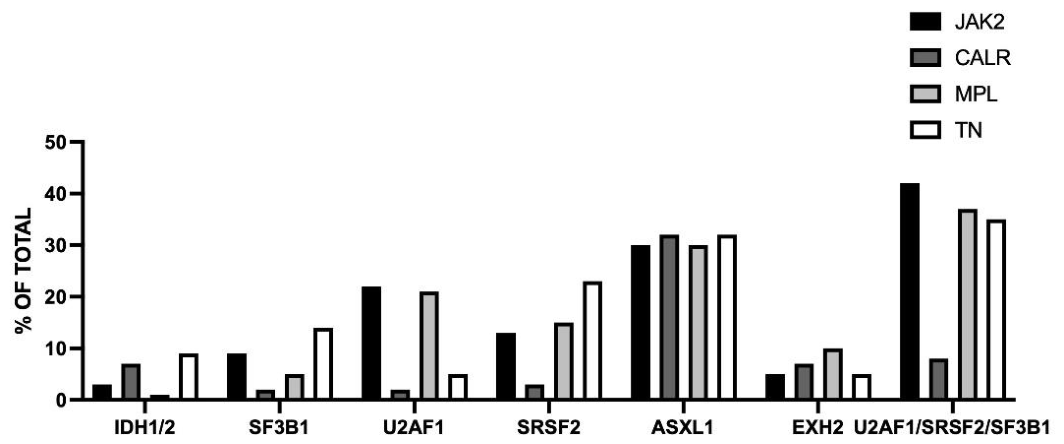
Figure 2.
Non driver gen mutations observed in PMF patients subdivided into four groups according to the presence or absence of driver mutations. Original data are reported in Tefferi et al. [39].
Figure 2.
Non driver gen mutations observed in PMF patients subdivided into four groups according to the presence or absence of driver mutations. Original data are reported in Tefferi et al. [39].
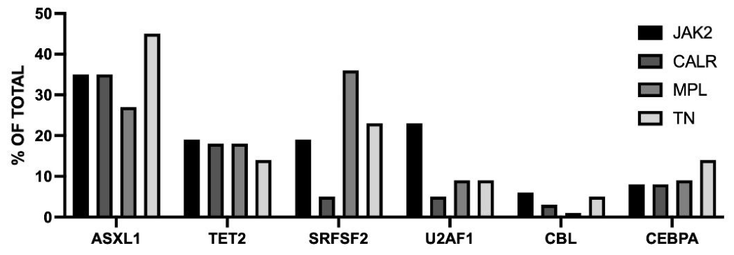

Table 1.
Histopathological grading of fibrosis according to the European consensus on the grading of bone marrow fibrosis [9].
Table 1.
Histopathological grading of fibrosis according to the European consensus on the grading of bone marrow fibrosis [9].
| Grading | Histopathology | Clinical Implication |
|---|---|---|
| MF-0 | Scattered linear reticulin fibers without intersections (cross-overs). | Normal bone marrow |
| MF-1 | A loose network of reticulin fibers with many intersections, primarily around blood vessels (perivascular). | Early-stage or minimal fibrosis |
| MF-2 | A diffuse and dense increase of reticulin with extensive intersections. May occasionally feature small, localized (focal) bundles of thick collagen fibers and/or mild bone hardering (focal osteosclerosis). | Moderate to advanced fibrosis; typical of the fibrotic phase of PMF. |
| MF-3 | A diffuse and dense network of reticulin fibers with extensive intersections, combined with coarse bundles of thick collagen fibers and significant bone hardering (ostesclerosis). | Severe, advanced fibrosis |
Table 2.
Cytogenetic abnormalities in PMF, classified in three risk categories, according to Tefferi et al [33].
Table 2.
Cytogenetic abnormalities in PMF, classified in three risk categories, according to Tefferi et al [33].
| Cytogenetic risk category | Cytogenetic abnormalities | Median survival |
|---|---|---|
| Favorable risk | Normal karyotype Sole sex abnormality, including -Y Sole chromosome 1 translocations/duplications Sole abnormalities of 20q-, 13q-, +9 |
4.4 years |
| Unfavorable risk | Sole abnormalities of +8 or 7q- Sole translocations not involving chromosome 1 Sole abnormalities not otherwise classified Monosomal karyotype without VHR abnormality Complex non-monosomal without VHR abnormality Single/multiple 5q- abnormality Two abnormalities without VHR abnormality |
2.9 years |
| Very high risk (VHR) | Single/multiple abnormalities of -7, inv(3)/q21, i(17q), 12p-/12p11.2, 11q-/11q23 Single/multiple autosomal trisomies other than +9 and +8 |
1.2 years |
Table 3.
Risk stratification models for PMF patients.
| Prognostic Model | Risk Factors | Risk Groups |
|---|---|---|
| International Prognostic Scoring System (IPSS) | Age>65 years (1 point) Constitutional symptoms (1 point) Hemoglobin <10 g/dL (1 point) WBC content >25x109/L (1 point) Circulating blasts ≥1% (1 point) |
Low-risk 0 point (MS 11.3 years) Intermediate-1 risk 1 point (MS 7.9 years) Intermediate-2 risk 2 points (MS 4.0 years) High-risk ≥3 points (MS 2.3 years) |
| Dynamic International Prognostic Scoring System (DIPSS) | Age>65 years (1 point) Constitutional symptoms (1 point) Hemoglobin <10 g/dL (2 points) WBC content >25x109/L (1 point) Circulating blasts ≥1% (1 point) |
Low-risk 0 point (MS Not Reached) Intermediate-1 risk 1 point (MS 14.2 years) Intermediate-2 risk 2 points (MS 4.0 years) High-risk ≥3 points (MS 1.5 years) |
| Dynamic International Prognostic Scoring System-Plus (DIPSS-Plus) | DIPSS score (DIPSS low = 0 point, DIPSS int-1 = 1 point, DIPSS int-2 = 2 points, DIPSS high = 3 points) Unfavorable karyotype (1 point) RBC transfusion (1 point) Platelet counts <100x109/L (1 point) |
Low-risk 0 point (MS 15.4 years) Intermediate-1 risk 1 point (MS 6.5 years) Intermediate-2 risk 2 points (MS 2.9 years) High-risk ≥3 points (MS 1.3 years) |
| Dynamic International Prognostic Scoring System-Revised (DIPSS-R) | Hemoglobin 8-10 g/dL (women) 9-11 g/dL (men) (1 point) Transfusion dependence or hemoglobin < 8g/dL (2 points) WBC 11-25x109/L (1 point) WBC >25x109/L (2 points) Platelet counts <150x109/L (1 point) Monocyte counts ≥1x10⁹/L (1 point) Circulating blasts ≥2% (1 point) |
Low-risk 0 point (MS not reached) Intermediate-1 risk 1 point (205 months) Intermediate-2 risk 2-3 points (96 months) High-risk 4-7points (44 months) |
| Mutation-Enhanced International Prognostic Score System (MIPSS-70) | Constitutional symptoms (1 point) Hemoglobin <10 g/dL (1 point) WBC content >25x109/L (2 points) Platelet counts <100x109/L (2 points) Bone marrow fibrosis ≥2 (1 point) One HMR mutation (1 point) ≥2 HMR mutation (2 points) Type 1 CALR absent (1 point) |
Low risk 0-1 points (MS 27.7 years) Intermediate risk 2-4 points (7.1 years) High risk ≥5 points (2.3 years) |
| Mutation-Enhanced International Prognostic Score System-Plus version 2.0 (MIPSS-70+ version 2.0) | VHR karyotype (4 points) Unfavorable karyotype (3 points) ≥2 HRM mutation (3 points) Type 1 CALR absent (2 points) Severe anemia (2 points) Moderate anemia (1 point) Circulating blasts ≥2% Constitutional symptoms (2 points) |
Very low risk 0 points (MS 26.4 years) Low risk 1-2 points (16.4 years) Intermediate risk 3-4 points (7.7 years) High risk 5-8 points (4.1 years) Very high risk ≥9 points (1.8 years) |
| Genetically Inspired Prognostic Scoring System (GIPSS) | VHR karyotype (2 points) Unfavorable karyotype (1 point) Type 1 CALR absent (1 point) ASXL1 mutation (1 point) SRSF2 mutation (1 point) U2AF1Q157 mutation (1 point) |
Low-risk 0 point (26.4 years) Intermediate risk-1 1 point (8 years) Intermediate risk-2 2 points (4.2 years) High risk ≥3 points (2 years) |
Disclaimer/Publisher’s Note: The statements, opinions and data contained in all publications are solely those of the individual author(s) and contributor(s) and not of MDPI and/or the editor(s). MDPI and/or the editor(s) disclaim responsibility for any injury to people or property resulting from any ideas, methods, instructions or products referred to in the content. |
© 2026 by the authors. Licensee MDPI, Basel, Switzerland. This article is an open access article distributed under the terms and conditions of the Creative Commons Attribution (CC BY) license (http://creativecommons.org/licenses/by/4.0/).
Copyright: This open access article is published under a Creative Commons CC BY 4.0 license, which permit the free download, distribution, and reuse, provided that the author and preprint are cited in any reuse.