

Submitted:
21 October 2026
Posted:
21 October 2026
You are already at the latest version
Abstract
Hydrogels are being investigated as delivery platforms for stem cells and extracellular vesicles (EVs) in regenerative medicine, but their relative advantages remain unclear. The objective of this critical narrative literature review is to compare stem‐cell‐loaded and EV‐loaded hydrogels in terms of biological activity, cell viability where applicable, retention and controlled release, stability, storage, safety, and clinical development. Published scientific studies were critically examined, considering experimental conditions, reported outcomes, methodological limitations, and levels of evidence. Stem‐cell‐loaded hydrogels can support living cells and their dynamic interactions with surrounding tissues but require maintenance of cell viability and control of cellular behavior. EV‐loaded hydrogels offer a cell‐free approach that avoids the administration of living therapeutic cells, although vesicle characterization, stability, dosing, and effective delivery remain challenges. Preclinical studies suggest improved local retention and sustained EV release, along with regenerative and immunomodulatory effects in specific experimental models. However, differences in hydrogel formulations, experimental conditions, and evaluation methods limit direct comparisons, while clinical effectiveness remains insufficiently established. Stimuli‐responsive hydrogels may provide additional control over EV delivery and regulate the microenvironment surrounding encapsulated stem cells, although their therapeutic superiority over conventional systems has not been demonstrated. Overall, neither strategy can currently be considered universally superior. Further research involving standardized characterization, direct comparative studies, long‐term safety evaluations, and clinical validation is necessary to determine their relative therapeutic potential.
Keywords:
hydrogels
; stem cells
; extracellular vesicles
; regenerative medicine
; stimuli-responsive hydrogels
Introduction
Regenerative medicine is an interdisciplinary field focused on restoring, replacing, or improving damaged tissues and organs through strategies that can activate endogenous repair mechanisms or introduce exogenous cells, stem-cell-derived products, tissues, or engineered constructs. Stem cells are central to many of these approaches because they are undifferentiated cells with self-renewal capacity and the ability to differentiate into specialized cell types in response to biological and environmental signals. Depending on the stem-cell population and its differentiation potential, these cells may contribute to the regeneration of tissues such as skin, bone, nervous tissue, cardiac tissue, and other organs. In addition to differentiation, stem cells may participate in tissue repair through anti-inflammatory activity, paracrine signaling, promotion of cell proliferation, and modulation of wound healing (Jin et al., 2023).
Although stem cells are considered an important development in regenerative medicine, several limitations continue to affect their clinical application. Stem cells require controlled conditions during in vitro cultivation to preserve growth, viability, phenotype, and biological function. After administration, the surrounding tissue may not provide adequate support for cellular attachment, survival, proliferation, or long-term function. These problems can reduce the number of viable cells that remain at the treatment site and may limit the effectiveness of stem-cell-based therapies. Therefore, maintaining cell viability and functionality remains an important consideration when developing regenerative treatments that use hydrogels as delivery or support systems (Choe et al., 2018; Xue et al., 2025).
To address some of these challenges, different hydrogel-based delivery strategies have been developed. Hydrogels can function as versatile carriers because their composition, preparation method, mechanical properties, degradation, and format can be modified according to the intended therapeutic application. Hydrogel systems can be prepared as injectable materials, dressings, microneedles, capsules, or other structures, allowing the delivery platform to be adapted to the characteristics of a specific disease or tissue. This versatility has made hydrogels an important area of research for improving the delivery of cells and cell-derived products in regenerative medicine (Zhang et al., 2024).
Although hydrogels can improve the delivery and local support of stem cells, these systems still depend on the use of viable cells. As a result, challenges related to cell survival, storage, handling, biological variability, and maintenance of cellular function remain relevant. These limitations have encouraged the development of cell-free approaches that attempt to preserve some of the therapeutic effects associated with stem cells without requiring administration of the complete living cell. Cell-free strategies may therefore reduce some of the complexity associated with maintaining viable cellular products while still providing regenerative biological signals (Hashemi et al., 2024).
In particular, increasing attention has been directed toward stem-cell-derived extracellular vesicles (EVs), which can transport bioactive molecules involved in cell-to-cell communication and may reproduce part of the paracrine activity associated with their cells of origin. Mesenchymal stromal cells (MSCs) are heterogeneous stromal cell populations with immunomodulatory properties and, in some cases, demonstrated capacity to differentiate into mesodermal lineages such as bone, cartilage, and adipose tissue under defined experimental conditions. The designation mesenchymal stem cells should be reserved for populations with evidence supporting stem-cell properties (Uccelli et al., 2008; Viswanathan et al., 2019). MSC-derived extracellular vesicles (MSC-EVs) may provide several advantages because they do not engraft or replicate, are substantially smaller than their parental cells, and may reduce some risks associated with abnormal tissue formation or vascular obstruction. Although these products do not require the preservation of viable cells, the manufacturing, characterization, storage, and transport of MSC-EVs still demand careful control to maintain vesicle integrity and biological activity (Hashemi et al., 2024; Welsh et al., 2024). In accordance with MISEV2023 recommendations, the term extracellular vesicles (EVs) is used as a general designation, whereas exosomes refers specifically to vesicles whose endosomal origin has been demonstrated (Welsh et al., 2024).
Although both approaches aim to improve tissue regeneration, their biological requirements and limitations differ. Stem-cell-loaded hydrogels must maintain the viability and functionality of living cells, whereas EV-loaded hydrogels must preserve vesicle stability, biological activity, and effective delivery. Furthermore, differences in hydrogel composition, EV isolation and characterization, experimental conditions, and evaluation methods make it difficult to directly compare their reported outcomes. These challenges highlight the need for a critical analysis of the available scientific evidence to identify the advantages and limitations of each strategy and distinguish findings from in vitro studies and animal models from demonstrated clinical outcomes (Choe et al., 2018; Hashemi et al., 2024; Welsh et al., 2024).
This review is to compare stem-cell-loaded hydrogels and extracellular-vesicle-loaded hydrogels based on the available scientific evidence. The comparison considers cell viability, where applicable, and biological activity, retention and controlled release, stability and storage, safety and biological risks, advantages and limitations, as well as their clinical applications and current stage of development. In addition, stimuli-responsive (smart) hydrogels are evaluated as a potential strategy to address some of the limitations associated with both systems, particularly by improving EV delivery and regulating the properties of the microenvironment surrounding stem cells and identifying the main differences between both technologies, the limitations of the available evidence, and opportunities for future research.
Methodology
2.1. Literature Search
A critical narrative literature review was conducted using ScienceDirect and scientific publications available through academic publishing platforms, including MDPI. Additional online resources were consulted to access the full texts of relevant publications. The literature search was conducted from September to October 2026 and focused on studies related to hydrogels, stem cells, extracellular vesicles, and their applications in regenerative medicine. Search terms included hydrogels, stem cells, mesenchymal stem cells, extracellular vesicles, EV-loaded hydrogels, cell-free therapy, and stimuli-responsive hydrogels. Publications from 2008 to 2026 were considered, according to the inclusion criteria (Table 1). The collected information included research articles, scientific reviews, scientific consensus and position statements, and an academic book chapter, which served as the basis for the comparative analysis of both therapeutic systems.
2.2. Selection and Refinement of Information
A total of 21 scientific publications were included in the final review based on their relevance to the research objectives. These sources included original research articles, scientific reviews, scientific consensus and position statements, and an academic book chapter related to hydrogels, stem cells, extracellular vesicles, and their biomedical applications.
The selection considered publications providing information on biological properties, cell viability, retention, controlled release, stability, and safety. The established inclusion criteria considered English-language publications from 2008 to 2026, thematic relevance, methodological clarity, and verifiable scientific information. The exclusion criteria addressed publications unrelated to the research objectives, duplicate content, unclear methodologies, and insufficient scientific support.
2.3. Selection of Subtopics
The collected information was organized into subtopics to facilitate the comparative analysis of hydrogel systems. Categories were established based on cell viability, biological response, retention and controlled release, storage, stability, safety and biological risks, advantages and limitations, and clinical applications and current stage of development.
Additionally, smart hydrogels were included as a potential strategy to improve the performance of both stem-cell-loaded and EV-loaded systems by providing a more responsive cellular microenvironment and improving the controlled release of extracellular vesicles.
2.4. Comparative and Critical Analysis
A comparative and critical analysis of stem-cell-loaded and extracellular-vesicle-loaded hydrogels was conducted, considering their properties, advantages, and limitations. Findings reported in the scientific literature were evaluated to identify differences in biological behavior, stability, safety, and therapeutic potential. The analysis also considered experimental conditions, quantitative results when available, control groups, and methodological limitations. Evidence from in vitro studies and animal models was distinguished from clinical evidence to avoid generalizing preclinical findings to human applications. Differences in experimental models, hydrogel formulations, and evaluation methods were also considered when interpreting the reported outcomes.
Additionally, the use of smart hydrogels was analyzed as a potential strategy to support stem-cell function and improve extracellular vesicle retention and controlled release. The effects of environmental stimuli, including temperature and pH, were examined in relation to the reported behavior of these hydrogel systems. Based on the collected evidence, the main limitations of both systems and areas requiring further research were identified.
General Hydrogel Classification
Hydrogels can be classified according to several characteristics, including their source, polymer composition, physical structure, electrical charge, and crosslinking method. When classified according to origin, the main groups generally discussed are natural and synthetic hydrogels, while hybrid systems combine features from both classes. Natural hydrogels are obtained from biologically derived materials and are often attractive for cell-based applications because they can provide high water content, biological compatibility, and chemical groups that resemble components of the extracellular environment. Examples of natural polymers used in hydrogel preparation include proteins such as collagen and gelatin and polysaccharides such as agarose, alginate, and chitosan (Khansari et al., 2017).
Natural hydrogels also present limitations that can affect reproducibility and engineering performance. Their mechanical strength may be lower than that of some synthetic systems, and biological source materials can show batch-to-batch variability. Materials obtained from animal sources may also require careful control of purity, immunogenicity, and biological safety. Synthetic hydrogels, in contrast, are produced using engineered polymers and chemical polymerization or crosslinking strategies. Materials such as polyacrylamide, polyethylene glycol, polyvinyl alcohol, and poly (2-hydroxyethyl methacrylate) allow greater control over network architecture, mechanical strength, water absorption, and other physicochemical properties. However, synthetic materials may require modification to improve biological interaction and can show degradation behavior that differs substantially from natural extracellular matrices (Khansari et al., 2017).
Using only a natural or synthetic hydrogel can therefore create trade-offs between biological performance and engineering control. Hybrid hydrogels have been developed to combine the adjustable chemical and mechanical characteristics of synthetic systems with the biological advantages of naturally derived components. These systems are intended to provide a more tunable microenvironment while preserving properties that support cell attachment, proliferation, survival, and differentiation. For stem-cell applications, an effective biomaterial should be biocompatible, reproduce relevant characteristics of native tissue, provide mechanical properties appropriate for the target tissue, permit mass transport, and allow modification of chemical and structural properties when necessary. Hydrogels remain promising because their polymer networks can be engineered to address several of these requirements simultaneously (Khansari et al., 2017).
As mentioned above, each hydrogel category presents its own advantages and limitations. These differences make hydrogel composition an important consideration for future investigations and biomedical applications. Synthetic materials are particularly useful when greater control over mechanical and chemical properties is required, while natural materials may provide stronger biological interaction with cells. Hybrid systems attempt to balance these characteristics. Regardless of the selected material, biocompatibility, degradation behavior, mechanical stability, reproducibility, and the biological effects of degradation products must be considered before the system can be translated into clinical use.
Cellular Viability and Biological Response
Stem cells offer considerable promise for tissue engineering because they can respond to biological signals and differentiate into specialized cell types. However, their clinical application requires maintaining cellular viability and function, while successful engraftment is particularly important for strategies involving direct tissue replacement. A significant fraction of transplanted cells may be lost during the early period after implantation when the host environment does not provide sufficient support for attachment, proliferation, nutrient availability, or biological function. Another important challenge is directing differentiation toward the desired lineage while avoiding unwanted cellular behavior. These limitations make survival and differentiation control central problems in the development of stem-cell-assisted regenerative therapies (Xue et al., 2025).
Similarly, EVs have emerged as an alternative approach because of several favorable biological properties. Extracellular vesicles are particles released by cells, delimited by a lipid bilayer, and unable to replicate independently. Because EV preparations may contain heterogeneous populations, terms such as exosomes and microvesicles should be used only when their corresponding biogenesis pathways have been demonstrated. When the subcellular origin is not established, the general term EVs or an operational descriptor such as small extracellular vesicles (sEVs), when supported by size characterization, is more appropriate (Welsh et al., 2024). EV cargo can include lipids, proteins, and nucleic acids, allowing these vesicles to participate in intercellular communication and influence the behavior of recipient cells (Doyle & Wang, 2019).
Unlike stem cells, EVs can transfer biologically active molecules without requiring a living therapeutic cell to remain viable, proliferate, or differentiate inside the patient. This makes them an important area of investigation for cell-free therapy. Their inability to proliferate or differentiate eliminates the specific risk of unwanted cellular differentiation from the therapeutic product itself. However, EV effectiveness still depends on maintaining biological activity and delivering an adequate concentration of vesicles to the intended tissue. Stability, retention, dose, biodistribution, and interaction with recipient cells therefore remain important variables when evaluating EV-based therapies (Doyle & Wang, 2019; Zhao et al., 2023).
An important application of hydrogels as bioactive materials is their use in stem-cell therapy. Hydrogel systems can help regulate stem-cell fate by creating a three-dimensional microenvironment that supports cellular attachment, viability, proliferation, and differentiation. The polymer network can be engineered through changes in stiffness, porosity, composition, compatibility, and degradation, and each of these characteristics can influence whether encapsulated stem cells survive or lose function. For this reason, hydrogel design is not only a structural issue but also a biological factor that can influence the success of stem-cell delivery (Tsou et al., 2016).
The scaffold structure and hydrophilic polymer chains can provide an environment that shares some characteristics with the extracellular matrix. Hydrogels may provide physical support for encapsulated cells and help maintain them at the treatment site while still allowing movement of water, nutrients, oxygen, metabolites, and waste through the network. Semipermeable hydrogel structures may also provide a degree of immunoisolation or protection from external biological stresses, depending on the material and pore characteristics. These properties can improve cell retention and viability, although successful use still requires a material that is compatible with the specific cell type and intended tissue (Choe et al., 2018).
When both approaches are considered, stem-cell-loaded hydrogels provide a dynamic biological system in which living cells can continuously respond to their surroundings, secrete biological factors, and potentially differentiate into tissue-specific cells. This activity may provide a potential regenerative benefit, but the same biological flexibility increases variability and makes post-implantation behavior more difficult to control. EV-loaded systems operate through a different mechanism because their effects depend on the transfer of previously produced bioactive molecules rather than the continued survival or differentiation of a living therapeutic cell. This may reduce certain biological risks, but EV activity still depends on maintaining stability, concentration, and effective interaction with the target tissue.
Retention and Controlled Release
Although EVs may have certain advantages over stem-cell-based therapies, one of their main limitations is achieving effective and sustained delivery at a specific target site. Free EVs can be cleared relatively rapidly from the circulatory system or treatment region, reducing the amount of therapeutic material that remains available for interaction with the target tissue. Incorporating EVs into hydrogels can improve local retention and provide a platform for more gradual delivery, which may increase the time during which the vesicles remain biologically available (Ju et al., 2023).
The porous three-dimensional structure of hydrogels provides the physical basis for EV loading and release. Mesh size is influenced by factors such as polymer concentration, crosslinking density, temperature, pH, and the chemical characteristics of the polymer network. Larger pores may make it easier to incorporate or diffuse EVs into the hydrogel, but they can also permit faster escape of the vesicles and contribute to an initial burst release. If release occurs too rapidly, the hydrogel may fail to significantly extend EV retention at the treatment site. Therefore, a useful EV-loaded hydrogel must balance loading efficiency with release control rather than maximizing porosity alone (Ju et al., 2023).
EV-loaded hydrogels therefore represent a promising strategy for improving localized and prolonged therapeutic activity, but their effectiveness depends on obtaining an appropriate balance among EV loading, retention, diffusion, and release. A hydrogel that retains the product too strongly could limit therapeutic availability, while a network that releases the EVs too quickly could fail to provide a sustained response. This relationship demonstrates that hydrogel design becomes an important determinant of whether an EV-based therapy can maintain sufficient exposure at the target tissue to produce a meaningful regenerative effect (Ju et al., 2023).
Pharmacokinetics and pharmacodynamics are also important concepts when evaluating controlled-delivery systems. Pharmacokinetics describes the processes that affect the concentration of a therapeutic agent over time, including absorption, distribution, metabolism, and elimination. Pharmacodynamics describes the biological effect produced by the therapeutic agent and the relationship between exposure and response. In controlled-delivery systems, hydrogel-mediated release can modify the time profile of therapeutic availability and may help maintain useful local concentrations for longer periods, particularly for products that could otherwise be rapidly lost or cleared after administration (Ramchandani et al., 2023).
Hydrogels can function as controlled-release systems by regulating the movement of encapsulated therapeutic agents through a polymeric network. In stimuli-responsive hydrogels, release can be affected by physicochemical factors such as molecular size, hydrophobicity, pH stability, concentration, and environmental conditions. Molecules or biological products can move into the hydrogel during loading and later diffuse or be released from the network as the material swells, degrades, or responds to environmental changes. These principles have supported the development of hydrogel systems for oral, nasal, buccal, rectal, vaginal, ophthalmic, parenteral, and other routes of administration (Ramchandani et al., 2023).
The comparison between stem-cell-loaded and EV-loaded hydrogels shows that retention is important for both approaches but for different biological reasons. Stem-cell-loaded systems must maintain viable cells within an environment that provides attachment, nutrient transport, and physical support. EV-loaded systems do not need to maintain living-cell survival, but they must prevent rapid loss of a finite therapeutic dose and provide an appropriate release profile. Hydrogel structure and release characteristics may therefore have an even more direct influence on the therapeutic availability of EVs than on therapies in which living cells can continue producing biological signals after administration.
Storage and Stability
Storage represents a major challenge for cell-loaded tissue-engineering products. Cryopreservation protocols for isolated cell cultures are relatively well established, but tissue-engineering products that contain both a cellular component and a scaffold are more complex. The storage method must preserve cell viability, proliferative capacity, and biological function while also maintaining the structure, mechanical behavior, and physicochemical properties of the hydrogel carrier. Only a limited number of studies have examined long-term cryostorage of these combined systems when compared with the extensive literature available for conventional cell preparations (Egorikhina et al., 2020).
Storage and stability are also important issues for EV-loaded hydrogels. EV biological activity can be affected by storage conditions, and low-temperature preservation is commonly used to reduce degradation. Repeated freeze-thaw cycles may negatively affect EV integrity or biological function, and many experimental studies emphasize freshly prepared or ready-to-use formulations rather than long-term stability. As a result, production, preservation, transportation, storage duration, handling, and economic considerations remain important factors for the clinical translation of EV-based hydrogel therapies (Hashemi et al., 2024).
When both approaches are compared, cell-loaded hydrogels present the additional requirement of preserving living cells throughout storage and subsequent administration. EV-loaded hydrogels avoid the requirement of maintaining cellular viability, which may simplify some aspects of handling and preservation. However, they still require preservation of EV integrity, cargo stability, and biological activity. Therefore, EV-loaded systems may provide practical advantages in storage and handling without eliminating long-term stability as an unresolved limitation for regenerative products (Egorikhina et al., 2020; Hashemi et al., 2024).
Safety and Biological Risks
Hydrogels are generally designed to be biocompatible, but safety depends on the material composition, degradation behavior, crosslinking chemistry, implantation site, duration of exposure, and interaction with surrounding tissue. Natural materials may contain biologically active components that can be advantageous for tissue integration, but they may also present variability or immune-related concerns depending on their source and purity. Synthetic systems may offer greater reproducibility but can introduce concerns related to residual monomers, initiators, crosslinkers, degradation products, or other functional components. For this reason, safety cannot be assumed solely from the general classification of a material as a hydrogel (Wu et al., 2026).
A more significant problem may arise when a hydrogel used for drug or biologic delivery triggers a persistent foreign-body response. If the immune response progresses into chronic inflammation, macrophages and other immune cells can contribute to fibrotic encapsulation around an implanted material. A fibrous layer may interfere with tissue integration and can also change diffusion pathways, affecting the amount and rate of therapeutic product that leaves the hydrogel. For treatments that require precise or sustained dosing, these changes could reduce therapeutic effectiveness and create variability between patients or implantation sites (Wu et al., 2026).
Long-term safety is therefore an important challenge in the clinical application of hydrogels. Favorable cytocompatibility in vitro or short-term performance in animal studies does not automatically establish long-term safety in humans. After implantation or repeated administration, hydrogels may undergo swelling, mechanical fatigue, incomplete degradation, or interactions with proteins, immune cells, and surrounding tissues. These processes may contribute to chronic inflammation, fibrotic encapsulation, foreign-body responses, delayed tissue remodeling, or local tissue compression. In addition, degradation products, residual crosslinkers, initiators, and functional nanocomponents may require systematic evaluation for accumulation, toxicity, and long-term biological effects, particularly when hydrogels are used repeatedly or implanted in sensitive tissues (Wu et al., 2026).
Stem-cell-loaded hydrogels introduce an additional layer of biological complexity because the therapeutic cells remain capable of proliferation, differentiation, secretion of signaling molecules, and interaction with the host environment. These characteristics contribute to their regenerative potential but can also create variability and risks related to uncontrolled or unintended cellular behavior. EV-loaded hydrogels avoid the specific problems of proliferation and differentiation because EVs are not living cells. However, the use of EVs does not eliminate risks related to the hydrogel material, immune response, degradation products, inappropriate EV concentration, or variability in EV composition and biological activity (Doyle & Wang, 2019; Wu et al., 2026).
For this reason, there is a need to develop hydrogel systems capable of reducing inflammation, infection, premature degradation, and other biological complications while still providing the intended therapeutic function. A hydrogel that can respond to a specific environmental signal may offer additional control over therapeutic release or material behavior. This concept is one of the reasons stimuli-responsive hydrogels are being investigated as more interactive delivery platforms rather than passive carriers (Samchenko et al., 2011; Wu et al., 2026).
Clinical Applications and Translation
Hydrogels are used or investigated in a wide range of medical applications. In gynecology, hydrogel systems have been studied for endometrial regeneration, localized treatment of infections, contraception, drug delivery, and tissue repair. In cardiology, hydrogels have shown potential in myocardial infarction therapy through injectable scaffolds, cardiac patches, and local delivery of therapeutic agents. In rheumatoid arthritis and other inflammatory joint conditions, hydrogels can function as localized drug-delivery systems, lubricating materials, scaffolds, and immunomodulatory platforms. Urological applications include the development of antimicrobial coatings, local therapeutic delivery systems, and protective barriers intended to reduce damage to healthy tissue during certain procedures (Rybicki et al., 2025).
Ophthalmology also benefits from hydrogel technologies because hydrated polymer networks are compatible with applications such as contact lenses, corneal bandages, and vitreous substitutes or implants. In cancer-related research, hydrogels have been explored for localized chemotherapy, chemoembolization, tumor models, and controlled drug delivery. In abdominal surgery, hydrogel-coated meshes and Janus-type materials have been developed to support tissue repair while reducing postoperative adhesions (Rybicki et al., 2025).
These clinical and preclinical applications demonstrate the versatility of hydrogels across different medical fields. At the same time, they show that not all hydrogel technologies are at the same level of clinical maturity. Mechanical stability, long-term safety, reproducibility, sterilization, manufacturing, regulatory requirements, and large-scale production remain important barriers for more advanced systems. These challenges become even more important when a hydrogel contains living cells or biologically sensitive EVs because the final product must maintain both material performance and biological activity (Rybicki et al., 2025).
The broad range of hydrogel applications demonstrates that the material platform already has significant biomedical potential. However, translating cell-loaded and EV-loaded hydrogels into clinical products introduces additional challenges related to preservation, characterization, manufacturing, standardization, controlled release, dosing, and long-term safety. These factors should be considered when determining which therapeutic strategy offers the most practical route toward clinical use. Although certain hydrogel-based medical devices are already used clinically, the evidence reviewed for stem-cell-loaded and EV-loaded hydrogels remains largely preclinical. Therefore, the established clinical use of conventional hydrogels should not be interpreted as evidence of the safety or therapeutic effectiveness of these combined systems, which require independent clinical validation (Choe et al., 2018; Hashemi et al., 2024). Table 2 summarizes the main differences between stem-cell-loaded and EV-loaded hydrogels, considering their biological activity, retention, stability, safety, advantages, limitations and clinical development.
Conventional hydrogels consist of crosslinked polymer networks capable of retaining large amounts of water while maintaining structural integrity. Many conventional systems exhibit relatively stable swelling and transport behavior under a defined environment. Smart or stimuli-responsive hydrogels are designed to change properties such as swelling, permeability, degradation, or structural organization when exposed to selected external or internal stimuli. These stimuli may be physical, such as temperature, pressure, light, electric fields, or magnetic fields, or chemical and biochemical, such as pH, ionic strength, specific molecules, or changes in local biological conditions (Samchenko et al., 2011). Figure 1 illustrates the general mechanism of a stimuli-responsive hydrogel, showing how changes in environmental conditions, such as pH or temperature, may alter the polymer network and promote the release of encapsulated components.
Conventional hydrogels can also provide controlled release through processes such as diffusion, swelling, and degradation. However, stimuli-responsive systems offer the additional ability to modify their properties in response to specific changes in the surrounding environment. This responsiveness may provide greater control over therapeutic release and allow the material to adapt to specific biological conditions. However, these advantages depend on the hydrogel composition, the selected stimulus, and the biological requirements of the therapeutic component. Therefore, greater responsiveness does not necessarily guarantee improved therapeutic effectiveness.
When considering EV delivery, one of the main concerns is the loss of therapeutic vesicles during administration and their limited retention at a specific target site. Conventional hydrogels can improve retention, but their release behavior often depends primarily on passive characteristics such as porosity, mesh size, swelling, and degradation. A smart hydrogel that changes its behavior in response to a relevant stimulus could potentially provide additional control over when and where EVs are released. Such a system may reduce premature loss while improving local availability of EVs at the intended tissue (Ju et al., 2023; Samchenko et al., 2011).
Temperature-responsive hydrogels provide an example of this approach. Wu et al. (2022) developed an injectable chitosan/β-glycerophosphate hydrogel that underwent a sol-to-gel transition at physiological temperature (37 °C), forming a localized system for sustained sEV delivery. This mechanism facilitates hydrogel formation after administration, while subsequent EV release depends on the characteristics of the polymer network. Therefore, temperature acts primarily as the trigger for hydrogel formation in this system, rather than demonstrating repeated temperature-triggered EV release. In contrast, pH-responsive systems can modify EV release in response to local acidity. Chewchuk et al. (2025) developed hydrogel microcapsules containing a CD9-binding peptide that enabled the release of CD9-positive EVs under acidic conditions. This example demonstrates the potential of pH-dependent interactions to regulate EV availability. However, these findings represent experimental evidence and do not establish superior clinical effectiveness.
The potential value of stimuli-responsive hydrogels becomes clearer when the limitations of conventional EV delivery are considered. Although EVs avoid some of the complications associated with administering viable stem cells, their therapeutic effectiveness can still be reduced by rapid clearance, insufficient retention, instability, inappropriate dosing, or uncontrolled release. A hydrogel capable of responding to environmental conditions may provide greater control over these factors and could combine the biological advantages of a cell-free therapy with more localized and sustained delivery. However, this potential should be considered promising rather than proven because additional evidence is still required to establish reproducibility, long-term safety, storage behavior, manufacturing feasibility, and clinical effectiveness.
Stimuli-responsive hydrogels may also benefit stem-cell-loaded systems by providing a dynamic cellular microenvironment. Changes in hydrogel stiffness, structural organization, or biochemical properties can influence cell attachment, proliferation, differentiation, and secretion of bioactive factors. In particular, stimuli-responsive materials have been investigated for their ability to modulate the immunomodulatory activity of mesenchymal stromal cells by regulating their interactions with the surrounding matrix and their secretion of signaling molecules. These responses may allow greater control over cellular behavior compared with conventional hydrogels with relatively fixed properties. However, the stimuli or resulting changes in material properties could also negatively affect cell viability or functionality if they are not compatible with the encapsulated cells. Nevertheless, the effects depend on the cell type, material composition, and applied stimulus, making it necessary to evaluate cellular viability, functionality, and safety under specific experimental conditions (Castilla-Casadiego et al., 2024).
Current Limitations and Future Research
Despite the progress achieved with stem-cell-loaded and EV-loaded hydrogels, several limitations remain before these technologies can achieve broader clinical application. Stem-cell-loaded systems continue to face challenges related to cell viability, storage, biological variability, differentiation control, and preservation of both the cellular and scaffold components. Cryopreservation must maintain cellular function while also preserving hydrogel structure, and the longer the product is stored, the more difficult it may become to maintain both components at an acceptable level (Egorikhina et al., 2020).
EV-loaded systems avoid some of the problems associated with maintaining living cells but still require improved methods for long-term storage, preservation of biological activity, controlled dosing, retention, and release. Standardization of EV production and characterization is also important because differences in cellular origin, isolation methods, purification, concentration, cargo, and loading procedure can influence the final therapeutic response. These factors make it difficult to compare studies directly and represent important challenges for reproducible manufacturing and clinical translation (Hashemi et al., 2024).
Hydrogel design introduces additional variables, including polymer composition, mesh size, degradation rate, mechanical properties, crosslinking conditions, sterilization, and biocompatibility. These characteristics can influence therapeutic loading, retention, release, biological response, and long-term safety. A formulation that performs well in vitro may not necessarily behave in the same way after implantation because tissue mechanics, proteins, enzymes, immune cells, and local inflammatory conditions can change the hydrogel over time (Ju et al., 2023; Wu et al., 2026).
Future investigations should therefore focus on optimizing the interaction between the therapeutic component and the hydrogel rather than evaluating each component independently. For EV-loaded systems in particular, the development of stimuli-responsive hydrogels may provide an opportunity to improve localized delivery and release control. However, additional studies are needed to determine whether these advantages can be maintained during long-term storage, reproducible large-scale manufacturing, sterilization, transportation, and clinically relevant use. More direct comparisons between cell-loaded and EV-loaded systems using similar disease models and outcome measures would also strengthen conclusions about which strategy is more appropriate for specific regenerative applications.
Comparative Analysis between EV-Loaded Hydrogels and Stem-Loaded Hydrogels
Experimental evidence supports the importance of appropriate delivery systems for EV-based therapies. Zhao et al. (2023) developed an injectable thermosensitive chitosan hydrogel containing bone-marrow-derived MSC-EVs, prepared through ionic crosslinking. In vitro experiments demonstrated sustained EV release, primarily regulated by hydrogel degradation, while preserving their biological activity. Following intranasal administration in an allergic-rhinitis mouse model, the treatment reduced eosinophil infiltration and helped reverse the Th1/Th2 imbalance. The authors also reported improved therapeutic effects when EVs were delivered using the hydrogel system. These findings suggest that hydrogel-mediated delivery may improve the local availability of EVs and contribute to their therapeutic effects. However, the study was conducted using experimental models, and its results do not establish long-term safety or clinical effectiveness in humans. Additionally, the outcomes were associated with inflammatory regulation rather than direct tissue regeneration, limiting their generalization to other regenerative applications (Zhao et al., 2023).
The relationship between hydrogel structure and EV delivery was also demonstrated by Wu et al. (2022), who evaluated a thermosensitive chitosan/β-glycerophosphate hydrogel containing bone-marrow-derived small extracellular vesicles (sEVs). In vitro release experiments showed that approximately 80% of the incorporated sEVs were released by day 8, followed by a slower release rate. This result indicates that the hydrogel allowed sustained EV delivery rather than immediate release of the entire dose. However, release was estimated using a protein-based assay, which does not independently confirm the integrity or biological activity of the released vesicles. In a rat calvarial-defect model, 18 rats were assigned to three experimental groups: PBS control, hydrogel alone, and hydrogel loaded with 200 μg of sEVs. After 12 weeks, the sEV-loaded hydrogel showed a greater area of newly formed bone than the hydrogel alone. Bone mineral density, bone volume fraction, and trabecular number were significantly higher in both hydrogel-treated groups than in the PBS control group. However, these comparisons do not establish that the EV-loaded hydrogel was significantly superior to the unloaded hydrogel for every measured parameter. Furthermore, the absence of a free-sEV-only group limits the ability to isolate the additional therapeutic contribution of hydrogel-mediated delivery. These findings support the potential of EV-loaded hydrogels for bone regeneration under experimental conditions, but they do not establish an optimal release profile or clinical efficacy in humans (Wu et al., 2022).
Storage also represents an important limitation for cell-loaded hydrogel systems. Egorikhina et al. (2020) evaluated fibrin/collagen hybrid hydrogel scaffolds containing human adipose-derived MSCs cryopreserved at −80 °C using Bambanker or 10% dimethyl sulfoxide (DMSO) as cryoprotectants. The scaffolds were evaluated after 3, 6, 9, and 12 months of storage, with cellular proliferation assessed after 96 hours. The study reported cell viability above 95% and preserved proliferative activity during the initial three-month storage period. However, proliferative activity decreased after six months, while nine months of storage resulted in reduced viability and proliferation. After twelve months, the authors reported approximately 34–48% dead cells, together with structural changes that were not fully reversed after thawing and subsequent culture. These findings demonstrate that preserving cell viability alone is insufficient to establish the long-term functionality of a cell-loaded hydrogel. More importantly, the reduction in proliferative activity after six months indicates that cells may remain viable while losing essential functional properties. Furthermore, the results depend on the specific materials and preservation conditions evaluated, and the study did not establish clinical regenerative effectiveness after transplantation (Egorikhina et al., 2020).
Taken together, these studies provide evidence of the potential benefits and limitations of both hydrogel-based strategies, but they do not establish the superiority of either approach. Wu et al. (2022) investigated bone regeneration in rats, Zhao et al. (2023) evaluated inflammatory regulation in an allergic-rhinitis mouse model, and Egorikhina et al. (2020) examined cell preservation within hydrogel scaffolds under laboratory conditions. Differences in therapeutic objectives, experimental models, evaluation methods, and measured outcomes prevent direct comparisons of their effectiveness. Moreover, these studies did not directly compare stem-cell-loaded and EV-loaded hydrogels under equivalent experimental conditions. Therefore, the available findings support the feasibility and potential benefits of individual systems rather than establishing their relative therapeutic superiority. Future investigations should include standardized experimental conditions, appropriate control groups, long-term biological evaluation, and clinical studies to determine the relative therapeutic potential of both technologies.
Conclusions
The available evidence suggests that both stem-cell-loaded and EV-loaded hydrogels have important potential for regenerative medicine, but neither strategy can currently be considered universally superior. Stem-cell-loaded hydrogels provide living cells capable of continuous biological activity and interaction with the surrounding environment, which may provide a dynamic regenerative response. However, their application requires careful control of cell viability, differentiation, storage, handling, and biological behavior. EV-loaded hydrogels avoid the need to administer living therapeutic cells and therefore avoid risks arising from the uncontrolled proliferation or differentiation of the administered cells, although other biological risks remain. However, their therapeutic effectiveness remains dependent on factors such as concentration, stability, retention, controlled release, and successful delivery to the target tissue. Therefore, the selection of either strategy should depend on the intended therapeutic application and the biological requirements of the target tissue rather than an assumed overall superiority.
Hydrogels can help address some of these limitations by improving local retention and providing more sustained delivery of EVs, while also supporting the cellular microenvironment in stem-cell-loaded systems. At the same time, their effectiveness depends strongly on structural and physicochemical characteristics such as porosity, mesh size, degradation, crosslinking, and interaction with the biological environment. Both strategies also face challenges related to storage, manufacturing, standardization, reproducibility, and long-term safety. Furthermore, much of the evidence discussed for these combined systems originates from in vitro experiments and animal models. Differences in experimental conditions and evaluation methods limit direct comparisons, emphasizing the need for additional studies to establish safety and therapeutic effectiveness in humans.
Based on these considerations, stimuli-responsive hydrogels may represent a promising strategy for improving EV delivery and regulating the microenvironment surrounding encapsulated stem cells. Their ability to respond to changes in the local environment could provide greater control over EV release and influence the biological behavior of encapsulated cells. However, these potential benefits do not establish the clinical superiority of smart hydrogels over conventional systems. Future research should prioritize direct comparisons under equivalent experimental conditions, standardized EV characterization, preservation of cellular and vesicular biological activity, and long-term safety evaluation. Additionally, clinical investigations are necessary to determine whether the improved control offered by stimuli-responsive hydrogels translates into meaningful therapeutic benefits for patients.
References
- Castilla-Casadiego, D. A.; Loh, D. H.; Pineda-Hernandez, A.; Rosales, A. M. Stimuli-responsive substrates to control the immunomodulatory potential of stromal cells. Biomacromolecules 2024, 25(10), 6319–6337. [Google Scholar] [CrossRef] [PubMed]
- Chewchuk, S.; Soucy, N.; Wan, F.; Harden, J.; Godin, M. pH controlled release of extracellular vesicles from a hydrogel scaffold for therapeutic applications. Biomed. Mater. 2025, 20(6), 065006. [Google Scholar] [CrossRef] [PubMed]
- Choe, G.; Park, J.; Park, H.; Lee, J. Y. Hydrogel biomaterials for stem cell microencapsulation. Polymers 2018, 10(9), 997. [Google Scholar] [CrossRef] [PubMed]
- Doyle, L. M.; Wang, M. Z. Overview of extracellular vesicles, their origin, composition, purpose, and methods for exosome isolation and analysis. Cells 2019, 8(7), 727. [Google Scholar] [CrossRef] [PubMed]
- Egorikhina, M. N.; Rubtsova, Y. P.; Aleynik, D. Y. Long-term cryostorage of mesenchymal stem cell-containing hybrid hydrogel scaffolds based on fibrin and collagen. Gels 2020, 6(4), 44. [Google Scholar] [CrossRef] [PubMed]
- Hashemi, A.; Ezati, M.; Partovi Nasr, M.; Zumberg, I.; Provaznik, V. Extracellular vesicles and hydrogels: An innovative approach to tissue regeneration. ACS Omega 2024, 9(6), 6184–6218. [Google Scholar] [CrossRef] [PubMed]
- Jin, Y.; Li, S.; Yu, Q.; Chen, T.; Liu, D. Application of stem cells in regeneration medicine. MedComm 2023, 4(4), e291. [Google Scholar] [CrossRef] [PubMed]
- Ju, Y.; Hu, Y.; Yang, P.; Xie, X.; Fang, B. Extracellular vesicle-loaded hydrogels for tissue repair and regeneration. Mater. Today Bio 18 2023, 100522. [Google Scholar] [CrossRef] [PubMed]
- Khansari, M. M.; Sorokina, L. V.; Mukherjee, P.; Mukhtar, F.; Shirdar, M. R.; Shahidi, M.; Shokuhfar, T. Classification of hydrogels based on their source: A review and application in stem cell regulation. JOM 2017, 69(8), 1340–1347. [Google Scholar] [CrossRef]
- Ramchandani, M.; Rath, G.; Goyal, A. K. Advances in hydrogel-based controlled drug-delivery systems. In Smart polymeric nano-constructs in drug delivery: Concept, design and therapeutic applications; Vyas, S. P., Agrawal, U., Sharma, R., Eds.; Academic Press, 2023; pp. 329–350. [Google Scholar] [CrossRef]
- Rybicki, M.; Czajkowska, K.; Grochowska, A.; Białas, B.; Dziatosz, M.; Karolczak, I.; Kot, J.; Wach, R. A.; Kłosiński, K. K. Innovative applications of hydrogels in contemporary medicine. Gels 2025, 11(10), 798. [Google Scholar] [CrossRef] [PubMed]
- Samchenko, Y.; Ulberg, Z.; Korotych, O. Multipurpose smart hydrogel systems. Adv. Colloid Interface Sci. 2011, 168(1–2), 247–262. [Google Scholar] [CrossRef] [PubMed]
- Tsou, Y.-H.; Khoneisser, J.; Huang, P.-C.; Xu, X. Hydrogel as a bioactive material to regulate stem cell fate. Bioact. Mater. 2016, 1(1), 39–55. [Google Scholar] [CrossRef] [PubMed]
- Uccelli, A.; Moretta, L.; Pistoia, F. Mesenchymal stem cells in health and disease. Nat. Rev. Immunol. 2008, 8(9), 726–736. [Google Scholar] [CrossRef] [PubMed]
- Viswanathan, S.; Shi, Y.; Galipeau, J.; Krampera, M.; Le Blanc, K.; Martin, I.; Nolta, J.; Phinney, D. G.; Sensebé, L. Mesenchymal stem versus stromal cells: International Society for Cell & Gene Therapy (ISCT®) Mesenchymal Stromal Cell committee position statement on nomenclature. Cytotherapy 2019, 21(10), 1019–1024. [Google Scholar] [CrossRef] [PubMed]
- Welsh, J. A.; Goberdhan, D. C. I.; O'Driscoll, L.; Buzas, E. I.; Blenkiron, C.; Bussolati, B.; Cai, H.; Di Vizio, D.; Driedonks, T. A. P.; Erdbrügger, U.; Falcon-Perez, J. M.; Fu, Q.-L.; Hill, A. F.; Lenassi, M.; Lim, S. K.; Mahoney, M. G.; Mohanty, S.; Möller, A.; Nieuwland, R.; …; Witwer, K. W. Minimal information for studies of extracellular vesicles (MISEV2023): From basic to advanced approaches. J. Extracell. Vesicles 2024, 13(2), e12404. [Google Scholar] [CrossRef] [PubMed]
- Wu, D.; Qin, H.; Wang, Z.; Yu, M.; Liu, Z.; Peng, H.; Liang, L.; Zhang, C.; Wei, X. Bone mesenchymal stem cell-derived sEV-encapsulated thermosensitive hydrogels accelerate osteogenesis and angiogenesis by release of exosomal miR-21. Front. Bioeng. Biotechnol. 9 2022, 829136. [Google Scholar] [CrossRef] [PubMed]
- Wu, H.; Piao, G.; Chen, Q.; Luo, W.; Liu, J.; Lan, Y.; Wang, Y.; Zhou, J.; Yang, K.; Xiao, J.; Lv, Q.; Yang, S.; Feng, W.; Zhu, G.; Tan, S.; Hu, X. Hydrogels: Current biomedical applications and future directions. Mol. Biomed. 7 2026, 123. [Google Scholar] [CrossRef] [PubMed]
- Xue, B.; Xu, Z.; Li, L.; Guo, K.; Mi, J.; Wu, H.; Li, Y.; Xie, C.; Jin, J.; Xu, J.; Jiang, C.; Gu, X.; Qin, M.; Jiang, Q.; Cao, Y.; Wang, W. Hydrogels with programmed spatiotemporal mechanical cues for stem cell-assisted bone regeneration. Nat. Commun. 2025, 16, 3633. [Google Scholar] [CrossRef] [PubMed]
- Zhang, X.; Liang, Y.; Luo, D.; Li, P.; Chen, Y.; Fu, X.; Yue, Y.; Hou, R.; Liu, J.; Wang, X. Advantages and disadvantages of various hydrogel scaffold types: A research to improve the clinical conversion rate of loaded MSCs-Exos hydrogel scaffolds. Biomed. Pharmacother. 2024, 179, 117386. [Google Scholar] [CrossRef] [PubMed]
- Zhao, C.; Wei, X.; Kong, W.; Zhao, Y.; Yang, J.; Cheng, J.; Wang, Z. Mesenchymal stem cell derived extracellular vesicles loaded thermosensitive chitosan-based hydrogel alleviates allergic rhinitis in mouse model. Mater. Des. 2023, 233, 112271. [Google Scholar] [CrossRef]
Figure 1.
Schematic representation of a smart hydrogel showing the controlled release of encapsulated components in response to changes in pH or temperature.
Figure 1.
Schematic representation of a smart hydrogel showing the controlled release of encapsulated components in response to changes in pH or temperature.
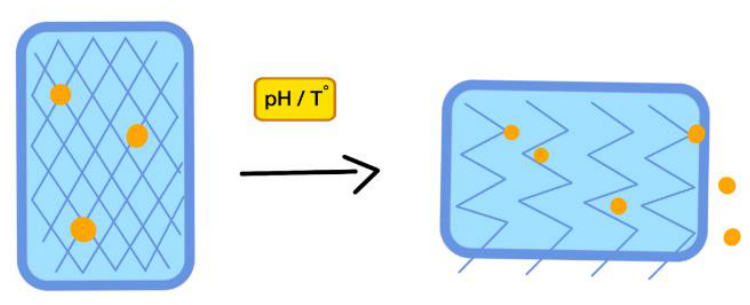

Table 1.
Inclusion and Exclusion Criteria of the Study.
| Category | Inclusion Criteria | Exclusion Criteria | |
| Document Type | Original research articles, scientific reviews, scientific consensus and position statements, and academic book chapters with verifiable scientific information. |
Non-academic publications, abstracts without full text, and documents lacking verifiable scientific support. | |
| Language | Scientific publications written in English. |
Publications in other languages. | |
| Time Frame | Publications between 2008 and 2026. |
Publications outside the established period. | |
| Content | Studies addressing hydrogels, stem cells, extracellular vesicles, and their applications in regenerative medicine, including biological activity, retention, controlled release, stability, safety, or clinical development. |
Studies unrelated to the research objectives or lacking relevant information for the comparative analysis. | |
| Quality | Publications with clearly described methodologies, verifiable scientific findings, or relevant scientific foundations supporting the analysis. | Publications with insufficient methodological information, unverifiable scientific support, or duplicate content. | |
Table 2.
Comparative Analysis of Stem-Cell-Loaded and Extracellular-Vesicle-Loaded Hydrogels.
| Criteria | Stem-Cell-Loaded Hydrogels | EV-Loaded Hydrogels |
| Cell Viability and Biological Activity | Require viable cells capable of responding to environmental signals, producing bioactive factors, and potentially differentiating. Their effectiveness depends on maintaining cellular survival and functionality. | Do not require living therapeutic cells. Their biological activity depends on EV integrity, bioactive cargo, and interaction with recipient cells. |
| Retention and Controlled Release | Hydrogels provide structural support and improve local cell retention. Therapeutic activity depends on cell survival and continued biological function. | Hydrogels improve EV retention and can regulate their release through diffusion, swelling, and degradation. Excessively rapid or limited release may reduce therapeutic availability. |
| Stability and Storage | Require preservation of cell viability, biological function, and hydrogel structure. Cryopreservation and long-term storage remain challenging. | Do not require cell viability but must preserve vesicle integrity, cargo stability, and biological activity. Storage conditions and standardization remain important limitations. |
| Safety and Biological Risks | Potential risks include unwanted differentiation, uncontrolled cellular behavior, immune responses, and complications associated with the hydrogel material. | Avoid risks associated with proliferation and differentiation of administered cells. However, immune responses, EV heterogeneity, inappropriate dosing, and hydrogel-related complications remain possible. |
| Advantages and Limitations | Provide a dynamic biological system capable of responding to the surrounding environment. However, cell survival, biological variability, and differentiation control remain limitations. | Offer a cell-free strategy without requiring living-cell administration. However, EV isolation, characterization, stability, dosing, and delivery efficiency remain challenges. |
| Clinical Applications and Current Development | Investigated for tissue regeneration, wound repair, cartilage, and bone applications. Many combined hydrogel–cell systems remain experimental or in clinical development. | Investigated for wound healing, bone regeneration, inflammatory conditions, and other applications. Evidence for combined hydrogel–EV systems remains predominantly preclinical, with limited clinical validation. |
Note. This table was developed by the authors based on the scientific literature reviewed (Choe et al., 2018; Egorikhina et al., 2020; Ju et al., 2023; Hashemi et al., 2024; Welsh et al., 2024).Stimuli-Responsive Hydrogels (Smart Hydrogels).
Disclaimer/Publisher’s Note: The statements, opinions and data contained in all publications are solely those of the individual author(s) and contributor(s) and not of MDPI and/or the editor(s). MDPI and/or the editor(s) disclaim responsibility for any injury to people or property resulting from any ideas, methods, instructions or products referred to in the content. |
© 2026 by the authors. Licensee MDPI, Basel, Switzerland. This article is an open access article distributed under the terms and conditions of the Creative Commons Attribution (CC BY) license (http://creativecommons.org/licenses/by/4.0/).
Copyright: This open access article is published under a Creative Commons CC BY 4.0 license, which permit the free download, distribution, and reuse, provided that the author and preprint are cited in any reuse.