

Submitted:
19 October 2026
Posted:
21 October 2026
You are already at the latest version
Abstract
Hepatocellular carcinoma (HCC) develops through cooperation between altered hepatocytes, chronic liver injury and a stromal and immune environment. Genomic alterations and signaling abnormalities provide a rationale for treatment development, but pathway activation rarely identifies therapeutic dependence alone. This narrative review connects hepatic stellate-cell states, tumor-cell plasticity, genomic instability and four major signaling pathways with antigen presentation, immune exclusion and clinical treatment evidence. Targeted searches were updated through 6 October 2026, retaining foundational work and prioritizing recent studies. Single-cell and spatial analyses reveal that stellate cells and cancer-associated fibroblasts can exert opposing effects according to their state and location. Similarly, CTNNB1-associated signaling can favor immune exclusion, but mutation status does not fully capture immune context or establish universal resistance to checkpoint combinations. Intratumoral dendritic-cell and lymphocyte organization, suppressive myeloid niches and humoral responses provide complementary explanations of treatment heterogeneity. Randomized trials support selected antiangiogenic and checkpoint combinations in unresectable HCC, while negative trials and updated adjuvant results demonstrate the limits of extrapolating biological rationale across regimens and disease stages. The synthesis distinguishes experimental causality, patient-level association and clinically validated benefit. Translation requires compartment-specific target assessment, representative sampling and prospective biomarker evaluation within a defined treatment setting, alongside assessment of liver reserve.
Keywords:
hepatocellular carcinoma
; hepatic carcinogenesis
; hepatic stellate cells
; genomic instability
; Wnt signaling
; PI3K–AKT–mTOR
; NF-κB
; tumor microenvironment
; immunotherapy
1. Introduction
Hepatocellular carcinoma (HCC) is the predominant form of primary liver cancer and develops most often in a background of chronic hepatic injury. In the GLOBOCAN 2022 estimates, liver cancer ranked sixth for incidence and third for cancer mortality worldwide. These rankings refer to primary liver cancer as a group and should not be attributed exclusively to HCC. The high mortality burden reflects the combination of aggressive tumor biology, frequent diagnosis at an advanced stage and the limitations imposed by underlying liver disease [1].
Chronic hepatitis B virus (HBV) and hepatitis C virus (HCV) infection, alcohol-associated liver disease and metabolic liver disease are major etiological drivers. The increasing burden of obesity and diabetes has strengthened the contribution of metabolic liver disease to HCC, including tumors arising without established cirrhosis [2]. Current terminology distinguishes metabolic dysfunction-associated steatotic liver disease (MASLD) and metabolic dysfunction-associated steatohepatitis (MASH); older publications use nonalcoholic fatty liver disease (NAFLD) and nonalcoholic steatohepatitis (NASH). The terminology has changed together with the diagnostic framework, so historical cohorts should not be assumed to be identical to cohorts defined by current criteria [3]. Earlier projections to 2030 anticipated increasing primary liver cancer burden in many countries, with variation according to sex, geography, risk-factor trends and population aging [4].
Type 2 diabetes mellitus connects systemic metabolic dysfunction with the hepatic processes that favor HCC. Insulin resistance, compensatory hyperinsulinemia and lipotoxic injury provide a plausible context for proliferative signaling, inflammation and fibrosis [2]. In a regional cohort of 137,158 people with type 2 diabetes, 902 HCC cases were identified; higher body mass index and glycated hemoglobin were associated with occurrence alongside viral hepatitis, alcohol exposure and cirrhosis [5]. Metabolic treatment is also being investigated as a prevention strategy. A retrospective cohort of 1,890,020 patients associated glucagon-like peptide-1 receptor agonist use with lower HCC incidence than insulin or sulfonylureas, while the comparison with metformin was not statistically significant [6]. Residual confounding and differences in treatment indication limit causal interpretation, so these findings do not establish these drugs as HCC chemoprevention.
Repeated hepatocyte death and regeneration favor selection of altered clones, while fibrosis, inflammatory signals and impaired immune surveillance support expansion. These interactions can contribute to heterogeneity and resistance to single-target therapy. Systemic treatment now includes checkpoint combinations and antiangiogenic approaches; selection also depends on stage, liver function, performance status and contraindications [7,8].
Recent reviews describe the broad immune ecosystem and its therapeutic opportunities [9]. The present review addresses a narrower interpretative question: why does evidence of oncogenic pathway activity not reliably identify a clinically actionable vulnerability? It connects four major signaling pathways with the state and location of hepatic stellate cell-derived stroma and immune cells and evaluates what each type of evidence can support. The contribution is a critical synthesis across these compartments, with explicit separation of mechanistic causality, patient-level association and treatment benefit.
1.1. Literature Search and Study Selection
This narrative review used structured, targeted searches of PubMed, updated on 6 October 2026. Search terms combined hepatocellular carcinoma with hepatic stellate cells, cancer-associated fibroblasts, genomic alterations, intratumoral heterogeneity, cellular plasticity, Wnt/β-catenin, CTNNB1, PI3K–AKT–mTOR, NF-κB, EGFR, MAPK signaling, natural killer cells, spatial or single-cell profiling, immunotherapy and molecular correlates. Additional searches addressed randomized trials, clinical guidelines, circulating biomarkers, type 2 diabetes and cardiovascular treatment safety. Recent evidence was prioritized using publication-date filters for 2021–2026; earlier foundational studies were retained where they established pathway mechanisms or clinical comparisons. Targeted article-title and DOI searches supplemented relevance-ranked retrieval.
Titles and abstracts were assessed for relevance to hepatocarcinogenesis, tumor–stroma interactions, immune function, therapeutic adaptation or biomarker interpretation. Eligible evidence included peer-reviewed functional studies in HCC models, human molecular and tissue cohorts, randomized trials and clinical guidelines. Reviews supplied context. Evidence from other disease settings was included only to clarify general signaling principles or limitations of biomarker specificity, with its population explicitly identified. Available full texts were examined to assess experimental context and the limits of the reported conclusions.
The qualitative synthesis considered study design, biological compartment, disease or treatment setting, intervention, endpoints and limitations. Mechanistic perturbation, patient-level association and randomized treatment benefit were interpreted separately. Bibliographic metadata were checked against PubMed records and publisher websites, and references were ordered by their first appearance. Selection was targeted rather than exhaustive; no formal risk-of-bias scoring or pooled statistical analysis was performed.
2. Cellular Modulators of Hepatic Carcinogenesis
2.1. Hepatic Stellate Cells and the Fibrotic Niche
Hepatic stellate cells (HSCs) are central effectors of fibrogenesis. Quiescent HSCs store vitamin A and support liver homeostasis; persistent injury promotes a contractile, proliferative and matrix-producing phenotype. Communication with damaged hepatocytes, macrophages, endothelial cells and extracellular matrix (ECM) sustains activation. Collagen accumulation changes tissue architecture and provides mechanical and biochemical signals to neighboring hepatocytes [10].
Activated HSCs produce or respond to transforming growth factor beta (TGF-β), platelet-derived growth factor (PDGF), hepatocyte growth factor (HGF) and other mediators. Immunocompetent mouse experiments demonstrated promotion of HCC development by activated HSCs [11]. Culture, spheroid and implantation models connected HSC-derived signals with tumor-cell proliferation, migration and growth through extracellular signal-regulated kinase (ERK) and nuclear factor kappa B (NF-κB) activity [12]. These findings link stroma with signaling in malignant hepatocytes, while remaining model-dependent.
TGF-β can restrain epithelial growth in some settings while promoting fibrosis, invasion or immunosuppression in established malignancy. Epithelial–mesenchymal transition describes acquisition of mesenchymal traits by epithelial cells; it does not imply that HSCs become malignant hepatocytes. Matrix remodeling and adhesion changes can also facilitate invasion, although specific experimental effects are not universal patient-level mechanisms [10,12].
HSC heterogeneity changes the interpretation of this stromal contribution. Filliol and colleagues combined single-cell analyses with genetic perturbation in mouse models to distinguish cytokine-producing, HGF-enriched HSC states from collagen-producing myofibroblastic states. HSC-derived HGF protected hepatocytes against death and reduced carcinogenesis in these models, whereas collagen promoted tumor development through stiffness-induced activation of the transcriptional coactivator TAZ and, in established tumors, discoidin domain receptor 1. Human associations supported a shift toward tumor-promoting stromal states as liver disease progressed [13]. HGF can therefore have different consequences in injured nonmalignant hepatocytes and established tumor cells. Figure 1 summarizes the injury and stromal interactions without implying that all HSC states promote HCC.
Earlier conditioned-medium experiments also found that quiescent HSCs could induce cancer-cell toxicity and increase doxorubicin sensitivity [14]. These findings remain experimental. In established human HCC, multimodal profiling subsequently distinguished spatially enriched cancer-associated fibroblast (CAF) subsets designated CAF-FAP and CAF-C7 associated with different stromal architectures and opposing outcomes. Combining CAF-FAP targeting with programmed cell death protein 1 (PD-1) blockade improved tumor regression in orthotopic mouse models [15]. The evidence favors selective characterization of stromal states; indiscriminate fibroblast depletion could remove protective functions, and the therapeutic findings require validation in patients.
2.2. Cancer Stem Cell Phenotypes and Cellular Plasticity
Cancer stem cells are functionally defined by self-renewal and sustained tumor propagation. Stem-like HCC states are associated with heterogeneity and resistance, but their phenotype does not establish the normal cell of origin. Both progenitor-cell transformation and acquisition of stem-like properties by differentiated cells are plausible routes [16].
Oncogenic manipulation of different hepatic cell types generated primary liver tumors with tumor-propagating properties in lineage-specific models [17]. These experiments support cellular plasticity without establishing a single origin for human HCC. The acquired alterations and surrounding niche can maintain resistant cell states, suggesting that control of proliferation alone may be insufficient for durable treatment.
Recent functional studies clarify the treatment specificity of this plasticity. In HCC models, chemotherapy-induced serine peptidase inhibitor Kazal type 1 (SPINK1) supported dedifferentiation, self-renewal and resistance through an EGFR–ERK–CDK4/6–E2F2 axis. SPINK1 inhibition increased sensitivity to the tested cytotoxic drugs, while the reported sorafenib experiments did not show the same benefit [18]. This supports a defined resistance mechanism rather than a universal consequence of stemness.
Plasticity can also restrain tumor initiation. Experiments that trapped injured mouse hepatocytes in partially reprogrammed states found reduced proliferation in response to mitogenic and oncogenic stimuli [19]. Such protective states in regenerating nonmalignant hepatocytes should be distinguished from stem-like states within established HCC. The direction of the effect depends on cellular identity, injury context and disease stage.
3. Genetic Alterations and Genomic Instability
3.1. Copy Number Changes and Allelic Loss
HCC contains somatic sequence variants, structural alterations and recurrent chromosomal gains or losses. Copy number studies identified candidate cancer genes and recurrent gains on chromosome 1q, including the 1q21 region [20,21]. CHD1L amplification has experimental oncogenic effects, while expression in resected tumors has been associated with adverse postoperative outcomes [22,23]. Chromosome 8q gain can increase MYC dosage; a fluorescence in situ hybridization study examined MYC amplification in relation to early relapse [24]. These observations support biological or prognostic hypotheses, without establishing drug-selection assays.
Broad chromosomal changes can influence several cooperating genes. Comparative genomic hybridization and integrative analyses of chromosome 8p linked regional abnormalities to tumor characteristics, progression or survival [25,26]. An allelic loss does not establish reduced expression of every gene in the region, and a regional association cannot assign causality to one locus. Cohort composition and assay definitions also limit direct comparison of alteration frequencies.
Recent whole-genome evidence expands this framework beyond coding mutations. The Chinese Liver Cancer Atlas analyzed 494 tumors at high sequencing depth, identifying additional coding and noncoding driver candidates, HBV integration within extrachromosomal circular DNA and subclonal structural events [27]. An independent analysis of HBV integration combined cross-cohort genomic mapping with mechanistic evaluation of telomerase reverse transcriptase (TERT) activation [28]. Whole-genome and long-read analyses of 124 HCCs also linked HBV integration with complex rearrangements and driver copy-number changes; genetic experiments supported the tumorigenic potential of selected integration-related amplifications [29]. These findings connect etiology with genome structure and clonal selection, while candidate-driver status and prognostic associations remain distinct from a validated therapeutic vulnerability.
3.2. Recurrent Driver Alterations
Whole-genome and exome sequencing have identified recurrent alterations in telomere maintenance, cell-cycle regulation, Wnt signaling, chromatin remodeling and oxidative-stress responses. Whole-genome sequencing identified recurrent mutations beyond the previously established HCC genes [30]. TERT promoter mutations are particularly relevant because they can occur during the transition from premalignant nodules to HCC in cirrhotic liver, providing evidence that telomerase reactivation is an early event in at least a subset of tumors [31].
Large sequencing studies confirmed the recurrent involvement of TP53, CTNNB1 and chromatin regulators, while identifying additional pathways and mutational signatures [32,33]. Integrated analysis of somatic mutations and focal copy number changes demonstrated that different genomic events could converge on the same functional networks [34]. The Cancer Genome Atlas extended this approach by integrating multiple molecular measurements, reinforcing the heterogeneity of HCC and the value of combining genomic, transcriptional and other data [35].
TP53 alterations can impair cell-cycle arrest, DNA-damage responses and apoptosis, allowing damaged clones to persist. CTNNB1 mutations can stabilize β-catenin and alter differentiation and transcriptional programs, whereas AXIN1 alterations disrupt a different component of the Wnt regulatory machinery. ARID1A and ARID2 affect chromatin regulation; NFE2L2 and KEAP1 influence the oxidative-stress response. Less frequent alterations affecting JAK1 or RPS6KA3 connect HCC with cytokine and mitogen signaling. Their interpretation requires distinction between an activating alteration, an inactivating alteration and a change of uncertain functional significance [32,34].
Table 1 summarizes recurrent gene groups without assigning universal mutation frequencies or deterministic outcomes. Established genomic classifications and recent deep sequencing show that tumor phenotype depends on cooperating alterations and on the population being studied [27,36]. Spatial sampling adds another limitation: sequencing 235 sectors from 13 HCCs revealed regional blocks with different genomic and transcriptional features [37]. A single biopsy can therefore miss relevant intratumoral variation, although the optimal clinical sampling strategy remains unresolved.
4. Major Signaling Pathways in Hepatic Carcinogenesis
4.1. Wnt/β-Catenin Signaling
Canonical Wnt signaling regulates liver development, homeostasis and regeneration and is frequently dysregulated in HCC. In the absence of ligand-dependent stimulation, cytoplasmic β-catenin is phosphorylated by a destruction complex containing AXIN, adenomatous polyposis coli protein (APC), casein kinase 1 (CK1) and glycogen synthase kinase 3 (GSK3). Phosphorylated β-catenin is recognized by the ubiquitination machinery and directed toward proteasomal degradation. This limits its availability for nuclear transcriptional signaling [38].
Wnt binding to Frizzled and LRP5/6 activates Dishevelled-dependent signaling, reduces destruction-complex activity and permits β-catenin accumulation. Nuclear β-catenin interacts with T-cell factor/lymphoid enhancer factor (TCF/LEF) transcription factors to change target-gene expression. Figure 2 contrasts these states and shows how mutations can bypass upstream regulation.
Mutations affecting CTNNB1 can impair the normal degradation of β-catenin. However, Wnt-related abnormalities are not biologically uniform, and AXIN1-mutant tumors should not automatically be treated as equivalent to CTNNB1-mutant tumors. Transcriptomic and protein-level analyses identified distinct Wnt-associated HCC classes, including a CTNNB1 class and a class associated with Wnt–TGF-β signaling [39]. The resulting phenotypes therefore depend on the route of pathway activation and on cooperating alterations.
The relationship between Wnt activation and immunity depends on the tumor context. A genetically engineered mouse study linked β-catenin activation with deficient dendritic-cell recruitment, reduced T-cell activity and anti-PD-1 resistance [40]. Human immunogenomic analysis subsequently identified both immune-excluded CTNNB1-enriched tumors and CTNNB1-mutant tumors with weaker Wnt activation or prominent interferon and antigen-presentation programs [41]. This supports a mechanism of immune exclusion without making CTNNB1 mutation a universal resistance biomarker. Mutation status, transcriptional output and the surrounding immune architecture measure related but nonidentical features; none should be substituted uncritically for a treatment-specific clinical assay.
Interventions can target ligand production, receptors or the nuclear transcriptional complex. TCF4/β-catenin antagonists inhibited HCC growth experimentally [42]; Frizzled antibodies showed preclinical activity across tumor types [43]. Patient benefit in HCC remains unestablished. Normal tissue functions and ligand-independent activation also limit extrapolation from upstream inhibition.
4.2. PI3K–AKT–mTOR Signaling
The phosphatidylinositol 3-kinase (PI3K)–AKT–mechanistic target of rapamycin (mTOR) pathway coordinates growth-factor responses, cellular survival and metabolism. Activated receptor tyrosine kinases can stimulate PI3K, which converts phosphatidylinositol 4,5-bisphosphate (PIP2) into phosphatidylinositol 3,4,5-trisphosphate (PIP3). PIP3 recruits proteins with pleckstrin-homology domains, including AKT, to the membrane. AKT activation involves phosphorylation by phosphoinositide-dependent kinase 1 and mTOR complex 2 (mTORC2). The lipid phosphatase PTEN opposes this signaling by converting PIP3 back to PIP2 [44]. The two signaling branches are summarized in Figure 3.
AKT regulates survival and metabolism and promotes mTORC1 activity through TSC–RHEB regulation. S6 kinase and 4E-binding protein phosphorylation support protein synthesis and growth. mTORC1 and mTORC2 have different substrates and functions.
Growth-factor inputs and PTEN downregulation can increase activity without mutations in each pathway component. Tissue and experimental studies linked PI3K–PTEN–AKT–mTOR signaling to invasion and matrix metalloproteinase expression [45]; PTEN loss was connected to an AKT–Sp1–matrix metalloproteinase 2 mechanism [46]. Expression or phosphorylation therefore measures different features from DNA sequencing.
Mechanical and metabolic inputs provide further routes to pathway activity. Matrix stiffness increased integrin β1–PI3K–AKT signaling and glucose-6-phosphate dehydrogenase expression in HCC models. Stiffer tumors were associated with sorafenib resistance in a 30-patient tissue analysis, while matrix and pathway perturbation improved drug sensitivity experimentally [47]. These findings connect stroma with metabolic adaptation, without establishing a clinical stiffness-based selection assay.
β-Catenin-driven models linked glutamine synthetase and glutamine availability with mTORC1 activation and showed sensitivity to mTORC1 inhibition [48]. However, a separate study found that hepatic glutamine synthetase ablation impaired ammonia handling, increased nonessential amino-acid production and accelerated mTORC1-dependent tumorigenesis [49]. The genetic manipulations and metabolic contexts differed. Enzyme abundance therefore cannot be assumed to establish that inhibiting the enzyme will benefit every tumor.
In the phase III EVOLVE-1 trial, everolimus did not improve overall survival after sorafenib failure [50]. The result separates biological pathway involvement from demonstrated therapeutic dependence in an unselected population. Biomarker selection, target engagement, feedback and parallel signaling remain relevant to future approaches.
4.3. NF-κB Signaling
Nuclear factor kappa B (NF-κB) connects inflammatory stimuli with transcriptional programs controlling immunity, survival and tissue repair. The family includes p50, p52, RelA/p65, RelB and c-Rel. Its members form different dimers, with activity determined by inhibitory proteins, upstream kinases and the cellular context. The canonical and noncanonical pathways have distinct initiation mechanisms and outputs, although they can interact within inflammatory and immune responses [51]. Figure 4 contrasts the two signaling branches.
Canonical stimuli include tumor necrosis factor, interleukin 1 and Toll-like receptor ligands. Receptor-specific cascades activate the IκB kinase machinery, commonly involving IKKβ and NEMO. IκB phosphorylation, ubiquitination and degradation release dimers such as p50–RelA for nuclear transcription of inflammatory and survival programs. The upstream steps vary by receptor.
Noncanonical stimuli include lymphotoxin beta receptor, BAFF receptor, CD40 and RANK. NIK stabilization activates IKKα and p100 processing into p52, enabling p52–RelB nuclear signaling in lymphoid organization and immune-cell differentiation [52].
NF-κB has compartment-dependent effects: hepatocyte survival can limit injury, while persistent macrophage or HSC signaling can sustain inflammation and fibrosis [53]. In a chemical carcinogenesis model, hepatocyte-restricted IKKβ deletion increased cell death and compensatory proliferation, whereas deletion extending to hematopoietic-derived cells reduced cytokine-driven tumor development [54]. These experiments caution against extrapolating a cell-specific effect to systemic inhibition in established human HCC.
Tumor-intrinsic noncanonical signaling can also support adaptation. DDX5-deficient HCC models linked β-catenin activity with NIK–p52/RelB activation and nuclear factor erythroid 2-related factor 2 (NRF2)-dependent antioxidant responses, facilitating escape from sorafenib-induced ferroptosis [55]. Ferroptosis is an iron-dependent form of cell death driven by lipid peroxidation. The findings connect Wnt, inflammatory transcription and oxidative-stress control, while the proposed therapeutic dependency remains preclinical.
4.4. RAS–RAF–MEK–ERK Signaling and Pathway Cooperation
RAS–RAF–MEK–ERK signaling transmits extracellular growth signals to proliferation and differentiation programs. Activated receptor tyrosine kinases recruit adaptors and exchange factors that generate RAS-GTP; RAF, MEK and ERK then transmit the signal. Upstream inputs or regulatory changes can increase activity in HCC without classical RAS or RAF mutations [44].
HSC-derived mediators activate ERK in HCC cells, connecting the fibrotic niche with proliferation and migration [12]. Receptors can also stimulate PI3K–AKT, while outputs cooperate with Wnt and inflammatory programs. Parallel signaling may therefore maintain survival during inhibition of one branch. A study combining a kinome-focused CRISPR screen with mechanistic experiments identified feedback activation of EGFR–PAK2–ERK5 after FGFR inhibition by lenvatinib. EGFR blockade restored sensitivity in experimental HCC models, with preliminary observations in 12 patients [56]. ERK5 is distinct from the canonical ERK1/2 branch, and the small nonrandomized patient series does not establish routine combination benefit.
Sorafenib targets RAF-related signaling and angiogenic receptor kinases, so its clinical effects cannot be assigned to ERK alone. Similarly, increased phospho-ERK may indicate adaptation without defining the sole resistance mechanism. Genomic, protein and longitudinal measurements must be interpreted together [36,44].
5. The Immune Microenvironment
5.1. Antitumor Immunity and Immune Escape
The liver must balance defense against pathogens with tolerance to repeated exposure to portal blood-derived antigens. HCC develops within this specialized immune environment and can exploit its regulatory mechanisms. Antitumor immunity requires antigen recognition, effective presentation, lymphocyte recruitment and preservation of effector function. CD8-positive T cells recognize antigenic peptides presented by major histocompatibility complex (MHC) class I molecules and can kill target cells through perforin–granzyme and death-receptor pathways [57].
Tumor-associated antigens studied in HCC include alpha-fetoprotein (AFP), glypican 3 and cancer–testis antigens. Patient studies found variation in antigen expression, antigen-specific responses and the functional competence of tumor-reactive CD8-positive T cells [58,59,60]. Antigen availability therefore does not guarantee effective tumor control. More recent neoadjuvant anti-PD-1 analyses linked response in T-cell-rich tumors to the organization of dendritic cells, CXCL13-positive CD4 helper cells and progenitor or effector-like CD8 cells. Nonresponders were enriched in terminally exhausted CD8 states [61]. These human correlates suggest that local differentiation niches can matter even when T cells reach the tumor.
Natural killer (NK) cells integrate activating and inhibitory inputs. NKG2D detects stress-associated ligands, while inhibitory killer-cell immunoglobulin-like receptors recognize self MHC class I. Loss of inhibitory signals can favor killing, but activating signals and cytokine context remain necessary. Local suppression of NK function weakens this complementary surveillance mechanism [57].
Functional studies illustrate how inhibitory context can limit antigen-directed killing. Disrupting TGF-β receptor signaling improved the activity of human induced pluripotent stem cell-derived NK cells in cell-line and HepG2 xenograft models; antigen-directed receptors alone were insufficient under the tested conditions [62]. A recent study combined human tissue profiling with mouse perturbation to implicate Nur77 in NK activation. Nur77 agonism reduced CD36 expression and increased resistance to oxidized-lipid suppression [63]. Both approaches remain preclinical.
B cells and plasma cells provide another component of this immune organization. A recent study associated neoadjuvant PD-1 response with expanded IgG1-positive plasma-cell populations and tumor-antigen-directed humoral responses, supported by additional cohorts [64]. The discovery cohort included 27 treated patients and 11 untreated controls. These data extend the immune framework beyond T-cell abundance, but they do not establish a routine serum-antibody test for selecting treatment.
5.2. Suppressive Myeloid Cells and Regulatory Lymphocytes
Tumor-associated macrophages influence angiogenesis, matrix remodeling and immune regulation. High macrophage density was associated with poor outcomes after HCC resection in an observational tissue study [65]. However, macrophage states form a continuum, and the traditional M1/M2 terminology cannot capture every functional state within a tumor. Density alone does not establish which macrophage population is responsible for progression or whether it is a predictive biomarker for a particular treatment. Figure 5 summarizes selected effector and suppressive relationships; it does not depict every immune population or treatment mechanism.
Myeloid-derived suppressor cells provide another route to diminished antitumor immunity. A study of patients with HCC identified increased CD14-positive, HLA-DR-low myeloid cells and examined their relationship to prognosis [66]. Regulatory T cells, suppressive cytokines and dysfunctional dendritic cells can further limit effector-cell activity. Their effects interact with stromal signals, altered metabolism and inhibitory checkpoint signaling. These processes therefore need to be interpreted as a coordinated microenvironment rather than as isolated changes in one immune-cell population [57].
Spatial analysis of post-chemoembolization tumors from 108 patients connected programmed death ligand 1 (PD-L1)-positive macrophage neighborhoods with stem-like malignant cells, CD8 exhaustion and poorer outcomes. Functional experiments implicated macrophage-derived TGF-β1 in residual tumor persistence; combined PD-L1 and TGF-β blockade eliminated residual stem-like cells in two mouse models [67]. This links tumor plasticity with a treatment-conditioned myeloid niche. The intervention remains preclinical, and the post-chemoembolization setting should not be assumed to represent untreated HCC or postoperative microscopic disease.
Etiology can further alter myeloid behavior. In experimental NASH-associated HCC, CXCR2 inhibition combined with PD-1 blockade reprogrammed tumor-associated neutrophils, increased dendritic-cell and CD8 activity and improved tumor control [68]. These results support context-specific myeloid targeting rather than simple depletion of every infiltrating neutrophil. Historical NASH model definitions and preclinical responses should not be used to exclude patients with MASLD or MASH from otherwise indicated immunotherapy.
5.3. Spatial Organization and Stromal Barriers
Immune-cell abundance alone does not show whether lymphocytes can reach malignant cells or maintain productive interactions. Imaging mass cytometry of tumors from 101 patients distinguished depleted, compartmentalized and enriched spatial immunotypes; the enriched pattern was associated with better progression-free survival under checkpoint therapy [69]. These findings complement the dendritic-cell and helper-cell niches described above. They are candidate biomarkers of tissue organization, requiring standardized measurement and prospective evaluation before directing routine treatment.
Stroma can influence access through cell-specific signaling as well as matrix deposition. Exploratory spatial profiling with a larger histological cohort associated peritumoral antigen-presenting CAF features and CXCL12–CXCR4-related communication with peripheral CD8 accumulation and poorer outcomes [70]. The analysis was correlational, and multicellular spatial spots limited assignment of ligand–receptor signals to individual cells. Another recent study implicated endothelial DLL4–NOTCH3 communication in matrix-producing CAF states and found improved immune infiltration and checkpoint responses after pathway perturbation in HCC models [71]. However, the gamma-secretase inhibitor used was not NOTCH3-specific, so its effects cannot be assigned exclusively to that receptor.
Together, these observations support investigation of anatomically and functionally defined stromal barriers. CAF nomenclature, spatial platforms and disease contexts differ across studies; similarly named subsets cannot automatically be treated as interchangeable. A high cytokine level also does not identify its cellular source. Therapeutic proposals should establish which compartment supplies the mediator, where the interaction occurs and whether selective perturbation preserves liver repair and useful immune functions.
6. Therapeutic Implications and Translational Limits
6.1. Clinical Evidence for Complementary Targets
Randomized trials demonstrate benefit from selected combinations in unresectable HCC. In IMbrave150, atezolizumab plus bevacizumab improved outcomes compared with sorafenib in previously untreated disease [72]. Integrated molecular analyses of 358 patients from GO30140 and IMbrave150 associated pre-existing T-cell immunity with better outcomes, while Treg-to-effector balance and oncofetal programs marked reduced benefit. Differences between the combination and atezolizumab alone were also associated with angiogenic and myeloid signatures [73]. These analyses support complementary immune and vascular effects, but the exploratory signatures are not established companion diagnostics. A subsequent single-cell-derived analysis of 422 RNA-sequencing samples distinguished immune-competent and angiogenesis-driven patterns of benefit, while resistance was associated with suppressive myeloid and Notch programs [74]. Treatment-interaction analyses supplied exploratory predictive evidence. However, the trial-linked datasets partly overlap earlier analyses, and prospective assay standardization and independent validation remain necessary.
The HIMALAYA trial established an alternative immune-based approach using a single priming dose of the cytotoxic T-lymphocyte-associated protein 4 (CTLA-4) inhibitor tremelimumab with durvalumab. The STRIDE regimen improved overall survival compared with sorafenib, with median survival of 16.43 versus 13.77 months in the trial population [75]. CheckMate 9DW subsequently reported improved overall survival with nivolumab plus ipilimumab compared with lenvatinib or sorafenib as first-line treatment for unresectable HCC [76]. These trials support complementary checkpoint targeting but also underline the need to weigh benefit against immune-mediated toxicity.
Therapeutic biomarkers must be judged according to the setting in which they were validated. REACH-2 demonstrated a survival benefit with ramucirumab after sorafenib in patients with AFP concentrations of at least 400 ng/mL [77]. This is a clinically defined enrichment strategy, not evidence that AFP directly identifies a particular DNA mutation. Conversely, the negative EVOLVE-1 result shows that targeting an active pathway without a validated selection strategy can fail to improve survival [50]. Table 2 contrasts clinically demonstrated approaches with experimental pathway-directed strategies.
The success of one combination does not validate every regimen or disease setting. LEAP-002 did not meet its prespecified significance thresholds for overall or progression-free survival when pembrolizumab was added to lenvatinib [78]. In the updated 2026 IMbrave050 analysis after resection or ablation, the initial recurrence-free survival benefit of adjuvant atezolizumab plus bevacizumab was not sustained (hazard ratio 0.90, 95% confidence interval 0.72–1.12); overall survival remained immature [79]. These findings distinguish first-line unresectable disease from adjuvant treatment and illustrate why mechanistic complementarity requires regimen-specific randomized confirmation.
6.2. Toward Functionally Informed Precision Treatment
A clinically useful biomarker must be measured reproducibly and evaluated in the treatment setting for which it is intended. An association with recurrence is prognostic; evidence that treatment benefit differs according to biomarker status is predictive. The 2025 TIMES study combined the spatial distribution of five markers into a recurrence-risk model, with multicohort validation and supporting NK-cell experiments [80]. This is evidence for risk stratification, rather than proof that a particular systemic therapy should be selected. Likewise, CHD1L, MYC and PTEN findings should not be promoted from biological or prognostic observations to drug-selection tests [23,24,46].
Circulating stromal markers also require biological specificity. Serum syndecan-1 was associated with outcomes in patients with HCC and alcoholic cirrhosis [81]. A separate prospective heart-failure cohort associated the same analyte with kidney and liver dysfunction [82]. This evidence from a different disease setting illustrates why a circulating remodeling signal cannot automatically be attributed to the tumor. HCC biomarker studies should account for concurrent organ injury and validate added value against established clinical assessment.
Combination strategies can address cooperating mechanisms, but they also introduce toxicity and must preserve sufficient liver function. Most pivotal systemic-treatment trials enrolled selected patients with preserved liver reserve; their findings have practical limits in decompensated cirrhosis. Guidance incorporates liver function, performance status, bleeding risk and other contraindications into treatment selection [7,8]. A molecular signature should add value to this clinical assessment. It cannot replace assessment of competing risks from tumor progression and underlying liver disease.
Cardiovascular disease adds a further constraint to the translation of HCC treatment mechanisms into clinical benefit. Inhibition of vascular endothelial growth factor (VEGF) signaling with bevacizumab or VEGF-targeting tyrosine kinase inhibitors can cause hypertension and vascular events, and some agents can impair left ventricular function. Immune checkpoint inhibitors carry a risk of myocarditis with potentially serious arrhythmias or conduction abnormalities. The 2022 European Society of Cardiology cardio-oncology guidelines support baseline clinical assessment and electrocardiography, with echocardiography and cardiac biomarkers selected according to cardiovascular risk and the planned regimen. Blood-pressure control and surveillance for new cardiac symptoms should accompany treatment, with prompt evaluation when toxicity is suspected. Cardiologist involvement is particularly relevant for patients with pre-existing cardiovascular disease, allowing oncological benefit, cardiac reserve and hepatic function to be considered together [83].
Tumor-derived models can test drug dependence, and paired tumor–stroma analyses can localize pathway activity. Longitudinal sampling helps distinguish baseline resistance from adaptation. Genomic, transcriptional, protein and immune measurements should inform independently validated, prospective biomarker studies [35,36].
7. Discussion
The combined evidence explains why a pathway catalogue has limited predictive power. A genomic alteration can establish one route to signaling, while stromal mediators activate the same pathway without that alteration. A transcriptomic signature can reflect several cell populations, and a lymphocyte-rich specimen can still lack productive antigen-presentation niches. Therapeutic dependence is therefore a separate question from mutation prevalence, pathway activity or immune-cell abundance. Table 3 summarizes the interpretation of representative findings without assigning a formal evidence grade.
7.1. Connecting Mechanisms with Treatment Hypotheses
The Wnt–immune connection illustrates the need to connect evidence across compartments. β-Catenin perturbation supplies experimental causality, human immunogenomic studies identify context-dependent immune exclusion, and trial-linked tissue analyses evaluate correlates within a defined regimen [40,41,73]. The resulting hypothesis is stronger than any one observation, yet it still does not establish a universal CTNNB1-based rule for treatment selection. A prospective test would need a prespecified assay, representative sampling and evidence that the biomarker changes relative benefit between clinically relevant alternatives.
Stromal studies expose a parallel problem. HSC-derived signals can protect injured hepatocytes during tumor initiation, while collagen-rich states or fibroblast–vascular communication support malignant growth or immune exclusion [13,15,71]. Thus, the same broad cell category may contain a therapeutic target and a protective population. The location, disease stage and mediator should define the proposed intervention. Molecular perturbation must also separate receptor-specific effects from broader pharmacological activity, as highlighted by nonselective NOTCH inhibition.
7.2. Priorities for Translational Studies
Translational studies should test a defined dependency. Spatial profiling can identify the relevant neighborhood; tumor–stroma cocultures, organoids and immune-competent models can examine selective disruption. Models should incorporate background liver disease when feasible and distinguish cancer prevention from treatment of established tumors. Serial sampling can separate pre-existing barriers from adaptation [37,67,68].
Clinical biomarker development should then use a prespecified measurement, account for within-tumor sampling variability and validate performance independently. Studies proposing a predictive assay need treatment-specific evaluation; prognostic accuracy alone is insufficient. Randomized negative trials and mature follow-up should remain part of the interpretation, because plausible targets and early response signals can fail to yield durable benefit [50,78,79]. Endpoints should also account for liver decompensation and treatment tolerability when translating a molecular strategy into patient benefit.
7.3. Limitations of the Review
This narrative review used structured, targeted PubMed searches and qualitative study selection. Relevance ranking and full-text availability can influence selection; no formal risk-of-bias assessment or pooled estimate was produced. Experimental models, resection cohorts, post-locoregional specimens and advanced-disease trials represent different settings. Spatial platforms vary in resolution, and ligand–receptor inference does not establish causality. These differences limit comparison. The focus on four pathways excludes a comprehensive account of epigenetics, noncoding RNAs, locoregional therapies and emerging systemic treatments.
8. Conclusions
Hepatocellular carcinogenesis results from cooperation between genetically altered hepatocytes, chronic liver injury and changing stromal and immune states. Wnt/β-catenin, PI3K–AKT–mTOR, NF-κB and RAS–RAF–MEK–ERK signaling provide important mechanistic links, but their clinical meaning depends on where they are active, and which functions remain required for tumor survival. Recent single-cell and spatial evidence shows why total fibroblast or lymphocyte abundance cannot capture this context.
The principal translational task is to demonstrate a treatment-relevant dependency in a defined cellular compartment and disease setting. Selective targeting of stromal or immune barriers is a rational experimental direction; routine selection requires prospective biomarker and clinical validation. Positive unresectable-HCC trials, negative combinations and updated adjuvant results should be interpreted together. Connecting these levels of evidence, while accounting for liver reserve, offers a practical basis for developing more durable and biologically informed treatment strategies.
Author Contributions
Conceptualization, A.-F.O. and M.F.; methodology, M.L., G.B. and I.-L.M.; validation, M.L., G.B. and I.-L.M.; investigation, A.B., A.V. and G.C.; resources, A.B., A.V. and G.C.; data curation, A.R., R.M. and D.M.T.; writing—original draft preparation, A.-F.O.; writing—review and editing, A.-F.O. and M.F.; visualization, A.R., R.M. and D.M.T.; supervision, G.B., E.C. and M.F.; project administration, A.-F.O., E.C. and M.F. All authors have read and agreed to the published version of the manuscript.
Funding
This research received no external funding.
Institutional Review Board Statement
Not applicable.
Informed Consent Statement
Not applicable.
Data Availability Statement
No new research datasets were generated.
Conflicts of Interest
The authors declare no conflicts of interest.
Abbreviations
AFP, alpha-fetoprotein; CAF, cancer-associated fibroblast; CTLA-4, cytotoxic T-lymphocyte-associated protein 4; ECM, extracellular matrix; ERK, extracellular signal-regulated kinase; HBV, hepatitis B virus; HCC, hepatocellular carcinoma; HCV, hepatitis C virus; HSC, hepatic stellate cell; IKK, IκB kinase; MASLD, metabolic dysfunction-associated steatotic liver disease; MASH, metabolic dysfunction-associated steatohepatitis; MDSC, myeloid-derived suppressor cell; MEK, mitogen-activated protein kinase kinase; mTOR, mechanistic target of rapamycin; NF-κB, nuclear factor kappa B; NK, natural killer; TAM, tumor-associated macrophage; Treg, regulatory T cell; PD-1, programmed cell death protein 1; PD-L1, programmed death ligand 1; PI3K, phosphatidylinositol 3-kinase; TGF-β, transforming growth factor beta; VEGF, vascular endothelial growth factor.
References
- Bray, F.; Laversanne, M.; Sung, H.; Ferlay, J.; Siegel, R.L.; Soerjomataram, I.; Jemal, A. Global cancer statistics 2022: GLOBOCAN estimates of incidence and mortality worldwide for 36 cancers in 185 countries. CA Cancer J. Clin. 2024, 74, 229–263. [Google Scholar] [CrossRef] [PubMed]
- Huang, D.Q.; El-Serag, H.B.; Loomba, R. Global epidemiology of NAFLD-related HCC: trends, predictions, risk factors and prevention. Nat. Rev. Gastroenterol. Hepatol. 2021, 18, 223–238. [Google Scholar] [CrossRef] [PubMed]
- Rinella, M.E.; Lazarus, J.V.; Ratziu, V.; Francque, S.M.; Sanyal, A.J.; Kanwal, F.; Romero, D.; Abdelmalek, M.F.; Anstee, Q.M.; Arab, J.P.; et al. A multisociety Delphi consensus statement on new fatty liver disease nomenclature. J. Hepatol. 2023, 79, 1542–1556. [Google Scholar] [CrossRef] [PubMed]
- Valery, P.C.; Laversanne, M.; Clark, P.J.; Petrick, J.L.; McGlynn, K.A.; Bray, F. Projections of primary liver cancer to 2030 in 30 countries worldwide. Hepatology 2018, 67, 600–611. [Google Scholar] [CrossRef] [PubMed]
- Giorda, C.B.; Picariello, R.; Tartaglino, B.; Nada, E.; Costa, G.; Manti, R.; Monge, L.; Gnavi, R. Hepatocellular carcinoma in a large cohort of type 2 diabetes patients. Diabetes Res. Clin. Pract. 2023, 200, 110684. [Google Scholar] [CrossRef] [PubMed]
- Wang, L.; Berger, N.A.; Kaelber, D.C.; Xu, R. Association of GLP-1 Receptor Agonists and Hepatocellular Carcinoma Incidence and Hepatic Decompensation in Patients With Type 2 Diabetes. Gastroenterology 2024, 167, 689–703. [Google Scholar] [CrossRef] [PubMed]
- Gordan, J.D.; Kennedy, E.B.; Abou-Alfa, G.K.; Beal, E.; Finn, R.S.; Gade, T.P.; Goff, L.; Gupta, S.; Guy, J.; Hoang, H.T.; et al. Systemic Therapy for Advanced Hepatocellular Carcinoma: ASCO Guideline Update. J. Clin. Oncol. 2024, 42, 1830–1850. [Google Scholar] [CrossRef] [PubMed]
- European Association for the Study of the Liver. EASL Clinical Practice Guidelines on the management of hepatocellular carcinoma. J. Hepatol. 2025, 82, 315–374. [Google Scholar] [CrossRef] [PubMed]
- Seyhan, D.; Allaire, M.; Fu, Y.; Conti, F.; Wang, X.W.; Gao, B.; Lafdil, F. Immune microenvironment in hepatocellular carcinoma: from pathogenesis to immunotherapy. Cell. Mol. Immunol. 2025, 22, 1132–1158. [Google Scholar] [CrossRef] [PubMed]
- Tsuchida, T.; Friedman, S.L. Mechanisms of hepatic stellate cell activation. Nat. Rev. Gastroenterol. Hepatol. 2017, 14, 397–411. [Google Scholar] [CrossRef] [PubMed]
- Zhao, W.; Zhang, L.; Yin, Z.; Su, W.; Ren, G.; Zhou, C.; You, J.; Fan, J.; Wang, X. Activated hepatic stellate cells promote hepatocellular carcinoma development in immunocompetent mice. Int. J. Cancer 2011, 129, 2651–2661. [Google Scholar] [CrossRef] [PubMed]
- Amann, T.; Bataille, F.; Spruss, T.; Mühlbauer, M.; Gäbele, E.; Schölmerich, J.; Kiefer, P.; Bosserhoff, A.K.; Hellerbrand, C. Activated hepatic stellate cells promote tumorigenicity of hepatocellular carcinoma. Cancer Sci. 2009, 100, 646–653. [Google Scholar] [CrossRef] [PubMed]
- Filliol, A.; Saito, Y.; Nair, A.; Dapito, D.H.; Yu, L.X.; Ravichandra, A.; Bhattacharjee, S.; Affo, S.; Fujiwara, N.; Su, H.; et al. Opposing roles of hepatic stellate cell subpopulations in hepatocarcinogenesis. Nature 2022, 610, 356–365. [Google Scholar] [CrossRef] [PubMed]
- Das, D.; Fayazzadeh, E.; Li, X.; Koirala, N.; Wadera, A.; Lang, M.; Zernic, M.; Panick, C.; Nesbitt, P.; McLennan, G. Quiescent hepatic stellate cells induce toxicity and sensitivity to doxorubicin in cancer cells through a caspase-independent cell death pathway: Central role of apoptosis-inducing factor. J. Cell. Physiol. 2020, 235, 6167–6182. [Google Scholar] [CrossRef] [PubMed]
- Cheng, Y.; Chen, X.; Feng, L.; Yang, Z.; Xiao, L.; Xiang, B.; Wang, X.; Liu, D.; Lin, P.; Shi, J.; et al. Stromal architecture and fibroblast subpopulations with opposing effects on outcomes in hepatocellular carcinoma. Cell Discov. 2025, 11, 1. [Google Scholar] [CrossRef] [PubMed]
- Anfuso, B.; El-Khobar, K.E.; Sukowati, C.H.C.; Tiribelli, C. The multiple origin of cancer stem cells in hepatocellular carcinoma. Clin. Res. Hepatol. Gastroenterol. 2015, 39 (Suppl. 1), S92–S97. [Google Scholar] [CrossRef] [PubMed]
- Holczbauer, Á.; Factor, V.M.; Andersen, J.B.; Marquardt, J.U.; Kleiner, D.E.; Raggi, C.; Kitade, M.; Seo, D.; Akita, H.; Durkin, M.E.; et al. Modeling pathogenesis of primary liver cancer in lineage-specific mouse cell types. Gastroenterology 2013, 145, 221–231. [Google Scholar] [CrossRef] [PubMed]
- Man, K.F.; Zhou, L.; Yu, H.; Lam, K.H.; Cheng, W.; Yu, J.; Lee, T.K.; Yun, J.P.; Guan, X.Y.; Liu, M.; et al. SPINK1-induced tumor plasticity provides a therapeutic window for chemotherapy in hepatocellular carcinoma. Nat. Commun. 2023, 14, 7863. [Google Scholar] [CrossRef] [PubMed]
- Strathearn, L.S.; Hayata, Y.; Illendula, A.; Hewett, C.K.; Chen, M.; He, G.; Escribano-Cebrián, M.; Jarboe, B.; Gómez-Tomé, L.; de Prisco, N.; et al. Plastic hepatocyte states limit liver cancer development. Nat. Commun. 2025, 16, 11647. [Google Scholar] [CrossRef] [PubMed]
- Jia, D.; Wei, L.; Guo, W.; Zha, R.; Bao, M.; Chen, Z.; Zhao, Y.; Ge, C.; Zhao, F.; Chen, T.; et al. Genome-wide copy number analyses identified novel cancer genes in hepatocellular carcinoma. Hepatology 2011, 54, 1227–1236. [Google Scholar] [CrossRef] [PubMed]
- Chen, L.; Chan, T.H.M.; Guan, X.Y. Chromosome 1q21 amplification and oncogenes in hepatocellular carcinoma. Acta Pharmacol. Sin. 2010, 31, 1165–1171. [Google Scholar] [CrossRef] [PubMed]
- Ma, N.F.; Hu, L.; Fung, J.M.; Xie, D.; Zheng, B.J.; Chen, L.; Tang, D.J.; Fu, L.; Wu, Z.; Chen, M.; et al. Isolation and characterization of a novel oncogene, amplified in liver cancer 1, within a commonly amplified region at 1q21 in hepatocellular carcinoma. Hepatology 2008, 47, 503–510. [Google Scholar] [CrossRef] [PubMed]
- Hyeon, J.; Ahn, S.; Park, C.K. CHD1L Is a Marker for Poor Prognosis of Hepatocellular Carcinoma after Surgical Resection. Korean J. Pathol. 2013, 47, 9–15. [Google Scholar] [CrossRef] [PubMed]
- Pedica, F.; Ruzzenente, A.; Bagante, F.; Capelli, P.; Cataldo, I.; Pedron, S.; Iacono, C.; Chilosi, M.; Scarpa, A.; Brunelli, M.; et al. A re-emerging marker for prognosis in hepatocellular carcinoma: the add-value of fishing c-myc gene for early relapse. PLoS ONE 2013, 8, e68203. [Google Scholar] [CrossRef] [PubMed]
- Moinzadeh, P.; Breuhahn, K.; Stützer, H.; Schirmacher, P. Chromosome alterations in human hepatocellular carcinomas correlate with aetiology and histological grade--results of an explorative CGH meta-analysis. Br. J. Cancer 2005, 92, 935–941. [Google Scholar] [CrossRef] [PubMed]
- Roessler, S.; Long, E.L.; Budhu, A.; Chen, Y.; Zhao, X.; Ji, J.; Walker, R.; Jia, H.L.; Ye, Q.H.; Qin, L.X.; et al. Integrative genomic identification of genes on 8p associated with hepatocellular carcinoma progression and patient survival. Gastroenterology 2012, 142, 957–966.e12. [Google Scholar] [CrossRef] [PubMed]
- Chen, L.; Zhang, C.; Xue, R.; Liu, M.; Bai, J.; Bao, J.; Wang, Y.; Jiang, N.; Li, Z.; Wang, W.; et al. Deep whole-genome analysis of 494 hepatocellular carcinomas. Nature 2024, 627, 586–593. [Google Scholar] [CrossRef] [PubMed]
- Lyu, X.; Sze, K.M.F.; Lee, J.M.F.; Husain, A.; Tian, L.; Imbeaud, S.; Zucman-Rossi, J.; Ng, I.O.L.; Ho, D.W.H. Disparity landscapes of viral-induced structural variations in HCC: Mechanistic characterization and functional implications. Hepatology 2025, 81, 1805–1821. [Google Scholar] [CrossRef] [PubMed]
- Qian, Z.; Liang, J.; Huang, R.; Song, W.; Ying, J.; Bi, X.; Zhao, J.; Shi, Z.; Liu, W.; Liu, J.; et al. HBV integrations reshaping genomic structures promote hepatocellular carcinoma. Gut 2024, 73, 1169–1182. [Google Scholar] [CrossRef] [PubMed]
- Kan, Z.; Zheng, H.; Liu, X.; Li, S.; Barber, T.D.; Gong, Z.; Gao, H.; Hao, K.; Willard, M.D.; Xu, J.; et al. Whole-genome sequencing identifies recurrent mutations in hepatocellular carcinoma. Genome Res. 2013, 23, 1422–1433. [Google Scholar] [CrossRef] [PubMed]
- Nault, J.C.; Calderaro, J.; Di Tommaso, L.; Balabaud, C.; Zafrani, E.S.; Bioulac-Sage, P.; Roncalli, M.; Zucman-Rossi, J. Telomerase reverse transcriptase promoter mutation is an early somatic genetic alteration in the transformation of premalignant nodules in hepatocellular carcinoma on cirrhosis. Hepatology 2014, 60, 1983–1992. [Google Scholar] [CrossRef] [PubMed]
- Schulze, K.; Imbeaud, S.; Letouzé, E.; Alexandrov, L.B.; Calderaro, J.; Rebouissou, S.; Couchy, G.; Meiller, C.; Shinde, J.; Soysouvanh, F.; et al. Exome sequencing of hepatocellular carcinomas identifies new mutational signatures and potential therapeutic targets. Nat. Genet. 2015, 47, 505–511. [Google Scholar] [CrossRef] [PubMed]
- Totoki, Y.; Tatsuno, K.; Covington, K.R.; Ueda, H.; Creighton, C.J.; Kato, M.; Tsuji, S.; Donehower, L.A.; Slagle, B.L.; Nakamura, H.; et al. Trans-ancestry mutational landscape of hepatocellular carcinoma genomes. Nat. Genet. 2014, 46, 1267–1273. [Google Scholar] [CrossRef] [PubMed]
- Guichard, C.; Amaddeo, G.; Imbeaud, S.; Ladeiro, Y.; Pelletier, L.; Ben Maad, I.; Calderaro, J.; Bioulac-Sage, P.; Letexier, M.; Degos, F.; et al. Integrated analysis of somatic mutations and focal copy-number changes identifies key genes and pathways in hepatocellular carcinoma. Nat. Genet. 2012, 44, 694–698. [Google Scholar] [CrossRef] [PubMed]
- The Cancer Genome Atlas Research Network. Comprehensive and Integrative Genomic Characterization of Hepatocellular Carcinoma. Cell 2017, 169, 1327–1341.e23. [CrossRef] [PubMed]
- Zucman-Rossi, J.; Villanueva, A.; Nault, J.C.; Llovet, J.M. Genetic Landscape and Biomarkers of Hepatocellular Carcinoma. Gastroenterology 2015, 149, 1226–1239.e4. [Google Scholar] [CrossRef] [PubMed]
- Liu, X.; Zhang, K.; Kaya, N.A.; Jia, Z.; Wu, D.; Chen, T.; Liu, Z.; Zhu, S.; Hillmer, A.M.; Wuestefeld, T.; et al. Tumor phylogeography reveals block-shaped spatial heterogeneity and the mode of evolution in Hepatocellular Carcinoma. Nat. Commun. 2024, 15, 3169. [Google Scholar] [CrossRef] [PubMed]
- Xu, C.; Xu, Z.; Zhang, Y.; Evert, M.; Calvisi, D.F.; Chen, X. β-Catenin signaling in hepatocellular carcinoma. J. Clin. Invest. 2022, 132, e154515. [Google Scholar] [CrossRef] [PubMed]
- Lachenmayer, A.; Alsinet, C.; Savic, R.; Cabellos, L.; Toffanin, S.; Hoshida, Y.; Villanueva, A.; Minguez, B.; Newell, P.; Tsai, H.W.; et al. Wnt-pathway activation in two molecular classes of hepatocellular carcinoma and experimental modulation by sorafenib. Clin. Cancer Res. 2012, 18, 4997–5007. [Google Scholar] [CrossRef] [PubMed]
- Ruiz de Galarreta, M.; Bresnahan, E.; Molina-Sánchez, P.; Lindblad, K.E.; Maier, B.; Sia, D.; Puigvehi, M.; Miguela, V.; Casanova-Acebes, M.; Dhainaut, M.; et al. β-Catenin Activation Promotes Immune Escape and Resistance to Anti-PD-1 Therapy in Hepatocellular Carcinoma. Cancer Discov. 2019, 9, 1124–1141. [Google Scholar] [CrossRef] [PubMed]
- Montironi, C.; Castet, F.; Haber, P.K.; Pinyol, R.; Torres-Martin, M.; Torrens, L.; Mesropian, A.; Wang, H.; Puigvehi, M.; Maeda, M.; et al. Inflamed and non-inflamed classes of HCC: a revised immunogenomic classification. Gut 2023, 72, 129–140. [Google Scholar] [CrossRef] [PubMed]
- Wei, W.; Chua, M.S.; Grepper, S.; So, S. Small molecule antagonists of Tcf4/beta-catenin complex inhibit the growth of HCC cells in vitro and in vivo. Int. J. Cancer 2010, 126, 2426–2436. [Google Scholar] [CrossRef] [PubMed]
- Gurney, A.; Axelrod, F.; Bond, C.J.; Cain, J.; Chartier, C.; Donigan, L.; Fischer, M.; Chaudhari, A.; Ji, M.; Kapoun, A.M.; et al. Wnt pathway inhibition via the targeting of Frizzled receptors results in decreased growth and tumorigenicity of human tumors. Proc. Natl. Acad. Sci. USA 2012, 109, 11717–11722. [Google Scholar] [CrossRef] [PubMed]
- Chen, C.; Wang, G. Mechanisms of hepatocellular carcinoma and challenges and opportunities for molecular targeted therapy. World J. Hepatol. 2015, 7, 1964–1970. [Google Scholar] [CrossRef] [PubMed]
- Chen, J.S.; Wang, Q.; Fu, X.H.; Huang, X.H.; Chen, X.L.; Cao, L.Q.; Chen, L.Z.; Tan, H.X.; Li, W.; Bi, J.; et al. Involvement of PI3K/PTEN/AKT/mTOR pathway in invasion and metastasis in hepatocellular carcinoma: Association with MMP-9. Hepatol. Res. 2009, 39, 177–186. [Google Scholar] [CrossRef] [PubMed]
- Sze, K.M.F.; Wong, K.L.T.; Chu, G.K.Y.; Lee, J.M.F.; Yau, T.O.; Ng, I.O.L. Loss of phosphatase and tensin homolog enhances cell invasion and migration through AKT/Sp-1 transcription factor/matrix metalloproteinase 2 activation in hepatocellular carcinoma and has clinicopathologic significance. Hepatology 2011, 53, 1558–1569. [Google Scholar] [CrossRef] [PubMed]
- Ren, R.; Zhang, S.; Peng, Z.; Ji, X.; Song, H.; Wang, Q.; Sun, X.; Wang, H.; Dong, Y. Matrix stiffness regulates glucose-6-phosphate dehydrogenase expression to mediate sorafenib resistance in hepatocellular carcinoma through the ITGB1-PI3K/AKT pathway. Cell Death Dis. 2025, 16, 538. [Google Scholar] [CrossRef] [PubMed]
- Adebayo Michael, A.O.; Ko, S.; Tao, J.; Moghe, A.; Yang, H.; Xu, M.; Russell, J.O.; Pradhan-Sundd, T.; Liu, S.; Singh, S.; et al. Inhibiting Glutamine-Dependent mTORC1 Activation Ameliorates Liver Cancers Driven by β-Catenin Mutations. Cell Metab. 2019, 29, 1135–1150.e6. [Google Scholar] [CrossRef] [PubMed]
- Dai, W.; Shen, J.; Yan, J.; Bott, A.J.; Maimouni, S.; Daguplo, H.Q.; Wang, Y.; Khayati, K.; Guo, J.Y.; Zhang, L.; et al. Glutamine synthetase limits β-catenin-mutated liver cancer growth by maintaining nitrogen homeostasis and suppressing mTORC1. J. Clin. Invest. 2022, 132, e161408. [Google Scholar] [CrossRef] [PubMed]
- Zhu, A.X.; Kudo, M.; Assenat, E.; Cattan, S.; Kang, Y.K.; Lim, H.Y.; Poon, R.T.P.; Blanc, J.F.; Vogel, A.; Chen, C.L.; et al. Effect of everolimus on survival in advanced hepatocellular carcinoma after failure of sorafenib: the EVOLVE-1 randomized clinical trial. JAMA 2014, 312, 57–67. [Google Scholar] [CrossRef] [PubMed]
- Zhang, Q.; Lenardo, M.J.; Baltimore, D. 30 Years of NF-κB: A Blossoming of Relevance to Human Pathobiology. Cell 2017, 168, 37–57. [Google Scholar] [CrossRef] [PubMed]
- Sun, S.C. The non-canonical NF-κB pathway in immunity and inflammation. Nat. Rev. Immunol. 2017, 17, 545–558. [Google Scholar] [CrossRef] [PubMed]
- Luedde, T.; Schwabe, R.F. NF-κB in the liver--linking injury, fibrosis and hepatocellular carcinoma. Nat. Rev. Gastroenterol. Hepatol. 2011, 8, 108–118. [Google Scholar] [CrossRef] [PubMed]
- Maeda, S.; Kamata, H.; Luo, J.L.; Leffert, H.; Karin, M. IKKbeta couples hepatocyte death to cytokine-driven compensatory proliferation that promotes chemical hepatocarcinogenesis. Cell 2005, 121, 977–990. [Google Scholar] [CrossRef] [PubMed]
- Li, Z.; Kim, W.; Utturkar, S.; Yan, B.; Lanman, N.A.; Elzey, B.D.; Kazemian, M.; Yeo, Y.; Andrisani, O. DDX5 deficiency drives non-canonical NF-κB activation and NRF2 expression, influencing sorafenib response and hepatocellular carcinoma progression. Cell Death Dis. 2024, 15, 583. [Google Scholar] [CrossRef] [PubMed]
- Jin, H.; Shi, Y.; Lv, Y.; Yuan, S.; Ramirez, C.F.A.; Lieftink, C.; Wang, L.; Wang, S.; Wang, C.; Dias, M.H.; et al. EGFR activation limits the response of liver cancer to lenvatinib. Nature 2021, 595, 730–734. [Google Scholar] [CrossRef] [PubMed]
- Nishida, N.; Kudo, M. Immunological Microenvironment of Hepatocellular Carcinoma and Its Clinical Implication. Oncology 2017, 92 (Suppl. 1), 40–49. [Google Scholar] [CrossRef] [PubMed]
- Mizukoshi, E.; Nakamoto, Y.; Arai, K.; Yamashita, T.; Sakai, A.; Sakai, Y.; Kagaya, T.; Yamashita, T.; Honda, M.; Kaneko, S. Comparative analysis of various tumor-associated antigen-specific t-cell responses in patients with hepatocellular carcinoma. Hepatology 2011, 53, 1206–1216. [Google Scholar] [CrossRef] [PubMed]
- Sideras, K.; Bots, S.J.; Biermann, K.; Sprengers, D.; Polak, W.G.; IJzermans, J.N.; de Man, R.A.; Pan, Q.; Sleijfer, S.; Bruno, M.J.; et al. Tumour antigen expression in hepatocellular carcinoma in a low-endemic western area. Br. J. Cancer 2015, 112, 1911–1920. [Google Scholar] [CrossRef] [PubMed]
- Flecken, T.; Schmidt, N.; Hild, S.; Gostick, E.; Drognitz, O.; Zeiser, R.; Schemmer, P.; Bruns, H.; Eiermann, T.; Price, D.A.; et al. Immunodominance and functional alterations of tumor-associated antigen-specific CD8+ T-cell responses in hepatocellular carcinoma. Hepatology 2014, 59, 1415–1426. [Google Scholar] [CrossRef] [PubMed]
- Magen, A.; Hamon, P.; Fiaschi, N.; Soong, B.Y.; Park, M.D.; Mattiuz, R.; Humblin, E.; Troncoso, L.; D’souza, D.; Dawson, T.; et al. Intratumoral dendritic cell-CD4+ T helper cell niches enable CD8+ T cell differentiation following PD-1 blockade in hepatocellular carcinoma. Nat. Med. 2023, 29, 1389–1399. [Google Scholar] [CrossRef] [PubMed]
- Thangaraj, J.L.; Coffey, M.; Lopez, E.; Kaufman, D.S. Disruption of TGF-β signaling pathway is required to mediate effective killing of hepatocellular carcinoma by human iPSC-derived NK cells. Cell Stem Cell 2024, 31, 1327–1343.e5. [Google Scholar] [CrossRef] [PubMed]
- Neo, S.Y.; Mei, Y.; Chong, J.; Lee, K.Y.; Lim, J.Y.; Rajapakse, M.P.; Shuen, T.W.H.; Zhao, J.; Tong, L.; Kajiji, T.; et al. Nur77 agonism invigorates Natural Killer cell immunity against hepatocellular carcinoma. Nat. Commun. 2026, 17, 8132. [Google Scholar] [CrossRef] [PubMed]
- Gonzalez-Kozlova, E.; Sweeney, R.; Figueiredo, I.; Tuballes, K.; Ozbey, S.; Hamon, P.; Park, M.D.; Ioannou, G.; Nose, Y.; Guo, R.; et al. Humoral IgG1 responses to tumor antigens underpin clinical outcomes in immune checkpoint blockade. Nat. Med. 2026, 32, 978–991. [Google Scholar] [CrossRef] [PubMed]
- Ding, T.; Xu, J.; Wang, F.; Shi, M.; Zhang, Y.; Li, S.P.; Zheng, L. High tumor-infiltrating macrophage density predicts poor prognosis in patients with primary hepatocellular carcinoma after resection. Hum. Pathol. 2009, 40, 381–389. [Google Scholar] [CrossRef] [PubMed]
- Arihara, F.; Mizukoshi, E.; Kitahara, M.; Takata, Y.; Arai, K.; Yamashita, T.; Nakamoto, Y.; Kaneko, S. Increase in CD14+HLA-DR -/low myeloid-derived suppressor cells in hepatocellular carcinoma patients and its impact on prognosis. Cancer Immunol. Immunother. 2013, 62, 1421–1430. [Google Scholar] [CrossRef] [PubMed]
- Lemaitre, L.; Adeniji, N.; Suresh, A.; Reguram, R.; Zhang, J.; Park, J.; Reddy, A.; Trevino, A.E.; Mayer, A.T.; Deutzmann, A.; et al. Spatial analysis reveals targetable macrophage-mediated mechanisms of immune evasion in hepatocellular carcinoma minimal residual disease. Nat. Cancer 2024, 5, 1534–1556. [Google Scholar] [CrossRef] [PubMed]
- Leslie, J.; Mackey, J.B.G.; Jamieson, T.; Ramon-Gil, E.; Drake, T.M.; Fercoq, F.; Clark, W.; Gilroy, K.; Hedley, A.; Nixon, C.; et al. CXCR2 inhibition enables NASH-HCC immunotherapy. Gut 2022, 71, 2093–2106. [Google Scholar] [CrossRef] [PubMed]
- Salié, H.; Wischer, L.; D’Alessio, A.; Godbole, I.; Suo, Y.; Otto-Mora, P.; Beck, J.; Neumann, O.; Stenzinger, A.; Schirmacher, P.; et al. Spatial single-cell profiling and neighbourhood analysis reveal the determinants of immune architecture connected to checkpoint inhibitor therapy outcome in hepatocellular carcinoma. Gut 2025, 74, 451–466. [Google Scholar] [CrossRef] [PubMed]
- Nakamura, D.; Nishio, T.; Oguma, Y.; Taura, K.; Koyama, Y.; Zhan, M.; Kurata, Y.; Okura, K.; Yoh, T.; Nishino, H.; et al. Spatial and functional polarization of cancer-associated fibroblasts with CXCR4-mediated immune modulation in hepatocellular carcinoma. Hepatol. Commun. 2026, 10, e0930. [Google Scholar] [CrossRef] [PubMed]
- Ji, F.; Li, H.; Wang, Q.; Wang, X.; Zhang, J.; Xiao, Y.; Li, H.; Liu, H.; Long, T.; Wu, B.; et al. Spatial multi-omics identifies a NOTCH3-mediated capillary-mCAF crosstalk driving immune exclusion in hepatocellular carcinoma. iMeta 2026, 5, e70117. [Google Scholar] [CrossRef] [PubMed]
- Finn, R.S.; Qin, S.; Ikeda, M.; Galle, P.R.; Ducreux, M.; Kim, T.Y.; Kudo, M.; Breder, V.; Merle, P.; Kaseb, A.O.; et al. Atezolizumab plus Bevacizumab in Unresectable Hepatocellular Carcinoma. N. Engl. J. Med. 2020, 382, 1894–1905. [Google Scholar] [CrossRef] [PubMed]
- Zhu, A.X.; Abbas, A.R.; Ruiz de Galarreta, M.; Guan, Y.; Lu, S.; Koeppen, H.; Zhang, W.; Hsu, C.H.; He, A.R.; Ryoo, B.Y.; et al. Molecular correlates of clinical response and resistance to atezolizumab in combination with bevacizumab in advanced hepatocellular carcinoma. Nat. Med. 2022, 28, 1599–1611. [Google Scholar] [CrossRef] [PubMed]
- Cappuyns, S.; Piqué-Gili, M.; Esteban-Fabró, R.; Philips, G.; Balaseviciute, U.; Pinyol, R.; Gris-Oliver, A.; Vandecaveye, V.; Abril-Fornaguera, J.; Montironi, C.; et al. Single-cell RNA sequencing-derived signatures define response patterns to atezolizumab + bevacizumab in advanced hepatocellular carcinoma. J. Hepatol. 2025, 82, 1036–1049. [Google Scholar] [CrossRef] [PubMed]
- Abou-Alfa, G.K.; Lau, G.; Kudo, M.; Chan, S.L.; Kelley, R.K.; Furuse, J.; Sukeepaisarnjaroen, W.; Kang, Y.K.; Van Dao, T.; De Toni, E.N.; et al. Tremelimumab plus Durvalumab in Unresectable Hepatocellular Carcinoma. NEJM Evid. 2022, 1, EVIDoa2100070. [Google Scholar] [CrossRef] [PubMed]
- Yau, T.; Galle, P.R.; Decaens, T.; Sangro, B.; Qin, S.; da Fonseca, L.G.; Karachiwala, H.; Blanc, J.F.; Park, J.W.; Gane, E.; et al. Nivolumab plus ipilimumab versus lenvatinib or sorafenib as first-line treatment for unresectable hepatocellular carcinoma (CheckMate 9DW): an open-label, randomised, phase 3 trial. Lancet 2025, 405, 1851–1864. [Google Scholar] [CrossRef] [PubMed]
- Zhu, A.X.; Kang, Y.K.; Yen, C.J.; Finn, R.S.; Galle, P.R.; Llovet, J.M.; Assenat, E.; Brandi, G.; Pracht, M.; Lim, H.Y.; et al. Ramucirumab after sorafenib in patients with advanced hepatocellular carcinoma and increased α-fetoprotein concentrations (REACH-2): a randomised, double-blind, placebo-controlled, phase 3 trial. Lancet Oncol. 2019, 20, 282–296. [Google Scholar] [CrossRef] [PubMed]
- Llovet, J.M.; Kudo, M.; Merle, P.; Meyer, T.; Qin, S.; Ikeda, M.; Xu, R.; Edeline, J.; Ryoo, B.Y.; Ren, Z.; et al. Lenvatinib plus pembrolizumab versus lenvatinib plus placebo for advanced hepatocellular carcinoma (LEAP-002): a randomised, double-blind, phase 3 trial. Lancet Oncol. 2023, 24, 1399–1410. [Google Scholar] [CrossRef] [PubMed]
- Yopp, A.; Chen, M.; Cheng, A.L.; Kaseb, A.; Kudo, M.; Lee, H.C.; Cha, E.; Hack, S.P.; Lian, Q.; Spahn, J.; et al. Updated data from IMbrave050: Adjuvant atezolizumab plus bevacizumab for high-risk hepatocellular carcinoma. J. Hepatol. 2026, 84, 1102–1111. [Google Scholar] [CrossRef] [PubMed]
- Jia, G.; He, P.; Dai, T.; Goh, D.; Wang, J.; Sun, M.; Wee, F.; Li, F.; Lim, J.C.T.; Hao, S.; et al. Spatial immune scoring system predicts hepatocellular carcinoma recurrence. Nature 2025, 640, 1031–1041. [Google Scholar] [CrossRef] [PubMed]
- Nault, J.C.; Guyot, E.; Laguillier, C.; Chevret, S.; Ganne-Carrie, N.; N’Kontchou, G.; Beaugrand, M.; Seror, O.; Trinchet, J.C.; Coelho, J.; et al. Serum proteoglycans as prognostic biomarkers of hepatocellular carcinoma in patients with alcoholic cirrhosis. Cancer Epidemiol. Biomark. Prev. 2013, 22, 1343–1352. [Google Scholar] [CrossRef] [PubMed]
- Miftode, R.S.; Costache, I.I.; Constantinescu, D.; Mitu, O.; Timpau, A.S.; Hancianu, M.; Leca, D.A.; Miftode, I.L.; Jigoranu, R.A.; Oancea, A.F.; et al. Syndecan-1: From a Promising Novel Cardiac Biomarker to a Surrogate Early Predictor of Kidney and Liver Injury in Patients with Acute Heart Failure. Life 2023, 13, 898. [Google Scholar] [CrossRef] [PubMed]
- Lyon, A.R.; López-Fernández, T.; Couch, L.S.; Asteggiano, R.; Aznar, M.C.; Bergler-Klein, J.; Boriani, G.; Cardinale, D.; Cordoba, R.; Cosyns, B.; et al. 2022 ESC Guidelines on cardio-oncology developed in collaboration with the European Hematology Association (EHA), the European Society for Therapeutic Radiology and Oncology (ESTRO) and the International Cardio-Oncology Society (IC-OS). Eur. Heart J. 2022, 43, 4229–4361. [Google Scholar] [CrossRef] [PubMed]
Figure 1.
Illustrated overview of cellular interactions in hepatocellular carcinogenesis. Chronic liver injury promotes hepatocyte death and regeneration, DNA damage and clonal selection, and hepatic stellate-cell activation. Activated stellate cells contribute collagen, extracellular matrix (ECM), TGF-β and other growth-factor signals. Persistent inflammation and stromal communication support a niche that can favor HCC initiation and progression. Arrows summarize functional relationships rather than an obligatory temporal sequence; the organ and cells are schematic and not to scale. HCC, hepatocellular carcinoma; TGF-β, transforming growth factor beta. Original illustration based on the cited literature [10,11,12,13] (created with BioRender; accessed on 6 October 2026).
Figure 1.
Illustrated overview of cellular interactions in hepatocellular carcinogenesis. Chronic liver injury promotes hepatocyte death and regeneration, DNA damage and clonal selection, and hepatic stellate-cell activation. Activated stellate cells contribute collagen, extracellular matrix (ECM), TGF-β and other growth-factor signals. Persistent inflammation and stromal communication support a niche that can favor HCC initiation and progression. Arrows summarize functional relationships rather than an obligatory temporal sequence; the organ and cells are schematic and not to scale. HCC, hepatocellular carcinoma; TGF-β, transforming growth factor beta. Original illustration based on the cited literature [10,11,12,13] (created with BioRender; accessed on 6 October 2026).
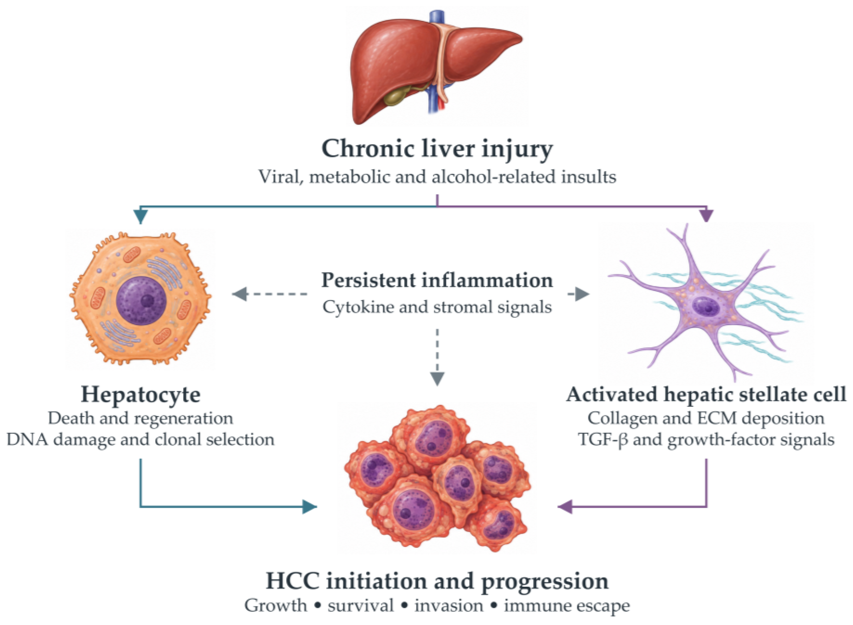
Figure 2.
Simplified canonical Wnt/β-catenin signaling. In the Wnt-off state, the AXIN–APC–CK1–GSK3 destruction complex promotes β-catenin phosphorylation and degradation. Wnt binding to Frizzled and LRP5/6 reduces destruction-complex activity, allowing β-catenin to accumulate and interact with nuclear TCF/LEF. Activating CTNNB1 mutations can stabilize β-catenin independently of ligand stimulation. The effect of AXIN1 loss is context dependent. APC, adenomatous polyposis coli; CK1, casein kinase 1; GSK3, glycogen synthase kinase 3; DVL, Dishevelled; TCF/LEF, T-cell factor/lymphoid enhancer factor. Original schematic based on published pathway descriptions [34,38].
Figure 2.
Simplified canonical Wnt/β-catenin signaling. In the Wnt-off state, the AXIN–APC–CK1–GSK3 destruction complex promotes β-catenin phosphorylation and degradation. Wnt binding to Frizzled and LRP5/6 reduces destruction-complex activity, allowing β-catenin to accumulate and interact with nuclear TCF/LEF. Activating CTNNB1 mutations can stabilize β-catenin independently of ligand stimulation. The effect of AXIN1 loss is context dependent. APC, adenomatous polyposis coli; CK1, casein kinase 1; GSK3, glycogen synthase kinase 3; DVL, Dishevelled; TCF/LEF, T-cell factor/lymphoid enhancer factor. Original schematic based on published pathway descriptions [34,38].
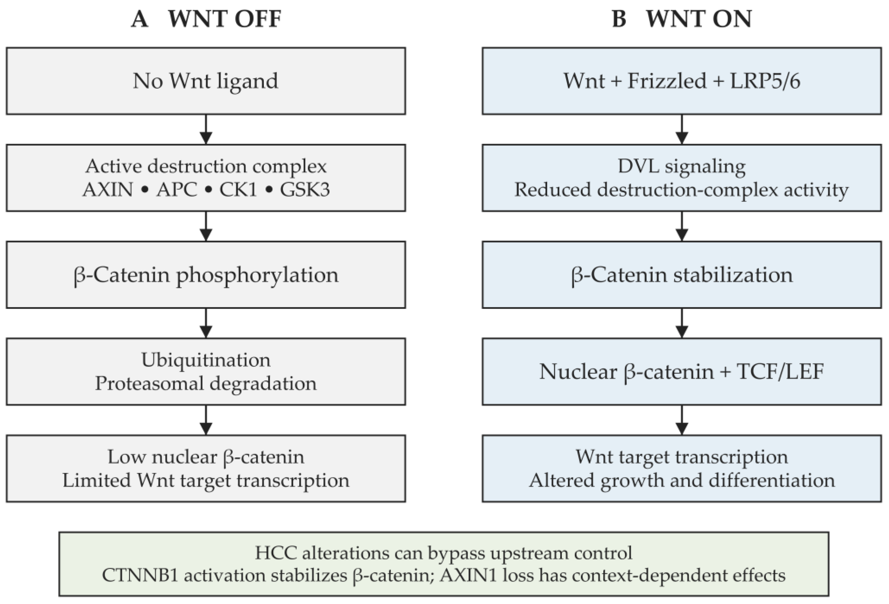
Figure 3.
Growth-factor signaling through PI3K–AKT–mTOR and RAS–RAF–MEK–ERK. Receptor tyrosine kinases provide upstream signals to two interacting branches. PI3K generates PIP3, PTEN reverses this conversion, and PDK1 and mTORC2 contribute to AKT activation. AKT restrains TSC activity, allowing RHEB to stimulate mTORC1. The RAS branch proceeds through RAF, MEK and ERK to transcriptional responses. Arrows indicate activation or forward signaling; bars indicate inhibition. RTK, receptor tyrosine kinase; MEK, mitogen-activated protein kinase kinase; PDK1, phosphoinositide-dependent kinase 1; TSC, tuberous sclerosis complex; mTORC, mechanistic target of rapamycin complex. Original schematic based on the cited literature [44,45].
Figure 3.
Growth-factor signaling through PI3K–AKT–mTOR and RAS–RAF–MEK–ERK. Receptor tyrosine kinases provide upstream signals to two interacting branches. PI3K generates PIP3, PTEN reverses this conversion, and PDK1 and mTORC2 contribute to AKT activation. AKT restrains TSC activity, allowing RHEB to stimulate mTORC1. The RAS branch proceeds through RAF, MEK and ERK to transcriptional responses. Arrows indicate activation or forward signaling; bars indicate inhibition. RTK, receptor tyrosine kinase; MEK, mitogen-activated protein kinase kinase; PDK1, phosphoinositide-dependent kinase 1; TSC, tuberous sclerosis complex; mTORC, mechanistic target of rapamycin complex. Original schematic based on the cited literature [44,45].
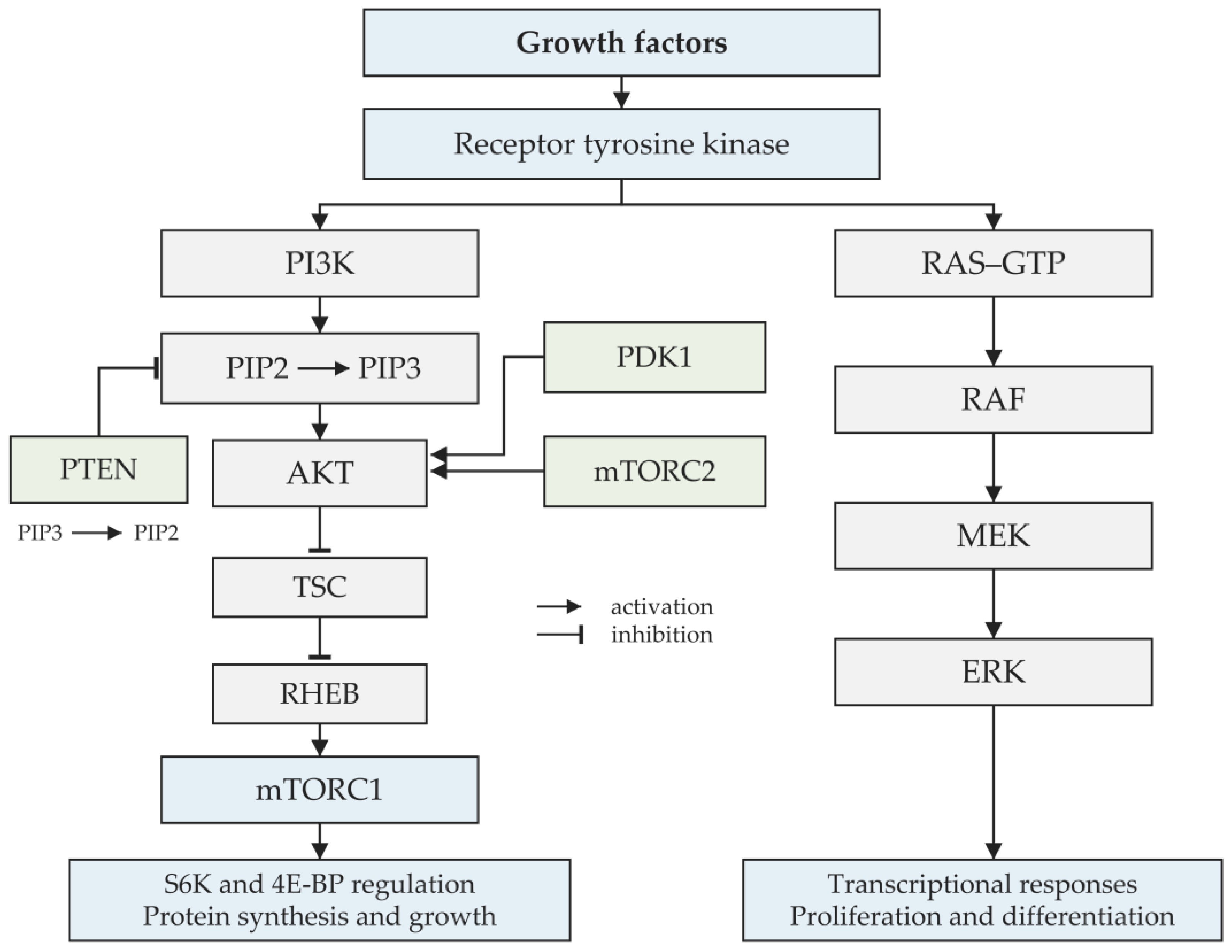
Figure 4.
Canonical and noncanonical NF-κB signaling. Canonical receptor signaling activates the IKK machinery, promotes IκB degradation and releases p50–RelA. Noncanonical signaling stabilizes NIK, activates IKKα and promotes p100 processing to p52, enabling p52–RelB nuclear activity. The schematic omits receptor-specific adaptor details. TNF, tumor necrosis factor; IL-1, interleukin 1; TLR, Toll-like receptor; IKK, IκB kinase; NEMO, NF-κB essential modulator; NIK, NF-κB-inducing kinase; LTβR, lymphotoxin beta receptor; BAFFR, B-cell activating factor receptor. Original schematic based on published pathway descriptions [51,52].
Figure 4.
Canonical and noncanonical NF-κB signaling. Canonical receptor signaling activates the IKK machinery, promotes IκB degradation and releases p50–RelA. Noncanonical signaling stabilizes NIK, activates IKKα and promotes p100 processing to p52, enabling p52–RelB nuclear activity. The schematic omits receptor-specific adaptor details. TNF, tumor necrosis factor; IL-1, interleukin 1; TLR, Toll-like receptor; IKK, IκB kinase; NEMO, NF-κB essential modulator; NIK, NF-κB-inducing kinase; LTβR, lymphotoxin beta receptor; BAFFR, B-cell activating factor receptor. Original schematic based on published pathway descriptions [51,52].
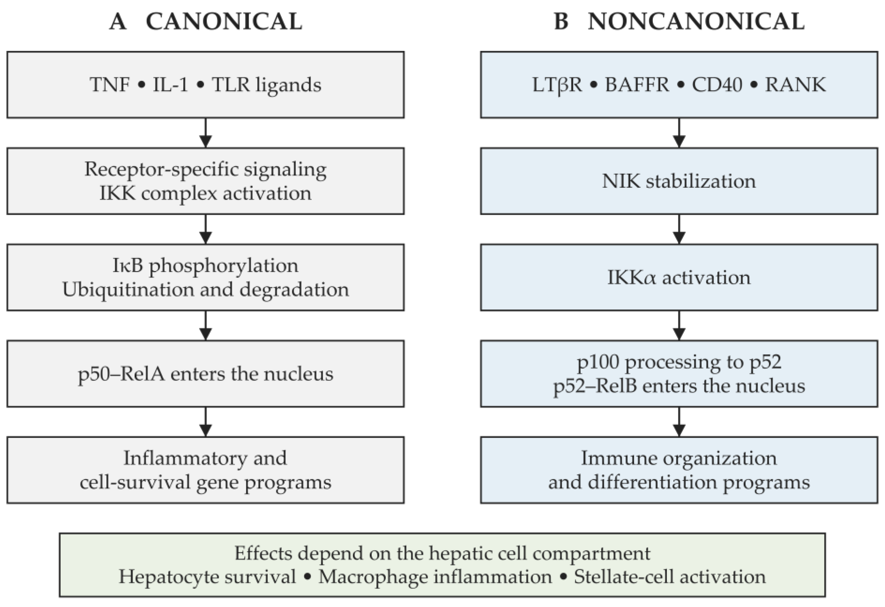
Figure 5.
Immune mechanisms in the HCC microenvironment. (A) Dendritic-cell antigen presentation supports CD8-positive T-cell responses; NK cells provide complementary tumor-cell recognition and killing. (B) Suppressive myeloid and regulatory lymphocyte states, cytokines and checkpoint signaling can impair effector function. PD-L1–PD-1 is shown as an example of an inhibitory interaction. Deficient dendritic-cell recruitment and fibrotic stroma create additional barriers. Cell morphology is schematic and does not imply fixed functional states or relative abundance. Arrows indicate functional relationships; bars indicate inhibition. TAM, tumor-associated macrophage; MDSC, myeloid-derived suppressor cell; Treg, regulatory T cell; NK, natural killer; PD-L1, programmed death ligand 1; PD-1, programmed cell death protein 1. Original illustration based on the cited literature [10,40,57,66] (created with BioRender; accessed on 5 October 2026).
Figure 5.
Immune mechanisms in the HCC microenvironment. (A) Dendritic-cell antigen presentation supports CD8-positive T-cell responses; NK cells provide complementary tumor-cell recognition and killing. (B) Suppressive myeloid and regulatory lymphocyte states, cytokines and checkpoint signaling can impair effector function. PD-L1–PD-1 is shown as an example of an inhibitory interaction. Deficient dendritic-cell recruitment and fibrotic stroma create additional barriers. Cell morphology is schematic and does not imply fixed functional states or relative abundance. Arrows indicate functional relationships; bars indicate inhibition. TAM, tumor-associated macrophage; MDSC, myeloid-derived suppressor cell; Treg, regulatory T cell; NK, natural killer; PD-L1, programmed death ligand 1; PD-1, programmed cell death protein 1. Original illustration based on the cited literature [10,40,57,66] (created with BioRender; accessed on 5 October 2026).
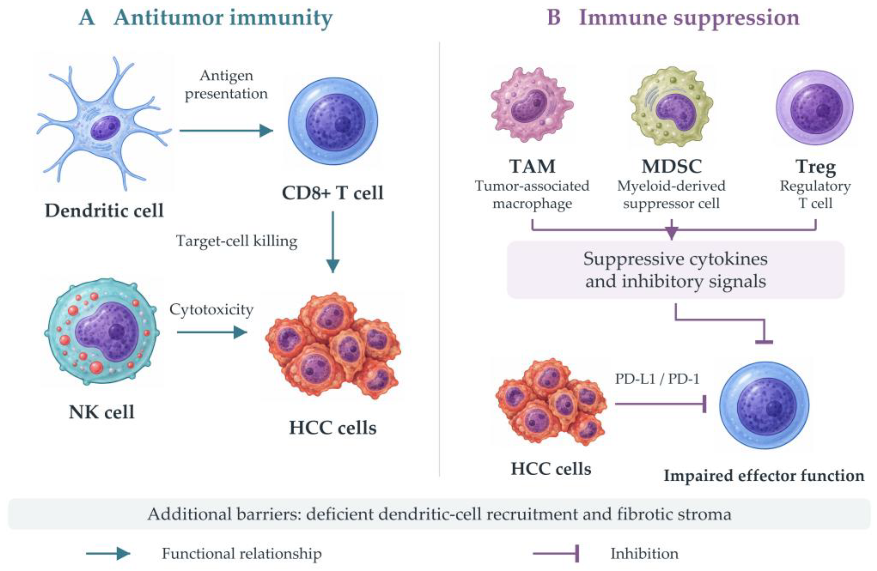

Table 1.
Recurrent genetic alterations and their principal functional implications in HCC.
| Gene or locus | Biological process | Functional interpretation | References |
| TERT promoter | Telomere maintenance | Telomerase reactivation; an early alteration in a subset of cirrhotic premalignant nodules. | [31] |
| TP53 | Cell-cycle and DNA-damage responses | Loss of checkpoint and apoptotic control; associations vary with etiology and tumor context. | [32,33] |
| CTNNB1 | Wnt/β-catenin signaling | β-Catenin stabilization and altered transcription; not a universal predictor of treatment response. | [34,35] |
| AXIN1 | Destruction-complex regulation | Loss of a regulatory scaffold; effects need not reproduce the CTNNB1-mutant phenotype. | [34] |
| ARID1A | Chromatin remodeling | Altered chromatin regulation and transcriptional control. | [32,35] |
| ARID2 | Chromatin remodeling | Disruption of a distinct chromatin-remodeling component. | [33,35] |
| NFE2L2 | Oxidative-stress response | NRF2 pathway activation can favor adaptation to cellular stress. | [32] |
| KEAP1 | Oxidative-stress regulation | Loss of negative regulation of NRF2 signaling. | [32] |
| JAK1 | JAK/STAT signaling | Less frequent alterations connect cytokine signaling with tumor-cell behavior. | [30] |
| RPS6KA3 | Mitogen signaling | Alteration of an ERK-associated kinase; functional effects depend on the variant. | [34] |
TERT, telomerase reverse transcriptase; TP53, tumor protein p53; CTNNB1, catenin beta 1; AXIN1, axin 1; ARID, AT-rich interaction domain; NRF2, nuclear factor erythroid 2-related factor 2; KEAP1, Kelch-like ECH-associated protein 1; JAK, Janus kinase; STAT, signal transducer and activator of transcription; RPS6KA3, ribosomal protein S6 kinase A3. Functional associations do not establish a validated predictive biomarker for routine therapy.
Table 2.
Clinical and experimental evidence for complementary therapeutic targets.
| Target | Intervention | Evidence and limitation | References |
| PD-L1 + VEGF-A | Atezolizumab + bevacizumab | Randomized phase III benefit versus sorafenib in IMbrave150. | [72] |
| CTLA-4 + PD-L1 | Tremelimumab + durvalumab | Overall-survival benefit versus sorafenib in HIMALAYA. | [75] |
| PD-1 + CTLA-4 | Nivolumab + ipilimumab | Overall-survival benefit versus lenvatinib or sorafenib in CheckMate 9DW. | [76] |
| VEGFR2 | Ramucirumab | Benefit after sorafenib in the AFP ≥400 ng/mL population of REACH-2. | [77] |
| mTOR | Everolimus | No overall-survival benefit after sorafenib failure in EVOLVE-1. | [50] |
| Wnt/β-catenin | Transcriptional-complex or Frizzled inhibition | Experimental antitumor activity; clinical benefit in HCC remains unestablished. | [42,43] |
| HSC and CAF states | Stromal-state or NOTCH pathway targeting | Human spatial associations and preclinical perturbation; no established HCC patient benefit. | [13,15,71] |
| PD-1 + multikinase inhibition | Pembrolizumab + lenvatinib | LEAP-002 did not meet prespecified overall- or progression-free-survival significance thresholds. | [78] |
| PD-L1 + VEGF-A after curative therapy | Adjuvant atezolizumab + bevacizumab | Updated IMbrave050: initial recurrence-free-survival benefit not sustained; overall survival immature. | [79] |
The clinical findings refer to the populations and comparators of the individual trials. They do not establish a ranking between regimens. PD-1, programmed cell death protein 1; PD-L1, programmed death ligand 1; CTLA-4, cytotoxic T-lymphocyte-associated protein 4; VEGF-A, vascular endothelial growth factor A; VEGFR2, VEGF receptor 2; AFP, alpha-fetoprotein; mTOR, mechanistic target of rapamycin; HSC, hepatic stellate cell.
Table 3.
Interpretation of mechanistic and clinical evidence across tumor and microenvironmental compartments.
Table 3.
Interpretation of mechanistic and clinical evidence across tumor and microenvironmental compartments.
| Question or compartment | Representative evidence | What the evidence supports |
| Oncogenic signaling | β-Catenin causes immune exclusion in a mouse model; human CTNNB1 immune states vary. [40,41] | Biological mechanism; not a universal checkpoint-resistance test. |
| Stromal dependency | HSC mediators have opposing effects; CAF or NOTCH perturbation improves control in models. [13,15,71] | Compartment- and stage-specific hypothesis requiring human validation. |
| Spatial immune context | Immune niches and immunotypes correlate with checkpoint outcomes. [61,69] | Candidate treatment-response biomarker; standardization and prospective testing needed. |
| Postoperative risk | Spatial marker combinations predict recurrence in validation cohorts. [80] | Prognostic stratification; does not establish relative benefit from a specific drug. |
| Treatment benefit | Positive unresectable-HCC trials coexist with negative combinations and updated adjuvant findings. [72,78,79] | Benefit applies to the tested regimen, population, comparator and follow-up. |
This qualitative framework is not a formal risk-of-bias assessment or evidence-grading system. Predictive refers to differential treatment benefit; prognostic refers to outcome risk. HSC, hepatic stellate cell; CAF, cancer-associated fibroblast.
Disclaimer/Publisher’s Note: The statements, opinions and data contained in all publications are solely those of the individual author(s) and contributor(s) and not of MDPI and/or the editor(s). MDPI and/or the editor(s) disclaim responsibility for any injury to people or property resulting from any ideas, methods, instructions or products referred to in the content. |
© 2026 by the authors. Licensee MDPI, Basel, Switzerland. This article is an open access article distributed under the terms and conditions of the Creative Commons Attribution (CC BY) license (http://creativecommons.org/licenses/by/4.0/).
Copyright: This open access article is published under a Creative Commons CC BY 4.0 license, which permit the free download, distribution, and reuse, provided that the author and preprint are cited in any reuse.