

Submitted:
19 October 2026
Posted:
20 October 2026
You are already at the latest version
Abstract
Background: Endometrial cancer remains a therapeutic challenge, particularly in advanced and recurrent cases. This study investigated the anticancer effects of three synthetic CDDO derivatives, RTA 404, RTA dh404, and RTA 408, in human endometrial cancer Ishikawa cells. Methods: Cell viability was assessed using the CCK-8 assay, while Ki67 expression, apoptosis, cell cycle distribution, mitochondrial membrane potential, and intracellular reactive oxygen species (ROS) levels were evaluated using immunofluorescence staining and flow cytometry-based assays. Results: All three CDDO derivatives inhibited cell viability in a time- and concentration-dependent manner and reduced Ki67 expression. They also induced apoptosis and cell cycle arrest, with RTA 404 and RTA dh404 promoting G1 phase arrest and RTA 408 inducing G2/M phase arrest. Additionally, all compounds decreased mitochondrial membrane potential and intracellular ROS levels. Conclusion: RTA 404, RTA dh404, and RTA 408 suppress Ishikawa cell growth by inhibiting proliferation, inducing apoptosis, altering cell cycle progression, and disrupting mitochondrial function. These findings support further investigation of CDDO derivatives as potential therapeutic candidates for endometrial cancer.
Keywords:
endometrial cancer
; CDDO derivatives
; apoptosis
; cell cycle arrest
; mitochondrial membrane potential
; oxidative stress
1. Introduction
Endometrial cancer is the most frequently diagnosed gynecologic malignancy in many developed countries and represents a significant cause of cancer-related morbidity and mortality among women [1,2]. The majority of patients are diagnosed at an early stage and achieve favorable outcomes after surgical intervention [3]; however, patients with advanced-stage or recurrent disease often exhibit poor prognosis due to limited therapeutic options and the development of treatment resistance [4,5]. Therefore, exploring novel compounds capable of suppressing tumor growth and improving therapeutic strategies remains an important research priority. The initiation and progression of endometrial cancer involve complex molecular alterations that promote uncontrolled proliferation, abnormal cell cycle progression, evasion of apoptosis, and changes in cellular metabolism [6,7]. Cancer cells frequently maintain an altered redox balance to support rapid growth and survival [8]. Although moderate ROS levels can promote tumor-associated signaling pathways, excessive ROS accumulation can induce oxidative damage and activate cell death pathways [9,10]. Consequently, regulation of intracellular ROS homeostasis has become an important target for anticancer intervention.
Mitochondria are central regulators of cellular energy metabolism and apoptosis. Alterations in mitochondrial membrane potential (ΔΨm) represent an early event during mitochondrial-mediated apoptosis, leading to activation of downstream apoptotic pathways [11,12]. In addition, mitochondrial dysfunction is closely associated with changes in oxidative stress signaling and cellular survival mechanisms [13]. Therefore, compounds capable of modulating mitochondrial function and redox balance may provide potential strategies for inhibiting cancer cell growth. Synthetic oleanane triterpenoids, particularly CDDO derivatives, have emerged as promising anticancer agents due to their broad biological activities [14,15]. CDDO derivatives have been shown to regulate inflammatory responses, oxidative stress pathways, and tumor suppressive mechanisms through interaction with multiple signaling molecules [16,17,18]. These compounds can modulate nuclear factor erythroid 2–related factor 2 (Nrf2)-dependent antioxidant signaling, inhibit pro-survival pathways, and promote apoptosis in cancer cells [17,19]. Among these derivatives, RTA 404, RTA dh404, and RTA 408 possess structural modifications that enhance biological activity and have demonstrated therapeutic potential in different cancer models [20,21,22,23,24].
However, despite increasing evidence supporting the anticancer properties of CDDO derivatives, their biological effects in endometrial cancer remain poorly understood. In particular, the influence of these compounds on endometrial cancer cell proliferation, apoptosis, cell cycle progression, mitochondrial membrane potential, and intracellular ROS regulation has not been fully investigated.
In this study, we evaluated the effects of three synthetic CDDO derivatives, RTA 404, RTA dh404, and RTA 408, on human endometrial cancer Ishikawa cells. We investigated their impact on cellular proliferation, Ki67 expression, apoptosis induction, cell cycle distribution, mitochondrial membrane potential, and ROS production. Our findings provide new insights into the potential molecular mechanisms underlying CDDO derivative-mediated inhibition of endometrial cancer cell growth and support further investigation of these compounds as candidate anticancer agents.
2. Materials and Methods
2.1. Reagents and Chemical Preparation
Synthetic oleanane triterpenoid derivatives, including RTA 404 (CAS number: 1191265-33-4), RTA dh404 (CAS number: 1191265-33-4), and RTA 408 (CAS number: 1474034-05-3), were obtained from Med Chem Express (MCE). Stock solutions of CDDO derivatives were prepared by dissolving compounds in dimethyl sulfoxide (DMSO) and stored according to the manufacturer's instructions. Working concentrations were freshly prepared by diluting stock solutions with complete culture medium before each experiment. The final concentration of DMSO in culture medium was maintained at a level that did not affect cell viability. All reagents were prepared using sterile techniques.
2.2. Cell Culture
Human endometrial cancer Ishikawa cells were obtained from an authenticated cell resource. Cells were maintained in Dulbecco’s modified Eagle’s medium (DMEM) supplemented with 10% fetal bovine serum (FBS) and antibiotics (100 U/mL penicillin and 100 μg/mL streptomycin). Cells were cultured in a humidified incubator containing 5% CO₂ at 37°C. Cells were routinely monitored for morphology and cultured under logarithmic growth conditions before experimental treatment.
2.3. Cell Viability Assay
Cell viability was evaluated using the Cell Counting Kit-8 (CCK-8) assay. Ishikawa cells were seeded into 96-well plates at an appropriate density and allowed to attach overnight. Cells were treated with different concentrations of RTA 404, RTA dh404, or RTA 408 for 24, 48, and 72 h. Following treatment, CCK-8 reagent was added to each well and incubated according to the manufacturer's protocol. Absorbance was measured at 450 nm using a microplate reader. Cell viability was calculated as the percentage relative to the untreated control group.
2.4. Cell Cycle Analysis
Cell cycle progression was analyzed by PI staining and flow cytometry. Ishikawa cells were treated with CDDO derivatives for 24 h and collected by trypsinization. Cells were washed with PBS and fixed with cold ethanol overnight at 4°C. Fixed cells were stained with PI-containing staining buffer supplemented with RNase A. DNA content was measured by flow cytometry, and the distribution of cells in G0/G1, S, and G2/M phases was analyzed using appropriate cell cycle analysis software. DNA content analysis by flow cytometry analysis on a FACS Calibur system (BD, USA). At least 104 cells were analyzed for each determination. Data was analyzed by WinMDI 2.8 free soft-ware (BD, USA).
2.5. Apoptosis Assay
Apoptotic cell death was analyzed using Annexin V-FITC/propidium iodide (PI) double staining followed by flow cytometry. Ishikawa cells were treated with RTA 404, RTA dh404, or RTA 408 for 4 and 24 h. Cells were harvested, washed with phosphate-buffered saline (PBS), and stained with Annexin V-FITC and PI according to the manufacturer's instructions. Cells were analyzed using a flow cytometer. The proportions of viable (Annexin V⁻/PI⁻), early apoptotic (Annexin V⁺/PI⁻), and late apoptotic or necrotic cells (Annexin V⁺/PI⁺) were quantified. Apoptosis analysis by flow cytometry analysis on a FACS Calibur system (BD, USA). At least 104 cells were analyzed for each determination. Data was analyzed by WinMDI 2.8 free soft-ware (BD, USA).
2.6. ROS Assay
Intracellular reactive oxygen species (ROS) levels were measured using a ROS detection assay kit. Ishikawa cells were treated with RTA 404, RTA dh404, or RTA 408 and incubated with ROS-sensitive fluorescent probes according to the manufacturer's instructions. After staining, cells were analyzed by flow cytometry. Fluorescence intensity representing intracellular ROS levels was quantified and compared between treated and control groups. ROS levels analysis by flow cytometry analysis on a FACS Calibur system (BD, USA). At least 104 cells were analyzed for each determination. Data was analyzed by WinMDI 2.8 free soft-ware (BD, USA).
2.7. Mitochondrial Membrane Potential (MMP) Assay
Changes in mitochondrial membrane potential (ΔΨm) were evaluated using JC-1 staining. Ishikawa cells were treated with RTA 404, RTA dh404, or RTA 408 for the indicated time periods and incubated with JC-1 dye according to the manufacturer's protocol. JC-1 forms red fluorescent aggregates in cells with normal mitochondrial membrane potential, whereas mitochondrial depolarization results in green fluorescent JC-1 monomers. Fluorescence signals were detected by flow cytometry, and the percentage of cells with decreased ΔΨm was quantified.
2.8. Immunocytochemistry Analysis
To evaluate cellular proliferation, Ki67 expression was examined using immunofluorescence staining. Ishikawa cells were seeded on coverslips and treated with RTA 404, RTA dh404, or RTA 408 for 24 h. Cells were fixed with paraformaldehyde and permeabilized with an appropriate permeabilization buffer. After blocking with blocking solution, cells were incubated with a primary antibody against Ki67 followed by incubation with a fluorescently labeled secondary antibody. Nuclei were counterstained with DAPI. Fluorescence images were captured using a fluorescence microscope, and Ki67-positive signals were quantified using image analysis software.
2.9. Statistical Analysis
All experiments were performed independently at least three times. Data are presented as mean ± standard error of the mean (SEM). Statistical analyses were performed using appropriate statistical software. Comparisons between multiple groups were performed using one-way analysis of variance (ANOVA) followed by an appropriate post hoc test. A value of p < 0.05 was considered statistically significant.
3. Results
3.1. CDDO Derivatives Suppress Proliferation of Endometrial Cancer Ishikawa Cells
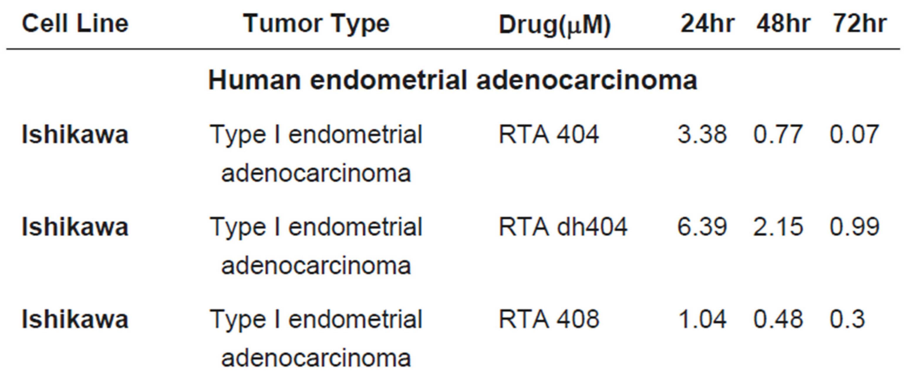
To investigate the anti-proliferative effects of synthetic oleanane triterpenoid derivatives, Ishikawa endometrial cancer cells were treated with increasing concentrations of CDDO derivatives, including RTA 404, RTA dh404, and RTA 408, for 24–72 h. Cell viability was evaluated using the CCK-8 assay. As shown in (Figure 1A, B), all three CDDO derivatives significantly inhibited Ishikawa cell growth in a concentration- and time-dependent manner. The calculated IC₅₀ values are summarized in Table 1, indicating that approximately 50% growth inhibition was achieved within the tested concentration range. Among the tested compounds, higher concentrations resulted in a more pronounced reduction in cell viability after prolonged treatment. These results demonstrate that CDDO derivatives exert potent growth-inhibitory effects on endometrial cancer cells. The text continues here.
3.2. CDDO Derivatives Reduce Ki67 Expression in Ishikawa Cells
To determine whether the inhibitory effect of CDDO derivatives was associated with suppression of cellular proliferation, Ki67 expression was examined by immunofluorescence staining. Following treatment with RTA 404, RTA dh404, or RTA 408 for 24 h, Ishikawa cells exhibited a marked decrease in Ki67-positive staining compared with untreated controls (Figure 2A). Quantitative analysis revealed a significant reduction in Ki67 fluorescence intensity following CDDO derivative treatment (Figure 2B). These findings indicate that CDDO derivatives suppress proliferative activity in Ishikawa endometrial cancer cells.
3.3. CDDO Derivatives Induce Apoptotic Cell Death in Ishikawa Cells
To determine whether CDDO derivatives affect programmed cell death, Annexin V-FITC/PI staining was performed followed by flow cytometric analysis. Treatment with RTA 404, RTA dh404, or RTA 408 increased the proportion of Annexin V-positive cells compared with the control group at both 4 and 24 h (Figure 3A). Quantification of apoptotic populations demonstrated that CDDO derivative treatment promoted apoptosis in Ishikawa cells (Figure 3B). These results suggest that induction of apoptosis contributes to the anti-tumor activity of CDDO derivatives.
3.4. CDDO Derivatives Alter Cell Cycle Progression in Ishikawa Cells
To examine whether CDDO derivatives affect cell cycle regulation, Ishikawa cells were treated with RTA 404, RTA dh404, or RTA 408 for 24 h and analyzed by propidium iodide staining and flow cytometry. As shown in Figure 4A, treatment with RTA 404 and RTA dh404 resulted in an increased proportion of cells in the G1 phase accompanied by a reduction in the G2/M population. In contrast, RTA 408 treatment caused an accumulation of cells in the G2/M phase with a corresponding decrease in the G1 population. Quantitative analysis confirmed significant alterations in cell cycle distribution following CDDO derivative exposure (Figure 4B). These findings indicate that CDDO derivatives inhibit Ishikawa cell proliferation through modulation of cell cycle progression.
3.5. CDDO Derivatives Disrupt Mitochondrial Membrane Potential in Ishikawa Cells
Loss of mitochondrial membrane potential (ΔΨm) is an important event associated with mitochondrial-mediated apoptosis. To evaluate mitochondrial dysfunction induced by CDDO derivatives, JC-1 staining was performed. Compared with untreated cells, Ishikawa cells treated with RTA 404, RTA dh404, or RTA 408 showed increased JC-1 monomer fluorescence and reduced JC-1 aggregate formation (Figure 5A), indicating mitochondrial membrane depolarization. Quantitative analysis further demonstrated an increased proportion of cells with reduced ΔΨm after treatment (Figure 5B). These results suggest that CDDO derivatives induce mitochondrial dysfunction during apoptosis induction.
3.6. CDDO Derivatives Decrease Intracellular ROS Levels in Ishikawa Cells
Because oxidative stress is frequently involved in cancer cell responses to therapeutic agents, intracellular ROS production was analyzed after CDDO derivative treatment. Ishikawa cells were treated with RTA 404, RTA dh404, or RTA 408 and stained with a ROS detection probe followed by flow cytometry analysis. Interestingly, all three CDDO derivatives significantly reduced intracellular ROS levels compared with untreated controls (Figure 6A–B). These findings suggest that the anti-proliferative effects of CDDO derivatives in Ishikawa cells are not mediated by ROS accumulation.
4. Discussion
Endometrial cancer remains a major gynecological malignancy, and the development of novel therapeutic strategies is required, particularly for patients with advanced-stage disease or limited treatment responses. In this study, we investigated the biological effects of three synthetic oleanane triterpenoid derivatives, RTA 404, RTA dh404, and RTA 408, in human endometrial cancer Ishikawa cells. Our findings demonstrate that CDDO derivatives significantly suppressed cell proliferation (Figure 1), reduced Ki67 expression (Figure 2), altered cell cycle progression, promoted apoptotic cell death (Figure 3 and Figure 4), disrupted mitochondrial membrane potential (Figure 5), and decreased intracellular ROS levels (Figure 6). These results suggest that CDDO derivatives inhibit endometrial cancer cell growth through multiple cellular mechanisms involving proliferation regulation, mitochondrial dysfunction, and apoptosis induction.
Uncontrolled cellular proliferation is a fundamental characteristic of cancer progression. Our CCK-8 assay demonstrated that RTA 404, RTA dh404, and RTA 408 reduced Ishikawa cell viability in a concentration- and time-dependent manner. Consistently, immunofluorescence analysis revealed decreased Ki67 expression following CDDO derivative treatment, indicating suppression of proliferative activity (Figure 1 and Figure 2). These findings suggest that the inhibitory effects of CDDO derivatives are associated with reduced cellular proliferation rather than a nonspecific cytotoxic response.
Cell cycle dysregulation plays an essential role in tumor development by allowing continuous cancer cell proliferation [25]. In our results, flow cytometric analysis demonstrated that different CDDO derivatives induced distinct alterations in cell cycle distribution. RTA 404 and RTA dh404 treatment resulted in increased G1 phase accumulation, suggesting inhibition of the G1/S transition. In contrast, RTA 408 promoted G2/M phase arrest, indicating that individual CDDO derivatives may regulate cell cycle progression through distinct molecular mechanisms (Figure 4). Previous studies have reported that synthetic triterpenoids can modulate cell cycle-related proteins, including cyclins and cyclin-dependent kinase regulators, which may contribute to their antiproliferative effects [21,26]. Further investigation of specific cell cycle regulatory molecules will be required to clarify the precise mechanisms involved in Ishikawa cells.
Apoptosis is a major mechanism responsible for elimination of cancer cells and is frequently targeted by anticancer compounds. Our Annexin V-FITC/PI staining results showed that treatment with RTA 404, RTA dh404, and RTA 408 increased apoptotic cell populations in Ishikawa cells. These findings indicate that apoptosis induction contributes to CDDO derivative-mediated growth inhibition. Furthermore, JC-1 staining demonstrated a reduction in mitochondrial membrane potential following CDDO derivative treatment, suggesting mitochondrial dysfunction as an important event associated with apoptosis activation (Figure 5). Since mitochondrial depolarization is an early indicator of mitochondrial-mediated apoptosis, these results suggest that CDDO derivatives may promote apoptosis through regulation of mitochondrial integrity.
Oxidative stress regulation is closely associated with cancer cell survival and therapeutic response. Many anticancer agents induce excessive ROS accumulation, resulting in oxidative damage and apoptosis [27]. However, in the present study, treatment with RTA 404, RTA dh404, and RTA 408 decreased intracellular ROS levels in Ishikawa cells. Although this observation may appear inconsistent with the concept that ROS elevation promotes cancer cell death, synthetic triterpenoids are known to regulate cellular redox balance through multiple mechanisms. CDDO derivatives have been reported to interact with the KEAP1–Nrf2 pathway, which plays a central role in antioxidant defense and maintenance of redox homeostasis [16,28]. Therefore, the reduction of ROS observed in this study may reflect activation of antioxidant responses or disruption of cancer-associated metabolic regulation rather than direct ROS-mediated cytotoxicity (Figure 6). Additional studies examining Nrf2 signaling, antioxidant enzymes, and mitochondrial metabolism are needed to further clarify this mechanism.
Interestingly, although RTA 408 has been reported as a potent Nrf2 activator with cytoprotective effects in non-cancer models [29], our results demonstrate that RTA 408 exhibits growth-inhibitory activity in Ishikawa endometrial cancer cells. This finding suggests that the cellular context, basal redox status, and genetic background of cancer cells may influence the biological outcome of CDDO derivative exposure. Differences in cellular signaling networks may determine whether Nrf2-related pathways contribute to survival or growth suppression in cancer cells.
Collectively, our study provides evidence that synthetic CDDO derivatives exert anticancer effects in endometrial cancer cells by targeting multiple cellular processes, including proliferation inhibition, cell cycle regulation, mitochondrial dysfunction, and apoptosis induction. Among the tested compounds, RTA 404, RTA dh404, and RTA 408 demonstrated comparable inhibitory effects but with different impacts on cell cycle regulation, suggesting compound-specific mechanisms of action.
Several limitations should be acknowledged. First, this study was performed using only one endometrial cancer cell line, and additional models, including other endometrial cancer subtypes and in vivo models, are required to validate these findings. Second, although we demonstrated changes in mitochondrial function and ROS levels, the upstream molecular pathways responsible for these effects remain unclear. Future studies investigating Nrf2 signaling, mitochondrial apoptotic proteins, and combination strategies with current therapeutic agents may provide further insight into the anticancer potential of CDDO derivatives.
5. Conclusions
In conclusion, our findings indicate that RTA 404, RTA dh404, and RTA 408 suppress the growth of Ishikawa endometrial cancer cells through coordinated regulation of proliferation, apoptosis, cell cycle progression, and mitochondrial function. These results support further investigation of CDDO derivatives as molecular candidates for endometrial cancer research.
Author Contributions
Conceptualization and design of the study: Kuan-Ting Lee; Data collection: Yu-Shan Yang; Statistical, Biostatistics analysis of data: Kuan-Ting Lee, Chih-Ying Wang; Writing, original draft: Kuan-Ting Lee, Yi-Chiang Hsu; Writing, review, and/or revision of the manuscript: Kuan-Ting Lee, Yi-Chiang Hsu; Supervision and Validation: Yi-Chiang Hsu, Chih-Yang Chang. All the authors approved the final version of the manuscript for publication.
Funding
This work was supported by the I-Shou University (grant no. ISU-109-IUC-01, ISU-111-IUC-08, ISU-114-01-09A), and E-Da Hospital (grant no. EDAHI115002).
Institutional Review Board Statement
Not applicable.
Informed Consent Statement
Not applicable.
Data Availability Statement
Data supporting the findings presented in this study are availablefrom the corresponding author upon reasonable request.
Acknowledgments
We thank all of the researchers and participants as well as E-Da Hospital for its research funding.
Conflicts of Interest
The author declares that there are no conflicts of interest.
References
- Tang, W.-Z.; Cai, Q.-Y.; Huang, K.-J.; Xu, W.-Z.; Li, J.-Z.; Pan, Y.-R.; Xu, H.-Y.; Zhao, Y.-F.; Sheng, T.-H.; Li, Z.-M.; et al. The global burden of polycystic ovary syndrome, endometriosis, uterine fibroids, cervical cancer, uterine cancer, and ovarian cancer from 1990 to 2021. BMC Public Health 2025, 25, 1774. [Google Scholar] [CrossRef]
- Crosbie, E.J.; Kitson, S.J.; McAlpine, J.N.; Mukhopadhyay, A.; Powell, M.E.; Singh, N. Endometrial cancer. Lancet 2022, 399, 1412–1428. [Google Scholar] [CrossRef]
- Baker-Rand, H.; Kitson, S.J. Recent advances in endometrial cancer prevention, early diagnosis and treatment. Cancers 2024, 16, 1028. [Google Scholar] [CrossRef]
- Matoba, Y.; Devins, K.M.; Milane, L.; Manning, W.B.; Mazina, V.; Yeku, O.O.; Rueda, B.R. High-grade endometrial cancer: molecular subtypes, current challenges, and treatment options. Reprod. Sci. 2024, 31, 2541–2559. [Google Scholar] [CrossRef]
- Gordhandas, S.; Zammarrelli, W.A.; Rios-Doria, E.V.; Green, A.K.; Makker, V. Current evidence-based systemic therapy for advanced and recurrent endometrial cancer. J. Natl. Compr. Canc Netw. 2023, 21, 217–226. [Google Scholar] [CrossRef]
- Bogaczyk, A.; Zawlik, I.; Zuzak, T.; Kluz, M.; Potocka, N.; Kluz, T. The role of miRNAs in the development, proliferation, and progression of endometrial cancer. Int. J. Mol. Sci. 2023, 24, 11489. [Google Scholar] [CrossRef]
- Mariadas, H.; Chen, J.-H.; Chen, K.-H. The molecular and cellular mechanisms of endometriosis: from basic pathophysiology to clinical implications. Int. J. Mol. Sci. 2025, 26, 2458. [Google Scholar] [CrossRef]
- Zhao, Y.; Ye, X.; Xiong, Z.; Ihsan, A.; Ares, I.; Martínez, M.; Lopez-Torres, B.; Martínez-Larrañaga, M.-R.; Anadón, A.; Wang, X.; et al. Cancer metabolism: the role of ROS in DNA damage and induction of apoptosis in cancer cells. Metabolites 2023, 13, 796. [Google Scholar] [CrossRef]
- Akter, S.; Madhuvilakku, R.; Kar, A.K.; Nila, I.S.; Liu, P.; Inuzuka, H.; Wei, W.; Hong, Y. Reactive oxygen species (ROS) in cancer: From mechanism to therapeutic implications. Signal Transduct. Target Ther. 2026, 11, 111. [Google Scholar] [CrossRef]
- Nigam, M.; Punia, B.; Dimri, D.B.; Mishra, A.P.; Radu, A.-F.; Bungau, G. Reactive oxygen species: A double-edged sword in the modulation of cancer signaling pathway dynamics. Cells 2025, 14, 1207. [Google Scholar] [CrossRef]
- Malla, S.; Neupane, R.; Sood, S.; Hussein, N.; Abou-Dahech, M.; Terrero, D.; Ashby, C.R., Jr.; Babu, R.J.; Tiwari, A.K. Mitochondria as regulators of nonapoptotic cell death in cancer. MedComm (2020) 2025, 6, e70244. [Google Scholar] [CrossRef]
- Penna, C.; Perrelli, M.-G.; Pagliaro, P. Mitochondrial pathways, permeability transition pore, and redox signaling in cardioprotection: therapeutic implications. Antioxid. Redox Signal. 2013, 18, 556–599. [Google Scholar] [CrossRef]
- Barbour, J.A.; Turner, N. Mitochondrial stress signaling promotes cellular adaptations. Int. J. Cell Biol. 2014, 2014, 156020. [Google Scholar] [CrossRef]
- Borella, R.; Forti, L.; Gibellini, L.; De Gaetano, A.; De Biasi, S.; Nasi, M.; Cossarizza, A.; Pinti, M. Synthesis and anticancer activity of CDDO and CDDO-Me, two derivatives of natural triterpenoids. Molecules 2019, 24, 4097. [Google Scholar] [CrossRef]
- Demir, N.; Ciftci, H.; Elamin, K.M.; Can, M.; Sever, B. Design, Synthesis and Biological Evaluation of Novel Oleanolic Acid Derivatives as Potential Anti-Leukemic Agents. Life 2026, 16, 1335. [Google Scholar] [CrossRef]
- Mathis, B.J.; Cui, T. CDDO and its role in chronic diseases. Adv. Exp. Med. Biol. 2016, 291–314. [Google Scholar] [CrossRef]
- Li, W.; Wang, M.; Du, P.; Han, A.; Feng, X.; Chen, L. CDDO-Me triggers ROS-dependent ferroptosis and apoptosis in cervical cancer via targeting PI3K/Nrf2 pathway. Food Chem. Toxicol. 2025, 115664. [Google Scholar] [CrossRef]
- Kim, J.-E.; Park, H.; Kang, T.-C. CDDO-Me distinctly regulates regional specific astroglial responses to status epilepticus via ERK1/2-Nrf2, PTEN-PI3K-AKT and NFκB signaling pathways. Antioxidants 2020, 9, 1026. [Google Scholar] [CrossRef]
- Schiavoni, V.; Di Crescenzo, T.; Membrino, V.; Alia, S.; Fantone, S.; Salvolini, E.; Vignini, A. Bardoxolone methyl: a comprehensive review of its role as a NRF2 activator in anticancer therapeutic applications. Pharmaceuticals 2025, 18, 966. [Google Scholar] [CrossRef]
- Tsai, T.-H.; Lieu, A.-S.; Wang, Y.-W.; Yang, S.-F.; Hsu, Y.-C.; Lin, C.-L. Therapeutic potential of RTA 404 in human brain malignant glioma cell lines via cell cycle arrest via p21/AKT signaling. BioMed Res. Int. 2021, 2021, 5552226. [Google Scholar] [CrossRef]
- Tsai, T.-H.; Su, Y.-F.; Tsai, C.-Y.; Wu, C.-H.; Lee, K.-T.; Hsu, Y.-C. RTA dh404 induces cell cycle arrest, apoptosis, and autophagy in glioblastoma cells. Int. J. Mol. Sci. 2023, 24, 4006. [Google Scholar] [CrossRef]
- Aminzadeh, M.A.; Reisman, S.A.; Vaziri, N.D.; Shelkovnikov, S.; Farzaneh, S.H.; Khazaeli, M.; Meyer, C.J. The synthetic triterpenoid RTA dh404 (CDDO-dhTFEA) restores endothelial function impaired by reduced Nrf2 activity in chronic kidney disease. Redox Biol. 2013, 1, 527–531. [Google Scholar] [CrossRef]
- Probst, B.L.; Trevino, I.; McCauley, L.; Bumeister, R.; Dulubova, I.; Wigley, W.C.; Ferguson, D.A. RTA 408, a novel synthetic triterpenoid with broad anticancer and anti-inflammatory activity. PLoS ONE 2015, 10, e0122942. [Google Scholar] [CrossRef]
- Lee, K.-T.; Lieu, A.-S.; Lin, C.-L.; Hsu, Y.-C.; Tsai, T.-H. Synthetic oleanolic acid derivative, RTA-408, overcome in TMZ-resistant glioblastoma cells by inducing apoptosis and G1 cell cycle arrest. Med. Oncol. 2025, 42, 353. [Google Scholar] [CrossRef]
- Jamasbi, E.; Hamelian, M.; Hossain, M.A.; Varmira, K. The cell cycle, cancer development and therapy. Mol. Biol. Rep. 2022, 49, 10875–10883. [Google Scholar] [CrossRef]
- Tsai, T.-H.; Lieu, A.-S.; Huang, T.-Y.; Kwan, A.-L.; Lin, C.-L.; Hsu, Y.-C. Induction of mitosis delay and apoptosis by CDDO-TFEA in glioblastoma multiforme. Front Pharmacol. 2021, 12, 756228. [Google Scholar] [CrossRef]
- Ivanova, D.; Zhelev, Z.; Aoki, I.; Bakalova, R.; Higashi, T. Overproduction of reactive oxygen species-obligatory or not for induction of apoptosis by anticancer drugs. Chin. J. Cancer Res. 2016, 28, 383. [Google Scholar] [CrossRef]
- Meng, X.; Waddington, J.C.; Tailor, A.; Lister, A.; Hamlett, J.; Berry, N.; Park, B.K.; Sporn, M.B. CDDO-imidazolide targets multiple amino acid residues on the Nrf2 adaptor, Keap1. J. Med. Chem. 2020, 63, 9965–9976. [Google Scholar] [CrossRef]
- Han, P.; Qin, Z.; Tang, J.; Xu, Z.; Li, R.; Jiang, X.; Yang, C.; Xing, Q.; Qi, X.; Tang, M.; et al. RTA-408 Protects Kidney from Ischemia-Reperfusion Injury in Mice via Activating Nrf2 and Downstream GSH Biosynthesis Gene. Oxid. Med. Cell. Longev. 2017, 7612182. [Google Scholar] [CrossRef]
Figure 1.
CDDO derivatives inhibit proliferation of Ishikawa endometrial cancer cells. (A) Chemical structures of synthetic oleanane triterpenoid derivatives RTA 404, RTA dh404, and RTA 408. (B) Ishikawa cells were treated with indicated concentrations of RTA 404, RTA dh404, or RTA 408 for 24, 48, and 72 h. Cell viability was determined using the CCK-8 assay. Data are presented as mean ± SEM from at least three independent experiments. Statistical significance was determined compared with the untreated control group.
Figure 1.
CDDO derivatives inhibit proliferation of Ishikawa endometrial cancer cells. (A) Chemical structures of synthetic oleanane triterpenoid derivatives RTA 404, RTA dh404, and RTA 408. (B) Ishikawa cells were treated with indicated concentrations of RTA 404, RTA dh404, or RTA 408 for 24, 48, and 72 h. Cell viability was determined using the CCK-8 assay. Data are presented as mean ± SEM from at least three independent experiments. Statistical significance was determined compared with the untreated control group.
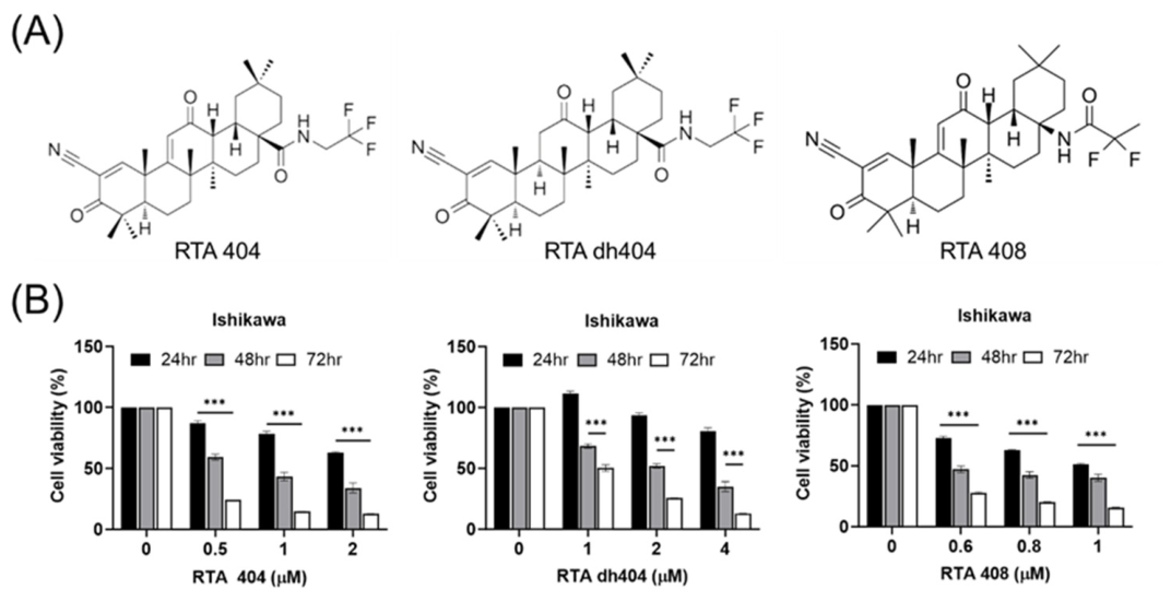
Figure 2.
CDDO derivatives suppress Ki67 expression in Ishikawa cells. (A) Representative immunofluorescence images showing Ki67 expression in Ishikawa cells treated with RTA 404, RTA dh404, or RTA 408 for 24 hr. Nuclei were counterstained with DAPI. (B) Quantification of Ki67 fluorescence intensity. Data represent mean ± SEM from three independent experiments. *P < 0.05, **P < 0.01 versus control.
Figure 2.
CDDO derivatives suppress Ki67 expression in Ishikawa cells. (A) Representative immunofluorescence images showing Ki67 expression in Ishikawa cells treated with RTA 404, RTA dh404, or RTA 408 for 24 hr. Nuclei were counterstained with DAPI. (B) Quantification of Ki67 fluorescence intensity. Data represent mean ± SEM from three independent experiments. *P < 0.05, **P < 0.01 versus control.
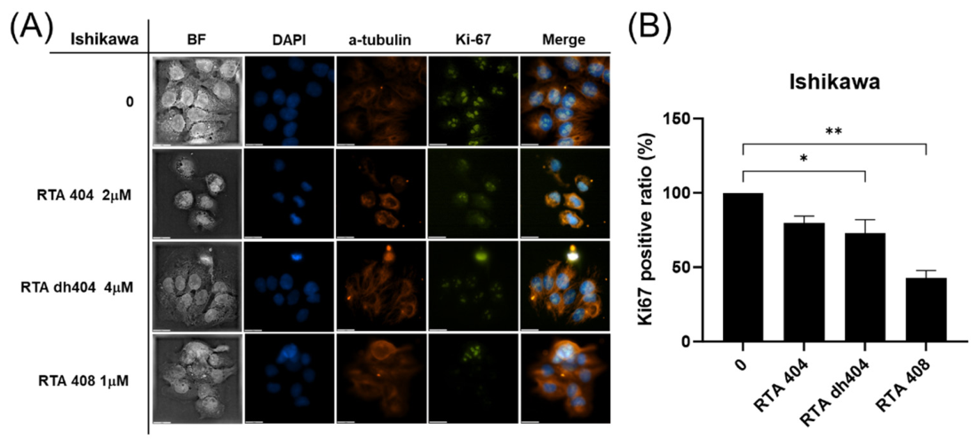
Figure 3.
CDDO derivatives induce apoptosis in Ishikawa cells. (A) Representative flow cytometry dot plots of Annexin V-FITC/PI staining in Ishikawa cells following treatment with RTA 404, RTA dh404, or RTA 408 for 4 and 24 hr. (B) Quantitative analysis of apoptotic cell populations. Results are presented as mean ± SEM from three independent experiments. *P < 0.05 versus control.
Figure 3.
CDDO derivatives induce apoptosis in Ishikawa cells. (A) Representative flow cytometry dot plots of Annexin V-FITC/PI staining in Ishikawa cells following treatment with RTA 404, RTA dh404, or RTA 408 for 4 and 24 hr. (B) Quantitative analysis of apoptotic cell populations. Results are presented as mean ± SEM from three independent experiments. *P < 0.05 versus control.
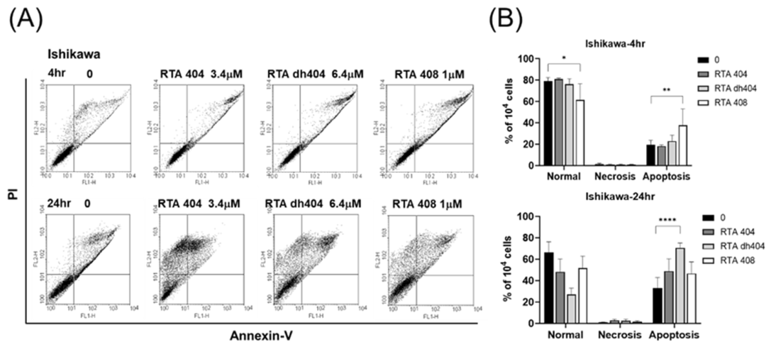
Figure 4.
CDDO derivatives regulate cell cycle progression in Ishikawa cells. (A) Representative flow cytometry histograms showing cell cycle distribution after treatment with RTA 404, RTA dh404, or RTA 408 for 24 h. (B) Quantification of cell populations in G0/G1, S, and G2/M phases. Data are expressed as mean ± SEM from three independent experiments. *P < 0.05 versus control.
Figure 4.
CDDO derivatives regulate cell cycle progression in Ishikawa cells. (A) Representative flow cytometry histograms showing cell cycle distribution after treatment with RTA 404, RTA dh404, or RTA 408 for 24 h. (B) Quantification of cell populations in G0/G1, S, and G2/M phases. Data are expressed as mean ± SEM from three independent experiments. *P < 0.05 versus control.
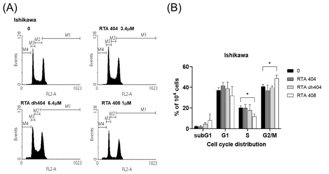
Figure 5.
CDDO derivatives induce mitochondrial membrane depolarization in Ishikawa cells. (A) Representative JC-1 staining profiles showing mitochondrial membrane potential changes following CDDO derivative treatment. Increased green fluorescence indicates mitochondrial depolarization. (B) Quantitative analysis of cells with reduced mitochondrial membrane potential. Data represent mean ± SEM from three independent experiments.
Figure 5.
CDDO derivatives induce mitochondrial membrane depolarization in Ishikawa cells. (A) Representative JC-1 staining profiles showing mitochondrial membrane potential changes following CDDO derivative treatment. Increased green fluorescence indicates mitochondrial depolarization. (B) Quantitative analysis of cells with reduced mitochondrial membrane potential. Data represent mean ± SEM from three independent experiments.
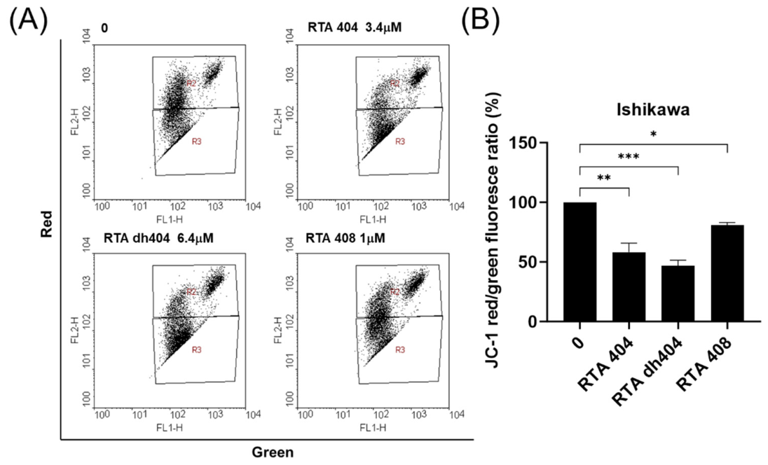
Figure 6.
CDDO derivatives reduce intracellular ROS levels in Ishikawa cells. (A) Representative flow cytometry profiles showing intracellular ROS production after treatment with RTA 404, RTA dh404, or RTA 408. (B) Quantification of ROS fluorescence intensity. Data are presented as mean ± SEM from three independent experiments. *P < 0.05 versus control.
Figure 6.
CDDO derivatives reduce intracellular ROS levels in Ishikawa cells. (A) Representative flow cytometry profiles showing intracellular ROS production after treatment with RTA 404, RTA dh404, or RTA 408. (B) Quantification of ROS fluorescence intensity. Data are presented as mean ± SEM from three independent experiments. *P < 0.05 versus control.
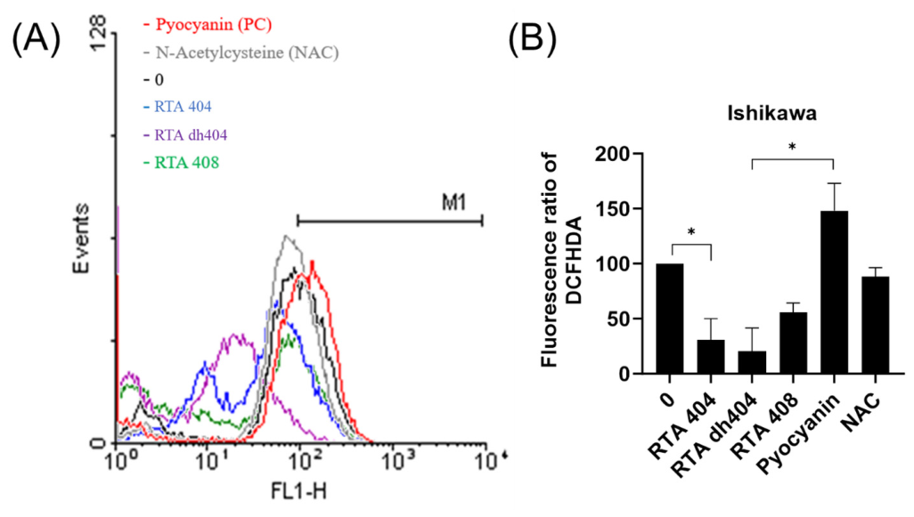

Table 1.
The IC50 of CDDO-Derived (RTA 404, RTA dh404, RTA 408) in Ishikawa cells.
Disclaimer/Publisher’s Note: The statements, opinions and data contained in all publications are solely those of the individual author(s) and contributor(s) and not of MDPI and/or the editor(s). MDPI and/or the editor(s) disclaim responsibility for any injury to people or property resulting from any ideas, methods, instructions or products referred to in the content. |
© 2026 by the authors. Licensee MDPI, Basel, Switzerland. This article is an open access article distributed under the terms and conditions of the Creative Commons Attribution (CC BY) license.
Copyright: This open access article is published under a Creative Commons CC BY 4.0 license, which permit the free download, distribution, and reuse, provided that the author and preprint are cited in any reuse.