
Submitted:
19 October 2026
Posted:
20 October 2026
You are already at the latest version
Abstract
During the progression of chronic liver disease, changes in the acidic microenvironment of liver tissue are closely correlated with the extent of pathological injury, making precise monitoring of these changes critical for disease assessment. Traditional detection methods (e.g., blood gas analysis, tissue pH electrodes) suffer from limitations such as high invasiveness and low spatiotemporal resolution, hindering dynamic reflection of hepatic acid environment evolution. PH-responsive probes (e.g., fluorescent probes, magnetic resonance probes), leveraging their non-invasive, real-time, and high-specificity advantages, have emerged as a research focus in this field. This review focuses on the application of such probes in rat chronic liver disease models: outlining their design principles and types, analyzing their quantitative detection capabilities for liver acidification levels, summarizing their value in correlating pathological processes such as liver inflammation, fibrosis, and cirrhosis, and demonstrating their ability to identify disease progression and evaluate intervention efficacy through early detection of acidic environment changes. Studies indicate that pH-responsive probes provide a tool for elucidating the mechanisms of the acidic microenvironment in chronic liver disease and lay the foundation for developing non-invasive monitoring technologies in clinical translation, holding significant academic and practical value.
Keywords:
pH-responsive probe
; chronic liver disease in rats
; acidic microenvironment of liver tissue
; evaluation of pathological progression
; fluorescent probe
; non-invasive monitoring
1. Introduction
Chronic liver disease is a leading cause of premature death and disability worldwide. According to the WHO Global Liver Disease Report (2025), approximately 2 million deaths annually are attributable to complications of chronic liver diseases, which now rank as the seventh leading cause of global mortality [1,2,3]. Chronic hepatitis B and C infections affect over 325 million individuals, while the prevalence of metabolic-associated fatty liver disease (MAFLD) exceeds 25% globally. In China, where liver disease prevalence is exceptionally high, MAFLD alone surpasses 30%, and together with viral and alcoholic liver diseases, constitutes a formidable public health challenge [2,3]. Without timely and effective intervention, these conditions progress through a common pathogenic sequence: hepatic steatosis and inflammation advance to liver fibrosis, cirrhosis, and ultimately hepatocellular carcinoma (HCC). Liver fibrosis is the pivotal intermediate stage, characterized by persistent activation of hepatic stellate cells (HSCs) and excessive deposition of extracellular matrix (ECM) [1,2,3,4,5]. Extensive evidence confirms that early- and mid-stage fibrosis can be reversed through etiology-targeted therapy, whereas decompensated cirrhosis and HCC carry a dramatically reduced 5-year survival and lack curative options. Consequently, early identification, dynamic monitoring, and precise staging of chronic liver disease are essential clinical imperatives to reduce end-stage liver disease incidence and mortality [4,7,8,9,10].
The liver maintains a tightly regulated tissue pH of 7.35–7.45 under physiological conditions, essential for its metabolic and detoxification functions [8,9,10]. During chronic liver disease, this homeostasis is profoundly disrupted, and the emergence of a locally acidic microenvironment has been identified as a hallmark feature throughout the disease course [8]. Multiple interrelated factors drive hepatic acidification. Injured hepatocytes and infiltrating inflammatory cells undergo aerobic glycolysis (the Warburg effect), producing large amounts of lactate and hydrogen ions even in the presence of oxygen [6,7,8,9]. Activated Kupffer cells, macrophages, and neutrophils release pro-inflammatory cytokines and chemokines that amplify local inflammation and exacerbate glycolytic dysfunction. Inflammation-induced vascular hyperpermeability and sinusoidal architectural disruption impair microcirculation and cause tissue hypoxia, further promoting anaerobic metabolism and hydrogen ion accumulation while hindering their clearance [12]. In fibrotic livers, excessive ECM deposition increases interstitial pressure and impedes lymphatic drainage, preventing the removal of acidic metabolites and establishing a vicious cycle of “metabolic dysregulation → exacerbated inflammation → acidic microenvironment → disease progression” [14]. The acidic microenvironment is not merely a byproduct of pathology but actively drives disease by promoting HSC activation and ECM production, polarizing macrophages toward a pro-inflammatory M1 phenotype, inducing hepatocyte apoptosis and epithelial-mesenchymal transition, and contributing to the immunosuppressive milieu that facilitates HCC invasion, migration, and drug resistance [2,10,11,12,13,14,15]. Thus, precise and dynamic monitoring of hepatic pH is of paramount importance for both basic research and clinical management of chronic liver diseases [13,15].
Traditional methods for assessing hepatic acid–base status have significant limitations. Liver biopsy, the gold standard for staging and grading, is invasive, carries procedural risks, suffers from sampling bias, and cannot provide dynamic, continuous pH measurements [16,17,18,20]. Blood biochemical analyses reflect systemic rather than local hepatic pH and are influenced by respiratory, circulatory, and metabolic factors, resulting in poor specificity and sensitivity for the liver microenvironment [24]. Conventional imaging modalities such as ultrasound, CT, and standard MRI detect only anatomical and morphological changes and are blind to early pH disturbances. Even functional imaging techniques like magnetic resonance spectroscopy and PET are limited by insufficient spatial resolution, high cost, ionizing radiation, and motion artifacts, precluding routine clinical application for chronic liver disease monitoring [22,23,24,25,26,27,28,29].
The rapid development of fluorescence imaging technology has positioned pH-responsive probes as a powerful alternative. These probes undergo changes in fluorescence intensity, emission wavelength, or lifetime in response to environmental pH, enabling quantitative, real-time, in situ detection with high sensitivity, selectivity, and spatiotemporal resolution without ionizing radiation [16,17,18,19,26,27,28,29,30,31,32]. Advances in chemical biology and materials science have yielded targeted probes capable of imaging specific liver cell populations and even subcellular compartments. The emergence of near-infrared II (NIR-II, 1000–1700 nm) probes has dramatically improved tissue penetration depth and reduced autofluorescence, permitting high-signal-to-noise, non-invasive, long-term dynamic imaging of the acidic microenvironment in living livers [27]. These innovations provide not only a visual tool for elucidating the molecular mechanisms of pH regulation in chronic liver disease but also a promising avenue to overcome current clinical diagnostic limitations, facilitating early diagnosis, precise staging, treatment optimization, and efficacy monitoring [25,26,27,28,29,30,31,32,33]. This review systematically examines the mechanisms of hepatic acidification during chronic liver disease progression, summarizes the design strategies and imaging performance of diverse pH-responsive probes, highlights their application advances across different chronic liver diseases, and analyzes current challenges and future directions to guide both basic research and clinical translation [27,28,29,30,31,32,33].See Figure 1.
2. Mechanisms of Acidic Microenvironment Changes in Chronic Liver Disease
2.1. Abnormal Cellular Metabolism
In the healthy liver, hepatocytes rely primarily on aerobic respiration. Chronic liver disease impairs mitochondrial function and inhibits the respiratory chain, shifting cellular metabolism toward anaerobic glycolysis and leading to intracellular and extracellular lactate accumulation with a concomitant decrease in local pH [6,7,8,9,10,11,12,13,14,15,16,17,18,19,20,21,22,23,24,25]. Lipid metabolism is also disrupted: impaired fatty acid β-oxidation results in triglyceride accumulation within hepatocytes, and the buildup of lipid intermediates further perturbs acid–base balance [10].
2.2. Inflammatory Response
Inflammation is central to the pathogenesis of chronic liver disease. In response to viral, toxic, or metabolic insults, macrophages, neutrophils, and other immune cells infiltrate the liver and release inflammatory mediators such as TNF-α, IL-1β, and IL-6 [21,26,27,28,29,30]. These cytokines directly injure hepatocytes, impair metabolic functions, and promote anaerobic metabolism within inflammatory cells by increasing local oxygen consumption and creating a hypoxic microenvironment. The resultant increase in lactate and proton production intensifies tissue acidification [27].
2.3. Circulatory Impairment
As fibrosis and cirrhosis develop, fibrous connective tissue disrupts the normal lobular architecture, compressing and distorting intrahepatic blood vessels and impairing hepatic circulation [29,30,31,32,33,34]. Portal venous obstruction leads to portal hypertension, reduced hepatic perfusion, inadequate oxygen and nutrient delivery, and impaired clearance of metabolic waste [30]. Local tissue hypoxia stimulates further acidic metabolite production, while the diminished buffering capacity of blood due to circulatory stasis compounds the acidification of the hepatic microenvironment [28,29,30,31,32,33,34].
2.4. Imbalance of Hepatobiliary Acid–Base Transporters
Hepatic pH homeostasis depends critically on a functional hepatobiliary transport system. Hepatocytes extrude excess hydrogen ions via the Na⁺/H⁺ exchanger (primarily NHE1) and vacuolar H⁺-ATPase, while cholangiocytes actively secrete bicarbonate (HCO₃⁻) through the Cl⁻/HCO₃⁻ exchanger (AE2) and Na⁺-HCO₃⁻ cotransporter (NBCe1), forming a protective “biliary HCO₃⁻ umbrella” that neutralizes acidic metabolites and shields the biliary epithelium [29,30,31,32,35,36,37,38]. In chronic liver disease, inflammatory cytokines (TGF-β1, TNF-α), hypoxia-inducible factor HIF-1α, and epigenetic modifications (e.g., AE2 promoter hypermethylation) suppress the expression and function of these transporters at transcriptional and translational levels [39]. The resulting failure of hepatocyte proton extrusion and impaired cholangiocyte HCO₃⁻ secretion directly disrupt pH homeostasis even in the absence of increased acid production. Downregulation of AE2 has been documented in primary biliary cholangitis (PBC), primary sclerosing cholangitis (PSC), viral hepatitis, and cirrhosis, and AE2 knockout mice spontaneously develop hepatic acidification, bile duct injury, and fibrosis, confirming that this transporter imbalance is an independent driver of the acidic microenvironment [27,28,29,30,31,32,33,34,35,36,37,38].
2.5. Extracellular Matrix Remodeling and Acid Clearance Barrier
Activated HSCs mediate pathological ECM remodeling, characterized by excessive deposition of type I/III collagen, sinusoidal capillarization, and fibrous septa formation, which creates a dense physical barrier to the diffusion of acidic metabolites from the parenchymal interstitium into the systemic circulation and biliary system [27,28,29,30,31,32,33,34,35,36]. This impeded clearance results in localized acid accumulation and focal acidic microenvironments. The severity of liver fibrosis correlates inversely with local tissue pH. Furthermore, an imbalance between matrix metalloproteinases (MMPs) and tissue inhibitors of metalloproteinases (TIMPs) perpetuates ECM deposition, establishing a vicious cycle of “ECM remodeling → impaired acid clearance → sustained acidic microenvironment → progressive fibrosis” [37,38,39,40].
3. Principles and Types of pH-Responsive Probes
3.1. Working Principle
pH-responsive probes detect environmental pH through interactions between fluorophores and hydrogen ions. pH changes alter the chemical structure or electron cloud distribution of the fluorophore, leading to measurable changes in fluorescence intensity, emission wavelength, or fluorescence lifetime [39]. For example, fluorophores containing protonatable groups undergo protonation in acidic conditions, modifying their conjugated structure and causing enhanced fluorescence intensity or a red shift; deprotonation in alkaline conditions restores the original signal. By quantifying these changes, environmental pH can be analyzed qualitatively or quantitatively [31,32,33,34,35,36,37,38,39,40].See Figure 2.
3.2. Types of Probes
3.2.1. Small-Molecule Fluorescent Probes
Small-molecule probes offer simple synthesis, good membrane permeability, and rapid cellular entry for intracellular pH detection. Common examples include luciferin- and rhodamine-based probes that exhibit marked pH-dependent changes in fluorescence intensity or emission wavelength [30]. However, they are susceptible to intracellular interference and have relatively limited stability.
3.2.2. Nano-Probes
Nano-probes are constructed by combining fluorophores with nanomaterials such as quantum dots, gold nanoparticles, and polymer nanoparticles. Quantum dots provide high fluorescence intensity, excellent photostability, and tunable emission wavelengths. When conjugated with pH-sensitive groups, they yield highly sensitive and selective pH sensors [25,26,27,28,29,30,31,32,33,34,35,36]. Gold nanoparticles exploit surface plasmon resonance to report pH changes via colorimetric or fluorescence signals upon binding pH-sensitive ligands. See Figure 3.Polymeric nanoparticles can be designed to respond within specific pH ranges and can be surface-modified for active targeting, enhancing accumulation in liver tissues [17,18,19,20,21,22,23,24].
3.2.3. Biomolecular Probes
Proteins and nucleic acids whose conformation changes with pH can be exploited as scaffolds. Labeling pH-sensitive proteins with fluorescent groups converts conformational changes into fluorescence signal variations [39]. Nucleic acid aptamers—single-stranded DNA or RNA molecules selected for specific pH recognition—can be conjugated with fluorophores for pH sensing. These probes exhibit excellent biocompatibility and minimal biological interference, although their preparation is relatively complex [35,36,37,38,39,40,41].
3.2.4. pH-Responsive Organic–Inorganic Hybrid Fluorescent Probes
This class integrates pH-responsive organic functional units with inorganic frameworks through coordination or covalent bonds at the molecular level, distinct from physical encapsulation approaches. These hybrids combine the structural stability and biocompatibility of inorganic materials with the high selectivity and sensitive pH response of organic moieties [36,37,38,39,40]. For instance, galactose-modified pH-responsive Zr-MOF probes exhibit a linear fluorescence intensity response over pH 5.0–7.4, enabling precise in vivo imaging of focal acidic microenvironments and staging of liver fibrosis in mice, with the added capability of loading anti-fibrotic drugs for theranostic applications.See Figure 4. Current limitations include poorly controllable in vivo degradation rates, unverified long-term biosafety, and batch-to-batch variability in large-scale synthesis, hindering clinical translation [27,28,29,30,31,32].
3.2.5. pH-Responsive Electrochemiluminescence (ECL) Probes
These probes exploit pH-dependent modulation of electrochemiluminescence efficiency. pH-sensitive groups are introduced into ECL-active molecules (e.g., ruthenium bipyridine, luminol, quantum dots, graphitic carbon nitride). Protonation/deprotonation alters the molecular redox potential, electron transfer efficiency, and excited-state energy, thus modulating ECL intensity [21,22,23,24,25,26,27,28,29,30,31,32,33,34,35]. Acidic conditions can also trigger probe aggregation or dispersion, producing signal changes suitable for high-sensitivity quantitative pH detection. Graphitic carbon nitride-based ECL probes have achieved precise pH quantification over the range 3.0–8.0 and have been successfully applied to measure pH in liver biopsy samples from patients with liver fibrosis, with results showing high concordance with immunohistochemical pathological staging, providing a novel in vitro method for accurate fibrosis staging [42].
4. Application of pH-Responsive Probes in Rat Models of Chronic Liver Disease
4.1. Liver Fibrosis
Liver fibrosis is defined by HSC activation and excessive ECM deposition [37]. In rat fibrosis models, pH-responsive probes have revealed a progressive decrease in liver tissue pH as fibrosis severity increases, attributable to active metabolism of activated HSCs producing acidic metabolites and heightened inflammatory infiltration [25,26,27,28,29,30,31,32,33,34,35,36,37,38,39,40,41]. Fluorescence imaging enables direct visualization, precise localization, and quantification of fibrotic lesions based on the enhanced probe signals in acidic regions. In one study, pH-sensitive nanoprobes injected intravenously into fibrotic rats showed significantly higher fluorescence intensity in fibrotic versus normal liver, with signal intensity positively correlating with fibrosis stage, supporting the use of these probes for early diagnosis and therapeutic monitoring [37,38,39,40,41,42,43].See Figure 5.
4.2. Liver Cirrhosis
Cirrhosis represents advanced structural and functional liver damage [5,6,7,8,9,10,11,12,13,14,15,16,17,18,19,20,21]. pH-responsive probes effectively map the heterogeneous acidic environment in cirrhotic rat livers, where impaired circulation, extensive necrosis, and persistent inflammation produce marked and regionally variable acidification [22,23,24,25,26,27,28,29,30]. The uneven distribution of enhanced fluorescence signals reflects the spatial heterogeneity of cirrhosis and can facilitate liver function assessment and therapeutic evaluation. See Figure 6.Following anti-fibrotic treatment, a reduction in probe signal corresponding to improved hepatic acidity indicates therapeutic efficacy [19,20,21,22,23,24].
4.3. Hepatocellular Carcinoma
HCC is among the most severe complications of chronic liver disease, with rapid tumor cell proliferation and aberrant metabolism generating a profoundly acidic tumor microenvironment [30]. In rat HCC models, pH-responsive probes show markedly enhanced fluorescence in tumor versus normal liver tissue, aiding early tumor detection, margin delineation, and targeted therapy [26,27,28,29]. Some studies have combined pH-responsive probes with chemotherapeutic agents to achieve pH-triggered drug release specifically within the acidic tumor milieu, enhancing therapeutic efficacy while reducing systemic toxicity. For example, a pH-responsive nanodrug delivery system encapsulating chemotherapeutics and surface-modified with pH-responsive probes undergoes structural changes in the acidic tumor microenvironment to release the drug, enabling precise HCC cell killing [23,35].
4.4. Alcoholic Liver Disease (ALD)
In rat models of alcoholic hepatitis, the degree of hepatic inflammatory infiltration and necrosis correlates closely with the extent of acidic microenvironment. pH-responsive probes can quantify inflammatory activity based on hepatic pH signal intensity, showing significant negative correlations with hepatic myeloperoxidase activity and pro-inflammatory cytokine levels (TNF-α, IL-6), thus enabling dynamic monitoring without repeated biopsies [37]. Dual-targeting gut–liver pH-responsive probes have been used to simultaneously observe alcohol-induced gut microbiota dysbiosis, intestinal barrier damage, and exogenous acidic load, alongside their temporal relationship with hepatic acidification, providing in vivo evidence of the gut–liver axis’s regulatory role in ALD progression [40,41,42].
4.5. Cholestatic Liver Disease
The earliest event in cholestasis is dysfunction of the cholangiocyte Cl⁻/HCO₃⁻ exchanger and disruption of the “biliary HCO₃⁻ umbrella,” generating a peribiliary acidic microenvironment that precedes elevations in serum bilirubin and alkaline phosphatase (ALP) or histological changes. pH-responsive probes targeting the biliary epithelium detect a marked reduction in peribiliary pH as early as 24 hours after bile duct ligation (BDL) in rats, while serum ALP is only mildly elevated and no significant structural damage is visible, enabling ultra-early detection of bile duct injury [42,43,44,45,46]. Probe signals allow non-invasive evaluation of biliary HCO₃⁻ secretory function and differentiation between hepatocellular and cholangiocyte injury, offering a novel visual tool for early diagnosis and pathological classification of PBC and PSC [39].
4.6. Acute-on-Chronic Liver Failure (ACLF)
ACLF is a syndrome of acute decompensation in chronic liver disease with high short-term mortality. Early warning is critical, yet traditional indicators fail to provide ultra-early prediction. pH-responsive probes have demonstrated that extensive hepatic acidosis occurs significantly before deterioration of clinical parameters such as serum bilirubin, INR, and MELD score [35,36,37,38,39]. In rat ACLF models, a rapid and substantial decline in liver pH can be detected within 12 hours post-modeling, enabling early prediction of ACLF onset and identification of the optimal window for intervention. The magnitude and distribution of pH decline correlate significantly with pathological liver injury grade, and post-treatment recovery of the hepatic acidic microenvironment shows a strong positive correlation with 28-day survival, establishing pH-responsive probes as non-invasive quantitative biomarkers for early prognostic assessment [42,43,44,45,46].
5. Current Research Status and Cutting-Edge Advances
5.1. International Research Landscape
International efforts in pH-responsive fluorescent probes for liver disease began in the early 2000s, with pioneering work on small-molecule probes for intracellular pH detection. After 2010, research shifted toward in vivo liver microenvironment imaging and application in chronic disease models. Seminal contributions from the NIH, Stanford, MIT, Harvard, Max Planck Institute, University of Amsterdam, University of Tokyo, and others have established a comprehensive framework spanning molecular design, performance optimization, in vivo validation, and preclinical translation [27,28,36].
Key innovations include the development of ratiometric probes that exploit dual-emission fluorophores to enable concentration-independent, accurate pH quantification, overcoming the limitations of single-intensity probes. For example, optimized rhodamine-derivative ratiometric probes with a response window locked to pH 5.0–7.4 achieve in vivo liver pH measurements with errors below 0.1 pH units [33,34,35]. BODIPY-based intramolecular charge transfer probes, near-infrared semicarbazine probes extending into the NIR-I region, and targeted modifications with galactose (for hepatocyte ASGPR), glycyrrhizin (for HSC GA receptor), and other ligands have achieved cell-specific imaging with high liver-to-background signal ratios [30,31,32,33,34,40]. Long-circulating probes stable over weeks have enabled longitudinal tracking of disease progression within individual animals [42].
At the application level, landmark multicenter studies led by the European Association for the Study of the Liver (EASL) used optimized rhodamine-based ratiometric probes to longitudinally track hepatic pH in CCl4-induced fibrosis models, establishing that a 0.3-unit pH decrease corresponds to a one-stage advancement in fibrosis, providing in vivo evidence that the acidic microenvironment is a key regulatory driver of HSC activation and ECM deposition [37]. Stanford researchers further differentiated compensated from decompensated cirrhosis based on probe signals, with pH decline correlating with portal pressure and ascites severity [32]. Importantly, international teams have demonstrated that the hepatic acidic microenvironment is an ultra-early event in metabolic-associated fatty liver disease (MAFLD/NASH), alcoholic liver disease, and cholestasis, occurring before traditional serological and histological abnormalities [29,30,31,32,33,34,35,36,37,38,39,40,47]. Dual-target probes have enabled simultaneous pH-specific imaging of macrophages and HSCs, validating the “immune metabolic reprogramming–acidic microenvironment–liver fibrosis” cascade in vivo [46,47,48,49].
5.2. Domestic Research Progress
In China, research on pH-responsive fluorescent probes for chronic liver disease began somewhat later but has advanced rapidly, achieving internationally influential breakthroughs, particularly in NIR-II probes, precise functional targeting, and novel response mechanisms [5,6,7,8,9,10,11,19,20].
Chinese teams have pioneered NIR-II small-molecule pH-responsive probes to address the penetration depth and autofluorescence limitations of visible and NIR-I probes. The Institute of Chemistry, Chinese Academy of Sciences, developed a benzothiadiazole-based NIR-II ratiometric probe covering the 1000–1700 nm bio-optically transparent window, achieving a penetration depth of up to 1.5 cm in rat liver and a signal-to-noise ratio fivefold higher than NIR-I probes [21,22,23,24,25]. The Changchun Institute of Applied Chemistry further optimized semicarbazole-based NIR-II probes with a pH response locked to 5.0–7.4, enabling continuous dynamic monitoring over four weeks. Targeted imaging for multiple hepatic cell subtypes has been achieved: mannose-modified probes for macrophage imaging in autoimmune hepatitis, glycyrrhizic acid-modified probes for HSC-specific imaging in fibrosis, and the integration of aggregation-induced emission (AIE) mechanisms to overcome aggregation-caused quenching, significantly enhancing stability and sensitivity in complex hepatic microenvironments [36,37].
Domestic application studies, aligned with the Chinese liver disease spectrum, have validated these probes in diverse etiology-specific rat models. In MCD diet-induced NASH, a focal reduction in liver pH was detected as early as 1 week, prior to ALT/AST elevation or histological inflammation [49]. In α-naphthylisothiocyanate-induced cholestasis, peribiliary acidification was identified at 12 hours, significantly earlier than serum bilirubin or ALP increases. In CCl4 fibrosis models, a linear regression model linking hepatic pH to fibrosis stage achieved quantitative error under 0.15 pH units [50]. In ACLF models, pH decline correlated with MELD score and 28-day survival, providing up to 24-hour early warning. Mechanistically, gut–liver dual-targeted probes provided the first in vivo evidence that gut–liver axis dysfunction-mediated exogenous acidic stress drives alcoholic liver injury, and cell-specific probes visualized the cascade of “steatosis → macrophage activation → acidic microenvironment → liver fibrosis” in NASH [48,49,50].
5.3. Cutting-Edge Advances
5.3.1. Intelligent pH-Responsive Probes
Integration of materials science and molecular engineering has produced intelligent probes with environmental adaptability. These probes automatically adjust fluorescence output in response to the biological microenvironment, overcoming fixed response windows and interference susceptibility. Stimulus-responsive polymer probes that transition from curled hydrophobic to extended hydrophilic states under acidic conditions disperse aggregated fluorophores, boosting signal contrast by orders of magnitude. “AND” logic-gate probes activate fluorescence only when both specific pH and characteristic enzyme activity are met, drastically improving specificity in the complex hepatic milieu. These intelligent systems can dynamically adapt to the broad pH range from mild early-disease acidification to severe advanced-stage acidosis, offering flexible tools for whole-course monitoring [46,47,48,49,50].
5.3.2. Multi-Target Detection Probes
Because chronic liver disease involves synergistic contributions from acidosis, oxidative stress, inflammation, and matrix remodeling, probes capable of simultaneous multi-parameter detection are essential. The most mature systems combine pH with reactive oxygen species (ROS) sensing, covalently linking a pH-responsive fluorophore with ROS-sensitive groups (e.g., borate esters, disulfide bonds) to yield independent signals for both parameters. Studies using such bifunctional probes have demonstrated high co-localization of acidic lesions and oxidative stress regions in NASH models, providing direct evidence of their positive feedback relationship. Preliminary progress has been made with tri-target probes for pH, ROS, glutathione (GSH), and MMPs, though signal crosstalk remains a challenge [39,40,41,42,43].
5.3.3. In Vivo Real-Time Dynamic Monitoring
Chronic liver disease progression spans months to years, and static single-time-point imaging cannot capture the dynamic evolution of the acidic microenvironment. Fluorescence lifetime imaging (FLIM) quantifies pH via excited-state lifetime, independent of probe concentration, photobleaching, or tissue scattering, enabling continuous absolute pH monitoring for hours in live rats. Hyperspectral fluorescence imaging combined with spectral unmixing algorithms precisely distinguishes probe signals at different pH levels, further improving accuracy in complex tissues [49,50]. Photoacoustic-fluorescence multimodal real-time imaging merges deep penetration with high sensitivity for whole-liver pH mapping. Implantable optical fiber fluorescence probes, placed via minimally invasive surgery, permit continuous dynamic monitoring over weeks, enabling longitudinal tracking of disease progression and therapeutic response within the same animal and significantly reducing inter-individual variability [36,37,38,39,40,41].
6. Conclusions and Future Perspectives
pH-responsive probes have demonstrated irreplaceable value in assessing acidic microenvironment changes during chronic liver disease progression in rodent models, opening novel technical pathways for fundamental research and clinical translation. These probes detect acidic signals before morphological changes or serological abnormalities, enabling non-invasive dynamic monitoring from simple steatosis through steatohepatitis, fibrosis, and cirrhosis. Through cell-specific imaging, they visually elucidate spatiotemporal pH distribution patterns and their intrinsic associations with hepatocyte injury, inflammatory infiltration, HSC activation, and matrix remodeling, while providing non-invasive, reproducible pharmacodynamic metrics for preclinical anti-fibrotic drug development, reducing animal usage and variability.
However, significant challenges remain in transitioning from basic research to clinical application. The performance of current probes is not yet fully optimized for the complex hepatic biological environment: visible and NIR-I probes suffer from autofluorescence interference from endogenous bilirubin and hemoglobin; some probe response windows inadequately match the actual pathological pH range; and cross-sensitivity to coexisting substances such as ROS and metal ions requires improved selectivity. Photobleaching and chemical degradation during in vivo circulation compromise long-term dynamic monitoring accuracy. The in vivo biological behavior and safety of probes are insufficiently characterized: as the liver is the primary metabolic and clearance organ, most small-molecule probes are rapidly taken up and cleared, resulting in short lesion accumulation times and potential nonspecific background signals. Systematic evaluation of long-term metabolite toxicity is lacking, representing a critical safety hurdle for clinical use. Critically, the translational pipeline from rodent models to human application contains major gaps: the vast majority of studies remain confined to rodent models without validation in non-human primates; synthesis protocols, imaging parameters, and data processing lack standardization across laboratories; and the complex clinical regulatory pathway for fluorescent probes necessitates large-scale trials to confirm safety and efficacy in humans.
Future advances, driven by the deep convergence of materials science, chemical biology, and medical imaging, will systematically enhance probe performance. Next-generation probes based on aggregation-induced emission and NIR-II emission are expected to further overcome tissue penetration and signal-to-noise constraints. Intelligent logic-gate and multi-target integrated probes will enable simultaneous detection of multiple liver microenvironment parameters, providing a more comprehensive reflection of disease complexity. Multimodal imaging will balance anatomical localization with functional readouts, delivering more precise diagnostic information. Beyond early diagnosis and efficacy monitoring in chronic liver diseases, the application scenarios of pH-responsive probes will expand to include intraoperative navigation for liver tumors, evaluation of immunotherapy efficacy, and detection of biliary tract disease microenvironments, and may extend to other systemic diseases associated with acidic microenvironments, fostering groundbreaking advances in biomedical science.
Author Contributions
ZL: Research and drafting – initial draft preparation, writing – review and editing. JL: Research and drafting – initial draft preparation, writing – review and editing. QL: Research and drafting – initial draft preparation, writing – review and editing. ZW: Supervision and writing – review and editing. FL: Supervision and writing – review and editing. JX: Supervision and writing – review and editing. JH: Supervision and writing – review and editing.
Acknowledgments
We would like to express our gratitude to Professor Song Bo and Professor Xing Chengjuan for their assistance and guidance in this research.
Conflict of Interest
The authors declare that the research was conducted in the absence of any commercial or financial relationships that could be construed as a potential conflict of interest.
References
- EASL-EASD-EASO Clinical Practice Guidelines on the management of metabolic dysfunction-associated steatotic liver disease (MASLD). J. Hepatol. 2024. [CrossRef] [PubMed]
- Guideline for the Prevention and Treatment of Metabolic Dysfunction-associated Fatty Liver Disease (Version 2024). J. Clin. Transl. Hepatol. 2025. [CrossRef] [PubMed]
- Hepatocellular carcinoma: updates on epidemiology, surveillance, diagnosis and treatment. Nat. Rev. Gastroenterol. Hepatol. 2025. [CrossRef] [PubMed]
- Role of lactate and lactate metabolism in liver diseases. Int. J. Mol. Med. 2024. [CrossRef] [PubMed]
- The acidic microenvironment in the perisinusoidal space critically determines bile salt-induced activation of hepatic stellate cells. Hepatology 2024. [CrossRef] [PubMed]
- CaM/CaMKII mediates activation and proliferation of hepatic stellate cells regulated by ASIC1a. Eur. J. Pharmacol. 2022. [CrossRef] [PubMed]
- Emerging roles of bile acids in chronic hepatitis, liver cirrhosis, and hepatocellular carcinoma. World J. Gastroenterol. 2023. [CrossRef] [PubMed]
- Lactic acid: The culprit behind the immunosuppressive microenvironment in hepatocellular carcinoma. J. Cancer 2025. [CrossRef] [PubMed]
- Progress in pH-Sensitive sensors: essential tools for organelle pH detection, spotlighting mitochondrion and diverse applications. Front. Cell Dev. Biol. 2024. [CrossRef] [PubMed]
- Electroanalytical Strategies for Local pH Sensing at Solid–Liquid Interfaces and Biointerfaces. ACS Sensors, 2024.
- pH-triggered hydrophility-adjustable fluorescent probes for simultaneously imaging lipid droplets and lysosomes and the application in fatty liver detection. Biosens. Bioelectron. 2025. [CrossRef] [PubMed]
- A microenvironment-adapted dual-cascade activated molecular probe for precise differentiation of liver disease types. Biomaterials 2025. [PubMed]
- Noninvasive Detection of Extracellular pH in Human Benign and Malignant Liver Tumors Using CEST MRI. Front. Oncol. 2025. [PubMed]
- A targeted fluorescent nanosensor for ratiometric pH sensing at the cell surface. Sci. Rep. 2024. [CrossRef] [PubMed]
- In Vivo Evaluation of Liver Injury and Arthritis Enabled by a Nitroxyl-Activated Near-Infrared Fluorescence and Photoacoustic Dual-Modality Imaging Probe. JACS Au 2025. [CrossRef] [PubMed]
- Advances in Organic Small Molecule-Based Fluorescent Probes for Precision Detection of Liver Diseases. J. Am. Chem. Soc. 2025. [CrossRef] [PubMed]
- A Cascade-Responsive AND-Logic-Activatable Nanoprobe for Intraoperative Fluorescence Imaging of Colorectal Cancer. Adv. Mater. 2026. [CrossRef]
- Visible-spectrum target-stabilizable fluorescent nanobodies for high-contrast imaging in living cells. Nat. Methods 2026. [PubMed]
- Fluorescence visualization of small molecular biomarkers and microenvironmental factors in chronic diseases. Coord. Chem. Rev. 2026. [CrossRef]
- Fluorescent Probes for Disease Diagnosis. In Chemical Reviews; 2024.
- Advances in Organic Small Molecule-Based Fluorescent Probes for Precision Detection of Liver Diseases. J. Am. Chem. Soc. 2025. [CrossRef] [PubMed]
- Theranostic probes for hepatocellular carcinoma: from bench to bedside. J. Hepatol. 2024. [PubMed]
- Molecular Imaging Probes for Early Detection and Staging of Liver Fibrosis. Bioconjugate Chem. 2025. [CrossRef] [PubMed]
- Molecular imaging of liver fibrosis: current state and future directions. Nat. Rev. Gastroenterol. Hepatol. 2023. [PubMed]
- Affibody-based optical imaging probe for noninvasive detection of liver fibrosis. Theranostics 2026. [CrossRef] [PubMed]
- Manganese-based type I collagen-targeting MRI probe for in vivo imaging of liver fibrosis. npj Imaging 2025. [CrossRef] [PubMed]
- pH-activatable PET probe for precise imaging of hepatocellular carcinoma with ultrahigh tumor-to-background ratio. Nat. Commun. 2025. [PubMed]
- Hepatic stellate cell-targeted theranostic probe for MRI-guided photothermal therapy of liver cirrhosis and prevention of hepatocarcinogenesis. Adv. Funct. Mater. 2024. [PubMed]
- MARTY, L.; FOUASSIER, L.; LEFEBVRE, T.; et al. Hepatic fibrosis and the microenvironment: pH, lactate, and metabolic reprogramming[J]. Int. J. Mol. Sci. 2024, 25(12), 6802. [Google Scholar]
- HUSSEIN, M.; EL-SAYED, M.; ABDEL-RAHMAN, M.; et al. Tissue pH changes in chronic liver disease: From fibrosis to cirrhosis[J]. J. Clin. Pathol. 2023, 76(8), 521-528. [Google Scholar]
- ZHANG, J.; LI, Y.; WANG, H.; et al. Overcoming therapeutic resistance of hepatomas by targeting the tumor microenvironment[J]. Front. Oncol. 2022, 12, 988956. [Google Scholar] [CrossRef] [PubMed]
- NISHIDA, N.; KUDO, M.; IKEDA, H.; et al. Current perspectives on the immunosuppressive niche and role of fibrosis in hepatocellular carcinoma[J]. Cancers 2021, 13(23), 5890. [Google Scholar] [PubMed]
- Intraperitoneal Delivery of Iopamidol to Assess Extracellular pH of Orthotopic Pancreatic Tumor Model by CEST-MRI. Contrast Media Mol. Imaging 2023, 36704211. [CrossRef] [PubMed]
- HENNINGER, B.; PLAIKNER, M.; ZOLLER, H.; ct al. Performance of different Dixon-based mmethods for MR liver iron assessment in comparison to a bbiopsy-validated R2- relaxometry method. Eur. Radiol. 2021, 31(4), 2252–2262. [Google Scholar] [PubMed]
- MUEHLER, M. R.; VIGEN, K.; HERNANDO, D. Reproducibility of liver R2* quantification for liver irou quantification from cardiac R2" acquisitions[J]. Abdom. Radiol. (NY 2021, 46(9), 4200–4209. [Google Scholar] [PubMed]
- CRAFT, M. L.; EDWARDS, M.; JAIN, T. P. R2 and R2* MRI assessment of liver iron content in ann undifferentiated diagnostic population with byperferritinaemia, and impact on clinical decision making[J]. Eur. JJ Radiol. 2021, 135, 109473. [Google Scholar] [CrossRef] [PubMed]
- XU, H.; ZHANG, N.; YANG, D. W.; et al. Scan Time Reduction in Intravoxel Incoherent Motion Diffusion-Weighted Imaging and Diffusion Kurosis Imaging of the Abdominal Organs: Using a Simmultaneous Multislice Technique With Different Acceleration Factors[J]. J. Compput Assist Tomogr. 2021, 45(4), 507–515. [Google Scholar] [CrossRef] [PubMed]
- CAO, L.; CHEN, J.; DUAN, T.; et al. Diffusion kurtosis imaging (DKI) of hcpatocellular carcinoma: correlaation with microvascular invasion and histologic grade[J]. Quant. Imaging MMed Surg. 2019, 9(4), 590–602. [Google Scholar]
- WANG, W. T.; YANG, L.; YANG, Z. X.; et al. Assessment of Microvascular Invasion of Hepaatocellular Carcinoma with Diffusion Kurtosis Imaging[J]. Radiology 2018, 286(2), 571–580. [Google Scholar] [CrossRef] [PubMed]
- IDILMAN, I.S.; LIJ; YIN, M.; et al. MR elastography of liver: cuurrent status and future perspectives[J]. Abdom. Radiol. (NY) 2020, 45(11), 34444–3462. [Google Scholar] [CrossRef] [PubMed]
- JAYAKUMAR, S.; MIDDLETON, M. S.; LAWITZ, E. J.; et al. Longitudinal correlations between MRE, MRI-PDFF, and liver histology in patients with non-alcoholic steatohe- patittis: Analysis of data from a phase II trial of selon- sertib[J]. J. Hepatol. 2019, 70((1), 133–141. [Google Scholar] [PubMed]
- SELVARAJ, E. A.; MÓZES, P.F. E.; JAYASWAL, A.; et al. Dia- gnostic accuracy of elastography and magnetic resonannce imaging in patients with NAFLD: A systematic review and meta-analysis[J]. J. Hepatol. 2021, 75(4), 770–785. [Google Scholar] [PubMed]
- REN, H.; YANG, D.; XU, H.; et al. Effect of breath bholding at the end of the inspiration annd expiration phases on liver stiffnesss measured by 2D-MR elastography[J]. Abdom. Radiol. (NY) 2021, 46(6), 2516–2526. [Google Scholar] [PubMed]
- In vivo imaging of the spatial heterogeneity of intratumoral acidosis (pH) as a marker of the metastatic phenotype in breast cancer. Breast Cancer Res. 2025, 39522065. [CrossRef] [PubMed]
- Multiscale imaging on proton pump-driven acidity for assessing tumor progression and metastasis. Nat. Commun. 2026, 41545373. [CrossRef] [PubMed]
- Overcoming Immune Exclusion in a Murine Model of Cirrhotic Hepatocellular Carcinoma via Cryoablation: Insights from Molecular Extracellular pH Imaging. Cancer Res. Commun. 2025, 39968742.
- BOSS, A.; BARTH, B.; FILLI, L.; et al. Simmultaneous multi- slice echo planar diffusion weighted imagiing of the liver and the pancreas: Optimization of signal-too-noise ratio and acquisition time and application to intravvoxel incoherent motion analysis[J]. Eur. J. Radiol. 2016, 85(11), 1948–1955. [Google Scholar] [PubMed]
- TANABE, M.; HIGASHI, M.; BENKERT, T.; ct al. Reduced Field-of-View Diffusion-Weighted Maggnetic Resonance Imaging of the Pancreas With Tilted Excitation Plane A Preliminary Study[J]. J. Magn. Reson Immagin. 2021, 54(3), 715–720. [Google Scholar] [CrossRef] [PubMed]
- HARDER, F. N.; KAMAL, O.; KAISSIS, G. A.; et al. Qualita- tive and Quantitative comparison of Respiratory Triggered Reduced Field-of-View (FOOV) Versus Full FOV Diffusion Weighted Imaging (DWI) in Pancreatic Pathologiees[J]. Acad. Radiol. 2021, 28 Suppl 1, S234–S243. [Google Scholar] [PubMed]
- DONATI, F.; CASINI, C.; CEERVELLI, R.; et al. Diffusion- weightted MRI of solid pancreatic lesions: comparison between reduced field-of-view and large field-of-viewsequences[J]. Eur. J. Radiol. 2021, 143, 109936. [Google Scholar] [CrossRef] [PubMed]
Figure 1.
(A) Proposed responsive mechanism of pH-Eu/Gd to pH fluctuations, illustrating a reversible, pH-dependent modulation of inner-sphere hydration that gradually shifts the coordination equilibrium between “MR-on/TGL-off” and “MR-off/TGL-on” states. (B) Schematic illustration of pH-responsive MR and TGL dual-modal imaging enabled by pH-Eu/Gd. Buffering of tumor extracellular acidosis has been investigated as a therapeutic strategy to limit acid-driven growth and metastasis.
Figure 1.
(A) Proposed responsive mechanism of pH-Eu/Gd to pH fluctuations, illustrating a reversible, pH-dependent modulation of inner-sphere hydration that gradually shifts the coordination equilibrium between “MR-on/TGL-off” and “MR-off/TGL-on” states. (B) Schematic illustration of pH-responsive MR and TGL dual-modal imaging enabled by pH-Eu/Gd. Buffering of tumor extracellular acidosis has been investigated as a therapeutic strategy to limit acid-driven growth and metastasis.
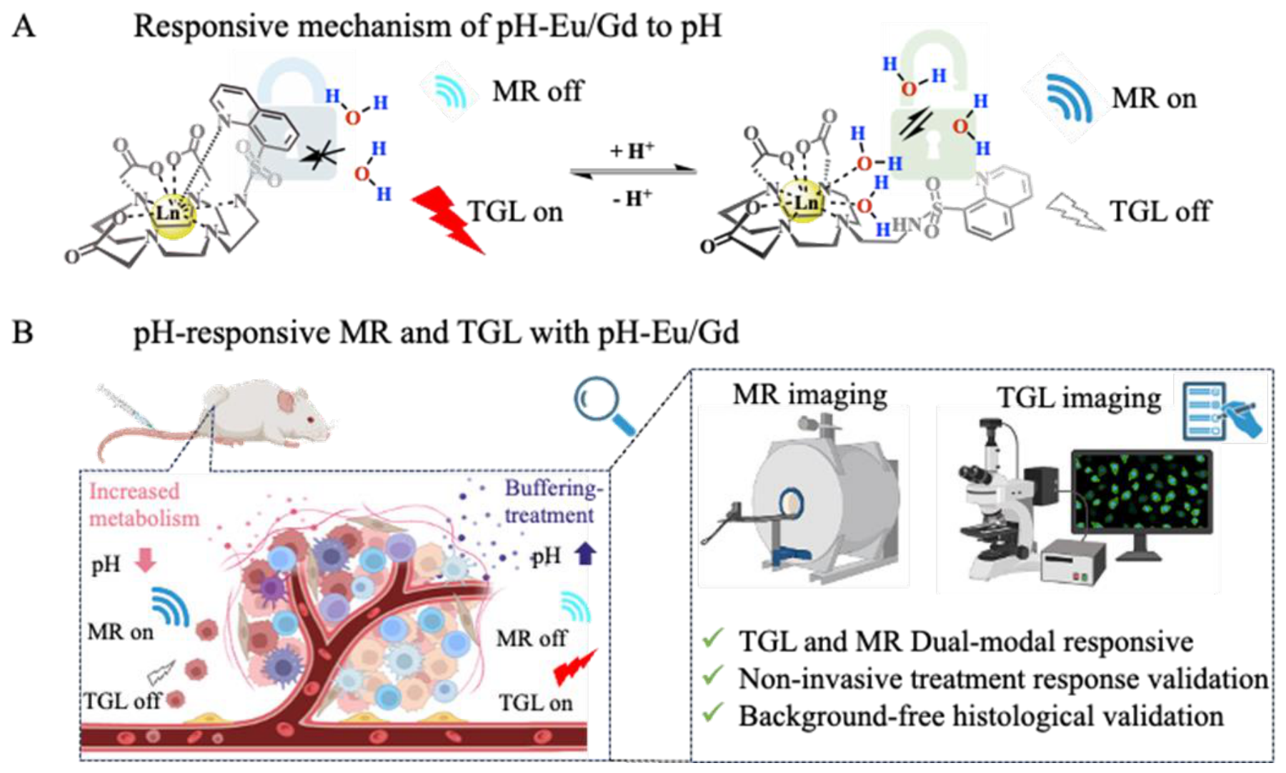
Figure 2.
Schematic illustration of a HClO-activatable bimodal TGLI/MRI probe, for in vivo and in vitro imaging.
Figure 2.
Schematic illustration of a HClO-activatable bimodal TGLI/MRI probe, for in vivo and in vitro imaging.
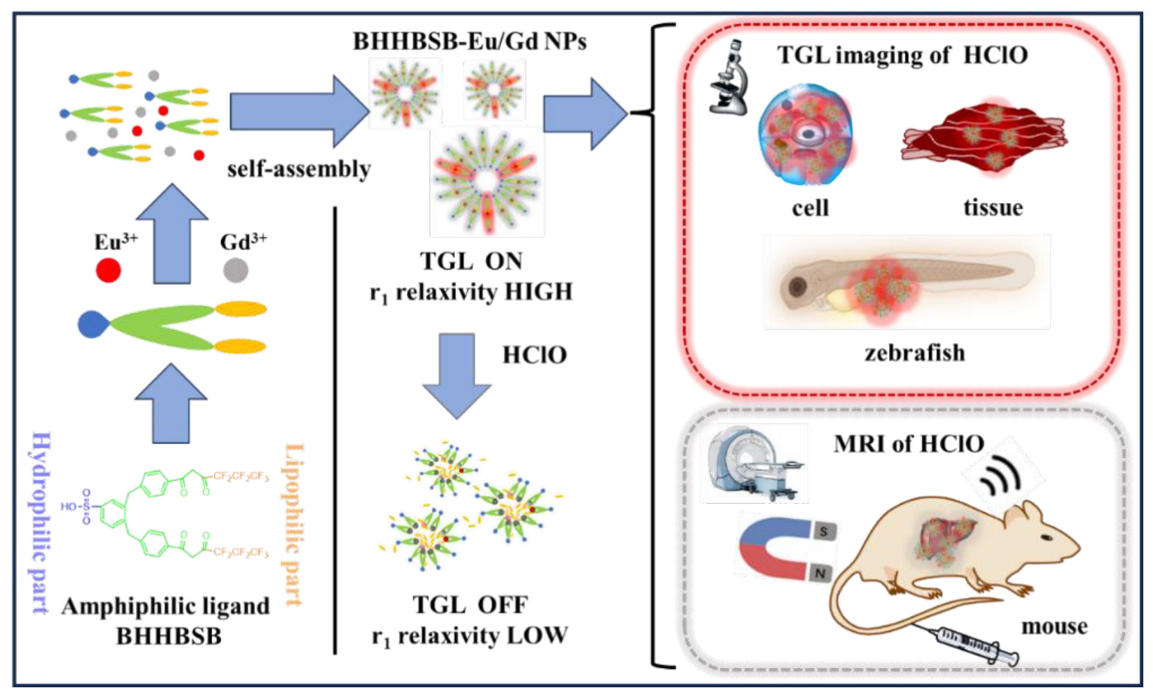
Figure 3.
(A) Representative T1-weighted MR images of vehicle- and NaHCO3-treated tumor-bearing mice at different time points following i.v. injection of pH-Eu/Gd (11 µmol/kg). Gray-scale images show anatomical structure, overlaid with false-color maps of tumor region.
Figure 3.
(A) Representative T1-weighted MR images of vehicle- and NaHCO3-treated tumor-bearing mice at different time points following i.v. injection of pH-Eu/Gd (11 µmol/kg). Gray-scale images show anatomical structure, overlaid with false-color maps of tumor region.
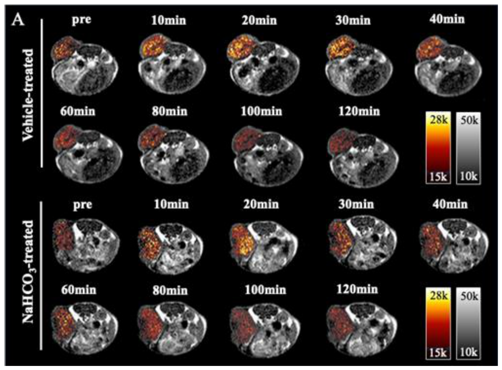
Figure 4.
(A) Longitudinal relaxation rates (1/T1) of pH-Eu/Gd in aqueous solutions at different pH values (37 °C, 0.5 T). (B) Correlation between longitudinal relaxivity (r1) and ambient pH. (C) Representative T1-weighted MR images of pH-Eu/Gd (330 µM) at different pH values (37 °C, 0.5 T). (D) Longitudinal relaxivity of pH-Eu/Gd at pH 6, 7.4, and 8 in the presence and absence of BSA. (E) Reversibility of the MR response of pH-Eu/Gd to sequential acid-base perturbations (NaOH/HCl). (F) Selectivity of the MR response of pH-Eu/Gd toward H⁺ in comparison with various competing species (500 µM) at pH 7.4: 1, pH = 7.4; 2, Zn2+; 3, Ca2+; 4, K+; 5, Mg2+; 6, cysteine; 7, homocysteine; 8, glutathione; 9, glucose; 10, pH = 4.
Figure 4.
(A) Longitudinal relaxation rates (1/T1) of pH-Eu/Gd in aqueous solutions at different pH values (37 °C, 0.5 T). (B) Correlation between longitudinal relaxivity (r1) and ambient pH. (C) Representative T1-weighted MR images of pH-Eu/Gd (330 µM) at different pH values (37 °C, 0.5 T). (D) Longitudinal relaxivity of pH-Eu/Gd at pH 6, 7.4, and 8 in the presence and absence of BSA. (E) Reversibility of the MR response of pH-Eu/Gd to sequential acid-base perturbations (NaOH/HCl). (F) Selectivity of the MR response of pH-Eu/Gd toward H⁺ in comparison with various competing species (500 µM) at pH 7.4: 1, pH = 7.4; 2, Zn2+; 3, Ca2+; 4, K+; 5, Mg2+; 6, cysteine; 7, homocysteine; 8, glutathione; 9, glucose; 10, pH = 4.
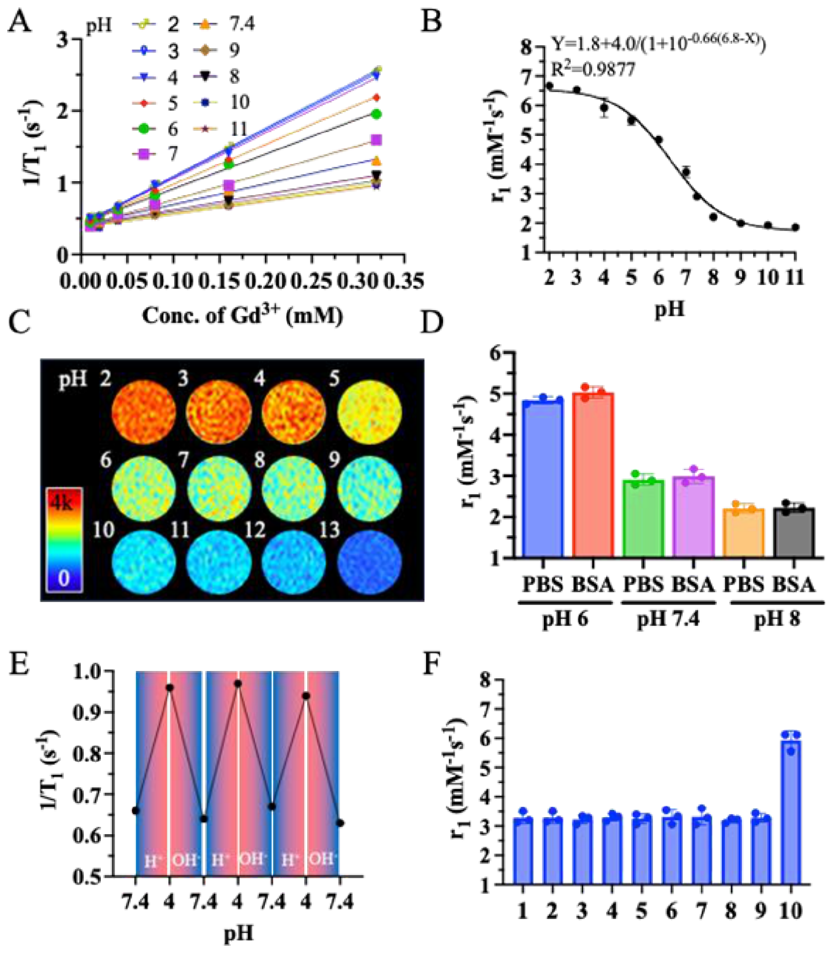
Figure 5.
Overview diagram of pH-responsive probes for evaluating the progression of fibrosis and detecting therapeutic interventions in liver fibrosis models.
Figure 5.
Overview diagram of pH-responsive probes for evaluating the progression of fibrosis and detecting therapeutic interventions in liver fibrosis models.
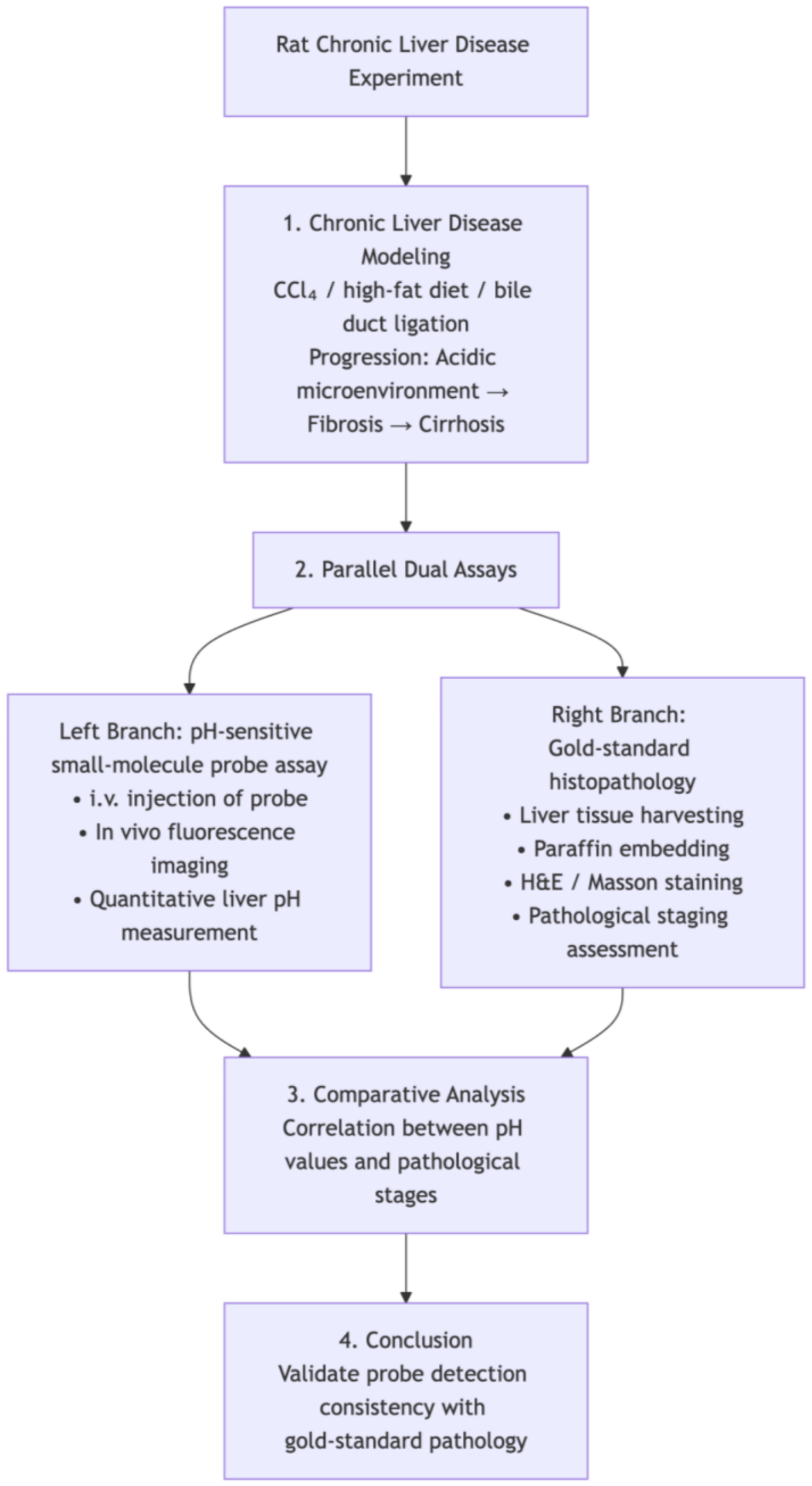
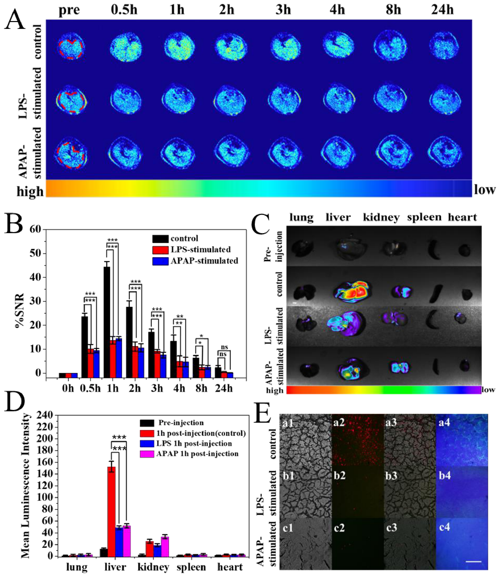
Figure 6.
(A) Representative T1-weighted MRI of healthy control, mouse model of LPS-, and APAP-induced acute liver injury after intravenously injected with BHHBSB-Eu/Gd NPs. (B) Quantitative analysis of liver %SNR in healthy control, LPS-, and APAP-treated mice after injected with BHHBSB-Eu/Gd NPs (Statistical analysis was performed using one-way ANOVA with Tukey’s post hoc test, *P < 0.05, **P < 0.01, ***P < 0.001). (C) Ex vivo luminescence images of the organs of healthy control, LPS-, and APAP-treated mice at 1h after injection of BHHBSB-Eu/Gd NPs (merged images of the luminescence and bright field imaging). (D) Quantitative analysis of luminescence intensities of ex vivo luminescence imaging (Statistical analysis was performed using one-way ANOVA with Tukey’s post hoc test, ***P < 0.001). (E) Luminescence imaging of frozen liver sections harvested from healthy control, LPS-, and APAP-treated mice 1 h after injection of BHHBSB-Eu/Gd NPs. (1: bright field; 2: time-gated luminescence; 3: steady-state luminescence; 4: overlap of bright field and time-gated luminescence. Scale bar: 50 μm).
Figure 6.
(A) Representative T1-weighted MRI of healthy control, mouse model of LPS-, and APAP-induced acute liver injury after intravenously injected with BHHBSB-Eu/Gd NPs. (B) Quantitative analysis of liver %SNR in healthy control, LPS-, and APAP-treated mice after injected with BHHBSB-Eu/Gd NPs (Statistical analysis was performed using one-way ANOVA with Tukey’s post hoc test, *P < 0.05, **P < 0.01, ***P < 0.001). (C) Ex vivo luminescence images of the organs of healthy control, LPS-, and APAP-treated mice at 1h after injection of BHHBSB-Eu/Gd NPs (merged images of the luminescence and bright field imaging). (D) Quantitative analysis of luminescence intensities of ex vivo luminescence imaging (Statistical analysis was performed using one-way ANOVA with Tukey’s post hoc test, ***P < 0.001). (E) Luminescence imaging of frozen liver sections harvested from healthy control, LPS-, and APAP-treated mice 1 h after injection of BHHBSB-Eu/Gd NPs. (1: bright field; 2: time-gated luminescence; 3: steady-state luminescence; 4: overlap of bright field and time-gated luminescence. Scale bar: 50 μm).
Disclaimer/Publisher’s Note: The statements, opinions and data contained in all publications are solely those of the individual author(s) and contributor(s) and not of MDPI and/or the editor(s). MDPI and/or the editor(s) disclaim responsibility for any injury to people or property resulting from any ideas, methods, instructions or products referred to in the content. |
© 2026 by the authors. Licensee MDPI, Basel, Switzerland. This article is an open access article distributed under the terms and conditions of the Creative Commons Attribution (CC BY) license.
Copyright: This open access article is published under a Creative Commons CC BY 4.0 license, which permit the free download, distribution, and reuse, provided that the author and preprint are cited in any reuse.