

Submitted:
18 October 2026
Posted:
20 October 2026
You are already at the latest version
Abstract
The acid growth hypothesis explains the role of apoplastic acidification in auxin-induced plant cell expansion; however, it does not, on its own, explain the conditions under which this process develops into sustained, irreversible growth. Apoplastic acidification, cell-wall loosening, and the presence of turgor pressure do not invariably result in the same growth response. This indicates that cell expansion should be understood as an outcome of interactions among hormonal regulation, cell-wall mechanical properties, water availability, and metabolic status. Here, we propose an integrative mechanistic model of plant cell expansion regulation in which the transition to a growth-permissive functional state is a central component. Within this framework, apoplastic pH is not considered a sufficient determinant of growth in itself, but rather a regulatory factor that influences the capacity of the cell wall to undergo mechanical deformation. The realization of growth depends on the coordination of this process with turgor pressure, water transport, metabolic capacity, and mechanosensory feedback mechanisms. This approach integrates the regulation of plasma membrane H⁺-ATPases, cell-wall remodelling, the TOR–SnRK1 signalling axis, and Ca²⁺- and reactive oxygen species-mediated pathways into a unified conceptual framework. The model describes cell expansion as a sequence of transitions among restricted, growth-permissive, actively expanding, and reinforcing states. Rather than rejecting the acid growth hypothesis, it extends its explanatory scope by linking local changes in apoplastic pH and cell-wall mechanics to the regulation of water relations, metabolism, and sustained growth. The proposed framework provides a basis for experimental investigation of the mechanisms through which plant cells convert regulatory signals into irreversible expansion.
Keywords:
plant cell expansion
; acid growth hypothesis
; growth-permissive state
; apoplastic pH
; cell-wall mechanics
; turgor
; expansins
; plasma membrane H⁺-ATPase
; TOR–SnRK1
; mechanosensing
; Ca²⁺–ROS feedback
; mechanochemical state transitions
1. Introduction: Why the Acid Growth Hypothesis Needs to Be Extended
1.1. The Classical Hypothesis and Its Molecular Elaboration
For half a century, plant cell expansion has been explained through a compact causal scheme: auxin activates the plasma membrane H⁺-ATPase, protons are extruded into the apoplast, the acidified wall becomes more extensible, and turgor-driven expansion follows [1,2]. Experimental studies have shown that acidic solutions induce extension of isolated walls, that auxin stimulates proton extrusion and elongation within minutes, and that expansins, wall proteins that are most active at acidic pH, can induce wall extension without detectable polysaccharide hydrolysis [3,4,5]. During the last decade the upstream steps of this scheme have been resolved at the molecular level. TIR1/AFB–Aux/IAA auxin perception is required for rapid apoplast acidification and hypocotyl growth [6]; SAUR proteins inhibit PP2C-D phosphatases and thereby maintain activation of plasma membrane H⁺-ATPases [7]; and transmembrane kinases (TMKs) of the cell-surface auxin signalling system interact with and phosphorylate H⁺-ATPases, promoting wall acidification and cell elongation [8]. Recent syntheses have reviewed this progress in detail [9,10].
The physical description of growth is equally established. In the formulation of Lockhart, irreversible expansion occurs when turgor pressure exceeds a yield threshold, and its rate is proportional to the excess pressure multiplied by a wall extensibility coefficient [11]. These parameters can be estimated experimentally, for example by in vivo stress relaxation [12], and the hydraulic dimension of growth has been formalized as well [13,14].
1.2. Limits of a Linear Scheme
The established observations summarized above are not in dispute. What is in question is whether a chain of the form auxin → H⁺ → wall loosening → growth is sufficient as an explanatory architecture. Several lines of evidence suggest that it is not. First, one and the same signal can produce opposite pH and growth outcomes: auxin promotes cell expansion in shoots but inhibits it in roots, and in roots two antagonistic pathways, a TMK1-dependent acidifying pathway and an intracellular TIR1/AFB-dependent pathway that causes net proton influx and apoplast alkalinization, converge on apoplastic pH [15,16]. Second, an intact pH response does not guarantee growth: in Arabidopsis hypocotyls, auxin- and fusicoccin-induced growth depends on CSI1- and microtubule-dependent wall architecture, even though crystalline cellulose content is not the determining factor [17]. Third, wall mechanics has proved more complex than softness: the response of epidermal walls to mechanical stretch differs from their response to growth-like loosening [18], and the older picture of load-bearing xyloglucan tethers has been revised toward a limited number of mechanically critical contact sites [19,20]. Fourth, sustained growth requires biosynthetic and energetic capacity that is governed by TOR and SnRK1 signalling rather than by auxin alone [21,22]. Finally, mechanical stress is not only a consequence of growth but feeds back on cytoskeletal organization and growth patterns [23,24].
The linear scheme therefore leaves at least six questions open: why identical pH can give different growth; why high turgor does not always produce expansion; why an extensible wall does not guarantee growth; why metabolic limitation can block long-term growth even when signalling is intact; why mechanical load changes the cellular response; and how a reversible signal results in irreversible deformation.
1.3. Distinguishing Established Knowledge from the Conceptual Contribution
To distinguish established scientific knowledge from the conceptual contribution of this work, throughout the manuscript we differentiate experimentally supported findings, interpretations grounded in available evidence, propositions developed within our proposed framework, and hypotheses that require further experimental testing. This distinction allows us to identify which elements of the proposed architecture are directly supported by existing evidence and which represent our interpretation or conceptual extension of previous models.
1.4. Central Problem and Central Hypothesis
The problem addressed here can be stated briefly: a growth signal is not growth. We propose viewing cell expansion as the outcome of a regulated transition of the cell into a growth-permissive mechanochemical state, in which hormonal permission, apoplastic pH, the molecular state of the wall, water balance, turgor, metabolic capacity and mechanosensory feedback are mutually consistent. We hypothesize that plant cell expansion is not directly generated by auxin, apoplastic acidification, wall loosening or turgor alone, but emerges when these components become sufficiently coordinated to establish and maintain such a state. This statement is a conceptual proposition of the present article; it is not presented as an experimentally demonstrated fact.
1.5. Conceptual Novelty
The conceptual novelty of this framework does not reside in proposing a new molecular component of cell expansion. Rather, it lies in reorganizing established molecular, hydraulic, metabolic and mechanical processes into a unified state-transition architecture in which a growth-permissive state determines whether regulatory signals can be converted into sustained irreversible expansion. In practical terms, we make four moves: we place hormones upstream as modifiers of permission rather than as generators of growth; we treat apoplastic pH as a gate on the molecular state of the wall instead of a direct driver; we treat the efficiency with which turgor is converted into irreversible deformation as a state-dependent variable; and we close the causal chain through a mechanosensory Ca²⁺–ROS feedback that returns the mechanical outcome of growth to the regulatory layer.
The framework also continues a line of reasoning developed in our earlier work. We have previously proposed that plant regulatory networks can be described as hypothetically multistable, with a TOR-dominant growth state, an ABA-dominant abiotic stress tolerance state and immune-dominant configurations, and that transitions among them are governed by hormonal switches and the TOR–SnRK1 energy-sensing module [25,26]; earlier syntheses described plant immunity as a multilevel signal–metabolic network [27] and jasmonate–ethylene signalling as a metabolic defence system [28]. The present article asks the complementary question at the level of a single expanding cell: what must be true, mechanically and biochemically, for a growth-dominant regulatory state to be converted into irreversible expansion?
2. From Acid Growth to a Mechanochemical Model
2.1. What the Classical Model Explains
The classical model accounts well for the rapid, auxin-dependent growth response of shoot tissues, for the pH sensitivity of isolated-wall extension, and for the existence of a molecular pH-sensitive wall agent, the expansin [1,5,9]. It also connects hormone perception with a membrane transport process, thereby linking signalling to the physical chemistry of the apoplast [6,8].
2.2. What It Does Not Explain
It does not specify what makes the same acidification effective in one wall and ineffective in another [17]; it does not distinguish the signalling step from the mechanical execution step; it treats the wall as a passive substrate and turgor as a constant; it contains no energetic term; and it contains no feedback from the mechanical result back to the cell. These omissions, rather than any error in its established components, motivate the extension proposed below.
2.3. Revised Roles of the Principal Components
We propose that apoplastic pH be treated as a regulatory gate rather than as a direct driver of growth. In this view, acidification shifts the probability that the wall occupies a mechanically permissive molecular state. Turgor is the mechanical driving force, but its effect depends on the state of the wall [11,13]. The wall is the mechanical constraint and, at the same time, a dynamic molecular system that is remodelled during growth [10,20]. Metabolic capacity is a separate condition for sustained growth, because wall polymers, membranes and proteins must be synthesized and the proton pump itself consumes ATP [21,29]. Finally, mechanical feedback is part of the regulation, not only its consequence [23,24].
2.4. A New Causal Architecture
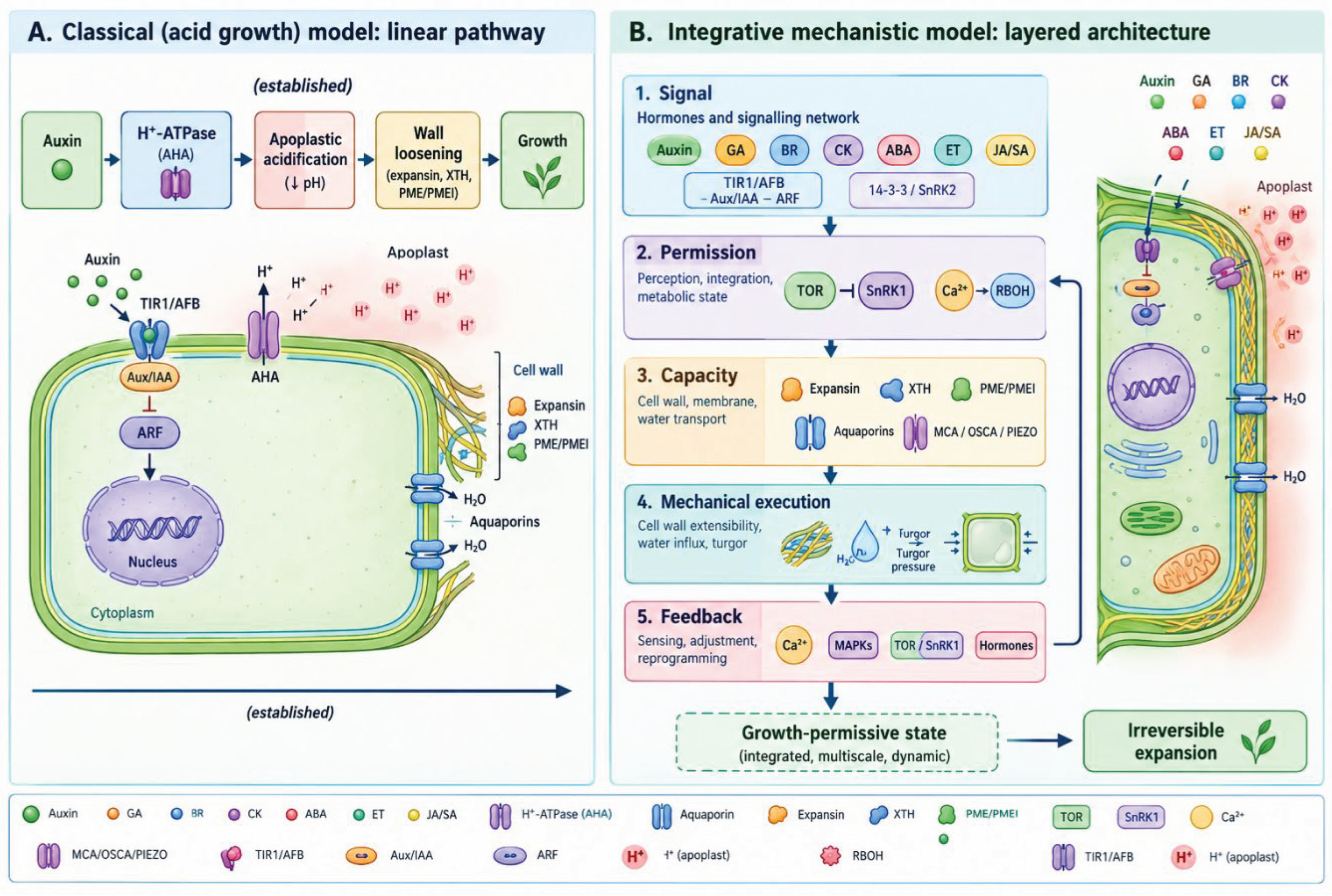
Within the framework developed here, we replace the linear chain by a layered architecture: Signal → Permission → Capacity → Mechanical execution → Feedback. Signals (hormonal and environmental) establish regulatory permission; permission acts on pH and on the molecular state of the wall, while capacity (metabolic and hydraulic) determines whether the cell can sustain what permission allows; mechanical execution converts turgor into deformation; and feedback returns the mechanical state to the regulatory layer through mechanosensing, Ca²⁺ and ROS.
Figure 1.
From the Acid Growth Hypothesis to an Integrative Mechanistic Model of Plant Cell Expansion. (A) The classical linear scheme: auxin → plasma membrane H⁺-ATPase → apoplastic acidification → wall loosening → growth. (B) The proposed layered architecture Signal → Permission → Capacity → Mechanical execution → Feedback, with the growth-permissive state at its centre. Elements in (A) are established; the layered organization in (B) is the conceptual proposition of this article.
Figure 1.
From the Acid Growth Hypothesis to an Integrative Mechanistic Model of Plant Cell Expansion. (A) The classical linear scheme: auxin → plasma membrane H⁺-ATPase → apoplastic acidification → wall loosening → growth. (B) The proposed layered architecture Signal → Permission → Capacity → Mechanical execution → Feedback, with the growth-permissive state at its centre. Elements in (A) are established; the layered organization in (B) is the conceptual proposition of this article.
3. The Growth-Permissive State as the Central Concept of the Model
3.1. Definition
We propose the following definition. A growth-permissive state is a transient cellular state in which regulatory signalling, wall permissiveness, water availability, turgor, metabolic capacity and mechanosensory feedback are sufficiently coordinated to permit sustained irreversible expansion. It is not equivalent to a high hormone level; it is an integrative functional property of the cell.
3.2. Regulatory and Mechanical Permission
Regulatory permission asks whether the cell’s signalling state allows growth: whether growth-promoting transcriptional programmes are derepressed (for example through Aux/IAA degradation and DELLA turnover) and whether the proton pump is switched on [30,31,32]. Mechanical permission asks whether the wall can convert turgor into deformation, which depends on cellulose organization, the matrix and the activity of wall-modifying proteins [17,20].
3.3. Metabolic and Hydraulic Capacity
3.4. State Versus Rate
A distinction that we regard as essential is that between state and rate. The growth-permissive state determines whether growth is possible; the growth rate determines how fast it proceeds. Using the Lockhart formalism as a starting point [11], we propose to write the relative expansion rate as
where φ is wall extensibility, Y the yield threshold, P turgor pressure, w the molecular state of the wall, F the mechanical history and load, and M the metabolic capacity. Equation (1) is a conceptual extension: the first expression is the classical relation, whereas the dependence of φ and Y on pH, wall state and mechanical history and the metabolic ceiling Rmax are our propositions. In this form, the growth-permissive state corresponds to the region of parameter space in which P > Y and Rmax is not limiting.
RER = φ(pH, w, F) · [P − Y(pH, w, F)] for P > Y, and RER ≤ Rmax(M)
4. Hormonal Architecture of Regulatory Permission
4.1. Auxin as the Primary Regulatory Signal
4.1.1. Perception and Transcriptional Permission
Auxin is perceived by the TIR1/AFB F-box proteins, which, in the presence of auxin, recruit Aux/IAA repressors for ubiquitin-dependent degradation, thereby releasing auxin response factors (ARFs) [30,31,35]. We interpret this as transcriptional permission for growth rather than as growth itself: derepression of ARF targets is necessary for a long-term growth programme, but it does not by itself generate deformation.
4.1.2. Fast, Non-Genomic and Cell-Surface Branches
Rapid auxin responses are mediated in part by TIR1/AFB-dependent perception that operates on a time scale of minutes [6,9], and in part by TMK-based cell-surface signalling [8]. In roots, an intracellular TIR1/AFB-dependent pathway promotes net proton influx and apoplast alkalinization while TMK1 acts in the opposite direction [15]. In root tissue, auxin has been shown to steer cell expansion through apoplastic pH regulation [16].
4.1.3. Regulation of the Plasma Membrane H⁺-ATPase
Activity of plasma membrane H⁺-ATPases (AHAs in Arabidopsis) is controlled mainly through their C-terminal autoinhibitory region, whose phosphorylation and subsequent 14-3-3 binding activate the pump [29]. Experimental studies have shown that PKS5 inhibits the pump by preventing its interaction with 14-3-3 [36], that SAUR proteins sustain activation through PP2C-D inhibition [7], that TMKs phosphorylate AHAs [8], and that the peptide hormone RALF1, acting through the receptor kinase FERONIA, triggers phosphorylation of AHA2 and apoplast alkalinization, thereby restricting root cell expansion [37]. Taken together, these data support the interpretation that the pump is a convergence node for several regulatory inputs, both activating and inhibitory.
4.1.4. Electrochemical Consequences
4.1.5. Limits of a Hormone-Centred Description
The auxin → H⁺-ATPase → pH axis is the best-characterized permission pathway, but the evidence summarized in Section 1.2 shows that it does not determine mechanical execution. Auxin creates a regulatory precondition for growth; whether this precondition is converted into expansion depends on wall state, water and metabolic capacity [15,17].
4.2. Gibberellins as Regulators of Growth Competence
Gibberellin binding to GID1 triggers degradation of DELLA proteins, which restrain growth by sequestering growth-promoting transcription factors [32]. DELLA proteins bind PIF transcription factors, and GA-dependent DELLA degradation releases them to promote cell elongation [38,39]; DELLAs also integrate signals from ethylene, ABA and auxin [40]. We interpret the GA–DELLA axis as setting growth competence: it determines whether the transcriptional machinery of elongation is available when other permission signals arrive.
4.3. Brassinosteroids as Amplifiers of the Permissive State
Brassinosteroid signalling through BRI1/BAK1 and BIN2 controls the transcription factors BZR1 and BES1 [41]. BZR1 forms, together with ARF6 and PIF4, a central circuit that regulates hypocotyl elongation [42], and BR, GA and light signals converge on a common transcriptional module [43]. Brassinosteroid treatment also induces phosphorylation of the plasma membrane H⁺-ATPase during hypocotyl elongation [44] and regulates the expression of cellulose synthase genes [45]. This is, to our knowledge, one of the few cases in which a single hormone has documented inputs into both the pH gate and the wall-rebuilding layer, which is why we describe BR as an amplifier of the permissive state rather than as an independent growth trigger. Its connection to carbon availability through TOR-dependent accumulation of BZR1 is discussed in Section 9 [46].
4.4. Cytokinins and Developmental Competence
In the root meristem, cytokinin acts through ARR1 to control the balance between cell division and differentiation, in part in antagonism to auxin [47]; cytokinin–auxin interactions more generally shape developmental programmes [48]. We consider these data to indicate that cytokinin influences the developmental context in which the same expansion machinery operates, which is relevant for Prediction 5 (Section 20).
4.5. ABA as a Regulator of Exit from the Permissive State
Abscisic acid is perceived by PYR/PYL/RCAR receptors, which inhibit clade A PP2C phosphatases and thereby release SnRK2 kinases [49,50]. Growth responses to ABA are context dependent: under water limitation, endogenous ABA is associated with maintenance of root growth and restriction of shoot growth, in interaction with ethylene [51]. ABA signalling is also connected with TOR: the TOR kinase and the ABA receptor regulate each other reciprocally, which balances growth with stress response [52]. In our previous work we proposed that ABA signalling defines a distinct ABA-dominant state of the regulatory network, with SnRK2 and RAF kinases connecting stress perception to TOR [26]. Within the framework developed here, ABA is treated as a regulator of exit from the growth-permissive state, mainly through hydraulic and metabolic restriction rather than through a direct block of the proton pump.
4.6. Ethylene as a Modifier of Mechanical and Stress State
Ethylene signalling proceeds through EIN2 and the EIN3/EIL transcription factors [53], and its effect on growth is organ- and context-dependent, promoting expansion in some systems and restricting it in others [54]. In deepwater rice, internode elongation is associated with ethylene- and gibberellin-regulated expansin expression [55]. We therefore consider ethylene a modifier of the mechanical and stress state of the cell, whose sign of effect on the permissive state depends on tissue and developmental context. Our earlier synthesis of jasmonate–ethylene signalling as a metabolic defence system [28] provides the immune-metabolic counterpart of this role.
4.7. Jasmonate and Salicylic Acid: Allocation Between Growth and Defence
Jasmonate perception through COI1 and JAZ repressors is a central determinant of the growth–defence balance [56], and jasmonate signalling can restrain growth by interfering with gibberellin signalling [57]. Growth–defence trade-offs are best understood as regulated resource allocation [58], and salicylic acid contributes to defence-state signalling [59]. We treat JA and SA as regulators that redistribute metabolic resources and thereby affect capacity (Section 9), a view consistent with our earlier network-level description of immunity [25,27].
4.8. Hormonal Crosstalk
4.9. Hormones as Modifiers of Permission, and Gene- And Genome-Level Organization of the Permission Layer
We propose that hormones be placed at the beginning of the causal chain as modifiers of the regulatory state, which in turn acts on pH, metabolism, water and wall mechanics; they should not be summed as inputs into a growth output. The permission layer also has a gene- and genome-level organization. Expansins form a multigene superfamily [62], and altered expansin expression changes growth in transgenic rice [63]; xyloglucan endotransglucosylase/hydrolase (XTH) proteins likewise form a large family [64]. Cell-type-specific maps of auxin responses [65] and high-resolution spatiotemporal expression maps of the root [66] show that hormone-responsive and wall-related genes are expressed in coordinated, spatially defined modules. Endoreduplication contributes to cell-size control [67], and genetic variation affects epidermal cell area and number in leaves [68]. These data are consistent with the possibility that genotype-dependent differences in growth reflect differences in the coupling between modules, and not only in hormone levels. We formulate this as a hypothesis: genotypes with the same hormonal response but different expression modules of wall-modifying genes should differ in the position of the pH and mechanical thresholds described in Section 14.
5. Apoplastic pH as a Molecular Gate of the Mechanical State
5.1. Formation of Apoplastic pH
Apoplastic pH is set by proton extrusion through H⁺-ATPases, by proton-coupled transport and by wall buffering [29]. It is dynamic and spatially variable: auxin-dependent changes of apoplastic pH have been resolved in roots [16], and oscillations of extracellular pH accompany tip growth of root hairs [69].
5.2. pH Dependence of Wall Proteins and Polymers
Expansins loosen walls in a pH-dependent manner, with activity favoured at acidic pH [3,4]. Solid-state NMR experiments identified a limited, cellulose-associated population of xyloglucan as the likely target of expansin [70]. XTH proteins catalyse endotransglucosylation and, in some members, hydrolysis of xyloglucan [64,71]; their contribution to growth and their pH dependence remain member-specific and debated, particularly since xyloglucan tethers appear less load-bearing than once thought [19].
Pectin provides a second, chemically distinct pH-sensitive layer. Homogalacturonan methylesterification is controlled by pectin methylesterases (PMEs) and their inhibitors (PMEIs) [72], and the PME–PMEI interaction itself has been shown to depend on pH [73]. Demethylesterification of pectin changes wall mechanics and underlies organ initiation at the shoot apex [74]. It has been proposed that acid growth requires dynamic remodelling of homogalacturonan through tight control of wall pH [75]. The consequences of demethylesterification are not unidirectional: Ca²⁺ cross-linking of demethylesterified blocks may stiffen the wall, whereas the same demethylesterification may make homogalacturonan susceptible to polygalacturonases [72,75]. We interpret this duality as the reason why pH cannot be mapped directly onto extensibility.
5.3. pH as a Switch of Wall State
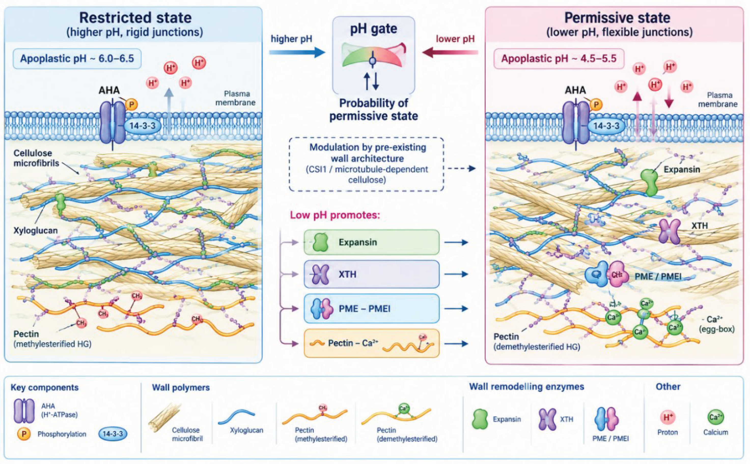
Within the framework developed here, we suggest the following reading. Low apoplastic pH does not equal growth; rather, low pH increases the probability of a mechanically permissive molecular state of the wall, by simultaneously changing expansin activity, PME/PMEI interaction, pectin–Ca²⁺ association and, potentially, XTH activity. The magnitude and even the sign of the mechanical consequence depend on the pre-existing state of the polymer network, which is why the model predicts that identical pH can result in different growth (Prediction 1, Section 20). The experimental observation that growth stimulated by auxin or fusicoccin depends on wall architecture [17] is consistent with this interpretation.
Figure 2.
pH-Gated Molecular and Mechanical Transition of the Plant Cell Wall. Apoplastic pH acts as a gate that shifts the probability of a mechanically permissive wall state. The gate acts in parallel on expansin activity, XTH, the PME–PMEI system and pectin–Ca²⁺ association, and its mechanical consequence depends on the pre-existing architecture of cellulose and matrix. The schematic is conceptual.
Figure 2.
pH-Gated Molecular and Mechanical Transition of the Plant Cell Wall. Apoplastic pH acts as a gate that shifts the probability of a mechanically permissive wall state. The gate acts in parallel on expansin activity, XTH, the PME–PMEI system and pectin–Ca²⁺ association, and its mechanical consequence depends on the pre-existing architecture of cellulose and matrix. The schematic is conceptual.
6. The Cell Wall as a Dynamic Molecular–Mechanical System
6.1. The Wall Is Not a Passive Substrate
6.2. Cellulose Architecture
Cellulose synthase complexes move along cortical microtubules [77], and microtubule-dependent, crossed-polylamellate wall architecture is required for acid growth in hypocotyls [17]. This is the strongest experimental support we are aware of for the idea that the mechanical outcome of acidification is conditioned by wall architecture.
6.3. Hemicellulose–Cellulose and Matrix Interactions
Biomechanical analysis with substrate-specific endoglucanases led to a revised architecture in which a limited number of cellulose–cellulose contacts, mediated by xyloglucan, rather than widespread xyloglucan tethers, determine wall mechanics [19]. Imaging and mechanical experiments on epidermal walls further showed that the structural response to stretch differs from that to loosening [18]. We take these results to indicate that wall mechanical state is an emergent property of the whole polymer network and not the function of a single protein.
6.4. Pectin, Cross-Linking and Oxidative Reinforcement
The pectin network contributes to mechanics in a methylesterification- and Ca²⁺-dependent manner [74,75]. Apoplastic class III peroxidases participate in cross-linking and in the production of reactive oxygen species and have documented roles in plant development [78]; hydroxyl radicals can in addition cause polysaccharide scission and wall loosening in vitro and in vivo [79,80]. These oxidative processes therefore have two opposite potential effects: loosening and reinforcement, a point that becomes central in Section 11.
6.5. Loosening is Not Destruction
A central proposition of the model is that wall loosening is not equivalent to wall destruction. Expansion requires a sequence of loosening, deformation, rebuilding and stabilization. The first step relies on expansin-type and, possibly, XTH-type activities [4,64]; deformation depends on the mechanical load [11,12]; rebuilding requires cellulose synthesis and matrix deposition [45,77]; and stabilization involves cross-linking and pectin remodelling [74,78]. A wall that is loosened without rebuilding would thin and fail, which cells avoid by coordinating these steps; how this coordination is achieved is, in our view, an open question (Section 23).
7. Turgor, Water and Hydraulic Capacity
7.1. Turgor as the Mechanical Driving Force
7.2. Osmotic Effectors, Vacuole and Transport
Turgor depends on osmotic solute accumulation. The H⁺-ATPase energizes ion uptake [29], and vacuolar cation/proton exchangers of the NHX family regulate vacuolar pH and K⁺ homeostasis and are required for normal growth [81]. The vacuole is not merely a solute store: auxin regulates SNARE-dependent vacuolar morphology, which restricts cell size [82], and extracellular matrix sensing by FERONIA and leucine-rich repeat extensins controls vacuolar expansion during elongation [83]. Anion channels of the SLAC1 type regulate turgor in guard cells [84]; whether anion-channel families (SLAC/SLAH, ALMT) play comparable roles in expanding cells has not, to our knowledge, been systematically addressed and is part of the experimental agenda in Section 21.
7.3. Aquaporins and Water Flux
Aquaporins of the PIP and TIP subfamilies are regulated at multiple levels and control plant water relations [34,85]. Auxin regulates aquaporin function during lateral root emergence [86], and expression of a tonoplast aquaporin (γ-TIP) correlates with cell enlargement [87]. These data suggest that the hydraulic conductance of the cell is a regulated variable which can be coordinated with hormonal permission.
7.4. Why High Turgor Does Not Guarantee Growth
In the Lockhart framework, turgor above the yield threshold is a necessary condition for irreversible expansion, but the expansion rate is determined by wall extensibility [11,12]. It follows that high turgor in combination with a mechanically restrictive wall is not expected to produce growth, a situation we consider in the scenarios of Section 17 and test in Prediction 2.
8. Turgor-to-Growth Coupling Efficiency
8.1. Definition
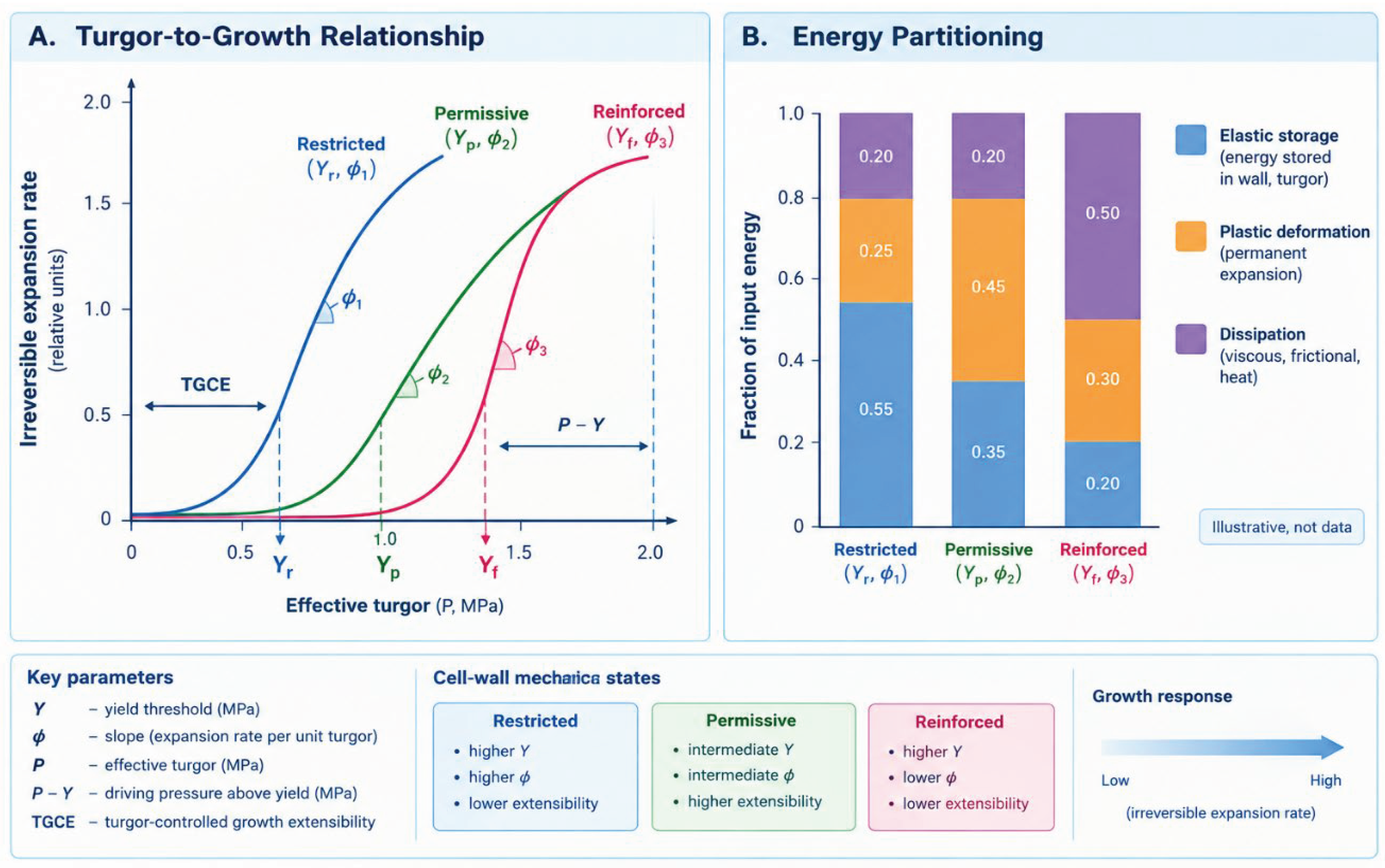
We introduce the notion of turgor-to-growth coupling efficiency (TGCE) to describe how effectively turgor is converted into irreversible expansion. We propose to define it as the irreversible fractional volumetric expansion rate per unit of effective turgor excess,
which, under the Lockhart formulation, corresponds to the extensibility φ [11]. We acknowledge this equivalence explicitly. The conceptual extension is that, in our model, TGCE is not a constant of the wall but an output of the permission layer and of mechanical history, and that the input energy is partitioned between elastic storage, plastic (irreversible) deformation and dissipation. Turgor × wall permissiveness → irreversible expansion.
TGCE = (1/V)(dV/dt)irr / (P − Y)
8.2. Elastic Deformation, Plastic Deformation and Creep
Cell walls display elastic (reversible) and plastic (irreversible) behaviour, and growth is associated with the latter [12,14]. The mechanical response of epidermal walls under strain suggests that elasticity is not the primary step of growth [18]. We propose to describe this by the plastic fraction of total deformation under a defined load, which can be measured by extensometry and stress relaxation [12].
8.3. Energy Dissipation
Not all mechanical energy supplied by turgor results in expansion: part is stored elastically and part is dissipated. We regard the efficiency of conversion as a property that can differ between walls with identical turgor and identical pH, and we place this idea at the centre of Prediction 8.
Figure 3.
Turgor-to-Growth Coupling as a Function of Cell-Wall Mechanical State. Conceptual relationship between turgor and irreversible expansion rate for walls in restricted, permissive and reinforced states, with partitioning of mechanical input between elastic storage, plastic deformation and dissipation. Slopes correspond to the state-dependent extensibility φ and thresholds to Y in Equation (1); the curves are illustrative and not experimental data.
Figure 3.
Turgor-to-Growth Coupling as a Function of Cell-Wall Mechanical State. Conceptual relationship between turgor and irreversible expansion rate for walls in restricted, permissive and reinforced states, with partitioning of mechanical input between elastic storage, plastic deformation and dissipation. Slopes correspond to the state-dependent extensibility φ and thresholds to Y in Equation (1); the curves are illustrative and not experimental data.
9. Metabolic Capacity as the Second Gate of Growth
9.1. Energy, Carbon and Biosynthetic Supply
Expansion consumes energy and building blocks: ATP for proton pumping and biosynthesis [29], carbon skeletons, amino acids for protein synthesis, lipids for membrane growth, and nucleotide sugars and other precursors for wall polysaccharides. We group these requirements under the term metabolic capacity.
9.2. TOR as a Regulator of Growth Capacity
The TOR kinase integrates nutrient and energy information and promotes anabolic processes [33]. Experimental studies have shown that glucose–TOR signalling reprograms the transcriptome and activates meristems [21], that auxin promotes TOR activation through the small GTPase ROP2 [88], and that TOR promotes accumulation of the brassinosteroid-responsive transcription factor BZR1 to balance growth with carbon availability [46]. These data are of particular interest for the present model because they link metabolic status to two of the permission-layer hormones (auxin and brassinosteroid).
9.3. SnRK1 as a Sensor of Energy Limitation
SnRK1 acts as a central integrator of transcriptional networks under stress and energy deficiency [22], and SnRK1 and TOR together modulate growth–defence trade-offs [89]. The TOR kinase and the ABA receptor regulate each other reciprocally [52], which provides a molecular connection between the metabolic gate and the stress-hormone layer.
9.4. The TOR–SnRK1 Balance
In our earlier synthesis we described the TOR–SnRK1 module, together with trehalose-6-phosphate, as a molecular switch between growth and stress adaptation, and proposed that a TOR-dominant growth state is one of several hypothetically stable regulatory configurations [25]. In the present framework we assume that the balance between TOR and SnRK1 activity determines whether the cell has the biosynthetic capacity to sustain growth. This is a proposition, and Section 23 lists the open question of where the boundary lies.
9.5. The Metabolic Gate in the Causal Architecture
Consider the situation in which auxin signalling is high, apoplastic pH is permissive and turgor is sufficient. The model predicts that if metabolic capacity is low, sustained growth is not supported, because wall rebuilding and the energy-consuming pH gate cannot be maintained. Initial acidification-driven expansion may occur, but its duration should be limited (Prediction 4). This prediction distinguishes the model from purely hormone-centred or purely mechanical descriptions.
10. Mechanosensing: How Mechanical Deformation Becomes a Molecular Signal
10.1. Membrane Tension and Mechanosensitive Channels
Plants possess several families of channels that respond to membrane tension or osmotic and mechanical stimuli [90]. Experimental studies have established that MCA proteins are involved in Ca²⁺ influx and touch sensing in roots [91] and that Arabidopsis MCAs are Ca²⁺-permeable, tension-sensitive channels [92]. MscS-like (MSL) proteins provide mechanosensitive channel activities in the root [93]; OSCA1 mediates osmotic-stress-evoked Ca²⁺ increases [94]; and a PIEZO channel is required for root mechanotransduction [95]. We include CNGC channels in the model as candidate Ca²⁺-permeable components, but their role as mechanosensors in expanding cells has yet to be established.
10.2. Wall-Sensing Receptor Kinases
The receptor-like kinase FERONIA is required for mechanical signal transduction in seedlings [96] and maintains cell wall integrity during salt stress through Ca²⁺ signalling [97]. FERONIA also participates in extracellular matrix sensing that controls vacuolar expansion [83] and, as noted above, in the RALF-dependent control of the proton pump [37]. We regard FERONIA as a candidate molecular link between the wall (and its mechanical state) and the pH and hydraulic layers of the model.
10.3. Ca²⁺ Decoding
10.4. Mechanics as Feedback
Mechanical stress prescribes cortical microtubule behaviour at subcellular and supracellular scales [23,100], and mechanical feedback restricts sepal growth and shape [24]. Elastic domains regulate growth and organogenesis in the shoot apical meristem [101]. These observations suggest that mechanics does not merely result from signalling: it returns to the regulatory system. Within the framework developed here, we describe this loop as growth → deformation → mechanosensing → Ca²⁺ → remodelling. The part of this loop that links mechanosensitive Ca²⁺ signalling to remodelling of the wall in diffusely expanding cells is, in our assessment, less directly supported than the cytoskeletal part and is therefore treated as a model proposition.
11. ROS as a Mechanochemical Feedback Signal
11.1. RBOH and Its Dual Activation by Ca²⁺ and Phosphorylation
Plant NADPH oxidases (RBOHs) produce apoplastic superoxide. Experimental evidence has shown that RBOHD is synergistically activated by Ca²⁺ binding and phosphorylation [102], and that a CPK–RBOH circuit supports rapid signal propagation [99]. ROS produced by NADPH oxidase regulate cell growth [103], and in root hairs a local positive feedback between ROS, Ca²⁺ and the growth machinery determines cell shape [104].
11.2. Ca²⁺, ROS and pH in Mechanosensing
In Arabidopsis roots, Ca²⁺ regulates ROS production and pH during mechanosensing [105], and in root hairs oscillations in extracellular pH and ROS modulate tip growth [69]. These studies are of central importance for the present model because they show that Ca²⁺, ROS and apoplastic pH are physically coupled in growing cells. We note that this evidence comes mainly from tip-growing cells and from the root, and its extension to diffusely expanding cells is, at this stage, an interpretation.
11.3. ROS-Dependent Wall Remodelling
Hydroxyl radicals loosen walls in vitro and in vivo [79,80], whereas peroxidase-dependent reactions are associated with cross-linking and reinforcement [78]. ROS are therefore signals and wall-modifying agents, not only damage markers. The model uses this ambivalence: depending on the chemical context, ROS may promote loosening during the permissive phase and reinforcement during the stabilization phase.
11.4. The Ca²⁺–ROS Loop
We propose the following feedback sequence: mechanical deformation → Ca²⁺ → RBOH → ROS → wall remodelling → altered mechanics. Its individual segments are supported by the studies cited above; the complete loop in an expanding cell is a conceptual proposition (Prediction 7).
12. The Mechanochemical Feedback Loop as an Integrating Circuit
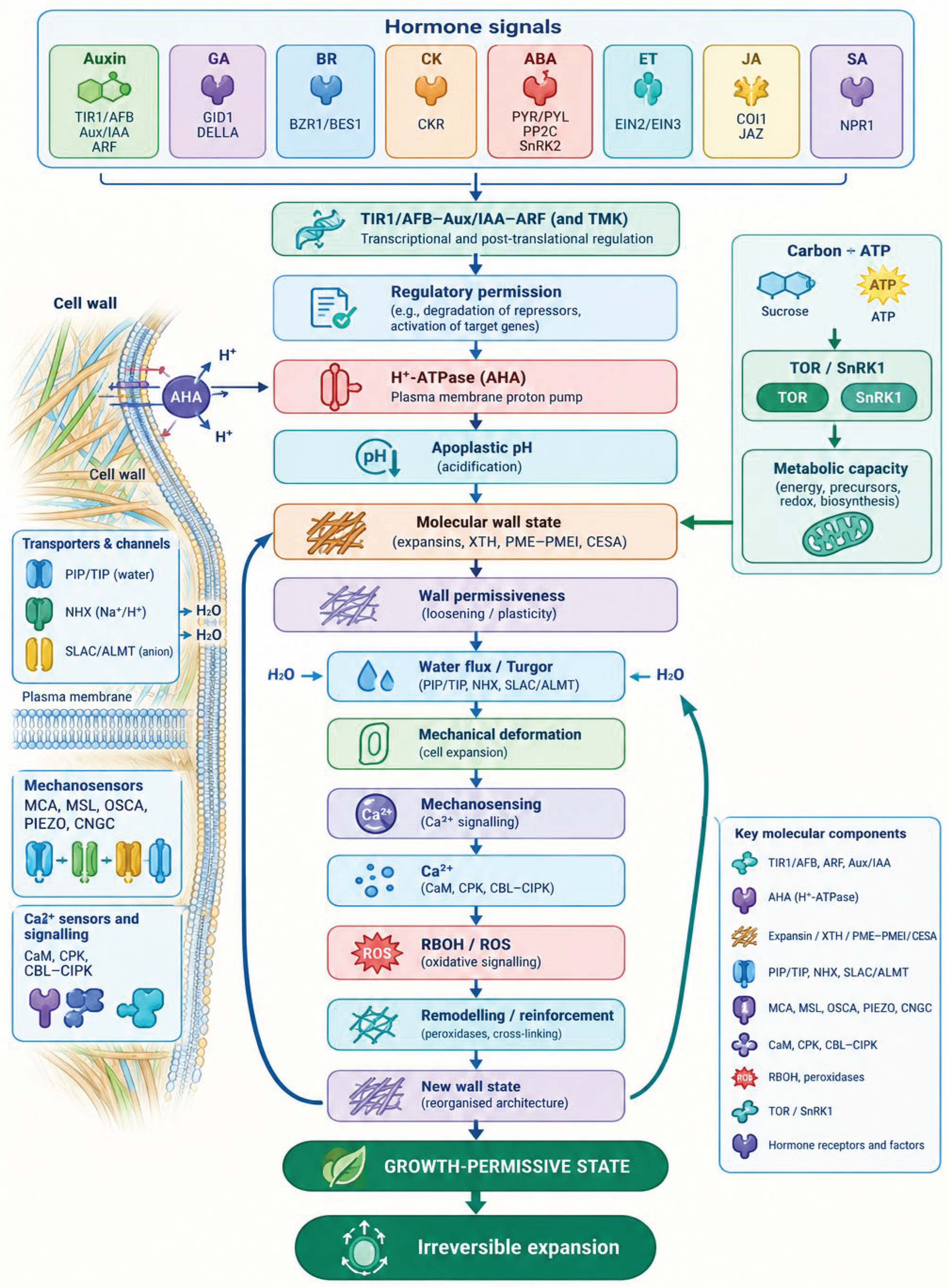
We propose that the elements discussed in Section 4 to 11 be viewed as a single circuit rather than a list of factors. Auxin and other signals act through TIR1/AFB–Aux/IAA–ARF and cell-surface pathways to establish regulatory permission, which controls the H⁺-ATPase and thereby apoplastic pH. Apoplastic pH acts on the molecular state of the wall (expansin, XTH, PME–PMEI, pectin–Ca²⁺ and polysaccharide interactions) and determines wall permissiveness. Wall permissiveness couples with water flux and osmotic regulation to generate turgor-driven mechanical deformation. Deformation is sensed by mechanosensitive channels and wall-sensing kinases and activates Ca²⁺ signalling, RBOH-derived ROS and wall remodelling or reinforcement, resulting in a new mechanochemical state of the wall. In parallel, carbon, ATP and TOR–SnRK1 signalling determine metabolic capacity. At the centre of the circuit lies the growth-permissive state, and its output is irreversible cell expansion.
Figure 4.
Integrated Molecular and Mechanistic Architecture of Plant Cell Expansion Regulation. Proposed causal circuit linking hormonal and environmental signals, TIR1/AFB–Aux/IAA–ARF and cell-surface signalling, the H⁺-ATPase and apoplastic pH, the molecular wall state, wall permissiveness, water flux and turgor, mechanical deformation, mechanosensing, Ca²⁺–RBOH–ROS and wall remodelling, with a parallel TOR–SnRK1 branch defining metabolic capacity. The growth-permissive state is placed at the centre. The scheme is a conceptual model.
Figure 4.
Integrated Molecular and Mechanistic Architecture of Plant Cell Expansion Regulation. Proposed causal circuit linking hormonal and environmental signals, TIR1/AFB–Aux/IAA–ARF and cell-surface signalling, the H⁺-ATPase and apoplastic pH, the molecular wall state, wall permissiveness, water flux and turgor, mechanical deformation, mechanosensing, Ca²⁺–RBOH–ROS and wall remodelling, with a parallel TOR–SnRK1 branch defining metabolic capacity. The growth-permissive state is placed at the centre. The scheme is a conceptual model.
13. From a Reversible Signal to Irreversible Growth
13.1. Which Steps Are Reversible
Hormonal signals, apoplastic pH and turgor are all reversible variables: pH can be restored by changing proton pumping or transport [15,29], and turgor depends on water flux and solute content [13]. By contrast, cell expansion leaves a persistent change in the geometry of the cell and in the arrangement of its wall.
13.2. Sources of Irreversibility
Irreversibility has, in our reading, three sources. The first is plastic deformation of the wall under stress: stress relaxation and creep produce a permanent change of dimensions [12,20]. The second is the deposition of new polymers during and after loosening, including oriented cellulose [45,77] and remodelled pectin [74]. The third is the stabilization of the new geometry by cross-linking [78]. Mechanical stress also guides cytoskeletal and therefore cellulose organization [23,100], which can propagate a mechanical history into the future wall architecture.
13.3. Structural Memory
We propose the term structural memory for the persistence of a past mechanical state in wall architecture. The evidence that stress guides the orientation of microtubules and, via cellulose synthase, of new cellulose is established [23,77,100]; the proposition that this constitutes a general mechanism by which short signals produce lasting growth outcomes is ours and remains to be tested.
13.4. Why a Short Signal Can Produce Lasting Growth
If a transient permissive state allows irreversible deformation and subsequent rebuilding, then a short signal can change the cell’s size and wall geometry for the rest of its life even though the signal itself decays. The model therefore predicts that responses to a second stimulus may depend on the outcome of the first (Prediction 6).
14. Threshold Transitions, Hysteresis and Mechanical Memory
14.1. Thresholds
The yield threshold Y is a classical element of growth physics [11,12]. We extend this notion and propose that growth involves several coupled thresholds: a pH threshold above which the wall is unlikely to be permissive, a mechanical threshold (wall yield), a turgor threshold, and a metabolic threshold below which growth cannot be sustained. The values of the pH and metabolic thresholds are not known and are among the open questions in Section 23. Importantly, we do not claim that such thresholds are sharp or universal; they may differ between cell types and developmental stages [65,68].
14.2. Hysteresis
We hypothesize that transitions between wall states are not symmetrical: the pH or turgor required to initiate a transition into the permissive state may differ from that required to return to the restricted state. This hypothesis is motivated by the irreversible components discussed in Section 13, but direct evidence for hysteresis in the growth response of plant cells is, to our knowledge, limited, and we therefore present it as a conceptual prediction and not as a general law.
14.3. Stabilization of the New State
15. Spatiotemporal Dynamics of the Growth-Permissive State
15.1. Spatial Heterogeneity
Apoplastic pH, wall composition, turgor and mechanical stress are not uniform. Apoplastic pH patterns have been resolved in roots [16] and as oscillations in root hairs [69]. Spatial heterogeneity of pectin methylesterification influences growth symmetry breaking in the hypocotyl [106], and elastic domains organize growth in the meristem [101]. Mechanical stress varies at subcellular and supracellular scales [100].
15.2. Growth Zones
We interpret these data as indicating that the permissive state is a local, rather than a whole-cell or whole-organ, property: different faces of a cell, or different cells of a tissue, may be at different positions in state space at the same moment, which would help explain differential growth [106,107].
15.3. Temporal Sequence
We propose the sequence Restricted → Permissive → Expanding → Reinforcing as the canonical temporal trajectory of a growing cell, with the possibility of returning from any state to an earlier one (for example from Expanding to Restricted when stress activates SnRK1 and ABA signalling [22,52]). The Reinforcing state corresponds to the stabilization phase of Section 6.5, and in a growing tissue cells can occupy different states simultaneously.
16. A Multidimensional State Space of the Cell
We propose to represent a cell at a given moment as a point in a multidimensional space whose axes include apoplastic pH, wall extensibility, turgor, water availability, metabolic capacity, Ca²⁺ state, ROS state, developmental state and mechanical load. The growth-permissive domain is the region of this space in which the conditions of Section 3 are jointly satisfied; the non-growing domain is its complement. Transitions between them are trajectories rather than jumps. A key feature of this representation is that stress can produce what we call stress-induced uncoupling: a displacement along one axis, for example reduced water availability, can move the cell out of the permissive domain even if the other axes remain favourable. This representation is a conceptual device and does not, at present, correspond to an experimentally measured coordinate system; measuring even a subset of its axes simultaneously is the main experimental goal outlined in Section 21.
Figure 5.
The Multidimensional Mechanochemical State Space of a Growing Plant Cell. Conceptual representation of the cell as a point in a space defined by apoplastic pH, wall extensibility, turgor, water availability, metabolic capacity, Ca²⁺ state, ROS state, developmental state and mechanical load, showing the growth-permissive and non-growing domains, transition trajectories and stress-induced uncoupling.
Figure 5.
The Multidimensional Mechanochemical State Space of a Growing Plant Cell. Conceptual representation of the cell as a point in a space defined by apoplastic pH, wall extensibility, turgor, water availability, metabolic capacity, Ca²⁺ state, ROS state, developmental state and mechanical load, showing the growth-permissive and non-growing domains, transition trajectories and stress-induced uncoupling.
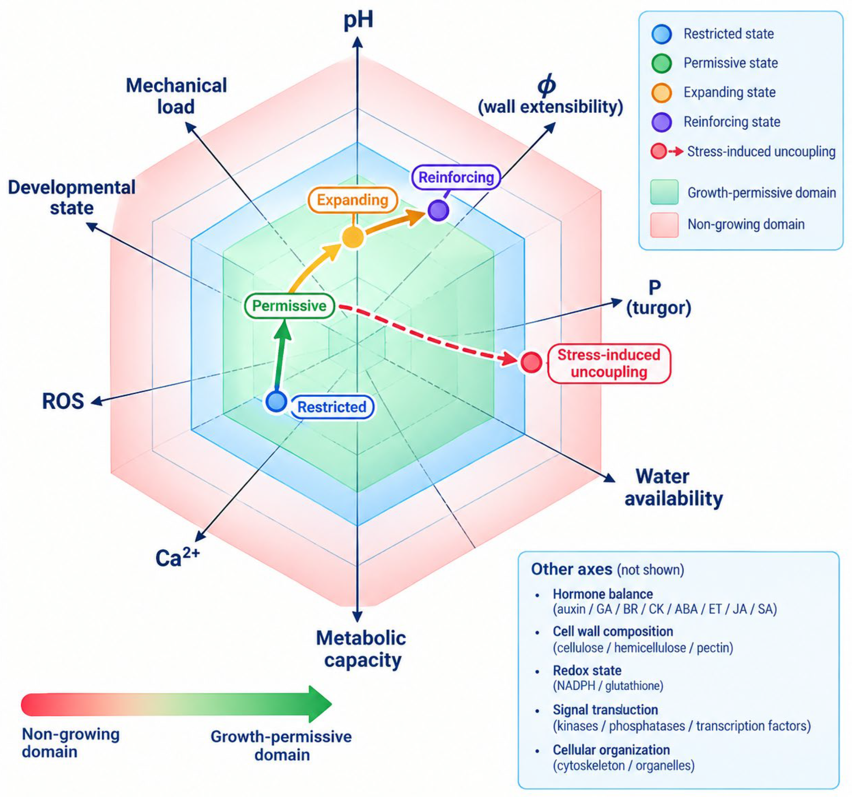
17. Functional Uncoupling: When Components of Growth Work Independently
A multi-component model is justified if it explains situations that one-dimensional descriptions cannot. Table 2 lists seven uncoupling scenarios and the evidence for each. Here we discuss the logic of each case briefly.
High auxin signalling with a rigid wall. When auxin- or fusicoccin-stimulated acidification occurs in a wall with disrupted CSI1-dependent architecture, growth is lost despite an intact stimulus [17]. This is the best-documented example of regulatory and pH permission without mechanical permission.
Permissive pH with low turgor, and high turgor with a rigid wall. The Lockhart relation predicts no expansion below the yield threshold regardless of extensibility, and no appreciable expansion at low extensibility regardless of turgor [11,12]. We are not aware of studies that have tested both variables independently and simultaneously with pH measurements in the same cells, which is why we list these as predictions and not as established cases.
Permissive wall with metabolic limitation. TOR activity links carbon status to growth [21,46] and SnRK1 is activated by energy deficit [22]. We interpret this as consistent with the prediction that metabolic capacity limits the duration of growth.
Sufficient metabolism with water limitation. Water limitation restricts growth through changes in cell proliferation and expansion, ethylene signalling and ABA [51,108,109].
18. Stress as a Disruption of the Growth-Permissive State
18.1. From “Stress Inhibits Growth” to Uncoupling
Stress is usually described as inhibiting growth. In the framework developed here, we propose a more specific statement: stress disrupts the coupling between the regulatory, hydraulic, metabolic and mechanical modules required for the growth-permissive state. Under this view, different stresses act on different modules and therefore produce different growth phenotypes.
18.2. Drought and Osmotic Stress
Under water limitation, plants balance growth and survival [108]; osmotic stress arrests cell proliferation in young leaves with a role for ethylene signalling [109]; and root and shoot growth respond differently to ABA and ethylene [51]. Walls of roots at low water potential show altered extensibility and wall proteins [110]. These data suggest that drought acts simultaneously on the hydraulic and mechanical modules and on the ABA-dependent regulatory state [26].
18.3. Salinity
Salt stress produces a spatio-temporal pattern of growth regulation in roots [113], and plants tune signalling and growth to survive salt [114]. Wall integrity signalling through FERONIA and Ca²⁺ is required during salt stress [97], and leucine-rich repeat extensins regulate salt tolerance [112]. Salinity is therefore a good test case for our proposition that mechanical-state sensing and the wall are integral parts of stress responses [115].
18.4. Heat, Cold, Nutrient Deficiency and Oxidative Stress
For heat, cold and nutrient deficiency, the literature that connects the specific components of our model (apoplastic pH, wall state, turgor and TOR–SnRK1) in a single experimental system is, in our assessment, much thinner than for drought and salinity. We therefore treat the application of the framework to these stresses as a hypothesis. Oxidative stress is of particular interest because ROS, which are part of the feedback loop (Section 11), can also act as damage signals, and the transition from signalling to damage may correspond to a loss of coupling.
18.5. Reinforcement, Energy Sensing and Ca²⁺/ROS Signalling Under Stress
Stress-induced wall reinforcement [110,115], activation of SnRK1 and suppression of TOR [22,52,89], and altered Ca²⁺ and ROS signalling [97,105] are the molecular expressions of the uncoupling in our model. In our earlier work we described these configurations as stress-adaptive regulatory states, including an ABA-dominant state [25,26,27]; the present article specifies the cell-level mechanical consequences of leaving the TOR-dominant growth state.
19. Comparison with Existing Models
We do not claim that the proposed model replaces earlier ones; we propose that it integrates their valid components into a single causal architecture. Table 3 compares seven classes of models according to their core variable, what they explain, what they leave unresolved, and whether they include feedback, thresholds or hysteresis and a metabolic term. The assessments in this table are our own interpretation of the literature cited in Sections 1 to 18 and should be read as an analytical device, not as a verdict on the models.
20. Predictions of the Model
The proposed model makes the following predictions. Each of them is testable and, as specified in Table 4 and Section 22, falsifiable.
Prediction 1. The same apoplastic pH can produce different growth depending on the wall state. This can be tested by comparing growth responses of walls with different cellulose or pectin architecture at matched, directly measured apoplastic pH (for example in CSI1-deficient and wild-type hypocotyls [17] or in lines with altered PME/PMEI activity [74]).
Prediction 2. High turgor does not guarantee expansion when the wall is mechanically restrictive. If the model is correct, one would expect cells with experimentally increased turgor and stiffened or cross-linked walls to show no proportional increase of irreversible expansion.
Prediction 3. Mechanical loading should change the pH-dependent growth response. A pH step applied under different imposed mechanical loads should produce different growth outcomes, which can be tested with tissue-stretching or micro-compression devices.
Prediction 4. Metabolic limitation should reduce the duration of sustained growth even in a permissive wall state. Initial acidification-driven growth may be preserved, but its duration should depend on carbon and energy status and on TOR–SnRK1 activity [21,22].
Prediction 5. The same hormonal stimulation should yield different outcomes in cells of different developmental state. Cell-type-resolved responses to auxin [65] provide an entry point for this test.
Prediction 6. After a transition of the wall into a new state, the response to subsequent stimulation should show hysteresis, that is, a dependence on the history of the preceding transition.
Prediction 7. Mechanical deformation should activate Ca²⁺ and ROS feedback that alters subsequent wall mechanics. Interference with mechanosensitive channels or RBOH activity should therefore change the later mechanical state of the wall, not only the immediate response [102,105].
Prediction 8. Turgor-to-growth coupling efficiency should depend on wall state, not only on absolute turgor. Cells with equal turgor but different wall states should differ in irreversible expansion per unit of turgor excess.
21. Experimental Programme
21.1. Variables to be Measured
The central methodological implication is that mechanism should not be inferred from a single parameter. We propose simultaneous or tightly paired measurements of apoplastic pH, turgor, wall extensibility, cell expansion rate, H⁺-ATPase activity and phosphorylation state, Ca²⁺ dynamics, ROS dynamics, ATP and metabolic state, water flux, activities of expansin, XTH and PME, and remodelling of cellulose and pectin.
21.2. Approaches
Established approaches exist for several of these variables: in vivo stress relaxation and extensometry for wall yield and extensibility [12], atomic force microscopy and related methods for wall structure and mechanics [18], ratiometric pH imaging in roots [16], imaging of pH and ROS in growing cells [69,105], and genetic tools for the components of the model, including mutants of mechanosensitive channels, aquaporins, H⁺-ATPase regulators and TOR–SnRK1 [34,52,91,95]. The experimental challenge is not the absence of individual tools but their combination in the same cells and at the same time.
21.3. Experimental Logic
We suggest a design in which a defined perturbation (for example an auxin, fusicoccin or RALF treatment, an osmotic step, a mechanical load or a change in carbon supply) is applied, and multiple state variables are recorded on a common time axis. The resulting dataset can be used to test whether the combination of variables, rather than any one of them, predicts expansion.
Figure 6.
Experimental Framework for Testing the Integrative Mechanistic Model. Proposed multi-variable experimental design in which a defined perturbation is applied and apoplastic pH, turgor, wall extensibility, expansion rate, H⁺-ATPase activity, Ca²⁺, ROS, ATP/metabolic state, water flux, wall-enzyme activities and wall remodelling are recorded on a common time axis.
Figure 6.
Experimental Framework for Testing the Integrative Mechanistic Model. Proposed multi-variable experimental design in which a defined perturbation is applied and apoplastic pH, turgor, wall extensibility, expansion rate, H⁺-ATPase activity, Ca²⁺, ROS, ATP/metabolic state, water flux, wall-enzyme activities and wall remodelling are recorded on a common time axis.
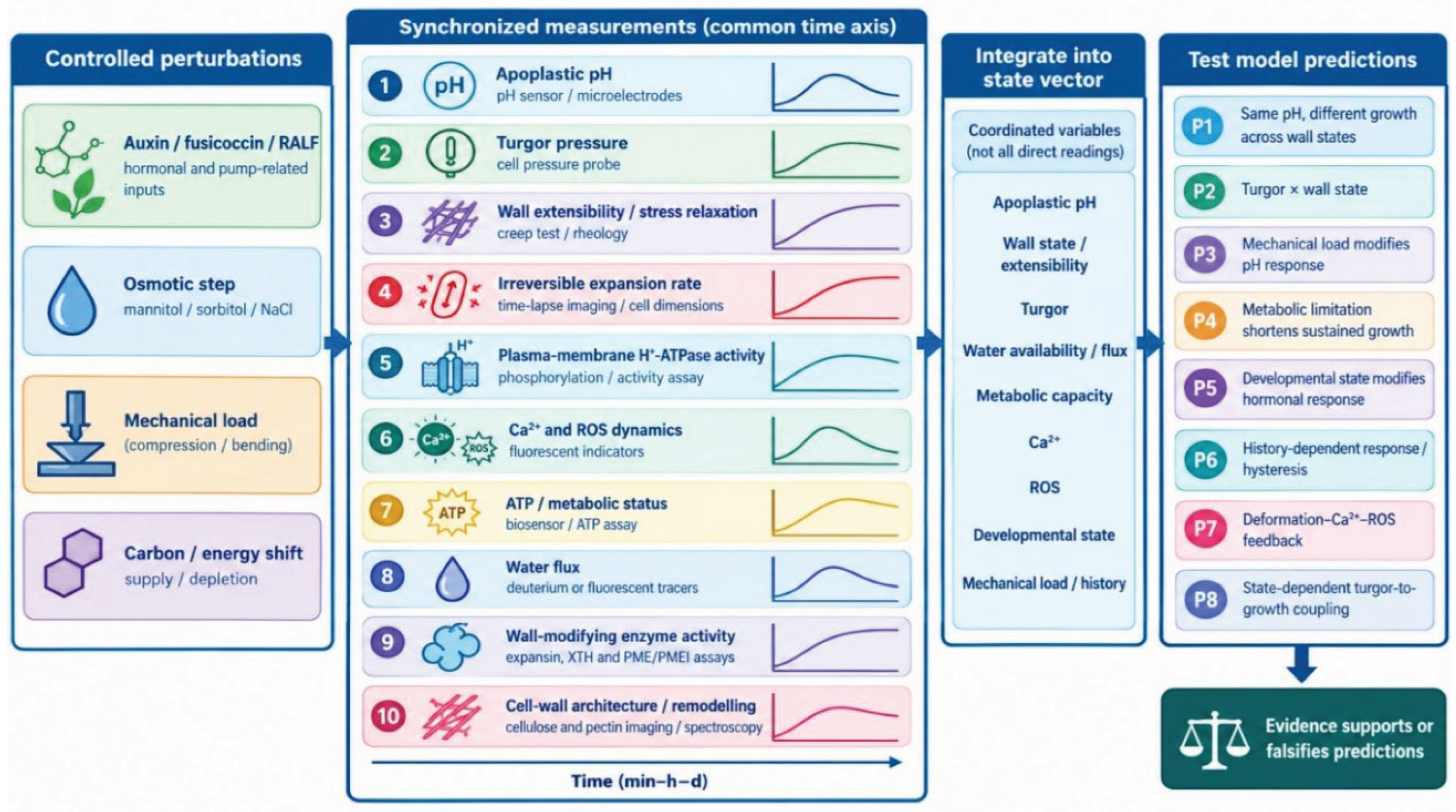
22. Criteria for Falsification of the Model
To protect the framework from being merely descriptive, we state the observations that would show it to be incomplete or wrong. The model should be considered incomplete or incorrect if (i) wall state does not modulate pH-dependent expansion; (ii) turgor explains expansion independently of wall state; (iii) mechanical loading has no influence on subsequent regulation; (iv) metabolic capacity does not determine the duration of sustained growth; (v) Ca²⁺/ROS feedback is unrelated to the mechanical state of the wall; or (vi) an identical molecular and mechanical state reproducibly leads to an identical outcome regardless of developmental context. The weakening of one criterion would require revision of the corresponding module and not necessarily the abandonment of the whole architecture; the simultaneous failure of several would, in our view, make the state-transition description unnecessary.
23. Open Questions
Where is the pH threshold for a permissive wall state, and does it differ between cell types? Which molecular components determine hysteresis, if hysteresis exists? How long does mechanical memory last, and does it reside in cellulose orientation, pectin state or both? How exactly does pectin state determine wall mechanics, given the opposing effects of demethylesterification described above? How does Ca²⁺/ROS feedback interact with auxin signalling? How does the TOR–SnRK1 balance define the boundary between permissive and non-permissive states? And, finally, can the growth-permissive state be measured experimentally as an integral property, for example through a composite index derived from the variables listed in Section 21? We regard the last question as decisive for whether the concept becomes an operational tool or remains a heuristic.
24. Significance of the Model for Plant Biology
24.1. A Reinterpretation of the Acid Growth Hypothesis
The model does not reject the acid growth hypothesis; it embeds it as a module within a larger system, in which acidification is a necessary-but-not-sufficient step of the permission layer.
24.2. Plant Mechanobiology and Cell-Wall Biology
24.3. Hormone–Mechanics Integration, Stress Physiology and Development
It proposes a concrete way in which hormone signalling and wall mechanics interact, namely through permission and gating, rather than through simple stimulation. It suggests that stress physiology can be described as uncoupling between modules [25,26], and that developmental context determines how signals are converted into expansion [47,48].
24.4. Cell Growth Theory
By generalizing the Lockhart relation so that extensibility and yield threshold are state-dependent [11], the model offers a bridge between classical growth physics and molecular regulation.
25. Implications for Breeding and Biotechnology
This section is intentionally brief and prospective. If the model proves useful, selection for growth rate alone could be complemented by mechanism-based phenotypes: responsiveness of apoplastic pH, wall extensibility, maintenance of turgor, hydraulic efficiency, metabolic resilience and mechanosensory response. The corresponding groups of potential molecular targets include H⁺-ATPases [29], expansins [62], XTHs [64], PME/PMEI [72], aquaporins [34], mechanosensitive channels [90], Ca²⁺ signalling and TOR–SnRK1 [33,89]. Altered expansin expression in rice, for example, changes growth and development [63]. In the logic of our earlier proposal for state-targeted crop management [25], mechanism-oriented selection and diagnostics would aim to identify the module that limits growth in a given genotype and environment. All such applications depend on the experimental validation of the framework.
26. Conclusions
How does a plant cell convert a reversible regulatory signal into an irreversible increase in cell size, and why do similar growth signals not always produce the same mechanical outcome? The acid growth hypothesis established the fundamental role of auxin-dependent activation of the plasma membrane H⁺-ATPase and apoplastic acidification. However, the mere presence of a growth signal, elevated turgor pressure, or an increased capacity for cell wall loosening does not explain under which conditions these factors promote sustained cell expansion and under which conditions growth remains constrained. The lack of an integrative explanation for this gap between regulatory signaling and mechanical outcomes limits our understanding of how plant cells coordinate growth under changing physiological and environmental conditions.
This study proposes an integrative mechanistic model of plant cell growth that conceptualizes cell expansion not as the direct consequence of an individual hormonal or physical factor, but as a state-dependent transition into a mechanochemical regime that permits sustained increases in cell size. The central conceptual proposition is that growth outcomes depend not only on the intensity of regulatory signaling but also on the coordination of cell wall physical properties, water availability, turgor pressure, mechanical deformation, and the cell’s metabolic capacity. Within this framework, apoplastic pH is regarded as a context-dependent regulator that alters the likelihood of establishing a cell wall state mechanically permissive for growth, without determining the outcome independently of the other components of the system.
The model integrates hormonal signaling, proton ATPase activity, cell wall remodeling, water transport, turgor mechanics, and the biosynthetic capacity required for growth. The TOR–SnRK1 regulatory module is incorporated as a component of metabolic control that may constrain the maintenance of sustained cell expansion under conditions of insufficient energy or resource availability. Mechanosensory signaling and the interplay between calcium signals and reactive oxygen species are considered a candidate feedback circuit capable of linking mechanical deformation to subsequent cell wall remodeling and reinforcement. Particular attention is given to distinguishing reversible regulatory changes from persistent morphological consequences of growth, as well as to the hypothesis that structural reorganization of the cell wall may preserve the effects of previous mechanical influences. To formalize this logic, growth is represented as a trajectory of transitions among four functional states: growth-restricted, growth-permissive, active-expansion, and structural-reinforcement states.
The principal conceptual contribution of this study is the shift in focus from the regulation of individual growth components to the mechanism by which regulatory signals are converted, in a state-dependent manner, into sustained cellular deformation. This approach provides a testable framework for explaining the context dependence of plant growth, identifying latent physiological constraints, and investigating why similar hormonal or mechanical stimuli can produce different growth outcomes. The proposed model does not replace the acid growth hypothesis but extends its explanatory scope by integrating regulatory, mechanical, hydraulic, and metabolic processes into a unified conceptual architecture amenable to experimental testing.
By linking molecular signaling with water transport, metabolic regulation, and cell wall mechanics, the proposed conceptual model establishes a foundation for experimentally investigating why similar growth signals can elicit different cellular responses and for identifying physiological state-dependent constraints on plant growth under changing environmental conditions.
Author Contributions
Conceptualization, S.H.K.; methodology, S.H.K. and L.M.B.; formal analysis, S.H.K.; investigation, S.H.K., Y.V.K., V.M.S. and Y.A.A.; writing—original draft preparation, S.H.K.; writing—review and editing, all authors; visualization, S.H.K.; supervision, S.H.K. All authors have read and agreed to the published version of the manuscript.
Funding
This research received no external funding.
Institutional Review Board Statement
Not applicable.
Informed Consent Statement
Not applicable.
Data Availability Statement
No new experimental data were generated in this conceptual article; all cited literature is publicly available via the DOIs listed in the References.
Acknowledgments
During the preparation of this work, the authors used Claude (Anthropic) (version/period of use: 2025–2026) for language refinement, grammar checking, and stylistic editing. The authors reviewed and edited the entire text, verified its accuracy, and take full responsibility for the integrity and originality of the final manuscript. No artificial intelligence tool is listed as an author.
Conflicts of Interest
The authors declare no conflict of interest.
Abbreviations
ABA – Abscisic Acid, ALMT – Aluminum-Activated Malate Transporter, ARF – Auxin Response Factor, AHA – Arabidopsis H⁺-ATPase, BAK1 – BRI1-Associated Receptor Kinase 1, BZR1 – Brassinazole-Resistant 1, BES1 – BRI1-EMS-Suppressor 1, Ca²⁺ – Calcium, CaM – Calmodulin, CDPK / CPK – Calcium-Dependent Protein Kinase, CESA – Cellulose Synthase, CBL – Calmodulin-Binding B-Like protein, CIPK – CBL-Interacting Protein Kinase, COI1 – Coronatine Insensitive 1, CSI1 – Cellulose Synthase Interactive 1, DELLA – DELLA protein, EIN2 / EIN3 – Ethylene-Insensitive 2 / 3, ET – Ethylene, FER – FERONIA, GA – Gibberellin, GID1 – GIBBERELLIN INSENSITIVE DWARF 1, H⁺ – Proton, JA – Jasmonic Acid, JAZ – Jasmonate ZIM-domain, MCA – Mid-Conductance Mechanosensitive Channel-like, MSL / MscS – Mechanosensitive Channel of Small Conductance-Like, NHX – Na⁺/H⁺ Exchanger, NPR1 – Nonexpressor of Pathogenesis-Related Genes 1, OSCA1 – Hyperosmolality-Gated Calcium-Permeable Channel 1, PIF – Phytochrome-Interacting Factor, PIP / TIP – Plasma membrane / Tonoplast Intrinsic Protein, PME / PMEI – Pectin Methylesterase / Pectin Methylesterase Inhibitor, PP2C – Protein Phosphatase 2C, PYR / PYL / RCAR – Pyrabactin Resistance 1 / PYL-like / Regulatory Components of ABA Receptor, RALF – Rapid Alkalinization Factor, RBOH – Respiratory Burst Oxidase Homolog, ROS – Reactive Oxygen Species, SA – Salicylic Acid, SAUR – Small Auxin Up RNA, SLAC / SLAH – Slow Anion Channel-Associated / SLAC1 Homolog, SnRK1 / SnRK2 – SNF1-Related Protein Kinase 1 / 2, TIR1/AFB – Transport Inhibitor Response 1 / Auxin F-Box, TMK – Transmarine Kinase, TOR – Target of Rapamycin, XTH – Xyloglucan Endotransglucosylase/Hydrolase.
References
- Rayle, D.L.; Cleland, R.E. The acid growth theory of auxin-induced cell elongation is alive and well. Plant Physiol. 1992, 99, 1271–1274. [Google Scholar] [CrossRef]
- Hager, A. Role of the plasma membrane H+-ATPase in auxin-induced elongation growth: Historical and new aspects. J. Plant Res. 2003, 116, 483–505. [Google Scholar] [CrossRef]
- McQueen-Mason, S.; Durachko, D.M.; Cosgrove, D.J. Two endogenous proteins that induce cell wall extension in plants. Plant Cell 1992, 4, 1425–1433. [Google Scholar] [CrossRef]
- Cosgrove, D.J. Loosening of plant cell walls by expansins. Nature 2000, 407, 321–326. [Google Scholar] [CrossRef]
- Cosgrove, D.J. Growth of the plant cell wall. Nat. Rev. Mol. Cell Biol. 2005, 6, 850–861. [Google Scholar] [CrossRef]
- Fendrych, M.; Leung, J.; Friml, J. TIR1/AFB-Aux/IAA auxin perception mediates rapid cell wall acidification and growth of Arabidopsis hypocotyls. eLife 2016, 5, e19048. [Google Scholar] [CrossRef]
- Spartz, A.K.; Ren, H.; Park, M.Y.; et al. SAUR inhibition of PP2C-D phosphatases activates plasma membrane H+-ATPases to promote cell expansion in. Arab. Plant Cell 2014, 26, 2129–2142. [Google Scholar] [CrossRef]
- Lin, W.; Zhou, X.; Tang, W.; et al. TMK-based cell-surface auxin signalling activates cell-wall acidification. Nature 2021, 599, 278–282. [Google Scholar] [CrossRef]
- Du, M.; Spalding, E.P.; Gray, W.M. Rapid auxin-mediated cell expansion. Annu. Rev. Plant Biol. 2020, 71, 379–402. [Google Scholar] [CrossRef]
- Cosgrove, D.J. Structure and growth of plant cell walls. Nat. Rev. Mol. Cell Biol. 2024, 25, 340–358. [Google Scholar] [CrossRef]
- Lockhart, J.A. An analysis of irreversible plant cell elongation. J. Theor. Biol. 1965, 8, 264–275. [Google Scholar] [CrossRef]
- Cosgrove, D.J. Cell wall yield properties of growing tissue: Evaluation by in vivo stress relaxation. Plant Physiol. 1985, 78, 347–356. [Google Scholar] [CrossRef]
- Boyer, J.S.; Silk, W.K. Hydraulics of plant growth. Funct. Plant Biol. 2004, 31, 761–773. [Google Scholar] [CrossRef]
- Geitmann, A.; Ortega, J.K.E. Mechanics and modeling of plant cell growth. Trends Plant Sci. 2009, 14, 467–478. [Google Scholar] [CrossRef]
- Li, L.; Verstraeten, I.; Roosjen, M.; et al. Cell surface and intracellular auxin signalling for H+ fluxes in root growth. Nature 2021, 599, 273–277. [Google Scholar] [CrossRef]
- Barbez, E.; Dünser, K.; Gaidora, A.; et al. Auxin steers root cell expansion via apoplastic pH regulation in. Arab. Thaliana Proc. Natl. Acad. Sci. USA 2017, 114, E4884–E4893. [Google Scholar] [CrossRef]
- Xin, X.; Lei, L.; Zheng, Y.; et al. Cellulose synthase interactive1- and microtubule-dependent cell wall architecture is required for acid growth in Arabidopsis hypocotyls. J. Exp. Bot. 2020, 71, 2982–2994. [Google Scholar] [CrossRef]
- Zhang, Y.; Yu, J.; Wang, X.; et al. Molecular insights into the complex mechanics of plant epidermal cell walls. Science 2021, 372, 706–711. [Google Scholar] [CrossRef]
- Park, Y.B.; Cosgrove, D.J. A revised architecture of primary cell walls based on biomechanical changes induced by substrate-specific endoglucanases. Plant Physiol. 2012, 158, 1933–1943. [Google Scholar] [CrossRef]
- Cosgrove, D.J. Building an extensible cell wall. Plant Physiol. 2022, 189, 1246–1277. [Google Scholar] [CrossRef]
- Xiong, Y.; McCormack, M.; Li, L.; et al. Glucose-TOR signalling reprograms the transcriptome and activates meristems. Nature 2013, 496, 181–186. [Google Scholar] [CrossRef]
- Baena-González, E.; Rolland, F.; Thevelein, J.M.; et al. A central integrator of transcription networks in plant stress and energy signalling. Nature 2007, 448, 938–942. [Google Scholar] [CrossRef]
- Hamant, O.; Heisler, M.G.; Jönsson, H.; et al. Developmental patterning by mechanical signals in. Arab. Sci. 2008, 322, 1650–1655. [Google Scholar] [CrossRef]
- Hervieux, N.; Dumond, M.; Sapala, A.; et al. A mechanical feedback restricts sepal growth and shape in. Arab. Curr. Biol. 2016, 26, 1019–1028. [Google Scholar] [CrossRef]
- Khablak, S.H.; Bondareva, L.M.; Abdullaieva, Y.A.; Spychak, V.M. From stress to growth dominance: Hormonal switches, energy sensing, and the concept of Regulatory Agronomy in plants. Plant Horm. 2026, 2, e017. [Google Scholar] [CrossRef]
- Khablak, S.H.; Bondareva, L.M.; Kolomiiets, Y.V.; Abdullaieva, Y.A.; Spychak, V.M. The ABA-dominant abiotic stress tolerance state: An integrative framework for abscisic acid-mediated abiotic stress tolerance in plants. Plant Horm. 2026, 2, e023. [Google Scholar] [CrossRef]
- Khablak, S.H.; Bondareva, L.M.; Dolia, M.M.; Spychak, V.M.; Lykholat, T.Y.; Sklyar, T.V.; Lykholat, Y.V. Integrative model of plant immunity to pathogens and stresses: A multilevel signal-metabolic network. Regul. Mech. Biosyst. 2025, 16, e25175. [Google Scholar] [CrossRef]
- Khablak, S.H.; Bondareva, L.M.; Lykholat, T.Y.; Sklyar, T.V.; Lykholat, Y.V.; Spychak, V.M.; Abdullaieva, Y.A. Jasmonate-ethylene (JA/ET) signaling – metabolic defense system in the integrative model of plant immunity against phytophagous insects. Regul. Mech. Biosyst. 2026, 17, e26013. [Google Scholar] [CrossRef]
- Falhof, J.; Pedersen, J.T.; Fuglsang, A.T.; et al. Plasma membrane H+-ATPase regulation in the center of plant physiology. Mol. Plant 2016, 9, 323–337. [Google Scholar] [CrossRef]
- Dharmasiri, N.; Dharmasiri, S.; Estelle, M. The F-box protein TIR1 is an auxin receptor. Nature 2005, 435, 441–445. [Google Scholar] [CrossRef]
- Tan, X.; Calderon-Villalobos, L.I.A.; Sharon, M.; et al. Mechanism of auxin perception by the TIR1 ubiquitin ligase. Nature 2007, 446, 640–645. [Google Scholar] [CrossRef]
- Davière, J.M.; Achard, P. Gibberellin signaling in plants. Development 2013, 140, 1147–1151. [Google Scholar] [CrossRef]
- Dobrenel, T.; Caldana, C.; Hanson, J.; et al. TOR signaling and nutrient sensing. Annu. Rev. Plant Biol. 2016, 67, 261–285. [Google Scholar] [CrossRef]
- Maurel, C.; Boursiac, Y.; Luu, D.T.; et al. Aquaporins in plants. Physiol. Rev. 2015, 95, 1321–1358. [Google Scholar] [CrossRef]
- Weijers, D.; Wagner, D. Transcriptional responses to the auxin hormone. Annu. Rev. Plant Biol. 2016, 67, 539–574. [Google Scholar] [CrossRef]
- Fuglsang, A.T.; Guo, Y.; Cuin, T.A.; et al. Arabidopsis protein kinase PKS5 inhibits the plasma membrane H+-ATPase by preventing interaction with 14-3-3 protein. Plant Cell 2007, 19, 1617–1634. [Google Scholar] [CrossRef]
- Haruta, M.; Sabat, G.; Stecker, K.; et al. A peptide hormone and its receptor protein kinase regulate plant cell expansion. Science 2014, 343, 408–411. [Google Scholar] [CrossRef]
- de Lucas, M.; Davière, J.M.; Rodríguez-Falcón, M.; et al. A molecular framework for light and gibberellin control of cell elongation. Nature 2008, 451, 480–484. [Google Scholar] [CrossRef]
- Feng, S.; Martinez, C.; Gusmaroli, G.; et al. Coordinated regulation of Arabidopsis thaliana development by light and gibberellins. Nature 2008, 451, 475–479. [Google Scholar] [CrossRef]
- Achard, P.; Cheng, H.; De Grauwe, L.; et al. Integration of plant responses to environmentally activated phytohormonal signals. Science 2006, 311, 91–94. [Google Scholar] [CrossRef]
- Nolan, T.M.; Vukašinović, N.; Liu, D.; et al. Brassinosteroids: Multidimensional regulators of plant growth, development, and stress responses. Plant Cell 2020, 32, 295–318. [Google Scholar] [CrossRef]
- Oh, E.; Zhu, J.Y.; Bai, M.Y.; et al. Cell elongation is regulated through a central circuit of interacting transcription factors in the Arabidopsis hypocotyl. eLife 2014, 3, e03031. [Google Scholar] [CrossRef]
- Bai, M.Y.; Shang, J.X.; Oh, E.; et al. Brassinosteroid, gibberellin and phytochrome impinge on a common transcription module in. Arab. Nat. Cell Biol. 2012, 14, 810–817. [Google Scholar] [CrossRef]
- Minami, A.; Takahashi, K.; Inoue, S.; et al. Brassinosteroid induces phosphorylation of the plasma membrane H+-ATPase during hypocotyl elongation in. Arab. Thaliana Plant Cell Physiol. 2019, 60, 935–944. [Google Scholar] [CrossRef]
- Xie, L.; Yang, C.; Wang, X. Brassinosteroids can regulate cellulose biosynthesis by controlling the expression of CESA genes in. Arab. J. Exp. Bot. 2011, 62, 4495–4506. [Google Scholar] [CrossRef]
- Zhang, Z.; Zhu, J.Y.; Roh, J.; et al. TOR signaling promotes accumulation of BZR1 to balance growth with carbon availability in. Arab. Curr. Biol. 2016, 26, 1854–1860. [Google Scholar] [CrossRef]
- Dello Ioio, R.; Nakamura, K.; Moubayidin, L.; et al. A genetic framework for the control of cell division and differentiation in the root meristem. Science 2008, 322, 1380–1384. [Google Scholar] [CrossRef]
- Schaller, G.E.; Bishopp, A.; Kieber, J.J. The yin-yang of hormones: Cytokinin and auxin interactions in plant development. Plant Cell 2015, 27, 44–63. [Google Scholar] [CrossRef]
- Cutler, S.R.; Rodriguez, P.L.; Finkelstein, R.R.; et al. Abscisic acid: Emergence of a core signaling network. Annu. Rev. Plant Biol. 2010, 61, 651–679. [Google Scholar] [CrossRef]
- Park, S.Y.; Fung, P.; Nishimura, N.; et al. Abscisic acid inhibits type 2C protein phosphatases via the PYR/PYL family of START proteins. Science 2009, 324, 1068–1071. [Google Scholar] [CrossRef]
- Sharp, R.E.; LeNoble, M.E. ABA, ethylene and the control of shoot and root growth under water stress. J. Exp. Bot. 2002, 53, 33–37. [Google Scholar] [CrossRef]
- Wang, P.; Zhao, Y.; Li, Z.; et al. Reciprocal regulation of the TOR kinase and ABA receptor balances plant growth and stress response. Mol. Cell 2018, 69, 100–112. [Google Scholar] [CrossRef]
- Binder, B.M. Ethylene signaling in plants. J. Biol. Chem. 2020, 295, 7710–7725. [Google Scholar] [CrossRef]
- Dubois, M.; Van den Broeck, L.; Inzé, D. The pivotal role of ethylene in plant growth. Trends Plant Sci. 2018, 23, 311–323. [Google Scholar] [CrossRef]
- Cho, H.T.; Kende, H. Expansins and internodal growth of deepwater rice. Plant Physiol. 1997, 113, 1145–1151. [Google Scholar] [CrossRef]
- Howe, G.A.; Major, I.T.; Koo, A.J. Modularity in Jasmonate Signaling for Multistress Resilience. Annu. Rev. Plant Biol. 2018, 69, 387–415. [Google Scholar] [CrossRef]
- Yang, D.L.; Yao, J.; Mei, C.S.; et al. Plant hormone jasmonate prioritizes defense over growth by interfering with gibberellin signaling cascade. Proc. Natl. Acad. Sci. USA 2012, 109, E1192–E1200. [Google Scholar] [CrossRef]
- Huot, B.; Yao, J.; Montgomery, B.L.; et al. Growth-defense tradeoffs in plants: A balancing act to optimize fitness. Mol. Plant 2014, 7, 1267–1287. [Google Scholar] [CrossRef]
- Vlot, A.C.; Dempsey, D.A.; Klessig, D.F. Salicylic acid, a multifaceted hormone to combat disease. Annu. Rev. Phytopathol. 2009, 47, 177–206. [Google Scholar] [CrossRef]
- Nemhauser, J.L.; Hong, F.; Chory, J. Different plant hormones regulate similar processes through largely nonoverlapping transcriptional responses. Cell 2006, 126, 467–475. [Google Scholar] [CrossRef]
- Depuydt, S.; Hardtke, C.S. Hormone signalling crosstalk in plant growth regulation. Curr. Biol. 2011, 21, R365–R373. [Google Scholar] [CrossRef]
- Sampedro, J.; Cosgrove, D.J. The expansin superfamily. Genome Biol. 2005, 6, 242. [Google Scholar] [CrossRef]
- Choi, D.; Lee, Y.; Cho, H.T.; et al. Regulation of expansin gene expression affects growth and development in transgenic rice plants. Plant Cell 2003, 15, 1386–1398. [Google Scholar] [CrossRef]
- Rose, J.K.C.; Braam, J.; Fry, S.C.; et al. The XTH family of enzymes involved in xyloglucan endotransglucosylation and endohydrolysis: Current perspectives and a new unifying nomenclature. Plant Cell Physiol. 2002, 43, 1421–1435. [Google Scholar] [CrossRef]
- Bargmann, B.O.R.; Vanneste, S.; Krouk, G.; et al. A map of cell type-specific auxin responses. Mol. Syst. Biol. 2013, 9, 688. [Google Scholar] [CrossRef]
- Brady, S.M.; Orlando, D.A.; Lee, J.Y.; et al. A high-resolution root spatiotemporal map reveals dominant expression patterns. Science 2007, 318, 801–806. [Google Scholar] [CrossRef]
- Sugimoto-Shirasu, K.; Roberts, K. Big it up”: Endoreduplication and cell-size control in plants. Curr. Opin. Plant Biol. 2003, 6, 544–553. [Google Scholar] [CrossRef]
- Tisné, S.; Reymond, M.; Vile, D.; et al. Combined genetic and modeling approaches reveal that epidermal cell area and number in leaves are controlled by leaf and plant developmental processes. 2008, 148, 1117–1127. [Google Scholar] [CrossRef]
- Monshausen, G.B.; Bibikova, T.N.; Messerli, M.A.; et al. Oscillations in extracellular pH and reactive oxygen species modulate tip growth of Arabidopsis root hairs. Proc. Natl. Acad. Sci. USA 2007, 104, 20996–21001. [Google Scholar] [CrossRef]
- Wang, T.; Park, Y.B.; Caporini, M.A.; et al. Sensitivity-enhanced solid-state NMR detection of expansin’s target in plant cell walls. Proc. Natl. Acad. Sci. USA 2013, 110, 16444–16449. [Google Scholar] [CrossRef]
- Fry, S.C.; Smith, R.C.; Renwick, K.F.; et al. Xyloglucan endotransglycosylase, a new wall-loosening enzyme activity from plants. Biochem. J. 1992, 282, 821–828. [Google Scholar] [CrossRef]
- Wolf, S.; Mouille, G.; Pelloux, J. Homogalacturonan methyl-esterification and plant development. Mol. Plant 2009, 2, 851–860. [Google Scholar] [CrossRef]
- Hocq, L.; Sénéchal, F.; Lefebvre, V.; et al. Combined experimental and computational approaches reveal distinct pH dependence of pectin methylesterase inhibitors. Plant Physiol. 2017, 173, 1075–1093. [Google Scholar] [CrossRef]
- Peaucelle, A.; Braybrook, S.A.; Le Guillou, L.; et al. Pectin-induced changes in cell wall mechanics underlie organ initiation in. Arab. Curr. Biol. 2011, 21, 1720–1726. [Google Scholar] [CrossRef]
- Hocq, L.; Pelloux, J.; Lefebvre, V. Connecting homogalacturonan-type pectin remodeling to acid growth. Trends Plant Sci. 2017, 22, 20–29. [Google Scholar] [CrossRef]
- Cosgrove, D.J. Plant cell wall extensibility: Connecting plant cell growth with cell wall structure, mechanics, and the action of wall-modifying enzymes. J. Exp. Bot. 2016, 67, 463–476. [Google Scholar] [CrossRef]
- Paredez, A.R.; Somerville, C.R.; Ehrhardt, D.W. Visualization of cellulose synthase demonstrates functional association with microtubules. Science 2006, 312, 1491–1495. [Google Scholar] [CrossRef]
- Francoz, E.; Ranocha, P.; Nguyen-Kim, H.; et al. Roles of cell wall peroxidases in plant development. Phytochemistry 2015, 112, 15–21. [Google Scholar] [CrossRef]
- Schopfer, P. Hydroxyl radical-induced cell-wall loosening in vitro and in vivo: Implications for the control of elongation growth. Plant J. 2001, 28, 679–688. [Google Scholar] [CrossRef]
- Müller, K.; Linkies, A.; Vreeburg, R.A.M.; et al. In vivo cell wall loosening by hydroxyl radicals during cress seed germination and elongation growth. Plant Physiol. 2009, 150, 1855–1865. [Google Scholar] [CrossRef]
- Bassil, E.; Tajima, H.; Liang, Y.C.; et al. The Arabidopsis Na+/H+ antiporters NHX1 and NHX2 control vacuolar pH and K+ homeostasis to regulate growth, flower development, and reproduction. Plant Cell 2011, 23, 3482–3497. [Google Scholar] [CrossRef]
- Löfke, C.; Dünser, K.; Scheuring, D.; et al. Auxin regulates SNARE-dependent vacuolar morphology restricting cell size. eLife 2015, 4, e05325. [Google Scholar] [CrossRef]
- Dünser, K.; Gupta, S.; Herger, A.; et al. Extracellular matrix sensing by FERONIA and leucine-rich repeat extensins controls vacuolar expansion during cellular elongation in. Arab. Thaliana EMBO J. 2019, 38, e100353. [Google Scholar] [CrossRef]
- Vahisalu, T.; Kollist, H.; Wang, Y.F.; et al. SLAC1 is required for plant guard cell S-type anion channel function in stomatal signalling. Nature 2008, 452, 487–491. [Google Scholar] [CrossRef]
- Chaumont, F.; Tyerman, S.D. Aquaporins: Highly regulated channels controlling plant water relations. Plant Physiol. 2014, 164, 1600–1618. [Google Scholar] [CrossRef]
- Péret, B.; Li, G.; Zhao, J.; et al. Auxin regulates aquaporin function to facilitate lateral root emergence. Nat. Cell Biol. 2012, 14, 991–998. [Google Scholar] [CrossRef]
- Ludevid, D.; Höfte, H.; Himelblau, E.; et al. The expression pattern of the tonoplast intrinsic protein γ-TIP in Arabidopsis thaliana is correlated with cell enlargement. Plant Physiol. 1992, 100, 1633–1639. [Google Scholar] [CrossRef]
- Schepetilnikov, M.; Makarian, J.; Srour, O.; et al. GTPase ROP2 binds and promotes activation of target of rapamycin, TOR, in response to auxin. EMBO J. 2017, 36, 886–903. [Google Scholar] [CrossRef]
- Margalha, L.; Confraria, A.; Baena-González, E. SnRK1 and TOR: Modulating growth-defense trade-offs in plant stress responses. J. Exp. Bot. 2019, 70, 2261–2274. [Google Scholar] [CrossRef]
- Hamilton, E.S.; Schlegel, A.M.; Haswell, E.S. United in diversity: Mechanosensitive ion channels in plants. Annu. Rev. Plant Biol. 2015, 66, 113–137. [Google Scholar] [CrossRef]
- Nakagawa, Y.; Katagiri, T.; Shinozaki, K.; et al. Arabidopsis plasma membrane protein crucial for Ca2+ influx and touch sensing in roots. Proc. Natl. Acad. Sci. USA 2007, 104, 3639–3644. [Google Scholar] [CrossRef]
- Yoshimura, K.; Iida, K.; Iida, H. MCAs in Arabidopsis are Ca2+-permeable mechanosensitive channels inherently sensitive to membrane tension. Nat. Commun. 2021, 12, 6074. [Google Scholar] [CrossRef]
- Haswell, E.S.; Peyronnet, R.; Barbier-Brygoo, H.; et al. Two MscS homologs provide mechanosensitive channel activities in the Arabidopsis root. Curr. Biol. 2008, 18, 730–734. [Google Scholar] [CrossRef]
- Yuan, F.; Yang, H.; Xue, Y.; et al. OSCA1 mediates osmotic-stress-evoked Ca2+ increases vital for osmosensing in. Arab. Nat. 2014, 514, 367–371. [Google Scholar] [CrossRef]
- Mousavi, S.A.R.; Dubin, A.E.; Zeng, W.Z.; et al. PIEZO ion channel is required for root mechanotransduction in. Arab. Thaliana Proc. Natl. Acad. Sci. USA 2021, 118, e2102188118. [Google Scholar] [CrossRef]
- Shih, H.W.; Miller, N.D.; Dai, C.; et al. The receptor-like kinase FERONIA is required for mechanical signal transduction in Arabidopsis seedlings. Curr. Biol. 2014, 24, 1887–1892. [Google Scholar] [CrossRef]
- Feng, W.; Kita, D.; Peaucelle, A.; et al. The FERONIA receptor kinase maintains cell-wall integrity during salt stress through Ca2+ signaling. Curr. Biol. 2018, 28, 666–675. [Google Scholar] [CrossRef]
- Steinhorst, L.; Kudla, J. Calcium and reactive oxygen species rule the waves of signaling. Plant Physiol. 2013, 163, 471–485. [Google Scholar] [CrossRef]
- Dubiella, U.; Seybold, H.; Durian, G.; et al. Calcium-dependent protein kinase/NADPH oxidase activation circuit is required for rapid defense signal propagation. Proc. Natl. Acad. Sci. USA 2013, 110, 8744–8749. [Google Scholar] [CrossRef]
- Sampathkumar, A.; Krupinski, P.; Wightman, R.; et al. Subcellular and supracellular mechanical stress prescribes cytoskeleton behavior in Arabidopsis cotyledon pavement cells. eLife 2014, 3, e01967. [Google Scholar] [CrossRef]
- Kierzkowski, D.; Nakayama, N.; Routier-Kierzkowska, A.L.; et al. Elastic domains regulate growth and organogenesis in the plant shoot apical meristem. Science 2012, 335, 1096–1099. [Google Scholar] [CrossRef]
- Ogasawara, Y.; Kaya, H.; Hiraoka, G.; et al. Synergistic activation of the Arabidopsis NADPH oxidase AtrbohD by Ca2+ and phosphorylation. J. Biol. Chem. 2008, 283, 8885–8892. [Google Scholar] [CrossRef]
- Foreman, J.; Demidchik, V.; Bothwell, J.H.F.; et al. Reactive oxygen species produced by NADPH oxidase regulate plant cell growth. Nature 2003, 422, 442–446. [Google Scholar] [CrossRef]
- Takeda, S.; Gapper, C.; Kaya, H.; et al. Local positive feedback regulation determines cell shape in root hair cells. Science 2008, 319, 1241–1244. [Google Scholar] [CrossRef]
- Monshausen, G.B.; Bibikova, T.N.; Weisenseel, M.H.; et al. Ca2+ regulates reactive oxygen species production and pH during mechanosensing in Arabidopsis roots. Plant Cell 2009, 21, 2341–2356. [Google Scholar] [CrossRef]
- Peaucelle, A.; Wightman, R.; Höfte, H. The control of growth symmetry breaking in the Arabidopsis hypocotyl. Curr. Biol. 2015, 25, 1746–1752. [Google Scholar] [CrossRef]
- Cao, M.; Chen, R.; Li, P.; et al. TMK1-mediated auxin signalling regulates differential growth of the apical hook. Nature 2019, 568, 240–243. [Google Scholar] [CrossRef]
- Claeys, H.; Inzé, D. The agony of choice: How plants balance growth and survival under water-limiting conditions. Plant Physiol. 2013, 162, 1768–1779. [Google Scholar] [CrossRef]
- Skirycz, A.; Claeys, H.; De Bodt, S.; et al. Pause-and-stop: The effects of osmotic stress on cell proliferation during early leaf development in Arabidopsis and a role for ethylene signaling in cell cycle arrest. Plant Cell 2011, 23, 1876–1888. [Google Scholar] [CrossRef]
- Wu, Y.; Cosgrove, D.J. Adaptation of roots to low water potentials by changes in cell wall extensibility and cell wall proteins. J. Exp. Bot. 2000, 51, 1543–1553. [Google Scholar] [CrossRef]
- Bacete, L.; Schulz, J.; Engelsdorf, T.; et al. THESEUS1 modulates cell wall stiffness and abscisic acid production in. Arab. Thaliana Proc. Natl. Acad. Sci. USA 2022, 119, e2119258119. [Google Scholar] [CrossRef]
- Zhao, C.; Zayed, O.; Yu, Z.; et al. Leucine-rich repeat extensin proteins regulate plant salt tolerance in. Arab. Proc. Natl. Acad. Sci. USA 2018, 115, 13123–13128. [Google Scholar] [CrossRef]
- Geng, Y.; Wu, R.; Wee, C.W.; et al. A spatio-temporal understanding of growth regulation during the salt stress response in. Arab. Plant Cell 2013, 25, 2132–2154. [Google Scholar] [CrossRef]
- Julkowska, M.M.; Testerink, C. Tuning plant signaling and growth to survive salt. Trends Plant Sci. 2015, 20, 586–594. [Google Scholar] [CrossRef]
- Rui, Y.; Dinneny, J.R. A wall with integrity: Surveillance and maintenance of the plant cell wall under stress. New Phytol. 2020, 225, 1428–1439. [Google Scholar] [CrossRef]
- Coen, E.; Cosgrove, D.J. The mechanics of plant morphogenesis. Science 2023, 379, eade8055. [Google Scholar] [CrossRef]

Table 1.
Molecular and mechanochemical wall states, transition thresholds and predicted cellular responses.
Table 1.
Molecular and mechanochemical wall states, transition thresholds and predicted cellular responses.
| State | Apoplastic pH and H⁺-ATPase | Molecular wall state | Mechanical signature | Hypothetical transition criterion | Predicted cellular response |
|---|---|---|---|---|---|
| Restricted | Pump activity low or inhibited (for example RALF–FERONIA-type inhibition [37]); pH comparatively high | Expansin largely ineffective; pectin–Ca²⁺ association and PME–PMEI balance may favour rigid junctions; cellulose contacts intact | High yield threshold Y; low extensibility φ | Persists until regulatory permission rises and the pH gate opens | Little or reversible deformation; turgor may accumulate without irreversible growth |
| Permissive | Pump activated through auxin, SAUR and TMK inputs [7,8]; pH shifted toward the acidic range | Higher probability of expansin action; altered pectin methylesterification; local loosening of cellulose contacts [10,75] | Lower Y and higher φ; wall can convert turgor into deformation | pH below a cell-type-specific threshold, P > Y, and metabolic capacity above threshold (all hypothetical) | Onset of plastic deformation; Ca²⁺ and ROS signals begin to rise |
| Expanding | Sustained, ATP-dependent proton pumping; pH maintained | Ongoing loosening coupled to polymer deposition (cellulose synthase, pectin secretion) [45,77] | Plastic creep and stress relaxation with water influx [12] | Maintained while regulatory, mechanical, hydraulic and metabolic gates remain open | Irreversible volume increase; strengthening of mechanosensory Ca²⁺/ROS feedback |
| Reinforcing | Pump activity may decline; pH may return toward resting range | Peroxidase-dependent cross-linking, cellulose deposition and pectin stabilization [78] | Y rises and φ falls; structural memory of the deformation | Mechanical feedback and/or stress signals (ABA, SnRK1) exceed a threshold | Growth slows or stops; the new wall state resists reversal (hysteresis predicted) |
Threshold values are hypotheses of the proposed framework; no numerical values are proposed because none have been established for the transitions as defined here.
Table 2.
Scenarios of functional uncoupling among hormonal, hydraulic, metabolic and mechanical components of growth.
Table 2.
Scenarios of functional uncoupling among hormonal, hydraulic, metabolic and mechanical components of growth.
| Scenario | Modules intact | Module limiting | Expected outcome | Evidence status |
|---|---|---|---|---|
| High auxin signalling and rigid wall | Regulatory, pH gate | Mechanical (wall architecture) | Acidification without proportional growth | Documented for CSI1-dependent wall architecture [17] |
| Permissive pH/wall and low turgor | Regulatory, wall | Hydraulic | No expansion below the yield threshold | Predicted by the Lockhart relation [11,12]; joint test with pH lacking |
| High turgor and rigid wall | Regulatory, hydraulic | Mechanical | Elastic loading without proportional irreversible expansion | Predicted; tested by Prediction 2 |
| Permissive wall and metabolic limitation | Regulatory, wall, hydraulic | Metabolic (low TOR, high SnRK1) | Initial growth that is not sustained | Consistent with TOR–SnRK1 data [21,22,46]; tested by Prediction 4 |
| Adequate metabolism and water limitation | Regulatory, metabolic | Hydraulic (aquaporins, osmotic effectors) | Growth restricted despite energy supply | Consistent with water-limitation studies [34,108,109] |
| High signalling and stress-induced reinforcement | Regulatory, hydraulic | Mechanical (stiffening) | Signal present, expansion absent | Consistent with altered wall extensibility under stress [110,111,112] |
| Mechanically permissive and transcriptionally restricted | Wall, hydraulic, metabolic | Regulatory (for example DELLA accumulation) | Capable wall, non-growing cell | Consistent with DELLA function [32,38]; direct joint test lacking |
Evidence status describes the relationship of published work to the scenario as defined here; the combined multi-variable conditions have rarely been tested in a single system.
Table 3.
Comparison of existing models of plant cell expansion with the proposed integrative mechanistic model.
Table 3.
Comparison of existing models of plant cell expansion with the proposed integrative mechanistic model.
| Model | Core variable | Explains well | Leaves unresolved | Feedback | Thresholds/hysteresis | Metabolic term |
|---|---|---|---|---|---|---|
| Acid growth hypothesis | Apoplastic acidification (H⁺-ATPase) | Rapid auxin response; pH-sensitive wall extension; expansin action [1,5] | Same pH with different growth [15,17]; wall mechanical state | Absent | Absent | Absent |
| Turgor-centred models | Turgor above yield threshold | Physical condition for irreversible expansion [11,13] | How molecular signals set Y and φ | Absent or limited | Yield threshold only | Absent |
| Wall-loosening models | Wall extensibility via expansin, XTH, PME | Molecular basis of loosening [10,19] | Hormonal permission, water and energy; loosening versus rebuilding | Limited | Not explicit | Absent |
| Hormone-centred models | Hormone level or signalling output | Growth-related transcription and competence [35,42] | Conversion into deformation; context dependence [15] | Limited | Not explicit | Indirect |
| Hydromechanical models | Coupled water flux and wall mechanics | Growth-induced water potential and wall–water coupling [13,14] | Molecular wall state, signalling, energy | Partial | Partial | Absent |
| Metabolic-growth models | TOR–SnRK1, carbon and energy status | Growth capacity and growth–stress trade-offs [21,25,89] | Mechanical execution; pH gate | Limited | Switch-like [25] | Central |
| Proposed integrative model | Growth-permissive state (state transitions) | Integrates the above; accounts for uncoupling scenarios | Quantitative parameters; direct measurement of the state; evidence for hysteresis | Mechanosensory Ca²⁺–ROS loop (proposed) | Explicit (hypothesized) | Explicit (capacity gate) |
Assessments are the authors’ interpretation of the cited literature and are intended as an analytical device.
Table 4.
Predictions of the integrative model and experimental criteria for falsification.
| Prediction | Experimental test | Outcome supporting the model | Outcome falsifying the model |
|---|---|---|---|
| P1. Same apoplastic pH, different growth depending on wall state | Matched, measured pH in walls with different cellulose/pectin architecture (for example CSI1-deficient versus wild-type, altered PME/PMEI) [17,74] | Growth differs at equal pH | Growth is explained by pH alone across wall states |
| P2. High turgor does not guarantee expansion with a restrictive wall | Raise turgor while stiffening or cross-linking the wall; measure irreversible expansion | No proportional increase of irreversible expansion | Expansion scales with turgor irrespective of wall state |
| P3. Mechanical loading modifies the pH-dependent response | pH steps under defined imposed loads (stretching or compression devices) | Dose–response to pH shifts with load | pH response independent of load |
| P4. Metabolic limitation shortens sustained growth | Vary carbon/energy supply or TOR–SnRK1 activity under permissive pH and wall [21,22] | Initial growth preserved, duration reduced | Duration unaffected by metabolic status |
| P5. Same hormone, different outcome by developmental state | Cell-type-resolved responses to identical hormonal input [65] | Response differs with developmental state | Identical outcome in all states |
| P6. Hysteresis after wall-state transition | Cyclic or sequential pH/turgor stimulation; compare ascending and descending thresholds | Asymmetric thresholds or history-dependent responses | Fully symmetric, history-independent response |
| P7. Deformation activates Ca²⁺/ROS feedback that alters later mechanics | Image Ca²⁺ and ROS during imposed deformation; perturb channels or RBOH; measure subsequent wall mechanics [105] | Perturbation changes later mechanical state | Ca²⁺/ROS unrelated to subsequent wall mechanics |
| P8. Coupling efficiency depends on wall state | Compare irreversible expansion per unit turgor excess between cells of equal turgor and different wall states | Efficiency differs between wall states | Efficiency constant across wall states |
Falsification criteria are elaborated in Section 22.
Disclaimer/Publisher’s Note: The statements, opinions and data contained in all publications are solely those of the individual author(s) and contributor(s) and not of MDPI and/or the editor(s). MDPI and/or the editor(s) disclaim responsibility for any injury to people or property resulting from any ideas, methods, instructions or products referred to in the content. |
© 2026 by the authors. Licensee MDPI, Basel, Switzerland. This article is an open access article distributed under the terms and conditions of the Creative Commons Attribution (CC BY) license (http://creativecommons.org/licenses/by/4.0/).
Copyright: This open access article is published under a Creative Commons CC BY 4.0 license, which permit the free download, distribution, and reuse, provided that the author and preprint are cited in any reuse.