

Submitted:
17 October 2026
Posted:
19 October 2026
You are already at the latest version
Abstract
Vasomotion refers to spontaneous rhythmic oscillations in microvascular tone that are independent of cardiac and respiratory cycles and contribute to the regulation of tissue perfusion. These oscillations arise from intrinsic vascular mechanisms and are modulated by autonomic, cardiometabolic and neuroendocrine signals. Although traditionally considered a local vascular phenomenon, emerging evidence suggests that microvascular oscillations may exhibit temporal relationships across different vascular beds. Whether shared physiological inputs contribute to the coordination of vasomotion between cerebral and systemic microcirculation remains largely unexplored. This review covers the cellular basis of vasomotion and the potential contributors of autonomic nervous system, cardiometabolic signals and neuroendocrine mediators in cerebral-systemic microvascular interactions. We propose that these extrinsic influences constitute a superimposed regulatory layer that may modulate the amplitude, frequency, and phase of locally generated vasomotor oscillations, potentially contributing to coordination across distributed vascular networks while preserving vascular bed-specific regulation. We further discuss experimental approaches for investigating vasomotion across spatial and temporal scales, including intravital imaging, wavelet analysis, phase-coherence assessment, and simultaneous measurements of cerebral and peripheral hemodynamic signals. Methodological limitations, including the difficulty of assigning specific mechanisms to individual frequency bands and inability to establish causality from coherence alone, are also considered. This integrative framework identifies key questions for determining whether coordinated vasomotion contributes to functional coupling between cerebral and systemic microcirculation and helps maintain microvascular homeostasis.
Keywords:
vasomotion
; microcirculation
; brain-body axis
; autonomic nervous system
; cardiometabolic signalling
; neuroendocrine regulation
; brain microcirculation
; neurovascular coupling
Introduction
More than 170 years after Thomas Wharton Jones first described it in the bat wing (Jones, 1852), vasomotion remains an intriguing yet incompletely understood feature of microvascular regulation. Defined as spontaneous rhythmic oscillations in vascular tone independent of cardiac and respiratory cycles, vasomotion arises from coordinated activity within the vascular wall and is influenced by neural, electrical and metabolic signals (Aalkjær & Nilsson, 2005; Pradhan & Chakravarthy, 2011). These oscillations may contribute to the dynamic regulation of microvascular perfusion and tissue oxygenation by modulating vascular resistance and blood flow distribution (Cracowski & Roustit, 2020). Despite considerable progress in elucidating its cellular and molecular basis, vasomotion has been variously interpreted as an intrinsic myogenic oscillator, endothelial-driven phenomenon, a mechanism facilitating tissue perfusion, or a coordinated vascular response to local metabolic demands (Peng et al., 2001; Aalkjær & Nilsson, 2005). Whether these mechanisms represent distinct functions or complementary components of a broader systems-level process governing microvascular network organization remains unresolved.
Recent advances in vascular physiology have challenged the traditional view of the microcirculation as a collection of independently regulated vascular units. Instead, the microvasculature is increasingly recognized as a dynamic, interconnected network in which endothelial signalling, vascular smooth muscle cell (VSMC) activity, autonomic regulation, neurovascular coupling, metabolic influences and systemic haemodynamic oscillations interact across multiple spatial and temporal scales (Spencer-Vargas et al., 2026). This perspective raises the possibility that vasomotion may contribute not only to local perfusion control but also to the coordination of microvascular activity within and across tissues and organs. This question is particularly relevant to the cerebral (micro)circulation, where local neurovascular mechanisms must operate within a system continuously influenced by cardiovascular, autonomic, endocrine, and metabolic signals (Iadecola, 2013; Claassen et al., 2021). As an integrative system, the central nervous system integrates afferent sensory inputs and generates efferent outputs, including vascular responses that influence systemic physiology and thus contribute to homeostasis. How cerebral and systemic microcirculation dynamics interact, and whether vasomotion could contribute to this coupling, remains largely unexplored.
This broader, integrative perspective motivates a closer examination of how vasomotion may operate beyond the local vascular environment. Building on its established cellular and molecular basis, we propose that vasomotion may emerge from the coordinated activity of interconnected microvascular networks and serve as a potential link between cerebral and systemic microcirculations. This review considers potential autonomic, cardiometabolic, and neurohumoral mechanisms that could support such coupling and discusses how to investigate their contributions. Finally, we address the methodological challenges and consider limitations of studying these multiscale interactions and outline realistic approaches for testing this proposed brain-body connection.
Cellular basis of vasomotion within neurovascular networks
Rather than arising from a single cellular component, vasomotion emerges from the coordinated activity of multiple cellular oscillators within the vascular wall and the neurovascular unit. While endothelial cells, VSMCs, and pericytes sense, generate, and transmit electrical, humoral and mechanical signals, astrocytes and neurons link vascular activity to local metabolic and neural demands (Muoio et al., 2014; Welsh et al., 2018). Together, these interactions provide the cellular foundation for the generation, propagation, and coordination of vasomotor oscillations across the microvascular network (Figure 1).
At the vascular level, endothelial signalling and intercellular coupling can propagate changes in vascular tone along the vasculature, while intracellular calcium (Ca²⁺) dynamics and myogenic mechanisms within VSMCs translate these signals into rhythmic contractions and relaxations (Koller & Toth, 2012; Welsh et al., 2018; Suzuki et al., 2024). Pericytes extend this regulatory interface into the capillary network and perivascular space, while astrocytic and neuronal interactions adjust vascular activity to local metabolic and neural demands (Armulik et al., 2010; Hall et al., 2014; Attwell et al., 2016). Microglia further contribute to the perivascular signalling environment, linking immune surveillance and inflammatory signalling with vascular and tissue homeostasis (Joost et al., 2019). Therefore, neuro-glial interactions in the brain modulate vascular dynamics through neurovascular coupling in a spatially and temporally organised manner (King et al., 2020). Thus, vasomotion may be viewed as an emergent characteristic of interacting cellular oscillators rather than isolated property of individual vascular cells.
These cellular mechanisms raise broader, systems-level questions: first, whether local vasomotor oscillations become coordinated across microvascular networks, and, if so, what mechanisms underlie such coordination? Second, could similar mechanism contribute to functional coupling between cerebral and systemic microcirculation? While conducted vasomotor responses and their underlying communication have been extensively reviewed (Mironova et al., 2024), and slow vasomotion has recently been considered in the context of brain clearance and disease (Dabija et al., 2025), we aim to elucidate how cellular oscillations may scale across vascular, neurovascular, and systemic levels, proposing potential mechanisms contributing to functional coupling between cerebral and systemic microcirculations. While local and conducted mechanisms may coordinate activity within vascular networks, broader-range coupling between cerebral and systemic microcirculations may depend more strongly on autonomic, humoral, endocrine and cardiometabolic pathways, which are considered in the following sections.
Autonomic nervous system as a potential modulator of vasomotion in cerebral-systemic microvascular coupling
Vasomotion has traditionally been regarded as a spontaneous phenomenon arising from intrinsic oscillatory mechanisms within the vascular wall (Aalkjaer & Nilsson, 2005). Yet, spontaneous does not necessarily imply independence. Microvascular oscillations span distinct frequency bands associated with different physiological regulatory mechanisms, including endothelial, neurogenic, myogenic, respiratory, and cardiac influences (Stefanovska et al., 1999), and may form a part of dynamically coupled, non-autonomous systems in which oscillatory frequencies and interactions vary over time (Brimble et al., 2026). Their dynamics are shaped by the coordinated interplay of endothelial and neuronal activity, autonomic nervous system (ANS) input, circulating mediators, and systemic haemodynamic fluctuations (Iadecola, 2013; Claassen et al., 2021). Whether these higher-order signals simply modulate locally generated rhythms or can also synchronize vasomotion across interconnected vascular networks remains unknown. Addressing this question may expand our understanding of vasomotion from a merely local vascular rhythm to a potential regulatory process, linking cellular oscillators with integrated brain-body physiology.
Sympathetic influence on vasomotion and cerebral-systemic coupling
Among extrinsic regulators, sympathetic activity provides one of the clearest pathways through which neural signals can influence microvascular dynamics. Sympathetic activation can increase VSMC Ca²⁺ through α1-adrenergic signalling, promoting vasoconstriction and thereby modifying the amplitude and frequency of vasomotor oscillations (Figure 2). Rather than generating vasomotion de novo, sympathetic input may thus alter the oscillatory state of local vascular networks and potentially their spatial organization across interconnected vascular segments. This possibility is particularly relevant given the distinct patterns of autonomic innervation across peripheral and cerebrovascular circulations (Mitsui et al., 2020; Koep et al., 2022). Direct experimental evidence remains limited and has been derived predominantly from animal models (Morita et al., 1995; Deriu et al., 1996). In healthy humans, sympathetic blockade with an α1-adrenergic antagonist increased phase synchronization between blood pressure and middle cerebral artery blood flow velocity, particularly within the low-frequency range, indicating a frequency-dependent contribution of sympathetic activity to spontaneous cerebral haemodynamic fluctuations (Saleem et al., 2016). These findings suggest that sympathetic input may act not simply as a tonic vasoconstrictor, but as a dynamic modulator of the oscillatory state of the microvasculature, and thus the temporal organisation or cerebral microvascular haemodynamic.
Human studies have provided complementary evidence for functional coupling between systemic and cerebral vascular oscillations (Song et al., 2015; Howells et al., 2025). Song et al. (2015) have demonstrated frequency dependent relationships between low-frequency oscillations in systemic cardiovascular and cerebral haemodynamic, with these relationships changing during a systemic haemodynamic challenge. Their findings indicate that oscillatory fluctuations in the systemic and cerebral circulations are not independent, but exhibit coordinated temporal dynamics sensitive to changes in systemic physiological state (Song et al., 2015). Importantly, these interrelationships do not establish direct physiological coupling or causality, and the low-frequency oscillations involved likely arise from the combined contribution of multiple regulatory mechanisms rather than sympathetic activity alone.
A decade later, Howells et al. (2025) placed this relationship more explicitly within the context of vasomotion and ANS control, showing that cerebral vascular fluctuations are closely related to the systemic ones. Their findings raise the possibility that oscillatory activity generated or modulated within the systemic circulation may provide a dynamic input to the cerebral microcirculation, while ANS and haemodynamic mechanisms may contribute to the transmission and regulation of these signals across different vascular beds (Howells et al., 2025). Consistent with this broader framework, neuroimaging-based evidence has linked central neural activity during pain anticipation with peripheral vasomotor fluctuations, supporting a relationship between brain state, sympathetic activity, and peripheral vascular dynamics (Xu et al., 2024). Taken together, these observations support a model in which vasomotion may act as a dynamic bridge between systemic and cerebral microvascular networks, rather than as an exclusively local vascular phenomenon. Within this framework, sympathetic signalling could modulate the amplitude and frequency of systemic vasomotor oscillations and thereby influence their coupling with cerebral vascular dynamics. Whether such autonomic modulation extends beyond local oscillatory changes to the spatial coordination of vasomotion across distributed vascular networks remains unresolved and represents an important direction for future investigation.
Parasympathetic modulation of cerebrovascular oscillations
Compared with sympathetic regulation, the influence of the parasympathetic nervous system on cerebral microvascular function is less pronounced but may contribute to cerebrovascular homeostasis. Parasympathetic fibres arising largely from the sphenopalatine ganglion can release acetylcholine, vasoactive intestinal peptide and nitric oxide (NO), promoting endothelial cell activation and subsequent vasodilation (Suzuki et al., 1990; Toda et al., 2009). These mediators interact with endothelial Ca²⁺ signalling and NO production, which contribute to the propagation of vasodilatory responses through conducted signalling and gap-junctional communication (Dora, 2010; Bagher & Segal, 2011). At the microvascular level, acetylcholine can directly modify endothelial function through muscarinic-receptor endothelial signalling. In human brain microvascular endothelial cells, acetylcholine-activated muscarinic receptors evoke an intracellular Ca²⁺ increase and NO release, providing a cellular mechanism through which cholinergic input can alter vascular reactivity and tone (Zuccolo et al., 2019), as shown in Figure 2. Although earlier experimental studies suggested that parasympathetic activity does not contribute substantially to the intrinsic rhythm of cerebral vasomotion (Morita et al., 1994), its broader role in the regulation of cerebral circulation remains incompletely understood (Claassen et al., 2021; Koep et al., 2022). Selective stimulation of postganglionic parasympathetic fibres has been shown to increase cortical microvascular blood flow, whereas parasympathetic denervation did not significantly alter the amplitude or frequency of spontaneous vasomotion in earlier experimental studies (Suzuki et al., 1990; Morita et al., 1994; Morita et al., 1995). Thus, parasympathetic input may influence cerebrovascular tone and reactivity without necessarily determining the intrinsic rhythmicity of vasomotion. Recent experimental evidence further supports a role for the pterygopalatine ganglion in regulating cerebral perfusion, with activation of its cholinergic neurons attenuating isoflurane-induced cerebral hyper-perfusion in mice (Zhang et al., 2025). This distinction is particularly relevant when vasomotion is considered not only as a local vascular oscillator but also as a potential component of vascular coupling. Although parasympathetic fibres are distributed throughout the cerebral circulation, their contribution to cerebrovascular regulation appears to be regionally specific and remains incompletely understood in humans (Koep et al., 2022). Their influence may therefore be spatially selective, modifying vascular responsiveness without necessarily altering the intrinsic vasomotor rhythm.
Recent evidence further supports a role for cholinergic signalling in cerebrovascular dynamics. Chuang et al. (2025) showed that cortical cholinergic innervation was associated with coupling between cerebral blood flow and cerebrospinal fluid flow in humans, while disruption of cholinergic neurons in the basal forebrain in mice altered arterial pulsations and coupling between cerebral blood oxygenation, cerebrospinal fluid flow, and glymphatic flux. These findings indicate that central cholinergic activity can influence vascular dynamics and its coordination with cerebrospinal fluid flow, but should not be interpreted as evidence of classical parasympathetic regulation (Chuang et al., 2025). Rather, they point to a broader role of central cholinergic neural circuits in modulating cerebrovascular dynamics independently of the classical ANS pathways.
Importantly, autonomic regulation does not necessarily impose a vascular rhythm itself. Rather, autonomic circuits continuously adjust tissue perfusion to organ-specific functional demands while maintaining systemic cardiovascular homeostasis (Colombari et al., 2026). In this context, vasomotion may represent a local dynamic mechanism by which neural regulation translates into changes in microvascular blood flow. Recent work further shows that vascular network architecture and physiological fluctuations strongly constrain cerebral microcirculation, and that coordinated changes across interconnected vessels are essential for effective blood flow control (Ji et al., 2026). Thus, autonomic signalling may influence not only vascular tone, but also the conditions under which local vasomotor activity is coordinated across the vascular network. Whether this provides a mechanism linking cerebral and systemic microvascular dynamics remains an important open question.
Cardiometabolic and neuroendocrine influences on vasomotor dynamics along the brain-body axis
Vasomotion operates within a continuously changing metabolic environment in which fluctuations in oxygen (O2) availability, carbon dioxide (CO2), glucose and purine metabolites link tissue metabolism with microvascular function. At the local level, changes in tissue O2 and CO2 partial pressure and adenosine concentration can influence vascular tone and vasomotor dynamics through endothelial and VSMC signalling, including NO-dependent and contractile pathways (Claassen et al., 2021). However, these local metabolic signals primarily reflect the demands of individual tissues, whereas circulating metabolites and endocrine signals may provide systemic information about the physiological state of the organism, which varies with conditions such as sleep, feeding, fasting and exercise, and thereby contribute to vasomotor dynamics across cerebral and systemic microvascular beds (Roy and Secomb, 2021).
Cardiometabolic modulation of vasomotion across vascular networks
Cardiometabolic signals originating in circulation, including nutrients and metabolic substrates may provide an important link between cerebral and systemic microvascular dynamics. Among the circulating signals that may modulate vascular oscillations across different microvascular beds, metabolic factors may influence vasomotion through changes in vascular regulation (Figure 3). Insulin, for example, modifies peripheral microvascular recruitment and vasomotion, thereby facilitating nutrient delivery, while systemic insulin administration also alters cerebrovascular dynamics in humans (de Boer et al., 2014; Shariffi et al., 2025). Glucose availability represents a complementary metabolic signal: acute glucose elevation has been shown to alter the spectral components of peripheral vasomotion, with transient changes in endothelial NO-independent and myogenic components (Kralj et al., 2025), while microvascular dysfunction associated with metabolic disturbances has been observed across peripheral and cerebral vascular beds (Zhao et al., 2024). Glucagon, in turn, contributes to the metabolic response to changes in nutrient availability and may also influence cerebral vascular dynamics; recent human data show that postprandial glucagon elevation alters regional cerebral blood flow in metabolically relevant brain regions (Wagner et al., 2026). Although direct effects of glucagon on cerebral vasomotion have not yet been established, its influence on vascular tone and metabolic substrate mobilisation suggests that counter-regulatory hormonal signals may contribute indirectly to the modulation of vasomotor activity during metabolic transitions.
Moreover, incretin signalling provides a further connection between nutritional state and microvascular regulation. Physiological concentrations of glucagon-like peptide-1 (GLP-1) promote peripheral microvascular recruitment in healthy humans (Subaran et al., 2015), whereas in rodents, experimental activation of GLP-1 receptors dilates cerebral arterioles and increases cerebral blood flow (Nizari et al., 2021). It was later shown that systemic GLP-1 administration in healthy humans induces arterial responses without significantly altering cerebral haemodynamic, while its relationship to systemic microvascular regulation and vasomotor dynamics remains poorly defined (Ghanizada et al., 2022). These vascular-bed-specific responses suggest that circulating metabolic hormones may modulate vasomotion beyond mean vascular tone, influencing intrinsic myogenic, endothelial and neurogenic oscillations. Thus, insulin, glucagon and incretin signalling may act as shared cardiometabolic inputs across cerebral and systemic microvascular networks, with local vascular mechanisms shaping the resulting oscillatory pattern.
These interactions are dynamically reorganised according to physiological states. During fasting, reduced glucose and insulin and increased counter-regulatory signalling, including cortisol, alter substrate availability and vascular regulation, whereas feeding rapidly reverses these changes through increases in glucose, insulin and incretin activity (Roberts-Thomson et al., 2022). Sleep represents another distinct state, characterised by coordinated changes in metabolic demand, ANS activity and respiratory regulation (Al-Shama et al., 2024). Changes in arterial O2 and CO2 partial pressure, together with alterations in cerebral metabolic activity and perfusion, may consequently reshape vasomotor dynamics (Claassen et al., 2021). Vasomotion may thus provide a dynamic interface through which changes in metabolic and physiological state translate into adjustments in microvascular flow.
Exercise may provide one of the clearest examples of this integrated regulation. Increased skeletal-muscle metabolism enhances O2 extraction, CO2 production, glucose utilisation and purine metabolism, generating local signals that alter vascular tone and flow (Smith et al., 2023). Concomitantly, ANS activation increases heart rate, cardiac output and arterial pressure, while the muscle metaboreflex contributes to the cardiovascular response and influences cerebral blood flow (Ogoh, 2025). These metabolic, autonomic and haemodynamic influences ultimately converge on VSMCs, shaping the pattern of vasomotion, which may represent a key microvascular mechanism linking cerebral and systemic microcirculation during exercise.
Neuroendocrine signals shaping cerebral-systemic vasomotion
Physiological perturbations and systemic stress are accompanied by coordinated changes in circulating (neuro)endocrine mediators, which can influence vascular tone across multiple vascular beds. In addition to insulin, glucagon, and incretins, glucocorticoids, catecholamines, and vasopressin contribute to vascular signalling during metabolic, cardiovascular, and stress-related challenges (Dunaway et al., 2025; Banerjee et al., 2025). These mediators act on VSMCs and endothelial signalling, with the sympathoadrenal system providing rapid neuroendocrine input, while circulating hormones provide slower systemic signals that may modulate vasomotor oscillations over time (Figure 3; Dunaway et al., 2025).
Glucocorticoids may represent an additional neuroendocrine input to vasomotor regulation, particularly during systemic stress. Although their direct effects on vasomotor oscillations remain poorly defined, glucocorticoid administration alters rhythmic diameter changes of pial arterioles and the vasomotor component associated with endothelial, neuronal, and myogenic activity (Lapi et al., 2019; Fredriksson et al., 2022). These findings suggest that glucocorticoid signalling can modify the dynamics of cerebral vascular tone, although its specific contribution to vasomotion remains to be established.
Vasopressin, on the other hand, may provide a more direct neuroendocrine input to vascular tone. Experimental studies have shown that vasopressin alters pial arteriolar diameter, with similar effects observed in systemic resistance vessels, including the coronary microcirculation (Kondo et al., 2022; Banerjee et al., 2025). Such effects indicate that circulating vasopressin can influence vascular dynamics across distinct microvascular beds, with the magnitude and nature of the response determined by local vascular mechanisms.
Thyroid hormones may provide another systemic endocrine link between metabolic state and microvascular regulation. Both hyperthyroidism and hypothyroidism have been shown to alter skin microvascular reactivity (Bajuk et al., 2015; Mihor et al., 2016). Although these findings concern the peripheral microcirculation, it would be interesting to determine whether similar effects contribute to cerebral–systemic vasomotor coupling.
Pain-related neural activation may influence cerebral vasomotion through autonomic pathways, while endogenous neuropeptides such as endorphins and enkephalins may provide additional modulatory links between neuronal activity and vascular function (Xu et al., 2024). Such signalling could therefore contribute to the modulation of vasomotor oscillations in the cerebral microcirculation, particularly during pain or other states of increased neural and autonomic activity.
Circadian endocrine signalling may provide an additional temporal dimension to vasomotor regulation. Melatonin exerts vascular effects that appear to differ across vascular beds. In cerebral vessels, experimental and human studies have reported effects on vascular tone and regulation, although acute melatonin supplementation does not appear to alter cerebrovascular reactivity in healthy young adults (Bang et al., 2012; Ramos Gonzalez et al., 2023). Although direct evidence linking melatonin to vasomotor oscillations remains limited, its rhythmic systemic release provides a potential temporal endocrine signal through which circadian state may influence local microvascular dynamics.
Together, these signals point to neuroendocrine state as a potential link between systemic physiology and the coordinated dynamics of cerebral and peripheral vasomotion.
Integration of vasomotion in cerebral–systemic microvascular coupling
The evidence reviewed above indicates that vasomotion arises predominantly from intrinsic vascular mechanisms, with myogenic and endothelial processes providing the primary basis for local oscillatory activity (Spencer-Vargas et al., 2026), while its dynamics is further shaped by autonomic, cardiometabolic and neuroendocrine influences. Recent work further demonstrates that cerebral vasomotion can propagate as long-wavelength waves along arterioles and modulate cortical perfusion, suggesting that its functional role extends beyond local changes in vascular diameter (Broggini et al., 2024). At the same time, direct relationships between cerebral and systemic vascular oscillations have only recently received attention. Thus, whether these regulatory mechanisms converge to coordinate vasomotion across cerebral and systemic microvascular networks remains largely unexplored.
We propose that extrinsic signals may provide an additional integrative layer through which intrinsically generated vasomotor oscillations are coordinated across cerebral and systemic microvascular networks. Importantly, such coordination would not imply a loss of local vascular autonomy. Individual vascular beds possess intrinsic regulatory mechanisms that adjust perfusion to tissue-specific metabolic demands, and these local mechanisms would remain central to generating and expressing vasomotion. Rather, systemic signals may act as a superimposed regulatory layer, providing common inputs that are locally translated according to vascular architecture.
Spatially, ANS, cardiometabolic, and neuroendocrine signals can influence multiple vascular beds simultaneously. Temporally, their effects span distinct timescales, from rapid fluctuations in ANS activity to slower cardiometabolic and neuroendocrine changes. We hypothesise that these convergent inputs may modulate the amplitude, frequency, and phase of local vasomotion over time. Coordination would therefore not require identical oscillatory patterns throughout the circulation; rather, shared physiological inputs could shape the dynamic state of distributed vascular oscillators while preserving vascular-bed-specific responses. These interactions may also be bidirectional, linking systemic signals with cerebral microvascular dynamics and, potentially, vice versa.
Testing this hypothesis will require simultaneous measurements of cerebral and peripheral models to resolve vasomotion together with autonomic, metabolic, and neuroendocrine signals. Such studies could determine whether cerebral-systemic relationships reflect shared physiological inputs or functional coordination between distributed vascular oscillators.
Experimental models to study multiscale vasomotion
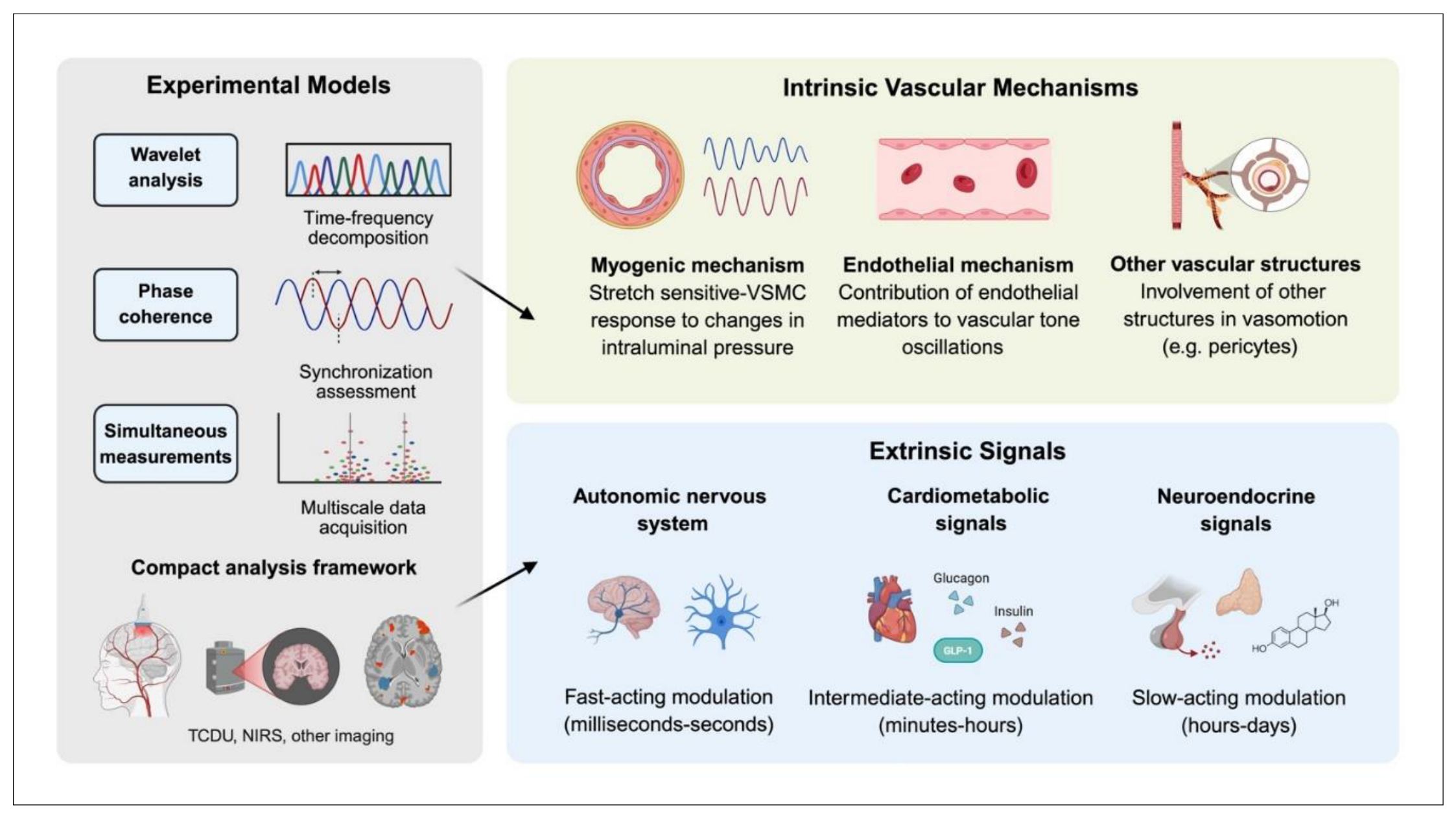
Testing vasomotion as a multiscale phenomenon requires complementary approaches that resolve its generation at the vascular-cell level, its organisation across vascular networks, and its relationship to tissue perfusion, and elucidate spatial and temporal oscillatory activity and the relationship thereof between different vascular beds. Isolated small vessel preparations, including pressure myography, provide high mechanistic resolution of membrane potential, intracellular Ca²⁺, vessel diameter, vascular tone, and oscillatory activity under controlled conditions (Lawton et al., 2019). These models, complemented with corresponding experiments in VSMC and endothelial cell cultures, are therefore well suited to testing intrinsic myogenic and endothelial mechanisms but cannot capture interactions between neighbouring vessels or influences of systemic signals (Figure 4).
Intravital and two-photon imaging of intact microvascular networks can bridge this gap by simultaneously resolving vessel diameter, mural-cell Ca2+ activity, and, where feasible, red blood cell velocity or flux across multiple vascular segments, establishing spatial characteristics of microvascular oscillatory phenomena (Glück et al., 2021; Zhang et al., 2024). Moreover, cerebral preparations allow examination of vascular dynamics alongside neuronal, astrocytic, and metabolic signals, providing an experimental framework for testing whether local oscillators become coordinated within the neurovascular network.
A complementary challenge is to characterise the temporal organisation of vasomotion. Wavelet transformation is particularly suited to assess microvascular signals because it resolves non-stationary oscillations in both time and frequency (Stefanovska et al., 1999; Kralj et al., 2023). For example, in cutaneous laser-Doppler flowmetry, wavelet analysis commonly separates oscillatory activity into characteristic frequency ranges associated with endothelial, neurogenic, myogenic, respiratory, and cardiac influences. These frequency spectra, however, should not be interpreted as direct markers of individual physiological mechanisms, as the underlying regulatory systems are strongly interconnected and their frequency bands overlap (Lenasi, 2011; Kralj & Lenasi, 2023; Kralj et al., 2026). Traditionally, specific frequency bands have been associated with distinct physiological mechanisms. However, substantial overlap between mechanisms precludes assigning a given frequency spectrum to a unique process (Kaneko et al., 2021; Lui et al., 2022; Kralj et al., 2026). Rather, integrating multiple signals at the same vascular level further supports our hypothesis that vasomotion arises from intrinsic VSMC oscillatory activity, onto which systemic influences are superimposed to modulate the final vascular response (Liu et al., 2022). Moreover, the spatial and temporal organisation of microvascular flow fluctuations, together with coherence between vascular signals, suggests that extrinsic influences may contribute to the synchronisation and coupling between cerebral and systemic microcirculation (Niizawa et al., 2023; Kaneko et al., 2021; Kralj et al., 2026). Combining wavelet analysis with time-localised phase coherence provides a more informative approach for testing interactions between distributed physiological systems, which has been detected following perturbations, such as arterial blood pressure changes or acute hyperglycaemia (Ticcinelli et al., 2017; Kralj et al., 2024). We propose extending this framework to the study of vasomotion in cerebral microcirculation. Although laser-Doppler flowmetry is not suitable for tracing cerebral microcirculation, near-infrared spectroscopy provides a non-invasive approximation of spontaneous fluctuations in cerebral tissue oxygenation, while transcranial Doppler ultrasound provides a complementary measurement of cerebral-blood flow velocity. Wavelet-based analyses of these signals may therefore assess frequency-specific relationships and phase coherence within and between peripheral and cerebral circulations (Tgavalekos et al., 2019; Holme et al., 2023; Thudium et al., 2025; Kralj et al., 2026). In vivo imaging can reveal distinct Ca²⁺ dynamics among cerebral mural-cell populations and link VSMC Ca²⁺ activity to spontaneous changes in arteriolar diameter (Glück et al., 2021; Zhang et al., 2024). Astrocytic Ca²⁺ signalling can also modulate ultraslow arteriole oscillations in vivo, providing evidence for bidirectional interactions between astrocytes and vascular dynamics (Haidey et al., 2021). These approaches nevertheless have important limitations: frequency bands cannot be equated with discrete mechanisms, coherence does not establish causality, and non-invasive human methods cannot resolve the cellular origin of vascular oscillations.
Computational modelling can help bridge these scales by incorporating experimentally measured oscillatory properties, VSMC dynamics, myogenic feedback and vessel-wall mechanics to explore how local oscillators generate collective vascular behaviour (Farina et al., 2022). Integrating such models with multiscale imaging, time-frequency analysis, and human haemodynamic measurements could generate experimentally testable predictions and help determine whether intrinsic vascular oscillators remain locally regulated or become functionally coupled across cerebral and systemic microvascular networks. Combining these measurements with tissue oxygenation or metabolic imaging, while experimentally manipulating inter-vessel coupling, could further establish whether coordinated vasomotion contributes functionally to tissue perfusion.
Conclusion and future perspective
We propose that vasomotion represents an intrinsic component of microvascular regulation continuously shaped by ANS, cardiometabolic and neuroendocrine signals. Rather than acting as isolated local oscillations, these influences may modulate the spatial and temporal organisation of vasomotion across vascular beds, potentially linking cerebral and systemic microvascular dynamics while preserving tissue specific local regulation. The functional significance of vasomotion may therefore depend not only on the generation of rhythmic changes in vascular tone, but also on their propagation, coordination, and coupling across the microvascular network. This perspective raises a fundamental question: do these oscillations merely reflect the physiological environment, or do they actively shape microvascular function? Addressing this question will require approaches that integrate cellular mechanisms with vascular network dynamics and tissue perfusion across spatial and temporal scales. Future studies should focus on combining simultaneous measurements of vascular oscillations, with advanced time-frequency and coherence analyses, experimental perturbations, and computational modelling to determine whether distributed vascular oscillators become functionally coupled across the cerebral-systemic axis.
Abbreviations
ANS – autonomic nervous system; Ca²⁺ – calcium; CO2 – carbon dioxide; GLP-1 – glucagon-like peptide-1; NO – nitric oxide; O2 – oxygen; VSMC – vascular smooth muscle cell
Author Contributions
T.M. conceived the review, developed the conceptual framework, performed the literature search, and drafted the manuscript. H.L. and T.L.T. contributed to the conception and critical revision of the manuscript. All authors approved the final version of the manuscript and agree to be accountable for all aspects of the work. All persons designated as authors qualify for authorship, and all those who qualify for authorship are listed as authors.
Funding
This work received no specific funding from any funding agency in the public, commercial or non-profit sectors.
Acknowledgments
The authors declare no acknowledgements for this manuscript.
Competing Interests
The authors declare no competing interests.
References
- Aalkjaer, C.; Nilsson, H. Vasomotion: cellular background for the oscillator and for the synchronization of smooth muscle cells. Br. J. Pharmacol. 2005, 144(5), 605–616. [Google Scholar] [CrossRef]
- Al-Shama, R. F. M.; Uleman, J. F.; Pereira, M.; Claassen, J. A. H. R.; Dresler, M. Cerebral blood flow in sleep: A systematic review and meta-analysis. Sleep Med. Rev. 2024, 77, 101977. [Google Scholar] [CrossRef]
- Armulik, A.; Genové, G.; Mäe, M.; Nisancioglu, M. H.; Wallgard, E.; Niaudet, C.; He, L.; Norlin, J.; Lindblom, P.; Strittmatter, K.; Johansson, B. R.; Betsholtz, C. Pericytes regulate the blood-brain barrier. Nature 2010, 468(7323), 557–561. [Google Scholar] [CrossRef]
- Attwell, D.; Mishra, A.; Hall, C. N.; O'Farrell, F. M.; Dalkara, T. What is a pericyte? Journal of cerebral blood flow and metabolism: official journal of the International Society of Cerebral Blood Flow and Metabolism 2016, 36(2), 451–455. [Google Scholar] [CrossRef]
- Bagher, P.; Segal, S. S. Regulation of blood flow in the microcirculation: role of conducted vasodilation. Acta Physiol. 2011, 202(3), 271–284. [Google Scholar] [CrossRef]
- Bajuk, N. B.; Zaletel, K.; Gaberšček, S.; Lenasi, H. Hyperthyroidism induced by Graves' disease reversibly affects skin microvascular reactivity. Clin. Hemorheol. Microcirc. 2015, 61(3), 459–470. [Google Scholar] [CrossRef]
- Banerjee, D.; Sabe, S. A.; Sodha, N. R.; Ehsan, A.; Cioffi, W. G.; Miner, T. J.; Li, J.; Abid, M. R.; Feng, J.; Sellke, F. W. Female patients exhibit altered vasopressin-induced coronary microvascular contractile response and molecular signaling following cardiac surgery. Am. J. Surg. 2025, 239, 116052. [Google Scholar] [CrossRef]
- Brimble, H.; Clemson, P. T.; Stefanovska, A. Cardiorespiratory Dynamics as a Non-Autonomous System of Coupled Oscillators with Time-Varying Frequency Modulation. Entropy 2026, 28(6), 685. [Google Scholar] [CrossRef]
- Broggini, T.; Duckworth, J.; Ji, X.; Liu, R.; Xia, X.; Mächler, P.; Shaked, I.; Munting, L. P.; Iyengar, S.; Kotlikoff, M.; van Veluw, S. J.; Vergassola, M.; Mishne, G.; Kleinfeld, D. Long-wavelength traveling waves of vasomotion modulate the perfusion of cortex. Neuron 2024, 112(14), 2349–2367.e8. [Google Scholar] [CrossRef]
- Chuang, K. H.; Zhou, X. A.; Xia, Y.; Li, Z.; Qian, L.; Eeles, E.; Ngiam, G.; Fripp, J.; Coulson, E. J. Cholinergic basal forebrain neurons regulate vascular dynamics and cerebrospinal fluid flux. Nat. Commun. 2025, 16(1), 5343. [Google Scholar] [CrossRef]
- Claassen, J. A. H. R.; Thijssen, D. H. J.; Panerai, R. B.; Faraci, F. M. Regulation of cerebral blood flow in humans: physiology and clinical implications of autoregulation. Physiol. Rev. 2021, 101(4), 1487–1559. [Google Scholar] [CrossRef]
- Colombari, E.; Pedrino, G. R.; Katayama, P. L.; Zoccal, D. B.; Mendonça, M. M.; Colombari, D. S. A.; Xavier, C. H. Neural Control of Tissue Perfusion: Emerging Evidence and Beyond. Curr. Hypertens. Rep. 2026, 28(1), 30. [Google Scholar] [CrossRef]
- Cracowski, J. L.; Roustit, M. Human Skin Microcirculation. Compr. Physiol. 2020, 10(3), 1105–1154. [Google Scholar] [CrossRef]
- Dabija, M. G.; Tataru, C. I.; Dumitru, A. V.; Munteanu, O.; Radoi, M. P.; Ciurea, A. V.; Petrescu, I. A. Noradrenergic Slow Vasomotion: The Hidden Fluid Pump Linking Sleep, Brain Clearance, and Dementia Pathogenesis. Int. J. Mol. Sci. 2025, 26(23), 11444. [Google Scholar] [CrossRef]
- de Boer, M. P.; Meijer, R. I.; Newman, J.; Stehouwer, C. D.; Eringa, E. C.; Smulders, Y. M.; Serné, E. H. Insulin-induced changes in microvascular vasomotion and capillary recruitment are associated in humans. In Microcirculation; New York, N.Y., 2014; Volume 21, 5, pp. 380–387. [Google Scholar]
- Deriu, F.; Roatta, S.; Grassi, C.; Urciuoli, R.; Micieli, G.; Passatore, M. Sympathetically-induced changes in microvascular cerebral blood flow and in the morphology of its low-frequency waves. J. Auton. Nerv. Syst. 1996, 59(1-2), 66–74. [Google Scholar] [CrossRef]
- Dora, K. A. Coordination of vasomotor responses by the endothelium. Circ. J. Off. J. Jpn. Circ. Soc. 2010, 74(2), 226–232. [Google Scholar] [CrossRef]
- Dunaway, L. S.; Mills, W. A., 3rd; Eyo, U. B.; Isakson, B. E. The Cells of the Vasculature: Advances in the Regulation of Vascular Tone in the Brain and Periphery. Basic Clin. Pharmacol. Toxicol. 2025, 136(5), e70023. [Google Scholar] [CrossRef]
- Farina, A.; Fasano, A.; Rosso, F. Modeling of vasomotion in arterioles. J. Theor. Biol. 2022, 544, 111124. [Google Scholar] [CrossRef]
- Ghanizada, H.; Christensen, R. H.; Al-Karagholi, M. A.; Elbahi, F. A.; Coskun, H.; Ashina, M. Arterial responses to infusion of glucagon-like peptide-1 in humans: A randomized trial study. Peptides 2022, 150, 170736. [Google Scholar] [CrossRef]
- Glück, C.; Ferrari, K. D.; Binini, N.; Keller, A.; Saab, A. S.; Stobart, J. L.; Weber, B. Distinct signatures of calcium activity in brain mural cells. eLife 2021, 10, e70591. [Google Scholar] [CrossRef]
- Hall, C. N.; Reynell, C.; Gesslein, B.; Hamilton, N. B.; Mishra, A.; Sutherland, B. A.; O'Farrell, F. M.; Buchan, A. M.; Lauritzen, M.; Attwell, D. Capillary pericytes regulate cerebral blood flow in health and disease. Nature 2014, 508(7494), 55–60. [Google Scholar] [CrossRef]
- Holme, N. L. A.; Zilakos, I.; Elstad, M.; Skytioti, M. Cerebral blood flow response to cardiorespiratory oscillations in healthy humans. Auton. Neurosci. Basic Clin. 2023, 245, 103069. [Google Scholar] [CrossRef]
- Howells, T.; Hånell, A.; Svedung Wettervik, T.; Enblad, P. Vasomotion and Cerebral Blood Flow in Patients With Traumatic Brain Injury and Subarachnoid Hemorrhage: Cerebrovascular Autoregulation Versus Autonomic Control. J. Neurotrauma 2025, 42(7-8), 700–713. [Google Scholar] [CrossRef]
- Iadecola, C. The pathobiology of vascular dementia. Neuron 2013, 80(4), 844–866. [Google Scholar] [CrossRef]
- Ji, X.; Zhao, Y.; Bai, L.; Wang, K.; Kleinfeld, D. Microvascular architecture and physiological fluctuations constrain the control of cerebral microcirculation. Proc. Natl. Acad. Sci. USA 2026, 123(3), e2521872123. [Google Scholar] [CrossRef]
- Jones, T. W. Discovery that the veins of the bat's wing are endowed with rhythmical contractility and that the outward flow of blood is accelerated by each contraction. Philos. Trans. R. Soc. Lond. 1852, 142, 131–136. [Google Scholar] [CrossRef]
- Joost, E.; Jordão, M. J. C.; Mages, B.; Prinz, M.; Bechmann, I.; Krueger, M. Microglia contribute to the glia limitans around arteries, capillaries and veins under physiological conditions, in a model of neuroinflammation and in human brain tissue. Brain Struct. Funct. 2019, 224(3), 1301–1314. [Google Scholar] [CrossRef]
- Kaneko, I.; Yoshida, Y.; Yuda, E.; Hayano, J. Sensing of Microvascular Vasomotion Using Consumer Camera. Sensors 2021, 21(18), 6256. [Google Scholar] [CrossRef]
- King, C. M.; Bohmbach, K.; Minge, D.; Delekate, A.; Zheng, K.; Reynolds, J.; Rakers, C.; Zeug, A.; Petzold, G. C.; Rusakov, D. A.; Henneberger, C. Local Resting Ca2+ Controls the Scale of Astroglial Ca2+ Signals. Cell Rep. 2020, 30(10), 3466–3477.e4. [Google Scholar] [CrossRef]
- Koep, J. L.; Taylor, C. E.; Coombes, J. S.; Bond, B.; Ainslie, P. N.; Bailey, T. G. Autonomic control of cerebral blood flow: fundamental comparisons between peripheral and cerebrovascular circulations in humans. J. Physiol. 2022, 600(1), 15–39. [Google Scholar] [CrossRef]
- Koller, A.; Toth, P. Contribution of flow-dependent vasomotor mechanisms to the autoregulation of cerebral blood flow. J. Vasc. Res. 2012, 49(5), 375–389. [Google Scholar] [CrossRef]
- Kondo, D.; Asano, N.; Ishiyama, T.; Shintani, N.; Matsukawa, T. Effects of adrenaline and vasopressin on cerebral microcirculation at baseline and during global brain ischemia and reperfusion in rabbits. BMC Anesthesiol. 2022, 22(1), 380. [Google Scholar] [CrossRef]
- Kralj, L.; Lenasi, H. Wavelet analysis of laser Doppler microcirculatory signals: Current applications and limitations. Front. Physiol. 2023, 13, 1076445. [Google Scholar] [CrossRef]
- Kralj, L.; Battelino, T.; Lenasi, H. Effects of oral glucose tolerance test on microvascular and autonomic nervous system regulation in young healthy individuals. Sci. Rep. 2025, 15(1), 32858. [Google Scholar] [CrossRef]
- Kralj, L.; Hultman, M.; Lenasi, H. Wavelet Analysis and the Cone of Influence: Does the Cone of Influence Impact Wavelet Analysis Results? Appl. Sci. 2024, 14(24), 11736. [Google Scholar] [CrossRef]
- Kralj, L.; Kurkin, S.; Lenasi, H.; Pavlov, A.; Stožer, A.; Hramov, A. E.; Perc, M.; Gosak, M. Wavelet analysis in biomedical research: A versatile tool for multiscale signal characterization and functional connectivity analysis. Phys. Life Rev. 2026, 59, 19–47. [Google Scholar] [CrossRef]
- Lawton, P. F.; Lee, M. D.; Saunter, C. D.; Girkin, J. M.; McCarron, J. G.; Wilson, C. VasoTracker, a Low-Cost and Open Source Pressure Myograph System for Vascular Physiology. Front. Physiol. 2019, 10, 99. [Google Scholar] [CrossRef]
- Lenasi, H. Assessment of Human Skin Microcirculation and Its Endothelial Function Using Laser Doppler Flowmetry. In Medical Imaging; InTech, 2011. [Google Scholar] [CrossRef]
- Liu, S.; Zhao, L.; Liu, Y. The Origin of Vasomotion and Stochastic Resonance in Vasomotion. Front. Bioeng. Biotechnol. 2022, 10, 819716. [Google Scholar] [CrossRef]
- Mihor, A.; Gergar, M.; Gaberšček, S.; Lenasi, H. Skin microvascular reactivity in patients with hypothyroidism. Clin. Hemorheol. Microcirc. 2016, 64(1), 105–114. [Google Scholar] [CrossRef]
- Mironova, G. Y.; Kowalewska, P. M.; El-Lakany, M.; Tran, C. H. T.; Sancho, M.; Zechariah, A.; Jackson, W. F.; Welsh, D. G. The conducted vasomotor response and the principles of electrical communication in resistance arteries. Physiol. Rev. 2024, 104(1), 33–84. [Google Scholar] [CrossRef]
- Mitsui, R.; Hashitani, H. Synchrony of spontaneous Ca2+ activity in microvascular mural cells. Journal of smooth muscle research = Nihon Heikatsukin Gakkai kikanshi 2020, 56(0), 1–18. [Google Scholar] [CrossRef]
- Bang, J.; Park, Y. S.; Jeong, S. M.; Song, J. G.; Kim, Y. K.; Hwang, G. S. Melatonin does not attenuate dynamic cardiovascular and cerebrovascular reflex responses to acute hypotension in healthy men. Korean J. Anesthesiol. 2012, 63(3), 245–252. [Google Scholar] [CrossRef]
- Morita, Y.; Hardebo, J. E.; Bouskela, E. Influence of cerebrovascular parasympathetic nerves on resting cerebral blood flow, spontaneous vasomotion, autoregulation, hypercapnic vasodilation and sympathetic vasoconstriction. J. Auton. Nerv. Syst. 1994, 49 Suppl, S9–S14. [Google Scholar] [CrossRef]
- Morita, Y.; Hardebo, J. E.; Bouskela, E. Influence of cerebrovascular sympathetic, parasympathetic, and sensory nerves on autoregulation and spontaneous vasomotion. Acta Physiol. Scand. 1995, 154(2), 121–130. [Google Scholar] [CrossRef]
- Muoio, V.; Persson, P. B.; Sendeski, M. M. The neurovascular unit - concept review. Acta Physiol. 2014, 210(4), 790–798. [Google Scholar] [CrossRef]
- Niizawa, T.; Sakuraba, R.; Kusaka, T.; Kurihara, Y.; Sugashi, T.; Kawaguchi, H.; Kanno, I.; Masamoto, K. Spatiotemporal analysis of blood plasma and blood cell flow fluctuations of cerebral microcirculation in anesthetized rats. Journal of cerebral blood flow and metabolism: official journal of the International Society of Cerebral Blood Flow and Metabolism 2023, 43(1), 138–152. [Google Scholar] [CrossRef]
- Nizari, S.; Basalay, M.; Chapman, P.; Korte, N.; Korsak, A.; Christie, I. N.; Theparambil, S. M.; Davidson, S. M.; Reimann, F.; Trapp, S.; Yellon, D. M.; Gourine, A. V. Glucagon-like peptide-1 (GLP-1) receptor activation dilates cerebral arterioles, increases cerebral blood flow, and mediates remote (pre)conditioning neuroprotection against ischaemic stroke. Basic Res. Cardiol. 2021, 116(1), 32. [Google Scholar] [CrossRef]
- Ogoh, S. Cardiac output-mediated regulation of cerebral blood flow during exercise: Clinical perspectives on the indirect impact of muscle metaboreflex. Exp. Physiol. 2025, 110(5), 686–693. [Google Scholar] [CrossRef]
- Peng, H.; Matchkov, V.; Ivarsen, A.; Aalkjaer, C.; Nilsson, H. Hypothesis for the initiation of vasomotion. Circ. Res. 2001, 88(8), 810–815. [Google Scholar] [CrossRef]
- Pradhan, R. K.; Chakravarthy, V. S. Informational dynamics of vasomotion in microvascular networks: a review. Acta Physiol. 2011, 201(2), 193–218. [Google Scholar] [CrossRef]
- Ramos Gonzalez, M.; Axler, M. R.; Kaseman, K. E.; Lobene, A. J.; Farquhar, W. B.; Witman, M. A.; Kirkman, D. L.; Lennon, S. L. Melatonin supplementation does not alter vascular function or oxidative stress in healthy normotensive adults on a high sodium diet. Physiol. Rep. 2023, 11(24), e15896. [Google Scholar] [CrossRef]
- Roberts-Thomson, K. M.; Parker, L.; Betik, A. C.; Wadley, G. D.; Gatta, P. A. D.; Marwick, T. H.; Keske, M. A. Oral and intravenous glucose administration elicit opposing microvascular blood flow responses in skeletal muscle of healthy people: role of incretins. J. Physiol. 2022, 600(7), 1667–1681. [Google Scholar] [CrossRef]
- Roy, T. K.; Secomb, T. W. Effects of impaired microvascular flow regulation on metabolism-perfusion matching and organ function. Microcirculation 2021, 28(3), e12673. [Google Scholar] [CrossRef]
- Saleem, S.; Teal, P. D.; Kleijn, W. B.; Ainslie, P. N.; Tzeng, Y. C. Identification of human sympathetic neurovascular control using multivariate wavelet decomposition analysis. Am. J. Physiol. Heart Circ. Physiol. 2016, 311(3), H837–H848. [Google Scholar] [CrossRef]
- Shariffi, B.; Harper, J. L.; McMillan, N. J.; Gonsalves, A. M.; Bond, B. J.; Pipkins, A. M.; Shoemaker, L. N.; Manrique-Acevedo, C.; Padilla, J.; Limberg, J. K. Effect of insulin on indices of cerebral blood flow and cerebrovascular compliance in young adults. Am. J. Physiol. Heart Circ. Physiol. 2025, 328(1), H21–H28. [Google Scholar] [CrossRef]
- Smith, J. A. B.; Murach, K. A.; Dyar, K. A.; Zierath, J. R. Exercise metabolism and adaptation in skeletal muscle. Nat. Rev. Mol. Cell Biol. 2023, 24(9), 607–632. [Google Scholar] [CrossRef]
- Song, S.; Kim, D.; Jang, D. P.; Lee, J.; Lee, H.; Lee, K. M.; Kim, I. Y. Low-frequency oscillations in cerebrovascular and cardiovascular hemodynamics: Their interrelationships and the effect of age. Microvasc. Res. 2015, 102, 46–53. [Google Scholar] [CrossRef]
- Spencer-Vargas, A. C.; Nelson, C.; Danchulis, L.; Xu, Y.; Walborn, L.; Mayrovitz, H. N. The Cellular and Biophysical Origins of Microvascular Vasomotion: A Scoping Review. Cureus 2026, 18(4), e107580. [Google Scholar] [CrossRef]
- Stefanovska, A.; Bracic, M.; Kvernmo, H. D. Wavelet analysis of oscillations in the peripheral blood circulation measured by laser Doppler technique. IEEE Trans. Bio-Med. Eng. 1999, 46(10), 1230–1239. [Google Scholar] [CrossRef]
- Subaran, S. C.; Sauder, M. A.; Chai, W.; Jahn, L. A.; Fowler, D. E.; Aylor, K. W.; Basu, A.; Liu, Z. GLP-1 at physiological concentrations recruits skeletal and cardiac muscle microvasculature in healthy humans. Clin. Sci. 2014, 127(3), 163–170. [Google Scholar] [CrossRef]
- Suzuki, N.; Hardebo, J. E.; Kåhrström, J.; Owman, C. Selective electrical stimulation of postganglionic cerebrovascular parasympathetic nerve fibers originating from the sphenopalatine ganglion enhances cortical blood flow in the rat. Journal of cerebral blood flow and metabolism: official journal of the International Society of Cerebral Blood Flow and Metabolism 1990, 10(3), 383–391. [Google Scholar] [CrossRef]
- Suzuki, Y.; Giles, W. R.; Zamponi, G. W.; Kondo, R.; Imaizumi, Y.; Yamamura, H. Ca2+ signaling in vascular smooth muscle and endothelial cells in blood vessel remodeling: a review. Inflamm. Regen. 2024, 44(1), 50. [Google Scholar] [CrossRef]
- Tgavalekos, K.; Pham, T.; Krishnamurthy, N.; Sassaroli, A.; Fantini, S. Frequency-resolved analysis of coherent oscillations of local cerebral blood volume, measured with near-infrared spectroscopy, and systemic arterial pressure in healthy human subjects. PLoS ONE 2019, 14(2), e0211710. [Google Scholar] [CrossRef]
- Thudium, M.; Kornilov, E.; Moestl, S.; Hoffmann, F.; Hoff, A.; Kulapatana, S.; Urechie, V.; Oremek, M.; Rigo, S.; Heusser, K.; Biaggioni, I.; Tank, J.; Diedrich, A. Continuous wavelet based transfer function analysis of cerebral autoregulation dynamics for neuromonitoring using near-infrared spectroscopy. Front. Physiol. 2025, 16, 1616125. [Google Scholar] [CrossRef]
- Ticcinelli, V.; Stankovski, T.; Iatsenko, D.; Bernjak, A.; Bradbury, A. E.; Gallagher, A. R.; Clarkson, P. B. M.; McClintock, P. V. E.; Stefanovska, A. Coherence and Coupling Functions Reveal Microvascular Impairment in Treated Hypertension. Front. Physiol. 2017, 8, 749. [Google Scholar] [CrossRef]
- Toda, N.; Ayajiki, K.; Okamura, T. Cerebral blood flow regulation by nitric oxide: recent advances. Pharmacol. Rev. 2009, 61(1), 62–97. [Google Scholar] [CrossRef]
- Wagner, R.; Kullmann, S.; Hummel, J.; Prystupa, K.; Hosenfeld, E.; Veit, R.; Birkenfeld, A. L.; Häring, H. U.; Preissl, H.; Fritsche, A.; Peter, A.; Heni, M. Postprandial Glucagon Action in the Human Brain. Diabetes Obes. Metab. 2026, 28(7), 5926–5938. [Google Scholar] [CrossRef]
- Welsh, D. G.; Tran, C. H. T.; Hald, B. O.; Sancho, M. The Conducted Vasomotor Response: Function, Biophysical Basis, and Pharmacological Control. Annu. Rev. Pharmacol. Toxicol. 2018, 58, 391–410. [Google Scholar] [CrossRef]
- Xu, Z.; Soh, Z.; Kurota, Y.; Kimura, Y.; Hirano, H.; Sasaoka, T.; Yoshino, A.; Tsuji, T. Neuroimaging-based evidence for sympathetic correlation between brain activity and peripheral vasomotion during pain anticipation. Sci. Rep. 2024, 14(1), 3383. [Google Scholar] [CrossRef]
- Yang, S.; Zhang, L. Glucocorticoids and vascular reactivity. Curr. Vasc. Pharmacol. 2004, 2(1), 1–12. [Google Scholar] [CrossRef]
- Zhang, P.; Yuan, D.; Luo, C.; Guo, W.; Li, F. Role of pterygopalatine ganglion in regulating isoflurane-induced cerebral hyper-perfusion. Journal of cerebral blood flow and metabolism: official journal of the International Society of Cerebral Blood Flow and Metabolism 2025, 45(2), 306–318. [Google Scholar] [CrossRef]
- Zhang, Y. Y.; Li, J. Z.; Xie, H. Q.; Jin, Y. X.; Wang, W. T.; Zhao, B.; Jia, J. M. High-resolution vasomotion analysis reveals novel arteriole physiological features and progressive modulation of cerebral vascular networks by stroke. Journal of cerebral blood flow and metabolism: official journal of the International Society of Cerebral Blood Flow and Metabolism 2024, 44(11), 1330–1348. [Google Scholar] [CrossRef]
- Zhao, X.; Schalkwijk, C.; Kroon, A.; Schram, M. T.; Stehouwer, C.; Houben, A. Different Measures of Hyperglycemia Are Negatively Associated With Skin Microvascular Flowmotion: The Maastricht Study. Microcirc. (New York N.Y. 2024, 31(7), e12882. [Google Scholar] [CrossRef]
- Zuccolo, E.; Laforenza, U.; Negri, S.; Botta, L.; Berra-Romani, R.; Faris, P.; Scarpellino, G.; Forcaia, G.; Pellavio, G.; Sancini, G.; Moccia, F. Muscarinic M5 receptors trigger acetylcholine-induced Ca2+ signals and nitric oxide release in human brain microvascular endothelial cells. J. Cell. Physiol. 2019, 234(4), 4540–4562. [Google Scholar] [CrossRef]
Figure 1.
Cellular basis of vasomotion within the neurovascular unit Vasomotion emerges from coordinated interactions among endothelial cells, vascular smooth muscle cells, pericytes, and neuroglial components of the neurovascular unit. Electrical, calcium-dependent, metabolic, and vasoactive signalling contributes to the generation, propagation, and coordination of local vasomotor oscillations. These cellular interactions provide the foundation for examining how local vascular dynamics may scale to network-level and brain-body microvascular regulation. Abbreviations: ATP – adenosine triphosphate; Ca2+ – calcium; EDHF – endothelium-derived hyperpolarizing factor; K+ – potassium; NO – nitric oxide; ROS – reactive oxygen species.
Figure 1.
Cellular basis of vasomotion within the neurovascular unit Vasomotion emerges from coordinated interactions among endothelial cells, vascular smooth muscle cells, pericytes, and neuroglial components of the neurovascular unit. Electrical, calcium-dependent, metabolic, and vasoactive signalling contributes to the generation, propagation, and coordination of local vasomotor oscillations. These cellular interactions provide the foundation for examining how local vascular dynamics may scale to network-level and brain-body microvascular regulation. Abbreviations: ATP – adenosine triphosphate; Ca2+ – calcium; EDHF – endothelium-derived hyperpolarizing factor; K+ – potassium; NO – nitric oxide; ROS – reactive oxygen species.
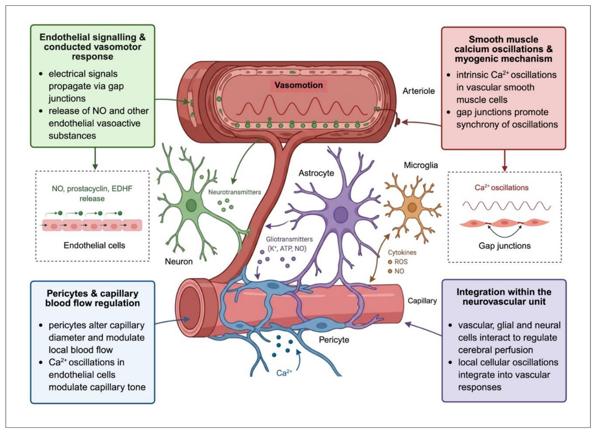
Figure 2.
Autonomic modulation of vasomotion and microvascular coordination. Schematic representation of the impact of autonomic nervous system on microvasculature and its regulation. Sympathetic and parasympathetic neural inputs can modulate vascular smooth muscle and endothelial signalling, thereby modulating the amplitude and frequency of vasomotor oscillations. Sympathetic signalling through α1-adrenergic receptors can increase vascular smooth muscle cell intracellular calcium and promote vasoconstriction, whereas parasympathetic fibre mediators, including acetylcholine, vasoactive intestinal peptide and nitric oxide, can promote endothelial signalling and subsequent vasodilation in susceptible vascular beds. Intercellular electrical coupling and conducted vascular responses contribute to the spatial coordination of vasomotor activity across interconnected vessels. Abbreviations: Ca2+ – calcium; eNOS – endothelial nitric oxide synthase; Gq – G protein-coupled receptor subunit; NO – nitric oxide; VIP – vasoactive intestinal peptide.
Figure 2.
Autonomic modulation of vasomotion and microvascular coordination. Schematic representation of the impact of autonomic nervous system on microvasculature and its regulation. Sympathetic and parasympathetic neural inputs can modulate vascular smooth muscle and endothelial signalling, thereby modulating the amplitude and frequency of vasomotor oscillations. Sympathetic signalling through α1-adrenergic receptors can increase vascular smooth muscle cell intracellular calcium and promote vasoconstriction, whereas parasympathetic fibre mediators, including acetylcholine, vasoactive intestinal peptide and nitric oxide, can promote endothelial signalling and subsequent vasodilation in susceptible vascular beds. Intercellular electrical coupling and conducted vascular responses contribute to the spatial coordination of vasomotor activity across interconnected vessels. Abbreviations: Ca2+ – calcium; eNOS – endothelial nitric oxide synthase; Gq – G protein-coupled receptor subunit; NO – nitric oxide; VIP – vasoactive intestinal peptide.
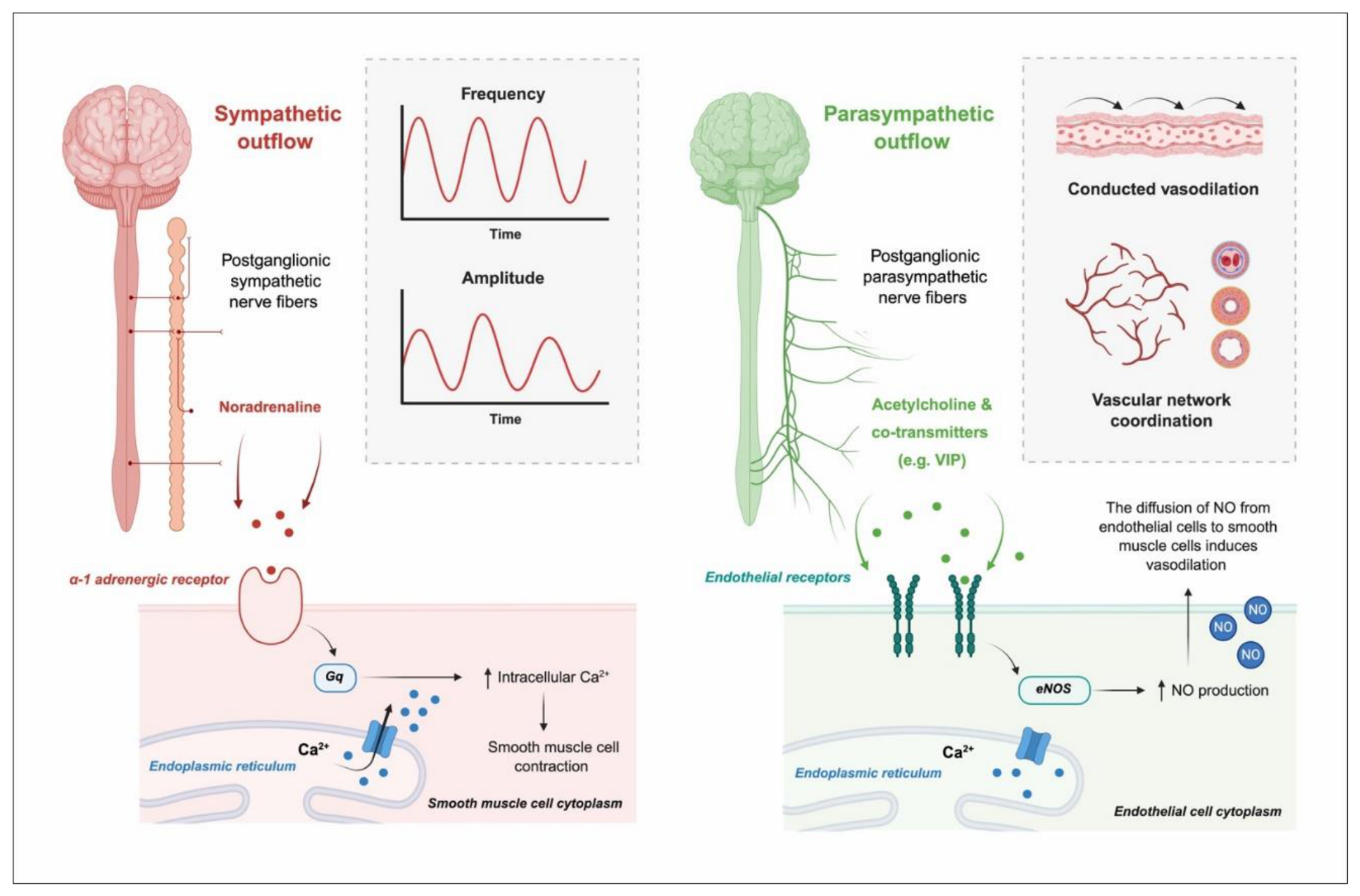
Figure 3.
Brain–body axis linking cerebral and systemic microcirculation through vasomotor dynamics Cerebral and systemic microvascular beds are interconnected through shared autonomic, cardiometabolic, and neuroendocrine signals. These extrinsic influences may modulate the amplitude, frequency, phase, and coordination of intrinsically generated vasomotor oscillations, arising from vascular smooth muscle cells, while local vascular mechanisms preserve tissue- and vascular-bed-specific responses, adjusted to tissue metabolic demands. The proposed framework allows for bidirectional interactions between cerebral and systemic microcirculation and does not imply identical oscillatory patterns across vascular beds. Abbreviations: CO2 – carbon dioxide; GLP-1 – glucagon-like peptide-1; O2 – oxygen.
Figure 3.
Brain–body axis linking cerebral and systemic microcirculation through vasomotor dynamics Cerebral and systemic microvascular beds are interconnected through shared autonomic, cardiometabolic, and neuroendocrine signals. These extrinsic influences may modulate the amplitude, frequency, phase, and coordination of intrinsically generated vasomotor oscillations, arising from vascular smooth muscle cells, while local vascular mechanisms preserve tissue- and vascular-bed-specific responses, adjusted to tissue metabolic demands. The proposed framework allows for bidirectional interactions between cerebral and systemic microcirculation and does not imply identical oscillatory patterns across vascular beds. Abbreviations: CO2 – carbon dioxide; GLP-1 – glucagon-like peptide-1; O2 – oxygen.
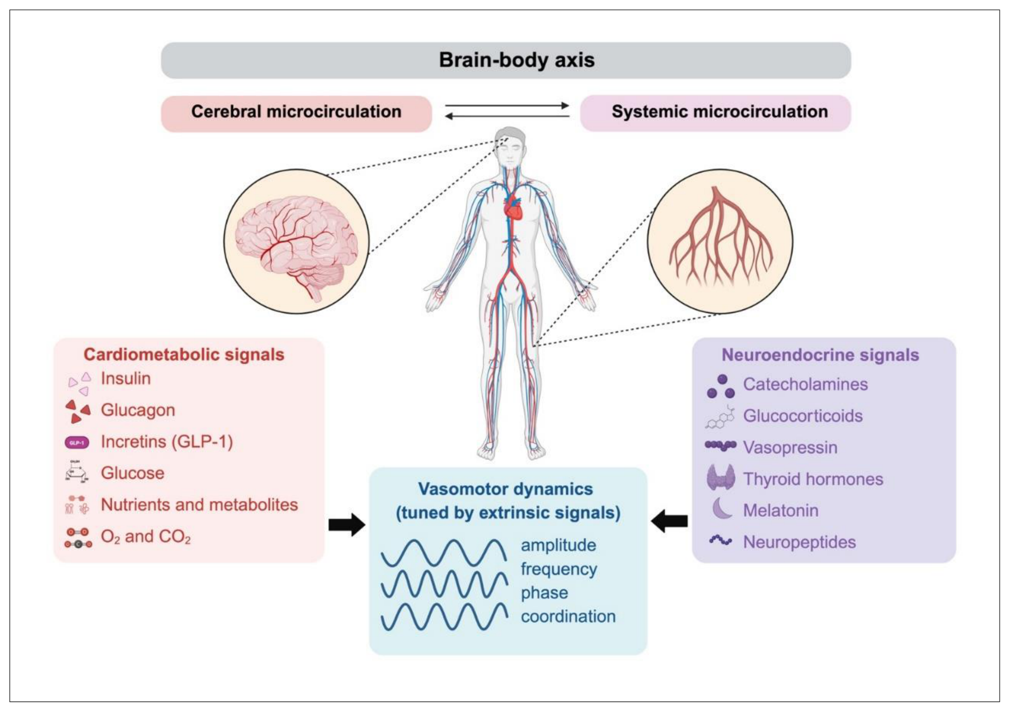
Figure 4.
Experimental framework for investigating vasomotion across spatial and temporal scales. Intrinsic vascular mechanisms, including myogenic and endothelial processes and contributions from other vascular structures such as pericytes, generate local vasomotor oscillations, whereas autonomic, cardiometabolic, and neuroendocrine signals provide extrinsic modulation. Complementary experimental approaches, including wavelet-based time–frequency analysis of the oscillatory signals emerging from microcirculation, phase-coherence assessment of various haemodynamic signals, and simultaneous measurements of cerebral and peripheral (micro)vascular signals, can be used to investigate the spatial and temporal organisation of vasomotion. Integrating cellular, vascular-network, and tissue-level measurements may help determine whether local vascular oscillators remain independently regulated or become functionally coordinated across cerebral and systemic microvascular networks. Abbreviations: NIRS – near-infrared spectroscopy; TCDU – transcranial doppler ultrasound.
Figure 4.
Experimental framework for investigating vasomotion across spatial and temporal scales. Intrinsic vascular mechanisms, including myogenic and endothelial processes and contributions from other vascular structures such as pericytes, generate local vasomotor oscillations, whereas autonomic, cardiometabolic, and neuroendocrine signals provide extrinsic modulation. Complementary experimental approaches, including wavelet-based time–frequency analysis of the oscillatory signals emerging from microcirculation, phase-coherence assessment of various haemodynamic signals, and simultaneous measurements of cerebral and peripheral (micro)vascular signals, can be used to investigate the spatial and temporal organisation of vasomotion. Integrating cellular, vascular-network, and tissue-level measurements may help determine whether local vascular oscillators remain independently regulated or become functionally coordinated across cerebral and systemic microvascular networks. Abbreviations: NIRS – near-infrared spectroscopy; TCDU – transcranial doppler ultrasound.
Disclaimer/Publisher’s Note: The statements, opinions and data contained in all publications are solely those of the individual author(s) and contributor(s) and not of MDPI and/or the editor(s). MDPI and/or the editor(s) disclaim responsibility for any injury to people or property resulting from any ideas, methods, instructions or products referred to in the content. |
© 2026 by the authors. Licensee MDPI, Basel, Switzerland. This article is an open access article distributed under the terms and conditions of the Creative Commons Attribution (CC BY) license.
Copyright: This open access article is published under a Creative Commons CC BY 4.0 license, which permit the free download, distribution, and reuse, provided that the author and preprint are cited in any reuse.