

Submitted:
15 October 2026
Posted:
19 October 2026
You are already at the latest version
Abstract
Background/Objectives: Immune checkpoint inhibitors (ICIs) have become part of standard systemic therapy for hepatocellular carcinoma (HCC), yet only a minority of patients achieve durable responses. The immunosuppressive tumour microenvironment (TME) is increasingly recognised as a central determinant of this resistance, but existing literature largely treats individual TME components – tumour-associated macrophages, neutrophils, myeloid-derived suppressor cells, regulatory T cells, cancer-associated fibroblasts, exhausted T cells and others – as independent contributors. This review aimed to synthesise current cellular, molecular and spatial evidence on the HCC TME into a unified, systems-level explanation of ICI resistance. Methods: A literature search was conducted in PubMed/MEDLINE, Scopus and Web of Science Core Collection, combining terms for HCC, the TME, individual TME cell populations, and methodological/therapeutic aspects, supplemented by a snowballing search of reference lists and a direct search of ClinicalTrials.gov for relevant trials. Results: The review characterises the physiological basis of hepatic immune tolerance and its pathological co-option in HCC, the principal immunosuppressive cell populations and their intercellular network, the contribution of the gut and intratumoural microbiome, and the spatial architecture of the TME, including the invasive tumour margin. Integrating these findings, we propose five interrelated principles of TME-centred ecosystem resistance – immune exclusion, distributed robustness, functional entrapment, multiscale feedback stabilisation, and surface–depth mismatch – that jointly explain why single-checkpoint blockade is frequently insufficient. Conclusions: Overcoming ICI resistance in HCC requires combination strategies that target multiple, interacting nodes of the suppressive TME network rather than the PD-1/PD-L1 axis alone, guided by aetiology-aware, spatially resolved biomarkers.
Keywords:
hepatocellular carcinoma
; tumour microenvironment
; immune checkpoint inhibitors
; immunotherapy resistance
; tumour-associated macrophages
; regulatory T cells
; cancer-associated fibroblasts
; T-cell exhaustion
; spatial transcriptomics
1. Introduction
Hepatocellular carcinoma (HCC) is the most common type of primary liver cancer and one of the key challenges in global oncology. According to GLOBOCAN 2020 data, HCC ranked sixth in incidence (4.7%; 906,000 new cases) and third in mortality (8.3%; 830,000 deaths) among all malignant neoplasms [1]. The high prevalence of hepatitis B virus (HBV) and hepatitis C virus (HCV) infection, which are the leading risk factors, accounts for the projected further increase in incidence [2]. Although approximately 72.5% of HCC cases occur in Asian countries [3], the global burden of the disease continues to rise, with incidence projected to increase further over the coming decades. A particularly worrying trend is the rise in incidence in regions with traditionally low rates – Europe, North and South America, Australia and New Zealand – predominantly due to the spread of obesity and type 2 diabetes mellitus [1].
When HCC is detected at early stages, radical treatment is possible: liver resection, orthotopic liver transplantation or local ablative therapy. However, the asymptomatic course of the disease means that HCC is in most cases diagnosed at late stages, when radical surgery is no longer feasible [4,5], and five-year survival under medical management does not exceed 31.5% [6]. Such patients are indicated for locoregional treatment methods (percutaneous ablation, hepatic arterial infusion chemotherapy (HAIC), lipiodol-based transarterial chemoembolisation (TACE), radioembolisation) and systemic therapy.
Immunotherapy has been regarded as one of the most promising directions in the treatment of HCC. Clinical trials of therapy with immune checkpoint inhibitors (ICIs) have been conducted, both as monotherapy and in combination regimens [7,8]. Based on the pivotal IMbrave150 trial, the combination of atezolizumab (a programmed death-ligand 1 (PD-L1) inhibitor) and bevacizumab (an anti-vascular endothelial growth factor (anti-VEGF) antibody) demonstrated superior overall and progression-free survival compared with sorafenib and has since been adopted as preferred first-line systemic therapy for unresectable HCC in international clinical practice guidelines, including those of EASL, AASLD and APASL [7,9,10,11]. A second approved first-line option is the STRIDE regimen – a single priming dose of tremelimumab (an anti-cytotoxic T-lymphocyte-associated protein 4 (anti-CTLA-4) antibody) followed by regular-interval durvalumab (a PD-L1 inhibitor) – which in the phase 3 HIMALAYA trial (NCT03298451) significantly improved overall survival versus sorafenib (HR = 0.78, 95% CI 0.65–0.92), with the benefit sustained at 5-year follow-up (19.6% vs 9.4% overall survival (OS)) [12]. Nevertheless, the results of these trials indicate that a significant proportion of patients do not achieve an objective response to ICI therapy, whether as monotherapy or in combination regimens [4,13].
Among the many factors determining the response to ICIs, the immunosuppressive tumour microenvironment (TME) occupies a special place. The TME comprises a complex network of interactions between immune and tumour cells, stromal elements and components of the extracellular matrix, and is regarded as one of the key factors limiting the efficacy of ICIs [14,15,16]. The development of methods for remodelling the TME and converting a "cold" tumour into a "hot" one–that is, a tumour with substantial infiltration by functionally active immune cells–is a promising area of research aimed at enhancing the efficacy of immunotherapy [16].
In addition to ICIs, other high-technology approaches to the treatment of HCC are being actively developed, including next-generation targeted multikinase inhibitors [17], CAR-T therapy directed against the tumour antigens glypican-3, alpha-fetoprotein and mucin 1 [18], oncolytic viruses and genetically engineered bacterial constructs capable of synthesising therapeutic antibodies directly within the TME [19,20], as well as nanosystems for the delivery of immunomodulatory drugs [21]. These directions, which are mostly at the preclinical or early clinical stage, could potentially substantially complement ICI therapy; however, their detailed consideration is beyond the scope of the present review.
The aim of this review is to systematise current understanding of the cellular and molecular mechanisms underlying the formation of the immunosuppressive TME in HCC and their impact on the efficacy of ICIs. The paper sequentially addresses: (1) the immune landscape of the liver under normal conditions and the mechanisms of physiological immune tolerance; (2) the cellular composition and intercellular interactions within the TME in HCC; (3) the principles of TME-centred ecosystem resistance; (4) promising strategies for remodelling the TME in the context of combination therapy.
Although numerous studies and recent reviews have addressed the role of the TME in HCC and its contribution to resistance to ICIs, these mechanisms are often discussed separately, with individual cellular populations, signalling pathways, metabolic alterations, or immune dysfunction considered as relatively independent determinants of treatment failure. Such an approach has substantially expanded our understanding of the molecular and cellular basis of ICI resistance, but it provides a limited explanation of how these mechanisms interact within the spatially and functionally heterogeneous HCC microenvironment and collectively maintain resistance under therapeutic pressure. In particular, the ability of different TME components to compensate for one another, reinforce suppressive feedback loops, restrict the functional accessibility of antitumour immunity, and undergo adaptive remodelling remains insufficiently integrated into a unified framework. Therefore, the present review goes beyond a component-based description of the HCC TME and proposes a systems-level concept of TME-centred ecosystem resistance, in which resistance to ICIs is considered an emergent property of the dynamic interaction between tumour-cell plasticity and the cellular, stromal, spatial, metabolic, and signalling components of the microenvironment. Within this framework, we synthesise current evidence on the major mechanisms of TME-mediated resistance and formulate five interrelated principles that may explain why inhibition of a single immune checkpoint is frequently insufficient to overcome an established resistant state. This conceptual framework is further used to identify potential therapeutic strategies and directions for clinical research aimed at disrupting the self-sustaining properties of the resistant tumour ecosystem.
2. Materials and Methods
A literature search was conducted in PubMed/MEDLINE, Scopus, and Web of Science Core Collection. Search queries were constructed as combinations of terms relating to (a) the disease entity, (b) the TME in general, (c) individual TME cell populations, and (d) methodological/therapeutic aspects:
(a) "hepatocellular carcinoma" OR "HCC"
(b) "tumor microenvironment" OR "TME" OR "tumour microenvironment"
(c) "tumor-associated macrophages" OR "TAMs", "regulatory T cells" OR "Tregs", "myeloid-derived suppressor cells" OR "MDSCs", "cancer-associated fibroblasts" OR "CAFs", "tumor-associated neutrophils" OR "TANs", "exhausted T cells" OR "T-cell exhaustion", "natural killer cells", "regulatory B cells" OR "Bregs", "dendritic cells", "tissue-resident memory T cells" OR "TRM", "mucosal-associated invariant T cells" OR "MAIT"
(d) "immune checkpoint inhibitor*" OR "ICI", "immunotherapy resistance", "spatial transcriptomics", "single-cell RNA sequencing" OR "scRNA-seq", "CODEX", "gut microbiome"
Terms from groups (c) and (d) were combined with (a) and (b) using the AND operator; within groups, the OR operator was used. A snowballing approach (manual review of reference lists of already-selected reviews and meta-analyses) was additionally applied to identify relevant work not captured by the search terms used. Data on clinical trials for Table 5 were obtained through a direct search of the ClinicalTrials.gov registry.
The inclusion criteria were:
Original studies, reviews, and meta-analyses in English and Russian addressing cellular, molecular, or spatial aspects of the TME in HCC;
Studies employing single-cell sequencing, spatial transcriptomics, or multiplex imaging methods – prioritised for the section on spatial organisation of the TME;
Clinical trials (registered on ClinicalTrials.gov) evaluating targeted or immune therapy directed at TME components;
Mechanistic/preclinical– included only where the extrapolation to HCC is explicitly flagged as such in the text (e.g., the example from melanoma in the section on compensatory upregulation of alternative checkpoints).
The exclusion criteria were:
Case reports and conference abstracts without a subsequent peer-reviewed publication, except where no alternative data source was available;
Publications that did not distinguish TME cell subpopulations (e.g., did not differentiate TAM subtypes) and could not be cross-referenced with other sources;
Duplicate analyses of the same patient cohort – the most complete/most recent publication was retained;
Studies conducted in other solid tumour models, except those whose findings were extrapolated to HCC due to the absence of relevant information in this field.
After duplicate removal, title and abstract screening, and subsequent in-depth analysis, 216 publications were included in this review.
3. Physiological Immune Tolerance of the Liver
For a better understanding of the TME, it is necessary to briefly examine the immune landscape maintained in the healthy liver. The liver has a dual blood supply: a smaller proportion of blood is delivered via the a. hepatica propria, and a larger proportion via the v. portae. Since blood flowing from the intestine via the portal vein contains a large number of dietary and microbial antigens, the liver is subjected to a considerable immunological burden [22,23]. Given the low reactivity to these antigens, as well as the fact that liver transplantation with a mismatched set of major histocompatibility complex antigens can be successful, some researchers consider the liver to be an "immune-privileged" organ [23]. Despite a number of advantages provided by immune tolerance, this organisation of the immune system is one of the liver's "weak points". As a result, carcinogenesis processes that evade immunobiological surveillance can develop rapidly in the liver [22].
The immune response in the liver is carried out through multiple cell populations, among which Kupffer cells, liver sinusoidal endothelial cells (LSECs), dendritic cells (DCs), hepatic stellate cells (HSCs), and various subpopulations of lymphocytes and macrophages are singled out [23,24].
Kupffer cells (KCs) belong to the group of resident liver macrophages. Studying human KCs is complicated by the difficulty of isolating them [24]. Most likely, the main pool of KCs is formed as early as embryogenesis from early and late erythromyeloid progenitors, although during life the hepatic macrophage pool is partially renewed through the transformation of monocytes from the bone marrow [25,26]. KCs perform a number of functions, such as phagocytosis of bacterial fragments and apoptotic cell debris and antigen presentation, and are also involved in iron and lipid metabolism [25]. There are several subpopulations of KCs, differing in the pattern of cytokine and chemokine secretion [24,25]. MARCO+ (macrophage receptor with collagenous structure) KCs most likely elicit anti-inflammatory and antitumour responses and regulate the intensity of the immune reaction [24,25]. There is evidence that KCs play a key role in establishing immune tolerance by activating regulatory T cells (Tregs) through IL-10 secretion [27]. It has also been established that KCs express immunosuppressive molecules, including PD-L1, on the outer surface of their membrane [28,29].
Liver sinusoidal endothelial cells are fenestrated cells lining the hepatic sinusoids. LSECs are responsible for recognising and endocytosing bacterial fragments in the blood, and therefore express a large number of pattern recognition receptors on their surface, including mannose and scavenger receptors [30,31]. Receptor-mediated endocytosis and subsequent antigen presentation by LSECs constitute one of the key mechanisms of physiological immunological tolerance in the liver. LSECtin is expressed by LSECs and functions as a negative regulator of hepatic T-cell immune responses [32]. Activated T cells expressing CD44 on their surface interact with these receptors, leading to a decrease in their activity and proliferation. LSECs also, via the Notch signalling pathway, drive the development of a distinct T-helper 1 (Th1) subpopulation secreting large amounts of anti-inflammatory IL-10 [33]. Antigen presentation by LSECs leads to transforming growth factor beta-dependent (TGF-β-dependent) conversion of forkhead box P3+ (FOXP3+) Tregs. LSECs also express immunosuppressive molecules, including PD-L1, on their surface [34]. However, it is important to note that, despite the described immunosuppressive features of LSECs, these cells have dual functions and also play an important coordinating role during an active immune response. Intracellular adhesion molecule-1 (ICAM-1) and vascular adhesion protein-1 (VAP-1) receptors are expressed on the LSEC membrane, promoting leukocyte adhesion. Their expression increases during an inflammatory reaction [35].
Dendritic cells also play a crucial role in the immune landscape of the liver. DCs are characterised by expression of major histocompatibility complex (MHC) I, MHC-II, CD209, and the CD11c integrin [36]. Three major subpopulations of DCs can be distinguished: classical DCs (conventional, myeloid DCs–cDC), plasmacytoid DCs (CD303+, CD304+), and inflammatory DCs; cDCs, in turn, consist of two groups: type 1 cDCs (CD141+/CD14-, which mainly activate CD8+ cells) and type 2 cDCs (CD1c+/CD14−, which mainly activate CD4+ cells) [34,36,37,38]. Type 2 cDCs are the most common DC subpopulation in the liver, whereas they are rarely encountered in peripheral blood or the spleen [36]. DCs are also involved in establishing immunological tolerance through the exhaustion and anergy of cytotoxic T cells or through stimulating Treg formation (particularly characteristic of plasmacytoid DCs) [34]. Hepatic DCs differ from analogous cells in other tissues: relatively immature DCs with low expression of MHC-II and co-stimulatory molecules are represented in the liver. It has been established that hepatic DCs secrete higher levels of IL-10 and TGF-β, lower levels of IL-12, express PD-L1 on their surface, and also produce prostaglandin E2 (PGE2), which has anti-inflammatory effects [34]. It is important to note that not only do DCs contribute to the induction of Tregs, but conversely, Tregs also suppress DC activity through mechanisms including CTLA-4 and leukocyte function-associated antigen-1 (LFA-1), which manifests, in particular, as reduced expression of CD80/CD86 even in the presence of stimulatory signals [34,39].
Regulatory T cells are probably the central element in the formation of peripheral immunological tolerance, including in the liver. Shimon Sakaguchi, Mary E. Brunkow, and Fred Ramsdell demonstrated the existence of a distinct subpopulation of CD25+ FOXP3+ immune cells performing immunosuppressive functions, and were awarded the 2025 Nobel Prize in Physiology or Medicine "for their discoveries concerning peripheral immune tolerance" [40,41,42].
FOXP3 serves as the key transcription factor required for the differentiation and maintenance of Treg function, whereby expression of the full-length isoform, rather than the truncated delta-2 form, is critically necessary for their identity [42,43]. Based on their origin, a distinction is made between naturally occurring Tregs (nTregs), which develop from precursors in the thymus, and inducible Tregs (iTregs), which form from naive peripheral blood T cells under the influence of microenvironmental signals, including TGF-β and tumour antigens [44]. Functionally, Tregs are subdivided into stable (FOXP3+, immunosuppressive action), fragile Tregs (FOXP3+, pro-inflammatory and antitumour action), and unstable (FOXP3-, effector-like properties).
In the healthy liver, Tregs ensure control over the intensity of the immune response to dietary and microbial antigens entering via the portal vein. The suppressive effect of nTregs is mediated through cell-to-cell contacts, secretion of cytokines (IL-10, TGF-β, IL-35) and chemokines, and also through effects on the metabolism of surrounding cells [44]. It is precisely these physiological mechanisms that subsequently become the basis of the immunosuppressive TME in HCC, where they are pathologically enhanced under the influence of tumour factors.
In brief conclusion, it can be noted that the immunosuppressive tumour microenvironment in HCC is created on the basis of physiological mechanisms functioning in the healthy organ. The tumour not only independently forms a distinct milieu around itself, but also enhances already existing mechanisms of immunological tolerance, as illustrated in Figure 1.
4. The Immunosuppressive Tumour Microenvironment in HCC
The immunosuppressive TME represents a complex ecosystem of cellular and biochemical interaction, the emergence of which in the liver is based on natural hepatic immune tolerance that transforms into a pro-tumoural one, as well as on the direct contributory action of the tumour itself. The TME comprises recruited cells that, under the influence of cancer cells, change their phenotype and function, protecting the tumour instead of the host organism, as well as cells that, while under the cellular and biochemical influence of the tumour and other TME components, lose or reduce their antitumour potential. Figure 2 provides a general overview of the key intercellular interactions within the TME, which are discussed in further detail below.
4.1. Tumour-Associated Macrophages
Tumour-associated macrophages (TAMs) represent one of the most numerous and heterogeneous cell populations in the TME [16,45]. Normally, depending on the stimulating cytokines and chemokines, macrophages undergo a process of activation and polarisation. This can result in the formation of classically activated M1 macrophages and alternatively activated M2 macrophages; these forms are regarded as extremes, and numerous intermediate forms also exist [46]. TAMs are considered to be macrophages predominantly belonging to the M2 (more specifically, M2d) phenotype, as they possess immunosuppressive and regulatory properties and express the corresponding markers (CD68, CD163, CD206) [47]. However, there is evidence that TAMs express markers characteristic of both M1 and M2 macrophages [48]. TAMs have an unstable expression profile, since under the influence of surrounding factors they are capable of converting towards the M1- or M2-phenotype, and therefore they must be considered and studied separately [48]. By origin, TAMs can be divided into 2 categories: resident (derived from KCs) and monocyte-derived. Clinically, a higher density of TAMs is associated with an unfavourable prognosis in HCC [49,50].
The cytokines IL-4, IL-10, IL-13, as well as TGF-β, promote the polarisation of TAMs towards the anti-inflammatory M2-phenotype [48]. TAMs, in turn, secrete TGF-β and IL-10, which have a whole range of immunosuppressive effects: they promote the growth and development of Th2, suppress the activity of Th1, activate Tregs, and promote the proliferation and invasion of tumour cells [51,52,53]. Th2 actively produce IL-4, IL-13, thus forming a positive feedback loop between Th2 and TAMs [48,54]. TAMs are also characterised by increased expression of macrophage colony-stimulating factor (CSF1), which causes a shift in the Th1/Th2 balance towards an immunosuppressive Th2-response [55,56]. TAMs secrete a range of matrix metalloproteinases (MMPs), particularly MMP-7, MMP-9, which contributes to the emergence of an immunosuppressive environment, tumour progression and metastasis, and resistance to treatment [53]. There is evidence that MMP-9+ TAMs represent the terminal stage of TAM differentiation; moreover, there are 2 pathways of TAM development towards the MMP-9+ phenotype [45].
Chemokines play a particular role in the recruitment and functioning of TAMs. CCL2 (C-C motif chemokine ligand 2) promotes the formation of TAMs from blood monocytes [57]. CCL15 promotes an increase in the number of CD163+ TAMs in the tumour [49]. TAMs secrete CCL22, which is a chemoattractant for tumour cells in HCC, as well as CXCL1 (C-X-C motif chemokine ligand 1), which promotes the acquisition of the M2-phenotype by macrophages [50,57]. TAMs in general, and CCL22 in particular, promote epithelial-mesenchymal transition of tumour cells, which leads to earlier metastasis [50,58]. There is a subpopulation of CXCL12+ TAMs which, via the CXCL12/CXCR4 (C-X-C motif chemokine receptor 4) axis, stimulate the expression of PD-L1 on regulatory B cells [59].
TAMs express large amounts of CD48 on their surface, which, by binding to the 2B4 receptor (CD244) on natural killer cells (NK cells), causes their subsequent exhaustion and death [60]. Both programmed cell death protein 1 (PD-1) and PD-L1 are present on the surface of TAMs [61]. Thus, TAMs promote tumour immune evasion.
4.2. Tumour-Associated Neutrophils
Tumour-associated neutrophils (TANs) also play an important role in shaping the TME in HCC. As with TAMs, two phenotypes are distinguished among TANs: N1 (anti-tumour) and N2 (pro-tumour) [57]. It has been established that TANs actively secrete CCL2 and CCL17 [62]. These chemokines actively recruit Tregs and pro-tumour M2-type macrophages into the tumour stroma, suppressing anti-tumour immunity [62]. TANs demonstrate increased expression of CCL2/CCL17 compared with peripheral neutrophils, which correlates with HCC aggressiveness, recurrence and reduced overall survival [62]. CXCL5 induces more active recruitment of TANs from neutrophils in the surrounding tissues [57]. CCL21 has been found to suppress the activity of N2 TANs and may therefore be considered a promising option for combination therapy with ICIs [63]. TANs are capable of secreting large quantities of factors with paracrine effects, including HGF (hepatocyte growth factor), oncostatin M, β2-integrin, neutrophil elastase, MMP-9 and VEGF [62].
Infiltration by TANs is closely associated with the accumulation of intratumoural immunosuppressive cells: CD68+/CD163+ TAMs and FOXP3+ Tregs [62]. This process enhances angiogenesis and creates a cycle that supports tumour growth [64]. A key mechanism is the "education" of neutrophils by tumour cells through activation of the PI3K/Akt and p38/MAPK signalling pathways, which switches them to a pro-tumour phenotype [62]. Hypoxia in the microenvironment further enhances TAN survival and secretory activity, promoting immunosuppression [62]. Through the secretion of arginase-1, as well as through the PD-L1/PD-1 axis, TANs reduce T-cell proliferation, and through activation of the TNFα pathway they induce their apoptosis [65].
Thus, TANs play an important role in the immunosuppressive network of HCC, making them a promising therapeutic target for restoring anti-tumour immunity.
4.3. Myeloid-Derived Suppressor Cells
Myeloid-derived suppressor cells (MDSCs) represent a heterogeneous group of immature myeloid cells and their precursors with immunosuppressive properties. MDSCs form when chronic processes are present in the body (infection, chronic inflammation, tissue damage, malignancies) accompanied by the release of large amounts of cyto- and chemokines [66,67]. In HCC, increased MDSC abundance is associated with poor prognosis and an elevated risk of disease progression [68].
Three populations of MDSCs are distinguished in humans: polymorphonuclear MDSCs (PMN-MDSCs, 80%), monocytic MDSCs (M-MDSCs), and early-stage MDSCs (e-MDSCs, less than 3%; some researchers do not distinguish these as a separate population) [63,67,69,70]. Phenotypically, these cells are characterised as follows: PMN-MDSCs are CD11b+ CD14- CD15+ or CD11b+ CD14- CD66b+; M-MDSCs are CD11b+ CD14+ HLA-DR-/lo CD15-; e-MDSCs are CD3− CD14- CD15- CD19- CD56- HLA-DR- CD33+ [69]. Difficulties arise in isolating MDSCs from samples and in differentiating them from cells that lack immunosuppressive properties. It has been established that lectin-like oxidised low-density lipoprotein receptor 1 (LOX-1), which belongs to the scavenger receptor family, can be used as a reliable marker of human PMN-MDSCs, while M-MDSCs can be distinguished from properly functioning human monocytes by the absence of MHC class II (HLA-DR-), which allows further studies of these cells to be carried out [67,71].
The combined effect of cytokines produced by the TME, such as PGE2, granulocyte-macrophage colony-stimulating factor (GM-CSF), granulocyte colony-stimulating factor (G-CSF), macrophage colony-stimulating factor (M-CSF), stem cell factor (SCF), S100 proteins, VEGF, IL-6, TGF-β and tumour necrosis factor-alpha (TNF-α), leads to disruptions in normal cell differentiation and to the formation of a whole spectrum of MDSCs with pro-tumour properties and variable phenotypes [72]. MDSCs accumulate in zones with specific conditions, such as the tumour microenvironment or zones of chronic inflammation [72]. Compared with physiologically differentiated cells, MDSCs demonstrate weak phagocytic activity, an immature phenotype, and more pronounced immunosuppressive effects [66].
PMN- and M-MDSCs possess pronounced immunosuppressive properties, although the mechanisms of action of these subpopulations may differ. M-MDSCs impair the function of T cells and NK cells through the production of NO, cytokines (IL-10, TGF-β) and expression of PD-L1, whereas PMN-MDSCs can exert a suppressive effect on T cells and NK cells through the production of reactive oxygen species (ROS), PGE2, indoleamine 2,3-dioxygenase (IDO) and peroxynitrite, as well as through the depletion of arginine required by T cells [67,73]. MDSCs are also characterised by reduced expression of IL-12, which is regulated by cell cycle-related kinase via the STAT3/E4BP4 signalling pathway [74]. Given the pro-inflammatory activity of IL-12 and its activating effect on NK cells and effector T cells, the reduction in its expression by MDSCs complements their immunosuppressive effects. MDSCs are characterised by the formation of a positive feedback loop that self-sustains the immunosuppressive effect.
There is complexity in separating and classifying the concepts of TANs and MDSCs [69]. Although N2 TANs and PMN-MDSCs possess similar immunosuppressive functions, their origin and stage of differentiation differ. N2 TANs represent mature neutrophils that were recruited from the blood by the tumour microenvironment and subsequently underwent changes, acquiring the pro-tumour N2 phenotype. PMN-MDSCs represent a broader group of cells that acquired immunosuppressive properties as early as the stage of myelopoiesis in the bone marrow, under the influence of tumour factors contained in the blood. PMN-MDSCs are found not only in the tumour microenvironment, but also in peripheral blood, the spleen and other organs. The tumour microenvironment constantly affects both TANs and PMN-MDSCs, so within the tumour the functional boundary becomes blurred and can only be established on the basis of surface markers.
4.4. Regulatory T Cells
Tregs are classically characterised as FOXP3+ CD25+ CD4+ cells and are rightly regarded as the "conductors" of the TME [75]. The pool of tumour-associated Tregs is replenished from several sources: nTregs are recruited from the blood, iTregs are formed directly within the TME from naïve CD4+ cells, and an additional contribution is made by IL-17A+ FOXP3+ cells and Foxp3+ cells derived from Th17 [76]. Recruitment of Tregs into the TME is mediated by the C-C motif chemokine receptor 4 (CCR4)–CCL17/22, CCR5–CCL5, CCR8–CCL1 and CCR10–CCL28 chemokine axes [77]. Intratumoural Tregs are characterised by a CD45RA- phenotype, i.e. a memory cell phenotype [77].
Clinical data indicate that an increased presence of Tregs in the TME is associated with more advanced stage, larger tumour size and unfavourable prognosis [78]. An increase in the proportion of Tregs in peripheral blood after hepatectomy also correlates with worse outcomes [79].
Tregs produce IL-10, TGF-β and IL-35, which inhibit CD8+ T cells and DCs [45]. In addition, Tregs are capable of direct cytotoxic action against effector cells: nTregs release granzyme A and perforin, while iTregs release granzyme B. Tregs also induce anergy and functional suppression of CD4- natural killer T cells (NKT cells) [44].
On the surface of Tregs in the TME, expression of CD25 (the high-affinity IL-2 receptor) is significantly higher than the corresponding levels on other T cells, which allows Tregs to efficiently "absorb" IL-2 – a critically important stimulator of effector T cells and NK cells [44,80,81]. In addition, Tregs express the ectonucleotidases CD39 and CD73, which sequentially hydrolyse ATP and ADP to adenosine, the accumulation of which in the extracellular matrix exerts an additional immunosuppressive effect [44]. A mechanism of direct metabolic interference via cAMP transport through gap junctions has also been described [44].
The heterogeneity and plasticity of Treg subpopulations play a key role in tumour progression [82]. More detailed typing of these cells makes it possible not only to identify groups with particular functional properties but also to improve the accuracy of predicting disease course. Table 1 presents the main markers of Treg subpopulations found in the TME in HCC.
A key feature of Tregs in the TME is the simultaneous high expression of several immune checkpoints, each of which makes an independent contribution to immunosuppression.
By binding to CD80/CD86 on the surface of DCs, Tregs suppress the costimulation of effector T cells via CTLA-4. Importantly, CTLA-4-mediated trans-endocytosis of CD80/CD86 molecules simultaneously serves as a mechanism of self-regulation of Treg numbers [43,85].
LAG-3 (lymphocyte activation gene 3, CD223) binds MHC class II with a significantly higher affinity than the CD4 co-receptor and comparable to T-cell receptors (TCRs), allowing it to outcompete CD4 for MHC-II binding on DCs and effectively "intercept" the activating signal from DCs [86]. Elevated LAG-3 levels are associated with an unfavourable prognosis [87]. In addition to the classical MHC class II-dependent pathway, LAG-3 interacts with fibrinogen-like protein 1 (FGL1), synthesised by hepatocytes [88], and also disrupts formation of the TCR–CD3 complex by directly binding to the TCR, causing local acidification of the microenvironment and, as a consequence, dissociation of lymphocyte-specific protein tyrosine kinase (Lck) from co-receptors [89]. The role of FGL1 in the TME in HCC remains a subject of debate: conflicting data have been published regarding the direction of change in its concentration [87,88,90,91], which requires further investigation.
TIGIT (T-cell immunoreceptor with immunoglobulin and immunoreceptor tyrosine-based inhibitory motif domains) is a co-inhibitory molecule characterised by increased expression on the surface of Tregs in the TME in HCC and interacts with the high-affinity ligand CD155, located on the surface of monocytes, DCs, fibroblasts and other cells [92,93,94]. Through the TIGIT/CD155 interaction, Tregs cause enhanced IL-10 production by DCs [92]. TIGIT+ Tregs display more pronounced expression of CTLA-4 and CD25, suppress Th1 and Th17 cells, and consequently exhibit more pronounced immunosuppressive properties than TIGIT- Tregs [95,96].
Both PD-1 and PD-L1 are expressed on the surface of Tregs; moreover, PD-L1 levels increase considerably upon activation [43]. Since Tregs in the TME are continuously subjected to stimulation, high PD-L1 expression enhances their capacity to inhibit effector cells. It is significant that PD-L1 blockade does not reduce the expression of FOXP3 or other Treg inhibitory molecules: thus, even against a background of anti-PD-L1 therapy, Tregs retain considerable immunosuppressive potential through alternative mechanisms [43].
The most important feature of Tregs in the TME is their capacity to form positive feedback loops that ensure autonomous maintenance of the immunosuppressive milieu [44]. IL-10, TGF-β and IL-35, secreted by Tregs, not only suppress effector cells but also promote the differentiation of new iTregs from naïve T cells. Adenosine, which accumulates as a result of CD39/CD73 activity, additionally inhibits effector T cells while simultaneously stabilising the Treg phenotype. Suppression of DCs through CTLA-4-dependent trans-endocytosis of CD80/CD86 reduces costimulation of effector cells, which in turn favours the relative predominance of Tregs in the TME [85]. Thus, the TME as a whole, and the Treg population in particular, acquire the properties of a self-reinforcing immunosuppressive system operating on the principle of positive feedback loops.
Despite their predominantly immunosuppressive and pro-tumourigenic role, Tregs are also capable of antitumour activity [97]. By suppressing chronic inflammation, Tregs limit damage to healthy tissues caused by aggressive immune response mediators, thereby reducing the risk of genetic mutations in surrounding cells that could potentially lead to their malignant transformation. Furthermore, fragile Tregs, while retaining FOXP3 expression, are able to produce proinflammatory and antitumour cytokines.
In summary, Tregs are a central coordinating element of the immunosuppressive TME in HCC. Their role extends beyond simply suppressing effector cells: Tregs form a self-sustaining network of interactions involving TAMs, MDSCs, DCs and tumour cells. The multiplicity of immunosuppressive mechanisms (secretion of inhibitory cytokines, metabolic competition, co-expression of immune checkpoints) explains the limited efficacy of ICI monotherapy targeting a single axis and justifies the need for combined approaches, including strategies specifically targeting Tregs.
4.5. Cancer-Associated Fibroblasts
Cancer-associated fibroblasts (CAFs) are the most numerous stromal components of the TME in HCC and play a key role in regulating immune cells and shaping a persistent TME [98]. Activated HSCs are considered the main source of CAFs, although mesenchymal stem cells, resident fibroblasts and other stromal elements, including endothelial cells, adipocytes and pericytes, also contribute to their population [99]. Tumour cells additionally promote the transformation of HSCs into CAFs through the secretion of exosomal miRNA-21 [100].
CAFs represent an extremely heterogeneous population, which complicates their classification and the development of targeted therapy. To date, more than 8 subtypes of CAFs have been identified, among which myofibroblastic CAF (myCAF), inflammatory CAF (iCAF) and cytokine/growth factor-expressing CAF (cyCAF) are the most common in HCC [99]. The most frequent subtype, myCAF, is characterised by expression of αSMA (α-smooth muscle actin, encoded by the ACTA2 (actin alpha 2, smooth muscle) gene), FAP (fibroblast activation protein), POSTN (periostin), ACTA1 (actin alpha 1, skeletal muscle), COL1A1 and COL1A2 (collagen type I alpha 1/2 chains), whereas cyCAF typically expresses HGF, extracellular matrix protein 1 (ECM1), regulator of G-protein signalling 5 (RGS5) and collectin subfamily member 11 (COLEC11) [98,99,101,102]. myCAF are predominantly localised in connective tissue septa, while cyCAF are found in the liver parenchyma [102]. Owing to the plasticity of their phenotypes and their dependence on the cytokine milieu, CAFs are more often classified by the markers they express rather than by strict subpopulations [98,102,103]. The myCAF/cyCAF ratio is likely to be of functional significance in hepatocarcinogenesis [102].
myCAF promote tumour progression through the synthesis of COL1, forming a physical barrier to immune cell infiltration and activating mechanisms associated with matrix stiffness (the COL1-stiffness-TAZ axis). These effects are particularly pronounced in liver fibrosis [102]. In addition, COL1 interacts with the discoidin domain receptor 1, expressed on hepatocytes and tumour cells, which also promotes HCC progression, especially at advanced stages. CAFs interact actively with cells of the TME via extracellular vesicles and secreted factors. Thus, CTGF (connective tissue growth factor) activates the Notch1/Snail1 pathway, enhancing proliferation and metastasis, while pleiotrophin, interacting with syndecan-2, produces similar effects [104,105].
Secretion of IL-6 by CAFs triggers the IL-6/IL6R/STAT3/Notch cascade, promoting epithelial-mesenchymal transition and the acquisition of a stem-like phenotype by tumour cells [106,107]. It is presumed that CAF-secreted IL-6 may stimulate the development of MDSCs, as has already been shown in other conditions [108,109]. CAFs produce CCL5, which activates the HIF1α (hypoxia-inducible factor 1-alpha)/ZEB1 (zinc finger e-box binding homeobox 1) pathway, leading to more active metastasis and, possibly, to the recruitment of Tregs [110]. In addition, CAFs synthesise CLCF1 (cardiotrophin-like cytokine factor 1), which enhances the secretion of TGF-β and CXCL6 by tumour cells. These factors, in turn, sustain the tumour's stem-like phenotype and attract TANs, and also, via ERK1/2, form a positive feedback loop that enhances CAF activity [111].
Interestingly, despite the well-known role of TGF-β in Treg recruitment, its effect on CAFs may lead to reduced Treg infiltration: a decrease in Treg migration into the TME and a negative correlation between αSMA and FOXP3 expression have been demonstrated [112]. CAFs also actively influence other immune cells: CXCL12 promotes M2 polarisation of TAMs, enhancing their pro-tumour functions, including the induction of epithelial-mesenchymal transition [113]. Via the migration inhibitory factor (MIF)/CD47 axis, CAFs stimulate the formation of PD-L1+ regulatory B cells, and the Endosialin-CD68 interaction likewise promotes M2 polarisation [59,114]. In addition, CAFs suppress NK cell activity via PGE2 and IDO, reducing their cytotoxic potential [115].
CAF activity is closely linked to the severity of liver fibrosis [112]. In HCC, fibrosis usually precedes the tumour, and it has been suggested that there is a threshold level of fibrosis beyond which COL1A1-mediated carcinogenesis is triggered [102]. The degree of fibrosis also correlates directly with the risk of postoperative recurrence, underscoring the important role of CAFs in disease progression [116].
Given the stimulatory effect of CAFs on HCC progression, epithelial-mesenchymal transition (EMT) and metastasis, it has been an established view that TME infiltration by CAFs in general, and by myCAFs in particular, is a factor negatively associated with HCC prognosis. Most studies (see Table 2), as well as a recent meta-analysis, confirm the negative influence of CAFs and the consequences of their activation on prognosis [117]. Nevertheless, there is evidence of a positive influence of certain CAF subtypes on OS [103]. In this regard, one of the most significant obstacles to research is the lack of clear criteria for differentiating CAF subtypes from one another, as well as their high capacity for phenotypic plasticity, which complicates drawing conclusions about specific subpopulations and their influence on prognosis.
4.6. Tissue-Resident Memory T Cells
Tissue-resident memory T cells (TRM) are a distinct subpopulation of T cells located directly within liver tissue. TRM develop from effector cells recruited to the site of infection under the action of chemokines and adhesion molecules. After resolution of the acute process, a proportion of effector cells remain in the organ to prevent reinfection [123]. TRM are characterised by increased expression of CD69 and CD103, which prevent TRM from migrating out of the organ [123].
The role of TRM in acute and chronic infectious processes has been actively studied, but recently researchers have become interested in the possible contribution of TRM to counteracting tumour progression. TRM in the TME are characterised by lower expression of granzyme B and IFN-γ compared with TRM in peritumoural tissue, but by higher expression of LAG3 [91]. As described in the section on Tregs, LAG3 has several ligands–MHC class II, FGL1 and TCR. A negative correlation has been found between FGL1 levels and CD103 expression in the TME in HCC, which was also associated with poor prognosis [91]. It is likely that the interaction of FGL1 with LAG3 leads to a reduction in the cytotoxic functions of TRM, thereby facilitating tumour immune evasion. TRM are also characterised by increased expression of PD-1 compared with other CD8+ T-cell subpopulations [91]. CD38 is an important marker of TRM exhaustion, as it is co-expressed with immune checkpoints (CTLA4, PD-1) [45].
4.7. Dysfunctional (Tolerogenic, Regulatory) Dendritic Cells
DCs, which normally play a central role in initiating and directing the adaptive immune response, become dysfunctional under TME conditions, leading to inadequate T-cell activation and tumour immune evasion. Several mechanisms of DC functional suppression have been identified.
Lipid overload of DCs, characterised by excessive intracellular lipid accumulation, leads to their functional impairment. This process is facilitated by PMN-MDSCs, which transfer oxidised lipids to cDC1 [38]. Lipid bodies, by binding to the heat shock chaperone protein 70 (HSP70), block the translocation of peptide-MHC-I complexes to the cell surface. This disrupts the cross-presentation function of DCs, resulting in reduced stimulation of effector T cells [124].
IL-10 and VEGF, widely represented in the TME, together with decreased expression of adhesion molecules and antigens and the influence of tumour cells, result in DCs remaining in an immature state. Such immature DCs promote Treg development via TGF-β, while suppressing effector T cells [125]. Immature DCs are characterised by low membrane expression of costimulatory molecules and MHC-II, which accounts for their inability to mount an adequate immune response [126]. Further contact of such a cell with tumour cell antigen leads to the formation of "semi-mature" DCs, whose interaction with effector cells results in anergy and exhaustion of these cells [36]. The contribution of pDCs to immunosuppression deserves separate mention: they promote the accumulation of Tregs in the TME, stimulate the formation of MDSCs indirectly via the IFN-α-CX3CL1-CX3CR1 pathway, and are associated with a poor prognosis [127,128]. pDCs are also characterised by secretion of IDO, which indirectly promotes Treg development [126].
DCs in the TME are characterised by increased expression of PD-L1 and GAL-9 (galectin-9, a TIM-3 ligand) compared with DCs of surrounding tissues [129]. This enables DCs to more actively suppress effector T cells and impede the antitumour immune response.
Recent studies have proposed distinguishing a particular DC cluster–mature DCs enriched in immunoregulatory molecules (mregDCs)–which have also been identified in HCC samples [130,131,132]. These cells are characterised by expression of LAMP3 (lysosomal-associated membrane protein 3), CCR7 and PD-L1 [130,132]. mregDCs suppress effector T cells through expression of inhibitory immune checkpoints and prolonged stimulation leading to exhaustion [131]. mregDCs also exert a reinforcing effect on the TME through cytokine and chemokine secretion, in particular by recruiting Tregs. However, there is also evidence of a possible antitumour activity of these cells [130]. Thus, mregDCs represent a promising new direction in which further research is needed, specifically in the field of HCC, to clarify the specific mechanisms involved and to develop corresponding therapy.
4.8. Natural Killer Cells
NK cells are widely distributed in liver tissue, where they constitute up to 30-50% of hepatic lymphocytes [133]. Two major populations of NK cells are present in the liver in approximately equal proportion, along with smaller subpopulations. CD56dimCD16high NK cells possess more pronounced cytotoxic properties, whereas CD56brightCD16low NK cells (also known as liver-resident NK cells, lrNK cells) predominantly perform cytokine-secreting functions, namely the release of IFN-γ, TNF-α, IL-1β, IL-2, IL-12, IL-15 and IL-18 [133]. LrNK cells are also characterised by expression of CCR5 and CXCR6 [134].
NK cells are highly dependent on the availability of a suitable energy substrate and are susceptible to the effects of microenvironmental factors [135]. Thus, treatment of NK cells with the secretome of the ovarian cancer TME led to the development of metabolic disturbances and a decrease in their antitumour activity [136].
IL-12, IL-15 and IL-18 exert a stimulatory effect on the cytotoxic and antitumour activity of NK cells, enhancing IFN-γ secretion [137]. Reduced interleukin expression in the TME, in particular IL-12, owing to the decreased functional activity of DCs, the suppressor phenotype of MDSCs and the predominance of M2 macrophages, also accounts for the reduced activity of NK cells in the TME.
TGF-β, whose concentration in the TME is maintained at a high level by numerous cells, exerts a negative effect on NK cell activity via several mechanisms [138]. In particular, TGF-β reduces IFN-γ secretion and the expression of the surface receptors natural killer group 2, member D (NKG2D) and natural killer cell p30 (NKp30), which are responsible for identifying malignant cells. TGF-β also regulates the expression of the inhibitory receptors CD96 and TIGIT on the surface of NK cells [135]. Under the influence of IL-10, NK cells increase expression of NKG2A, which, together with TIGIT, is a marker of an exhausted cellular phenotype [139]. NK cells are subject to direct and indirect suppressive influence from Tregs and TAMs, which leads to a decrease in effector activity [44,60]. At the same time, it has been found that infiltration by certain NK cell subpopulations is associated with a more favourable prognosis [140].
Thus, under the aggressive conditions of nutrient deprivation, the immunosuppressive cytokine profile of the TME and inhibitory intercellular interactions, NK cells are in a "suppressed" state and are unable to provide adequate antitumour immune defence.
4.9. Exhausted CD8+ T Cells
Effector CD8+ cytotoxic T cells are the principal effectors of the antitumour immune response, capable of recognising and directly lysing cancer cells. However, under conditions of chronic antigenic stimulation and exposure to the TME in HCC, functionally active T cells undergo a process of exhaustion and are converted into exhausted T cells (TEX). TEX are associated with a poorer prognosis and response to therapy in HCC and represent the main target of ICI treatment [141,142].
Under the influence of tumour antigens, T cells undergo a two-stage epigenetic transformation of chromatin that begins within the first days or even hours of tumourigenesis [143,144]. The first stage is reversible, giving rise to progenitor TEX, which retain the capacity for self-renewal [145]. It is progenitor TEX that are able to respond well to ICI therapy [146,147]. Over time, the exhausted phenotype becomes irreversibly fixed, and the cells become terminal TEX, which accumulate in the TME and respond poorly to ICI therapy [145]. TOX (thymocyte selection-associated high mobility group box protein) plays a key role in the epigenetic remodelling process, promoting the expression of immune checkpoint genes and impeding the normal functioning of cytotoxic T cells [142]. These processes represent a serious obstacle to effective ICI therapy. During the first, "reversible" stage, TEX are characterised by CD38low, CD101low, CD30Llow and CD5high expression, whereas the opposite phenotype is observed during the second, "irreversible" stage [143]. This fact can be used to predict the reversibility of T-cell exhaustion. The development of CD8+ T-cell exhaustion in HCC is a multifactorial process, driven both by chronic stimulation by tumour antigens and by the direct suppressive effects of TME components [148].
A recent study by Wang et al. in Nature describes a proteotoxic stress response (PSR) specific to TEX cells, characterised by a paradoxical increase in global translational activity, accumulation of protein aggregates, activation of specialised chaperones (such as BiP and gp96), and enhanced autophagy [145]. This process is linked to chronic activation of the AKT signalling pathway, which not only sustains the survival of TEX cells but also contributes to their functional dysfunction through disruption of proteostasis. Induction of proteotoxic stress (for example, by introducing misfolded proteins) is, on its own, able to convert effector T cells into exhausted T cells even without repeated TCR stimulation, indicating a causal role for proteostasis dysregulation in the development of T-cell exhaustion. Notably, TEX-PSR is associated with resistance to immunotherapy, including PD-1/PD-L1 blockade [145].
TEX are characterised by a progressive and sustained decline in effector functions, extending to their complete loss, which is brought about by a whole complex of mechanisms. It is well established that TEX display reduced secretion of IFN-γ, IL-2, TNF-α, granzyme B and perforin, impaired proliferative capacity, an altered transcriptional profile, and disrupted TCR signalling, which together substantially reduce their ability to prevent tumour emergence and growth [142,148]
A key feature of exhaustion, one that substantially influences all surrounding elements of the TME, is the co-expression of immune checkpoints. In addition to the classical PD-1/PD-L1 pair, expression of molecules such as CTLA-4, LAG-3, TIM-3 and TIGIT is markedly increased on exhausted CD8+ T cells in the TME in HCC [141,142,149]. Each of these molecules contributes to the suppression of TCR signalling and T-cell activation through partially overlapping mechanisms. A high level of co-expression of these receptors correlates with a deeper degree of functional exhaustion and an unfavourable prognosis [150,151].
The inability of DCs in the TME to provide adequate co-stimulatory signals (via CD80/CD86) while simultaneously expressing inhibitory ligands (PD-L1, GAL-9) promotes the deviation of naive T-cell differentiation towards an exhausted rather than an effector phenotype [129].
Tregs, MDSCs and TAMs actively suppress the functions of CD8+ T cells through the secretion of immunosuppressive cytokines (IL-10, TGF-β), expression of inhibitory ligands (PD-L1), and competition for the critically important cytokine IL-2 [44,67,72]. The predominance of TGF-β and IL-10 over activating cytokines such as IL-2, IL-12 and IL-15 in the TME promotes the transition of T cells into a state of anergy and exhaustion. Moreover, anergic T cells, upon rapid repeated antigen stimulation, are themselves able to acquire features of Tregs, although in this case they are characterised as FOXP3- IL10+, i.e. they belong to the subpopulation of unstable Tregs [152].
It is important to note that exhaustion does not represent a binary state but rather a spectrum of dysfunctional phenotypes–ranging from cells with partially preserved effector function to a completely non-functional population. ICI therapy is aimed at "reviving" precisely those TEX that have retained the potential for recovery. However, in HCC, some tumour-infiltrating CD8+ T cells may be in a state of terminal exhaustion, characterised by irreversible epigenetic changes and resistance to reactivation even under immune checkpoint blockade. This is one of the key reasons for resistance to ICI monotherapy.
4.10. B cells
B cells are important components of adaptive humoral immunity, however their role in the TME in HCC is ambiguous. B cells include many distinct subpopulations, including different differentiation stages of common precursors, in particular naive B cells, mature B cells, plasmablasts, plasma cells, memory B cells (IgM+), regulatory B cells (Bregs, next Section), as well as cell populations characterised by expression of specific markers, such as CXCR3+ or NR4A2+ (nuclear receptor subfamily 4 group A member 2) B cells [153,154,155,156]. The impact on prognosis depends on the specific B-cell subtype and its state (exhausted or active). There is evidence that increased levels of naive and mature B cells, as well as IgM+ memory B cells, in the tumour are associated with a more favourable prognosis [153,157,158], whereas IgG+ plasma cells, in contrast, are a prognostically unfavourable factor [156]. On the other hand, when it comes to specific subtypes, such as CD20+ B cells, their impact on prognosis remains contradictory [155,159,160,161]. During tumour development, under the influence of the TME, B cells undergo changes in their quantitative composition. Overall, a decrease in the number of B cells is observed in the TME [158]. With progression to later tumour stages, the proportion of naive B cells increases, whereas the proportion of plasmablasts and memory B cells decreases significantly [157].
Given the growing interest in the spatial organisation of the TME, B cells located at the invasive margin are of particular interest. CD20+ and CD79a+ B cells are located predominantly at the invasive margin and are characterised by improved prognosis [159,161]. At the same time, CD20+ B cells at the invasive margin are IgD- IgG+ CD27- CD38-, possess an atypical memory-cell phenotype, demonstrate cytotoxic potential through secretion of IFN-γ, IL-12p40, granzyme B, TRAIL (TNF-related apoptosis-inducing ligand), and actively interact with T cells [159]. The degree of interaction between B cells and T cells is an important factor restraining tumour progression [160]. However, CXCR3+ B cells with pro-tumour activity and an unfavourable prognosis are also found at the invasive margin [154].
The direction of B-cell activity depends on the specific cell subpopulation, its location and many other factors. Despite the anti-tumour activity identified for CD20+ B cells at the invasive margin, within the tumour they are associated with an increased presence of Tregs and PD-L1 expression [155]. CXCR3+ B cells promote M2b polarisation of macrophages and tumour progression [154]. CXCL10 is able to interact with CXCR3 and induce the transition of B cells into IgG+ plasma cells, which stimulate TAMs and likewise possess pro-tumour activity [154].
4.11. Regulatory B cells
Regulatory B cells are a distinct subpopulation of B lymphocytes that, by analogy with Tregs, coordinate the activity of effector B lymphocytes, predominantly suppressing their aggressiveness. The term "regulatory B cells" was first proposed by Mizoguchi et al. in 2002 to designate IL-10-producing B cells that suppress inflammatory reactions [162].
In the TME in HCC, a distinct subpopulation of PD-1high B cells has been identified, comprising approximately 10% of all B cells at advanced stages of HCC [119]. These cells are characterised by a CD5hi CD24-/+ CD27hi CD38dim phenotype, which differs from that of classical peripheral Bregs (CD24hi CD38hi). Induction of PD-1high Bregs is mediated by factors of the TME in HCC, in particular hyaluronic acid fragments, via Toll-like receptor 4 (TLR4)-dependent activation of B-cell lymphoma 6 (BCL6). Upon contact with PD-L1+ cells, PD-1high Bregs produce IL-10, suppressing the tumour-specific T-cell response.
Secretion of HMGB1 (high-mobility group box 1) by tumour cells promotes activation of TIM-1+ (T-cell immunoglobulin and mucin domain-containing protein 1) Bregs, whose increased presence in the tumour is associated with an unfavourable prognosis owing to increased IL-10 secretion and suppression of CD8+ T cells [122].
Atypical memory B cells (nonswitched memory B cells) in the TME in HCC acquire a Breg phenotype characterised by TBET+ (T-box expressed in T cells) CD11c+ expression and elevated levels of PD-L1, CD25 and granzyme B [120]. HCC tumour cells are able to induce suppressive functions in peripheral B lymphocytes, which, in turn, suppress the T-cell response.
The principal mechanism of Breg-mediated immunosuppression is secretion of IL-10, which inhibits the activation of cytotoxic CD8+ T cells. In addition to IL-10, Bregs produce IL-35 and TGF-β, which promotes activation of the Th2 response and suppression of the cytotoxic functions of T cells [163].
Importantly, Bregs are integrated into a complex network of intercellular interactions within the TME. It is known that there is a regulatory cascade in which CAFs, via the CD74/MIF axis, recruit CXCL12+ TAMs, which, in turn, via the CXCL12/CXCR4 axis, attract PD-L1+ Bregs [59]. Thus, CAFs, TAMs and Bregs form a triple immunosuppressive axis, in which each component enhances the action of the others.
Clinically, an increased level of circulating Bregs in patients with HCC correlates with advanced stage, multiplicity of tumour nodules, and venous invasion. Shang et al. demonstrated that Bregs promote HCC growth independently of Tregs, and their migration from the blood into the tumour has been confirmed in mouse models. The mechanism of the direct pro-tumour action of Bregs is realised via the CD40/CD154 signalling pathway [164].
Significantly, anti-PD-1/PD-L1 antibody therapy may exert a dual effect: in addition to the classical reactivation of T cells, it also blocks the suppressive functions of PD-1high Bregs, eliminating IL-10-mediated suppression of anti-tumour immunity. This mechanism contributes additionally to the efficacy of ICIs, which must be taken into account when developing combination treatment regimens.
4.12. NKT Cells and MAIT Cells
In addition to classical adaptive and innate immune cells, the liver contains substantial populations of unconventional T cells, including NKT cells and MAIT cells (mucosal-associated invariant T cells). Both populations possess unique characteristics that combine features of innate and adaptive immunity, and they play a significant role in the immune landscape of the liver.
4.12.1. NKT Cells
NKT cells recognise lipid antigens presented via the CD1d molecule and are capable of rapid cytokine production, including IFN-γ and IL-4. Two main subpopulations are distinguished: invariant NKT cells (iNKT, type I), expressing a semi-invariant TCR (Vα24-Jα18 in humans), and type II NKT cells with a more diverse TCR repertoire. In the healthy liver, iNKT cells participate in maintaining immune homeostasis and can exhibit both anti-tumour and pro-tumour activity depending on microenvironmental signals [165,166].
In the TME in HCC, NKT cells undergo functional suppression and apoptosis [165]. Type II NKT cells, activated by tumour lipid antigens, are able to suppress the anti-tumour functions of iNKT cells through cross-regulation, creating an immunosuppressive skew. Moreover, in the presence of TGF-β, IL-10 and other TME factors, the cytotoxic functions of iNKT cells are significantly reduced. As described in the section on Tregs, Tregs directly induce anergy and suppression of CD4- NKT cells [44]. scRNA-seq data confirm the presence of NKT cells enriched for the CD16 marker among T-cell populations in the TME in HCC.
It has been found that the presence of NKT cells at the carcinogenesis stage tends to promote the development of HCC rather than prevent it [165]. Combined with contradictory findings regarding the impact of NKT cells on disease prognosis [167,168], this raises questions about a more complex and contradictory role of NKT cells in this process.
4.12.2. MAIT Cells
MAIT cells recognise bacterial riboflavin metabolites presented via the MR1 molecule (MHC-I-related protein 1) and are characterised by expression of a semi-invariant TCR (TRAV1-2) [169]. MAIT cells are normally capable of rapid production of IFN-γ, TNF-α and granzyme B, which underlies their potential in antitumour immunity; however, in the TME their antitumour activity is markedly reduced [170].
It has been established that MAIT cells barely infiltrate the tumour itself but instead concentrate at the invasive margin of HCC–the zone of contact between tumour and non-tumour tissue. In this zone, CSF1R+ (colony stimulating factor 1 receptor) PD-L1+ TAMs make direct contact with MAIT cells, inducing their dysfunction via a PD-L1-dependent mechanism. This interaction leads to reduced IFN-γ production and loss of MAIT cell cytotoxic functions [171].
The study findings emphasise that MAIT cell dysfunction occurs predominantly at the tumour border, which is consistent with the concept of the "invasive zone" as a critical immunoregulatory niche in HCC. Restoration of MAIT cell function through blockade of PD-L1 on TAMs or modulation of their metabolic state is regarded as a promising avenue for combination immunotherapy [171].
MAIT cells also produce IL-17A, which links them to Th17-dependent pro-inflammatory cascades in the TME [171]. Given the association of MAIT cells with the gut microbiota (through recognition of bacterial riboflavin metabolites), modulation of the microbiome composition may indirectly influence the functional status of MAIT cells in the liver, creating an additional therapeutic opportunity.
A specific subtype of MAIT cells has also been identified – regulatory MAIT cells, which are characterised by a FOXP3+ CXCR3+ phenotype, high immunosuppressive potential, and are associated with a negative impact on prognosis [172].
Despite the antitumour potential of MAIT cells, their increased infiltration into the TME in HCC is associated with worse overall survival, which warrants further investigation [170].
Collectively, the cell populations reviewed in the Section 4 form a densely interconnected immunosuppressive network rather than a set of independent actors, with considerable overlap in the cytokines, checkpoints and metabolic pathways they exploit. Their key characteristics are consolidated in Table 3.
5. The Role of the Microbiome in Modulating the TME in HCC
The gut–liver axis represents a system of bidirectional communication between the gastrointestinal tract and the liver, mediated via the portal circulation and the biliary tract [173]. Dysregulation of this axis, caused by disturbances in the composition of the gut microbiota (dysbiosis), is recognised as one of the important factors modulating the TME and the response to immunotherapy in HCC [174].
5.1. Mechanisms by Which the Microbiome Influences the TME
5.1.1. Bacterial Translocation and Innate Immunity
Disruption of intestinal barrier integrity, characteristic of chronic liver diseases (cirrhosis, metabolic dysfunction-associated steatotic liver disease (MASLD)/ metabolic dysfunction-associated steatohepatitis (MASH)), leads to increased translocation of bacteria and their components (lipopolysaccharide, LPS; peptidoglycan) into the portal circulation [173]. LPS activates TLR4 on KCs and HSCs, inducing the production of pro-inflammatory cytokines (IL-6, TNF-α, IL-1β) via the NF-κB signalling pathway [175]. Chronic stimulation of this cascade promotes fibrogenesis and the formation of a pro-inflammatory, and subsequently immunosuppressive, TME. Bacterial translocation in a murine model of steatohepatitis-associated HCC leads to expansion of monocytic MDSCs and suppression of CD8+ T-cell proliferation [176].
5.1.2. Microbial Metabolites
Metabolites of the gut microbiota exert divergent effects on the TME in HCC. Secondary bile acids (deoxycholic acid, lithocholic acid), formed by bacterial metabolism of primary bile acids, accumulate in the liver and, through activation of farnesoid X receptor (FXR) and Takeda G protein-coupled receptor 5 (TGR5), modulate immune functions and hepatocyte metabolism [173]. Secondary bile acids have been shown to induce HSC senescence and the formation of a senescence-associated secretory phenotype (SASP), comprising pro-inflammatory cytokines and growth factors that promote HCC progression [177].
Short-chain fatty acids (SCFAs), produced by fermentation of dietary fibre, interact with G-protein coupled receptor 41 (GPR41) and GPR43 and may likewise exert a pro-tumoural effect, as has been shown for prostate cancer [178].
5.2. Impact of the Microbiome on ICI Efficacy
Cumulative evidence indicates that the composition of the gut microbiota substantially influences the response of patients with HCC to ICI therapy [174,179]. In patients responding to anti-PD-1 therapy, the microbiome is characterised by greater diversity and enrichment in certain taxa, including Akkermansia muciniphila and Ruminococcaceae [180]. A. muciniphila enhances the efficacy of anti-PD-1 therapy in MAFLD-associated (metabolic dysfunction-associated fatty liver disease) HCC by modulating bile acid metabolism and increasing tauroursodeoxycholic acid (TUDCA) levels, leading to enhanced CD8+ T-cell infiltration in the TME [180].
In contrast, antibiotic use during ICI therapy is associated with reduced survival in patients with HCC, underscoring the negative impact of iatrogenic dysbiosis on antitumour immunity. These observations support the need for a cautious approach to antibacterial therapy in patients receiving ICIs, as well as the potential use of prebiotics and probiotics [181].
5.3. Intratumoural Microbiota
In addition to the gut microbiota, the intratumoural microbiota – microorganisms directly present within tumour tissue – is attracting increasing attention. It has been shown that microbiome heterogeneity in HBV-associated HCC tumours correlates with clinicopathological features and TME characteristics [182]. The intratumoural microbiota is regarded as a potential component of the TME capable of modulating the antitumour immune response.
Given the substantial influence of the microbiome on the TME and on treatment efficacy, targeting bacteria is considered a promising strategy for enhancing the efficacy of immunotherapy in HCC. In particular, the main strategies include faecal microbiota transplantation, probiotic therapy, dietary interventions, and the development of genetically engineered bacteria [174].
Thus, the gut-liver axis and the microbiome form an additional layer of TME regulation in HCC that has, until recently, remained poorly studied. Integrating microbiome data into clinical practice could substantially improve the precision of immunotherapy, making it possible to stratify patients by their likelihood of response to ICIs and to develop combined microbiome-modulating treatment regimens.
6. Spatial Organisation of the TME
Traditional methods for analysing the TME, including flow cytometry and bulk RNA-seq, provide information on cellular composition but lose spatial context–data on the relative positioning of cells within the tissue. The introduction of spatial transcriptomics (ST) and multiplexed imaging technologies (co-detection by indexing - CODEX, imaging mass cytometry) has opened up fundamentally new possibilities for studying TME architecture in HCC.
6.1. Spatial Heterogeneity of the TME in HCC
The application of ST to HCC samples has revealed pronounced intratumoural spatial heterogeneity. The tumour core is enriched in CCL15, which promotes the recruitment and M2 polarisation of macrophages, forming an immunosuppressive core of HCC [49]. In contrast, zones with increased expression of CCL19 and CCL21, associated with the recruitment of B and T cells and an antitumour immune response, were found at the tumour periphery.
6.2. The Invasive Zone
The concept of the "invasive zone"–a region approximately 500 μm wide at the border between the tumour and normal tissue–is of particular interest [183]. Studies have demonstrated that this zone is characterised by pronounced immunosuppression, metabolic reprogramming, and hepatocyte damage. It is within this zone that the critical intercellular interactions determining the fate of the antitumour immune response take place.
As described in the sections on barrier mechanisms and MAIT cells, cooperation has been found at the invasive margin between SPP1+ (secreted phosphoprotein 1) TAMs and CAFs [98,184], between POSTN+ CAFs and SPP1+ TAMs [121], as well as between CSF1R+ (colony stimulating factor 1 receptor) PD-L1+ TAMs and the MAIT cells they suppress. Overexpression of serum amyloid A1 and A2 (SAA1/SAA2) in the invasive zone correlates with immunosuppression and an unfavourable prognosis [185].
6.3. Spatial Cellular Niches and Prognosis
Using CODEX imaging, four groups of patients were stratified according to different patterns of spatial interaction between tumour and immune cells. Patients in whom CD8+ T cells were located in immediate spatial proximity to tumour cells demonstrated significantly better overall survival, whereas spatial isolation of effector cells from the tumour was associated with an unfavourable outcome [186]. The study also revealed that spatial proximity of Arg1hi macrophages to CD8+ T cells correlates with reduced effector functions of T cells, which is particularly pronounced in patients who do not respond to immunotherapy.
A distinct subpopulation of myofibroblasts was identified–hypoxic metabolic myofibroblasts (hmmyCAFs)–which concentrate at the invasive margin [187]. hmmyCAFs form a collagen-rich barrier that physically excludes CD8+ T cells, while simultaneously secreting POSTN and inducing hypoxia-mediated metabolic reprogramming of T cells. hmmyCAF activity correlated with reduced progression-free survival and resistance to immunotherapy.
6.4. Perspectives and Clinical Significance
Spatial transcriptomics is transforming our understanding of the TME in HCC, shifting the analysis from a "list of cells" to a "map of interactions". Key findings from spatial studies include: (1) immunosuppression is organised zonally, with the core and periphery exhibiting distinct immune profiles; (2) the invasive zone is a critical niche determining the efficacy of immunotherapy; (3) the spatial proximity of immune and tumour cells is an independent predictor of prognosis; (4) spatial patterns may form the basis for the personalised selection of therapeutic strategies.
7. Core Principles of TME-Centred Ecosystem Resistance to ICIs
Despite the large number of studies devoted to individual mechanisms of TME-mediated resistance to immune checkpoint inhibitors, the existing data remain predominantly fragmented and more often consider cellular populations, signalling axes, or metabolic disturbances as independent phenomena. This approach has substantially expanded understanding of individual components of resistance, but has not produced a unified conceptual model explaining how these processes jointly confer tumour resistance to immunotherapy as a systemic property. In the present review, an attempt has been made to synthesise disparate observations into a coherent theoretical framework that considers TME-centred ecosystem resistance not as the sum of individual suppressive mechanisms, but as a property of a complex adaptive system organised according to its own principles (Table 4). Within this concept, the TME is regarded not as an isolated source of immunosuppression, but as a central regulator capable of sustaining, amplifying, or compensating for tumour-associated mechanisms of resistance.
7.1. Immune Exclusion as Protective Spatial Shielding
Components of the TME begin to protect the tumour even before it encounters the threat posed by a drug, forming a four-level barrier system that includes immune, mechanical, metabolic and informational barriers.
The immune barrier consists of cellular associations that, through the secretion of biologically active substances or intercellular interactions, prevent infiltration by cells with antitumour activity. In particular, it has been found that in HCC, specific spatial structures form – macrophage-coated tumour clusters (MCTCs) – representing an accumulation of macrophages at the periphery of a conglomerate of tumour cells [188]. Moreover, even if a tumour is "hot", i.e., infiltrated by a large number of CD8+ T cells, there is in fact an extensive macrophage barrier at the border with the tumour that prevents the realisation of the cytotoxic functions of immune cells. MCTC+ patients are characterised by a more unfavourable prognosis and reduced sensitivity to ICI therapy [188].
The mechanical barrier results from increased CAF activity in cooperation with other cells, leading to compaction and remodelling of the extracellular matrix [103]. A denser environment slows cell migration, in particular reducing the rate of CD8+ T-cell infiltration. In this case, even if ICIs reactivate TEX, they penetrate more slowly into the depth of the tumour, remain subject to the immunosuppressive influences of the TME for a longer period, and, consequently, realise their cytotoxic functions less effectively. In particular, the identified cooperation between SPP1+ TAMs and CAFs, as well as between FAP+ CAFs and DAB2+ (disabled homolog 2) TAMs located at the invasive margin of the tumour, leads to compaction of the extracellular matrix and hinders T-cell infiltration of the tumour [90,170]. POSTN+ (periostin) CAFs are capable of recruiting SPP1+ TAMs, which indicates their substantial contribution to barrier formation [121].
The metabolic barrier represents a set of changes in the metabolism of TME cells that lead to the formation of microecological niches unsuitable for effective effector functions of immune cells, or to the direct induction of immunosuppressive phenotypes. The formation of this barrier is based on the phenomenon of metabolic reprogramming of tumour cells, which restructure their bioenergetics towards aerobic glycolysis (the Warburg effect), consuming significant amounts of glucose and releasing excess lactate even in the presence of oxygen [189]. A key factor in the metabolic barrier is excessive production of lactate and protons, leading to acidification of the TME. Lactate, accumulating in the TME, not only suppresses the cytotoxic activity of CD8+ T cells but also actively recruits Tregs [190]. Moreover, lactate is capable of inducing histone lactylation (in particular, H3K18la) in macrophages, reprogramming them towards an M2 phenotype with pro-tumour activity, which closes a positive feedback loop that sustains immunosuppression [191].
Another crucial aspect of the metabolic barrier is competition for nutrients between tumour and immune cells. HCC tumour cells, by expressing elevated levels of transporters (for example, SLC1A5 for glutamine and SLC43A2 for methionine and leucine), efficiently absorb critically important amino acids, creating a deficiency of them in the microenvironment [192]. In particular, the TAM-transported protein circPETH-147aa enhances HuR-dependent stability of SLC43A2 mRNA, which leads to depletion of the methionine and leucine reserves required for the full activation and proliferation of cytotoxic CD8+ T cells [193]. Similarly, competitive uptake of glutamine by tumour cells induces endoplasmic reticulum stress in myeloid cells, activating the IRE1α/XBP1 axis, which promotes the formation of GPR109A+ MDSCs that suppress the T-cell response [192].
Ultimately, the metabolic barrier operates through two interrelated mechanisms: the creation of a toxic acidic environment that selectively attracts immunosuppressive populations (Tregs, M2 macrophages) and the direct metabolic depletion of effector cells, which, even upon successful reactivation by ICIs, deprives them of the functional resource needed to fully realise their cytotoxic potential.
The TME may contribute to resistance not only through direct suppression of effector functions but also through a more fundamental mechanism – the filtering, attenuation and distortion of immune signals required for an adequate response to ICIs.
The tumour and its microenvironment are capable of disrupting the transmission of immunological information at several levels. At the priming stage, this manifests as a decline in the quality of antigen presentation due to dendritic cell dysfunction, deficiency of co-stimulatory signals, and suppression of cross-presentation of antigens [129,194]. Under these conditions, even with partial relief of PD-1/PD-L1-mediated inhibition, reactivation of T cells may occur under conditions of insufficient or distorted antigenic information, which limits the completeness of the antitumour response.
An additional level of distortion is created by the tumour itself through disruption of interferon signalling, loss of components of the antigen-presenting apparatus, selective reduction of immunogenicity, and immunoediting [195,196]. In this case, the TME acts not only as a suppressor but also as a filter that alters the content of the signals available to the immune system. The immune response may thus be reactivated, but directed at incomplete, outdated, or functionally irrelevant antigenic information.
Thus, the TME affects the efficacy of ICIs against target cells not only inside but also at the edge of the tumour. The TME does not merely counteract the consequences of treatment – it initially prevents effective interaction between activated effector cells and tumour cells and blocks their penetration into the depth of the tumour, shifting the effect as far as possible towards the periphery. At the same time, the mechanisms by which human immune cells could adapt are considerably fewer than those available to HCC cells or the TME, which are subject to a whole spectrum of genetic and epigenetic transformations. In this connection, the development of combination therapy with components that reduce the severity of the barrier functions of the TME is of particular interest.
7.2. Distributed Robustness through Network Redundancy and Adaptive Evolution under Therapeutic Pressure
Within the TME and the tumour itself, the cellular composition is heterogeneous and comprises multiple subpopulations expressing different surface markers and intracellular substances [45,196,197]. This considerable diversity may enable the TME and the tumour to compensate for the loss or suppression of individual cell populations through the relative expansion or functional activation of alternative cellular states, resulting in increased resistance to treatment and an unfavourable disease course [198]. Thus, the heterogeneity of tumour cells and TME components creates distributed robustness within the system, whereby targeting a single mechanism can be compensated for by alternative cellular states and signalling pathways.
An important characteristic of tumour heterogeneity that has recently attracted considerable attention from researchers is neoantigen load. The total number of somatic neoantigens is associated with improved survival [199]. This is because neoantigens represent excellent targets for the lymphocyte-mediated immune response [200]. Under the pressure of ICIs, this can lead to the elimination of individual, more sensitive clones, with the preservation of resistant subpopulations and clonal evolution [197]. Paradoxically, however, there is also evidence that high intratumoural transcriptomic diversity is associated with a more aggressive tumour phenotype and poor prognosis in HCC [199,201]. This apparent logical discrepancy can be explained by a more immunosuppressive TME phenotype, which compensates for the increased neoantigen load through a reduction in the antigen-presenting capacity of immune cells, although these associations may reflect different aspects of tumour evolution and should not be regarded as directly opposing phenomena. This is further supported by the fact that HCC samples with low intratumoural heterogeneity are infiltrated by larger numbers of active immune cells and therefore have a weaker TME [202].
Not only can samples from different patients differ in their characteristics, but so can different regions of the same tumour [200]. This heterogeneity, manifesting at the level of the malignant cells themselves as well as at the level of immune and stromal elements, is a fundamental cause of tumour resistance to both the action of the immune system and to treatment.
During anti-PD-1/PD-L1 therapy, selective pressure is directed at a single axis of the inhibitory signal within the TME, which may promote increased expression of alternative co-inhibitory immune checkpoints (TIM-3, TIGIT, CTLA-4). Unfortunately, we were unable to find studies on this subject specifically concerning HCC; however, this has been demonstrated in samples from melanoma patients treated with nivolumab [197]. We believe that such changes represent a general biological process and may be partly applicable to HCC as well, although more in-depth studies specifically on HCC are required to increase the accuracy of these conclusions.
7.3. Functional Entrapment of Antitumour Immunity
CD8+ T cells, the principal target of ICI therapy, are under constant pressure from the TME and chronic antigen hyperstimulation, which leads to their transformation into TEX [149]. However, as already mentioned above, the TEX population is heterogeneous and consists of progenitor TEX and terminal TEX, which differ in their capacity for cytokine secretion, proliferation, effector function execution and response to ICIs [145,147,203,204].
Terminally differentiated TEX are characterised by the production of IFNγ and Granzyme b, whereas progenitor TEX are characterised by lower IFN-γ production, as well as secretion of TNF and IL-2. Terminally differentiated TEX possess more pronounced direct cytotoxicity but a lower proliferative capacity [146]. Thus, the subpopulation of terminally differentiated TEX performs direct antitumour functions, albeit in reduced volume, while the subpopulation of progenitor TEX acts as a functional reserve capable, given appropriate stimulation and reactivation, of inflicting substantial damage on the tumour.
There is evidence that anti-PD-1 therapy is able to increase the content of active T cells in the tumour not through active reactivation of resident TEX, but through the recruitment of new subpopulations or the reactivation of T cells that have recently arrived in the TME, which substantially revises the understanding of the mechanisms of action of ICIs [205].
Given that the process of T-cell exhaustion begins from the earliest stages of tumourigenesis and, in the case of terminal differentiation, is irreversible [143,144,206], the main efforts should be directed towards recruiting new active immune cells to the site and restoring the functions of progenitor TEX to the greatest extent possible.
Thus, the main problem of ICI therapy lies not in the physical absence of the target, but in the functional impossibility of returning terminal TEX to their previous active state.
7.4. Multiscale Feedback Stabilisation
One of the fundamental reasons for the resistance of the TME to ICI therapy is not only the presence of individual immunosuppressive cell populations, but also their integration into a multilevel network of mutually reinforcing interactions that form a self-stabilising suppressive state. Unlike linear resistance mechanisms, in which a single factor produces a single effect, the TME in HCC is largely organised as a system of positive feedback loops, in which resistance is generated through the mutual reinforcement of interactions between tumour cells and TME components.
Continuous mutual co-stimulation between tumour cells and TME components is one of the key principles underlying this stabilisation. HCC cells actively recruit and reprogramme microenvironmental elements through the secretion of cytokines, chemokines, growth factors and exosomal signals [207]. In particular, tumour cells induce M2 polarisation of macrophages via CSF1 and exosomal miR-452-5p, and also promote the formation of CAFs from HSCs via miRNA-21 [100,208,209]. In response, TAMs and CAFs begin to support tumour growth, suppress effector lymphocytes and enhance the recruitment of new suppressive populations, closing a self-sustaining loop of mutual support.
Within the TME, there are numerous pathways of stimulatory signalling, which were addressed in the main text of the article and in Figure 2. These interactions constitute a complex cooperative network, in which the activity of various elements is directed towards increasing the overall stability of the system. In this process, cells exchange signals through the secretion of biologically active substances, as well as through direct intercellular receptor interactions.
Despite the pronounced mutual cooperation within the TME, it is also important to note that competition for resources and possible mutual suppression may occur among cells with a pro-tumoural phenotype. An example of this is the aforementioned negative effect of the CAF secretome on the conversion of Tregs within the TME [112]. Future discoveries in this area are of undoubted practical interest, since their external enhancement could form the basis for the development of a fundamentally new therapeutic approach.
In turn, the TME components themselves can directly and indirectly contribute to the survival, development and metastasis of HCC. These processes belong to the tumour-extrinsic mechanisms of resistance to ICI therapy [210]. In particular, CAFs, via the CLCF1-CXCL6 mechanism, through the secretion of IL-6, CCL5, CXCL11 and the release of extracellular vesicles, promote the acquisition of a stem-like phenotype by tumour cells, epithelial-mesenchymal transition, and activation of tumour cell proliferation [104,105,106,110,111]. TAMs also make a substantial contribution by secreting cytokines and MMPs that promote the progression and metastasis of HCC [53]. TAMs also secrete the circular RNA circPETH, which is taken up by tumour cells, leading to enhanced glycolysis in the tumour and its metastasis [193].
An additional level of stabilisation of these changes may be provided by the transcriptional-epigenetic fixation of these interactions. Chronic stimulation of suppressive circuits can entrench stable programmes in TEX, TAMs and tolerogenic DCs, rendering the system not only self-sustaining but also partially irreversible.
Thus, continuous processes of mutual protection and stimulation take place between tumour cells and the TME. Treatment aimed at weakening only the tumour or only the TME may lack the necessary efficacy, since its effect can be compensated for by "supporting" signals directed towards or from the tumour.
7.5. Superficial Targeting of Deep System States
ICI therapy is aimed at inhibiting superficial intercellular interactions, whereas recruitment of a cell into the TME is, to a considerable extent, driven by intrinsic (epigenetic) changes. Checkpoint blockade may be insufficient to fully reverse the stable transcriptional, epigenetic and metabolic states maintained by the interaction between tumour cells and the TME. In this regard, ICIs act on the consequences of these changes rather than on their cause, which leads to a temporary and limited effect and also gives TME components the opportunity and time for internal restructuring and adaptation.
Although PD-L1 blockade reduces the immunosuppressive potential of Tregs, blockade of PD-1, PD-L1, CD80 and CD86 on the Treg surface did not affect FOXP3 expression [78]. In addition, blockade of the PD-1/PD-L1 axis probably does not affect TOX, which is key to the development of TEX [143]. Overall, it has been found that PD-1 blockade has virtually no effect on the epigenetically exhausted profile of T cells [211,212].
Thus, ICI therapy combats the manifestations of deeper intrinsic processes, without the ability to influence the epigenetic changes that are themselves the cause of the cellular rearrangements.
Summarising the proposed mechanisms, TME-centred ecosystem resistance does not imply that all resistance mechanisms are exclusively microenvironment-dependent; on the contrary, resistance is regarded as an emergent property of the interaction between tumour cells and the cellular, spatial, metabolic and signalling components of the TME (Figure 3).
8. Future Directions
The five principles of TME-centred ecosystem resistance proposed in this review generate several testable directions for future research. These directions concern the identification of patient-specific ecosystem states, the spatial and temporal dynamics of resistance, the development of biomarkers that capture multiple interacting mechanisms, and the design of therapeutic strategies capable of disrupting several components of the resistant ecosystem simultaneously.
First, the aetiology of HCC may shape the initial configuration and subsequent adaptation of the TME, thereby influencing several principles of ecosystem resistance, particularly distributed robustness and functional entrapment. MASLD/MASH-associated HCC has been shown to be characterised by reduced PD-L1 expression and a diminished diversity of the T-cell receptor repertoire compared with virus-associated HCC, while the accumulation of CXCR6+ PD-1high exhausted CD8+ T cells forms a profile of autoreactivity and impaired immune surveillance characteristic of the MASH-TME [213]. A retrospective meta-analysis summarised in the same work showed that, in the MASLD/MASH-associated HCC subgroup, lenvatinib outperforms the atezolizumab+bevacizumab combination in terms of OS (HR 0.46) and PFS (HR 0.55), whereas no differences were found in the subgroup without MASLD/MASH [213]. This finding supports the central thesis of the present review regarding the systemic, rather than single-target, nature of TME resistance and points to the need to consider the aetiology of liver disease as an obligatory stratification factor when choosing between ICI- and tyrosine kinase inhibitor-containing first-line regimens. At the same time, definitive conclusions on aetiology-based treatment selection should be based on the results of prospective randomised clinical trials confirming this hypothesis. Future studies should therefore determine whether aetiology-specific TME states are associated with distinct combinations of resistance mechanisms rather than with isolated biomarkers.
Second, spatial and multi-omic characterisation of the TME provides a direct approach for testing the proposed systems-level principles, particularly immune exclusion, functional entrapment, and multiscale feedback stabilisation. Single-cell RNA sequencing of paired HCC biopsies before and after atezolizumab+bevacizumab therapy has shown a simultaneous enhancement of effector and exhausted CD8+ T-cell signatures, with the inhibitory TIGIT–PVR/NECTIN2 axis (PVR - poliovirus receptor, identical to CD155, already mentioned in the section on Tregs in this review) proving most pronounced precisely at the tumour margin–in the "invasive zone" described above [214]. Longitudinal spatial profiling will be particularly important for determining whether these features represent stable ecosystem states or adaptive changes emerging under therapeutic pressure. These data provide empirical confirmation of the simultaneous action of the immune barrier and functional exhaustion (principles I and III of the systemic model proposed in this review) at the level of a specific spatial niche. Similar approaches–spatial multi-omic profiling of the TME under triple combinations of targeted therapy, ICIs and radiotherapy–are already being applied to map the dynamics of cellular niches under treatment [215], opening a path to direct verification of the "positive feedback network" model proposed in this review.
Third, non-invasive monitoring of TME status remains a promising direction. Circulating tumour DNA, extracellular vesicles and their associated microRNAs (in particular, miR-21 and miR-122) are being considered as potential dynamic biomarkers of TME remodelling and treatment response–including in relation to MASLD/MASH-associated HCC, for which specific biomarkers remain to date largely undeveloped [213]. The synthesis of data from 2023–2026 further indicates the need for aetiology-specific thresholds for tissue and serum biomarkers (PD-L1, the CRAFITY index, the neutrophil-to-lymphocyte ratio), since HBV-associated tumours are inherently characterised by higher infiltration and a different CD8+ T-cell exhaustion profile compared with non-viral HCC; that is, a single biomarker panel developed without regard to aetiology risks systematically losing predictive value [216]. Accordingly, future biomarker development should move from single-marker assessment towards dynamic, multi-parameter signatures reflecting immune exclusion, exhaustion, tumour heterogeneity, and other interacting components of the resistant ecosystem.
Finally, the multilevel architecture of TME resistance presented in this review points to the need for rational multitargeted combinations that act simultaneously on different nodes of the suppressive network, rather than for the sequential blockade of individual axes. Such approaches are already undergoing clinical testing: a platform trial of neoadjuvant therapy for resectable HCC includes arms with simultaneous blockade of PD-L1, VEGF and TIGIT, as well as a bispecific CD3/PD-L1 antibody [213]. Verification of such multitargeted regimens in combination with spatial and liquid-biopsy monitoring of response appears to be the most logical next step on the path from the theoretical model proposed here to personalised HCC treatment regimens.
Clinical trials of combined and multitargeted HCC therapy regimens acting on several components of the suppressive network simultaneously have been actively developed and widely published in recent years. One of the most up-to-date reviews of this clinical field, including neoadjuvant, adjuvant and perioperative contexts for the use of ICI-containing regimens, is presented in the recent work by Kapelanski-Lamoureux et al. [213]. Table 5 brings together a number of the most illustrative regimens from this actively developing field, including those discussed in the review cited above. They demonstrate that multitargeted action on several independent mechanisms of TME resistance simultaneously is already gradually moving from theory into clinical practice. Importantly, the therapeutic implications of the proposed framework should be interpreted according to the strength and directness of the available evidence. While individual TME mechanisms are supported by substantial experimental and translational data, the integrated ecosystem model remains a testable conceptual framework rather than a clinically validated model. Future studies should therefore combine longitudinal clinical sampling, spatial and multi-omic profiling, and response monitoring to determine whether the five proposed principles can serve as reproducible predictors of ICI resistance and guide mechanism-based combination therapy.

Table 5.
Candidate targets for modulating the tumour microenvironment in hepatocellular carcinoma.
| TME component | Therapeutic target | Expected effect | Trial phase | Trial identifier |
|---|---|---|---|---|
| TAMs | CSF-1/CSF-1R | Blockade of TAM recruitment and survival, repolarisation towards M1 | Phase I/II | NCT03245190 |
| TAMs | CCL2/CCR2/5 | Inhibition of monocyte influx and TAM formation | Phase II | NCT04123379 |
| TAMs + Tregs | PD-L1 + VEGF + TIGIT ± bispecific anti-CD3/PD-L1 | Simultaneous blockade of several independent immunosuppressive axes of the TME with direct T-cell activation | Phase Ib/II | NCT05908786 |
| Tregs | LAG-3 | Blockade of co-inhibitory signalling | Phase I | NCT03538028 |
| Tregs | CCR4 | Treg depletion | Phase I/II | NCT02705105 |
| Tregs | TIGIT | Blockade of co-inhibitory signalling | Phase II | NCT05724563 |
| Tregs + TEX | LAG-3 + PD-1 | Reduction of checkpoint co-expression on exhausted T cells and intratumoural Tregs | Phase I | NCT04658147 |
| Tregs + TEX | CTLA-4 + PD-L1 | Simultaneous depletion of intratumoural Tregs and relief of PD-1/PD-L1-mediated inhibition of effector cells | Phase III | NCT03298451 |
The table summarises candidate targets for modulating the tumour microenvironment in hepatocellular carcinoma. Abbreviations: TME, Tumour Microenvironment; TAMs, Tumour-Associated Macrophages; Tregs, Regulatory T Cells; TEX, Exhausted T Cells; CSF-1/CSF-1R, Colony-Stimulating Factor 1/Colony-Stimulating Factor 1 Receptor; PD-L1, Programmed Death-Ligand 1; PD-1, Programmed Cell Death Protein 1; VEGF, Vascular Endothelial Growth Factor; TIGIT, T-cell Immunoreceptor with Ig and ITIM Domains; LAG-3, Lymphocyte Activation Gene 3; CTLA-4, Cytotoxic T-Lymphocyte-Associated Protein 4.
9. Conclusions
The scientific data accumulated to date indicate that, despite the revolutionary breakthrough in the treatment of many solid tumours, the efficacy of ICI monotherapy in HCC remains limited. The analysis conducted in this review convincingly demonstrates that the key factor limiting the success of immunotherapy is the unique immunosuppressive TME.
The formation of the TME in HCC represents a complex, multistage process underpinned by the physiological immunological tolerance of the healthy organ. Malignant cells do not merely exploit a ready-made "platform" of tolerance but actively remodel it, recruiting and reprogramming a broad range of cellular populations. TAMs, TANs, MDSCs, CAFs, Tregs and other cellular components form an extensive network of cooperative interactions. This network, operating on the principle of positive feedback loops, ensures sustained suppression of the effector arms of antitumour immunity.
Thus, overcoming resistance to ICIs requires moving beyond the "single-target blockade" paradigm. The data presented in this review form a solid foundation for the development of combined treatment strategies. Targeted intervention on key components of the TME in combination with ICIs appears the most promising approach. Potential directions for such combinations may include: normalisation of antigen-presenting cell function, inhibition of the recruitment or polarisation of TAMs and MDSCs, depletion or functional inactivation of Tregs, and disruption of the mechanical barrier created by CAFs and extracellular matrix components. Strategies aimed at the metabolic reprogramming of the TME (reducing lactate production, blocking competition for amino acids) deserve particular attention, as do methods that promote the recruitment of new, non-exhausted clones of effector cells into the tumour.
Further study of the spatial organisation of the TME, together with a deeper understanding of the dynamics of epigenetic changes in immune cells, will make it possible to move from the empirical selection of combinations towards the creation of personalised treatment regimens. Only a comprehensive approach, aimed both at reactivating antitumour immunity and at eliminating the suppressive microenvironment, can overcome resistance and substantially improve treatment outcomes for patients with hepatocellular carcinoma.
Author Contributions
A.M.: conceptualization, methodology, investigation, writing–original draft preparation, visualization. D.K.: conceptualization, methodology, supervision, validation, writing–review and editing. All authors have read and agreed to the published version of the manuscript.
Funding
This research received no external funding.
Institutional Review Board Statement
Not applicable.
Informed Consent Statement
Not applicable.
Data Availability Statement
No new data were created or analyzed in this study. Data availability is not applicable to this article.
Conflicts of Interest
The authors declare no conflicts of interest.
Abbreviations
The following abbreviations are used in this manuscript:
| α-SMA | α-smooth muscle actin |
| ACTA1 | actin alpha 1, skeletal muscle |
| ACTA2 | actin alpha 2, smooth muscle |
| BCL6 | B-cell lymphoma 6 |
| Bregs | regulatory B cells |
| CAFs | cancer-associated fibroblasts |
| CAR-T | chimeric antigen receptor T cell |
| CCL | C-C motif chemokine ligand |
| CCR | C-C motif chemokine receptor |
| CD | cluster of differentiation |
| cDC | classical (conventional) dendritic cell |
| CLCF1 | cardiotrophin-like cytokine factor 1 |
| CODEX | co-detection by indexing |
| COL1 | collagen type I |
| COL1A1/2 | collagen type I alpha 1/2 chains |
| COLEC11 | collectin subfamily member 11 |
| CRAFITY | C-reactive protein and alpha-fetoprotein in immunotherapy score |
| CSF1 | colony-stimulating factor 1 |
| CSF1R | colony stimulating factor 1 receptor |
| CTGF | connective tissue growth factor |
| CTLA-4 | cytotoxic T-lymphocyte-associated protein 4 |
| CXCL | C-X-C motif chemokine ligand |
| CXCR | C-X-C motif chemokine receptor |
| cyCAF | cytokine/growth factor-expressing cancer-associated fibroblast |
| DAB2 | disabled homolog 2 |
| DCs | dendritic cells |
| e-MDSCs | early-stage myeloid-derived suppressor cells |
| ECM1 | extracellular matrix protein 1 |
| EMT | epithelial-mesenchymal transition |
| FAP | fibroblast activation protein |
| FGL1 | fibrinogen-like protein 1 |
| FOXP3 | forkhead box P3 |
| FXR | farnesoid X receptor |
| G-CSF | granulocyte colony-stimulating factor |
| GAL-9 | galectin-9 |
| GM-CSF | granulocyte-macrophage colony-stimulating factor |
| GPR | G-protein coupled receptor |
| HAIC | hepatic arterial infusion chemotherapy |
| HBV | hepatitis B virus |
| HCC | hepatocellular carcinoma |
| HCV | hepatitis C virus |
| HGF | hepatocyte growth factor |
| HIF1α | hypoxia-inducible factor 1-alpha |
| HLA-DR | human leukocyte antigen-DR isotype |
| HMGB1 | high-mobility group box 1 |
| hmmyCAFs | hypoxic metabolic myofibroblastic cancer-associated fibroblasts |
| HSCs | hepatic stellate cells |
| HSP70 | heat shock protein 70 |
| iCAF | inflammatory cancer-associated fibroblast |
| ICAM-1 | intracellular adhesion molecule-1 |
| ICIs | immune checkpoint inhibitors |
| IDO | indoleamine 2,3-dioxygenase |
| iNKT | invariant natural killer T cell |
| iTreg | induced regulatory T cell |
| KCs | Kupffer cells |
| LAG-3 | lymphocyte activation gene 3 |
| LAMP3 | lysosomal-associated membrane protein 3 |
| Lck | lymphocyte-specific protein tyrosine kinase |
| LFA-1 | leukocyte function-associated antigen-1 |
| LOX-1 | lectin-like oxidised low-density lipoprotein receptor 1 |
| LPS | lipopolysaccharide |
| lrNK | liver-resident natural killer cell |
| LSECs | liver sinusoidal endothelial cells |
| M-CSF | macrophage colony-stimulating factor |
| M-MDSCs | monocytic myeloid-derived suppressor cells |
| MAFLD | metabolic dysfunction-associated fatty liver disease |
| MAIT | mucosal-associated invariant T cell |
| MARCO | macrophage receptor with collagenous structure |
| MASH | metabolic dysfunction-associated steatohepatitis |
| MASLD | metabolic dysfunction-associated steatotic liver disease |
| MCTCs | macrophage-coated tumour clusters |
| MDSCs | myeloid-derived suppressor cells |
| MHC | major histocompatibility complex |
| MIF | migration inhibitory factor |
| MMPs | matrix metalloproteinases |
| MR1 | MHC class I-related protein 1 |
| mregDCs | mature dendritic cells enriched in immunoregulatory molecules |
| myCAF | myofibroblastic cancer-associated fibroblast |
| NK | natural killer |
| NKG2 | natural killer group 2 |
| NKp30 | natural killer cell p30 |
| NKT | natural killer T cell |
| NR4A2 | nuclear receptor subfamily 4 group A member 2 |
| nTreg | naturally occurring (thymus-derived) regulatory T cell |
| OS | overall survival |
| PD-1 | programmed cell death protein 1 |
| PD-L1 | programmed death-ligand 1 |
| PFS | progression-free survival |
| PGE2 | prostaglandin E2 |
| PMN-MDSCs | polymorphonuclear myeloid-derived suppressor cells |
| POSTN | periostin |
| PSR | proteotoxic stress response |
| PVR | poliovirus receptor (CD155) |
| RGS5 | regulator of G-protein signalling 5 |
| ROS | reactive oxygen species |
| SAA1/SAA2 | serum amyloid A1 and A2 |
| SASP | senescence-associated secretory phenotype |
| SCF | stem cell factor |
| SCFAs | short-chain fatty acids |
| SPP1 | secreted phosphoprotein 1 |
| ST | spatial transcriptomics |
| TACE | transarterial chemoembolisation |
| TAMs | tumour-associated macrophages |
| TANs | tumour-associated neutrophils |
| TBET | T-box expressed in T cells |
| TCR | T-cell receptor |
| TEX | exhausted T cells |
| TGF-β | transforming growth factor beta |
| TGR5 | Takeda G protein-coupled receptor 5 |
| Th | T-helper cell |
| TIGIT | T-cell immunoreceptor with immunoglobulin and immunoreceptor tyrosine-based in-hibitory motif domains |
| TIM | T-cell immunoglobulin and mucin domain-containing protein |
| TLR | Toll-like receptor |
| TME | tumour microenvironment |
| TNF-α | tumour necrosis factor-alpha |
| TOX | thymocyte selection-associated high mobility group box protein |
| TRAIL | TNF-related apoptosis-inducing ligand |
| Tregs | regulatory T cells |
| TRM | tissue-resident memory T cells |
| TUDCA | tauroursodeoxycholic acid |
| VAP-1 | vascular adhesion protein-1 |
| VEGF | vascular endothelial growth factor |
| ZEB1 | zinc finger e-box binding homeobox 1 |
References
- Sung, H.; Ferlay, J.; Siegel, R.L.; Laversanne, M.; Soerjomataram, I.; Jemal, A.; Bray, F. Global Cancer Statistics 2020: GLOBOCAN Estimates of Incidence and Mortality Worldwide for 36 Cancers in 185 Countries. CA Cancer J. Clin. 2021, 71, 209–249. [Google Scholar] [CrossRef] [PubMed]
- World Health Organization. World health statistics 2025: monitoring health for the SDGs, Sustainable Development Goals; World Health Organization: Geneva, 2025. [Google Scholar]
- Liu, Y.; Liu, L. Changes in the Epidemiology of Hepatocellular Carcinoma in Asia. Cancers 2022, 14, 4473. [Google Scholar] [CrossRef] [PubMed]
- Qin, S.; Chen, Z.; Fang, W.; Ren, Z.; Xu, R.; Ryoo, B.Y.; Meng, Z.; Bai, Y.; Chen, X.; Liu, X.; Xiao, J.; Ho, G.F.; Mao, Y.; Wang, X.; Ying, J.; Li, J.; Zhong, W.; Zhou, Y.; Siegel, A.B.; Hao, C. Pembrolizumab Versus Placebo as Second-Line Therapy in Patients From Asia With Advanced Hepatocellular Carcinoma: A Randomized, Double-Blind, Phase III Trial. J. Clin. Oncol. 2023, 41, 1434–1443. [Google Scholar] [CrossRef] [PubMed]
- Dai, X.; Guo, Y.; Hu, Y.; Bao, X.; Zhu, X.; Fu, Q.; Zhang, H.; Tong, Z.; Liu, L.; Zheng, Y.; Zhao, P.; Fang, W. Immunotherapy for targeting cancer stem cells in hepatocellular carcinoma. Theranostics 2021, 11, 3489–3501. [Google Scholar] [CrossRef] [PubMed]
- Pelizzaro, F.; Vitale, A.; Sartori, A.; Vieno, A.; Penzo, B.; Russo, F.P.; Frigo, A.C.; Giannini, E.G.; Piccinnu, M.; Rapaccini, G.L.; Di Marco, M.; Caturelli, E.; Zoli, M.; Sacco, R.; Celsa, C.; Marra, F.; Mega, A.; Guarino, M.; Gasbarrini, A.; Svegliati-Baroni, G.; Foschi, F.G.; Olivani, A.; Masotto, A.; Coccoli, P.; Raimondo, G.; Azzaroli, F.; Vidili, G.; Brunetto, M.R.; Trevisani, F.; Farinati, F. On Behalf Of Ita Li Ca Study Group. Surveillance as Determinant of Long-Term Survival in Non-Transplanted Hepatocellular Carcinoma Patients. Cancers 2021, 13, 897. [Google Scholar] [CrossRef] [PubMed]
- Finn, R.S.; Qin, S.; Ikeda, M.; Galle, P.R.; Ducreux, M.; Kim, T.Y.; Kudo, M.; Breder, V.; Merle, P.; Kaseb, A.O.; Li, D.; Verret, W.; Xu, D.Z.; Hernandez, S.; Liu, J.; Huang, C.; Mulla, S.; Wang, Y.; Lim, H.Y.; Zhu, A.X.; Cheng, A.L. IMbrave150 Investigators. Atezolizumab plus Bevacizumab in Unresectable Hepatocellular Carcinoma. N Engl. J. Med. 2020, 382, 1894–1905. [Google Scholar] [CrossRef] [PubMed]
- Yau, T.; Park, J.W.; Finn, R.S.; Cheng, A.L.; Mathurin, P.; Edeline, J.; Kudo, M.; Harding, J.J.; Merle, P.; Rosmorduc, O.; Wyrwicz, L.; Schott, E.; Choo, S.P.; Kelley, R.K.; Sieghart, W.; Assenat, E.; Zaucha, R.; Furuse, J.; Abou-Alfa, G.K.; El-Khoueiry, A.B.; Melero, I.; Begic, D.; Chen, G.; Neely, J.; Wisniewski, T.; Tschaika, M.; Sangro, B. Nivolumab versus sorafenib in advanced hepatocellular carcinoma (CheckMate 459): a randomised, multicentre, open-label, phase 3 trial. Lancet Oncol. 2022, 23, 77–90. [Google Scholar] [CrossRef] [PubMed]
- European Association for the Study of the Liver. EASL Clinical Practice Guidelines on the management of hepatocellular carcinoma. J. Hepatol. 2025, 82, 315–374. [Google Scholar] [CrossRef] [PubMed]
- Lau, G.; Obi, S.; Zhou, J.; Tateishi, R.; Qin, S.; Zhao, H.; Otsuka, M.; Ogasawara, S.; George, J.; Chow, P.K.H.; Cai, J.; Shiina, S.; Kato, N.; Yokosuka, O.; Oura, K.; Yau, T.; Chan, S.L.; Kuang, M.; Ueno, Y.; Chen, M.; Cheng, A.L.; Cheng, G.; Chuang, W.L.; Baatarkhuu, O.; Bi, F.; Dan, Y.Y.; Gani, R.A.; Tanaka, A.; Jafri, W.; Jia, J.D.; Kao, J.H.; Hasegawa, K.; Lau, P.; Lee, J.M.; Liang, J.; Liu, Z.; Lu, Y.; Pan, H.; Payawal, D.A.; Rahman, S.; Seong, J.; Shen, F.; Shiha, G.; Song, T.; Sun, H.C.; Masaki, T.; Sirachainan, E.; Wei, L.; Yang, J.M.; Sallano, J.D.; Zhang, Y.; Tanwandee, T.; Dokmeci, A.; Zheng, S.S.; Fan, J.; Fan, S.T.; Sarin, S.K.; Omata, M. APASL clinical practice guidelines on systemic therapy for hepatocellular carcinoma-2024. Hepatol. Int. 2024, 18, 1661–1683. [Google Scholar] [CrossRef] [PubMed]
- Singal, A.G.; Llovet, J.M.; Yarchoan, M.; Mehta, N.; Heimbach, J.K.; Dawson, L.A.; Jou, J.H.; Kulik, L.M.; Agopian, V.G.; Marrero, J.A.; Mendiratta-Lala, M.; Brown, D.B.; Rilling, W.S.; Goyal, L.; Wei, A.C.; Taddei, T.H. AASLD Practice Guidance on prevention, diagnosis, and treatment of hepatocellular carcinoma. Hepatology 2023, 78, 1922–1965. [Google Scholar] [CrossRef] [PubMed]
- Abou-Alfa, G.K.; Lau, G.; Kudo, M.; Chan, S.L.; Kelley, R.K.; Furuse, J.; Sukeepaisarnjaroen, W.; Kang, Y.K.; Van Dao, T.; De Toni, E.N.; Rimassa, L.; Breder, V.; Vasilyev, A.; Heurgué, A.; Tam, V.C.; Mody, K.; Thungappa, S.C.; Ostapenko, Y.; Yau, T.; Azevedo, S.; Varela, M.; Cheng, A.L.; Qin, S.; Galle, P.R.; Ali, S.; Marcovitz, M.; Makowsky, M.; He, P.; Kurland, J.F.; Negro, A.; Sangro, B. Tremelimumab plus Durvalumab in Unresectable Hepatocellular Carcinoma. NEJM Evid. 2022, 1, EVIDoa2100070. [Google Scholar] [CrossRef] [PubMed]
- El-Khoueiry, A.B.; Sangro, B.; Yau, T.; Crocenzi, T.S.; Kudo, M.; Hsu, C.; Kim, T.Y.; Choo, S.P.; Trojan, J.; Welling, T.H. Rd; Meyer, T.; Kang, Y.K.; Yeo, W.; Chopra, A.; Anderson, J.; Dela Cruz, C.; Lang, L.; Neely, J.; Tang, H.; Dastani, H.B.; Melero, I. Nivolumab in patients with advanced hepatocellular carcinoma (CheckMate 040): an open-label, non-comparative, phase 1/2 dose escalation and expansion trial. Lancet 2017, 389, 2492–2502. [Google Scholar] [CrossRef] [PubMed]
- Fu, T.; Dai, L.J.; Wu, S.Y.; Xiao, Y.; Ma, D.; Jiang, Y.Z.; Shao, Z.M. Spatial architecture of the immune microenvironment orchestrates tumor immunity and therapeutic response. J. Hematol. Oncol. 2021, 14, 98. [Google Scholar] [CrossRef] [PubMed]
- Zhang, Y.Z.; Ma, Y.; Ma, E.; Chen, X.; Zhang, Y.; Yin, B.; Zhao, J. Sophisticated roles of tumor microenvironment in resistance to immune checkpoint blockade therapy in hepatocellular carcinoma. Cancer Drug Resist 2025, 8, 10. [Google Scholar] [CrossRef] [PubMed]
- Shen, K.Y.; Zhu, Y.; Xie, S.Z.; Qin, L.X. Immunosuppressive tumor microenvironment and immunotherapy of hepatocellular carcinoma: current status and prospectives. J. Hematol. Oncol. 2024, 17, 25. [Google Scholar] [CrossRef] [PubMed]
- Tella, S.H.; Kommalapati, A.; Mahipal, A.; Jin, Z. First-Line Targeted Therapy for Hepatocellular Carcinoma: Role of Atezolizumab/Bevacizumab Combination. Biomedicines 2022, 10, 1304. [Google Scholar] [CrossRef] [PubMed]
- Hendrickson, P.G.; Olson, M.; Luetkens, T.; Weston, S.; Han, T.; Atanackovic, D.; Fine, G.C. The promise of adoptive cellular immunotherapies in hepatocellular carcinoma. Oncoimmunology 2019, 9, 1673129. [Google Scholar] [CrossRef] [PubMed]
- Zhang, Y.; He, B.; Zou, P.; Wu, M.; Wei, M.; Xu, C.; Dong, J.; Wei, J. Targeted release of a bispecific fusion protein SIRPα/Siglec-10 by oncolytic adenovirus reinvigorates tumor-associated macrophages to improve therapeutic outcomes in solid tumors. J. Immunother. Cancer 2025, 13, e010767. [Google Scholar] [CrossRef] [PubMed]
- Li, Y.; Wang, X.; Ye, F.; Hong, X.; Chen, Y.; Huang, J.; Liu, J.; Huang, X.; Liang, L.; Guo, Y.; Shi, F.; Zhu, K.; Lin, L.; Huang, W. Acid-responsive engineered bacteria with aberrant In-Situ anti-PD-1 expression for post-ablation immunotherapy of hepatocellular carcinoma. BioMed Pharmacother. 2025, 186, 118046. [Google Scholar] [CrossRef] [PubMed]
- Xu, H.; Li, S.; Liu, Y.; Sung, Y.Y.; Zhou, Y.; Wu, H. A novel pH-sensitive nanoparticles encapsulating anti-PD-1 antibody and MDK-siRNA overcome immune checkpoint blockade resistance in HCC via reshaping immunosuppressive TME. J. Exp. Clin. Cancer Res. 2025, 44, 148. [Google Scholar] [CrossRef] [PubMed]
- Zheng, M.; Tian, Z. Liver-Mediated Adaptive Immune Tolerance. Front Immunol. 2019, 10, 2525. [Google Scholar] [CrossRef] [PubMed]
- Guardascione, M.; Toffoli, G. Immune Checkpoint Inhibitors as Monotherapy or Within a Combinatorial Strategy in Advanced Hepatocellular Carcinoma. Int. J. Mol. Sci. 2020, 21, 6302. [Google Scholar] [CrossRef] [PubMed]
- MacParland, S.A.; Liu, J.C.; Ma, X.Z.; Innes, B.T.; Bartczak, A.M.; Gage, B.K.; Manuel, J.; Khuu, N.; Echeverri, J.; Linares, I.; Gupta, R.; Cheng, M.L.; Liu, L.Y.; Camat, D.; Chung, S.W.; Seliga, R.K.; Shao, Z.; Lee, E.; Ogawa, S.; Ogawa, M.; Wilson, M.D.; Fish, J.E.; Selzner, M.; Ghanekar, A.; Grant, D.; Greig, P.; Sapisochin, G.; Selzner, N.; Winegarden, N.; Adeyi, O.; Keller, G.; Bader, G.D.; McGilvray, I.D. Single cell RNA sequencing of human liver reveals distinct intrahepatic macrophage populations. Nat. Commun. 2018, 9, 4383. [Google Scholar] [CrossRef] [PubMed]
- Li, W.; Chang, N.; Li, L. Heterogeneity and Function of Kupffer Cells in Liver Injury. Front Immunol. 2022, 13, 940867. [Google Scholar] [CrossRef] [PubMed]
- Scott, C.L.; Zheng, F.; De Baetselier, P.; Martens, L.; Saeys, Y.; De Prijck, S.; Lippens, S.; Abels, C.; Schoonooghe, S.; Raes, G.; Devoogdt, N.; Lambrecht, B.N.; Beschin, A.; Guilliams, M. Bone marrow-derived monocytes give rise to self-renewing and fully differentiated Kupffer cells. Nat. Commun. 2016, 7, 10321. [Google Scholar] [CrossRef] [PubMed]
- Breous, E.; Somanathan, S.; Vandenberghe, L.H.; Wilson, J.M. Hepatic regulatory T cells and Kupffer cells are crucial mediators of systemic T cell tolerance to antigens targeting murine liver. Hepatology 2009, 50, 612–21. [Google Scholar] [CrossRef] [PubMed]
- Triantafyllou, E.; Gudd, C.L.; Mawhin, M.A.; Husbyn, H.C.; Trovato, F.M.; Siggins, M.K.; O'Connor, T.; Kudo, H.; Mukherjee, S.K.; Wendon, J.A.; Bernsmeier, C.; Goldin, R.D.; Botto, M.; Khamri, W.; McPhail, M.J.; Possamai, L.A.; Woollard, K.J.; Antoniades, C.G.; Thursz, M.R. PD-1 blockade improves Kupffer cell bacterial clearance in acute liver injury. J. Clin. Invest 2021, 131, e140196. [Google Scholar] [CrossRef] [PubMed]
- Wu, K.; Kryczek, I.; Chen, L.; Zou, W.; Welling, T.H. Kupffer cell suppression of CD8+ T cells in human hepatocellular carcinoma is mediated by B7-H1/programmed death-1 interactions. Cancer Res. 2009, 69, 8067–75. [Google Scholar] [CrossRef] [PubMed]
- Shetty, S.; Lalor, P.F.; Adams, D.H. Liver sinusoidal endothelial cells - gatekeepers of hepatic immunity. Nat. Rev. Gastroenterol. Hepatol. 2018, 15, 555–567. [Google Scholar] [CrossRef] [PubMed]
- Kremer, K.N.; Khammash, H.A.; Miranda, A.M.; Rutt, L.N.; Twardy, S.M.; Anton, P.E.; Campbell, M.L.; Garza-Ortiz, C.; Orlicky, D.J.; Pelanda, R.; McCullough, R.L.; Torres, R.M. Liver sinusoidal endothelial cells regulate the balance between hepatic immunosuppression and immunosurveillance. Front Immunol. 2025, 15, 1497788. [Google Scholar] [CrossRef] [PubMed]
- Tang, L.; Yang, J.; Liu, W.; Tang, X.; Chen, J.; Zhao, D.; Wang, M.; Xu, F.; Lu, Y.; Liu, B.; Sun, Q.; Zhang, L.; He, F. Liver sinusoidal endothelial cell lectin, LSECtin, negatively regulates hepatic T-cell immune response. Gastroenterology 2009, 137, 1498-508.e1-5. [Google Scholar] [CrossRef] [PubMed]
- Neumann, K.; Rudolph, C.; Neumann, C.; Janke, M.; Amsen, D.; Scheffold, A. Liver sinusoidal endothelial cells induce immunosuppressive IL-10-producing Th1 cells via the Notch pathway. Eur. J. Immunol. 2015, 45, 2008–16. [Google Scholar] [CrossRef] [PubMed]
- Horst, A.K.; Neumann, K.; Diehl, L.; Tiegs, G. Modulation of liver tolerance by conventional and nonconventional antigen-presenting cells and regulatory immune cells. Cell Mol. Immunol. 2016, 13, 277–92. [Google Scholar] [CrossRef] [PubMed]
- Poisson, J.; Lemoinne, S.; Boulanger, C.; Durand, F.; Moreau, R.; Valla, D.; Rautou, P.E. Liver sinusoidal endothelial cells: Physiology and role in liver diseases. J. Hepatol. 2017, 66, 212–227. [Google Scholar] [CrossRef] [PubMed]
- Lurje, I.; Hammerich, L.; Tacke, F. Dendritic Cell and T Cell Crosstalk in Liver Fibrogenesis and Hepatocarcinogenesis: Implications for Prevention and Therapy of Liver Cancer. Int. J. Mol. Sci. 2020, 21, 7378. [Google Scholar] [CrossRef] [PubMed]
- Villani, A.C.; Satija, R.; Reynolds, G.; Sarkizova, S.; Shekhar, K.; Fletcher, J.; Griesbeck, M.; Butler, A.; Zheng, S.; Lazo, S.; Jardine, L.; Dixon, D.; Stephenson, E.; Nilsson, E.; Grundberg, I.; McDonald, D.; Filby, A.; Li, W.; De Jager, P.L.; Rozenblatt-Rosen, O.; Lane, A.A.; Haniffa, M.; Regev, A.; Hacohen, N. Single-cell RNA-seq reveals new types of human blood dendritic cells, monocytes, and progenitors. Science 2017, 356, eaah4573. [Google Scholar] [CrossRef] [PubMed]
- Ugolini, A.; Tyurin, V.A.; Tyurina, Y.Y.; Tcyganov, E.N.; Donthireddy, L.; Kagan, V.E.; Gabrilovich, D.I.; Veglia, F. Polymorphonuclear myeloid-derived suppressor cells limit antigen cross-presentation by dendritic cells in cancer. JCI Insight 2020, 5, e138581. [Google Scholar] [CrossRef] [PubMed]
- Onishi, Y.; Fehervari, Z.; Yamaguchi, T.; Sakaguchi, S. Foxp3+ natural regulatory T cells preferentially form aggregates on dendritic cells in vitro and actively inhibit their maturation. Proc. Natl. Acad. Sci. U S A 2008, 105, 10113–8. [Google Scholar] [CrossRef] [PubMed]
- Sakaguchi, S.; Sakaguchi, N.; Asano, M.; Itoh, M.; Toda, M. Immunologic self-tolerance maintained by activated T cells expressing IL-2 receptor alpha-chains (CD25). Breakdown of a single mechanism of self-tolerance causes various autoimmune diseases. J. Immunol. 1995, 155, 1151–64. [Google Scholar] [CrossRef]
- Bennett, C.L.; Christie, J.; Ramsdell, F.; Brunkow, M.E.; Ferguson, P.J.; Whitesell, L.; Kelly, T.E.; Saulsbury, F.T.; Chance, P.F.; Ochs, H.D. The immune dysregulation, polyendocrinopathy, enteropathy, X-linked syndrome (IPEX) is caused by mutations of FOXP3. Nat. Genet 2001, 27, 20–1. [Google Scholar] [CrossRef] [PubMed]
- Hori, S.; Nomura, T.; Sakaguchi, S. Control of regulatory T cell development by the transcription factor Foxp3. Science 2003, 299, 1057–61. [Google Scholar] [CrossRef] [PubMed]
- Sato, Y.; Osada, E.; Manome, Y. Full-Length Transcriptome Sequencing Reveals Treg-Specific Isoform Expression upon Activation. Int. J. Mol. Sci. 2025, 26, 6302. [Google Scholar] [CrossRef] [PubMed]
- Li, C.; Jiang, P.; Wei, S.; Xu, X.; Wang, J. Regulatory T cells in tumor microenvironment: new mechanisms, potential therapeutic strategies and future prospects. Mol. Cancer 2020, 19, 116. [Google Scholar] [CrossRef] [PubMed]
- Reolo, M.J.Y.; Otsuka, M.; Seow, J.J.W.; Lee, J.; Lee, Y.H.; Nguyen, P.H.D.; Lim, C.J.; Wasser, M.; Chua, C.; Lim, T.K.H.; Leow, W.Q.; Chung, A.; Goh, B.K.P.; Chow, P.K.H.; DasGupta, R.; Yeong, J.P.S.; Chew, V. CD38 marks the exhausted CD8+ tissue-resident memory T cells in hepatocellular carcinoma. Front Immunol. 2023, 14, 1182016. [Google Scholar] [CrossRef] [PubMed]
- Xue, J.; Schmidt, S.V.; Sander, J.; Draffehn, A.; Krebs, W.; Quester, I.; De Nardo, D.; Gohel, T.D.; Emde, M.; Schmidleithner, L.; Ganesan, H.; Nino-Castro, A.; Mallmann, M.R.; Labzin, L.; Theis, H.; Kraut, M.; Beyer, M.; Latz, E.; Freeman, T.C.; Ulas, T.; Schultze, J.L. Transcriptome-based network analysis reveals a spectrum model of human macrophage activation. Immunity 2014, 40, 274–88. [Google Scholar] [CrossRef] [PubMed]
- Wu, R.; Wu, R.; Kong, X.; Wang, X.; Duan, Y.; Cao, S.; Yu, S.; Zhao, Y.; Li, S.; Zhou, J.; Duan, L. Neutrophil-macrophage crosstalk via NETs-IL-17/VEGF/S100A9 axis promotes hepatocellular carcinoma progression. J. Exp. Clin. Cancer Res. 2025, 45, 27. [Google Scholar] [CrossRef] [PubMed]
- Huang, J.; Wu, Q.; Geller, D.A.; Yan, Y. Macrophage metabolism, phenotype, function, and therapy in hepatocellular carcinoma (HCC). J. Transl. Med. 2023, 21, 815. [Google Scholar] [CrossRef] [PubMed]
- Wang, Y.F.; Yuan, S.X.; Jiang, H.; Li, Z.X.; Yin, H.Z.; Tan, J.; Dai, Z.H.; Ge, C.M.; Sun, S.H.; Yang, F. Spatial maps of hepatocellular carcinoma transcriptomes reveal spatial expression patterns in tumor immune microenvironment. Theranostics 2022, 12, 4163–4180. [Google Scholar] [CrossRef] [PubMed]
- Yeung, O.W.; Lo, C.M.; Ling, C.C.; Qi, X.; Geng, W.; Li, C.X.; Ng, K.T.; Forbes, S.J.; Guan, X.Y.; Poon, R.T.; Fan, S.T.; Man, K. Alternatively activated (M2) macrophages promote tumour growth and invasiveness in hepatocellular carcinoma. J. Hepatol. 2015, 62, 607–16. [Google Scholar] [CrossRef] [PubMed]
- Deng, L.; Huang, S.; Chen, B.; Tang, Y.; Huang, F.; Li, D.; Tang, D. Tumor-Linked Macrophages Promote HCC Development by Mediating the CCAT1/Let-7b/HMGA2 Signaling Pathway. Onco Targets Ther. 2020, 13, 12829–12843. [Google Scholar] [CrossRef] [PubMed]
- Zhang, Q.; Huang, F.; Yao, Y.; Wang, J.; Wei, J.; Wu, Q.; Xiang, S.; Xu, L. Interaction of transforming growth factor-β-Smads/microRNA-362-3p/CD82 mediated by M2 macrophages promotes the process of epithelial-mesenchymal transition in hepatocellular carcinoma cells. Cancer Sci. 2019, 110, 2507–2519. [Google Scholar] [CrossRef] [PubMed]
- Li, Z.; Duan, D.; Li, L.; Peng, D.; Ming, Y.; Ni, R.; Liu, Y. Tumor-associated macrophages in anti-PD-1/PD-L1 immunotherapy for hepatocellular carcinoma: recent research progress. Front Pharmacol. 2024, 15, 1382256. [Google Scholar] [CrossRef] [PubMed] [PubMed Central]
- Zhu, J. T helper 2 (Th2) cell differentiation, type 2 innate lymphoid cell (ILC2) development and regulation of interleukin-4 (IL-4) and IL-13 production. Cytokine 2015, 75, 14–24. [Google Scholar] [CrossRef] [PubMed]
- Maldonado, M.D.M.; Schlom, J.; Hamilton, D.H. Blockade of tumor-derived colony-stimulating factor 1 (CSF1) promotes an immune-permissive tumor microenvironment. Cancer Immunol. Immunother. 2023, 72, 3349–3362. [Google Scholar] [CrossRef] [PubMed]
- Budhu, A.; Forgues, M.; Ye, Q.H.; Jia, H.L.; He, P.; Zanetti, K.A.; Kammula, U.S.; Chen, Y.; Qin, L.X.; Tang, Z.Y.; Wang, X.W. Prediction of venous metastases, recurrence, and prognosis in hepatocellular carcinoma based on a unique immune response signature of the liver microenvironment. Cancer Cell 2006, 10, 99–111. [Google Scholar] [CrossRef] [PubMed]
- Ruishi, X.; Linyi, X.; Yunfan, B.; Wenbo, Y.; Xiaoying, Z.; Xiaoxue, F.; Difu, Z.; Xintian, L.; Ming, Z.; Haoming, L. New perspectives on chemokines in hepatocellular carcinoma therapy: a critical pathway for natural products regulation of the tumor microenvironment. Front Immunol. 2024, 15, 1456405. [Google Scholar] [CrossRef] [PubMed]
- Fan, Q.M.; Jing, Y.Y.; Yu, G.F.; Kou, X.R.; Ye, F.; Gao, L.; Li, R.; Zhao, Q.D.; Yang, Y.; Lu, Z.H.; Wei, L.X. Tumor-associated macrophages promote cancer stem cell-like properties via transforming growth factor-beta1-induced epithelial-mesenchymal transition in hepatocellular carcinoma. Cancer Lett. 2014, 352, 160–8. [Google Scholar] [CrossRef] [PubMed]
- Lian, S.L.; Lu, Y.T.; Lu, Y.J.; Yao, Y.L.; Wang, X.L.; Jiang, R.Q. Tumor-associated macrophages promoting PD-L1 expression in infiltrating B cells through the CXCL12/CXCR4 axis in human hepatocellular carcinoma. Am. J. Cancer Res. 2024, 14, 832–853. [Google Scholar] [CrossRef] [PubMed]
- Wu, Y.; Kuang, D.M.; Pan, W.D.; Wan, Y.L.; Lao, X.M.; Wang, D.; Li, X.F.; Zheng, L. Monocyte/macrophage-elicited natural killer cell dysfunction in hepatocellular carcinoma is mediated by CD48/2B4 interactions. Hepatology 2013, 57, 1107–16. [Google Scholar] [CrossRef] [PubMed]
- Zheng, H.; Peng, X.; Yang, S.; Li, X.; Huang, M.; Wei, S.; Zhang, S.; He, G.; Liu, J.; Fan, Q.; Yang, L.; Li, H. Targeting tumor-associated macrophages in hepatocellular carcinoma: biology, strategy, and immunotherapy. Cell Death Discov. 2023, 9, 65. [Google Scholar] [CrossRef] [PubMed]
- Zhou, S.L.; Zhou, Z.J.; Hu, Z.Q.; Huang, X.W.; Wang, Z.; Chen, E.B.; Fan, J.; Cao, Y.; Dai, Z.; Zhou, J. Tumor-Associated Neutrophils Recruit Macrophages and T-Regulatory Cells to Promote Progression of Hepatocellular Carcinoma and Resistance to Sorafenib. Gastroenterology 2016, 150, 1646–1658.e17. [Google Scholar] [CrossRef] [PubMed]
- Xu, W.; Weng, J.; Xu, M.; Zhou, Q.; Liu, S.; Hu, Z.; Ren, N.; Zhou, C.; Shen, Y. Chemokine CCL21 determines immunotherapy response in hepatocellular carcinoma by affecting neutrophil polarization. Cancer Immunol. Immunother. 2024, 73, 56. [Google Scholar] [CrossRef] [PubMed]
- Kuang, D.M.; Zhao, Q.; Wu, Y.; Peng, C.; Wang, J.; Xu, Z.; Yin, X.Y.; Zheng, L. Peritumoral neutrophils link inflammatory response to disease progression by fostering angiogenesis in hepatocellular carcinoma. J. Hepatol. 2011, 54, 948–55. [Google Scholar] [CrossRef] [PubMed]
- Masucci, M.T.; Minopoli, M.; Carriero, M.V. Tumor Associated Neutrophils. Their Role in Tumorigenesis, Metastasis, Prognosis and Therapy. Front Oncol. 2019, 9, 1146. [Google Scholar] [CrossRef] [PubMed]
- He, S.; Zheng, L.; Qi, C. Myeloid-derived suppressor cells (MDSCs) in the tumor microenvironment and their targeting in cancer therapy. Mol. Cancer 2025, 24, 5. [Google Scholar] [CrossRef] [PubMed]
- Veglia, F.; Sanseviero, E.; Gabrilovich, D.I. Myeloid-derived suppressor cells in the era of increasing myeloid cell diversity. Nat. Rev. Immunol. 2021, 21, 485–498. [Google Scholar] [CrossRef] [PubMed]
- Zhang, X.; Fu, X.; Li, T.; Yan, H. The prognostic value of myeloid derived suppressor cell level in hepatocellular carcinoma: A systematic review and meta-analysis. PLoS ONE 2019, 14, e0225327. [Google Scholar] [CrossRef] [PubMed]
- Gabrilovich, D.I. Myeloid-Derived Suppressor Cells. Cancer Immunol. Res. 2017, 5, 3–8. [Google Scholar] [CrossRef] [PubMed]
- Bronte, V.; Brandau, S.; Chen, S.H.; Colombo, M.P.; Frey, A.B.; Greten, T.F.; Mandruzzato, S.; Murray, P.J.; Ochoa, A.; Ostrand-Rosenberg, S.; Rodriguez, P.C.; Sica, A.; Umansky, V.; Vonderheide, R.H.; Gabrilovich, D.I. Recommendations for myeloid-derived suppressor cell nomenclature and characterization standards. Nat. Commun. 2016, 7, 12150. [Google Scholar] [CrossRef] [PubMed]
- Condamine, T.; Dominguez, G.A.; Youn, J.I.; Kossenkov, A.V.; Mony, S.; Alicea-Torres, K.; Tcyganov, E.; Hashimoto, A.; Nefedova, Y.; Lin, C.; Partlova, S.; Garfall, A.; Vogl, D.T.; Xu, X.; Knight, S.C.; Malietzis, G.; Lee, G.H.; Eruslanov, E.; Albelda, S.M.; Wang, X.; Mehta, J.L.; Bewtra, M.; Rustgi, A.; Hockstein, N.; Witt, R.; Masters, G.; Nam, B.; Smirnov, D.; Sepulveda, M.A.; Gabrilovich, D.I. Lectin-type oxidized LDL receptor-1 distinguishes population of human polymorphonuclear myeloid-derived suppressor cells in cancer patients. Sci. Immunol. 2016, 1, aaf8943. [Google Scholar] [CrossRef] [PubMed]
- Law, A.M.K.; Valdes-Mora, F.; Gallego-Ortega, D. Myeloid-Derived Suppressor Cells as a Therapeutic Target for Cancer. Cells 2020, 9, 561. [Google Scholar] [CrossRef] [PubMed]
- Medina-Echeverz, J.; Eggert, T.; Han, M.; Greten, T.F. Hepatic myeloid-derived suppressor cells in cancer. Cancer Immunol. Immunother. 2015, 64, 931–40. [Google Scholar] [CrossRef] [PubMed]
- Zhou, J.; Wang, H.; Shu, T.; Wang, J.; Yang, W.; Li, J.; Ding, L.; Liu, M.; Sun, H.; Wong, J.; Lai, P.B.; Tsang, S.W.; Ward, S.E.; Chow, K.L.; Sung, J.J.; Sze-Lok Cheng, A. Myeloid-intrinsic cell cycle-related kinase drives immunosuppression to promote tumorigenesis. iScience 2023, 26, 107626. [Google Scholar] [CrossRef] [PubMed]
- Sun, R.; Lee, K.Y.; Mei, Y.; Nickles, E.; Le Lin, J.; Xia, R.; Liu, H.; Schwarz, H. Induction of cell death in malignant cells and regulatory T cells in the tumor microenvironment by targeting CD137. Oncoimmunology 2025, 14, 2443265. [Google Scholar] [CrossRef] [PubMed]
- Downs-Canner, S.; Berkey, S.; Delgoffe, G.M.; Edwards, R.P.; Curiel, T.; Odunsi, K.; Bartlett, D.L.; Obermajer, N. Suppressive IL-17A+Foxp3+ and ex-Th17 IL-17AnegFoxp3+ Treg cells are a source of tumour-associated Treg cells. Nat. Commun. 2017, 8, 14649. [Google Scholar] [CrossRef] [PubMed]
- Yin, Y.; Feng, W.; Chen, J.; Chen, X.; Wang, G.; Wang, S.; Xu, X.; Nie, Y.; Fan, D.; Wu, K.; Xia, L. Immunosuppressive tumor microenvironment in the progression, metastasis, and therapy of hepatocellular carcinoma: from bench to bedside. Exp. Hematol. Oncol. 2024, 13, 72. [Google Scholar] [CrossRef] [PubMed]
- Liu, Q.; Du, F.; Huang, W.; Ding, X.; Wang, Z.; Yan, F.; Wu, Z. Epigenetic control of Foxp3 in intratumoral T-cells regulates growth of hepatocellular carcinoma. Aging 2019, 11, 2343–2351. [Google Scholar] [CrossRef] [PubMed]
- Sun, H.; Cao, Z.; Zhao, B.; Zhou, D.; Chen, Z.; Zhang, B. An elevated percentage of CD4⁺CD25⁺CD127low regulatory T cells in peripheral blood indicates a poorer prognosis in hepatocellular carcinoma after curative hepatectomy. BMC Gastroenterol. 2025, 25, 340. [Google Scholar] [CrossRef] [PubMed]
- Pandiyan, P.; Zheng, L.; Ishihara, S.; Reed, J.; Lenardo, M.J. CD4+CD25+Foxp3+ regulatory T cells induce cytokine deprivation-mediated apoptosis of effector CD4+ T cells. Nat. Immunol. 2007, 8, 1353–62. [Google Scholar] [CrossRef] [PubMed]
- Buszko, M.; Jones, M.; Chempati, S.; Morina, L.; Ward, K.; Habte, H.; Dziegielewski, M.; Shevach, E.M. A novel anti-human CD25 mAb with preferential reactivity to activated T regulatory cells depletes them from the tumor microenvironment. Oncotarget 2025, 16, 545–558. [Google Scholar] [CrossRef] [PubMed]
- Liang, Y.; Qiao, L.; Qian, Q.; Zhang, R.; Li, Y.; Xu, X.; Xu, Z.; Bu, Q.; Wang, H.; Li, X.; Huang, T.; Zhou, J.; Lu, L.; Chen, Q. Integrated single-cell and spatial transcriptomic profiling reveals that CD177+ Tregs enhance immunosuppression through apoptosis and resistance to immunotherapy in hepatocellular carcinoma. Oncogene 2025, 44, 1578–1591. [Google Scholar] [CrossRef] [PubMed]
- Bacher, P.; Heinrich, F.; Stervbo, U.; Nienen, M.; Vahldieck, M.; Iwert, C.; Vogt, K.; Kollet, J.; Babel, N.; Sawitzki, B.; Schwarz, C.; Bereswill, S.; Heimesaat, M.M.; Heine, G.; Gadermaier, G.; Asam, C.; Assenmacher, M.; Kniemeyer, O.; Brakhage, A.A.; Ferreira, F.; Wallner, M.; Worm, M.; Scheffold, A. Regulatory T Cell Specificity Directs Tolerance versus Allergy against Aeroantigens in Humans. Cell 2016, 167, 1067–1078.e16. [Google Scholar] [CrossRef] [PubMed]
- Kim, M.C.; Borcherding, N.; Ahmed, K.K.; Voigt, A.P.; Vishwakarma, A.; Kolb, R.; Kluz, P.N.; Pandey, G.; De, U.; Drashansky, T.; Helm, E.Y.; Zhang, X.; Gibson-Corley, K.N.; Klesney-Tait, J.; Zhu, Y.; Lu, J.; Lu, J.; Huang, X.; Xiang, H.; Cheng, J.; Wang, D.; Wang, Z.; Tang, J.; Hu, J.; Wang, Z.; Liu, H.; Li, M.; Zhuang, H.; Avram, D.; Zhou, D.; Bacher, R.; Zheng, S.G.; Wu, X.; Zakharia, Y.; Zhang, W. CD177 modulates the function and homeostasis of tumor-infiltrating regulatory T cells. Nat. Commun. 2021, 12, 5764. [Google Scholar] [CrossRef] [PubMed]
- Marangoni, F.; Zhakyp, A.; Corsini, M.; Geels, S.N.; Carrizosa, E.; Thelen, M.; Mani, V.; Prüßmann, J.N.; Warner, R.D.; Ozga, A.J.; Di Pilato, M.; Othy, S.; Mempel, T.R. Expansion of tumor-associated Treg cells upon disruption of a CTLA-4-dependent feedback loop. Cell 2021, 184, 3998–4015.e19. [Google Scholar] [CrossRef] [PubMed]
- MacLachlan, B.J.; Mason, G.H.; Greenshields-Watson, A.; Triebel, F.; Gallimore, A.; Cole, D.K.; Godkin, A. Molecular characterization of HLA class II binding to the LAG-3 T cell co-inhibitory receptor. Eur. J. Immunol. 2021, 51, 331–341. [Google Scholar] [CrossRef] [PubMed]
- Guo, M.; Yuan, F.; Qi, F.; Sun, J.; Rao, Q.; Zhao, Z.; Huang, P.; Fang, T.; Yang, B.; Xia, J. Expression and clinical significance of LAG-3, FGL1, PD-L1 and CD8+T cells in hepatocellular carcinoma using multiplex quantitative analysis. J. Transl. Med. 2020, 18, 306. [Google Scholar] [CrossRef] [PubMed]
- Wang, J.; Sanmamed, M.F.; Datar, I.; Su, T.T.; Ji, L.; Sun, J.; Chen, L.; Chen, Y.; Zhu, G.; Yin, W.; Zheng, L.; Zhou, T.; Badri, T.; Yao, S.; Zhu, S.; Boto, A.; Sznol, M.; Melero, I.; Vignali, D.A.A.; Schalper, K.; Chen, L. Fibrinogen-like Protein 1 Is a Major Immune Inhibitory Ligand of LAG-3. Cell 2019, 176, 334–347.e12. [Google Scholar] [CrossRef] [PubMed]
- Guy, C.; Mitrea, D.M.; Chou, P.C.; Temirov, J.; Vignali, K.M.; Liu, X.; Zhang, H.; Kriwacki, R.; Bruchez, M.P.; Watkins, S.C.; Workman, C.J.; Vignali, D.A.A. LAG3 associates with TCR-CD3 complexes and suppresses signaling by driving co-receptor-Lck dissociation. Nat. Immunol. 2022, 23, 757–767. [Google Scholar] [CrossRef] [PubMed]
- Hua, N.; Chen, A.; Yang, C.; Dong, H.; He, X.; Ru, G.; Tong, X.; Zhou, F.; Wang, S. The correlation of fibrinogen-like protein-1 expression with the progression and prognosis of hepatocellular carcinoma. Mol. Biol. Rep. 2022, 49, 7911–7919. [Google Scholar] [CrossRef] [PubMed]
- Yang, C.; Qian, Q.; Zhao, Y.; Huang, B.; Chen, R.; Gong, Q.; Ji, H.; Wang, C.; Xia, L.; You, Z.; Zhang, J.; Chen, X. Fibrinogen-like protein 1 promotes liver-resident memory T-cell exhaustion in hepatocellular carcinoma. Front Immunol. 2023, 14, 1112672. [Google Scholar] [CrossRef] [PubMed]
- Zhang, C.; Wang, Y.; Xun, X.; Wang, S.; Xiang, X.; Hu, S.; Cheng, Q.; Guo, J.; Li, Z.; Zhu, J. TIGIT Can Exert Immunosuppressive Effects on CD8+ T Cells by the CD155/TIGIT Signaling Pathway for Hepatocellular Carcinoma In Vitro. J. Immunother. 2020, 43, 236–243. [Google Scholar] [CrossRef] [PubMed]
- Guo, Y.; Yang, X.; Xia, W.L.; Zhu, W.B.; Li, F.T.; Hu, H.T.; Li, H.L. Relationship between TIGIT expression on T cells and the prognosis of patients with hepatocellular carcinoma. BMC Cancer 2024, 24, 1120. [Google Scholar] [CrossRef] [PubMed]
- Bai, Y.; Chen, D.; Cheng, C.; Li, Z.; Chi, H.; Zhang, Y.; Zhang, X.; Tang, S.; Zhao, Q.; Ang, B.; Zhang, Y. Immunosuppressive landscape in hepatocellular carcinoma revealed by single-cell sequencing. Front Immunol. 2022, 13, 950536. [Google Scholar] [CrossRef] [PubMed]
- Bozward, A.G.; Davies, S.P.; Fiancette, R.; Wootton, G.E.; Faustini, S.; Kwok, H.F.; Richardson, N.; Morris, S.M.; Kayani, K.; Middleton, G.; Oo, Y.H. The dual role of TIGIT in regulatory and effector T cells in chronic liver disease. JHEP Rep. 2025, 7, 101405. [Google Scholar] [CrossRef] [PubMed]
- Joller, N.; Lozano, E.; Burkett, P.R.; Patel, B.; Xiao, S.; Zhu, C.; Xia, J.; Tan, T.G.; Sefik, E.; Yajnik, V.; Sharpe, A.H.; Quintana, F.J.; Mathis, D.; Benoist, C.; Hafler, D.A.; Kuchroo, V.K. Treg cells expressing the coinhibitory molecule TIGIT selectively inhibit proinflammatory Th1 and Th17 cell responses. Immunity 2014, 40, 569–81. [Google Scholar] [CrossRef] [PubMed]
- Li, Y.; Zhang, C.; Jiang, A.; Lin, A.; Liu, Z.; Cheng, X.; Wang, W.; Cheng, Q.; Zhang, J.; Wei, T.; Luo, P. Potential anti-tumor effects of regulatory T cells in the tumor microenvironment: a review. J. Transl. Med. 2024, 22, 293. [Google Scholar] [CrossRef] [PubMed]
- Long, F.; Zhong, W.; Zhao, F.; Xu, Y.; Hu, X.; Jia, G.; Huang, L.; Yi, K.; Wang, N.; Si, H.; Wang, J.; Wang, B.; Rong, Y.; Yuan, Y.; Yuan, C.; Wang, F. DAB2+ macrophages support FAP+ fibroblasts in shaping tumor barrier and inducing poor clinical outcomes in liver cancer. Theranostics 2024, 14, 4822–4843. [Google Scholar] [CrossRef] [PubMed]
- Shi, X.; Zhu, W.; Zhang, J.; Fan, C.; Zhang, J. Cancer-associated fibroblasts in hepatocellular carcinoma: origins, heterogeneity, and therapeutic implications. Front Immunol. 2025, 16, 1620075. [Google Scholar] [CrossRef] [PubMed]
- Zhou, Y.; Ren, H.; Dai, B.; Li, J.; Shang, L.; Huang, J.; Shi, X. Hepatocellular carcinoma-derived exosomal miRNA-21 contributes to tumor progression by converting hepatocyte stellate cells to cancer-associated fibroblasts. J. Exp. Clin. Cancer Res. 2018, 37, 324. [Google Scholar] [CrossRef] [PubMed]
- Cheng, J.T.; Deng, Y.N.; Yi, H.M.; Wang, G.Y.; Fu, B.S.; Chen, W.J.; Liu, W.; Tai, Y.; Peng, Y.W.; Zhang, Q. Hepatic carcinoma-associated fibroblasts induce IDO-producing regulatory dendritic cells through IL-6-mediated STAT3 activation. Oncogenesis 2016, 5, e198. [Google Scholar] [CrossRef] [PubMed]
- Filliol, A.; Saito, Y.; Nair, A.; Dapito, D.H.; Yu, L.X.; Ravichandra, A.; Bhattacharjee, S.; Affo, S.; Fujiwara, N.; Su, H.; Sun, Q.; Savage, T.M.; Wilson-Kanamori, J.R.; Caviglia, J.M.; Chin, L.; Chen, D.; Wang, X.; Caruso, S.; Kang, J.K.; Amin, A.D.; Wallace, S.; Dobie, R.; Yin, D.; Rodriguez-Fiallos, O.M.; Yin, C.; Mehal, A.; Izar, B.; Friedman, R.A.; Wells, R.G.; Pajvani, U.B.; Hoshida, Y.; Remotti, H.E.; Arpaia, N.; Zucman-Rossi, J.; Karin, M.; Henderson, N.C.; Tabas, I.; Schwabe, R.F. Opposing roles of hepatic stellate cell subpopulations in hepatocarcinogenesis. Nature 2022, 610, 356–365. [Google Scholar] [CrossRef] [PubMed]
- Ge, J.; Jiang, H.; Chen, J.; Chen, X.; Zhang, Y.; Shi, L.; Zheng, X.; Jiang, J.; Chen, L. TGF-β signaling orchestrates cancer-associated fibroblasts in the tumor microenvironment of human hepatocellular carcinoma: unveiling insights and clinical significance. BMC Cancer 2025, 25, 113. [Google Scholar] [CrossRef] [PubMed]
- Zheng, M.; Liu, L.; Cui, H.; Zhao, Y.; Chen, W.; Bai, S.; Peng, W.; Wang, Y.; Li, Y.; Wang, R.; Wang, X.; Cheng, B. Cancer-associated fibroblast-derived extracellular vesicles facilitate metastasis in hepatocellular carcinoma by delivering CTGF. Cell Oncol. (Dordr) 2025, 48, 1413–1432. [Google Scholar] [CrossRef] [PubMed]
- Lin, W.; Tang, L.; Zhuo, C.; Mao, X.; Shen, J.; Huang, S.; Li, S.; Qin, Y.; Liao, J.; Chen, Y.; Zhang, X.; Li, Y.; Song, J.; Meng, L.; Dong, X.; Li, Y. scRNA-Seq Analysis Revealed CAFs Regulating HCC Cells via PTN Signaling. J. Hepatocell. Carcinoma 2024, 11, 2269–2281. [Google Scholar] [CrossRef] [PubMed]
- Xiong, S.; Wang, R.; Chen, Q.; Luo, J.; Wang, J.; Zhao, Z.; Li, Y.; Wang, Y.; Wang, X.; Cheng, B. Cancer-associated fibroblasts promote stem cell-like properties of hepatocellular carcinoma cells through IL-6/STAT3/Notch signaling. Am. J. Cancer Res. 2018, 8, 302–316. [Google Scholar] [PubMed]
- Jia, C.; Wang, G.; Wang, T.; Fu, B.; Zhang, Y.; Huang, L.; Deng, Y.; Chen, G.; Wu, X.; Chen, J.; Pan, Y.; Tai, Y.; Liang, J.; Li, X.; Hu, K.; Xie, B.; Li, S.; Yang, Y.; Chen, G.; Zhang, Q.; Liu, W. Cancer-associated Fibroblasts induce epithelial-mesenchymal transition via the Transglutaminase 2-dependent IL-6/IL6R/STAT3 axis in Hepatocellular Carcinoma. Int. J. Biol. Sci. 2020, 16, 2542–2558. [Google Scholar] [CrossRef] [PubMed]
- Lin, Y.; Cai, Q.; Chen, Y.; Shi, T.; Liu, W.; Mao, L.; Deng, B.; Ying, Z.; Gao, Y.; Luo, H.; Yang, X.; Huang, X.; Shi, Y.; He, R. CAFs shape myeloid-derived suppressor cells to promote stemness of intrahepatic cholangiocarcinoma through 5-lipoxygenase. Hepatology 2022, 75, 28–42. [Google Scholar] [CrossRef] [PubMed]
- Zhao, Q.; Huang, L.; Qin, G.; Qiao, Y.; Ren, F.; Shen, C.; Wang, S.; Liu, S.; Lian, J.; Wang, D.; Yu, W.; Zhang, Y. Cancer-associated fibroblasts induce monocytic myeloid-derived suppressor cell generation via IL-6/exosomal miR-21-activated STAT3 signaling to promote cisplatin resistance in esophageal squamous cell carcinoma. Cancer Lett. 2021, 518, 35–48. [Google Scholar] [CrossRef] [PubMed]
- Xu, H.; Zhao, J.; Li, J.; Zhu, Z.; Cui, Z.; Liu, R.; Lu, R.; Yao, Z.; Xu, Q. Cancer associated fibroblast-derived CCL5 promotes hepatocellular carcinoma metastasis through activating HIF1α/ZEB1 axis. Cell Death Dis. 2022, 13, 478. [Google Scholar] [CrossRef] [PubMed]
- Song, M.; He, J.; Pan, Q.Z.; Yang, J.; Zhao, J.; Zhang, Y.J.; Huang, Y.; Tang, Y.; Wang, Q.; He, J.; Gu, J.; Li, Y.; Chen, S.; Zeng, J.; Zhou, Z.Q.; Yang, C.; Han, Y.; Chen, H.; Xiang, T.; Weng, D.S.; Xia, J.C. Cancer-Associated Fibroblast-Mediated Cellular Crosstalk Supports Hepatocellular Carcinoma Progression. Hepatology 2021, 73, 1717–1735. [Google Scholar] [CrossRef] [PubMed]
- Dituri, F.; Mancarella, S.; Serino, G.; Chaoul, N.; Lupo, L.G.; Villa, E.; Fabregat, I.; Giannelli, G. Direct and Indirect Effect of TGFβ on Treg Transendothelial Recruitment in HCC Tissue Microenvironment. Int. J. Mol. Sci. 2021, 22, 11765. [Google Scholar] [CrossRef] [PubMed]
- Chen, S.; Morine, Y.; Tokuda, K.; Yamada, S.; Saito, Y.; Nishi, M.; Ikemoto, T.; Shimada, M. Cancer-associated fibroblast-induced M2-polarized macrophages promote hepatocellular carcinoma progression via the plasminogen activator inhibitor-1 pathway. Int. J. Oncol. 2021, 59, 59. [Google Scholar] [CrossRef] [PubMed]
- Yang, F.; Wei, Y.; Han, D.; Li, Y.; Shi, S.; Jiao, D.; Wu, J.; Zhang, Q.; Shi, C.; Yang, L.; Song, W.; Zhang, J.; Han, Y.; Zhang, R.; Yang, A.G.; Dimitrov, D.S.; Zhao, A.; Qin, W.; Wen, W. Interaction with CD68 and Regulation of GAS6 Expression by Endosialin in Fibroblasts Drives Recruitment and Polarization of Macrophages in Hepatocellular Carcinoma. Cancer Res. 2020, 80, 3892–3905. [Google Scholar] [CrossRef] [PubMed]
- Li, T.; Yang, Y.; Hua, X.; Wang, G.; Liu, W.; Jia, C.; Tai, Y.; Zhang, Q.; Chen, G. Hepatocellular carcinoma-associated fibroblasts trigger NK cell dysfunction via PGE2 and IDO. Cancer Lett. 2012, 318, 154–61. [Google Scholar] [CrossRef] [PubMed]
- Wang, J.; Shan, Q.; Liu, Y.; Yang, H.; Kuang, S.; He, B.; Zhang, Y.; Chen, J.; Zhang, T.; Glaser, K.J.; Zhu, C.; Chen, J.; Yin, M.; Venkatesh, S.K.; Ehman, R.L. 3D MR Elastography of Hepatocellular Carcinomas as a Potential Biomarker for Predicting Tumor Recurrence. J. Magn. Reson Imaging 2019, 49, 719–730. [Google Scholar] [CrossRef] [PubMed]
- Wei, Y.H.; Jiang, W.J.; Wang, S.Q.; Cai, Y.L.; Ma, X.L. Cancer-associated fibroblasts, clinicopathological characteristics and prognosis of liver cancer: A systematic review and meta-analysis based on real-world research. World J. Gastrointest. Oncol. 2025, 17, 110395. [Google Scholar] [CrossRef] [PubMed]
- Song, L.; Li, Q.; Lu, Y.; Feng, X.; Yang, R.; Wang, S. Cancer Progression Mediated by CAFs Relating to HCC and Identification of Genetic Characteristics Influencing Prognosis. J. Oncol. 2022, 2022, 2495361. [Google Scholar] [CrossRef] [PubMed]
- Xiao, X.; Lao, X.M.; Chen, M.M.; Liu, R.X.; Wei, Y.; Ouyang, F.Z.; Chen, D.P.; Zhao, X.Y.; Zhao, Q.; Li, X.F.; Liu, C.L.; Zheng, L.; Kuang, D.M. PD-1hi Identifies a Novel Regulatory B-cell Population in Human Hepatoma That Promotes Disease Progression. Cancer Discov. 2016, 6, 546–59. [Google Scholar] [CrossRef] [PubMed]
- Neo, S.Y.; Shuen, T.W.H.; Khare, S.; Chong, J.; Lau, M.; Shirgaonkar, N.; Chua, L.; Zhao, J.; Lee, K.; Tan, C.; Ba, R.; Lim, J.; Chua, J.; Cheong, H.S.; Chai, H.M.; Chan, C.Y.; Chung, A.Y.F.; Cheow, P.C.; Jeyaraj, P.R.; Teo, J.Y.; Koh, Y.X.; Chok, A.Y.; Chow, P.K.H.; Goh, B.; Wan, W.K.; Leow, W.Q.; Loh, T.J.Z.; Tang, P.Y.; Karunanithi, J.; Ngo, N.T.; Lim, T.K.H.; Xu, S.; Dasgupta, R.; Toh, H.C.; Lam, K.P. Atypical memory B cells acquire Breg phenotypes in hepatocellular carcinoma. JCI Insight 2025, 10, e187025. [Google Scholar] [CrossRef] [PubMed]
- Wang, H.; Liang, Y.; Liu, Z.; Zhang, R.; Chao, J.; Wang, M.; Liu, M.; Qiao, L.; Xuan, Z.; Zhao, H.; Lu, L. POSTN+ cancer-associated fibroblasts determine the efficacy of immunotherapy in hepatocellular carcinoma. J. Immunother. Cancer 2024, 12, e008721. [Google Scholar] [CrossRef] [PubMed]
- Ye, L.; Zhang, Q.; Cheng, Y.; Chen, X.; Wang, G.; Shi, M.; Zhang, T.; Cao, Y.; Pan, H.; Zhang, L.; Wang, G.; Deng, Y.; Yang, Y.; Chen, G. Tumor-derived exosomal HMGB1 fosters hepatocellular carcinoma immune evasion by promoting TIM-1+ regulatory B cell expansion. J. Immunother. Cancer 2018, 6, 145. [Google Scholar] [CrossRef] [PubMed]
- Crowl, J.T.; Heeg, M.; Ferry, A.; Milner, J.J.; Omilusik, K.D.; Toma, C.; He, Z.; Chang, J.T.; Goldrath, A.W. Tissue-resident memory CD8+ T cells possess unique transcriptional, epigenetic and functional adaptations to different tissue environments. Nat. Immunol. 2022, 23, 1121–1131. [Google Scholar] [CrossRef] [PubMed]
- Veglia, F.; Tyurin, V.A.; Mohammadyani, D.; Blasi, M.; Duperret, E.K.; Donthireddy, L.; Hashimoto, A.; Kapralov, A.; Amoscato, A.; Angelini, R.; Patel, S.; Alicea-Torres, K.; Weiner, D.; Murphy, M.E.; Klein-Seetharaman, J.; Celis, E.; Kagan, V.E.; Gabrilovich, D.I. Lipid bodies containing oxidatively truncated lipids block antigen cross-presentation by dendritic cells in cancer. Nat. Commun. 2017, 8, 2122. [Google Scholar] [CrossRef] [PubMed]
- Ghiringhelli, F.; Puig, P.E.; Roux, S.; Parcellier, A.; Schmitt, E.; Solary, E.; Kroemer, G.; Martin, F.; Chauffert, B.; Zitvogel, L. Tumor cells convert immature myeloid dendritic cells into TGF-beta-secreting cells inducing CD4+CD25+ regulatory T cell proliferation. J. Exp. Med. 2005, 202, 919–29. [Google Scholar] [CrossRef] [PubMed]
- Raker, V.K.; Domogalla, M.P.; Steinbrink, K. Tolerogenic Dendritic Cells for Regulatory T Cell Induction in Man. Front Immunol. 2015, 6, 569. [Google Scholar] [CrossRef] [PubMed]
- Pang, L.; Yeung, O.W.H.; Ng, K.T.P.; Liu, H.; Zhu, J.; Liu, J.; Yang, X.; Ding, T.; Qiu, W.; Wang, Y.; Chiu, T.L.S.; Chen, Z.; Lo, C.M.; Man, K. Postoperative Plasmacytoid Dendritic Cells Secrete IFNα to Promote Recruitment of Myeloid-Derived Suppressor Cells and Drive Hepatocellular Carcinoma Recurrence. Cancer Res. 2022, 82, 4206–4218. [Google Scholar] [CrossRef] [PubMed]
- Pang, L.; Ng, K.T.; Liu, J.; Yeung, W.O.; Zhu, J.; Chiu, T.S.; Liu, H.; Chen, Z.; Lo, C.M.; Man, K. Plasmacytoid dendritic cells recruited by HIF-1α/eADO/ADORA1 signaling induce immunosuppression in hepatocellular carcinoma. Cancer Lett. 2021, 522, 80–92. [Google Scholar] [CrossRef] [PubMed]
- Zhou, G.; Sprengers, D.; Boor, P.P.C.; Doukas, M.; Schutz, H.; Mancham, S.; Pedroza-Gonzalez, A.; Polak, W.G.; de Jonge, J.; Gaspersz, M.; Dong, H.; Thielemans, K.; Pan, Q.; IJzermans, J.N.M.; Bruno, M.J.; Kwekkeboom, J. Antibodies Against Immune Checkpoint Molecules Restore Functions of Tumor-Infiltrating T Cells in Hepatocellular Carcinomas. Gastroenterology 2017, 153, 1107–1119.e10. [Google Scholar] [CrossRef] [PubMed]
- Li, J.; Zhou, J.; Huang, H.; Jiang, J.; Zhang, T.; Ni, C. Mature dendritic cells enriched in immunoregulatory molecules (mregDCs): A novel population in the tumour microenvironment and immunotherapy target. Clin. Transl. Med. 2023, 13, e1199. [Google Scholar] [CrossRef] [PubMed]
- Sun, Y.; Wu, L.; Zhong, Y.; Zhou, K.; Hou, Y.; Wang, Z.; Zhang, Z.; Xie, J.; Wang, C.; Chen, D.; Huang, Y.; Wei, X.; Shi, Y.; Zhao, Z.; Li, Y.; Guo, Z.; Yu, Q.; Xu, L.; Volpe, G.; Qiu, S.; Zhou, J.; Ward, C.; Sun, H.; Yin, Y.; Xu, X.; Wang, X.; Esteban, M.A.; Yang, H.; Wang, J.; Dean, M.; Zhang, Y.; Liu, S.; Yang, X.; Fan, J. Single-cell landscape of the ecosystem in early-relapse hepatocellular carcinoma. Cell 2021, 184, 404–421.e16. [Google Scholar] [CrossRef] [PubMed]
- Zhang, Q.; He, Y.; Luo, N.; Patel, S.J.; Han, Y.; Gao, R.; Modak, M.; Carotta, S.; Haslinger, C.; Kind, D.; Peet, G.W.; Zhong, G.; Lu, S.; Zhu, W.; Mao, Y.; Xiao, M.; Bergmann, M.; Hu, X.; Kerkar, S.P.; Vogt, A.B.; Pflanz, S.; Liu, K.; Peng, J.; Ren, X.; Zhang, Z. Landscape and Dynamics of Single Immune Cells in Hepatocellular Carcinoma. Cell 2019, 179, 829–845.e20. [Google Scholar] [CrossRef] [PubMed]
- Ruf, B.; Heinrich, B.; Greten, T.F. Immunobiology and immunotherapy of HCC: spotlight on innate and innate-like immune cells. Cell Mol. Immunol. 2021, 18, 112–127. [Google Scholar] [CrossRef] [PubMed]
- Harmon, C.; Robinson, M.W.; Fahey, R.; Whelan, S.; Houlihan, D.D.; Geoghegan, J.; O'Farrelly, C. Tissue-resident Eomes(hi) T-bet(lo) CD56(bright) NK cells with reduced proinflammatory potential are enriched in the adult human liver. Eur. J. Immunol. 2016, 46, 2111–20. [Google Scholar] [CrossRef] [PubMed]
- Zhang, C.; Liu, Y. Targeting NK Cell Checkpoint Receptors or Molecules for Cancer Immunotherapy. Front Immunol. 2020, 11, 1295. [Google Scholar] [CrossRef] [PubMed]
- Poznanski, S.M.; Singh, K.; Ritchie, T.M.; Aguiar, J.A.; Fan, I.Y.; Portillo, A.L.; Rojas, E.A.; Vahedi, F.; El-Sayes, A.; Xing, S.; Butcher, M.; Lu, Y.; Doxey, A.C.; Schertzer, J.D.; Hirte, H.W.; Ashkar, A.A. Metabolic flexibility determines human NK cell functional fate in the tumor microenvironment. Cell Metab. 2021, 33, 1205–1220.e5. [Google Scholar] [CrossRef] [PubMed]
- Romee, R.; Schneider, S.E.; Leong, J.W.; Chase, J.M.; Keppel, C.R.; Sullivan, R.P.; Cooper, M.A.; Fehniger, T.A. Cytokine activation induces human memory-like NK cells. Blood 2012, 120, 4751–60. [Google Scholar] [CrossRef] [PubMed]
- Batlle, E.; Massagué, J. Transforming Growth Factor-β Signaling in Immunity and Cancer. Immunity 2019, 50, 924–940. [Google Scholar] [CrossRef] [PubMed]
- Sun, C.; Xu, J.; Huang, Q.; Huang, M.; Wen, H.; Zhang, C.; Wang, J.; Song, J.; Zheng, M.; Sun, H.; Wei, H.; Xiao, W.; Sun, R.; Tian, Z. High NKG2A expression contributes to NK cell exhaustion and predicts a poor prognosis of patients with liver cancer. Oncoimmunology 2016, 6, e1264562. [Google Scholar] [CrossRef] [PubMed]
- Wu, Y.; Peng, H.; Chen, G.; Tu, Y.; Yu, X. Bulk and single-cell RNA sequencing identify prognostic signatures related to FGFBP2+ NK cell in hepatocellular carcinoma. PeerJ 2025, 13, e19337. [Google Scholar] [CrossRef] [PubMed]
- Barsch, M.; Salié, H.; Schlaak, A.E.; Zhang, Z.; Hess, M.; Mayer, L.S.; Tauber, C.; Otto-Mora, P.; Ohtani, T.; Nilsson, T.; Wischer, L.; Winkler, F.; Manne, S.; Rech, A.; Schmitt-Graeff, A.; Bronsert, P.; Hofmann, M.; Neumann-Haefelin, C.; Boettler, T.; Fichtner-Feigl, S.; van Boemmel, F.; Berg, T.; Rimassa, L.; Di Tommaso, L.; Saeed, A.; D'Alessio, A.; Pinato, D.J.; Bettinger, D.; Binder, H.; John Wherry, E.; Schultheiss, M.; Thimme, R.; Bengsch, B. T-cell exhaustion and residency dynamics inform clinical outcomes in hepatocellular carcinoma. J. Hepatol. 2022, 77, 397–409. [Google Scholar] [CrossRef] [PubMed]
- Khan, O.; Giles, J.R.; McDonald, S.; Manne, S.; Ngiow, S.F.; Patel, K.P.; Werner, M.T.; Huang, A.C.; Alexander, K.A.; Wu, J.E.; Attanasio, J.; Yan, P.; George, S.M.; Bengsch, B.; Staupe, R.P.; Donahue, G.; Xu, W.; Amaravadi, R.K.; Xu, X.; Karakousis, G.C.; Mitchell, T.C.; Schuchter, L.M.; Kaye, J.; Berger, S.L.; Wherry, E.J. TOX transcriptionally and epigenetically programs CD8+ T cell exhaustion. Nature 2019, 571, 211–218. [Google Scholar] [CrossRef] [PubMed]
- Philip, M.; Fairchild, L.; Sun, L.; Horste, E.L.; Camara, S.; Shakiba, M.; Scott, A.C.; Viale, A.; Lauer, P.; Merghoub, T.; Hellmann, M.D.; Wolchok, J.D.; Leslie, C.S.; Schietinger, A. Chromatin states define tumour-specific T cell dysfunction and reprogramming. Nature 2017, 545, 452–456. [Google Scholar] [CrossRef] [PubMed]
- Rudloff, M.W.; Zumbo, P.; Favret, N.R.; Roetman, J.J.; Detrés Román, C.R.; Erwin, M.M.; Murray, K.A.; Jonnakuti, S.T.; Dündar, F.; Betel, D.; Philip, M. Hallmarks of CD8+ T cell dysfunction are established within hours of tumor antigen encounter before cell division. Nat. Immunol. 2023, 24, 1527–1539. [Google Scholar] [CrossRef] [PubMed]
- Wang, Y.; Ma, A.; Song, N.J.; Shannon, A.E.; Amankwah, Y.S.; Chen, X.; Wu, W.; Wang, Z.; Saadey, A.A.; Yousif, A.; Ghosh, G.; Mandula, J.K.; Velegraki, M.; Xiao, T.; Wen, H.; Huang, S.C.; Wang, R.; Beusch, C.M.; Dawood, A.S.; Gordon, D.E.; Abdel-Hakeem, M.S.; Ghoneim, H.E.; Xin, G.; Searle, B.C.; Li, Z. Proteotoxic stress response drives T cell exhaustion and immune evasion. Nature 2025, 647, 1025–1035. [Google Scholar] [CrossRef] [PubMed]
- Miller, B.C.; Sen, D.R.; Al Abosy, R.; Bi, K.; Virkud, Y.V.; LaFleur, M.W.; Yates, K.B.; Lako, A.; Felt, K.; Naik, G.S.; Manos, M.; Gjini, E.; Kuchroo, J.R.; Ishizuka, J.J.; Collier, J.L.; Griffin, G.K.; Maleri, S.; Comstock, D.E.; Weiss, S.A.; Brown, F.D.; Panda, A.; Zimmer, M.D.; Manguso, R.T.; Hodi, F.S.; Rodig, S.J.; Sharpe, A.H.; Haining, W.N. Subsets of exhausted CD8+ T cells differentially mediate tumor control and respond to checkpoint blockade. Nat. Immunol. 2019, 20, 326–336. [Google Scholar] [CrossRef] [PubMed]
- Siddiqui, I.; Schaeuble, K.; Chennupati, V.; Fuertes Marraco, S.A.; Calderon-Copete, S.; Pais Ferreira, D.; Carmona, S.J.; Scarpellino, L.; Gfeller, D.; Pradervand, S.; Luther, S.A.; Speiser, D.E.; Held, W. Intratumoral Tcf1+PD-1+CD8+ T Cells with Stem-like Properties Promote Tumor Control in Response to Vaccination and Checkpoint Blockade Immunotherapy. Immunity 2019, 50, 195–211.e10. [Google Scholar] [CrossRef] [PubMed]
- Su, X.; Zhang, M.; Zhu, H.; Cai, J.; Wang, Z.; Xu, Y.; Wang, L.; Shen, C.; Cai, M. Mechanisms of T-cell Depletion in Tumors and Advances in Clinical Research. Biol. Proced. Online 2025, 27, 5. [Google Scholar] [CrossRef] [PubMed]
- Zhu, Y.; Tan, H.; Wang, J.; Zhuang, H.; Zhao, H.; Lu, X. Molecular insight into T cell exhaustion in hepatocellular carcinoma. Pharmacol. Res. 2024, 203, 107161. [Google Scholar] [CrossRef] [PubMed]
- Ma, J.; Zheng, B.; Goswami, S.; Meng, L.; Zhang, D.; Cao, C.; Li, T.; Zhu, F.; Ma, L.; Zhang, Z.; Zhang, S.; Duan, M.; Chen, Q.; Gao, Q.; Zhang, X. PD1Hi CD8+ T cells correlate with exhausted signature and poor clinical outcome in hepatocellular carcinoma. J. Immunother. Cancer 2019, 7, 331. [Google Scholar] [CrossRef] [PubMed]
- Kim, H.D.; Song, G.W.; Park, S.; Jung, M.K.; Kim, M.H.; Kang, H.J.; Yoo, C.; Yi, K.; Kim, K.H.; Eo, S.; Moon, D.B.; Hong, S.M.; Ju, Y.S.; Shin, E.C.; Hwang, S.; Park, S.H. Association Between Expression Level of PD1 by Tumor-Infiltrating CD8+ T Cells and Features of Hepatocellular Carcinoma. Gastroenterology 2018, 155, 1936–1950.e17. [Google Scholar] [CrossRef] [PubMed]
- Thomann, A.S.; Schneider, T.; Cyran, L.; Eckert, I.N.; Kerstan, A.; Lutz, M.B. Conversion of Anergic T Cells Into Foxp3- IL-10+ Regulatory T Cells by a Second Antigen Stimulus In Vivo. Front Immunol. 2021, 12, 704578. [Google Scholar] [CrossRef] [PubMed]
- Zhang, Z.; Ma, L.; Goswami, S.; Ma, J.; Zheng, B.; Duan, M.; Liu, L.; Zhang, L.; Shi, J.; Dong, L.; Sun, Y.; Tian, L.; Gao, Q.; Zhang, X. Landscape of infiltrating B cells and their clinical significance in human hepatocellular carcinoma. Oncoimmunology 2019, 8, e1571388. [Google Scholar] [CrossRef] [PubMed]
- Liu, R.X.; Wei, Y.; Zeng, Q.H.; Chan, K.W.; Xiao, X.; Zhao, X.Y.; Chen, M.M.; Ouyang, F.Z.; Chen, D.P.; Zheng, L.; Lao, X.M.; Kuang, D.M. Chemokine (C-X-C motif) receptor 3-positive B cells link interleukin-17 inflammation to protumorigenic macrophage polarization in human hepatocellular carcinoma. Hepatology 2015, 62, 1779–90. [Google Scholar] [CrossRef] [PubMed]
- Feng, Y.; Liu, L.; Li, J.; Huang, J.; Xie, J.H.; Menard, L.; Shi, Y.; Zhao, X.; Xie, S.; Zang, W.; Tan, H.; Yang, Z.; Ni, L. Systematic characterization of the tumor microenvironment in Chinese patients with hepatocellular carcinoma highlights intratumoral B cells as a potential immunotherapy target. Oncol. Rep. 2022, 47, 38. [Google Scholar] [CrossRef] [PubMed]
- Wei, Y.; Lao, X.M.; Xiao, X.; Wang, X.Y.; Wu, Z.J.; Zeng, Q.H.; Wu, C.Y.; Wu, R.Q.; Chen, Z.X.; Zheng, L.; Li, B.; Kuang, D.M. Plasma Cell Polarization to the Immunoglobulin G Phenotype in Hepatocellular Carcinomas Involves Epigenetic Alterations and Promotes Hepatoma Progression in Mice. Gastroenterology 2019, 156, 1890–1904.e16. [Google Scholar] [CrossRef] [PubMed]
- Sun, Q.; Gao, R.; Lin, Y.; Zhou, X.; Wang, T.; He, J. Leveraging single-cell RNA-seq for uncovering naïve B cells associated with better prognosis of hepatocellular carcinoma. MedComm (2020) 2024, 5, e563. [Google Scholar] [CrossRef] [PubMed]
- Zou, J.; Luo, C.; Xin, H.; Xue, T.; Xie, X.; Chen, R.; Zhang, L. The role of tumor-infiltrating B cells in the tumor microenvironment of hepatocellular carcinoma and its prognostic value: a bioinformatics analysis. J. Gastrointest. Oncol. 2022, 13, 1959–1966. [Google Scholar] [CrossRef] [PubMed]
- Shi, J.Y.; Gao, Q.; Wang, Z.C.; Zhou, J.; Wang, X.Y.; Min, Z.H.; Shi, Y.H.; Shi, G.M.; Ding, Z.B.; Ke, A.W.; Dai, Z.; Qiu, S.J.; Song, K.; Fan, J. Margin-infiltrating CD20(+) B cells display an atypical memory phenotype and correlate with favorable prognosis in hepatocellular carcinoma. Clin. Cancer Res. 2013, 19, 5994–6005. [Google Scholar] [CrossRef] [PubMed]
- Garnelo, M.; Tan, A.; Her, Z.; Yeong, J.; Lim, C.J.; Chen, J.; Lim, K.H.; Weber, A.; Chow, P.; Chung, A.; Ooi, L.L.; Toh, H.C.; Heikenwalder, M.; Ng, I.O.; Nardin, A.; Chen, Q.; Abastado, J.P.; Chew, V. Interaction between tumour-infiltrating B cells and T cells controls the progression of hepatocellular carcinoma. Gut 2017, 66, 342–351. [Google Scholar] [CrossRef] [PubMed]
- Brunner, S.M.; Itzel, T.; Rubner, C.; Kesselring, R.; Griesshammer, E.; Evert, M.; Teufel, A.; Schlitt, H.J.; Fichtner-Feigl, S. Tumor-infiltrating B cells producing antitumor active immunoglobulins in resected HCC prolong patient survival. Oncotarget 2017, 8, 71002–71011. [Google Scholar] [CrossRef] [PubMed]
- Mizoguchi, A.; Mizoguchi, E.; Takedatsu, H.; Blumberg, R.S.; Bhan, A.K. Chronic intestinal inflammatory condition generates IL-10-producing regulatory B cell subset characterized by CD1d upregulation. Immunity 2002, 16, 219–30. [Google Scholar] [CrossRef] [PubMed]
- Shang, J.; Zha, H.; Sun, Y. Phenotypes, Functions, and Clinical Relevance of Regulatory B Cells in Cancer. Front Immunol. 2020, 11, 582657. [Google Scholar] [CrossRef] [PubMed]
- Shao, Y.; Lo, C.M.; Ling, C.C.; Liu, X.B.; Ng, K.T.; Chu, A.C.; Ma, Y.Y.; Li, C.X.; Fan, S.T.; Man, K. Regulatory B cells accelerate hepatocellular carcinoma progression via CD40/CD154 signaling pathway. Cancer Lett. 2014, 355, 264–72. [Google Scholar] [CrossRef] [PubMed]
- Papanastasatou, M.; Gioulbasani, M.; Nakou, E.; Galaras, A.; Rubio-Tomás, T.; Talianidis, I.; Eliopoulos, A.G.; Hatzis, P.; Verykokakis, M. Dual regulatory role of natural killer T cells during development of hepatocellular carcinoma. Commun. Biol. 2025, 8, 1478. [Google Scholar] [CrossRef] [PubMed]
- Kato, S.; Berzofsky, J.A.; Terabe, M. Possible Therapeutic Application of Targeting Type II Natural Killer T Cell-Mediated Suppression of Tumor Immunity. Front Immunol. 2018, 9, 314. [Google Scholar] [CrossRef] [PubMed]
- Cariani, E.; Pilli, M.; Zerbini, A.; Rota, C.; Olivani, A.; Pelosi, G.; Schianchi, C.; Soliani, P.; Campanini, N.; Silini, E.M.; Trenti, T.; Ferrari, C.; Missale, G. Immunological and molecular correlates of disease recurrence after liver resection for hepatocellular carcinoma. PLoS ONE 2012, 7, e32493. [Google Scholar] [CrossRef] [PubMed]
- Xiao, Y.S.; Gao, Q.; Xu, X.N.; Li, Y.W.; Ju, M.J.; Cai, M.Y.; Dai, C.X.; Hu, J.; Qiu, S.J.; Zhou, J.; Fan, J. Combination of intratumoral invariant natural killer T cells and interferon-gamma is associated with prognosis of hepatocellular carcinoma after curative resection. PLoS ONE 2013, 8, e70345. [Google Scholar] [CrossRef] [PubMed]
- Ruf, B.; Greten, T.F.; Korangy, F. Innate lymphoid cells and innate-like T cells in cancer - at the crossroads of innate and adaptive immunity. Nat. Rev. Cancer 2023, 23, 351–371. [Google Scholar] [CrossRef] [PubMed]
- Duan, M.; Goswami, S.; Shi, J.Y.; Wu, L.J.; Wang, X.Y.; Ma, J.Q.; Zhang, Z.; Shi, Y.; Ma, L.J.; Zhang, S.; Xi, R.B.; Cao, Y.; Zhou, J.; Fan, J.; Zhang, X.M.; Gao, Q. Activated and Exhausted MAIT Cells Foster Disease Progression and Indicate Poor Outcome in Hepatocellular Carcinoma. Clin. Cancer Res. 2019, 25, 3304–3316. [Google Scholar] [CrossRef] [PubMed]
- Ruf, B.; Bruhns, M.; Babaei, S.; Kedei, N.; Ma, L.; Revsine, M.; Benmebarek, M.R.; Ma, C.; Heinrich, B.; Subramanyam, V.; Qi, J.; Wabitsch, S.; Green, B.L.; Bauer, K.C.; Myojin, Y.; Greten, L.T.; McCallen, J.D.; Huang, P.; Trehan, R.; Wang, X.; Nur, A.; Murphy Soika, D.Q.; Pouzolles, M.; Evans, C.N.; Chari, R.; Kleiner, D.E.; Telford, W.; Dadkhah, K.; Ruchinskas, A.; Stovroff, M.K.; Kang, J.; Oza, K.; Ruchirawat, M.; Kroemer, A.; Wang, X.W.; Claassen, M.; Korangy, F.; Greten, T.F. Tumor-associated macrophages trigger MAIT cell dysfunction at the HCC invasive margin. Cell 2023, 186, 3686–3705.e32. [Google Scholar] [CrossRef] [PubMed]
- Fu, S.; Liu, M.; Zhu, C.; Zhang, H.; Zhao, C.; Xie, Y.; Chen, G.; Sheng, D.; Pan, J.; He, Z.; Dai, Y.; Gao, Y.; Li, X.; Chen, L.; Qian, Y.; Jin, T.; Sun, C.; Tian, Z.; Wang, H.; Bai, L. Regulatory mucosa-associated invariant T cells controlled by β1 adrenergic receptor signaling contribute to hepatocellular carcinoma progression. Hepatology 2023, 78, 72–87. [Google Scholar] [CrossRef] [PubMed]
- Xiao, K.; Li, K.; Xiao, K.; Yang, J.; Zhou, L. Gut Microbiota and Hepatocellular Carcinoma: Metabolic Products and Immunotherapy Modulation. Cancer Med. 2025, 14, e70914. [Google Scholar] [CrossRef] [PubMed]
- Huang, M.; Ji, Q.; Huang, H.; Wang, X.; Wang, L. Gut microbiota in hepatocellular carcinoma immunotherapy: immune microenvironment remodeling and gut microbiota modification. Gut Microbes 2025, 17, 2486519. [Google Scholar] [CrossRef] [PubMed]
- Seki, E.; De Minicis, S.; Osterreicher, C.H.; Kluwe, J.; Osawa, Y.; Brenner, D.A.; Schwabe, R.F. TLR4 enhances TGF-beta signaling and hepatic fibrosis. Nat. Med. 2007, 13, 1324–32. [Google Scholar] [CrossRef] [PubMed]
- Schneider, K.M.; Mohs, A.; Gui, W.; Galvez, E.J.C.; Candels, L.S.; Hoenicke, L.; Muthukumarasamy, U.; Holland, C.H.; Elfers, C.; Kilic, K.; Schneider, C.V.; Schierwagen, R.; Strnad, P.; Wirtz, T.H.; Marschall, H.U.; Latz, E.; Lelouvier, B.; Saez-Rodriguez, J.; de Vos, W.; Strowig, T.; Trebicka, J.; Trautwein, C. Imbalanced gut microbiota fuels hepatocellular carcinoma development by shaping the hepatic inflammatory microenvironment. Nat. Commun. 2022, 13, 3964. [Google Scholar] [CrossRef] [PubMed]
- Yoshimoto, S.; Loo, T.M.; Atarashi, K.; Kanda, H.; Sato, S.; Oyadomari, S.; Iwakura, Y.; Oshima, K.; Morita, H.; Hattori, M.; Honda, K.; Ishikawa, Y.; Hara, E.; Ohtani, N. Obesity-induced gut microbial metabolite promotes liver cancer through senescence secretome. Nature 2013, 499, 97–101. [Google Scholar] [CrossRef] [PubMed]
- Liu, Y.; Zhou, Q.; Ye, F.; Yang, C.; Jiang, H. Gut microbiota-derived short-chain fatty acids promote prostate cancer progression via inducing cancer cell autophagy and M2 macrophage polarization. Neoplasia 2023, 43, 100928. [Google Scholar] [CrossRef] [PubMed]
- Zheng, Y.; Wang, T.; Tu, X.; Huang, Y.; Zhang, H.; Tan, D.; Jiang, W.; Cai, S.; Zhao, P.; Song, R.; Li, P.; Qin, N.; Fang, W. Gut microbiome affects the response to anti-PD-1 immunotherapy in patients with hepatocellular carcinoma. J. Immunother. Cancer 2019, 7, 193. [Google Scholar] [CrossRef] [PubMed]
- Wu, X.Q.; Ying, F.; Chung, K.P.S.; Leung, C.O.N.; Leung, R.W.H.; So, K.K.H.; Lei, M.M.L.; Chau, W.K.; Tong, M.; Yu, J.; Wei, D.; Tai, W.C.S.; Ma, S.; Lu, Y.Y.; Lee, T.K.W. Intestinal Akkermansia muciniphila complements the efficacy of PD1 therapy in MAFLD-related hepatocellular carcinoma. Cell Rep. Med. 2025, 6, 101900. [Google Scholar] [CrossRef] [PubMed]
- Gao, G.; Ma, T.; Zhang, T.; Jin, H.; Li, Y.; Kwok, L.Y.; Zhang, H.; Sun, Z. Adjunctive Probiotic Lactobacillus rhamnosus Probio-M9 Administration Enhances the Effect of Anti-PD-1 Antitumor Therapy via Restoring Antibiotic-Disrupted Gut Microbiota. Front Immunol. 2021, 12, 772532. [Google Scholar] [CrossRef] [PubMed]
- Dang, Y.; Huang, J.; Peng, X.; Wang, Y.; Wang, J. Identification of intratumoral bacteria correlated with hepatitis B virus (HBV) levels: a prognostic indicator for patient outcomes in hepatocellular carcinoma patients. Microbiol. Spectr. 2025, 13, e0118825. [Google Scholar] [CrossRef] [PubMed]
- Yan, J.; Jiang, Z.; Zhang, S.; Yu, Q.; Lu, Y.; Miao, R.; Tang, Z.; Fan, J.; Wu, L.; Duda, D.G.; Zhou, J.; Yang, X. Spatial‒temporal heterogeneities of liver cancer and the discovery of the invasive zone. Clin. Transl. Med. 2025, 15, e70224. [Google Scholar] [CrossRef] [PubMed]
- Liu, Y.; Xun, Z.; Ma, K.; Liang, S.; Li, X.; Zhou, S.; Sun, L.; Liu, Y.; Du, Y.; Guo, X.; Cui, T.; Zhou, H.; Wang, J.; Yin, D.; Song, R.; Zhang, S.; Cai, W.; Meng, F.; Guo, H.; Zhang, B.; Yang, D.; Bao, R.; Hu, Q.; Wang, J.; Ye, Y.; Liu, L. Identification of a tumour immune barrier in the HCC microenvironment that determines the efficacy of immunotherapy. J. Hepatol. 2023, 78, 770–782. [Google Scholar] [CrossRef] [PubMed]
- Wu, L.; Yan, J.; Bai, Y.; Chen, F.; Zou, X.; Xu, J.; Huang, A.; Hou, L.; Zhong, Y.; Jing, Z.; Yu, Q.; Zhou, X.; Jiang, Z.; Wang, C.; Cheng, M.; Ji, Y.; Hou, Y.; Luo, R.; Li, Q.; Wu, L.; Cheng, J.; Wang, P.; Guo, D.; Huang, W.; Lei, J.; Liu, S.; Yan, Y.; Chen, Y.; Liao, S.; Li, Y.; Sun, H.; Yao, N.; Zhang, X.; Zhang, S.; Chen, X.; Yu, Y.; Li, Y.; Liu, F.; Wang, Z.; Zhou, S.; Yang, H.; Yang, S.; Xu, X.; Liu, L.; Gao, Q.; Tang, Z.; Wang, X.; Wang, J.; Fan, J.; Liu, S.; Yang, X.; Chen, A.; Zhou, J. An invasive zone in human liver cancer identified by Stereo-seq promotes hepatocyte-tumor cell crosstalk, local immunosuppression and tumor progression. Cell Res. 2023, 33, 585–603. [Google Scholar] [CrossRef] [PubMed]
- Maestri, E.; Kedei, N.; Khatib, S.; Forgues, M.; Ylaya, K.; Hewitt, S.M.; Wang, L.; Chaisaingmongkol, J.; Ruchirawat, M.; Ma, L.; Wang, X.W. Spatial proximity of tumor-immune interactions predicts patient outcome in hepatocellular carcinoma. Hepatology 2024, 79, 768–779. [Google Scholar] [CrossRef] [PubMed]
- Li, Y.; Huan, C.; Sun, H.; Zhang, W.; Guo, Z.; Li, C.; Yao, J.; Zhang, Z.; Yu, S.; Dong, Q.; Qin, L.; Li, J.; Zhou, L. Spatial Transcriptomics and snRNA-seq Expose CAF Niches Orchestrating Dual Stromal-Immune Barriers in Hepatocellular Carcinoma. Adv. Sci. (Weinh) 2025, 12, e14661. [Google Scholar] [CrossRef] [PubMed]
- Ning, J.; Ye, Y.; Shen, H.; Zhang, R.; Li, H.; Song, T.; Zhang, R.; Liu, P.; Chen, G.; Wang, H.; Zang, F.; Li, X.; Yu, J. Macrophage-coated tumor cluster aggravates hepatoma invasion and immunotherapy resistance via generating local immune deprivation. Cell Rep. Med. 2024, 5, 101505. [Google Scholar] [CrossRef] [PubMed]
- Zhu, H.; Li, J.; Zhang, J.; Niu, D.; Zheng, W.; Jin, C. Molecular regulatory network of glycolytic reprogramming in hepatocellular carcinoma and its clinical implications. Biol. Proced. Online 2025, 27, 51. [Google Scholar] [CrossRef] [PubMed]
- Yasukawa, K.; Shimada, S.; Akiyama, Y.; Taniai, T.; Igarashi, Y.; Tsukihara, S.; Tanji, Y.; Umemura, K.; Kamachi, A.; Nara, A.; Yamane, M.; Akahoshi, K.; Shimizu, A.; Soejima, Y.; Tanabe, M.; Tanaka, S. ACVR2A attenuation impacts lactate production and hyperglycolytic conditions attracting regulatory T cells in hepatocellular carcinoma. Cell Rep. Med. 2025, 6, 102038. [Google Scholar] [CrossRef] [PubMed]
- Cai, J.; Song, L.; Zhang, F.; Wu, S.; Zhu, G.; Zhang, P.; Chen, S.; Du, J.; Wang, B.; Cai, Y.; Yang, Y.; Wan, J.; Zhou, J.; Fan, J.; Dai, Z. Targeting SRSF10 might inhibit M2 macrophage polarization and potentiate anti-PD-1 therapy in hepatocellular carcinoma. Cancer Commun. (Lond) 2024, 44, 1231–1260. [Google Scholar] [CrossRef] [PubMed]
- Yang, Y.; Pei, T.; Liu, C.; Cao, M.; Hu, X.; Yuan, J.; Chen, F.; Guo, B.; Hong, Y.; Liu, J.; Li, B.; Li, X.; Wang, H. Glutamine metabolic competition drives immunosuppressive reprogramming of intratumour GPR109A+ myeloid cells to promote liver cancer progression. Gut 2025, 74, 255–269. [Google Scholar] [CrossRef] [PubMed]
- Lan, T.; Gao, F.; Cai, Y.; Lv, Y.; Zhu, J.; Liu, H.; Xie, S.; Wan, H.; He, H.; Xie, K.; Liu, C.; Wu, H. The protein circPETH-147aa regulates metabolic reprogramming in hepatocellular carcinoma cells to remodel immunosuppressive microenvironment. Nat. Commun. 2025, 16, 333. [Google Scholar] [CrossRef] [PubMed]
- Guo, J.; Dai, B.; Zhu, H.; Sui, C.; Dong, Z.; Chen, K.; Duan, K.; Fang, K.; Li, A.; Xie, F.; Geng, L. APOA2 mediates immune therapy tolerance in hepatocellular carcinoma by inhibiting the antigen-presenting function of dendritic cells through the PPAR signaling pathway. Transl. Cancer Res. 2025, 14, 5777–5791. [Google Scholar] [CrossRef] [PubMed]
- Shigematsu, Y.; Amori, G.; Tanaka, K.; Kitahama, K.; Kanda, H.; Takahashi, Y.; Takazawa, Y.; Takeuchi, K.; Inamura, K. MHC class I loss is associated with biliary/progenitor cell features and "cold" tumor-immune microenvironment in hepatocellular carcinoma. Virchows Arch. 2023, 483, 177–186. [Google Scholar] [CrossRef] [PubMed]
- Dong, L.Q.; Peng, L.H.; Ma, L.J.; Liu, D.B.; Zhang, S.; Luo, S.Z.; Rao, J.H.; Zhu, H.W.; Yang, S.X.; Xi, S.J.; Chen, M.; Xie, F.F.; Li, F.Q.; Li, W.H.; Ye, C.; Lin, L.Y.; Wang, Y.J.; Wang, X.Y.; Gao, D.M.; Zhou, H.; Yang, H.M.; Wang, J.; Zhu, S.D.; Wang, X.D.; Cao, Y.; Zhou, J.; Fan, J.; Wu, K.; Gao, Q. Heterogeneous immunogenomic features and distinct escape mechanisms in multifocal hepatocellular carcinoma. J. Hepatol. 2020, 72, 896–908. [Google Scholar] [CrossRef] [PubMed]
- Riaz, N.; Havel, J.J.; Makarov, V.; Desrichard, A.; Urba, W.J.; Sims, J.S.; Hodi, F.S.; Martín-Algarra, S.; Mandal, R.; Sharfman, W.H.; Bhatia, S.; Hwu, W.J.; Gajewski, T.F.; Slingluff, C.L., Jr.; Chowell, D.; Kendall, S.M.; Chang, H.; Shah, R.; Kuo, F.; Morris, L.G.T.; Sidhom, J.W.; Schneck, J.P.; Horak, C.E.; Weinhold, N.; Chan, T.A. Tumor and Microenvironment Evolution during Immunotherapy with Nivolumab. Cell 2017, 171, 934–949.e16. [Google Scholar] [CrossRef] [PubMed]
- Sharma, P.; Hu-Lieskovan, S.; Wargo, J.A.; Ribas, A. Primary, Adaptive, and Acquired Resistance to Cancer Immunotherapy. Cell 2017, 168, 707–723. [Google Scholar] [CrossRef] [PubMed]
- Li, Z.; Chen, G.; Cai, Z.; Dong, X.; He, L.; Qiu, L.; Zeng, Y.; Liu, X.; Liu, J. Profiling of hepatocellular carcinoma neoantigens reveals immune microenvironment and clonal evolution related patterns. Chin. J. Cancer Res. 2021, 33, 364–378. [Google Scholar] [CrossRef] [PubMed]
- Losic, B.; Craig, A.J.; Villacorta-Martin, C.; Martins-Filho, S.N.; Akers, N.; Chen, X.; Ahsen, M.E.; von Felden, J.; Labgaa, I.; DʹAvola, D.; Allette, K.; Lira, S.A.; Furtado, G.C.; Garcia-Lezana, T.; Restrepo, P.; Stueck, A.; Ward, S.C.; Fiel, M.I.; Hiotis, S.P.; Gunasekaran, G.; Sia, D.; Schadt, E.E.; Sebra, R.; Schwartz, M.; Llovet, J.M.; Thung, S.; Stolovitzky, G.; Villanueva, A. Intratumoral heterogeneity and clonal evolution in liver cancer. Nat. Commun. 2020, 11, 291. [Google Scholar] [CrossRef] [PubMed]
- Ma, L.; Hernandez, M.O.; Zhao, Y.; Mehta, M.; Tran, B.; Kelly, M.; Rae, Z.; Hernandez, J.M.; Davis, J.L.; Martin, S.P.; Kleiner, D.E.; Hewitt, S.M.; Ylaya, K.; Wood, B.J.; Greten, T.F.; Wang, X.W. Tumor Cell Biodiversity Drives Microenvironmental Reprogramming in Liver Cancer. Cancer Cell 2019, 36, 418–430.e6. [Google Scholar] [CrossRef] [PubMed]
- Nguyen, P.H.D.; Ma, S.; Phua, C.Z.J.; Kaya, N.A.; Lai, H.L.H.; Lim, C.J.; Lim, J.Q.; Wasser, M.; Lai, L.; Tam, W.L.; Lim, T.K.H.; Wan, W.K.; Loh, T.; Leow, W.Q.; Pang, Y.H.; Chan, C.Y.; Lee, S.Y.; Cheow, P.C.; Toh, H.C.; Ginhoux, F.; Iyer, S.; Kow, A.W.C.; Young Dan, Y.; Chung, A.; Bonney, G.K.; Goh, B.K.P.; Albani, S.; Chow, P.K.H.; Zhai, W.; Chew, V. Intratumoural immune heterogeneity as a hallmark of tumour evolution and progression in hepatocellular carcinoma. Nat. Commun. 2021, 12, 227. [Google Scholar] [CrossRef] [PubMed]
- Collier, J.L.; Weiss, S.A.; Pauken, K.E.; Sen, D.R.; Sharpe, A.H. Not-so-opposite ends of the spectrum: CD8+ T cell dysfunction across chronic infection, cancer and autoimmunity. Nat. Immunol. 2021, 22, 809–819. [Google Scholar] [CrossRef] [PubMed]
- Blank, C.U.; Haining, W.N.; Held, W.; Hogan, P.G.; Kallies, A.; Lugli, E.; Lynn, R.C.; Philip, M.; Rao, A.; Restifo, N.P.; Schietinger, A.; Schumacher, T.N.; Schwartzberg, P.L.; Sharpe, A.H.; Speiser, D.E.; Wherry, E.J.; Youngblood, B.A.; Zehn, D. Defining 'T cell exhaustion'. Nat. Rev. Immunol. 2019, 19, 665–674. [Google Scholar] [CrossRef] [PubMed]
- Yost, K.E.; Satpathy, A.T.; Wells, D.K.; Qi, Y.; Wang, C.; Kageyama, R.; McNamara, K.L.; Granja, J.M.; Sarin, K.Y.; Brown, R.A.; Gupta, R.K.; Curtis, C.; Bucktrout, S.L.; Davis, M.M.; Chang, A.L.S.; Chang, H.Y. Clonal replacement of tumor-specific T cells following PD-1 blockade. Nat. Med. 2019, 25, 1251–1259. [Google Scholar] [CrossRef] [PubMed]
- Schietinger, A.; Philip, M.; Krisnawan, V.E.; Chiu, E.Y.; Delrow, J.J.; Basom, R.S.; Lauer, P.; Brockstedt, D.G.; Knoblaugh, S.E.; Hämmerling, G.J.; Schell, T.D.; Garbi, N.; Greenberg, P.D. Tumor-Specific T Cell Dysfunction Is a Dynamic Antigen-Driven Differentiation Program Initiated Early during Tumorigenesis. Immunity 2016, 45, 389–401. [Google Scholar] [CrossRef] [PubMed]
- Muliawan, G.K.; Lee, T.K. The roles of cancer stem cell-derived secretory factors in shaping the immunosuppressive tumor microenvironment in hepatocellular carcinoma. Front Immunol. 2024, 15, 1400112. [Google Scholar] [CrossRef] [PubMed]
- Zongqiang, H.; Jiapeng, C.; Yingpeng, Z.; Chuntao, Y.; Yiting, W.; Jiashun, Z.; Li, L. Exosomal miR-452-5p Induce M2 Macrophage Polarization to Accelerate Hepatocellular Carcinoma Progression by Targeting TIMP3. J. Immunol. Res. 2022, 2022, 1032106. [Google Scholar] [CrossRef] [PubMed]
- Cai, H.; Zhu, X.D.; Ao, J.Y.; Ye, B.G.; Zhang, Y.Y.; Chai, Z.T.; Wang, C.H.; Shi, W.K.; Cao, M.Q.; Li, X.L.; Sun, H.C. Colony-stimulating factor-1-induced AIF1 expression in tumor-associated macrophages enhances the progression of hepatocellular carcinoma. Oncoimmunology 2017, 6, e1333213. [Google Scholar] [CrossRef] [PubMed]
- De Lorenzo, S.; Tovoli, F.; Trevisani, F. Mechanisms of Primary and Acquired Resistance to Immune Checkpoint Inhibitors in Patients with Hepatocellular Carcinoma. Cancers 2022, 14, 4616. [Google Scholar] [CrossRef] [PubMed]
- Ghoneim, H.E.; Fan, Y.; Moustaki, A.; Abdelsamed, H.A.; Dash, P.; Dogra, P.; Carter, R.; Awad, W.; Neale, G.; Thomas, P.G.; Youngblood, B. De Novo Epigenetic Programs Inhibit PD-1 Blockade-Mediated T Cell Rejuvenation. Cell 2017, 170, 142–157.e19. [Google Scholar] [CrossRef] [PubMed]
- Pauken, K.E.; Sammons, M.A.; Odorizzi, P.M.; Manne, S.; Godec, J.; Khan, O.; Drake, A.M.; Chen, Z.; Sen, D.R.; Kurachi, M.; Barnitz, R.A.; Bartman, C.; Bengsch, B.; Huang, A.C.; Schenkel, J.M.; Vahedi, G.; Haining, W.N.; Berger, S.L.; Wherry, E.J. Epigenetic stability of exhausted T cells limits durability of reinvigoration by PD-1 blockade. Science 2016, 354, 1160–1165. [Google Scholar] [CrossRef] [PubMed]
- Kapelanski-Lamoureux, A.; Lazaris, A.; Meti, N.; Metrakos, P. Current Immunotherapy Strategies and Emerging Biomarkers for the Treatment of Hepatocellular Carcinoma. Cancers 2025, 17, 3870. [Google Scholar] [CrossRef] [PubMed]
- Iwasaki, H.; Itoh, S.; Toshida, K.; Mita, J.; Ishikawa, T.; Iseda, N.; Yugawa, K.; Yoshiya, S.; Motomura, T.; Toshima, T.; Aishima, S.; Oda, Y.; Yoshizumi, T. Spatiotemporal dynamics of the tumor microenvironment in hepatocellular carcinoma during combination immunotherapy. Hepatol. Commun. 2026, 10, e1014. [Google Scholar] [CrossRef] [PubMed]
- Ji, F.; Xia, H.; Xiao, Y.; Zhang, J.; Chen, H.; Tang, B.; Li, H.; Liu, H.; Wu, B.; Wang, X.; Yang, S.; Dong, J. Spatial multi-omics mapping of tumor microanatomy dynamics following radiotherapy combined with targeted-immunotherapy in hepatocellular carcinoma. Biomark. Res. 2026, 14, 7. [Google Scholar] [CrossRef] [PubMed]
- Sharma, A.; Tan, J.K.; Sisodia, R.; Kumar, H.; Bharadwaj, H.R.; Sonaiya, S.; Shah, Y.; Ali, H.; Dhali, A.; Abdul, R.H.M.; Khan, S.A.; Alsakarneh, S.; Dahiya, D.S.; Mandal, S. Tissue and blood-based predictive biomarkers in hepatocellular carcinoma immunotherapy: synthesis of 2023-2026 evidence and a proposed clinical integration framework. Front Oncol. 2026, 16, 1894185. [Google Scholar] [CrossRef] [PubMed]
Figure 1.
Evolution of the hepatic immune landscape from physiological tolerance to the tumour microenvironment: (a) In the physiological state, the liver maintains immune tolerance through a network of specialised cells. LSECs and Kupffer cells establish an anti-inflammatory milieu via the expression of immunosuppressive cytokines and checkpoint ligands, while also promoting Treg accumulation. This environment is characterised by low MHC-II expression and high levels of anti-inflammatory mediators, balanced by a population of cells with cytotoxic potential. (b) During hepatocarcinogenesis, the landscape remodels into a TME. Neoangiogenesis supports tumour growth, while TAMs and Tregs dominate. Cytotoxic functions of T cells and NK cells are suppressed through direct contact, immunosuppressive cytokines (IL-10, TGF-β), and the downregulation of antigen-presentation machinery and impaired co-stimulatory signalling, contributing to T-cell dysfunction. Abbreviations: TME, Tumour Microenvironment; LSEC, Liver Sinusoidal Endothelial Cell; L-SIGN, Liver/Lymph Node-Specific ICAM-3-Grabbing Non-integrin; LSECtin, Liver and Lymph Node Sinusoidal Endothelial Cell C-type Lectin; PD-L1, Programmed Death-Ligand 1; IL-10, Interleukin-10; Treg, Regulatory T Cell; TGF-β, Transforming Growth Factor Beta; TAMs, Tumour-Associated Macrophages; TANs, Tumour-Associated Neutrophils; MDSCs, Myeloid-Derived Suppressor Cells; CTLA-4, Cytotoxic T-Lymphocyte-Associated Protein 4; NK, Natural Killer; MHC-II, Major Histocompatibility Complex Class II; Gzm A/B, Granzyme A/B; TEX, Exhausted T Cell; DCs, Dendritic Cells.
Figure 1.
Evolution of the hepatic immune landscape from physiological tolerance to the tumour microenvironment: (a) In the physiological state, the liver maintains immune tolerance through a network of specialised cells. LSECs and Kupffer cells establish an anti-inflammatory milieu via the expression of immunosuppressive cytokines and checkpoint ligands, while also promoting Treg accumulation. This environment is characterised by low MHC-II expression and high levels of anti-inflammatory mediators, balanced by a population of cells with cytotoxic potential. (b) During hepatocarcinogenesis, the landscape remodels into a TME. Neoangiogenesis supports tumour growth, while TAMs and Tregs dominate. Cytotoxic functions of T cells and NK cells are suppressed through direct contact, immunosuppressive cytokines (IL-10, TGF-β), and the downregulation of antigen-presentation machinery and impaired co-stimulatory signalling, contributing to T-cell dysfunction. Abbreviations: TME, Tumour Microenvironment; LSEC, Liver Sinusoidal Endothelial Cell; L-SIGN, Liver/Lymph Node-Specific ICAM-3-Grabbing Non-integrin; LSECtin, Liver and Lymph Node Sinusoidal Endothelial Cell C-type Lectin; PD-L1, Programmed Death-Ligand 1; IL-10, Interleukin-10; Treg, Regulatory T Cell; TGF-β, Transforming Growth Factor Beta; TAMs, Tumour-Associated Macrophages; TANs, Tumour-Associated Neutrophils; MDSCs, Myeloid-Derived Suppressor Cells; CTLA-4, Cytotoxic T-Lymphocyte-Associated Protein 4; NK, Natural Killer; MHC-II, Major Histocompatibility Complex Class II; Gzm A/B, Granzyme A/B; TEX, Exhausted T Cell; DCs, Dendritic Cells.
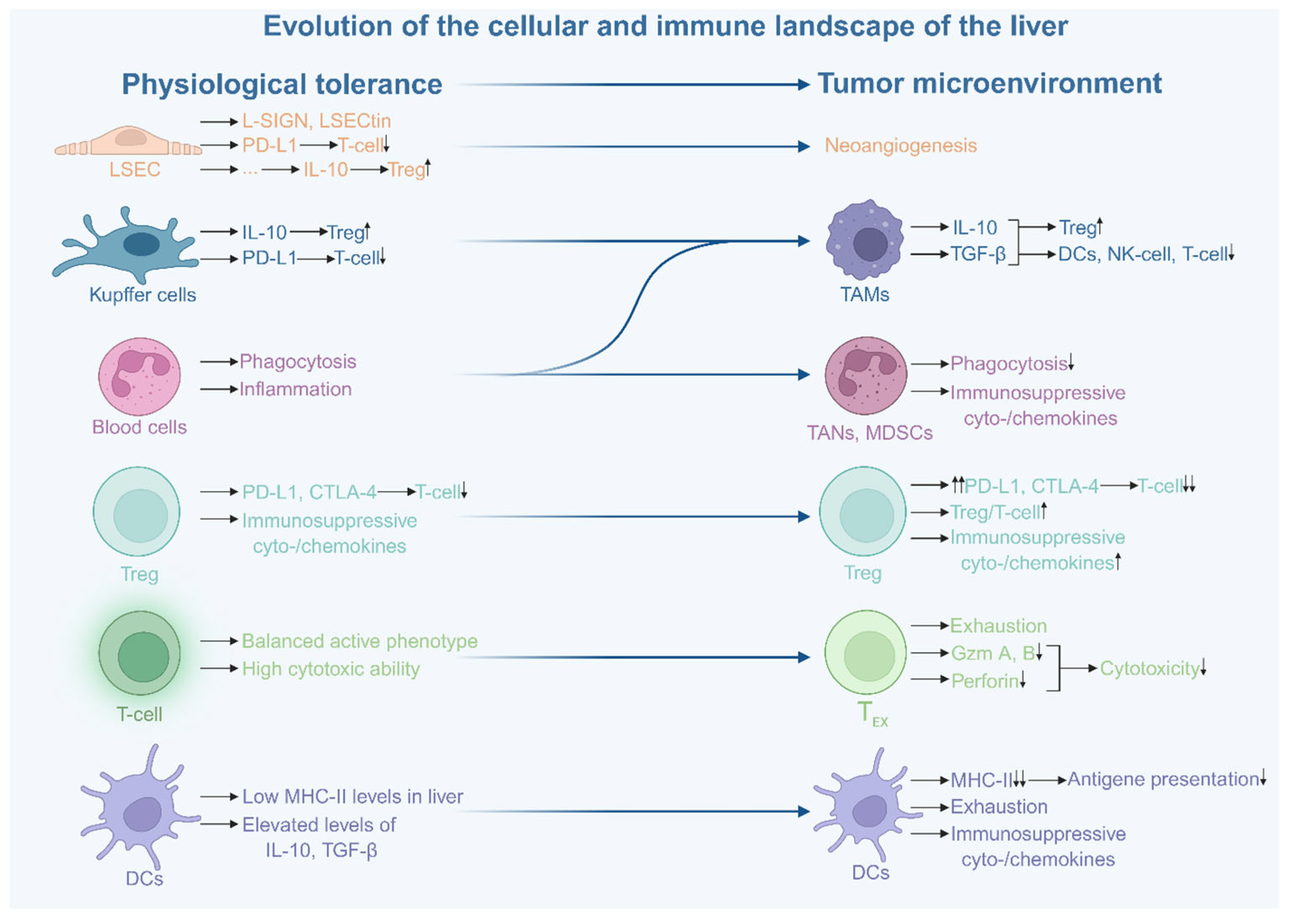
Figure 2.
Multilayered immunosuppressive network in the hepatocellular carcinoma microenvironment. The HCC microenvironment operates as a closed loop of reciprocal support. Myeloid and stromal cells orchestrate a milieu in which effector cells are rendered dysfunctional. This architecture ensures that every attempt at immune activation is met with a multilayered counter-response, effectively "locking" the tumour in a state of immunological inertia. The PD-1/PD-L1 axis (highlighted in red on the figure) plays an important role in this process, and most cells are involved in its formation. ICI therapy is aimed specifically at blocking these interactions, but such treatment cannot affect the other extensive areas of cellular support within the TME. For clarity, the effector compartment is represented by T cells as the most extensively characterised and therapeutically targeted population; other effector cell types discussed elsewhere in this review are omitted for visual simplicity. Abbreviations: TME, Tumour Microenvironment; HCC, Hepatocellular Carcinoma; ICI, Immune Checkpoint Inhibitor; TAMs, Tumour-Associated Macrophages; TANs, Tumour-Associated Neutrophils; PMN-MDSCs/M-MDSCs, Polymorphonuclear/Monocytic Myeloid-Derived Suppressor Cells; DCs, Dendritic Cells; Treg, Regulatory T Cell; NK, Natural Killer; TEX, Exhausted T Cell; CAFs, Cancer-Associated Fibroblasts; MMP, Matrix Metalloproteinase; CSF1, Colony-Stimulating Factor 1; HGF, Hepatocyte Growth Factor; IDO, Indoleamine 2,3-Dioxygenase; CTGF, Connective Tissue Growth Factor; CLCF1, Cardiotrophin-Like Cytokine Factor 1; COL1A1, Collagen Type I Alpha 1 Chain; ATP, Adenosine Triphosphate; PD-1, Programmed Cell Death Protein 1; PD-L1, Programmed Death-Ligand 1; MHC-II, Major Histocompatibility Complex Class II; TCR, T-Cell Receptor; LAG-3, Lymphocyte Activation Gene 3; CTLA-4, Cytotoxic T-Lymphocyte-Associated Protein 4; CCR4/5, C-C Motif Chemokine Receptor 4/5; TIGIT, T-cell Immunoreceptor with Ig and ITIM Domains; GAL-9, Galectin-9; TIM-3, T-cell Immunoglobulin and Mucin-domain Containing-3.
Figure 2.
Multilayered immunosuppressive network in the hepatocellular carcinoma microenvironment. The HCC microenvironment operates as a closed loop of reciprocal support. Myeloid and stromal cells orchestrate a milieu in which effector cells are rendered dysfunctional. This architecture ensures that every attempt at immune activation is met with a multilayered counter-response, effectively "locking" the tumour in a state of immunological inertia. The PD-1/PD-L1 axis (highlighted in red on the figure) plays an important role in this process, and most cells are involved in its formation. ICI therapy is aimed specifically at blocking these interactions, but such treatment cannot affect the other extensive areas of cellular support within the TME. For clarity, the effector compartment is represented by T cells as the most extensively characterised and therapeutically targeted population; other effector cell types discussed elsewhere in this review are omitted for visual simplicity. Abbreviations: TME, Tumour Microenvironment; HCC, Hepatocellular Carcinoma; ICI, Immune Checkpoint Inhibitor; TAMs, Tumour-Associated Macrophages; TANs, Tumour-Associated Neutrophils; PMN-MDSCs/M-MDSCs, Polymorphonuclear/Monocytic Myeloid-Derived Suppressor Cells; DCs, Dendritic Cells; Treg, Regulatory T Cell; NK, Natural Killer; TEX, Exhausted T Cell; CAFs, Cancer-Associated Fibroblasts; MMP, Matrix Metalloproteinase; CSF1, Colony-Stimulating Factor 1; HGF, Hepatocyte Growth Factor; IDO, Indoleamine 2,3-Dioxygenase; CTGF, Connective Tissue Growth Factor; CLCF1, Cardiotrophin-Like Cytokine Factor 1; COL1A1, Collagen Type I Alpha 1 Chain; ATP, Adenosine Triphosphate; PD-1, Programmed Cell Death Protein 1; PD-L1, Programmed Death-Ligand 1; MHC-II, Major Histocompatibility Complex Class II; TCR, T-Cell Receptor; LAG-3, Lymphocyte Activation Gene 3; CTLA-4, Cytotoxic T-Lymphocyte-Associated Protein 4; CCR4/5, C-C Motif Chemokine Receptor 4/5; TIGIT, T-cell Immunoreceptor with Ig and ITIM Domains; GAL-9, Galectin-9; TIM-3, T-cell Immunoglobulin and Mucin-domain Containing-3.
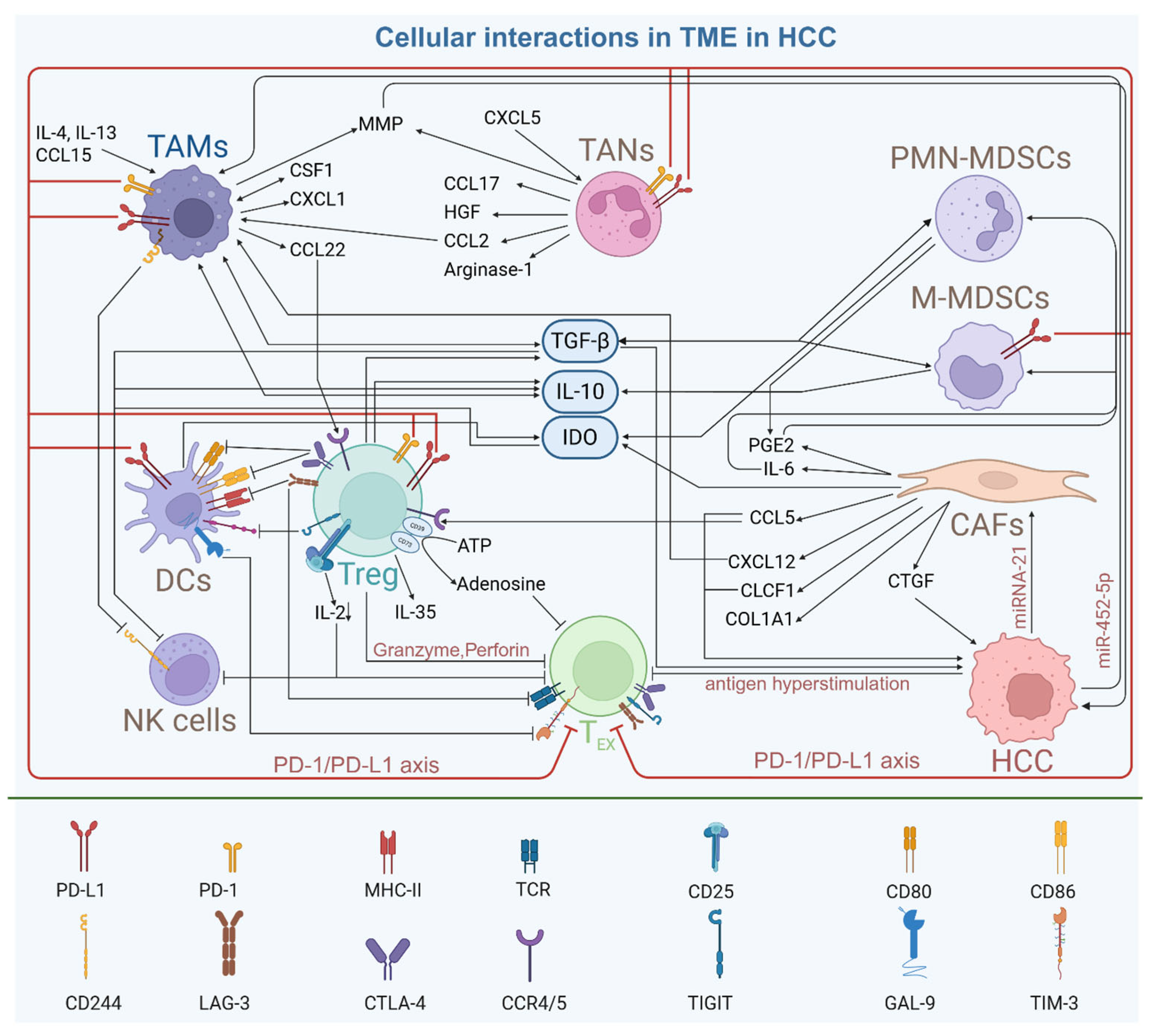
Figure 3.
Key principles of TME-centred ecosystem resistance to immune checkpoint blockade in HCC. (1) Immune exclusion: physical, metabolic and cellular barriers block reactivated T cells despite immune checkpoint inhibitor (ICI) therapy. (2) Distributed robustness: antigenic heterogeneity allows resistant cancer cell subpopulations to survive T-cell pressure. (3) Functional entrapment: hyperstimulation drives progenitor exhausted T cells (TEX) to a terminal state irreversible by ICIs. (4) Feedback stabilisation: tumour-associated macrophages (TAMs), regulatory T cells (Tregs), cancer-associated fibroblasts (CAFs) and cancer cells sustain a suppressive circuit via CSF1, miR-452-5p, miRNA-21, IL-6, CCL5, CXCL11, matrix metalloproteinases (MMPs) and circPETH. (5) Surface–depth mismatch: checkpoint blockade acts on surface PD-L1/CD80/CD86/PD-1 but not on the underlying FOXP3/TOX transcriptional programme.
Figure 3.
Key principles of TME-centred ecosystem resistance to immune checkpoint blockade in HCC. (1) Immune exclusion: physical, metabolic and cellular barriers block reactivated T cells despite immune checkpoint inhibitor (ICI) therapy. (2) Distributed robustness: antigenic heterogeneity allows resistant cancer cell subpopulations to survive T-cell pressure. (3) Functional entrapment: hyperstimulation drives progenitor exhausted T cells (TEX) to a terminal state irreversible by ICIs. (4) Feedback stabilisation: tumour-associated macrophages (TAMs), regulatory T cells (Tregs), cancer-associated fibroblasts (CAFs) and cancer cells sustain a suppressive circuit via CSF1, miR-452-5p, miRNA-21, IL-6, CCL5, CXCL11, matrix metalloproteinases (MMPs) and circPETH. (5) Surface–depth mismatch: checkpoint blockade acts on surface PD-L1/CD80/CD86/PD-1 but not on the underlying FOXP3/TOX transcriptional programme.
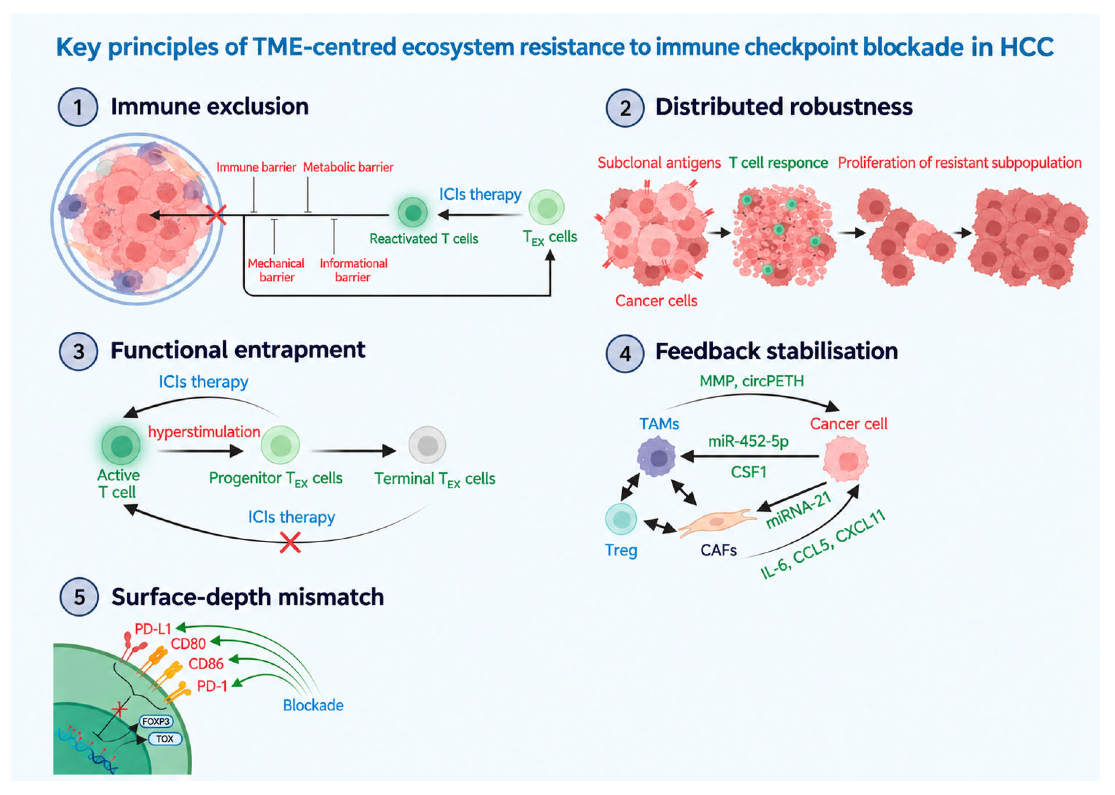
Table 1.
Key markers and immune checkpoints of regulatory T cells in the TME of HCC.
| Marker | Ligands/targets | Functional significance in the TME | References |
|---|---|---|---|
| CD137 (4-1BB) | 4-1BBL (DCs, macrophages) | Reliable marker of intratumoural Tregs. CD137+ Tregs exhibit more pronounced suppressive activity than CD137- Tregs. | [74,83] |
| CD177 | – | Previously considered a neutrophil marker. In the HCC TME, specific to CD4+FOXP3+ cells. CD177+ Tregs are characterised by increased secretion of TGF-β, IL-10, IL-35 and active proliferation. Associated with unfavourable prognosis. | [82,84] |
| CTLA-4 | CD80, CD86 (DCs) | Suppression of DC costimulation. Serves as a mechanism of self-regulation of Treg numbers. | [43,85] |
| LAG-3 | MHC-II, FGL1, TCR | Interception of the activating signal from DCs (higher affinity for MHC-II than TCR). Disruption of TCR–CD3 complex formation. Increased presence in the TME is associated with unfavourable prognosis. | [86,88,89,90,91] |
| TIGIT | CD155 (monocytes, DCs, fibroblasts) | Enhanced IL-10 production via TIGIT/CD155 interaction with DCs. Stimulation of iTreg proliferation. TIGIT+ Tregs show more pronounced expression of CTLA-4, CD25; suppress Th1 and Th17. | [92,93,94,95,96] |
| PD-1 / PD-L1 | PD-L1/PD-1 (interaction) | PD-L1 increases upon Treg activation. PD-L1 blockade does not affect FOXP3 expression, i.e. Tregs retain suppressive activity via alternative pathways. | [43] |
The table summarises key markers and immune checkpoints of regulatory T cells in the TME of HCC. Abbreviations: TME, Tumour Microenvironment; HCC, Hepatocellular Carcinoma; Tregs, Regulatory T Cells; DCs, Dendritic Cells; CTLA-4, Cytotoxic T-Lymphocyte-Associated Protein 4; LAG-3, Lymphocyte Activation Gene 3; MHC-II, Major Histocompatibility Complex Class II; FGL1, Fibrinogen-Like Protein 1; TCR, T-Cell Receptor; TIGIT, T-cell Immunoreceptor with Ig and ITIM domains; PD-1, Programmed Cell Death Protein 1; PD-L1, Programmed Death-Ligand 1; FOXP3, Forkhead Box P3; iTreg, Induced Regulatory T Cell; Th1/Th17, T-helper 1/17 cell.
Table 2.
Studies addressing the influence of cancer-associated fibroblasts on the prognosis of patients with hepatocellular carcinoma.
Table 2.
Studies addressing the influence of cancer-associated fibroblasts on the prognosis of patients with hepatocellular carcinoma.
| Reference | Study country | Study year | Methods | Number of samples | Marker | CAF type | Prognostic impact |
|---|---|---|---|---|---|---|---|
| Studies directly assessing CAFs | |||||||
| [110] | China | 2020 | 2D-DIGE, MALDI-TOF/TOF MS, proteomics, ChIP, luciferase assay, in vivo mouse models. | 124 HCC (clinical cohort), cell lines | α-SMA | CAFs | Shorter OS and PFS |
| [118] | China | 2022 | WGCNA, differential expression analysis, univariable and multivariable Cox regression, ssGSEA, drug sensitivity prediction (pRRophetic), qRT-PCR | 371 HCC (TCGA), 243 (ICGC) | CAF-related genes (ACTA2, IGJ, CTHRC1, CXCL12, LAMB1) | CAFs | Shorter OS |
| [128] | China | 2022 | DESeq2, GEO2R, LASSO, Cox regression, cluster analysis, GSVA, PPI, chemosensitivity prediction (pRRophetic) and immunotherapy response prediction | 374 HCC (TCGA), 268 (GEO), 232 (ICGC) | CAF-related genes (ARHGAP11A, DLGAP5, RFPL4B, TOP2A, TTK) | CAFs | Shorter OS |
| [119] | China | 2023 | scRNA-seq, unsupervised clustering, ESTIMATE, MCPcounter, EPIC, drug sensitivity prediction | scRNA-seq data (GSE146409), TCGA, ICGC, CHCC, GEO | 39 CAF-related genes (including COL3A1, ACTA2, TPM2) | CAFs | Shorter OS |
| [98] | China | 2024 | scRNA-seq, ST, WGCNA, CIBERSORTx, CellChat, NicheNet, CellTrek, IHC, IF | 156 scRNA-seq samples (120 patients), 1518 bulk RNA-seq samples, 90 HCC (IHC) | FAP | FAP+ CAFs | Shorter OS |
| [121] | China | 2024 | scRNA-seq, ST, mIHC, GSEA, CellPhoneDB, NicheNet, pySCENIC, CIBERSORTx, in vivo models | 220 scRNA-seq samples (6 datasets), TCGA, GEO, spatial transcriptomics (ST) | POSTN, α-SMA, IL-6, SPP1 | POSTN+ CAFs | Shorter OS |
| [104] | China | 2025 | iTRAQ proteomics, LC-MS/MS, EV isolation, Transwell assay, colony formation assay, orthotopic mouse model, Co-IP, molecular docking | 47 HCC (clinical cohort), cell lines | CTGF, α-SMA, FAP | CAFs (myCAFs) | Shorter OS |
| [103] | China | 2025 | scRNA-seq (GSE149614), mIHC, GSEA, CellPhoneDB, Cox regression | 94 HCC (TMA), scRNA-seq data | FAP, α-SMA, ACTA2 | iCAFs (FAP+) | No association with OS |
| [103] | China | 2025 | scRNA-seq (GSE149614), mIHC, GSEA, CellPhoneDB, Cox regression | 94 HCC (TMA), scRNA-seq data | FAP, α-SMA, ACTA2 | myCAFs (α-SMA+) | Longer OS |
| Studies based on indirect CAF-related data | |||||||
| [106] | China | 2018 | qRT-PCR, western blot, sphere formation assay, colony formation assay, Transwell assay, in vivo NOD/SCID mouse models, IHC. | 88 HCC (clinical cohort), cell lines | nNICD | CAFs | Shorter OS |
| [114] | China, USA | 2020 | IHC, IF, MS, co-IP, RNA-seq, ELISA, flow cytometry, in vivo xenograft models. | 125 HCC (tissues), 88 non-tumour, cell lines | Endosialin | CAFs | Shorter OS |
| [122] | Japan | 2020 | miRNA microarray, exosome isolation, Transwell assay, scratch assay, qRT-PCR, in vivo models. | 82 HCC (tissues), 40 (plasma), cell lines | MiR-150-3p was significantly reduced in CAF-derived exosomes | CAFs | Shorter OS |
| [111] | China | 2021 | PCR array, ELISA, IHC, IF, sphere formation assay, chromatin IP, in vivo BALB/c nude and NOD/SCID mouse models. | 287 HCC (tissues), 60 (serum), cell lines | CLCF1 | CAFs | Shorter OS and PFS |
The table summarises studies addressing the influence of cancer-associated fibroblasts on the prognosis of patients with hepatocellular carcinoma. Abbreviations: HCC, Hepatocellular Carcinoma; CAFs, Cancer-Associated Fibroblasts; myCAFs/iCAFs, Myofibroblastic/Inflammatory Cancer-Associated Fibroblasts; FAP, Fibroblast Activation Protein; α-SMA, α-Smooth Muscle Actin; POSTN, Periostin; SPP1, Secreted Phosphoprotein 1; CTGF, Connective Tissue Growth Factor; OS, Overall Survival; PFS, Progression-Free Survival; TCGA, The Cancer Genome Atlas; ICGC, International Cancer Genome Consortium; GEO, Gene Expression Omnibus; scRNA-seq, Single-Cell RNA Sequencing; ST, Spatial Transcriptomics; IHC, Immunohistochemistry; IF, Immunofluorescence.
Table 3.
Main immunosuppressive cell populations in the tumour microenvironment of hepatocellular carcinoma.
Table 3.
Main immunosuppressive cell populations in the tumour microenvironment of hepatocellular carcinoma.
| Cell population |
Key markers |
Main sources | Main secreted factors | Key immunosuppressive mechanisms | Impact on prognosis in HCC |
References |
|---|---|---|---|---|---|---|
| TAMs | CD68, CD163, CD206 | Kupffer cells, blood monocytes | TGF-β, IL-10, CSF1, MMP-9, CCL22 | Suppression of Th1 cells and NK cells, promotion of Tregs, EMT, PD-L1 expression | Unfavourable | [45,46,47,48,49,50,51,52,53,54,55,56,57,58,59,60,61] |
| TANs | CD66b | Blood neutrophils | CCL2, CCL17, CXCL5, CCL21 | Recruitment of Tregs and TAMs, suppression of T cells | Unfavourable | [62,63,64,65] |
| MDSCs | CD11b, CD33, HLA-DR, CD14, CD15, LOX-1 | Immature myeloid precursors in the bone marrow | PGE2, IDO, TGF-β, IL-10 | Impairment of T-cell and NK-cell function, PD-L1 expression | Unfavourable | [66,67,68,69,70,71,72,73,74] |
| Tregs | FOXP3, CD25, CD4, CD137 | Precursors in the thymus and directly in the TME from naïve CD4+ T cells, Th17 cells | IL-10, TGF-β, IL-35, adenosine, granzymes | Suppression of T cells and NK cells, expression of multiple immune checkpoints, accumulation of adenosine in the matrix | Unfavourable | [43,44,45,75,76,77,78,79,80,81,82,85,86,87,88,89,90,91,92,93,94,95,96,97] |
| CAFs | αSMA, FAP, COL1A1, PDGFRα/β | HSCs, mesenchymal stem cells | IL-6, CTGF, CCL5, CLCF1, CXCL12 | Formation of a mechanical barrier, recruitment of TAMs, promotion of tumour cell proliferation, metastasis and stemness | Unfavourable | [98,99,100,101,102,103,104,105,106,107,108,109,110,111,112,113,114,115,116,117] |
| TEX | PD-1, TIM-3, LAG-3, TIGIT, TOX | Effector CD8+ T cells under chronic stimulation | IFN-γ, IL-2, TNF-α, granzyme B, perforin | Remain in a suppressed state, fail to adequately perform antitumour functions | Unfavourable | [141,142,143,144,145,146,147,148,149,150,151,152] |
| TRM | CD69, CD103 | Effector CD8+ T cells | IFN-γ, granzyme B | Remain in a suppressed state, fail to adequately perform antitumour functions | Favourable | [45,91,123] |
| NK cells | CD16, CD56 | Resident and circulating NK cells | IFN-γ, TNF-α, IL-12, IL-15, IL-18 | Remain in a suppressed state, fail to adequately perform antitumour functions | Favourable | [133,134,135,136,137,138,139,140] |
| mregDCs | LAMP3, CCR7, PD-L1 | Mature DCs in the TME | TGF-β, IDO | Remain in a suppressed state, fail to adequately perform antitumour functions, recruit Tregs | Unfavourable | [130,131,132] |
| B cells | CD19, CD20, CD79a | Naive, mature and memory B cells | IFN-γ, IL-12p40, granzyme B, TRAIL | Decline in overall numbers under TME suppressive pressure; pro-tumour promotion of Tregs, TAMs, PD-L1 and M2b polarisation confined to a minority subset | Subtype-dependent | [153,154,155,156,157,158,159,160,161] |
| Bregs | PD-1, CD5, CD24, CD27, CD38, TIM-1 | Peripheral and atypical memory B cells | IL-10, IL-35, TGF-β | Suppression of CD8+ T cells, promotion of TAMs | Unfavourable | [59,119,120,122,162,163,164] |
| NKT cells | CD1d, CD16, semi-invariant TCR (iNKT) | Liver-resident NKT cells | IFN-γ, IL-4 | Cross-suppression of iNKT cells, susceptibility to Treg-induced anergy | Contradictory | [165,166,167,168] |
| MAIT cells | CD45RO, CD95, TRAV1-2, MR1 | Liver-resident unconventional T cells | IFN-γ, TNF-α, granzyme B, IL-17A | Suppression by CSF1R+PD-L1+ TAMs, loss of cytotoxic function | Unfavourable | [169,170,171,172] |
The table summarises main immunosuppressive cell populations in the tumour microenvironment of hepatocellular carcinoma. Abbreviations: TME, Tumour Microenvironment; HCC, Hepatocellular Carcinoma; TAMs, Tumour-Associated Macrophages; TANs, Tumour-Associated Neutrophils; MDSCs, Myeloid-Derived Suppressor Cells; HLA-DR, Human Leukocyte Antigen – DR Isotype; LOX-1, Lectin-like Oxidised Low-Density Lipoprotein Receptor-1; PGE2, Prostaglandin E2; IDO, Indoleamine 2,3-Dioxygenase; Tregs, Regulatory T Cells; CSF1, Colony-Stimulating Factor 1; MMP-9, Matrix Metalloproteinase 9; CAFs, Cancer-Associated Fibroblasts; FAP, Fibroblast Activation Protein; CTGF, Connective Tissue Growth Factor; CLCF1, Cardiotrophin-Like Cytokine Factor 1; PDGFRα/β, Platelet-Derived Growth Factor Receptor Alpha/Beta; TEX, Exhausted T Cells; TOX, Thymocyte Selection-Associated High Mobility Group Box Protein; TRM, Tissue-Resident Memory T Cells; NK, Natural Killer; mregDCs, Mature Dendritic Cells Enriched in Immunoregulatory Molecules; Bregs, Regulatory B Cells; NKT, Natural Killer T Cell; MAIT, Mucosal-Associated Invariant T Cell; PD-1, Programmed Cell Death Protein 1; PD-L1, Programmed Death-Ligand 1; LAG-3, Lymphocyte Activation Gene 3; TIGIT, T-cell Immunoreceptor with Ig and ITIM Domains; TIM-3, T-cell Immunoglobulin and Mucin-domain containing-3.
Table 4.
Systems-level principles of TME-centred ecosystem resistance to immune checkpoint blockade in HCC.
Table 4.
Systems-level principles of TME-centred ecosystem resistance to immune checkpoint blockade in HCC.
| Principle of resistance | System property | Dominant biological manifestation | Why ICIs alone fail? | Therapeutic implication | Testable prediction |
|---|---|---|---|---|---|
| Immune exclusion | Physical and functional exclusion | Immune, stromal, metabolic and informational barriers restrict effector access | Reinvigorated T cells cannot reach or penetrate tumour niches | Combine ICIs with TME-normalising strategies | Patients with exclusion signatures show reduced ICI response despite inflamed phenotype |
| Distributed robustness | Network redundancy and adaptive evolution under therapeutic pressure | Multiple suppressive circuits and heterogeneous tumour/TME states provide alternative routes to resistance and enable selection of resistant states | Blocking one axis leaves alternative circuits intact, while therapeutic pressure promotes selection of resistant states | Multi-node and evolution-informed combination therapy | Multi-axis suppression scores and dynamic resistance signatures outperform single biomarkers |
| Functional entrapment | Stable dysfunctional immune states | Exhaustion, suppressive polarisation, metabolic paralysis | Checkpoint blockade reverses inhibition incompletely | Pair ICIs with state-reprogramming therapies | Exhaustion-state plasticity predicts reversibility |
| Feedback stabilisation | Self-reinforcing suppressive loops | Reciprocal tumour–immune–stromal reinforcement | Resistant ecosystem restores itself after perturbation | Disrupt reinforcing loops, not single nodes | Disruption of feedback hubs sensitises to ICIs |
| Surface–depth mismatch | Therapeutic targeting mismatch | Surface checkpoint blockade vs deep epigenetic/system states | Intervention does not collapse underlying resistant architecture | Combine ICIs with deeper reprogramming strategies | Epigenetic co-targeting should outperform checkpoint monotherapy |
The table summarises systems-level principles of TME-centred ecosystem resistance to immune checkpoint blockade in HCC. Abbreviations: ICIs, immune checkpoint inhibitors; TME, tumour microenvironment.
Disclaimer/Publisher’s Note: The statements, opinions and data contained in all publications are solely those of the individual author(s) and contributor(s) and not of MDPI and/or the editor(s). MDPI and/or the editor(s) disclaim responsibility for any injury to people or property resulting from any ideas, methods, instructions or products referred to in the content. |
© 2026 by the authors. Licensee MDPI, Basel, Switzerland. This article is an open access article distributed under the terms and conditions of the Creative Commons Attribution (CC BY) license (http://creativecommons.org/licenses/by/4.0/).
Copyright: This open access article is published under a Creative Commons CC BY 4.0 license, which permit the free download, distribution, and reuse, provided that the author and preprint are cited in any reuse.