

Submitted:
15 October 2026
Posted:
19 October 2026
You are already at the latest version
Abstract
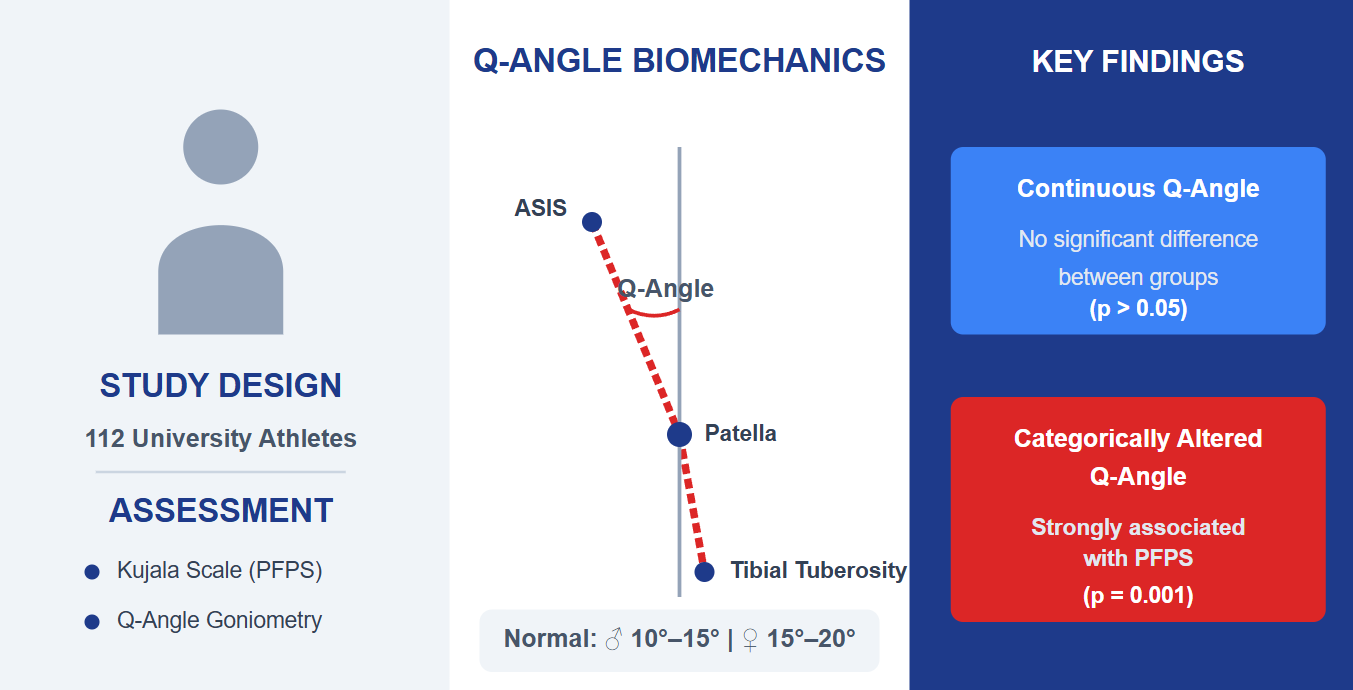
Background and Objectives: Patellofemoral Pain Syndrome (PFPS) is a highly preva-lent knee injury in sports. Despite its mechanical plausibility, the clinical utility of the quadriceps angle (Q-angle) as a risk predictor remains controversial. This study aims to analyze the association between the Q-angle and PFPS in university athletes, evalu-ating the variable based on its parameters of morphological normality according to bi-ological sex. Methods: An analytical observational cross-sectional study was conduct-ed with 112 university athletes. The presence of PFPS was determined using the Kujala Scale. Anthropometric variables, body composition, and Q-angle goniometry were measured. The Q-angle was analyzed both as a continuous quantitative variable and as a dichotomized categorical variable (Normal vs. Altered), based on sex-specific physiological limits (10°–15° for men; 15°–20° for women). Bivariate analyses were performed using Chi-square, Student’s t, and Mann-Whitney U tests. Results: The prevalence of PFPS was 30.3% (n=34). No significant differences were found between groups regarding sex, sports discipline, or body composition (p > 0.05). When evaluat-ing the Q-angle as a continuous variable, absolute means did not differ significantly between asymptomatic and affected individuals (p > 0.05). However, the clinical cate-gorization demonstrated a highly significant association: an altered Q-angle was strongly associated with the presence of PFPS in both the right and left knees (p = 0.001). Conclusions: Clinical alteration of the Q-angle, rather than its absolute contin-uous value or the athlete's general body phenotype, is directly associated with PFPS. Stratified categorical goniometry remains a highly effective tool for preventive screening in university athletes.
Keywords:
Patellofemoral Pain Syndrome
; athletes
; knee joint
; anthropometry
; biomechanical phenomena
1. Introduction
Patellofemoral Pain Syndrome (PFPS) is a highly prevalent musculoskeletal condition described as pain or dysfunction in the anterior knee compartment [1]. This pathology results from an alteration in load distribution across the patellofemoral joint, frequently exacerbated by activities demanding high mechanical stress on the lower extremities, such as jumping, running, or ascending and descending stairs [2,3]. Worldwide, its prevalence ranges from 22.7% to 28.9% [4]; in the clinical setting, it accounts for 25% to 40% of all knee problems, and similarly, represents 33% and 18% of all knee injuries in sports practice [5,6]. Its impact is particularly severe in young and active populations, affecting over 70% of individuals aged 16 to 25 years, which substantially increases the risk of functional decline, impaired performance, and early dropout from sports participation [7].
Despite its high incidence and extensive clinical management, the etiopathogenesis of PFPS remains a subject of debate due to its multifactorial nature [4,8]. Its onset is mediated by the interaction of extrinsic components, such as joint overuse associated with training load [4,9], and intrinsic factors, linked to structural and neuromuscular alterations [4,8,10]. Among the most extensively studied local anatomical determinants is the quadriceps angle (Q-angle), a morphological parameter that evaluates the alignment of the force vectors of the extensor mechanism and the tendency of the patella to track laterally upon muscular contraction [11,12]. From a biomechanical perspective, it is assumed that an increased Q-angle compromises joint congruence, lateralizes the patella, increases contact pressures on the lateral femoral condyle, and predisposes the individual to pain [13]. The physiological values of this angle differ significantly according to sex-linked pelvic anatomy and the consulted literature. While some studies estimate normal values between 8° and 10° in men and up to 15° in women [14], other authors broaden these physiological tolerance margins, placing them between 10° and 15° for men and 15° to 20° for women [15]; in any case, deviations from these limits could indicate a functional anomaly.
However, despite the mechanical plausibility of this association, the clinical utility of the Q-angle as a risk predictor remains controversial. Previous studies have reported contradictory findings: while some research correlates an increased angle with excessive pronation and internal tibial rotation that promote the syndrome [16,17,18], other authors argue that a larger Q-angle has a minimal effect on lower extremity kinematics during athletic movement and fails to correlate directly with patellar tracking [19,20]. This lack of clarity appears to be exacerbated by the methodological approach to the variable, compounded by the fact that a large part of the research has focused on professional athletes or recreational runners, leaving a significant gap in knowledge regarding the dynamics of anterior knee pain in university athletes.
To address these gaps, the present study aims to analyze the association between the Q-angle and PFPS in university athletes, evaluating the variable based on its parameters of morphological normality. This relationship will be explored through a multidimensional approach that integrates not only goniometric characteristics but also clinical factors and load variables (training frequency). The findings will substantiate the relevance of structural markers in the functional evaluation of athletes, providing sports medicine professionals with solid evidence for the design of more precise and personalized prevention strategies and rehabilitation programs.
2. Materials and Methods
2.1. Study Design and Participants
An analytical observational cross-sectional study was conducted. The study population was derived from the retrospective records of the Comprehensive Athlete Care Model (MAID-UDES, by its Spanish acronym) extension program at the Universidad de Santander, Bucaramanga campus [21]. Clinical and biomechanical measurements of the athletes affiliated with the Institutional Welfare team were collected during the period between February 2024 and October 2025. During this timeframe, a total of 129 records were obtained; however, following an initial database depuration process to ensure information integrity, the number was reduced to 122 records. Finally, after the strict application of eligibility criteria, the final analyzed sample consisted of 112 university athletes.
For the definitive confirmation of this sample, the inclusion criteria were established as a university athlete enrolled at the institution, being between 18 and 30 years of age, and maintaining regular participation in sports activities, defined as a minimum of twice a week. Conversely, individuals with a history of lower extremity surgery (including knee surgical interventions within the past year) or a history of acute knee injuries in the last six months were excluded. Likewise, cases presenting neuromuscular, neurological, or rheumatological pathologies affecting mobility, as well as pregnant women, were also excluded.
2.2. Instruments and Procedure
Data collection was structured based on a multidimensional approach that included sociodemographic and anthropometric variables (such as weight, height, body fat percentage, and muscle mass, among others), sports practice characteristics, and clinical aspects. For the clinical evaluation of Patellofemoral Pain Syndrome (PFPS), the Kujala Scale (Anterior Knee Pain Scale) was used, a validated 13-item questionnaire that allows for the quantification of symptoms and functional limitations associated with anterior knee pain [22]. For analytical purposes, the total score obtained on this scale was dichotomized, establishing a PFPS diagnosis for those athletes who recorded a score below 85 points, while scores ranging between 85 and 100 points were categorized as the absence of the syndrome [23].
Furthermore, the evaluation of the main morphological parameter consisted of measuring the quadriceps angle (Q-angle) using a conventional goniometry technique. The standardized procedure required the athlete to be positioned in a supine posture, with the knees fully extended and the musculature relaxed. In this position, a goniometer was used to accurately record the angle formed by the intersection of two anatomical axes: an initial line drawn from the anterior superior iliac spine to the geometric center of the patella, and a second line projected from the center of the patella to the anterior tibial tuberosity [24].
For data processing, the goniometric measurement was methodologically approached in a dual manner: as a continuous quantitative variable and as a dichotomized categorical variable. This clinical categorization was strictly based on the specific anatomical and physiological reference parameters for each biological sex. Thus, values between 10° and 15° in men, and between 15° and 20° in women were defined as "Normal"; consequently, any goniometric record falling outside these physiological tolerance limits was classified as "Altered” [15].
2.3. Statistical Analysis
Data processing and analysis were performed using IBM SPSS Statistics software, version 27. Descriptive statistics were applied by calculating absolute frequencies and percentages for qualitative variables. For quantitative variables, normality was determined using the Kolmogorov-Smirnov test. Data with a normal distribution were expressed as mean and standard deviation, whereas those without a normal distribution were reported using the median and interquartile range.
In the bivariate analysis, the Chi-square test or Fisher's exact test was employed to establish the association between categorical variables (such as Q-angle alteration) and the presence of PFPS. To compare continuous variables between affected and unaffected groups, the independent samples Student’s T-test or the Mann-Whitney U test was used, as appropriate. The level of statistical significance was set at a p-value < 0.05.
3. Results
3.1. Sociodemographic and Sports Characteristics of Participants
The analyzed sample consisted of 112 university athletes, characterized by a slight predominance of females (53.57%). Regarding general sociodemographic and sports practice variables, the largest proportion of participants belonged to the Faculty of Natural Sciences (21.43%) and were part of the basketball and futsal teams (21.43% each), with a heterogeneous academic distribution where the fourth semester stood out (13.39%). Upon evaluating the presence of Patellofemoral Pain Syndrome (PFPS), documented in 34 individuals (30.3% of the sample), a higher frequency of the pathology was observed in men (55.9%), students from the Faculty of Engineering and Technologies (26.5%), and volleyball players (23.5%). However, the bivariate analysis determined that the participants' baseline characteristics did not represent determining factors for suffering from the injury, as no statistically significant associations were found between the presence of PFPS and biological sex (p = 0.185), sports discipline (p = 0.868), faculty (p = 0.406), or current academic semester (p = 0.711) (Table 1).
3.2. Anthropometric and Body Composition Characteristics of Participants
When analyzing the anthropometric and body composition characteristics, the sample of university athletes evidenced great structural homogeneity (Table 2). Overall, participants presented a median age of 21 years, a weight of 63 kg, and an average height of 168.24 ± 8.18 cm. The comparative evaluation between individuals with and without Patellofemoral Pain Syndrome (PFPS) yielded no statistically significant differences in these variables. Fundamental parameters such as body fat percentage (17.18% without PFPS versus 16.48% with PFPS; p = 0.487), muscle mass percentage (p = 0.337), bone mass (p = 0.256), and estimated daily caloric intake (DCI) (p = 0.286) maintained a statistically and clinically similar distribution in both groups.
Likewise, the specific anthropometric evaluation of the lower limbs demonstrated no variations, suggesting an anatomical predisposition or asymmetry linked to these variables. The lengths of the right and left extremities were comparable between the groups, with means of 81.36 cm and 81.32 cm respectively in affected subjects, compared to averages without significant variations in healthy participants (p > 0.05). In turn, the bivariate analysis confirmed that the muscle circumferences of the thigh and calf, measured bilaterally, do not possess statistically significant differences (p > 0.05 for all circumferences), indicating that the volume and distribution of the lower appendicular mass do not act as differentiating factors for the development of the syndrome in this population.
On the other hand, when evaluating the quantitative behavior of the Q-angle as a continuous variable, no statistically significant differences were observed in the absolute averages between athletes with and without PFPS (p = 0.759 for the right limb and p = 0.928 for the left). Nevertheless, the dispersion analysis (Figure 1) revealed greater variability and amplitude in the goniometric ranges of the PFPS group, evidencing a higher tendency to present extreme values far from the physiological mean.
Finally, upon clinically categorizing the quadriceps Q-angle, the association analysis demonstrated that presenting an altered Q-angle, outside the parameters of morphological normality according to sex, is highly significantly associated with the presence of the syndrome (p = 0.001 in both extremities). While in the asymptomatic group the vast majority of individuals (nearly 80%) preserved a normal physiological alignment, the affected group was predominated by biomechanical alteration, present in 70.6% and 61.8% for the right and left knees, respectively (Table 3).
4. Discussion
The present study primarily analyzed the association between the Q-angle and Patellofemoral Pain Syndrome (PFPS) in university athletes, approaching this variable from a multidimensional morphological perspective. The most relevant finding of this research demonstrates that, although the continuous absolute values of the Q-angle do not differ significantly between healthy and affected subjects, the alteration of patellofemoral alignment, clinically defined as falling outside the physiological normal ranges according to sex, presents a highly significant association with the presence of the syndrome.
First, the prevalence of PFPS in our cohort of university athletes was 30.3%, a finding that aligns with global estimates reported in the literature, which position this pathology as the cause of up to one-third of all knee injuries in sports practice [5,25,26]. Interestingly, unlike previous studies suggesting a higher predisposition in females due to structural and neuromuscular pelvic differences [27,28,29], no statistically significant association was found in our sample between PFPS and biological sex (p = 0.185), nor with the sports discipline practiced [5,30]. This homogeneity suggests that, in the context of university sports, where mechanical load demands and training frequency (minimum twice a week) tend to be standardized, local mechanical factors assume greater etiopathogenic prominence than sociodemographic variables or the specific type of athletic movement.
This predominance of local factors is reinforced when analyzing anthropometric and body composition characteristics. The rigorous control of variables such as muscle mass percentage, body fat, and asymmetries in muscle circumferences demonstrated no significant differences between the groups. This allows us to infer that the volume or distribution of lower appendicular mass does not act as a primary differentiating factor for the development of the syndrome, as described by other studies [31,32]. Consequently, the etiology of anterior knee pain in these athletes appears to rely less on macroscopic structural variations and more on specific micro-biomechanical alterations of the patellofemoral joint, such as the alteration of quadriceps force vectors.
The analysis of the Q-angle yielded the most clarifying finding of the study, offering a response to the divergences present in current literature. When evaluating the Q-angle as a continuous quantitative variable, no differences in absolute means were found between the groups, consistent with other studies [33,34,35]. Nevertheless, the dispersion analysis revealed that athletes with PFPS exhibited greater goniometric variability, with extreme values far from the mean; this explains why the traditional analytical approach (comparison of means) often fails to predict risk: biomechanical alterations can occur due to both an excessive increase in the angle (which lateralizes the patella and increases pressure on the lateral femoral condyle) and an abnormal decrease in it, which compensatorily alters joint congruence [13].
Upon categorizing the Q-angle based on sex-specific physiological tolerance parameters (10°-15° in men; 15°-20° in women) [15], the true functional anomaly was exposed. The highly significant association (p = 0.001 in both extremities) between a clinically "Altered" Q-angle and the presence of PFPS confirms that the utility of this morphological marker lies in its categorical and individualized interpretation. This finding supports the biomechanical rationale that any sustained deviation from the anatomical limits of the extensor mechanism, subjected to the high repetitive training loads typical of university sports, disrupts joint homeostasis and triggers pain [36].
Despite the robustness of these findings, the present study is not without limitations. Cross-sectional design restricts the ability to establish direct causal relationships; that is, it is not possible to determine whether the Q-angle alteration is the primary cause of the syndrome or if antalgic adaptations have modified the static posture of the lower limb. Likewise, evaluation using static goniometry in a supine position, although standardized, cost-effective, and of great clinical utility, does not capture the three-dimensional kinematics of the lower extremity (such as internal tibial rotation or dynamic knee valgus) during weight-bearing or athletic movement [19,20].
Nevertheless, its strengths include strict methodological rigor in sample depuration, the multidimensional approach to confounding factors (anthropometric and load-related), and its focus on an understudied yet highly vulnerable population: the university athlete. The findings have direct implications for sports medicine practice and comprehensive physical therapy, demonstrating that conventional Q-angle goniometry remains an invaluable preventive screening tool, provided its results are interpreted using clinical normality thresholds stratified by sex.
5. Conclusions
The present study demonstrates a statistically significant association between clinical alteration of the Q-angle and the presence of Patellofemoral Pain Syndrome (PFPS) in university athletes. Although the continuous quantitative analysis of the angle yielded no differences in absolute averages between asymptomatic and affected individuals, its qualitative categorization—based on sex-specific physiological normal ranges—confirmed that extreme anatomical deviations of the extensor mechanism directly predispose individuals to the development of the pathology. Furthermore, the remarkable anthropometric and body composition homogeneity found in the sample allows us to conclude that, under the mechanical demands of university sports, local biomechanical alterations of the patellofemoral joint constitute a greater risk factor than the general body phenotype or the specific sports discipline practiced.
On a practical level, these findings reaffirm the utility of conventional Q-angle goniometry as an accessible and highly effective tool for preventive screening, provided its results are interpreted from a categorical and individualized approach. It is recommended that sports medicine and physical therapy teams integrate this stratified morphological evaluation into routine pre-participation examinations, which will facilitate the early identification of athletes with structural vulnerability. The design of personalized neuromuscular conditioning programs focused on compensating for these force vector alterations, complemented by future longitudinal research incorporating dynamic kinematic analyses, will be essential to reduce the incidence of anterior knee pain and mitigate early sports dropouts.
Author Contributions
Conceptualization, J.D.B.M. and A.S.V.V.; methodology, J.A.B.B.; formal analysis, J.A.B.B..; investigation, J.D.B.M. and A.S.V.V.; data curation, J.A.B.B.; writing—original draft preparation, J.D.B.M., J.A.B.B. and A.S.V.V.; writing—review and editing, J.A.B.B. and A.S.V.V.; visualization, J.A.B.B. All authors have read and agreed to the published version of the manuscript.
Funding
This research received no external funding.
Institutional Review Board Statement
The development of this research was governed by the fundamental principles of the World Medical Association's Declaration of Helsinki and Resolution 8430 of 1993 of the Colombian Ministry of Health, under which it was classified as research with no risk. Confidentiality, privacy, and the anonymized handling of information were guaranteed by utilizing the records of participants linked to the MAID-UDES program. The entire data collection protocol and study design were officially evaluated and endorsed by the Institutional Bioethics Committee of the Universidad de Santander, issuing a favorable concept through notification VII-29-BUC, corresponding to Minutes No. 12 of April 13, 2026.
Informed Consent Statement
Formal informed consent was waived for this study as it was based on the retrospective analysis of secondary clinical data. The Institutional Bioethics Committee approved this waiver, guaranteeing the strict confidentiality and anonymization of all participant records.
Data Availability Statement
The data presented in this study are openly available in the Mendeley Data repository at https://data.mendeley.com/datasets/wdcdtsyf5c/1
Acknowledgments
The authors would like to thank the Comprehensive Athlete Care Model (MAID-UDES) program for providing access to the data, the Physiotherapy program for the use of their laboratories, and the Universidad de Santander for facilitating these research spaces. During the preparation of this manuscript, the authors used Gemini for the purposes of English translation, drafting adjustments, and improving paragraph coherence. The authors have reviewed and edited the output and take full responsibility for the content of this publication.
Conflicts of Interest
The authors declare no conflicts of interest.
Abbreviations
The following abbreviations are used in this manuscript:
| PFPS | Patellofemoral Pain Syndrome |
| LL | Lower Limb |
| MAID | Comprehensive Athlete Care Model |
| UDES | University of Santander |
| PHY | Physique Rating |
| DCI | Daily Caloric Intake |
References
- Santilli G, Martino M, Pacini P, Agostini F, Bernetti A, Giuliani L, et al. Patellofemoral Pain Syndrome: Focused Vibrations Plus Kinesiotaping with Insights into Radiological Influences—An Observational Study. Journal of Functional Morphology and Kinesiology 2025, Vol 10, Page 2 2024;10:2. [CrossRef]
- Martínez-Cano JP, Rojas-Canizales S, Cortés-Cabrera S. Current Concepts in the Management of Patellofemoral Pain Syndrome. Revista Colombiana de Ortopedia y Traumatología 2022;36:20–6. [CrossRef]
- Mayo Clinic. Patellofemoral pain syndrome 2023. https://www.mayoclinic.org/diseases-conditions/patellofemoral-pain-syndrome/symptoms-causes/syc-20350792 (accessed October 5, 2026).
- Sanchis GJB, de Souto Barbosa JV, Cavalcanti RL, Bezerra JP, Santos ML dos, Guedes TSR, et al. Patellofemoral pain syndrome in children and adolescents: A cross-sectional study. PLoS One 2024;19:e0300683. [CrossRef]
- Halabchi F, Abolhasani M, Mirshahi M, Alizadeh Z. Patellofemoral pain in athletes: clinical perspectives. Open Access J Sports Med 2017;8:189. [CrossRef]
- Smith BE, Selfe J, Thacker D, Hendrick P, Bateman M, Moffatt F, et al. Incidence and prevalence of patellofemoral pain: A systematic review and meta-analysis. PLoS One 2018;13:e0190892. [CrossRef]
- Pappas E, Wong-Tom WM. Prospective Predictors of Patellofemoral Pain Syndrome: A Systematic Review With Meta-analysis. Sports Health 2012;4:115. [CrossRef]
- Gragnani BC, Hart HF, Forsyth A, Barton CJ, De Oliveira Silva D. Patellofemoral Pain is a Multifactorial Complex Condition; Are We Missing a Multidisciplinary Approach to its Management? Time for a Paradigm Shift. Open Access J Sports Med 2026;16:517971. [CrossRef]
- Petersen W, Ellermann A, Gösele-Koppenburg A, Best R, Rembitzki IV, Brüggemann GP, et al. Patellofemoral pain syndrome. Knee Surgery, Sports Traumatology, Arthroscopy 2013;22:2264. [CrossRef]
- Sanchis GJB, Nascimento JAS do, Santana R de C, Santos VM dos, Cunha VL da, Assis SJC de, et al. Biomechanical factors associated with patellofemoral pain in children and adolescents. Sci Rep 2024;14:15490. [CrossRef]
- Ketut N, Widhiantari E, Widnyana M, Jawi IM, Indrayani AW. Analysis of Q-Angle as a Risk Factor for Patellofemoral Pain Syndrome in Volleyball Athletes: A Cross-Sectional Study. Majalah Ilmiah Fisioterapi Indonesia 2025;13:256–61. [CrossRef]
- Gant H, Ghimire N, Min K, Musa I, Ashraf M, Lawan A. Impact of the Quadriceps Angle on Health and Injury Risk in Female Athletes. International Journal of Environmental Research and Public Health 2024, Vol 21, Page 1547 2024;21:1547. [CrossRef]
- Bai D, Cao X, Feng S. Effects of hip stability training versus standard care on Q-angle and pain in marathon runners with patellofemoral pain syndrome: A retrospective cohort study. PLoS One 2026;21:e0357057. [CrossRef]
- Unuvar BS, Torlak MS, Gercek H, Tufekci O, Erdagi K, Işik B. Comparison and Relationship of Quadriceps Femoris Angle, Muscle Strength, and Balance in Athletes and Non-Athletes. Indian J Orthop 2023;57:1243. [CrossRef]
- Skouras AZ, Kanellopoulos AK, Stasi S, Triantafyllou A, Koulouvaris P, Papagiannis G, et al. Clinical Significance of the Static and Dynamic Q-angle. Cureus 2022;14:e24911. [CrossRef]
- Phatama KY, Isma SPP, Devi LK, Siahaan LD, Pribadi A, Pradana AS, et al. Relationship of Anterior Knee Pain with Quadriceps Angle and Anthropometric Measurements in an Asian Female Population. Malays Orthop J 2022;16:95. [CrossRef]
- Caylor D, Fites R, Worrell TW. The relationship between quadriceps angle and anterior knee pain syndrome. J Orthop Sports Phys Ther 1993;17:11–6. [CrossRef]
- Moss RI, DeVita P, Dawson ML. A Biomechanical Analysis of Patellofemoral Stress Syndrome. J Athl Train 1992;27:64.
- Freedman BR, Brindle TJ, Sheehan FT. Re-evaluating the functional implications of the Q-angle and its relationship to in-vivo patellofemoral kinematics. Clinical Biomechanics 2014;29:1139–45. [CrossRef]
- Örtqvist M, Moström EB, Roos EM, Lundell P, Janarv PM, Werner S, et al. Reliability and reference values of two clinical measurements of dynamic and static knee position in healthy children. Knee Surgery, Sports Traumatology, Arthroscopy 2011;19:2060–6. [CrossRef]
- Vacca-Valencia A, Rojas Cano LE, Buitrago-Buitrago J, Lopez Gomez FA, Acosta Salazar HU, Méndez Diaz LM, et al. Comprehensive Care Model for University Athletes of the University of Santander. An Institutional Experience. Kinesiologia 2023:328–32.
- Ittenbach RF, Huang G, Foss KDB, Hewett TE, Myer GD. Reliability and Validity of the Anterior Knee Pain Scale: Applications for Use as an Epidemiologic Screener. PLoS One 2016;11:e0159204. [CrossRef]
- Martínez-Cano JP, Arango AS, Castro AM, Piña AM, Martínez-Rondanelli A, Martínez-Cano JP, et al. Validación de la escala de Kujala para dolor patelofemoral en su versión en español. CES Medicina 2017;31:47–57. [CrossRef]
- Malone TR, Pfeifle AL. Patellofemoral Disorders. Orthopaedic Physical Therapy Secrets: Third Edition 2017:536–46. [CrossRef]
- Smith BE, Selfe J, Thacker D, Hendrick P, Bateman M, Moffatt F, et al. Incidence and prevalence of patellofemoral pain: A systematic review and meta-analysis. PLoS One 2018;13:e0190892. [CrossRef]
- Hudgins TH, Verma A, Kupperman W, Alleva JT. Patellofemoral Syndrome. Essentials of Physical Medicine and Rehabilitation: Musculoskeletal Disorders, Pain, and Rehabilitation 2023:414–8. [CrossRef]
- Boling M, Padua D, Marshall S, Guskiewicz K, Pyne S, Beutler A. Gender differences in the incidence and prevalence of patellofemoral pain syndrome. Scand J Med Sci Sports 2010;20:725. [CrossRef]
- Lankhorst NE, Bierma-Zeinstra SMA, Van Middelkoop M. Risk Factors for Patellofemoral Pain Syndrome: A Systematic Review. Https://DoiOrg/102519/Jospt20123803 2012;42. [CrossRef]
- Pallone L, P. Nunes C, F. Gonzalez F, Generoso TO, Saad-Berreta R, Joyce KM, et al. Biomechanical differences between females and males with patellofemoral pain: A systematic review and meta-analysis. J Exp Orthop 2026;13:e70880. [CrossRef]
- Ferreira AS, de Oliveira Silva D, Priore LB Del, Garcia CLG, Ducatti MHM, Botta AFB, et al. Differences in pain and function between adolescent athletes and physically active non-athletes with patellofemoral pain. Physical Therapy in Sport 2018;33:70–5. [CrossRef]
- D’Ambrosi R, Meena A, Raj A, Ursino N, Hewett TE. Anterior Knee Pain: State of the Art. Sports Med Open 2022;8:98. [CrossRef]
- Szybist S, Houser A, Corletto J, Maloy W. Patellofemoral Biomechanics Considerations: Analysis of Factors Contributing to Patellofemoral Pain. Curr Sports Med Rep 2025;24:275–80. [CrossRef]
- Almeida GPL, Silva AP de MCC e, França FJR, Magalhães MO, Burke TN, Marques AP. Q-angle in patellofemoral pain: relationship with dynamic knee valgus, hip abductor torque, pain and function. Rev Bras Ortop (Sao Paulo) 2016;51:181. [CrossRef]
- Thijs Y, Pattyn E, Van Tiggelen D, Rombaut L, Witvrouw E. Is hip muscle weakness a predisposing factor for patellofemoral pain in female novice runners? A prospective study. Am J Sports Med 2011;39:1877–82. [CrossRef]
- Pérez-Morcillo A, Gómez-Bernal A, Gil-Guillen VF, Alfaro-Santafé J, Alfaro-Santafé JV, Quesada JA, et al. Association between the Foot Posture Index and running related injuries: A case-control study. Clinical Biomechanics 2019;61:217–21. [CrossRef]
- Thomeé R, Augustsson J, Karlsson J. Patellofemoral pain syndrome: a review of current issues. Sports Med 1999;28:245–62. [CrossRef]
Figure 1.
Clinical dispersion of goniometric Q-angle values according to the presence of PFPS.
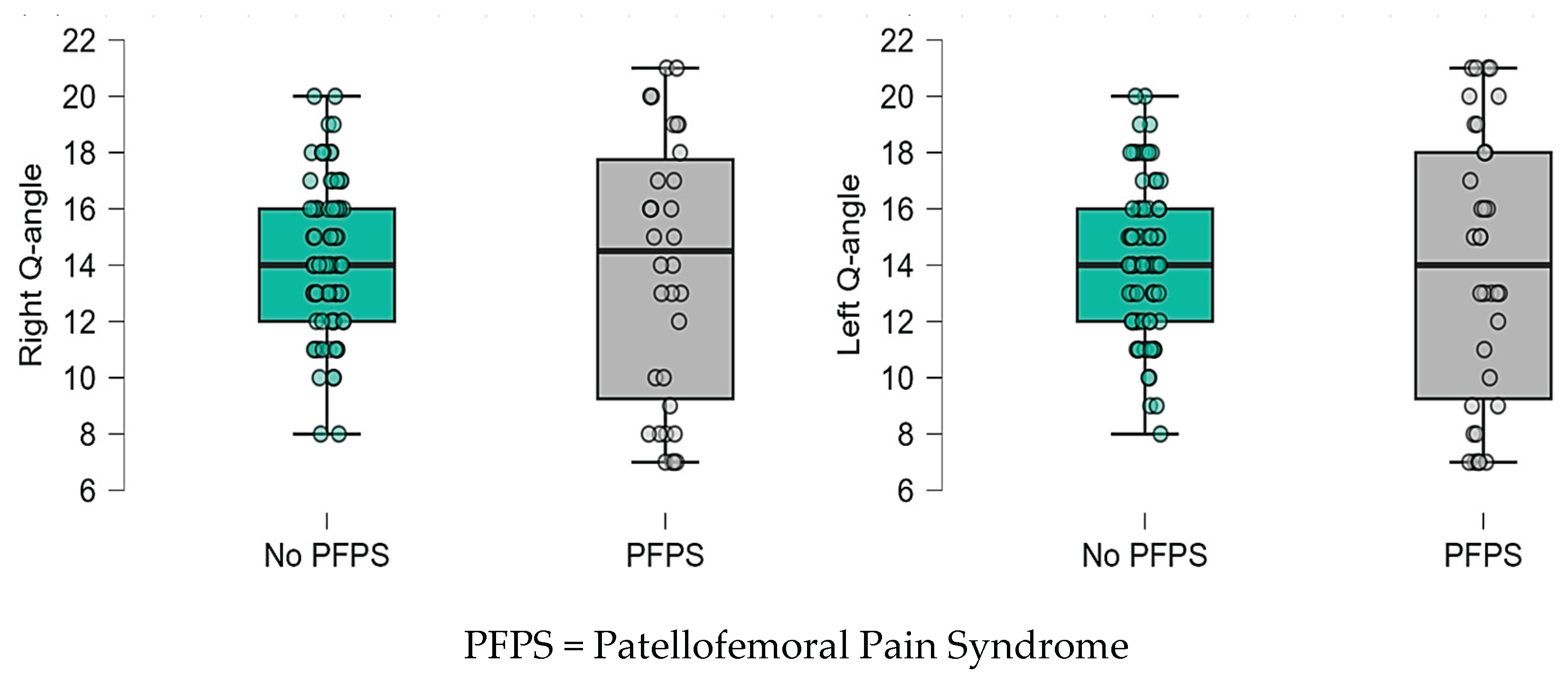

Table 1.
Sociodemographic and sports characteristics of the study population according to the presence of Patellofemoral Pain Syndrome (PFPS).
Table 1.
Sociodemographic and sports characteristics of the study population according to the presence of Patellofemoral Pain Syndrome (PFPS).
| Variable | Description | No PFPS | PFPS | Total | p-value |
|---|---|---|---|---|---|
| n (%) | n (%) | n (%) | |||
| Sex | Female | 45 (57,7) | 15 (44,1) | 60 (53,57) | 0,185 (a) |
| Male | 33 (42,3) | 19 (55,9) | 52 (46,43) | ||
| Sports team | Basketball | 17 (21,8) | 7 (20,6) | 24 (21,43) | 0,868 (b) |
| Soccer | 9 (11,5) | 2 (5,9) | 11 (9,82) | ||
| Futsal | 17 (21,8) | 7 (20,6) | 24 (21,43) | ||
| Swimming | 10 (12,8) | 6 (17,6) | 16 (14,29) | ||
| Ultimate | 12 (15,4) | 4 (11,8) | 16 (14,29) | ||
| Volleyball | 13 (16,7) | 8 (23,5) | 21 (18,75) | ||
| Faculty | Faculty of Agricultural and Veterinary Sciences | 13 (16,7) | 4 (11,8) | 17 (15,18) | 0,406 (b) |
| Faculty of Economic, Administrative, and Accounting Sciences | 8 (10,3) | 4 (11,8) | 12 (10,71) | ||
| Faculty of Medical and Health Sciences | 17 (21,8) | 3 (8,8) | 20 (17,86) | ||
| Faculty of Natural Sciences | 17 (21,8) | 7 (20,6) | 24 (21,43) | ||
| Faculty of Social Sciences | 12 (15,4) | 7 (20,6) | 19 (16,96) | ||
| Faculty of Engineering and Technologies | 11 (14,1) | 9 (26,5) | 20 (17,86) | ||
| Academic semester | First semester | 8 (10,3) | 4 (11,8) | 12 (10,71) | 0,711 (b) |
| Second semester | 6 (7,7) | 2 (5,9) | 8 (7,14) | ||
| Third semester | 7 (9,0) | 3 (8,8) | 10 (8,93) | ||
| Fourth semester | 9 (11,5) | 6 (17,6) | 15 (13,39) | ||
| Fifth semester | 5 (6,4) | 3 (8,8) | 8 (7,14) | ||
| Sixth semester | 4 (5,1) | 2 (5,9) | 6 (5,36) | ||
| Seventh semester | 3 (3,8) | 3 (8,8) | 6 (5,36) | ||
| Eighth semester | 8 (10,3) | - | 8 (7,14) | ||
| Ninth semester | 5 (6,4) | 2 (5,9) | 7 (6,25) | ||
| Tenth semester | 10 (12,8) | 2 (5,9) | 12 (10,71) | ||
| Eleventh semester | 8 (10,3) | 3 (8,8) | 11 (9,82) | ||
| Twelfth semester | 5 (6,4) | 4 (11,8) | 9 (8,04) |
PFPS = Patellofemoral Pain Syndrome; n = Frequency; % = Percentage; (a) = Chi-Square test; (b) = Fisher's exact test.
Table 2.
Anthropometric and body composition characteristics of university athletes according to the presence of PFPS.
Table 2.
Anthropometric and body composition characteristics of university athletes according to the presence of PFPS.
| Variable | No PFPS | PFPS | TOTAL | p-value | |
|---|---|---|---|---|---|
| Age (b) | 21 (19 - 23) | 22 (19 - 23) | 21 (19 - 23) | 0,602 U | |
| Height (cm) (a) | 167,60 ± 8,03 | 169,7 ± 8,45 | 168,24 ± 8,18 | 0,213 t | |
| Weight (kg) (b) | 62,15 (57,88 - 73,23) | 67,80 (59,08 - 76,1) | 63 (58,4 - 74,18) | 0,222 U | |
| Fat % (a) | 17,18 ± 5,21 | 16,48 ± 3,99 | 16,96 ± 4,87 | 0,487 t | |
| Water % (a) | 56,89 ± 2,75 | 57,71 ± 3,65 | 57,14 ± 3,05 | 0,195 t | |
| Muscle % (a) | 40,94 ± 4,25 | 41,75 ± 4,78 | 41,19 ± 4,41 | 0,337 t | |
| PHY Rate (b) | 7 (6 - 8) | 8 (6 - 9) | 7 (6 - 8) | 0,552 U | |
| DCI (kcal) (a) | 2528,77 ± 433,13 | 2619,18 ± 352,57 | 2556,21 ± 410,9 | 0,286 t | |
| Body Age (b) | 20 (18 - 22) | 20,50 (18 - 22) | 20 (18 - 22) | 0,667 U | |
| Bone (kg) (b) | 3,1 (2,9 – 3,63) | 3,4 (2,8 – 3,8) | 3,1 (2,9 - 3,7) | 0,256 U | |
| Visceral Fat (b) | 2 (1 – 3) | 2 (1,75 – 4) | 2 (1,25 - 3) | 0,739 U | |
| Right LL Length (cm) (a) | 80,49 ± 3,85 | 81,36 ± 4,38 | 80,75 ± 4,02 | 0,294 t | |
| Left LL Length (cm) (a) | 80,46 ± 3,85 | 81,32 ± 4,37 | 80,72 ± 4,02 | 0,299 t | |
| Right Thigh Circumference (cm) (b) | 49,30 (44,03 - 58,48) | 51,85 (45,98 - 59,13) | 50,15 (45,18 - 59) | 0,275 U | |
| Left Thigh Circumference (cm) (b) | 49,15 (43,95 - 58,38) | 51,90 (45,93 - 58,95) | 50,2 (45,08 - 58,68) | 0,272 U | |
| Right Calf Circumference (cm) (b) | 32,05 (28,25 - 38) | 34,65 (30,08 - 38,70) | 32,8 (28,83 - 38,2) | 0,227 U | |
| Left Calf Circumference (cm) (b) | 31,90 (28,28 - 38,13) | 34,4 (30,30 - 38,70) | 33 (28,93 - 38,28) | 0,221 U |
PFPS = Patellofemoral Pain Syndrome; LL = Lower Limb; (a) = Mean and standard deviation; (b) = Median and Interquartile range; t = Independent samples t-test; U = Mann-Whitney U test.
Table 3.
Clinical association between Q-angle alteration and the presence of Patellofemoral Pain Syndrome.
Table 3.
Clinical association between Q-angle alteration and the presence of Patellofemoral Pain Syndrome.
| Clinical Variable | No PFPS (n=78) | PFPS (n=34) |
Chi-Square Test | p-value |
|---|---|---|---|---|
| Right Q-angle | 25,861 | 0,001* | ||
| Normal | 62 (79,5%) | 10 (29,4%) | ||
| Altered | 16 (20,5%) | 24 (70,6%) | ||
| Left Q-angle | 16,875 | 0,001* | ||
| Normal | 61 (78,2%) | 13 (38,2%) | ||
| Altered | 17 (21,8%) | 21 (61,8%) | ||
PFPS = Patellofemoral Pain Syndrome.
Disclaimer/Publisher’s Note: The statements, opinions and data contained in all publications are solely those of the individual author(s) and contributor(s) and not of MDPI and/or the editor(s). MDPI and/or the editor(s) disclaim responsibility for any injury to people or property resulting from any ideas, methods, instructions or products referred to in the content. |
© 2026 by the authors. Licensee MDPI, Basel, Switzerland. This article is an open access article distributed under the terms and conditions of the Creative Commons Attribution (CC BY) license (http://creativecommons.org/licenses/by/4.0/).
Copyright: This open access article is published under a Creative Commons CC BY 4.0 license, which permit the free download, distribution, and reuse, provided that the author and preprint are cited in any reuse.