

Submitted:
16 October 2026
Posted:
16 October 2026
You are already at the latest version
Abstract
Salmonella enterica (S. enterica) is a leading cause of foodborne illness worldwide, and the accelerating spread of multidrug-resistant serovars has renewed interest in bacteriophages as therapeutic agents. Phages infecting S. enterica exhibit an extraordinary diversity of virion architectures, ranging from compact, non-tailed microviruses with circular, single-stranded DNA genomes to jumbo phages with large, double-stranded DNA genomes and elaborately layered capsid–tail assemblies. Here, we review this architecture by morphotype: microviruses (Microviridae), podoviruses, siphoviruses, myoviruses and jumbo phages. Each section centers on representative, structurally characterized phages as focal case studies. By integrating available cryo-electron microscopy (cryo-EM) reconstructions with complementary biochemical and genomic evidence, we distinguish features conserved within a morphotype from those reflecting lineage-specific adaptation. For each morphotype, we further discuss how these architectures address key functional imperatives, including protection of the genome in the extracellular environment and recognition of diverse Salmonella surface receptors. By linking virion architecture to function, this review identifies key gaps for future mechanistic studies and informs the rational selection and engineering of phages against multidrug-resistant Salmonella.
Keywords:
Salmonella enterica
; bacteriophage
; virion architecture
; cryo-electron microscopy
; receptor-binding protein
1. Introduction
S. enterica remains among the leading causes of bacterial foodborne illness worldwide, and the burden attributable to antimicrobial-resistant Salmonella has continued to grow [1,2]. S. enterica serovar Typhi (S. Typhi) is an exclusively human-adapted pathogen, in contrast to nontyphoidal Salmonella (NTS), which is maintained in diverse animal reservoirs [3]. Globally, S. Typhi is estimated to cause 16–33 million infections and 500,000–600,000 deaths annually, whereas NTS accounts for approximately 90 million infections and 155,000 deaths each year [3]. Bacteriophages are among the most developed alternatives for controlling this host, and are under active development as therapeutic and biocontrol agents [4,5]. Phages are also, independently of any applied motive, one of the richest systems available for studying how protein shells are built, and Salmonella phages are unusual in combining the two attractions. Within this single, well characterized host, phages span nearly the entire known morphological range of bacterial viruses, from tailless icosahedral Microviridae to contractile-tailed jumbo phages.
The infectious phage particle is more than a container for genetic material. It is a coordinated molecular assembly in which genome protection, host recognition, and genome delivery must be reconciled with the requirements of assembly and environmental persistence. Capsid proteins form protective shells, while receptor-binding proteins mediate interactions with bacterial surface determinants. In tailed phages, portals, head-to-tail connectors, tail tubes, and distal attachment complexes organize the route between the packaged genome and the host cell. Contractile systems additionally incorporate a sheath whose conformational transition contributes to infection. Non-tailed phages lack this extended apparatus and instead rely on differently organized capsid-associated and genome-delivery components. The relevant comparison is therefore not simply whether a particular structure is present or absent, but how different architectures accomplish equivalent biological tasks. Receptor-binding proteins and receptors of the Salmonella phages discussed in this review are listed in Table 1.
Structural coverage across this spectrum is uneven, and the unevenness is not random: cryo-EM resolves small, symmetric and mechanically rigid particles most readily, and that is where near-atomic structures exist. The microviruses PJNS001 and PJNS002 are determined at 2.68 Å and 2.59 Å [6]; the podoviruses are covered by Epsilon15, determined first at 20 Å and then at validated near-atomic resolution [10,21], and by P22, characterized at multiple stages from capsid assembly through the portal and tail machine to infection initiation [22,23,24,25]; and among siphoviruses the flagellotropic Chi (χ) has been resolved nearly completely, from capsid to tail tip [11]. SPN3US is the best-characterized Salmonella jumbo phage, although its mature capsid is resolved only to ~30 Å (Figure 1) [26].
2. Microviridae: The Minimal Architecture
The Microviridae is the most abundant single-stranded DNA phage lineage on Earth and a substantial component of the human gut virome, yet its diversity has only recently been organized phylogenetically [27]. The family falls into two subfamilies, Bullavirinae and Gokushovirinae, which differ in host range, lifecycle tempo and assembly strategy [28]. High-resolution structures of diverse Microviridae phages have been well characterized [6,29], including the bullaviruses φX174 and α3, among the gokushoviruses, φEC6098 and Ebor [30,31]. However, the detailed assembly mechanisms of Microviridae phages infecting Salmonella remained poorly defined until 2025, when the cryo-EM structures of PJNS001 and PJNS002 were resolved at 2.68 Å and 2.59 Å, providing the first clear insights into their capsid assembly and pentameric adaptations (Figure 2A) [6]. PJNS001 and PJNS002 are Bullavirinae phages infecting S. enterica; their circular ssDNA genomes of 5,386 nt (45% GC) and 5,486 nt (46.1% GC) encode no virulence, antimicrobial-resistance or transduction genes and support a purely lytic lifecycle [6]. Both phages were isolated by receptor-guided screening against lipopolysaccharide (LPS)-truncated hosts [6]. Their capsids are T = 1 icosahedra whose asymmetric unit contains the capsid protein F, the spike protein G and the DNA-binding protein J, together with several ordered nucleotides in PJNS002 (Figure 2B) [6].
2.1. The F Capsid Protein: Building the T = 1 Shell
The F protein is the principal structural constituent of the capsid, sixty copies of which build the entire shell. Across the Microviridae it adopts a conserved eight-stranded antiparallel β-barrel of jelly-roll topology, from which long insertions between strands, most prominently the EF and HI loops, project outward to form the particle surface and the contacts between neighboring subunits (Figure 2C) [29]. Assembly is scaffolding-dependent throughout the family, but the underlying morphogenetic pathway has diverged: members of the Bullavirinae (φX174, G4 and α3) use two scaffolding proteins, whereas gokushoviruses rely on a single internal scaffolding protein [32,33,34,35]. PJNS001 and PJNS002 follow the bullavirus pattern [6]. At the five-fold axis, the F protein carries a conserved DGTDQ motif (F residues 251–255), which generates a pore of approximately 4 Å at the junction of five F subunits, constricted at Thr253, a feature conserved throughout the Bullavirinae but variable in both sequence and dimension among gokushoviruses (Figure 2D) [6]. Thermodynamic dissection confirmed the family-wide hierarchy in which pentameric interfaces dominate capsid stability, and revealed that F–G affinity is intrinsically weak, rationalizing the absolute requirement for scaffolding proteins during assembly [6].
2.2. The G Spike Protein: Pentameric Host Recognition
The G protein is the receptor-binding element of the Bullavirinae. Sixty copies assemble into twelve pentamers, one above each five-fold vertex, each subunit a compact β-barrel whose strands run radially outward in a topology echoing that of F; the five together enclose a hydrophilic channel [29]. The subfamilies diverge sharply here. Gokushoviruses carry no G protein at all, presenting instead mushroom-shaped protrusions built from the capsid protein itself at the three-fold axes [30,31,35]. Spike-borne recognition is thus a bullavirus solution rather than a family-wide one. PJNS001 and PJNS002 add atomic detail to this framework. Although both G proteins comprise 175 residues and retain the conserved bullavirus β-barrel, each pentamer is stitched together by a species-specific contact network: in PJNS001, subunit G1 forms nine hydrogen bonds with G2, one with G3 and five with F subunits, whereas in PJNS002 it forms twelve with G2 and only two with F [6]. The central hydrophilic channel is tuned differently as well: negatively charged Asp117 and positively charged Arg115 in PJNS001 are replaced by uncharged Ser118/Asn119 and Thr116 in PJNS002, which may alter pore geometry and charge selectivity (Figure 2D) [6]. These molecular differences map onto distinct receptor dependencies of PJNS001 and PJNS002, placing their epitopes at successive truncation levels of the LPS core [6].
2.3. The J DNA-Binding Protein: Organizing the Packaged Genome
The J protein varies considerably in length, from 26 to 38 residues among the Bullavirinae and longer in some relatives, yet the C-terminal region is invariably the part that contacts F [28]. J is a strongly basic protein that occupies the capsid interior, where it helps neutralize the electrostatic environment of the packaged genome against the slightly alkaline inner surface of F [6]. How much of it is resolved varies markedly: the N-terminus is disordered in φEC6098, partly ordered in SpV4, and remains unresolved in Ebor [30,31,35]. This variation cannot be explained by genome size alone and may instead reflect differences in particle integrity. The PJNS structures provide the most complete views. PJNS001 resolves all 38 residues, including a 12-residue N-terminal extension that traverses the β-barrel of the adjacent F; PJNS002 resolves 26 residues, and its J further stabilizes nine ordered nucleotides through interactions involving Lys3, Arg7, Arg17 and Tyr20 [6]. In both structures, the J C-terminus is anchored to the inner surface of F through specific hydrogen-bonding interactions [6].
What remains unresolved in PJNS001 and PJNS002 is the structural pathway linking receptor engagement to capsid opening and genome delivery: although PJNS002 G protein has been proved to bind LPS in vitro, the corresponding G–LPS interaction has not been resolved at high resolution, and the molecular trigger coupling receptor engagement to spike release and capsid opening remains unclear. In situ reconstructions of PJNS001 and PJNS002 have likewise not resolved the putative H-derived genome-delivery tube, leaving the transition from host attachment to genome translocation structurally undefined.
3. Podoviruses: The Compact Injector
Podoviruses are the short-tailed members of the Caudoviricetes: adsorption and genome ejection are handled by a compact machine at one vertex, without the long pipe of the siphoviruses or the contractile sheath of the myoviruses. Across all tailed phages, as well as certain archaeal viruses and the Herpesviridae, the capsid adopts an icosahedral (occasionally prolate) geometry in which a single vertex is occupied by a connector protein—conventionally termed the portal [36]. Bacteriophages of the podovirus morphotype form a large polyphyletic group, but their structural and functional paradigms have predominantly been established by the T7, P22, φ29, and N4 lineages which themselves differ in capsid geometry (prolate T = 3, Q = 5 in φ29, isometric T = 7 in T7 and P22, and T = 9 in N4), genome size (19.3 kb in φ29 to 70.2 kb in N4) and receptor-binding strategy (enzymatic tailspikes in P22, non-catalytic fibers in T7) [36,37,38,39]. Several well-studied podoviruses can effectively infect Salmonella, including P22 (serogroup B), Epsilon15 (serogroup E1) and the T7-like SP6. Among them, P22 is the founding model of Salmonella phage biology, anchoring work on lysogeny, generalized transduction and virion assembly for half a century [40,41]. Its mature virion and isolated tail machine have both been characterized in detail by cryo-electron microscopy (cryo-EM) (Figure 3A) [36,42,43,44,45,46]. Cryo-electron tomography of P22 infecting Salmonella cells captured the successive states of infection initiation: tailspike engagement of the O-antigen, virion reorientation, needle penetration of the outer membrane, and assembly of a trans-envelope channel that delivers the genome into the cytoplasm [25].
3.1. Capsid and Assembly
In podoviruses, capsid assembly typically begins with the formation of a metastable precursor particle termed the procapsid (PC), prohead, or prehead [47]. The major capsid protein forms the protective shell that encloses the viral genome and adopts a highly conserved HK97 fold across structurally characterized tailed bacteriophages [48]. What varies is the lattice the fold is arranged into, and how the expanded lattice is kept from failing. The capsid of P22 is an isometric T = 7 laevo icosahedron, ~64 nm across, built from 415 copies of the HK97-fold coat protein gp5, with a dodecameric portal in place of one penton. What distinguishes P22 is how the expanded lattice is stabilized. Stability arises from within the subunit: a telokin-like immunoglobulin domain inserted into the coat protein rigidifies the monomer and contributes to capsid size determination, while the N-arm refolds during expansion to lock the enlarged lattice without covalent crosslinks [49,50]. Assembly occurs through the sequential addition of individual subunits rather than pre-formed capsomers and is facilitated by an internal scaffolding protein that is released before expansion [45].
3.2. Portal and Packaging
A defining feature of the portal vertex in tailed phages is the C5–C12 symmetry mismatch between the capsid vertex and the dodecameric portal. Rather than being a purely geometric irregularity, this mismatch creates a dynamic interface that allows the portal to coordinate capsid assembly, genome packaging, tail attachment and genome ejection [51]. Its three-dimensional architecture is highly conserved across tailed phages and herpesviruses despite negligible sequence similarity (Figure 3B) [36]. The portal is laid down at the very start of assembly rather than fitted into a finished shell, and it helps organize the shell around itself. Scaffolding protein acts first on portal monomers, driving them to oligomerize; what then enters a nascent procapsid is the assembled twelve-membered ring rather than individual subunits, and incorporation favors that ring in its procapsid rather than its mature conformation [52]. Once seated, the portal serves as the nucleation point from which procapsid assembly proceeds [44].
Packaging itself follows the headful route. In phage P22, cutting begins at a pac site, but every subsequent cleavage is determined by the amount of DNA the head can accommodate rather than by any specific sequence [53]. The reaction is carried out by the large terminase gp2, which comprises an N-terminal ATPase module connected through a flexible linker to a C-terminal nuclease domain [54]. Crystallography shows that nuclease to be autoinhibited in isolation: its catalytic metals are correctly placed, but the surface that must engage DNA is occluded, and activity emerges only within the intact large terminase, where communication with the ATPase module lifts the block [54].
The autoinhibition fits the problem the phage has to solve. Because more than one genome length is drawn into the head, no sequence is available at which to stop, so the decision must derive from the physical state of the shell and the nuclease is built so that it cannot act before that decision reaches it.
3.3. Tail Machine and Host Recognition
The P22 tail is a short stack of rings on the portal vertex: twelve copies of the adaptor gp4, six of the hub gp10, and the trimeric needle gp26, with six trimeric tailspikes gp9 attached laterally [36]. The assembly absorbs a stepwise symmetry mismatch, from twelvefold at the adaptor to threefold at the needle, whose triple-stranded coiled coil plugs the DNA exit channel once packaging is complete [55]. This core is conserved among tailed phages, while the distal recognition module varies with the surface it crosses.
Salmonella presents an extended O-antigen, and the two Salmonella podoviruses with resolved structures, P22 and Epsilon15, both employ enzymatic tailspikes. P22 gp9 is a thermostable trimer built around a right-handed parallel β-helix, its head-binding and endorhamnosidase domains refined at 2.3 Å and 1.56 Å [56]. Epsilon15 gp20 is the same enzyme aimed elsewhere, cleaving the group E1 repeat unit that lies outside P22's range [57,58]. The same catalytic chemistry has thus been retargeted to a different serovar, specificity residing in the binding groove. Other lineages mark the range: T7 uses non-enzymatic fibers, and N4 infection involves both the surface glycan NGR and the outer-membrane protein NfrA [59,60,61,62]. The module is also portable: Det7 mounts a P22-type tailspike on a contractile tail [16]. Together, these examples illustrate how phage tail appendages can be remodelled across lineages to recognize chemically and structurally distinct receptors, while retaining their central role in the initial stages of host recognition.
The tail cannot complete delivery step on its own. It stops short of the periplasm, so the conduit across that space must be assembled after contact with the cell. Tomography of P22 shows that the virion lands obliquely on two tailspikes, turns to sit square against the envelope, drives its needle through, and ejects gp7* and gp20, which lay a channel across the outer membrane while gp16 opens the final passage into the cytoplasm [25]. Epsilon15 spans the periplasm with an equivalent tube [63], and T7 ejectosome is resolved at 2.7 Å [64]. N4 presses the same logic further, ejecting its ~380 kDa virion RNA polymerase ahead of the genome, a strategy that jumbo phages scale up [65]. Thus, although these phages share the requirement for a functional genome-delivery pathway, the composition and assembly of their penetration machinery are lineage-specific.
4. Siphoviruses: The Long Flexible Injector
Siphoviruses carry a long, flexible, non-contractile tail. That single architectural feature fundamentally changes the delivery problem: whereas a podovirus must assemble a trans-envelope conduit after receptor engagement, a siphovirus carries a preassembled, hollow tail that serves as the physical route from the capsid toward the host surface. Its tail must combine sufficient length and flexibility to accommodate adsorption with a stable, sealed channel that prevents premature genome release. Structures of numerous siphophages have now revealed the modular organization of this long-tail machine, including λ and T5 infecting Escherichia coli [66,67], SPP1 infecting Bacillus subtilis [68], and mycobacteriophages Bxb1 and Douge [69,70], together illustrating a conserved tail tube framework coupled to highly diversified distal receptor-binding architectures. Among Salmonella phages, the best-characterized structural examples are the flagellotropic YSD1 of S. Typhi and the archetypal χ, which face an additional challenge: the virion must engage and travel down a long, rotating flagellar filament before reaching the cell surface, withstanding the drag and torque that motion imposes [11,71].
4.1. Capsid and Tail Architecture
Siphoviral capsids are built from the HK97 fold, but the means of locking an expanded lattice differ by class: HK97 itself forms covalent isopeptide catenanes [72], whereas the λ/χ-like capsids recruit an external cementing protein [71]. YSD1 provides the model for that class at 3.8 Å. Its auxiliary protein YSD1_16, a homotrimer of β-tulip fold homologous to λ gpD, contributes an N-terminal hook that inserts as a fourth β-strand into the E-loop stem of a neighboring hexon, stapling the two together [71]. The major capsid protein and the auxiliary protein thus form two nested layers of chain mail, interlocked without covalent bonds (Figure 4A,B). The same clamping arrangement recurs in the herpesvirus triplex, suggesting an ancestral origin for these stabilizing elements [71].
The tail tube is equally conserved at its core—hexameric rings of the major tail protein (Figure 4C), built on a β-sandwich fold shared with contractile tails, pyocins and secretion-system tubes, threaded onto the tape-measure protein that fixes their number [66,71]. In YSD1 the lumen is paved with regularly spaced dyads of aspartate and glutamate forming a right-handed helical track, proposed to ratchet the genome onward once that initial impetus wanes [71]. Equivalent patterns occur in 80α, SPP1 and T5 but share no sequence conservation, suggesting independent solutions to a common mechanical constraint [73]. Head and tail are married at the neck: in χ, a dodecameric portal is sealed by head-completion and connector rings plus a tail terminator, and the topmost tail hexamer refolds its otherwise-disordered D3 domains into a socket that docks the neck [11].
4.2. Tail-Tip Diversification and Host Recognition
If the tail tube is conserved, the tail tip is where siphophages diversify. A common core underlies most of them: the tape-measure protein C-terminus, the distal tail protein (Dit) and the tail-associated lysin (Tal) form a triplet onto which variable receptor-binding proteins are mounted [74,75]. Bacteriophage T5 illustrates the canonical two-step solution. Three L-shaped side fibers (pb1) bind polymannose O-antigen reversibly, allowing the particle to sample the surface; the straight central fiber then engages the outer-membrane transporter FhuA irreversibly, and that binding not the reversible step triggers conformational change in the tip and opens the channel [76,77,78]. The division of labor is explicit: fibers search, the tip commits.
The Salmonella flagellotropes condense that division of labor into a single device. Rather than an array of side fibers, YSD1 and χ present one long, flexible tail fiber attached to the tail tip, which wraps around the flagellar filament to establish first contact [11,79]. Electron microscopy first showed the fiber coiled about the filament in 1967 [79], and the prevailing model holds that flagellar rotation then drives the virion along the filament to the cell body like a nut on a bolt [80], converting a swimming organelle into a guide rail. The χ structure now reveals what such a minimal tip is made of. Four protein species build it: the distal tail protein (DTP), the tail-assembly protein TAP and the baseplate hub proteins BH1P and BH2P [11]. DTP departs from the siphophage consensus by forming a trimer rather than a hexamer, so it alone bridges the hexametric tail to the trimeric tip, performing the symmetry reduction that additional hub components accomplish elsewhere (Figure 4D) [11]. The contrast is instructive: T5 elaborates its tip because a scarce receptor on a static surface must be found, whereas χ can afford a minimal one because the flagellum performs the search on the phage's behalf.
5. Myoviruses: Contractile-Tailed Phages of Salmonella
The term “myovirus” is used here to describe a contractile-tailed morphotype rather than a family-level taxon in the current classification [81]. These particles typically comprise a head, a head-to-tail connector and a tail. The contractile sheath surrounds an inner tail tube, while the distal tail apparatus includes a baseplate and receptor-binding structures [82]. This architecture integrates genome containment, host recognition and mechanical penetration within a single particle. Members differ in head morphology, baseplate organization and the composition of their distal receptor-binding apparatus. Studies of Salmonella phages provide distinct levels of evidence, including particle morphology, functional identification of host receptors [14] and local protein structure [16]. These evidence types should be distinguished when relating individual components to the architecture and function of the complete virion.
5.1. Head and Tail Machinery
For most of these phages, head architecture remains at the level of particle morphology, with electron micrographs the only structural evidence. Morphological observations place some members within the T-even morphological group [14], whose heads are polyhedra elongated along one axis [83]; the remaining members adopt an isometric geometry, Felix O1 and its relatives having a head of ~73 nm and a tail of 113 × 17 nm [84], and the multi-tailspike phages a head of ~90 nm and a tail of 110 × 18 nm [17]. For elongated heads, T4 provides a comparative reference for capsid geometric organization: its prolate capsid comprises icosahedral end caps and an elongated midsection, with Tend = 13 and Q = 20; the mature capsid contains approximately 930 subunits of the major capsid protein gp23* and 55 subunits of the vertex protein gp24*, the latter forming 11 pentamers, while the single remaining vertex is the portal vertex, occupied by the gp20 dodecamer that connects to the tail [83]. These structural features illustrate how an HK97-like capsid protein can support an elongated head architecture, although the precise geometry and subunit composition may differ among Salmonella phages. For Felix O1-related phages, the capsid decoration and neck whisker structures determined for the congeneric phage Bas63 [85] provide a comparative reference for the organization of accessory components, although whether the composition of the decoration proteins, their binding sites and the mode of whisker attachment are conserved must be judged from sequence and structural evidence for the target phage itself.
The conformational mechanism of the contractile tail likewise rests on T4: reversible binding of the long tail fibers to their receptor precedes irreversible anchoring by the short tail fibers; once the signal has been transmitted to the baseplate, the baseplate converts from a hexagonal to a star-shaped conformation, a transition coupled to contraction of the tail sheath, which shortens and thickens through subunit rearrangement and drives the tail tube through the cell envelope [86]. By contrast, for the contractile-tailed phages that infect Salmonella, current genomic studies assign candidate structural proteins of the tail sheath, tail tube and baseplate mainly on the basis of sequence homology [87,88]; how these components together constitute the complete tail apparatus, and the stoichiometry, assembly relationships and conformational states of the portal–neck–tail connector region, still require direct structural evidence. The T4 system provides a structural reference for framing these open questions: its baseplate has been structurally characterized in both pre- and post-attachment conformations [86], assembly intermediates of the portal–neck have been resolved [89], and the remodeling of the virion and the host membranes at the initiation of infection has been recorded by in situ tomography [90]. Comparison with T4 therefore supplies only a mechanistic framework; the order of triggering, the conformational intermediates, and the spatial relationship between the tail and the cell envelope still require structural evidence from these phages themselves.
5.2. Host-Recognition Module
Salmonella phage Det7 provides direct structural evidence that a receptor-binding module can be shared across tail morphotypes. What has been solved is an N-terminally truncated form of its tailspike protein, a homotrimer whose body is an extended β-helical domain [16], homologous to the tailspike of the podovirus P22 [7]. In the P22 tailspike, both the oligosaccharide-binding site and the catalytic residues are located in a surface groove of the central β-helix domain [8]; the three active-site carboxylate residues are conserved in Det7 [16]. A receptor-binding module can thus be shared between different tail morphotypes, yet the N-terminal region of Det7 lacks detectable sequence homology to that of P22, suggesting that the mechanisms of attachment to their respective virions may differ [16].
Det7 and ViI belong to the genus Kuttervirus, whose members can carry multiple distinct tailspike proteins on a single tail apparatus, thereby combining different surface-polysaccharide recognition specificities [17]. Work on the Escherichia coli O157:H7 phage CBA120 has resolved the architecture of such a branched receptor-binding complex [91]; by contrast, the composition and spatial arrangement of the complete tailspike array in the Salmonella members remain unresolved. Chimeric engineering based on homologous tailspikes has been used to alter phage host range [92], indicating that this recognition module is modular and amenable to engineering.
S16 represents an alternative recognition strategy that uses an outer membrane protein as its receptor: its host recognition depends on outer membrane protein C (OmpC) [14]. The gp38 adhesin at the distal end of the long tail fiber is attached to gp37 and adopts an unusual fold of stacked polyglycine type II helices, termed a "PG II sandwich" by the original authors; its sequence variability is confined to surface-exposed helices and distal loops, the latter being considered to constitute the receptor-binding site [15]. This division of labour between subunits differs from that in T4: in T4 the receptor-binding domain of the long tail fiber resides at the C-terminal tip of gp37 [93], with gp38 acting as a folding chaperone required for gp37 trimerization [94,95], whereas in phages such as T2 and S16 gp38 is itself the adhesin that caps the fiber tip and binds the host receptor [94,95]. Structural inferences drawn from the T4 long tail fiber therefore transfer only partly to S16: in S16 the determinants of receptor specificity must be sought in gp38 rather than in the gp37 tip.
Recent work in E. coli systems has further constrained the recognition relationship between adhesins of this type and their receptors: gp38-mediated recognition in some Straboviridae members involves specific extracellular loops of outer membrane protein receptors [95], and the structure of the gp38 receptor-binding domain of PP01 further localizes the contacts to corresponding residues in its hypervariable loops and in the extracellular loops of O157-type OmpC [96]. These results support the interpretation that the variable loops of gp38 contribute to receptor specificity. However, the specific receptor-binding interactions inferred from these results cannot be directly extrapolated to S16. Given the sequence differences between the extracellular loops of Salmonella OmpC and those of the E. coli OmpC variants referred to above, the S16 gp38–OmpC interface awaits a structure of the complex together with functional validation in S16.
Felix O1 represents a third receptor-recognition strategy: the N-acetylglucosamine residue of the LPS common core oligosaccharide was identified as an essential component of its receptor, and shortening of the O chain is associated with higher adsorption rates, consistent with O-chain shielding of this core site [19]. Among the Salmonella contractile-tailed phages discussed above, structural characterization of receptor-binding proteins has concentrated on O-antigen-recognizing and -degrading tailspikes and on gp38-type adhesins. However, these ligand-free structures do not establish the corresponding receptor-binding interfaces. For Felix O1, how the receptor-binding apparatus recognizes and gains access to the LPS core site awaits direct structural evidence.
6. Jumbo Phages
Jumbo phages are conventionally defined as phages with genomes larger than 200 kb; the term rests on genome size and does not correspond to a discrete phylogenetic group [97]. Among the jumbo phages that infect Salmonella, SPN3US is the best-characterized representative, with a genome of approximately 240 kb [98]. Cryo-electron microscopy has been used to examine its capsid and a putative maturation intermediate [26], and genetic and proteomic analyses have addressed its essential genes, head assembly and the course of infection [20,88,99,100]. For comparison, the Pseudomonas aeruginosa jumbo phage phiKZ has been characterized both by early three-dimensional reconstructions [101,102] and by subsequent high-resolution work [103,104], which together provide the reference framework for the structural organization, assembly and stabilization of jumbo phages.
6.1. Capsid Structure, Assembly and Maturation
Among jumbo phages of Salmonella, SPN3US provides the principal model for the assembly and maturation of a large capsid. Its "mottled" capsid was reconstructed at approximately 16 Å and the corresponding mature virion at approximately 30 Å; the former revealed a T = 1 inner shell enclosed within a T = 27 outer shell, and the particle was interpreted as a probable maturation intermediate rather than a prohead [26].
Beyond these structural observations, genetic and protein-level analyses indicate that head assembly in SPN3US involves not only the construction of structural components but also the incorporation of internal functional components, and that it depends on a protease-mediated maturation step. Sequencing of 18 amber mutants identified 13 essential genes, whose products include head proteins (gp47, gp64 and gp214), tail proteins (gp25, gp171, gp203 and gp238), an SbcC subunit (gp186), and the β subunit of the virion RNA polymerase (gp241, which is likewise head-associated) [88]. Protease mutants established the link between proteolytic processing and head maturation: the prohead protease gp245 cleaves 8 of approximately 50 head proteins, and in its absence proheads are retained and accumulate at the Salmonella inner membrane, so that DNA packaging cannot proceed [99]. The mature head, moreover, carries not only the genome but also more than 40 MDa of ejection proteins [99]. Taken together, these findings indicate that head morphogenesis in SPN3US must be considered in terms of capsid formation, incorporation of the internal protein cargo and proteolytic processing, and that the maturation state of a particle cannot be inferred from the morphology of its outer shell alone.
Homology of the core components does not, however, imply an identical assembly and maturation pathway. Among the components involved in head morphogenesis, the recognizable correspondence between SPN3US and T4 is limited to the portal and major capsid proteins required for prohead shell formation and to the protease and large terminase subunit required for head maturation; although giant phages generally use a homologous protease to process the major capsid and portal proteins, whether the ejection proteins are processed is not a stable feature of this group [20]. Beyond this difference in processing substrates, head assembly in SPN3US also requires a distinction between components retained in the mature particle and transient assembly factors. Proteomic profiling over the first 0–60 min of infection detected 232 phage proteins, corresponding to approximately 88% of the predicted genes, together with 1994 host proteins; the four most abundant phage proteins were the major capsid protein, the two ejection proteins gp53 and gp54, and gp22. Unlike the first three, gp22 was detected at high abundance in infected cells but not in mature virions, and has therefore been proposed as a candidate scaffolding protein [100].
6.2. Portal, Tail and Baseplate Architecture
In SPN3US, the functional assignment of these proteins currently rests on virion proteomics combined with homology detection and structure prediction, and lacks direct structural validation. The protein gp81 is assigned as the portal protein (12 copies per virion; its closest predicted structural homologue is T4 gp20), gp256 and gp255 as the tail sheath and tail tube, respectively (~264 copies each), and the paralog family C members gp168, gp169 and gp170 are implicated in baseplate or tail-fiber organization based on their similarity to phiKZ gp131 and their absence from tailless mutant particles [20].
Recent structural work on phiKZ provides a comparative framework for these assignments: the portal protein gp129 is indeed dodecameric, sharing a conserved core with the T4 portal while diverging in its wing and barrel domains; the tail comprises 44 stacked hexameric sheath rings (gp29) wrapping 43 stacked hexameric tube rings (gp30); and the baseplate resolves into central, inner-peripheral and outer-peripheral regions, with the central hub protein gp101 being a homologue of T4 gp27 and the outer region forming a star-shaped assembly of ~1070 Å in diameter built from six lobes, beneath which twelve peripheral fibers are anchored. At ~6 Å, the basal complex of each fiber is tentatively assigned as a hetero-hexamer comprising three copies of gp131 together with gp133, gp134 and gp135, and the C-terminal seven-bladed propeller of gp131 has been proposed to participate in host recognition [104]. These structural features of phiKZ provide a comparative basis for evaluating the predicted portal, tail sheath, tail tube and baseplate-associated proteins of SPN3US. In particular, the similarity between SPN3US gp168–gp170 and phiKZ gp131 suggests a potential role in distal tail organization, although their precise arrangement and interactions remain unresolved.
6.3. Head Protein Cargo and Intracellular Compartmentalization
As noted above, the SPN3US head accommodates not only the genome but also ejection proteins and components of the virion RNA polymerase, and thus also serves as a carrier of internal functional proteins [88,99]. The reconstruction of the mottled capsid revealed an internal shell layer and attributed it mainly to the ejection proteins gp53 and gp54 [26]; at this resolution, however, that assignment rests on compositional inference, and the arrangement of these proteins within the shell still requires direct localization evidence. For comparison, bubblegram imaging revealed a cylindrical inner body in mature phiKZ virions [105]. It should be noted that the SPN3US inner shell was observed in a putative maturation intermediate and therefore cannot be equated directly with the inner body of the mature phiKZ particle.
The organization of proteins within the virion is also distinct from the intracellular compartment formed after infection. Several Pseudomonas jumbo phages assemble a "phage nucleus" in the host cell, which separates the replicating genome from the host cytoplasm [106,107]. Subsequent work has resolved the shell lattice, built mainly from chimallin [108], and the selective import of proteins across that shell [109]. These findings provide a reference for the intracellular spatial organization of jumbo phages, but the same mechanism cannot be inferred for SPN3US from genome size or head protein cargo alone; to date, no such compartment has been reported for SPN3US in Salmonella.
7. Conclusions
This review has explored the structural diversity of phages infecting S. enterica, from the minimal T = 1 capsids of microviruses to the elaborate architectures of contractile-tailed and jumbo phages, drawing on both experimentally determined structures and comparative evidence. The organizing axis is morphological rather than taxonomic: the traditional families Myoviridae, Podoviridae and Siphoviridae, together with the order Caudovirales, were abolished in a proposal advanced in 2021 and ratified in 2022, their subfamilies and genera moving directly into the pre-existing class Caudoviricetes. Structural cores are conserved: the HK97-like fold is widely conserved among structurally characterized tailed phages; portals nucleate assembly and coordinate packaging, and tail tubes share a β-sandwich fold with pyocins and secretion systems. The periphery is anything but conserved: receptor-binding modules are exchanged, retargeted or minimized to fit each host surface—from the pentameric G spikes of PJNS001 and PJNS002, through the enzymatic tailspikes of P22 and Epsilon15, to the flagellum-binding tail fibers of χ and YSD1 and the multi-tailspike baseplates of myoviruses. Genome delivery diverges just as sharply, but the three strategies do not stand on equal evidence. The preassembled siphovirus tail and the contact-built podovirus conduit have both been visualized directly, whereas the microvirus H-protein tube remains structurally unresolved in PJNS001 and PJNS002.
The remaining gaps are as informative as the structures. A major challenge for the next phase of Salmonella phage structural biology is therefore not simply to obtain higher resolution, but to connect static structures with the dynamic events of infection. Continued improvements in cryo-EM instrumentation, image processing and particle classification should make it increasingly possible to resolve smaller conformational differences and heterogeneous states, allowing assembly, receptor engagement and genome release to be followed as structural trajectories rather than isolated snapshots. For several Salmonella phages, cryo-electron tomography (cryo-ET) and subtomogram averaging may be particularly valuable because the unresolved steps occur at the interface between virions and intact cells. In situ structures could reveal how receptor binding triggers tail or capsid rearrangements, how penetration structures cross the cell envelope, and how viral proteins and genomes are positioned during genome delivery. By linking these structural observations to the molecular mechanisms that govern host recognition, genome delivery and virion maturation, future studies may begin to identify general principles that explain and ultimately predict how phage architecture determines infection. Such principles could provide a structural foundation for translating comparative phage biology into rational design rules for therapeutic phages.
Author Contributions
Y.G. conceptualized the review theme and designed the manuscript framework; C.F. and Y.W. wrote the initial draft; W.Z., H.Y. and Y.G. revised and edited the manuscript. All authors have read and agreed to the published version of the manuscript.
Funding
The research was funded by Shanghai Municipal Science and Technology Major Project (grant No. ZD2021CY001 to H.Y.) and the State Key Laboratory of Medicinal Chemical Biology, Nankai University (grant No. 2025013 to W.Z.).
Data Availability Statement
No new data were created or analyzed in this study. Data sharing is not applicable to this article.
Conflicts of Interest
The authors declare no conflicts of interest.
References
- Kirk, M.D.; Pires, S.M.; Black, R.E.; Caipo, M.; Crump, J.A.; Devleesschauwer, B.; Dopfer, D.; Fazil, A.; Fischer-Walker, C.L.; Hald, T.; et al. World Health Organization Estimates of the Global and Regional Disease Burden of 22 Foodborne Bacterial, Protozoal, and Viral Diseases, 2010: A Data Synthesis. PLoS Med. 2015, 12, e1001921. [Google Scholar] [CrossRef] [PubMed]
- Collaborators. Global burden of bacterial antimicrobial resistance 1990-2021: a systematic analysis with forecasts to 2050. Lancet 2024, 404, 1199–1226. [Google Scholar] [CrossRef] [PubMed]
- Bula-Rudas, F.J.; Rathore, M.H.; Maraqa, N.F. Salmonella Infections in Childhood. Adv. Pediatr. 2015, 62, 29–58. [Google Scholar] [CrossRef] [PubMed]
- Cui, L.; Kiga, K.; Kondabagil, K.; Wegrzyn, A. Current and future directions in bacteriophage research for developing therapeutic innovations. Sci. Rep. 2024, 14, 24404. [Google Scholar] [CrossRef] [PubMed]
- Li, L.; Fan, R.; Chen, Y.; Zhang, Q.; Zhao, X.; Hu, M.; Lv, Q.; Luo, Y.; Xu, X.; Cai, Y.; et al. Characterization, genome analysis, and therapeutic evaluation of a novel Salmonella phage vB_SalS_JNS02: a candidate bacteriophage for phage therapy. Poult. Sci. 2024, 103, 103845. [Google Scholar] [CrossRef] [PubMed]
- Hu, W.; Liu, Z.; Wei, Y.; Bian, Q.; Lan, W.; Fan, C.; Song, J.; Sun, Q.; Zhang, X.; Liu, Y.; et al. Structural basis for Salmonella infection by two Microviridae phages. Commun. Biol. 2025, 8, 1166. [Google Scholar] [CrossRef] [PubMed]
- Steinbacher, S.; Seckler, R.; Miller, S.; Steipe, B.; Huber, R.; Reinemer, P. Crystal structure of P22 tailspike protein: interdigitated subunits in a thermostable trimer. Science 1994, 265, 383–386. [Google Scholar] [CrossRef] [PubMed]
- Steinbacher, S.; Baxa, U.; Miller, S.; Weintraub, A.; Seckler, R.; Huber, R. Crystal structure of phage P22 tailspike protein complexed with Salmonella sp. O-antigen receptors. Proc. Natl. Acad. Sci. U S A 1996, 93, 10584–10588. [Google Scholar] [CrossRef] [PubMed]
- Seoane-Blanco, M.; Pereda, A.; Broeker, N.; McConnell, M.; Cañada, F.J.; Barbirz, S.; van Raaij, M.J. The structure of the. Salmonella phage epsilon15 tailspike reveals multiple O-antigen binding sites and a protruding esterase domain 2026. [Google Scholar] [CrossRef]
- Jiang, W.; Chang, J.; Jakana, J.; Weigele, P.; King, J.; Chiu, W. Structure of epsilon15 bacteriophage reveals genome organization and DNA packaging/injection apparatus. Nature 2006, 439, 612–616. [Google Scholar] [CrossRef] [PubMed]
- Sonani, R.R.; Esteves, N.C.; Scharf, B.E.; Egelman, E.H. Cryo-EM structure of flagellotropic bacteriophage Chi. Structure 2024, 32, 856–865 e853. [Google Scholar] [CrossRef] [PubMed]
- Esteves, N.C.; Bigham, D.N.; Scharf, B.E. Phages on filaments: A genetic screen elucidates the complex interactions between Salmonella enterica flagellin and bacteriophage Chi. PLoS Pathog. 2023, 19, e1011537. [Google Scholar] [CrossRef] [PubMed]
- Dunstan, R.A.; Pickard, D.; Dougan, S.; Goulding, D.; Cormie, C.; Hardy, J.; Li, F.; Grinter, R.; Harcourt, K.; Yu, L.; et al. The flagellotropic bacteriophage YSD1 targets Salmonella Typhi with a Chi-like protein tail fibre. Mol. Microbiol. 2019, 112, 1831–1846. [Google Scholar] [CrossRef] [PubMed]
- Marti, R.; Zurfluh, K.; Hagens, S.; Pianezzi, J.; Klumpp, J.; Loessner, M.J. Long tail fibres of the novel broad-host-range T-even bacteriophage S16 specifically recognize Salmonella OmpC. Mol. Microbiol. 2013, 87, 818–834. [Google Scholar] [CrossRef] [PubMed]
- Dunne, M.; Denyes, J.M.; Arndt, H.; Loessner, M.J.; Leiman, P.G.; Klumpp, J. Salmonella Phage S16 Tail Fiber Adhesin Features a Rare Polyglycine Rich Domain for Host Recognition. Structure 2018, 26, 1573–1582 e1574. [Google Scholar] [CrossRef] [PubMed]
- Walter, M.; Fiedler, C.; Grassl, R.; Biebl, M.; Rachel, R.; Hermo-Parrado, X.L.; Llamas-Saiz, A.L.; Seckler, R.; Miller, S.; van Raaij, M.J. Structure of the receptor-binding protein of bacteriophage det7: a podoviral tail spike in a myovirus. J. Virol. 2008, 82, 2265–2273. [Google Scholar] [CrossRef] [PubMed]
- Sorensen, A.N.; Brondsted, L. Renewed insights into Ackermannviridae phage biology and applications. npj Viruses 2024, 2, 37. [Google Scholar] [CrossRef] [PubMed]
- Pickard, D.; Toribio, A.L.; Petty, N.K.; van Tonder, A.; Yu, L.; Goulding, D.; Barrell, B.; Rance, R.; Harris, D.; Wetter, M.; et al. A conserved acetyl esterase domain targets diverse bacteriophages to the Vi capsular receptor of Salmonella enterica serovar Typhi. J. Bacteriol. 2010, 192, 5746–5754. [Google Scholar] [CrossRef] [PubMed]
- Lindberg, A.A.; Holme, T. Influence of O side chains on the attachment of the Felix O-1 bacteriophage to Salmonella bacteria. J. Bacteriol. 1969, 99, 513–519. [Google Scholar] [CrossRef] [PubMed]
- Ali, B.; Desmond, M.I.; Mallory, S.A.; Benitez, A.D.; Buckley, L.J.; Weintraub, S.T.; Osier, M.V.; Black, L.W.; Thomas, J.A. To Be or Not To Be T4: Evidence of a Complex Evolutionary Pathway of Head Structure and Assembly in Giant Salmonella Virus SPN3US. Front Microbiol. 2017, 8, 2251. [Google Scholar] [CrossRef] [PubMed]
- Baker, M.L.; Hryc, C.F.; Zhang, Q.; Wu, W.; Jakana, J.; Haase-Pettingell, C.; Afonine, P.V.; Adams, P.D.; King, J.A.; Jiang, W.; et al. Validated near-atomic resolution structure of bacteriophage epsilon15 derived from cryo-EM and modeling. Proc. Natl. Acad. Sci. U S A 2013, 110, 12301–12306. [Google Scholar] [CrossRef] [PubMed]
- Xiao, H.; Zhou, J.; Yang, F.; Liu, Z.; Song, J.; Chen, W.; Liu, H.; Cheng, L. Assembly and Capsid Expansion Mechanism of Bacteriophage P22 Revealed by High-Resolution Cryo-EM Structures. Viruses 2023, 15, 355. [Google Scholar] [CrossRef] [PubMed]
- Olia, A.S.; Prevelige, P.E., Jr.; Johnson, J.E.; Cingolani, G. Three-dimensional structure of a viral genome-delivery portal vertex. Nat. Struct. Mol. Biol. 2011, 18, 597–603. [Google Scholar] [CrossRef] [PubMed]
- Lander, G.C.; Khayat, R.; Li, R.; Prevelige, P.E.; Potter, C.S.; Carragher, B.; Johnson, J.E. The P22 tail machine at subnanometer resolution reveals the architecture of an infection conduit. Structure 2009, 17, 789–799. [Google Scholar] [CrossRef] [PubMed]
- Wang, C.; Tu, J.; Liu, J.; Molineux, I.J. Structural dynamics of bacteriophage P22 infection initiation revealed by cryo-electron tomography. Nat. Microbiol. 2019, 4, 1049–1056. [Google Scholar] [CrossRef] [PubMed]
- Heymann, J.B.; Wang, B.; Newcomb, W.W.; Wu, W.; Winkler, D.C.; Cheng, N.; Reilly, E.R.; Hsia, R.C.; Thomas, J.A.; Steven, A.C. The Mottled Capsid of the Salmonella Giant Phage SPN3US, a Likely Maturation Intermediate with a Novel Internal Shell. Viruses 2020, 12, 910. [Google Scholar] [CrossRef] [PubMed]
- Kirchberger, P.C.; Martinez, Z.A.; Ochman, H. Organizing the Global Diversity of Microviruses. mBio 2022, 13, e0058822. [Google Scholar] [CrossRef] [PubMed]
- Doore, S.M.; Fane, B.A. The microviridae: Diversity, assembly, and experimental evolution. Virology 2016, 491, 45–55. [Google Scholar] [CrossRef] [PubMed]
- McKenna, R.; Xia, D.; Willingmann, P.; Ilag, L.L.; Krishnaswamy, S.; Rossmann, M.G.; Olson, N.H.; Baker, T.S.; Incardona, N.L. Atomic structure of single-stranded DNA bacteriophage phi X174 and its functional implications. Nature 1992, 355, 137–143. [Google Scholar] [CrossRef] [PubMed]
- Bardy, P.; MacDonald, C.I.W.; Kirchberger, P.C.; Jenkins, H.T.; Botka, T.; Byrom, L.; Alim, N.T.B.; Traore, D.A.K.; Koenig, H.C.; Nicholas, T.R.; et al. Penton blooming, a conserved mechanism of genome delivery used by disparate microviruses. mBio 2025, 16, e0371324. [Google Scholar] [CrossRef] [PubMed]
- Lee, H.; Baxter, A.J.; Bator, C.M.; Fane, B.A.; Hafenstein, S.L. Cryo-EM Structure of Gokushovirus PhiEC6098 Reveals a Novel Capsid Architecture for a Single-Scaffolding Protein, Microvirus Assembly System. J. Virol. 2022, 96, e0099022. [Google Scholar] [CrossRef] [PubMed]
- Dokland, T.; McKenna, R.; Ilag, L.L.; Bowman, B.R.; Incardona, N.L.; Fane, B.A.; Rossmann, M.G. Structure of a viral procapsid with molecular scaffolding. Nature 1997, 389, 308–313. [Google Scholar] [CrossRef] [PubMed]
- McKenna, R.; Bowman, B.R.; Ilag, L.L.; Rossmann, M.G.; Fane, B.A. Atomic structure of the degraded procapsid particle of the bacteriophage G4: induced structural changes in the presence of calcium ions and functional implications. J. Mol. Biol. 1996, 256, 736–750. [Google Scholar] [CrossRef] [PubMed]
- Bernal, R.A.; Hafenstein, S.; Olson, N.H.; Bowman, V.D.; Chipman, P.R.; Baker, T.S.; Fane, B.A.; Rossmann, M.G. Structural studies of bacteriophage alpha3 assembly. J. Mol. Biol. 2003, 325, 11–24. [Google Scholar] [CrossRef] [PubMed]
- Mietzsch, M.; Kailasan, S.; Bennett, A.; Chipman, P.; Fane, B.; Huiskonen, J.T.; Clarke, I.N.; McKenna, R. The Structure of Spiroplasma Virus 4: Exploring the Capsid Diversity of the Microviridae. Viruses 2024, 16. [Google Scholar] [CrossRef] [PubMed]
- Wang, C.; Yu, H.; Park, T.; Molineux, I.J.; Liu, J. Structural Basis for Bacteriophage P22 Assembly and Infection Initiation. J. Mol. Biol. 2026, 438, 169647. [Google Scholar] [CrossRef] [PubMed]
- Tao, Y.; Olson, N.H.; Xu, W.; Anderson, D.L.; Rossmann, M.G.; Baker, T.S. Assembly of a tailed bacterial virus and its genome release studied in three dimensions. Cell 1998, 95, 431–437. [Google Scholar] [CrossRef] [PubMed]
- Guo, F.; Liu, Z.; Fang, P.A.; Zhang, Q.; Wright, E.T.; Wu, W.; Zhang, C.; Vago, F.; Ren, Y.; Jakana, J.; et al. Capsid expansion mechanism of bacteriophage T7 revealed by multistate atomic models derived from cryo-EM reconstructions. Proc. Natl. Acad. Sci. U S A 2014, 111, E4606-4614. [Google Scholar] [CrossRef] [PubMed]
- Choi, K.H.; McPartland, J.; Kaganman, I.; Bowman, V.D.; Rothman-Denes, L.B.; Rossmann, M.G. Insight into DNA and protein transport in double-stranded DNA viruses: the structure of bacteriophage N4. J. Mol. Biol. 2008, 378, 726–736. [Google Scholar] [CrossRef] [PubMed]
- Susskind, M.M.; Botstein, D. Molecular genetics of bacteriophage P22. Microbiol. Rev. 1978, 42, 385–413. [Google Scholar] [CrossRef] [PubMed]
- Casjens, S.R.; Molineux, I.J. Short noncontractile tail machines: adsorption and DNA delivery by podoviruses. Adv. Exp. Med. Biol. 2012, 726, 143–179. [Google Scholar] [CrossRef] [PubMed]
- Iglesias, S.M.; Lokareddy, R.K.; Yang, R.; Li, F.; Yeggoni, D.P.; David Hou, C.F.; Leroux, M.N.; Cortines, J.R.; Leavitt, J.C.; Bird, M.; et al. Molecular Architecture of Salmonella Typhimurium Virus P22 Genome Ejection Machinery. J. Mol. Biol. 2023, 435, 168365. [Google Scholar] [CrossRef] [PubMed]
- McNulty, R.; Cardone, G.; Gilcrease, E.B.; Baker, T.S.; Casjens, S.R.; Johnson, J.E. Cryo-EM Elucidation of the Structure of Bacteriophage P22 Virions after Genome Release. Biophys. J. 2018, 114, 1295–1301. [Google Scholar] [CrossRef] [PubMed]
- Motwani, T.; Teschke, C.M. Architect of Virus Assembly: the Portal Protein Nucleates Procapsid Assembly in Bacteriophage P22. J. Virol. 2019, 93. [Google Scholar] [CrossRef] [PubMed]
- Xiao, H.; Chen, W.; Pang, H.; Zheng, J.; Wang, L.; Feng, H.; Song, J.; Cheng, L.; Liu, H. Structure of the scaffolding protein and portal within the bacteriophage P22 procapsid provides insights into the self-assembly process. PLoS Biol. 2025, 23, e3003104. [Google Scholar] [CrossRef] [PubMed]
- Asija, K.; Teschke, C.M. Of capsid structure and stability: The partnership between charged residues of E-loop and P-domain of the bacteriophage P22 coat protein. Virology 2019, 534, 45–53. [Google Scholar] [CrossRef] [PubMed]
- Hendrix, R.W. Symmetry mismatch and DNA packaging in large bacteriophages. Proc. Natl. Acad. Sci. U S A 1978, 75, 4779–4783. [Google Scholar] [CrossRef] [PubMed]
- Duda, R.L.; Teschke, C.M. The amazing HK97 fold: versatile results of modest differences. Curr. Opin. Virol. 2019, 36, 9–16. [Google Scholar] [CrossRef] [PubMed]
- Suhanovsky, M.M.; Teschke, C.M. Bacteriophage P22 capsid size determination: roles for the coat protein telokin-like domain and the scaffolding protein amino-terminus. Virology 2011, 417, 418–429. [Google Scholar] [CrossRef] [PubMed]
- Parent, K.N.; Khayat, R.; Tu, L.H.; Suhanovsky, M.M.; Cortines, J.R.; Teschke, C.M.; Johnson, J.E.; Baker, T.S. P22 coat protein structures reveal a novel mechanism for capsid maturation: stability without auxiliary proteins or chemical crosslinks. Structure 2010, 18, 390–401. [Google Scholar] [CrossRef] [PubMed]
- San Martin, C. Structure and assembly of complex viruses. Subcell. Biochem 2013, 68, 329–360. [Google Scholar] [CrossRef] [PubMed]
- Motwani, T.; Lokareddy, R.K.; Dunbar, C.A.; Cortines, J.R.; Jarrold, M.F.; Cingolani, G.; Teschke, C.M. A viral scaffolding protein triggers portal ring oligomerization and incorporation during procapsid assembly. Sci. Adv. 2017, 3, e1700423. [Google Scholar] [CrossRef] [PubMed]
- Casjens, S.R.; Gilcrease, E.B. Determining DNA packaging strategy by analysis of the termini of the chromosomes in tailed-bacteriophage virions. Methods Mol. Biol. 2009, 502, 91–111. [Google Scholar] [CrossRef] [PubMed]
- Roy, A.; Cingolani, G. Structure of p22 headful packaging nuclease. J. Biol. Chem. 2012, 287, 28196–28205. [Google Scholar] [CrossRef] [PubMed]
- Bhardwaj, A.; Walker-Kopp, N.; Casjens, S.R.; Cingolani, G. An evolutionarily conserved family of virion tail needles related to bacteriophage P22 gp26: correlation between structural stability and length of the alpha-helical trimeric coiled coil. J. Mol. Biol. 2009, 391, 227–245. [Google Scholar] [CrossRef] [PubMed]
- Steinbacher, S.; Miller, S.; Baxa, U.; Budisa, N.; Weintraub, A.; Seckler, R.; Huber, R. Phage P22 tailspike protein: crystal structure of the head-binding domain at 2.3 A, fully refined structure of the endorhamnosidase at 1.56 A resolution, and the molecular basis of O-antigen recognition and cleavage. J. Mol. Biol. 1997, 267, 865–880. [Google Scholar] [CrossRef] [PubMed]
- Takeda, K.; Uetake, H. In vitro interaction between phage and receptor lipopolysaccharide: a novel glycosidase associated with Salmonella phage epsilon15. Virology 1973, 52, 148–159. [Google Scholar] [CrossRef]
- Kanegasaki, S.; Wright, A. Studies on the mechanism of phage adsorption: interaction between phage epsilon15 and its cellular receptor. Virology 1973, 52, 160–173. [Google Scholar] [CrossRef] [PubMed]
- Narwal, M.; Shin, Y.; Murakami, K.S. Cryo-EM study of bacteriophage N4 virion RNA polymerase. J. Bacteriol. 2026, 208, e0020826. [Google Scholar] [CrossRef] [PubMed]
- Garcia-Doval, C.; van Raaij, M.J. Structure of the receptor-binding carboxy-terminal domain of bacteriophage T7 tail fibers. Proc. Natl. Acad. Sci. U S A 2012, 109, 9390–9395. [Google Scholar] [CrossRef] [PubMed]
- Kiino, D.R.; Singer, M.S.; Rothman-Denes, L.B. Two overlapping genes encoding membrane proteins required for bacteriophage N4 adsorption. J. Bacteriol. 1993, 175, 7081–7085. [Google Scholar] [CrossRef] [PubMed]
- Sellner, B.; Prakapaitė, R.; van Berkum, M.; Heinemann, M.; Harms, A.; Jenal, U. A New Sugar for an Old Phage: a c-di-GMP-Dependent Polysaccharide Pathway Sensitizes Escherichia coli for Bacteriophage Infection. mBio 2021, 12, e0324621. [Google Scholar] [CrossRef] [PubMed]
- Chang, J.T.; Schmid, M.F.; Haase-Pettingell, C.; Weigele, P.R.; King, J.A.; Chiu, W. Visualizing the structural changes of bacteriophage Epsilon15 and its Salmonella host during infection. J. Mol. Biol. 2010, 402, 731–740. [Google Scholar] [CrossRef] [PubMed]
- Swanson, N.A.; Lokareddy, R.K.; Li, F.; Hou, C.D.; Leptihn, S.; Pavlenok, M.; Niederweis, M.; Pumroy, R.A.; Moiseenkova-Bell, V.Y.; Cingolani, G. Cryo-EM structure of the periplasmic tunnel of T7 DNA-ejectosome at 2.7 A resolution. Mol. Cell 2021, 81, 3145–3159 e3147. [Google Scholar] [CrossRef] [PubMed]
- Bellis, N.F.; Lokareddy, R.K.; Pavlenok, M.; Jacobs, R.Q.; Cooper Horton, S.L.; Kizziah, J.L.; Forti, F.; Schneider, D.A.; Niederweis, M.; Briani, F.; et al. Structural choreography of bacteriophage N4 ejection proteins and the giant virion-associated RNA polymerase. Nat. Commun. 2026, 17. [Google Scholar] [CrossRef] [PubMed]
- Pell, L.G.; Kanelis, V.; Donaldson, L.W.; Howell, P.L.; Davidson, A.R. The phage lambda major tail protein structure reveals a common evolution for long-tailed phages and the type VI bacterial secretion system. Proc. Natl. Acad. Sci. U S A 2009, 106, 4160–4165. [Google Scholar] [CrossRef] [PubMed]
- Arnaud, C.A.; Effantin, G.; Vives, C.; Engilberge, S.; Bacia, M.; Boulanger, P.; Girard, E.; Schoehn, G.; Breyton, C. Bacteriophage T5 tail tube structure suggests a trigger mechanism for Siphoviridae DNA ejection. Nat. Commun. 2017, 8, 1953. [Google Scholar] [CrossRef] [PubMed]
- Zinke, M.; Sachowsky, K.A.A.; Oster, C.; Zinn-Justin, S.; Ravelli, R.; Schroder, G.F.; Habeck, M.; Lange, A. Architecture of the flexible tail tube of bacteriophage SPP1. Nat. Commun. 2020, 11, 5759. [Google Scholar] [CrossRef] [PubMed]
- Freeman, K.G.; Mondal, S.; Macale, L.S.; Podgorski, J.; White, S.J.; Silva, B.H.; Ortiz, V.; Huet, A.; Perez, R.J.; Narsico, J.T.; et al. Structure and infection dynamics of mycobacteriophage Bxb1. Cell 2025, 188, 2925–2942 e2917. [Google Scholar] [CrossRef] [PubMed]
- Maharana, J.; Wang, C.H.; Tsai, L.A.; Liao, Y.T.; Yang, C.H.; Shen, M.C.; Macale, L.S.; Tran, T.N.; Narsico, J.; Perez, R.J.; et al. Cryo-EM and cryo-ET reveal the molecular architecture and host interactions of mycobacteriophage Douge. Cell Rep. 2025, 44, 116057. [Google Scholar] [CrossRef] [PubMed]
- Hardy, J.M.; Dunstan, R.A.; Grinter, R.; Belousoff, M.J.; Wang, J.; Pickard, D.; Venugopal, H.; Dougan, G.; Lithgow, T.; Coulibaly, F. The architecture and stabilisation of flagellotropic tailed bacteriophages. Nat. Commun. 2020, 11, 3748. [Google Scholar] [CrossRef] [PubMed]
- Wikoff, W.R.; Liljas, L.; Duda, R.L.; Tsuruta, H.; Hendrix, R.W.; Johnson, J.E. Topologically linked protein rings in the bacteriophage HK97 capsid. Science 2000, 289, 2129–2133. [Google Scholar] [CrossRef] [PubMed]
- Hardy, J.M.; Dunstan, R.A.; Lithgow, T.; Coulibaly, F. Tall tails: cryo-electron microscopy of phage tail DNA ejection conduits. Biochem Soc. Trans. 2022, 50, 459–422W. [Google Scholar] [CrossRef] [PubMed]
- Flayhan, A.; Vellieux, F.M.; Lurz, R.; Maury, O.; Contreras-Martel, C.; Girard, E.; Boulanger, P.; Breyton, C. Crystal structure of pb9, the distal tail protein of bacteriophage T5: a conserved structural motif among all siphophages. J. Virol. 2014, 88, 820–828. [Google Scholar] [CrossRef] [PubMed]
- Xiao, H.; Tan, L.; Tan, Z.; Zhang, Y.; Chen, W.; Li, X.; Song, J.; Cheng, L.; Liu, H. Structure of the siphophage neck-Tail complex suggests that conserved tail tip proteins facilitate receptor binding and tail assembly. PLoS Biol. 2023, 21, e3002441. [Google Scholar] [CrossRef] [PubMed]
- Linares, R.; Arnaud, C.A.; Effantin, G.; Darnault, C.; Epalle, N.H.; Boeri Erba, E.; Schoehn, G.; Breyton, C. Structural basis of bacteriophage T5 infection trigger and E. coli cell wall perforation. Sci. Adv. 2023, 9, eade9674. [Google Scholar] [CrossRef] [PubMed]
- Degroux, S.; Effantin, G.; Linares, R.; Schoehn, G.; Breyton, C. Deciphering Bacteriophage T5 Host Recognition Mechanism and Infection Trigger. J. Virol. 2023, 97, e0158422. [Google Scholar] [CrossRef] [PubMed]
- Heller, K.; Braun, V. Polymannose O-antigens of Escherichia coli, the binding sites for the reversible adsorption of bacteriophage T5+ via the L-shaped tail fibers. J. Virol. 1982, 41, 222–227. [Google Scholar] [CrossRef] [PubMed]
- Schade, S.Z.; Adler, J.; Ris, H. How bacteriophage chi attacks motile bacteria. J. Virol. 1967, 1, 599–609. [Google Scholar] [CrossRef] [PubMed]
- Samuel, A.D.; Pitta, T.P.; Ryu, W.S.; Danese, P.N.; Leung, E.C.; Berg, H.C. Flagellar determinants of bacterial sensitivity to chi-phage. Proc. Natl. Acad. Sci. U S A 1999, 96, 9863–9866. [Google Scholar] [CrossRef] [PubMed]
- Turner, D.; Shkoporov, A.N.; Lood, C.; Millard, A.D.; Dutilh, B.E.; Alfenas-Zerbini, P.; van Zyl, L.J.; Aziz, R.K.; Oksanen, H.M.; Poranen, M.M.; et al. Abolishment of morphology-based taxa and change to binomial species names: 2022 taxonomy update of the ICTV bacterial viruses subcommittee. Arch. Virol. 2023, 168, 74. [Google Scholar] [CrossRef] [PubMed]
- Leiman, P.G.; Shneider, M.M. Contractile Tail Machines of Bacteriophages. Adv. Exp. Med. Biol. 2012, Volume 726, 93–114. [Google Scholar] [CrossRef] [PubMed]
- Fokine, A.; Chipman, P.R.; Leiman, P.G.; Mesyanzhinov, V.V.; Rao, V.B.; Rossmann, M.G. Molecular architecture of the prolate head of bacteriophage T4. Proc. Natl. Acad. Sci. U S A 2004, 101, 6003–6008. [Google Scholar] [CrossRef] [PubMed]
- Villegas, A.; She, Y.M.; Kropinski, A.M.; Lingohr, E.J.; Mazzocco, A.; Ojha, S.; Waddell, T.E.; Ackermann, H.W.; Moyles, D.M.; Ahmed, R.; et al. The genome and proteome of a virulent Escherichia coli O157:H7 bacteriophage closely resembling Salmonella phage Felix O1. Virol. J. 2009, 6, 41. [Google Scholar] [CrossRef] [PubMed]
- Hodgkinson-Bean, J.; Ayala, R.; McJarrow-Keller, K.; Cassin, L.; Rutter, G.L.; Crowe, A.J.M.; Wolf, M.; Bostina, M. Cryo-EM structure of bacteriophage Bas63 reveals structural conservation and diversity in the Felixounavirus genus. Sci. Adv. 2025, 11, eadx0790. [Google Scholar] [CrossRef] [PubMed]
- Taylor, N.M.; Prokhorov, N.S.; Guerrero-Ferreira, R.C.; Shneider, M.M.; Browning, C.; Goldie, K.N.; Stahlberg, H.; Leiman, P.G. Structure of the T4 baseplate and its function in triggering sheath contraction. Nature 2016, 533, 346–352. [Google Scholar] [CrossRef] [PubMed]
- Whichard, J.M.; Weigt, L.A.; Borris, D.J.; Li, L.L.; Zhang, Q.; Kapur, V.; Pierson, F.W.; Lingohr, E.J.; She, Y.M.; Kropinski, A.M.; et al. Complete genomic sequence of bacteriophage felix o1. Viruses 2010, 2, 710–730. [Google Scholar] [CrossRef] [PubMed]
- Thomas, J.A.; Benitez Quintana, A.D.; Bosch, M.A.; Coll De Pena, A.; Aguilera, E.; Coulibaly, A.; Wu, W.; Osier, M.V.; Hudson, A.O.; Weintraub, S.T.; et al. Identification of Essential Genes in the Salmonella Phage SPN3US Reveals Novel Insights into Giant Phage Head Structure and Assembly. J. Virol. 2016, 90, 10284–10298. [Google Scholar] [CrossRef] [PubMed]
- Han, L.; Mao, Q.; Zhu, J.; Jin, X.; Wang, X.; Liu, Z.; Upreti, N.; Wu, X.; Fang, Q.; Fokine, A.; et al. Cryo-EM structures of bacteriophage T4 portal-neck assembly intermediates reveal a viral genome retention mechanism. Nat. Commun. 2026, 17, 1964. [Google Scholar] [CrossRef] [PubMed]
- Hu, B.; Margolin, W.; Molineux, I.J.; Liu, J. Structural remodeling of bacteriophage T4 and host membranes during infection initiation. Proc. Natl. Acad. Sci. U S A 2015, 112, E4919-4928. [Google Scholar] [CrossRef] [PubMed]
- Plattner, M.; Shneider, M.M.; Arbatsky, N.P.; Shashkov, A.S.; Chizhov, A.O.; Nazarov, S.; Prokhorov, N.S.; Taylor, N.M.I.; Buth, S.A.; Gambino, M.; et al. Structure and Function of the Branched Receptor-Binding Complex of Bacteriophage CBA120. J. Mol. Biol. 2019, 431, 3718–3739. [Google Scholar] [CrossRef] [PubMed]
- Gil, J.; Paulson, J.; Brown, M.; Zahn, H.; Nguyen, M.M.; Eisenberg, M.; Erickson, S. Tailoring the Host Range of Ackermannviridae Bacteriophages through Chimeric Tailspike Proteins. Viruses 2023, 15, 286. [Google Scholar] [CrossRef] [PubMed]
- Bartual, S.G.; Otero, J.M.; Garcia-Doval, C.; Llamas-Saiz, A.L.; Kahn, R.; Fox, G.C.; van Raaij, M.J. Structure of the bacteriophage T4 long tail fiber receptor-binding tip. Proc. Natl. Acad. Sci. U S A 2010, 107, 20287–20292. [Google Scholar] [CrossRef] [PubMed]
- Trojet, S.N.; Caumont-Sarcos, A.; Perrody, E.; Comeau, A.M.; Krisch, H.M. The gp38 adhesins of the T4 superfamily: a complex modular determinant of the phage's host specificity. Genome Biol. Evol. 2011, 3, 674–686. [Google Scholar] [CrossRef] [PubMed]
- Lutz, V.T.; Klein- Sousa, V.; Bojer, M.S.; Van Overfelt, S.S.F.; Nugen, S.R.; Taylor, N.M.I.; Gambino, M.; Brøndsted, L. Gp38 adhesins of Straboviridae phages recognize specific extracellular loops of outer membrane protein receptors. npj Viruses 2025, 3, 37. [Google Scholar] [CrossRef] [PubMed]
- Terasaki, H.; Zdravkovic, A.; Niwa, T.; Washizaki, A.; Kawaguchi, M.; Yonesaki, T.; Kanamaru, S.; Otsuka, Y. Structural and functional insights into the interaction between a PP01 phage gp38 tail fiber tip and an Escherichia coli OmpC receptor. mBio 2026, 17, e0211025. [Google Scholar] [CrossRef] [PubMed]
- Yuan, Y.; Gao, M. Jumbo Bacteriophages: An Overview. Front Microbiol. 2017, 8, 403. [Google Scholar] [CrossRef] [PubMed]
- Lee, J.H.; Shin, H.; Kim, H.; Ryu, S. Complete genome sequence of Salmonella bacteriophage SPN3US. J. Virol. 2011, 85, 13470–13471. [Google Scholar] [CrossRef] [PubMed]
- Reilly, E.R.; Abajorga, M.K.; Kiser, C.; Mohd Redzuan, N.H.; Haidar, Z.; Adams, L.E.; Diaz, R.; Pinzon, J.A.; Hudson, A.O.; Black, L.W.; et al. A Cut above the Rest: Characterization of the Assembly of a Large Viral Icosahedral Capsid. Viruses 2020, 12, 725. [Google Scholar] [CrossRef] [PubMed]
- Weintraub, S.T.; Mohd Redzuan, N.H.; Barton, M.K.; Md Amin, N.A.; Desmond, M.I.; Adams, L.E.; Ali, B.; Pardo, S.; Molleur, D.; Wu, W.; et al. Global Proteomic Profiling of Salmonella Infection by a Giant Phage. J. Virol. 2019, 93, e01833-01818. [Google Scholar] [CrossRef] [PubMed]
- Fokine, A.; Kostyuchenko, V.A.; Efimov, A.V.; Kurochkina, L.P.; Sykilinda, N.N.; Robben, J.; Volckaert, G.; Hoenger, A.; Chipman, P.R.; Battisti, A.J.; et al. A three-dimensional cryo-electron microscopy structure of the bacteriophage phiKZ head. J. Mol. Biol. 2005, 352, 117–124. [Google Scholar] [CrossRef] [PubMed]
- Fokine, A.; Battisti, A.J.; Bowman, V.D.; Efimov, A.V.; Kurochkina, L.P.; Chipman, P.R.; Mesyanzhinov, V.V.; Rossmann, M.G. Cryo-EM study of the Pseudomonas bacteriophage phiKZ. Structure 2007, 15, 1099–1104. [Google Scholar] [CrossRef] [PubMed]
- Yang, Y.; Shao, Q.; Guo, M.; Han, L.; Zhao, X.; Wang, A.; Li, X.; Wang, B.; Pan, J.A.; Chen, Z.; et al. Capsid structure of bacteriophage PhiKZ provides insights into assembly and stabilization of jumbo phages. Nat. Commun. 2024, 15, 6551. [Google Scholar] [CrossRef] [PubMed]
- Xiao, H.; Peng, Z.; Zhou, J.; Chen, Y.; Peng, Y.; Tang, Y.; Li, T.; Chen, W.; Huang, S.Y.; Cheng, L.; et al. Structural atlas of the intact jumbo phage phiKZ. Nat. Commun. 2026, 17, 5148. [Google Scholar] [CrossRef] [PubMed]
- Wu, W.; Thomas, J.A.; Cheng, N.; Black, L.W.; Steven, A.C. Bubblegrams reveal the inner body of bacteriophage phiKZ. Science 2012, 335, 182. [Google Scholar] [CrossRef] [PubMed]
- Chaikeeratisak, V.; Nguyen, K.; Khanna, K.; Brilot, A.F.; Erb, M.L.; Coker, J.K.; Vavilina, A.; Newton, G.L.; Buschauer, R.; Pogliano, K.; et al. Assembly of a nucleus-like structure during viral replication in bacteria. Science 2017, 355, 194–197. [Google Scholar] [CrossRef] [PubMed]
- Chaikeeratisak, V.; Nguyen, K.; Egan, M.E.; Erb, M.L.; Vavilina, A.; Pogliano, J. The Phage Nucleus and Tubulin Spindle Are Conserved among Large Pseudomonas Phages. Cell Rep. 2017, 20, 1563–1571. [Google Scholar] [CrossRef] [PubMed]
- Laughlin, T.G.; Deep, A.; Prichard, A.M.; Seitz, C.; Gu, Y.; Enustun, E.; Suslov, S.; Khanna, K.; Birkholz, E.A.; Armbruster, E.; et al. Architecture and self-assembly of the jumbo bacteriophage nuclear shell. Nature 2022, 608, 429–435. [Google Scholar] [CrossRef] [PubMed]
- Kokontis, C.; Klein, T.A.; Silas, S.; Bondy-Denomy, J. Multi-interface licensing of protein import into a phage nucleus. Nature 2025, 639, 456–462. [Google Scholar] [CrossRef] [PubMed]
Figure 1.
Schematic overview of structural architecture across the Salmonella phage size spectrum. Representative cryo-EM density maps of five phage morphotypes, arranged left to right by increasing genome size. For each morphotype the major capsid protein fold and principal structural features are indicated. From left: a tailless T = 1 microvirus with jelly-roll capsid protein F and twelve pentameric G spikes (PJNS002); a T = 7 HK97-fold podovirus with a dodecameric portal and short tail (P22); a T = 7 HK97-fold siphovirus with a long flexible tail built on a central tape-measure protein (χ); a prolate-headed myovirus (Tend = 13, Q = 20) with a contractile sheath (T4); and a T = 27 jumbo phage with a contractile tail of markedly greater dimensions (phiKZ). Salmonella phages are shown wherever high-resolution structures are available (PJNS002, P22, χ). As no near-atomic structure of a Salmonella myovirus or jumbo phage has been reported, the coliphage T4 and the Pseudomonas aeruginosa phage phiKZ are shown as structural surrogates for these two morphotypes. This figure is a schematic intended to illustrate architectural trends; individual maps are not rendered to a common scale. Density maps: PJNS002, EMD-62023; P22, EMD-41791/8606; χ, EMD-43282/43243/43278/43281; T4, EMD-32109/48462/48459; phiKZ, EMD-39002/67102/67125.
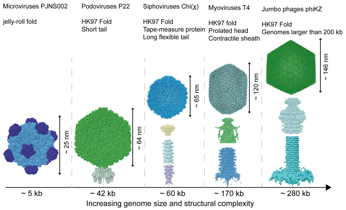
Figure 2.
Cryo-EM structure of the PJNS002 and conservation of protein F among φX174-like microviruses. (A) Cryo-EM density map of PJNS002 at a threshold of 0.5. Pentagons, triangles and diamonds mark the fivefold, threefold and twofold symmetry axes, respectively. Capsid protein F and spike protein G are colored yellow and light orange, respectively. To visualize the interactions within one G spike and its underlying capsid, the five F subunits surrounding the fivefold axis and the associated G pentamer are differentially colored. (B) Cartoon representation of the asymmetric unit (ASU) within the dashed triangle indicated in panel (A), comprising one copy each of F, G and J, colored yellow, light orange and rose, respectively. (C) Cut-away view of the PJNS002 fivefold channel, shown as Coulombic electrostatic surface potential (red, negative; blue, positive). The channel forms at the junction of five F subunits and is lined by the conserved DGTDQ motif (F residues 251–255), which constricts the pore to ~4 Å at Thr253, a feature conserved across the Bullavirinae but variable among gokushoviruses. Thr116, Ser118 and Asn119 (labeled) replace the charged Arg115 and Asp117 of PJNS001, which may alter pore geometry and charge selectivity. (D) Superposition of protein F from six microviruses: Ebor, SPV4, φEC6098, φX174, PJNS001 and PJNS002, colored as indicated. Data sources: PJNS002, PDB 9K3N and EMD-62023; PJNS001, PDB 9K3M; φX174, PDB 2BPA; φEC6098, PDB 8DES; SPV4, PDB 9CGM; Ebor, PDB 9FFG.
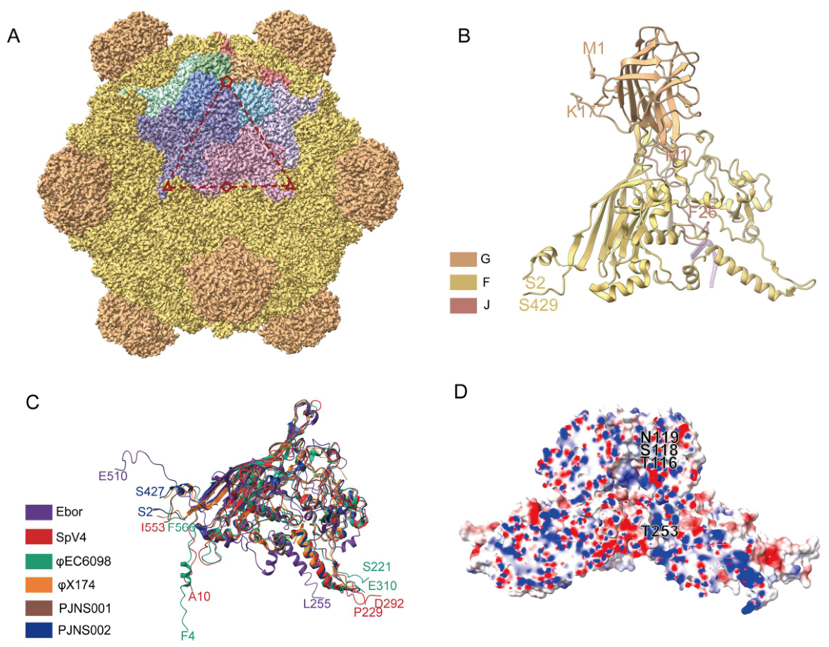
Figure 3.
Architecture of bacteriophage P22. (A) Composite cryo-EM map of the mature P22 virion and clipped along the tail axis to expose the capsid interior together with the portal and tail. Components are colored as follows: capsid (pale yellow), portal (light purple), adaptor (pink), hub (cyan), tailspikes (red), and needle (lime green). (B) Ribbon representation of the dodecameric portal protein, with each subunit in a different color. The C-terminal helices of the 12 subunits form an elongated barrel that projects into the capsid interior. Data sources: capsid, EMD-8606; portal–tail, EMD-41791; portal model, PDB 3LJ5.
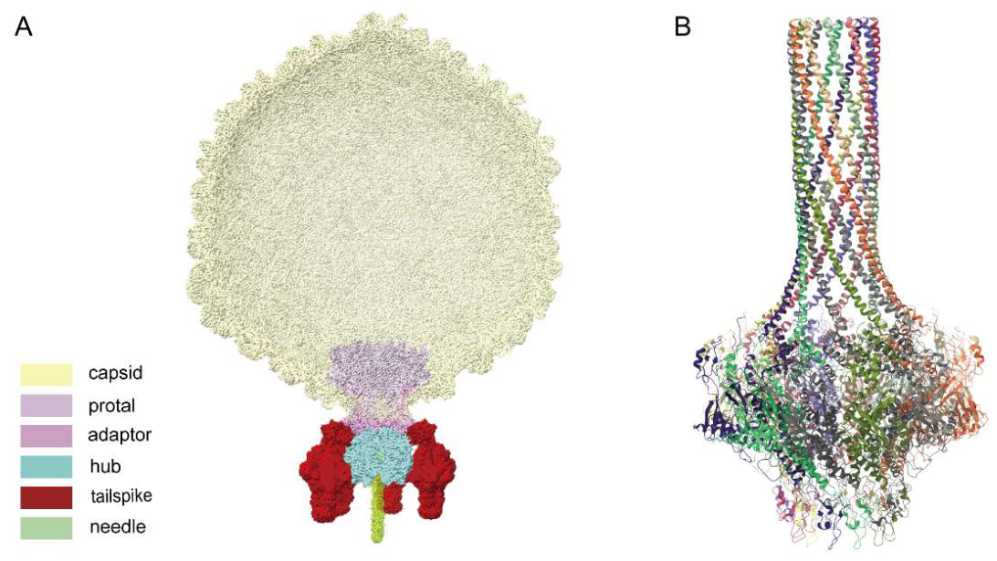
Figure 4.
Capsid and tail architecture of Salmonella flagellotropic siphophages. (A) Homotrimer of the auxiliary protein YSD1_16, with each protomer in a different color. Each protomer contains a β-tulip domain and an extended N-terminal hook (PDB: 6XGQ). (B) Top views of icosahedral asymmetric unit of head from χ (left, PDB: 8VJI) and YSD1 (right, PDB: 6XGQ). (C) Top view of the hexameric ring of tail tube protein from YSD1, with each subunit shown in a different color (PDB: 6XGR). (D) Side view of the χ tail tip. Left, cryo-EM map colored by protein; right, asymmetric unit fitted into the cryo-EM map (PDB: 8VJH, EMD-43281).
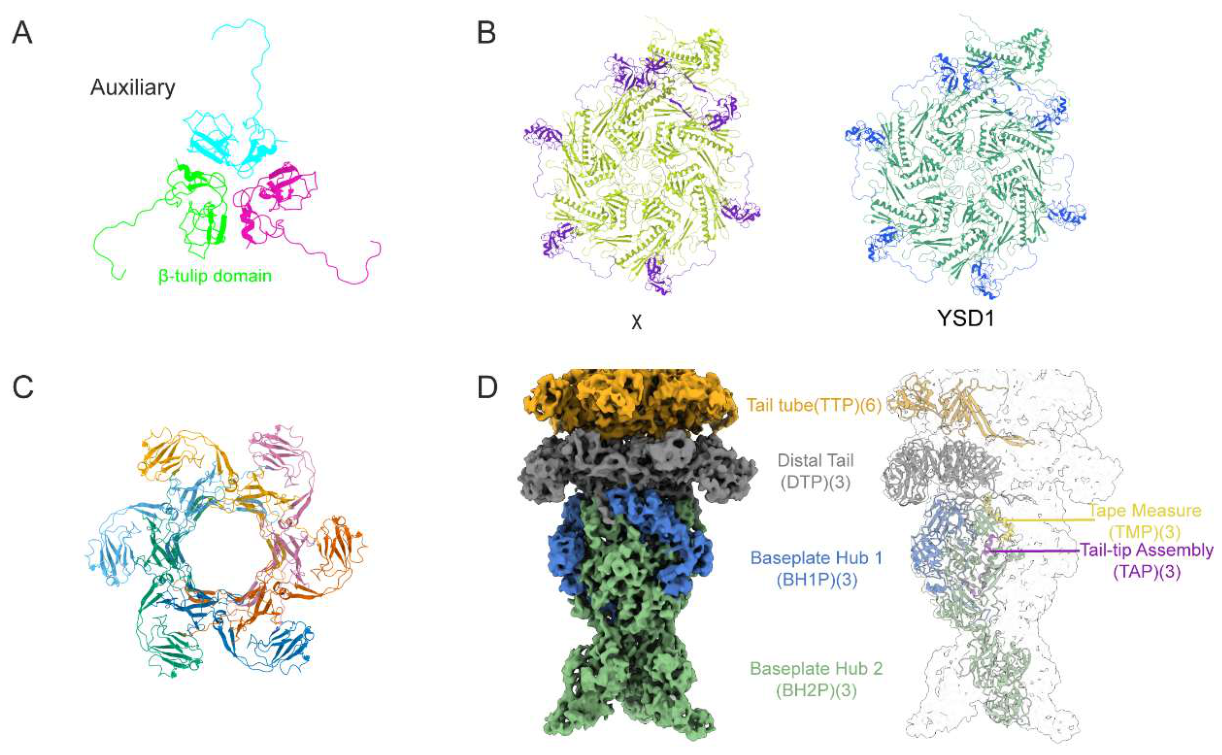

Table 1.
Receptor-binding proteins and receptors of the Salmonella phages discussed in this review, listed by tail morphotype.
Table 1.
Receptor-binding proteins and receptors of the Salmonella phages discussed in this review, listed by tail morphotype.
| Morphotype | Phage | Host (serovar) | Receptor-binding protein | Receptor | PDB |
| Microvirus | PJNS001, PJNS002 [6] |
S. Typhimurium | Spike protein G | LPS | 9K3M, 9K3N |
| Podovirus | P22 [7,8] | S. Typhimurium | Tailspike gp9 | O-antigen | 1TSP, 1TYU, 1TYV |
| Podovirus | ε15 [9,10] |
S. Anatum (serogroup E1) |
Tailspike gp20 | O-antigen | 9SIM, 9SIN |
| Siphovirus | Χ (Chi) [11,12] | S. Typhimurium | Tail fiber | Flagellar filament | None |
| Siphovirus | YSD1 [13] | S. Typhi | Tail fiber YSD1_29 | Flagellar filament | None |
| Myovirus | S16 [14,15] | Broad host range across Salmonella | Long tail fiber tip adhesin gp38 | OmpC | 6F45 |
| Myovirus | Det7 [16] | S. Typhimurium | Tailspike | O-antigen | 2V5I |
| Myovirus | ViI (Vi01) [17,18] | S. Typhi | Tailspike | Vi capsular polysaccharide | None |
| Myovirus | Felix O1 [19] | Broad across Salmonella | Not identified | LPS core oligosaccharide | None |
| Jumbo (phiKZ-related) | SPN3US [20] | S. Typhimurium | Not identified; gp168–gp170 are candidates | Unknown | None |
Disclaimer/Publisher’s Note: The statements, opinions and data contained in all publications are solely those of the individual author(s) and contributor(s) and not of MDPI and/or the editor(s). MDPI and/or the editor(s) disclaim responsibility for any injury to people or property resulting from any ideas, methods, instructions or products referred to in the content. |
© 2026 by the authors. Licensee MDPI, Basel, Switzerland. This article is an open access article distributed under the terms and conditions of the Creative Commons Attribution (CC BY) license.
Copyright: This open access article is published under a Creative Commons CC BY 4.0 license, which permit the free download, distribution, and reuse, provided that the author and preprint are cited in any reuse.